


Abstract
Polystyrene nanoplastics (PS-NPs) are emerging environmental contaminants increasingly linked to male reproductive toxicity; however, the molecular mechanisms underlying testicular damage remain unclear. This study evaluated PS-NP-associated testicular damage in rats after 55 days of exposure and assessed the modulatory effects of Nelumbo nucifera leaf, flower, and rhizome extracts, with quercetin as a reference. PS-NP exposure reduced spermatogenic cell populations, testicular, epididymal weights, and sperm motility. These changes were accompanied by increased NOX4 and NF-κB expression, upregulation of intrinsic apoptosis-related genes (Tp53, Bax, Caspase-9, and Caspase-3), elevated caspase-3 and caspase-9 protein levels, and enhanced cleaved caspase-3 immunoreactivity. In contrast, Fas and Caspase-8 were downregulated, confirming intrinsic mitochondrial apoptosis. PS-NP exposure also altered reproductive hormone receptor expression (LHr, FSHr, and AR) and dysregulated chromatin-regulatory genes, with increased Dnmt1, Dnmt3a, and Ehmt2 (G9a) and decreased Hdac1 and Ep300. Co-administration of N. nucifera attenuated most of these alterations, with the rhizome extract exhibiting the most pronounced protective effect. GO and PPI network analyses suggested functional connectivity among stress-responsive, apoptotic, and chromatin-modifying proteins. Docking simulations indicated phytochemical-apoptosis-related protein interactions. PS-NPs may impair testicular homeostasis through coordinated stress, apoptosis, endocrine disturbance, and epigenetic dysregulation, with possible relevance to male reproductive health, while N. nucifera shows promise as a protective modulator.
1. Introduction
Plastic pollution represents a major global environmental challenge, and the fragmentation of macroplastics into micro- and nanoplastics (NPs) increases particle surface area, reactivity, and biological availability, hence amplifying toxicological relevance [1,2,3]. Polystyrene nanoplastics (PS-NPs) pose a risk due to their nanoscale size, physicochemical stability, and high surface reactivity, which promote systemic absorption and accumulation in susceptible organs, including the testis [4,5]. Considering growing evidence that plastic-derived particles may cause male reproductive dysfunction, this problem is crucial to experimental toxicology and public health [4,6]. Although direct extrapolation from animal models to humans is limited, growing evidence suggests that PS-NP exposure may disrupt cellular homeostasis through stress-responsive and apoptosis-related pathways, affecting spermatogenesis, sperm quality, endocrine balance, and long-term male reproductive health [7,8].
Oxidative stress is widely recognized as an early interface between NP exposure and tissue injury [9,10]. In the testis, NADPH oxidases particularly NOX4, serve as important enzymatic sources of reactive oxygen species (ROS) and are increasingly viewed as redox regulatory nodes that couple oxidative load to downstream signaling responses [11,12]. NOX4-derived ROS contribute to compartmentalized redox signaling, affecting transcriptional programs, stress adaptability, and cell-fate decisions related to germ cell survival and steroidogenic activity [13]. Recent studies indicate that NP exposure may elevate oxidative stress and disturb NOX-related redox equilibrium in reproductive tissues, hence increasing vulnerability to transcriptional dysregulation and apoptosis [14,15,16]. Prolonged redox imbalance may also trigger excessive germ cell apoptosis, while activation of the redox-sensitive NF-κB pathway links oxidative stress to broader inflammatory and transcriptional responses in the testis [17,18,19,20,21]. Beyond its classical inflammatory role, NF-κB integrates oxidative stress signaling with apoptotic and chromatin-regulatory programs, positioning it as a central hub in environmentally induced reproductive toxicity [22]. Nevertheless, the relationship between PS-NP-associated NF-κB stress signaling and the transcriptional profile of apoptosis-related mediators in the testis remains incompletely characterized.
In addition to acute cytotoxicity, NP exposure may exert longer-term effects through disruption of epigenetic regulation [23]. DNA methylation and histone modifications are essential for germ cell differentiation and spermatogenic progression and are highly sensitive to environmental stressors [20,24,25]. Despite reports of epigenetic disruptions following exposure to diverse nanomaterials, a thorough evaluation of essential chromatin regulators, including Dnmt1, Dnmt3a, Ehmt2 (G9a), Hdac1, and Ep300, concerning PS-NP–induced testicular toxicity is yet insufficiently explored [26]. This gap is significant, as disruption of these regulators may be associated with altered chromatin regulatory processes and increased germ cell susceptibility to apoptosis, thereby contributing to persistent impairment of spermatogenesis [27].
Due to the complex nature of NP-related damage, mitigation strategies to modulate oxidative stress, stress-responsive signaling, apoptosis, and epigenetic regulation are important [28]. N. nucifera is a medicinal plant with documented antioxidant and anti-inflammatory activities. Its bioactive constituents include flavonoids (isoquercetin and kaempferol), alkaloids such as nuciferine, and phytosterols including stigmasterol. These compounds are variably distributed among plant organs and may influence organ-specific biological effects [29,30,31]. Our previous studies have shown organ-dependent protective effects of N. nucifera against testicular oxidative and inflammatory injury [21]. The molecular basis for these distinct effects under PS-NP-associated stress, including any potential links to apoptosis-related pathways and chromatin-regulatory gene, remains poorly characterized.
Therefore, this study tested the hypothesis that PS-NP exposure is associated with testicular injury accompanied by alterations in NOX4-related redox signals, NF-κB-associated stress signaling, apoptosis-related gene expression, and transcriptional changes in selected chromatin-regulatory enzymes. We further hypothesized that N. nucifera extracts partially normalize these responses in an organ-dependent manner. By clarifying these mechanisms in an animal model, the present study may also provide insight into biological pathways pertinent to environmentally linked male reproductive risks in humans.
2. Results
2.1. PS-NPs Induced Reproductive Parameters and Mitigated by N. nucifera
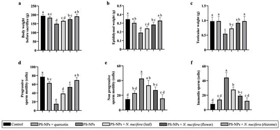
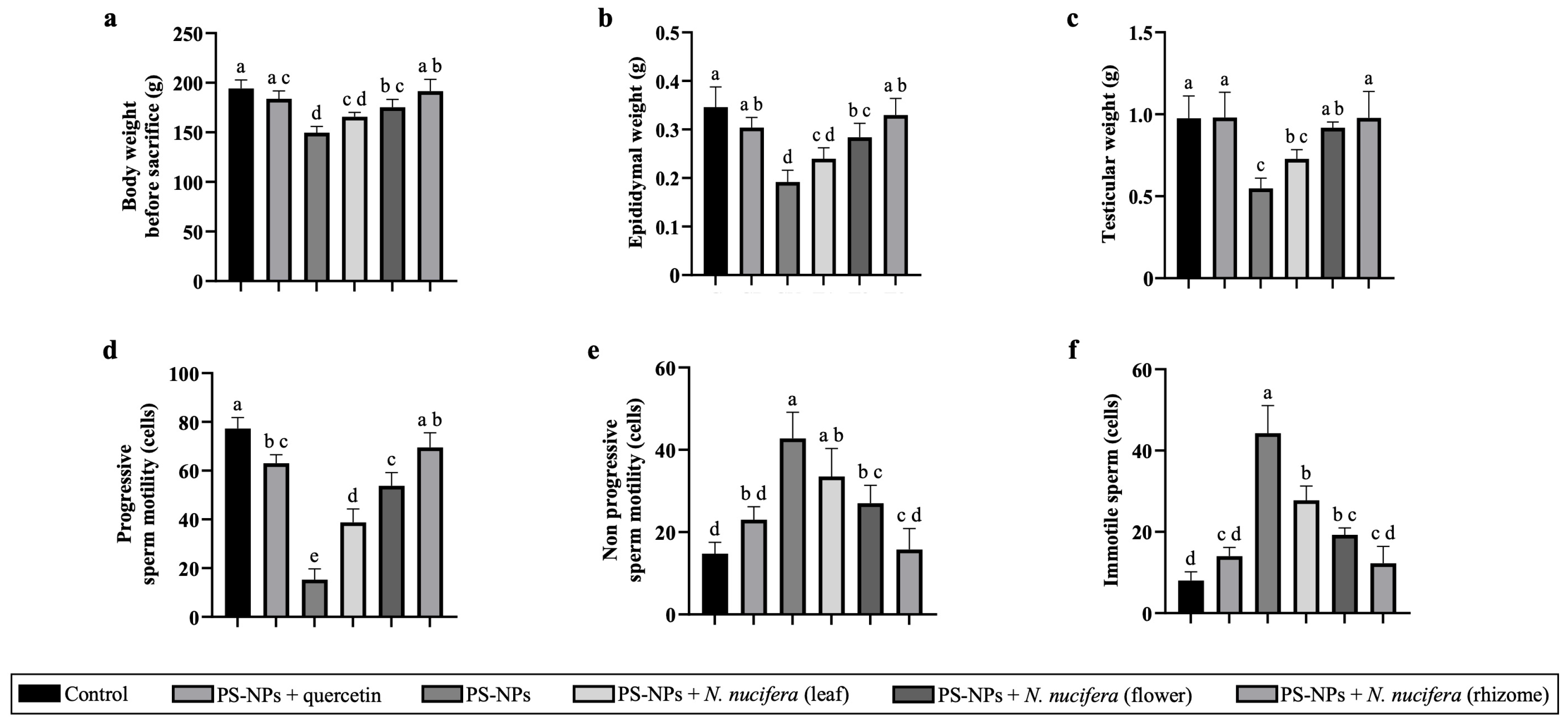
PS-NP exposure was associated with a significant reduction in final body weight compared with the control group (Figure 1a). Co-administration with N. nucifera extracts partially restored body weight, with the rhizome extract showing the greatest recovery. In addition, PS-NP exposure significantly reduced epididymal (Figure 1b) and testicular weight (Figure 1c). These changes were attenuated by N. nucifera treatment in an extract-dependent manner. PS-NP exposure also significantly altered sperm motility parameters. The PS-NP group showed reduced progressive motility (Figure 1d) and increased non-progressive (Figure 1e) and immotile sperm populations (Figure 1f) relative to the control. These changes were attenuated by N. nucifera treatment in an organ-dependent manner. Among the extracts, the rhizome group showed the strongest improvement, with higher progressive motility and lower immotile sperm proportions than the PS-NP group.
Figure 1.
PS-NPs induced alterations in reproductive organ weight and sperm motility were mitigated by N. nucifera. (a) Final body weight, (b) epididymal weight, (c) testicular weight, (d) progressive sperm motility, (e) non-progressive sperm motility, and (f) immotile sperm percentage in the control (C), PS-NPs (CN), PS-NPs + quercetin (CP), and PS-NPs + N. nucifera leaf (T1), flower (T2), or rhizome (T3) groups. Data are presented as the mean ± SD (n = 8 animals per group). Different letters indicate significant differences among groups (p < 0.05).
2.2. PS-NP Exposure Disrupts Spermatogenic Structure and Mitigated by N. nucifera
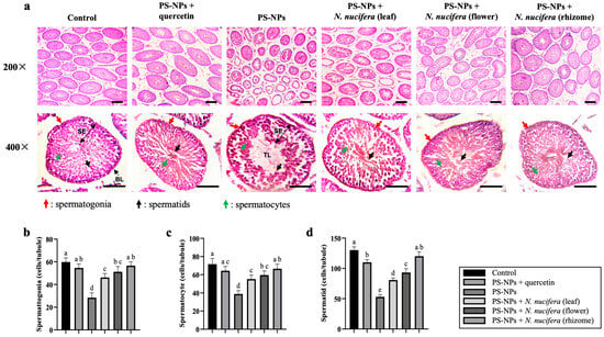
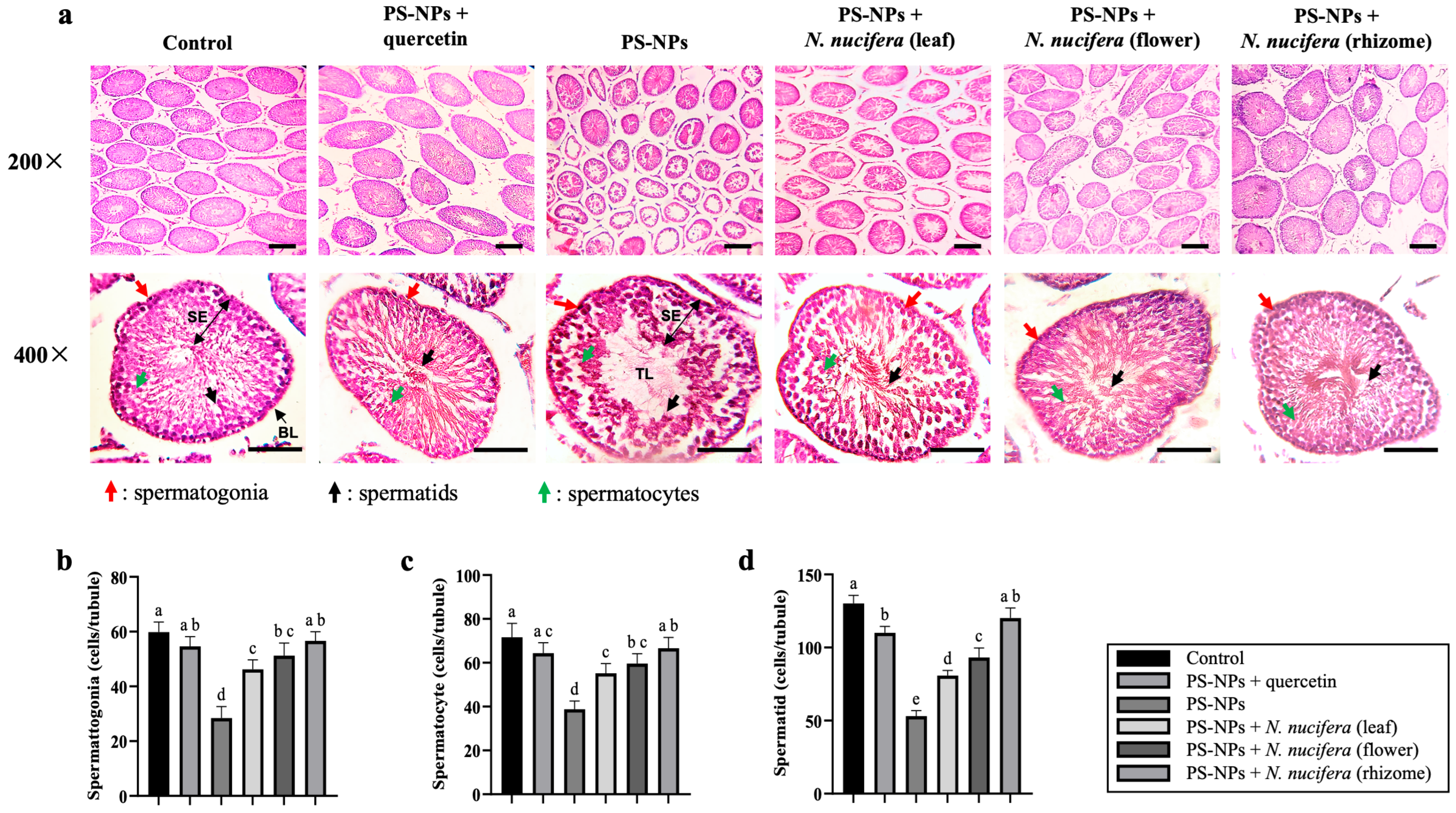
Representative H&E-stained sections showed normal seminiferous tubule organization in the control group, with a compact seminiferous epithelium and dense germ-cell layering at both 200× and 400× magnification (Figure 2a). In contrast, PS-NP exposure induced marked histopathological abnormalities, including disorganization of the seminiferous epithelium, disruption of germ-cell layering, reduced germ-cell density, and enlargement of the tubular lumen, consistent with spermatogenic depletion. At higher magnification (400×), these alterations were further reflected by reduced numbers of spermatogonia, spermatocytes, and spermatids. Quantitative analysis confirmed significant reductions in all three spermatogenic cell populations compared with the control group (Figure 2b–d). Co-treatment with N. nucifera extracts significantly mitigated these histological changes, as evidenced by elevated spermatogenic cell counts at relative to the PS-NP group (p < 0.05). The rhizome group exhibited the most significant preservation and normalization of spermatogenic cell populations, corresponding to the values observed in the control and quercetin groups.
Figure 2.
Effects of PS-NPs and N. nucifera extracts on testicular histology and spermatogenic cell populations. (a) Representative hematoxylin and eosin (H&E)-stained sections of seminiferous tubules from the control (C), PS-NP-exposed (CN), PS-NPs + quercetin (CP), and PS-NPs + N. nucifera leaf (T1), flower (T2), or rhizome (T3) groups, shown at 200× and 400× magnification. Arrowheads indicate spermatogonia (red), spermatocytes (green), and spermatids (black). TL, tubular lumen; SE, seminiferous epithelium; BL, basal lamina. Scale bars = 50 μm. (b) Quantitative analysis of spermatogonia, (c) spermatocytes, and (d) spermatids per seminiferous tubule. Data are presented as the mean ± SD (n = 8 biological replicates per group). Different letters indicate significant differences among groups (p < 0.05).
2.3. PS-NP Exposure Elevates NF-κB Immunoreactivity and Transcript Levels
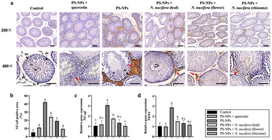
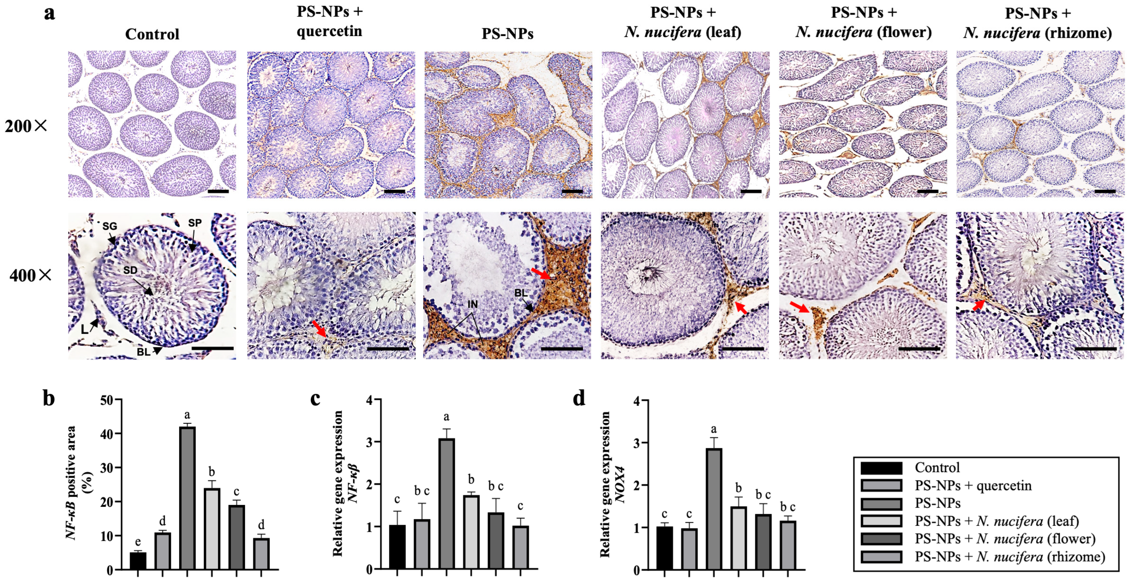
NF-κB immunohistochemistry revealed minimal basal staining in the control group, with weak and diffuse distribution within the seminiferous tubules (Figure 3a). In contrast, the PS-NP group showed markedly increased NF-κB staining, characterized by intense brown DAB signals predominantly localized in interstitial regions, with both nuclear and cytoplasmic distribution. At higher magnification (400×), staining was more pronounced surrounding the basal lamina and within interstitial regions. Quantitative analysis confirmed a significant increase in NF-κB-positive area in the PS-NP group compared with the control (Figure 3b), which was paralleled by elevated NF-κB gene expression (Figure 3c) and increased NOX4 expression (Figure 3d). Co-treatment with N. nucifera reduced NF-κB immunoreactivity and gene expression compared with the PS-NP group. Among the treatments, the rhizome extract showed the greatest reduction, with staining intensity and distribution approaching those of the control groups.
Figure 3.
Effects of PS-NPs and N. nucifera on NF-κB and NOX4-associated stress signaling in testis. (a) Representative NF-κB immunohistochemical staining in the control (C), PS-NPs + quercetin (CP), PS-NP-exposed (CN), and PS-NPs + N. nucifera leaf (T1), flower (T2), or rhizome (T3) groups, shown at 200× and 400× magnification. NF-κB-positive immunoreactivity is shown by brown DAB staining and was observed predominantly in interstitial regions (red arrows). Sections were counterstained with hematoxylin. SG, spermatogonia; SP, spermatocyte; SD, spermatid; L, Leydig cell; IN, interstitial region; BL, basal lamina. Scale bars = 50 μm. (b) Quantification of NF-κB-positive area, expressed as the percentage of immunoreactive area relative to total tissue area. (c) Relative NF-κB gene expression. (d) Relative NOX4 gene expression. Data are presented as the mean ± SD (n = 8 per group). Different letters indicate significant differences among groups (p < 0.05).
2.4. PS-NP-Associated Upregulation of NOX4 Expression and Attenuated by N. nucifera
To assess redox-associated stress signaling, NOX4 gene expression was quantified in testicular tissue. Compared with the control group, PS-NP exposure resulted in a significant increase in NOX4 expression levels (Figure 3d). Co-administration of N. nucifera extracts significantly attenuated PS-NP–induced NOX4 upregulation (p < 0.05), with clear organ-dependent efficacy. The rhizome and flower extracts resulted in the most significant reductions in NOX4 gene expression.
2.5. PS-NPs–Induced Testicular Apoptosis and Attenuated by N. nucifera
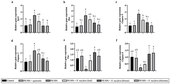
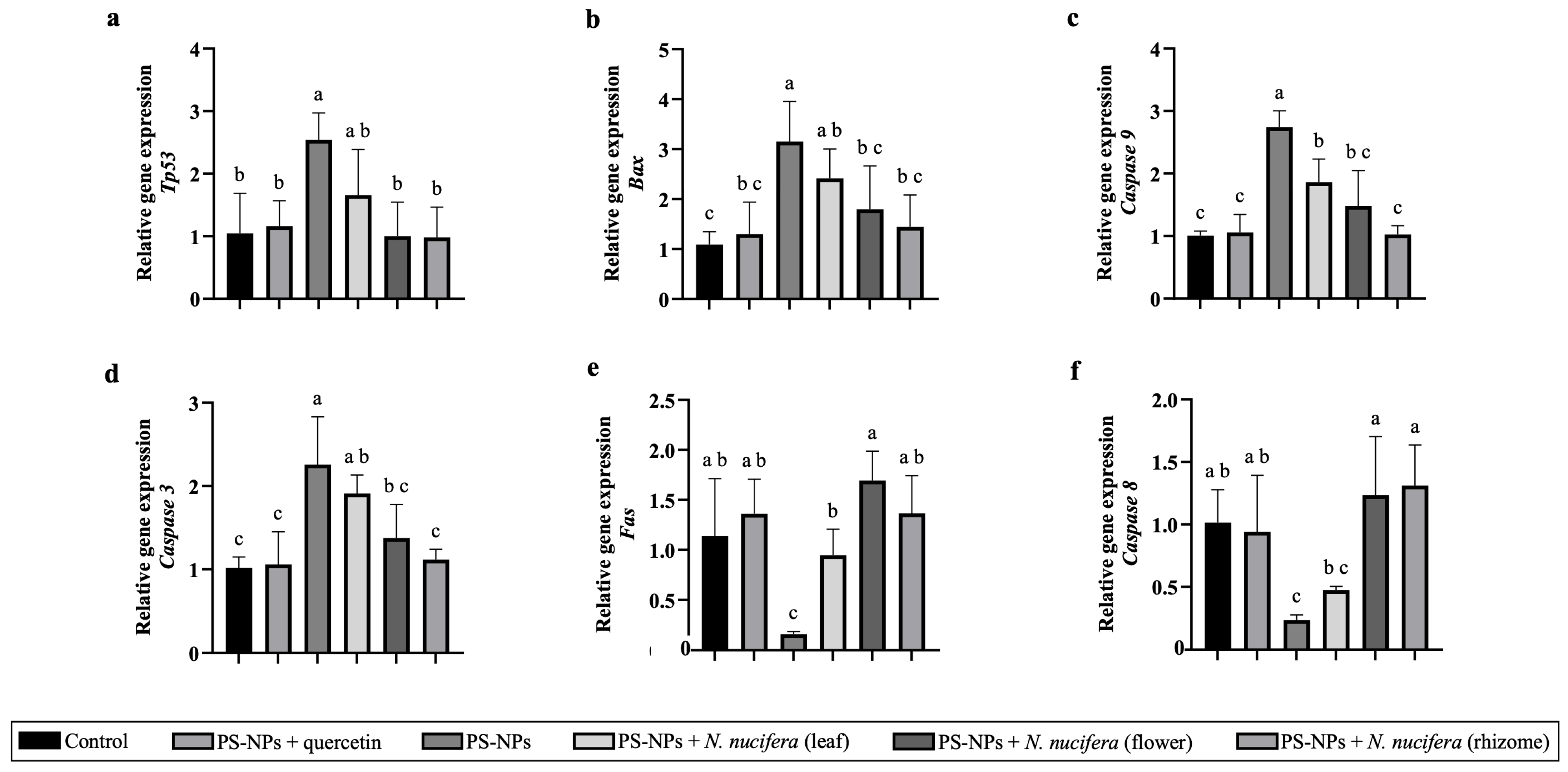
PS-NP exposure caused a distinct apoptosis-associated transcriptional pattern in testicular tissue (Figure 4). The intrinsic apoptosis-associated genes Tp53 and Bax were significantly upregulated compared to controls (Figure 4a,b), along with elevated transcript levels of Caspase-9 and Caspase-3 (Figure 4c,d). In contrast, extrinsic pathway-associated mediators Fas and Caspase-8 were significantly downregulated (Figure 4e,f).
Figure 4.
Effects of N. nucifera on apoptosis-related gene expression in PS-NP–exposed testes. Relative gene expression of intrinsic apoptosis-related genes: (a) Tp53, (b) Bax, (c) Caspase-9, and (d) Caspase-3, and extrinsic apoptosis-related genes: (e) Fas and (f) Caspase-8 in the control (C), PS-NPs + quercetin (CP), PS-NP-exposed (CN), and PS-NPs + N. nucifera leaf (T1), flower (T2), or rhizome (T3) groups. Data are presented as the mean ± SD (n = 8). Different letters indicate significant differences (p < 0.05).
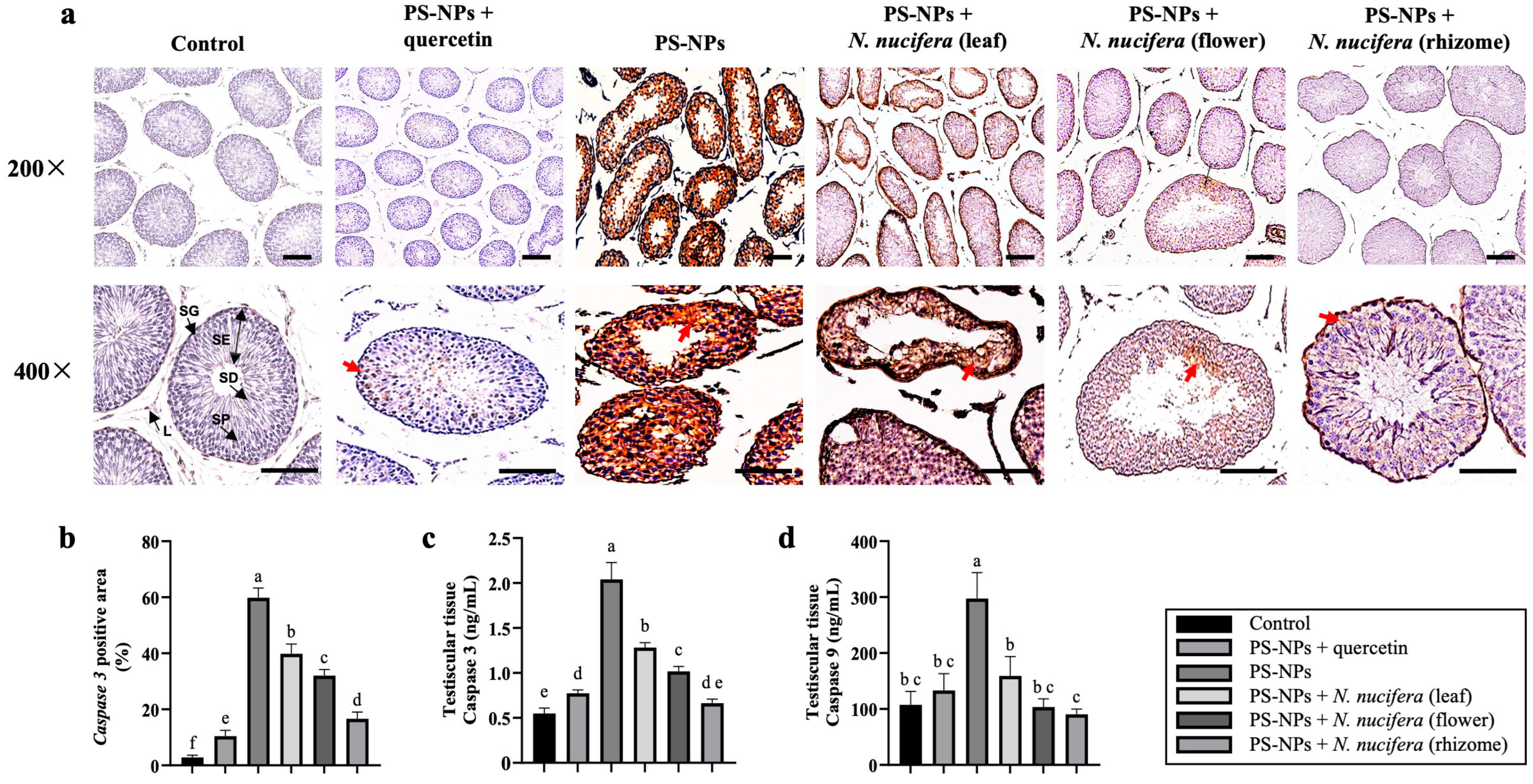
Moreover, protein-level analysis supported the apoptosis-related transcriptional changes. Cleaved caspase-3 immunohistochemistry showed minimal staining in the control group, whereas PS-NP exposure markedly increased brown DAB-positive immunoreactivity within the seminiferous epithelium (Figure 5a,b). At higher magnification (400×), staining was concentrated along the seminiferous epithelium. ELISA further confirmed significantly increased testicular caspase-3 and caspase-9 protein levels in the PS-NP group relative to the control (Figure 5c,d). Co-treatment with N. nucifera attenuated these changes, with the rhizome extract showing the greatest reduction in cleaved caspase-3 staining and caspase protein levels, approaching the control and quercetin groups.
Figure 5.
Effects of N. nucifera on apoptosis-related protein markers in PS-NP–exposed testes. (a) Representative cleaved caspase-3 immunohistochemical staining in the control (C), PS-NPs + quercetin (CP), PS-NPs (CN), and PS-NPs + N. nucifera leaf (T1), flower (T2), or rhizome (T3) group, shown at 200× and 400× magnification. Cleaved caspase-3-positive immunoreactivity is shown by brown DAB staining, predominantly localized in the seminiferous epithelium (red arrows). SG, spermatogonia; SP, spermatocyte; SD, spermatid; L, Leydig cell; SE, seminiferous epithelium. Sections were counterstained with hematoxylin. Scale bar = 50 μm. (b) Quantification of cleaved caspase-3–positive area (%). (c) Testicular caspase-3 protein levels. (d) Testicular caspase-9 protein levels. Data are presented as the mean ± SD (n = 8). Different letters indicate significant differences (p < 0.05).
2.6. PS-NP Induced Alterations in Reproductive Hormone Receptor Gene Expression
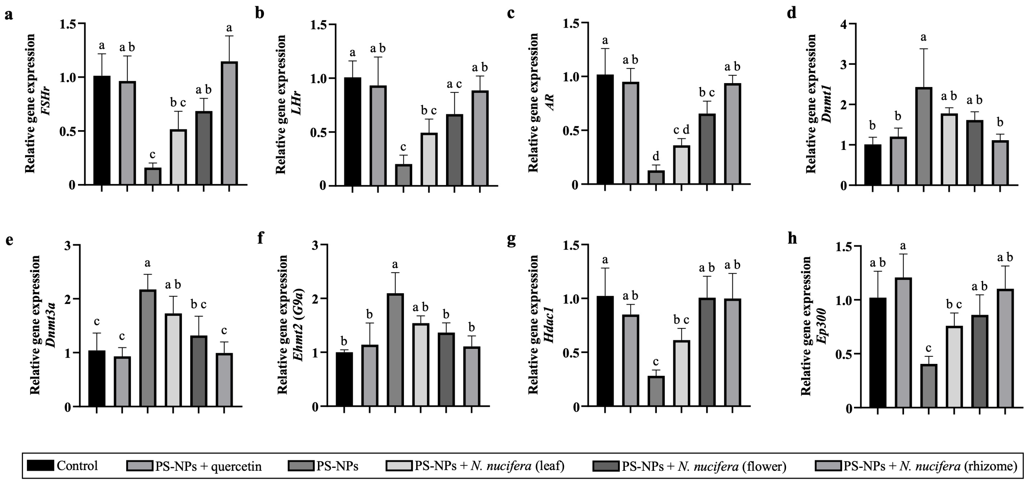
PS-NP exposure was associated with significant alterations in the expression of reproductive hormone receptors in testicular tissue (Figure 6a–c). Specifically, the expression levels of LHr, FSHr, and AR were reduced in the PS-NP group compared with the control. Co-administration with N. nucifera extracts partially restored the expression of these receptors in a treatment-dependent manner. Among the treatments, the rhizome extract showed the greatest recovery, with expression levels approaching those observed in the control and quercetin groups.
Figure 6.
Effects of N. nucifera on hormone receptor and chromatin-regulatory gene expression in PS-NP–exposed testes. Relative gene expression of hormone receptor genes, including (a) LHr, (b) FSHr, (c) AR, and chromatin-regulatory genes, including (d) Dnmt1, (e) Dnmt3a, (f) Ehmt2 (G9a), (g) Hdac1, and (h) Ep300 in the control (C), PS-NPs + quercetin (CP), PS-NP-exposed (CN), and PS-NPs + N. nucifera leaf (T1), flower (T2), or rhizome (T3) groups. Gene expression was measured by quantitative real-time PCR and normalized to GAPDH as the internal reference gene. Data are presented as fold-change relative to the control group using the 2−ΔΔCt method and shown as the mean ± SD (n = 8). Different letters indicate significant differences (p < 0.05).
2.7. PS-NP Associated with Dysregulation of Chromatin-Regulatory Genes
PS-NP exposure significantly altered the expression of multiple chromatin-regulatory genes in testicular tissue (Figure 6d–h). PS-NPs significantly elevated gene levels of the DNA methylation-related enzymes Dnmt1 and Dnmt3a, and upregulated the histone methyltransferase Ehmt2. In contrast, Hdac1 and Ep300 expression was significantly reduced compared with controls. Co-treatment with N. nucifera significantly mitigated these transcriptional alterations (p < 0.05). The rhizome extract exhibited the most significant normalization of Dnmt1, Dnmt3a, and Ehmt2, Hdac1, and Ep300 expression.
2.8. Total Flavonoid Content and Antioxidant Activity of N. nucifera
Total flavonoid content (TFC) and DPPH scavenging activity were evaluated to facilitate the evaluation of organ-specific extract performance (Table 1). The leaf extract demonstrated the highest total flavonoid content, followed by the flower and rhizome. Nonetheless, antioxidant activity (DPPH) demonstrated an organ-specific pattern, with the rhizome extract exhibiting the strongest antioxidant activity (lowest IC50).
Table 1.
Antioxidant activity and Total Flavonoid Content (TFC) of N. nucifera.
2.9. Gene Ontology Study Promotes Stress-Induced Apoptosis and Epigenetic Mechanisms
GO Biological Process enrichment analysis of the measured genes revealed significant overrepresentation (FDR < 0.05) of pathways related to cellular stress response, apoptotic signaling, epigenetic/chromatin regulation, DNA methylation, testosterone response, and spermatogenesis (Table 2). The strongest enrichment was observed for cellular response to stress (GO:0033554; FDR = 6.55 × 10⁻⁵), represented by Bax, Fas, Tp53, Caspase-8, Caspase-9, and Caspase-3, and linking stress signaling with chromatin regulators such as NF-κB, Ehmt2, Hdac1, and Dnmt3a. Epigenetic-related terms included regulation of DNA methylation (GO:0044030; FDR = 3.30 × 10⁻⁴), represented mainly by Dnmt1, Dnmt3a, and Ehmt2, and regulation of histone modification (GO:0031056; FDR = 1.76 × 10⁻²), represented by Tp53, Ehmt2, and Dnmt1. These epigenetic signatures were closely linked to reproductive processes, as indicated by enrichment of spermatogenesis (GO:0007283; FDR = 2.65 × 10⁻⁵), represented by Bax, Fas, Tp53, AR, FSHr, Dnmt3a, and Ehmt2, and response to testosterone (GO:0033574; FDR = 2.83 × 10⁻³), represented by AR, LHr, and Dnmt1. Together, these results suggest a potential functional connection between stress signaling, epigenetic regulation, and androgen-responsive pathways in male reproductive function.
Table 2.
STRING-based GO Biological Process Enrichment.
2.10. PASS Online Screening
PASS online screening of representative N. nucifera phytochemicals predicted antioxidant-related activity for multiple compounds (Table 3). Among compounds with Pa > Pi, the highest predicted antioxidant probabilities included ascorbic acid (Pa = 0.928; rhizome), isoquercetin (Pa = 0.913; rhizome and flower), and kaempferol-3-glucuronide (Pa = 0.906; flower, leaf, rhizome). These predictions were used as supportive information to contextualize observed organ-specific extract performance.
Table 3.
PASS antioxidant activity prediction of selected N. nucifera compounds.
2.11. Network-Based Integration of Stress, Apoptotic, and Epigenetic-Related Proteins
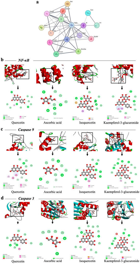
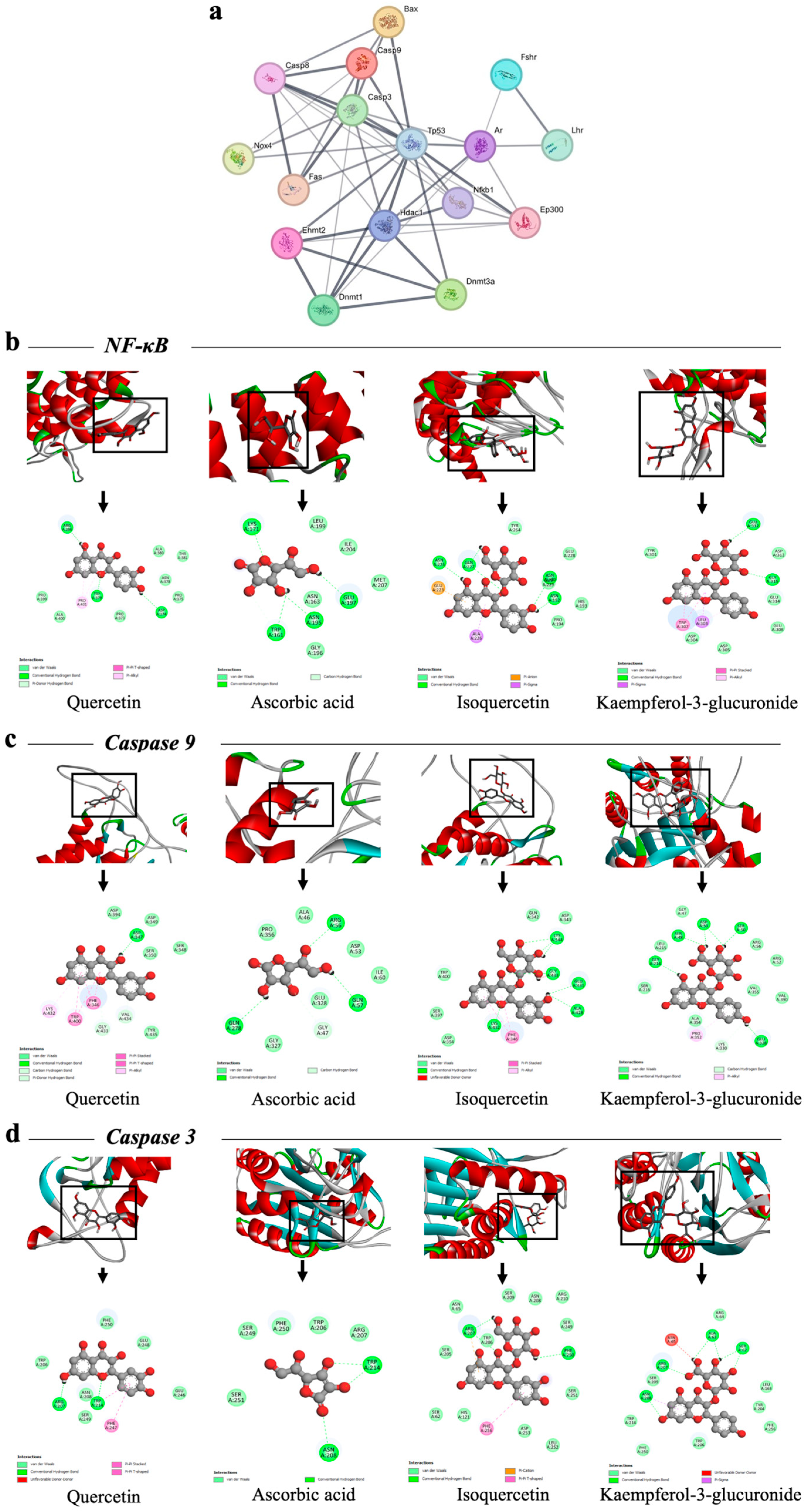
STRING analysis produced a densely connected PPI network among the measured proteins (Figure 7a). Tp53 formed the central hub, bridging stress-associated nodes (NOX4 and NF-κB) with core apoptotic mediators (Bax, Casp3, Casp9, and Casp8/Fas). A second connectivity cluster comprised chromatin regulators (Dnmt1, Dnmt3a, Ehmt2/G9a, Hdac1, and Ep300), which showed multiple links to Tp53 and NF-κB, supporting functional coupling between stress-responsive signaling, apoptotic execution, and transcriptional/chromatin regulatory machinery. Network interaction enrichment was significant (PPI enrichment p < 1.0 × 10⁻¹⁶).
Figure 7.
PPI network and molecular docking of representative N. nucifera phytochemicals. (a) STRING-derived protein–protein interaction (PPI) network generated from the experimentally measured stress-, apoptosis-, and chromatin-regulatory proteins (NOX4, NF-κB, Tp53, Bax, Fas, Caspase-8, Caspase-9, Caspase-3, LHr, FSHr, AR, Dnmt1, Dnmt3a, Ehmt2/G9a, Hdac1, and Ep300). The network comprised 13 nodes and 39 edges and showed significant interaction enrichment (p < 1.0 × 10⁻¹⁶), supporting functional connectivity among stress-responsive, apoptotic, and chromatin-regulatory pathways. (b–d) Molecular docking of quercetin, ascorbic acid, isoquercetin, and kaempferol-3-glucuronide with (b) NF-κB, (c) caspase-9, and (d) caspase-3. Upper panels show representative three-dimensional binding poses, and lower panels show corresponding two-dimensional interaction maps. Docking results are presented as supportive in silico evidence.
2.12. Molecular Docking and Dynamics Simulation
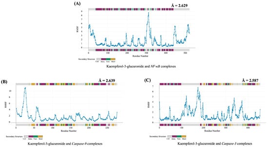
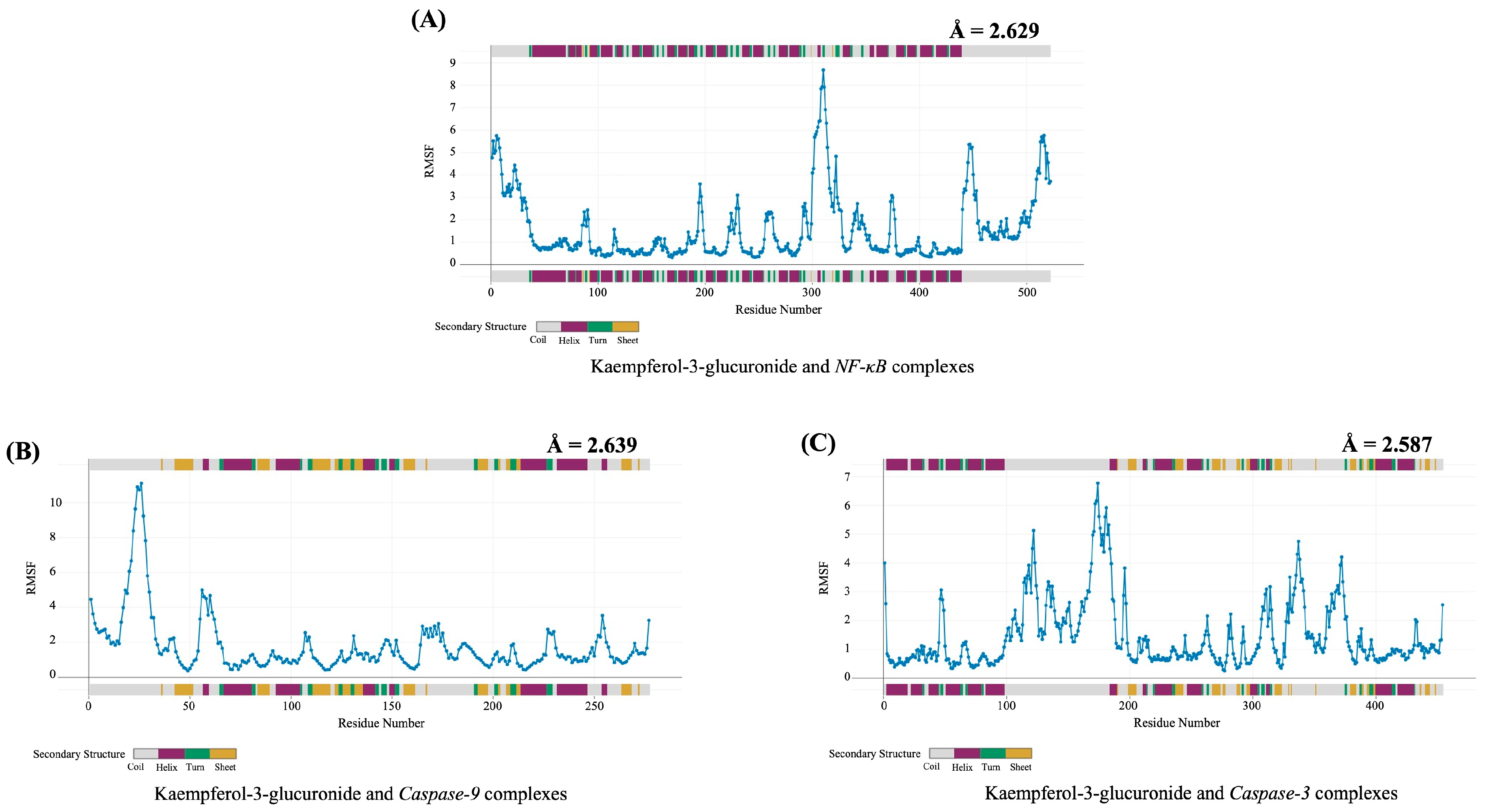
Based on the experimental stress–apoptosis signatures and the PPI network context, docking simulations were performed using NF-κB, caspase-9, and caspase-3 as representative targets of stress signaling and intrinsic apoptosis execution. Among the screened ligands, kaempferol-3-glucuronide showed the most favorable docking scores across all three targets, with binding energies ranging from −7.1 to −6.9 kcal/mol (Figure 7b–d, Table 4). Isoquercetin showed comparable binding energies, whereas ascorbic acid exhibited weaker predicted interactions. Dynamics simulations (CABS-Flex) indicated stable conformational behavior of kaempferol-3-glucuronide–protein complexes, with average RMSF values of 2.58–2.64 Å for NF-κB, caspase-3, and caspase-9, consistent with overall structural stability and localized residue flexibility (Figure 8A–C).
Table 4.
Molecular docking result.
Figure 8.
RMSF profiles of kaempferol-3-glucuronide–protein complexes. (A–C) Root mean square fluctuation (RMSF) plots showing residue-level flexibility of kaempferol-3-glucuronide complexes with (A) NF-κB, (B) caspase-9, and (C) caspase-3. Average RMSF values (Å) are shown in each panel. Lower RMSF values indicate greater predicted structural stability, whereas higher peaks indicate localized residue flexibility. Secondary structure distribution is displayed above and below each plot.
3. Discussion
This study demonstrated that oral exposure to PS-NPs disrupts spermatogenic structural integrity, resulting in redox-associated stress, NF-κB-linked transcriptional responses, apoptosis-related alterations, and chromatin-regulatory gene expression dysregulation. In parallel, PS-NP exposure was associated with impaired sperm motility, indicating functional deterioration of male reproductive capacity. Sperm motility is highly dependent on cellular integrity and is particularly sensitive to oxidative stress and mitochondrial dysfunction [27], the decline in progressive motility is consistent with the histological evidence of germ cell loss and suggests that PS-NP-induced testicular injury extends beyond structural damage to functional impairment of sperm quality [8]. These findings are consistent with previous reports showing that nanoplastics accumulate in testicular tissue and impair germ cell homeostasis, sperm quality, and fertility-related parameters through oxidative stress-, inflammation-, and apoptosis-associated mechanisms [6,16,21,32]. However, because the present study did not include fertility-related endpoints such as mating success, fertilization rate, or detailed sperm functional assays, the reproductive consequences of these changes should be interpreted as likely but not definitive outcomes in this animal model. The PS-NP dose used in this study (200 μg/kg bw) was selected as a controlled experimental exposure to produce measurable biological responses, consistent with previous nanoplastic toxicity studies [21]. Although this dose enabled mechanistic evaluation within a practical study period, it does not directly represent environmentally relevant human exposure, which is expected to be lower but more chronic and variable [9,25]. Oral administration was chosen to approximate ingestion as a plausible route of exposure [16]. Repeated gavage may introduce mild physiological stress. However, all groups were handled identically, reducing the likelihood that procedural stress alone explains the observed group differences [1]. Nevertheless, future studies using environmentally relevant exposure scenarios, voluntary ingestion models, and multiple dose levels will be necessary to better define dose-response relationships and establish toxicological thresholds such as NOAEL and LOAEL.
PS-NP exposure was associated with significant upregulation of NOX4 gene expression, suggesting activation of redox-associated stress signaling in testicular tissue [33]. As a constitutively active member of the NADPH oxidase family, NOX4 generates sustained intracellular ROS and contributes to redox-sensitive signaling and transcriptional regulation [34,35,36]. Therefore, the observed elevation of NOX4 gene expression may reflect a persistent oxidative stress environment induced by PS-NP exposure. While direct ROS measurements were not performed, the elevation of NOX4 transcripts, together with downstream NF-κB and apoptosis-related changes, supports the interpretation that PS-NPs induce a redox-associated stress environment in testicular tissue [1]. Sustained NOX4 activity has been linked to cellular stress responses, mitochondrial dysfunction, and apoptotic signaling in multiple tissues, including the reproductive system [34,35,37]. In the testis, excessive ROS disrupt the balance between germ-cell proliferation and apoptosis, impair sertoli cell support functions, and ultimately compromise spermatogenic progression [38]. Thus, NOX4 upregulation may represent an early molecular occurrence linking PS-NP exposure to downstream apoptotic and structural damage observed in the seminiferous epithelium. Nevertheless, due to the absence of quantitative oxidative stress biomarkers, additional research will be necessary in the future to assess ROS levels and antioxidant defense mechanisms to elucidate the specific involvement of NOX4-mediated signaling in PS-NP–induced testicular damage.
Increased NF-κB immunoreactivity and gene expression levels were observed following PS-NP exposure, suggesting enhancement of stress-responsive transcriptional signaling in testicular tissue. NF-κB is a redox- and injury-responsive transcription factor that integrates oxidative stress, inflammatory cues, and cellular damage signals [39]. In the present study, the elevation of NOX4 and NF-κB is consistent with a possible relationship between redox-associated stress and NF-κB–linked signaling, as ROS-generating systems are known to influence NF-κB regulatory pathways [33]. Increased NF-κB may indicate a stress-response pathway, along with PS-NP-induced apoptosis and chromatin-related transcriptional alterations in testes [40]. The enhanced nuclear and cytoplasmic staining, especially in interstitial regions around seminiferous tubules, suggesting PS-NP–induced stress responses involve somatic and germinal compartments [41]. Although these findings do not establish direct pathway activation or cell-specific signaling, they support the involvement of NF-κB-associated responses in PS-NP-induced testicular stress. This is consistent with previous studies showing that PS-NP exposure enhances testicular inflammatory responses and NF-κB activation, particularly under co-exposure conditions, together with oxidative stress and increased pro-inflammatory signaling [42,43]. Moreover, micro- and nanoplastics have been implicated in reproductive toxicity through oxidative stress, inflammation, apoptosis, and blood–testis barrier disruption [44,45]. Thus, the NF-κB elevation observed in this study may reflect part of a broader adaptive or injury-related transcriptional response to PS-NP-associated oxidative stress.
PS-NP exposure induced apoptosis-related transcriptional pattern characterized by upregulation of intrinsic mediators (Tp53, Bax, Caspase-9, Caspase-3) and downregulation of extrinsic mediators (Fas, Caspase-8). This demonstrates that PS-NP-associated testicular injury is linked to intrinsic apoptotic signaling instead of extrinsic pathway mediator transcription [46]. Upregulation of Tp53 may indicate a cellular stress response which favors apoptosis [3], whereas increased Bax expression indicates mitochondrial outer membrane permeabilization and downstream initiator caspase signaling [47]. Similarly, increased Caspase-9 and Caspase-3 gene expressions support transcriptional activation of the intrinsic apoptotic cascade [48]. These transcriptional changes were paralleled by protein-level findings. PS-NP exposure increased testicular caspase-9 and caspase-3 concentrations and enhanced cleaved caspase-3 immunoreactivity within the seminiferous epithelium, indicating that the altered gene expression profile was accompanied by downstream apoptotic execution. Together, these findings are consistent with the histological evidence of spermatogenic cell loss and support the interpretation that PS-NP-associated testicular injury involves activation of intrinsic mitochondrial apoptosis [46].
In addition, alterations in the expression of hormone-related receptors, including LHr, FSHr, and AR, suggest disruption of endocrine signaling within the testis. These receptors are central to the hypothalamic–pituitary–gonadal (HPG) axis, where LHr regulates testosterone production in Leydig cells, FSHr supports Sertoli cell function and germ cell development, and AR mediates androgen-dependent maintenance of spermatogenesis [49]. Dysregulation of these receptors may therefore impair cellular responsiveness to hormonal cues and compromise coordinated spermatogenic processes [50]. This finding is consistent with previous studies showing that micro- and nanoplastic exposure disturb endocrine homeostasis, alter androgen signaling, and impair spermatogenesis and sperm quality [6,51]. Although circulating hormone levels were not measured in the present study, the observed receptor-level changes provide biologically relevant evidence of altered local endocrine responsiveness within testicular tissue. Together with the accompanying abnormalities in seminiferous tubule structure and sperm motility, these findings support the involvement of both endocrine-related and cellular stress mechanisms in PS-NP-associated reproductive toxicity [52]. Future studies integrating circulating testosterone, LH, and FSH with local receptor analysis are needed to clarify the endocrine basis of PS-NP-induced reproductive toxicity.
PS-NP exposure significantly altered the expression of chromatin-regulatory genes, including upregulation of Dnmt1, Dnmt3a, and Ehmt2, and downregulation of Hdac1 and Ep300. These genes encode key enzymes involved in DNA methylation and histone modification, two core regulatory systems that maintain transcriptional homeostasis during germ-cell differentiation and spermatogenic progression [53]. In the testis, meticulous regulation of chromatin-modifying pathways is crucial for maintaining stem cells, advancing meiosis, and facilitating post-meiotic maturation [54,55]. Thus, any disruption in this expression may suggest that PS-NP-associated injury transcends acute cellular stress and leads to extensive alterations in transcriptional regulation [56]. More recent research links male reproductive toxicity caused by micro-/NPs to oxidative stress, apoptosis, altered gene regulation, DNA damage, and epigenetic instability [6]. According to a review of male reproductive toxicity, MPs/NPs bioaccumulate in the testis and epididymis and cause oxidative stress, inflammation, and gene regulation disruption [45]. Increased Dnmt1 and Dnmt3a may indicate an alteration in DNA methylation–related regulatory capacity, while elevated Ehmt2 suggests enhanced transcriptional circumstances favoring repressive histone methylation [57,58]. On the other hand, decreased Hdac1 and Ep300 expression implies histone deacetylation/acetylation imbalance, which is essential for spermatogenic gene expression [59,60]. Together, these changes suggest that PS-NP exposure disrupts the chromatin-regulatory environment required for normal germ cell development [61], consistent with reports of altered DNA methylation and transcriptional networks in PS-NP-exposed testes [8,20].
Network-level analyses further supported that PS-NP exposure is associated with systems-level disruption rather than isolated gene-level changes. GO enrichment revealed overrepresentation of apoptotic signaling, cellular stress responses, DNA methylation, histone regulation, and spermatogenesis, indicating convergence of the measured genes on biologically coherent pathways relevant to testicular injury. This pattern is consistent with evidence that PS-NP reproductive toxicity involves integrated perturbation of oxidative stress, apoptosis, and germ-cell regulatory processes [4,52,62]. Consistent with these functional annotations, STRING analysis produced a densely interconnected PPI network among the measured proteins. STRING incorporates both physical and functional protein interactions, making such network connection valuable for elucidating pathway-level organization within user-defined gene sets [63]. Tp53 emerged as a central hub linking stress-associated nodes (NOX4, NF-κB) with apoptotic mediators (Bax, Casp3, Casp9, Casp8/Fas), in line with its role as a stress-responsive regulator of cell-fate decisions [64]. A second major module consisted of chromatin-regulatory proteins (Dnmt1, Dnmt3a, Ehmt2/G9a, Hdac1, Ep300), which showed multiple links to Tp53 and NF-κB. In addition, the inclusion of LHr, FSHr and AR in the broader network context, together with enrichment of testosterone-response and spermatogenesis-related terms, further supports the idea that PS-NP exposure affects not only cellular stress and apoptosis but also hormone-responsive regulatory pathways relevant to male reproductive function [49,50]. This architecture suggests that stress-responsive signaling and chromatin-regulatory machinery do not act independently, but are integrated within the broader testicular response to PS-NP exposure [65]. Together, the GO and PPI analyses are consistent with a model in which PS-NP exposure may disrupt interconnected networks governing stress adaptation, apoptosis, endocrine responsiveness, and chromatin regulation, thereby contributing to impaired spermatogenic homeostasis [4,6,51].
Treatment with N. nucifera, particularly the rhizome extract, attenuated key PS-NP–induced network alterations, including reduced Tp53- and NF-κB-associated responses and partial normalization of chromatin-regulatory transcripts. This pattern indicates that the protective effect extends across interconnected stress-response pathways rather than a single downstream target [66]. Given that NF-κB functions as a redox-sensitive hub linking oxidative stress to inflammatory, apoptotic, and transcriptional responses [40], its reduction suggests suppression of stress-associated signaling. This aligns with findings indicating that N. nucifera possesses antioxidant and anti-inflammatory phytochemicals, which indicate that oxidative stress and NF-κB signaling are pivotal in nanoplastic-induced testicular damage [67,68]. Although the leaf extract contains higher total flavonoids, the rhizome extract exhibits stronger radical-scavenging activity, indicating that protective efficacy depends on phytochemical composition rather than total content alone [21]. Recent phytochemical investigations of the rhizome have revealed the presence of phenolics, flavonoids, tannins, alkaloids, steroids, and terpenoids, suggesting a favorable combination of redox-active compounds [67,69]. The improved normalized of NOX4, NF-κB, apoptosis-related markers, and chromatin-regulatory transcripts by the rhizome extract is mechanistically aligned with the extensive modulation of stress-responsive regulatory systems [70]. In silico analyses further supported this interpretation. PASS prediction and docking suggested potential interactions of rhizome-associated compounds, such as kaempferol-3-glucuronide and isoquercetin, with NF-κB and intrinsic apoptosis-related proteins [71]. Because the extract was administered as a crude preparation and the active constituents were not quantified in vivo, the protective effects cannot yet be attributed to a single compound or mechanism. Likewise, the selected extract dose (200 mg/kg bw) should be regarded as a pharmacological test dose rather than a direct analogue of typical dietary intake [21]. The consistent normalization across multiple endpoints supports that N. nucifera, particularly the rhizome extract, exerts multi-level protective effects against PS-NP-associated testicular stress.
In summary, PS-NP exposure was associated with disruption of redox signaling, NF-κB activation, intrinsic apoptosis, endocrine regulation, and chromatin remodeling, leading to impaired spermatogenic homeostasis. Importantly, N. nucifera, particularly the rhizome extract, significantly attenuated many of these structural and molecular alterations across multiple biological levels, supporting a protective effect that may extend beyond conventional antioxidant activity. The observed structural, molecular, and functional alterations may plausibly be associated with longer-term reproductive consequences, including reduced sperm quality, impaired spermatogenesis, and altered endocrine responsiveness. However, these results were not directly measured in the present study and should therefore be interpreted as potential or likely consequences rather than definitive effects. While these findings provide mechanistic insight into PS-NP-associated reproductive toxicity, their relevance to human exposure requires cautious interpretation and further investigation.
4. Materials and Methods
4.1. Plant Material Collection and Extract Preparation
Fresh organs of N. nucifera (leaf, flower, and rhizome) were collected from the Jotosanur Reservoir, East Java, Indonesia. Plant identity was authenticated by the Generasi Biologi Indonesia Foundation, and a voucher specimen was deposited (Voucher No. BT.08.2482). Each organ was washed with distilled water, air-dried at room temperature to constant weight, and milled into a fine powder. Crude extracts were prepared by maceration in 96% ethanol (Merck, Darmstadt, Germany) at a solvent-to-material ratio 10:1, v/w for 72 h at room temperature with periodic agitation. The extracts were filtered, and the combined filtrates were concentrated under reduced pressure using a rotary evaporator (Buchi, Flawil, Switzerland) at 40 °C. Concentrated extracts were stored at −20 °C until use. Extracts were used as crude organ-specific preparations, and their total flavonoid content and antioxidant activity were subsequently determined as supportive indicators of redox-modulating potential.
4.2. Determination of Total Flavonoid Content and Antioxidant Activity of N. nucifera
Total flavonoid content (TFC) and antioxidant capacity of N. nucifera extracts were assessed as supportive indicators of redox-modulating potential. TFC was quantified using the aluminum chloride (AlCl3) colorimetric method. Briefly, 0.5 mL of extract solution was mixed with 0.1 mL of 10% (w/v) AlCl3 (Merck, Germany), 0.1 mL of 1 M potassium acetate (Merck, Germany), and 4.3 mL distilled water. After incubation for 30 min at room temperature, absorbance was measured at 415 nm using a UV–Vis spectrophotometer (Shimadzu, Kyoto, Japan). Quercetin was used to generate a standard calibration curve, and TFC was expressed as mg quercetin equivalents per gram of extract (mg QE/g). Antioxidant activity was evaluated using the DPPH radical scavenging assay with minor modifications [30]. Extracts (20, 40, 60, 80, and 100 µg/mL) were mixed with 0.1 mM DPPH in methanol and incubated in the dark for 30 min at room temperature. Absorbance was measured at 517 nm, with DPPH solution in methanol without extract used as the control. Radical scavenging activity was calculated as:
where is the absorbance of the DPPH solution without extract and is the absorbance of the reaction mixture containing the extract. The IC50 value (µg/mL), defined as the concentration required to scavenge 50% of DPPH radicals, was estimated by nonlinear regression using GraphPad Prism version 10. All measurements were performed in triplicate, and results are presented as the mean ± SD.
4.3. Preparation and Characterization of PS-NPs and Quercetin
Polystyrene nanoplastics (PS-NPs; ~100 nm; NIST1963A, Sigma-Aldrich, St. Louis, MO, USA) were freshly prepared prior to dosing. The exposure dose (200 µg/kg body weight, bw) was selected based on prior in vivo work reporting reproducible testicular toxicity without overt systemic lethality [20]. PS-NPs were suspended in sterile distilled water and sonicated for 10 min immediately before oral administration to improve dispersion and minimize aggregation, suspensions were gently mixed prior to each gavage to limit sedimentation. Quercetin (≥95% purity; Q4951, Sigma-Aldrich) was used as a reference protective compound due to its well-established ability to modulate oxidative stress and intrinsic apoptotic pathways in testicular tissue [72,73]. Quercetin was prepared in 0.5% sodium carboxymethylcellulose (Na-CMC; Merck, Germany) and administered at 20 mg/kg bw, consistent with doses reported to mitigate oxidative stress-associated tissue injury in rats [21].
4.4. Animals and Study Design
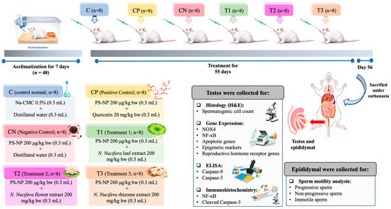
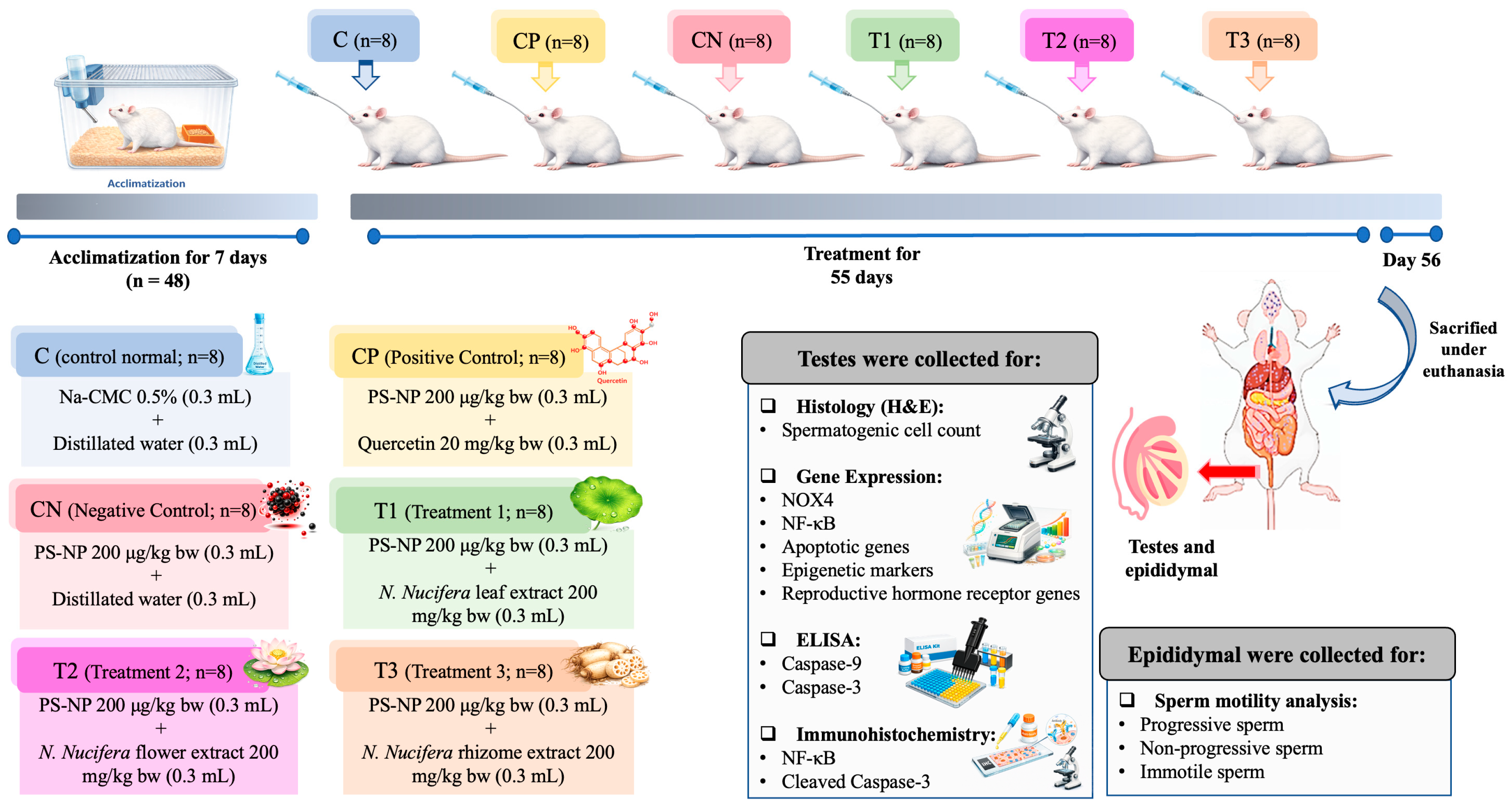
Forty-eight male Wistar rats (8–10 weeks old, 180–200 g) were obtained from Universitas Gadjah Mada and housed under standard conditions with ad libitum food and water. All procedures were approved by the Ethics Committee of Universitas Brawijaya (No. 138-KEP-UB-2024), approved on 24 July 2024. Rats were randomized into six groups (n = 8/group): control (0.5% Na-CMC; C), PS-NPs (200 µg/kg bw; CN), PS-NPs + quercetin (20 mg/kg bw; CP), and PS-NPs + N. nucifera leaf (T1), flower (T2), or rhizome (T3) extracts (200 mg/kg bw). Extract doses were selected based on prior studies and tolerability observations [21] and were freshly prepared in 0.5% Na-CMC. All treatments were administered orally once daily (0.3 mL/animal) for 55 days, covering one spermatogenic cycle. Body weight was recorded prior to sacrifice. Animals were euthanized under ketamine/xylazine anesthesia (80/10 mg/kg bw, intraperitoneal injection; ketamine, Sigma-Aldrich, USA; xylazine, Sigma-Aldrich, USA), and reproductive organs, including testes and epididymis, were carefully excised and weighed. The epididymis was used for sperm collection and subsequent evaluation of sperm motility. Testicular tissues were subsequently processed for histology, immunohistochemistry, ELISA, and qPCR (Figure 9). For all analyses, each animal was treated as an independent biological replicate (n = 8 per group), and molecular/biochemical measurements were performed in technical duplicate.
Figure 9.
Experimental design of PS-NP exposure and N. nucifera intervention in rats. Male Wistar rats (n = 48) were acclimatized for 7 days and then randomly assigned to six groups (n = 8 per group): control (C), PS-NPs + quercetin (CP), PS-NP-exposed (CN), PS-NPs + N. nucifera leaf (T1), flower (T2), or rhizome (T3). All treatments were administered orally once daily for 55 days. The control group received 0.5% Na-CMC and distilled water, the PS-NP group received PS-NPs (200 µg/kg bw), the positive control group received PS-NPs (200 µg/kg bw) plus quercetin (20 mg/kg bw), and the treatment groups received PS-NPs (200 µg/kg bw) plus N. nucifera leaf, flower, or rhizome extract (200 mg/kg bw). On day 56, animals were euthanized and the testes and epididymides were collected. Testes were used for histology, immunohistochemistry, ELISA, and gene expression analysis, whereas epididymides were used for sperm motility assessment.
4.5. Sperm Motility Analysis
Spermatozoa were collected from the cauda epididymis and suspended in physiological saline prewarmed to 37 °C. Immediately after preparation, a drop of the sperm suspension was placed on a glass slide and examined under a light microscope at 400× magnification to minimize time-dependent deterioration in motility. Spermatozoa were classified into three categories according to their movement characteristics: progressive motility (active forward movement in a straight trajectory or large circular path), non-progressive motility (motile without effective forward progression), and immotile (absence of movement). A minimum of 200 spermatozoa were assessed per sample, and the proportion of each motility category was calculated.
4.6. Histological and Immunohistochemical Analysis
Testicular tissues from each group (n = 8) were fixed in 10% neutral-buffered formalin, paraffin-embedded, sectioned at 4–5 µm, and stained with hematoxylin and eosin (H&E; Merck, Germany). Histopathological evaluation focused on spermatogenic cell populations within seminiferous tubules. Spermatogonia, spermatocytes, and spermatids were quantified in at least 100 randomly selected, non-overlapping seminiferous tubules per animal using a light microscope (Olympus BH-2, Olympus Corp., Tokyo, Japan) at 200× and 400× magnification. Cell identification was based on morphological criteria and relative position within the seminiferous epithelium: spermatogonia were identified as round to oval cells with darkly stained nuclei located along the basal membrane, spermatocytes as larger cells with prominent nuclei located in the adluminal compartment, and spermatids as smaller, more condensed cells situated near the luminal region. Only tubules with clear cross-sectional morphology were included for analysis. Cell counts were expressed as the mean number of cells per tubule for each animal and used for statistical analysis. Histological assessment was performed using standardized criteria, randomized field selection, and blinded evaluation.
For immunohistochemistry, sections were deparaffinized, rehydrated, subjected to antigen retrieval and endogenous peroxidase blocking, and incubated overnight at 4 °C with primary antibodies against cleaved caspase-3 (Asp175) (rabbit monoclonal, Cell Signaling Technology, Danvers, MA, USA; clone 5A1E, cat. no. 9664) and NF-κB p65 (mouse monoclonal, Santa Cruz Biotechnology, Dallas, TX, USA; cat. no. sc-514451). Sections were then incubated with HRP-conjugated goat anti-rabbit IgG (Santa Cruz Biotechnology, USA; cat. no. sc-2004) or goat anti-mouse IgG (Santa Cruz Biotechnology, USA; cat. no. sc-2005), followed by visualization with 3,3′-diaminobenzidine (DAB; Sigma-Aldrich, USA) and hematoxylin counterstaining. Five randomly selected, non-overlapping fields per animal were analyzed. Immunoreactivity was quantified as the percentage of DAB-positive area relative to total tissue area using ImageJ software version 1.48q (National Institutes of Health, Bethesda, MD, USA). Mean values per animal were used for statistical analysis, and image acquisition and analysis were performed under identical settings for all groups.
4.7. ELISA-Based Analysis of Apoptotic Markers
Testicular tissues were homogenized in cold phosphate-buffered saline and centrifuged to obtain the supernatants for protein analysis. Caspase-3 and caspase-9 concentrations were determined using commercial ELISA kits (BT-Lab, Shanghai, China; cat. no. E1648Ra for caspase-3 and E0627Ra for caspase-9) according to the manufacturers’ instructions. Each experimental group comprised eight biological replicates (n = 8), and all samples were analyzed in technical duplicate. Absorbance was measured using a microplate reader (Thermo Scientific, Waltham, MA, USA), and protein concentrations were calculated from standard curves.
4.8. Gene Expression Analysis by Quantitative Real-Time PCR
Total RNA was extracted from testicular tissue using the RTD050 Total RNA Mini Kit (Geneaid, New Taipei City, Taiwan), and cDNA was synthesized using ReverTra Ace™ qPCR RT Master Mix with gDNA Remover (Toyobo, Japan; FSQ-301), according to the manufacturers’ instructions. Quantitative real-time PCR (qPCR) was performed using THUNDERBIRD™ Next SYBR qPCR Mix (Toyobo, Japan; QPX-201) in a two-step amplification protocol on a real-time PCR system. The amplification conditions consisted of an initial denaturation at 95 °C for 30 s, followed by 40 cycles of denaturation at 95 °C for 5 s and annealing/extension at 60 °C for 30 s [74]. A melting curve analysis was performed at the end of each run to confirm amplification specificity. Target genes included the redox-associated marker (NOX4), apoptosis/inflammation-related genes (Fas, Caspase-8, Tp53, Bax, Caspase-9, Caspase-3, NF-κB), and epigenetic regulators (Dnmt1, Dnmt3a, Ehmt2/G9a, Hdac1, Ep300), and hormone-related receptors (LHr, FSHr, and AR). Primer sequences are listed in Table 5, and all primers were synthesized by Integrated DNA Technologies (IDT, USA). Each reaction was performed in technical duplicate, and the mean Ct value for each animal was used for downstream analysis. Relative gene expression was calculated using the 2−ΔΔCt method with GAPDH as the reference gene and is presented as fold-change relative to the control group. GAPDH was selected based on its reported expression stability and was further confirmed in the present study by minimal Ct variation across experimental groups [75].
4.9. Gene Ontology Enrichment and Protein–Protein Interaction (PPI) Analysis
Gene ontology (GO) enrichment and protein–protein interaction (PPI) analyses were conducted to provide functional context for the experimentally measured gene set. Genes quantified by qPCR were uploaded to STRING v12.0, and interaction networks were generated using a high-confidence threshold (minimum interaction score = 0.7) [76]. GO Biological Process enrichment was performed within STRING and corrected for multiple testing using the false discovery rate (FDR), with significance defined as FDR <0.05. These analyses were used to support pathway-level interpretation of coordinated stress-responsive, apoptotic, and chromatin-regulatory signatures and were not intended to establish new molecular interactions or causal relationships.
Table 5.
Primer sequence.
4.10. Network Visualization
STRING-derived PPI networks were imported into Cytoscape v3.9.1 for visualization and layout optimization. Nodes represent proteins encoded by the analyzed gene set, and edges represent known or predicted functional associations. Edge thickness was scaled to STRING interaction confidence scores to facilitate visual interpretation of network connectivity.
4.11. Molecular Docking and Dynamics
Molecular docking was performed as a supportive in silico approach to explore potential interactions between representative N. nucifera phytochemicals and apoptosis-/stress-related targets, including NF-κB, caspase-3, and caspase-9. Target protein sequences (NF-κB: Q63369; caspase-3: P55213; caspase-9: Q9JHK1) were obtained from UniProt. Ligand structures were retrieved from PubChem and Dr. Duke’s Phytochemical and Ethnobotanical Databases; quercetin (PubChem CID: 5280343) was included as a reference compound. PASS online was used to predict antioxidant-related biological activities of the selected compounds, and outputs were used as supplementary information for mechanistic interpretation. Docking simulations were performed using PyRx 0.8, and docking poses and interactions were visualized using PyMol version 3.1 and BIOVIA Discovery Studio version 2025. To assess the relative stability and flexibility of selected ligand–target complexes, molecular dynamics simulations were conducted using CABS-Flex, with root mean square fluctuation (RMSF) values computed to evaluate residue-level flexibility.
4.12. Data and Statistical Analysis
All data are presented as the mean ± standard deviation (SD). Data normality was assessed using the Shapiro–Wilk test, and homogeneity of variances was evaluated prior to parametric analysis. Differences among groups were analyzed using one-way analysis of variance (ANOVA) followed by Tukey’s multiple-comparisons post hoc test using GraphPad Prism GraphPad Software version 9.0, San Diego, CA, USA). A two-tailed p value < 0.05 was considered statistically significant. Statistical analyses were performed using mean values obtained from individual animals as biological replicates (n = 8 per group).
Author Contributions
Conceptualization, P.A.I.S., F.S. and A.H.; methodology, P.A.I.S. and F.S.; software, M.A.H.; validation, F.S.; formal analysis, P.A.I.S.; investigation, P.A.I.S., A.U.R., F.A.N. and M.A.H.; data curation, P.A.I.S., A.U.R., F.A.N. and A.H.; writing—original draft preparation, P.A.I.S. and F.S.; writing—review and editing, F.S., V.L. and A.H.; visualization, P.A.I.S. and F.S.; supervision, V.L. and A.H. All authors have read and agreed to the published version of the manuscript.
Funding
This research was funded by the Indonesian Endowment Fund for Education (LPDP) under the Ministry of Higher Education, Science, and Technology of the Republic of Indonesia, through the EQUITY Program (Contract No. 4300/B3/DT.03.08/2025 and 297/UN3/HK.07.00/2025).
Institutional Review Board Statement
The study was reviewed and approved by Ethics Committee of Universitas Brawijaya (approval code: No. 138-KEP-UB-2024 and approval date: 29 July 2024).
Informed Consent Statement
Not applicable.
Data Availability Statement
The data obtained in this study are available upon reasonable request from the corresponding author.
Acknowledgments
The authors express gratitude to the Indonesian Education Scholarship (BPI), Center for Higher Education Funding and Assessment (PPAPT), Ministry of Higher Education, Science, and Technology of Republic Indonesia.
Conflicts of Interest
The authors declare no conflicts of interest.
References
- Liu, Y.; Hao, F.; Liang, H.; Liu, W.; Guo, Y. Exposure to polystyrene nanoplastics impairs sperm metabolism and pre-implantation embryo development in mice. Front. Cell. Dev. Biol. 2025, 13, 1562331. [Google Scholar] [CrossRef] [Scilit]
- Kiran, B.R.; Kopperi, H.; Venkata Mohan, S. Micro/nano-plastics occurrence, identification, risk analysis and mitigation: Challenges and perspectives. Rev. Environ. Sci. Bio/Technol 2022, 21, 169–203. [Google Scholar] [CrossRef] [Scilit]
- Saputra, F.; Tsao, Y.-T.; Soegianto, A.; Pramata, A.D.; Hu, S.-Y. Developmental cardiovascular disruption triggered by polystyrene nanoplastics in zebrafish mediated through oxidative stress. Life Sci. 2025, 382, 124056. [Google Scholar] [CrossRef] [Scilit]
- Zhang, X.; Wu, Y.; Fu, X.; He, S.; Shi, L.; Xu, H.; Shi, X.; Yang, Y.; Zhu, Y.; Wang, Y.; et al. Toxicity to the Male Reproductive System after Exposure to Polystyrene Nanoplastics: A Macrogenomic and Metabolomic Analysis. Toxics 2024, 12, 531. [Google Scholar] [CrossRef] [Scilit] [PubMed]
- Li, S.; Ma, Y.; Ye, S.; Su, Y.; Hu, D.; Xiao, F. Endogenous hydrogen sulfide counteracts polystyrene nanoplastics-induced mitochondrial apoptosis and excessive autophagy via regulating Nrf2 and PGC-1α signaling pathway in mouse spermatocyte-derived GC-2spd(ts) cells. Food Chem. Toxicol. 2022, 164, 113071. [Google Scholar] [CrossRef] [Scilit]
- Alex, S.A.; George, N.K.; Guardiola, J.; Clegg, D. The Threat of Micro-/Nanoplastics to Male Fertility: A Review of the Data and the Importance of Future Research. Int. J. Mol. Sci. 2025, 26, 11457. [Google Scholar] [CrossRef] [Scilit]
- Lv, J.; Liu, G.; Wang, Z.; Zhang, J.; Li, Y.; Wang, Y.; Liu, N.; Altyn, S.; Jiang, Z. Internalized polystyrene nanoplastics trigger testicular damage and promote ferroptosis via CISD1 downregulation in mouse spermatocyte. J. Nanobiotechnol. 2025, 23, 537. [Google Scholar] [CrossRef] [Scilit] [PubMed]
- Feng, H.; Huang, Y.; Zhang, H.; Li, Z.; Cen, X.; Zhuang, Y.; Zhang, X.; Ma, K.; Ruan, S.; Yi, R.; et al. Multi-omics analysis of testis after long-term exposure to polystyrene nanoplastics reveals premature testicular aging and age-dependent damage in mice. Part. Fibre Toxicol. 2025, 22, 27. [Google Scholar] [CrossRef] [Scilit] [PubMed]
- Saputra, F.; Pramata, A.D.; Soegianto, A.; Hu, S.-Y. Polystyrene nanoplastics cause developmental abnormalities, oxidative damage and immune toxicity in early zebrafish development. Comp. Biochem. Physiol. C 2025, 295, 110216. [Google Scholar] [CrossRef] [Scilit]
- Bridgeman, L.; Cimbalo, A.; López-Rodríguez, D.; Pamies, D.; Frangiamone, M. Exploring toxicological pathways of microplastics and nanoplastics: Insights from animal and cellular models. J. Hazard. Mater. 2025, 490, 137795. [Google Scholar] [CrossRef] [Scilit]
- Almarzouq, D.; Al-Maghrebi, M. NADPH Oxidase-Mediated Testicular Oxidative Imbalance Regulates the TXNIP/NLRP3 Inflammasome Axis Activation after Ischemia Reperfusion Injury. Antioxidants 2023, 12, 145. [Google Scholar] [CrossRef] [Scilit] [PubMed]
- Al-Saleh, F.; Khashab, F.; Fadel, F.; Al-Kandari, N.; Al-Maghrebi, M. Inhibition of NADPH oxidase alleviates germ cell apoptosis and ER stress during testicular ischemia reperfusion injury. Saudi J. Biol. Sci. 2020, 27, 2174–2184. [Google Scholar] [CrossRef] [Scilit]
- Schieber, M.; Chandel, N.S. ROS function in redox signaling and oxidative stress. Curr. Biol. 2014, 24, R453–R462. [Google Scholar] [CrossRef] [Scilit] [PubMed]
- Chulkov, V.; Gasanov, M.; Isakov, V.; Denisenko, A.; Nwosu, C.; Rodkin, S. Molecular and Cellular Effects of Microplastics and Nanoplastics in the Pathogenesis of Cardiovascular, Nervous, Urinary, Digestive, and Reproductive System Diseases: A Global Systematic Review. Int. J. Mol. Sci. 2025, 26, 11194. [Google Scholar] [CrossRef] [Scilit] [PubMed]
- Hou, B.; Wang, F.; Liu, T.; Wang, Z. Reproductive toxicity of polystyrene microplastics: In vivo experimental study on testicular toxicity in mice. J. Hazard. Mater. 2021, 405, 124028. [Google Scholar] [CrossRef] [Scilit]
- Xu, W.; Yuan, Y.; Tian, Y.; Cheng, C.; Chen, Y.; Zeng, L.; Yuan, Y.; Li, D.; Zheng, L.; Luo, T. Oral exposure to polystyrene nanoplastics reduced male fertility and even caused male infertility by inducing testicular and sperm toxicities in mice. J. Hazard. Mater. 2023, 454, 131470. [Google Scholar] [CrossRef] [Scilit]
- Kaltsas, A. Oxidative Stress and Male Infertility: The Protective Role of Antioxidants. Medicina 2023, 59, 1769. [Google Scholar] [CrossRef] [Scilit]
- Sharma, P.; Kaushal, N.; Saleth, L.R.; Ghavami, S.; Dhingra, S.; Kaur, P. Oxidative stress-induced apoptosis and autophagy: Balancing the contrary forces in spermatogenesis. Biochim. Biophys. Acta Mol. Basis Dis. 2023, 1869, 166742. [Google Scholar] [CrossRef] [Scilit]
- Asadi, A.; Ghahremani, R.; Abdolmaleki, A.; Rajaei, F. Role of sperm apoptosis and oxidative stress in male infertility: A narrative review. Int. J. Reprod. Biomed. 2021, 19, 493–504. [Google Scholar] [CrossRef] [Scilit]
- Maulidah, S.; Saputra, F.; Hayati, A.; Herdiansyah, M.A.; Pramudya, M.; Primula Dewi, F.R.; Kuncoroningrat Susilo, R.J.; Lim, V.; Pramata, A.D. Disruption of gluconeogenesis as a key mediator of male infertility in rats exposed to nanoplastics: In vivo evidence and molecular docking analysis. Emerg. Contam. 2025, 11, 100581. [Google Scholar] [CrossRef] [Scilit]
- Setiyowati, P.A.I.; Saputra, F.; Dewi, F.R.P.; Herdiansyah, M.A.; Lim, V.; Hayati, A. Metabolic–endocrine remodelling of the testis under polystyrene nanoplastic exposure: Intervention by organ-specific phytocomplexes of Nelumbo nucifera. Reprod. Biol. 2026, 26, 101183. [Google Scholar] [CrossRef] [Scilit]
- Hassanein, E.H.M.; Alotaibi, M.F.; Alruhaimi, R.S.; Sabry, M.; Sayed, G.A.; Atwa, A.M.; Mahmoud, A.M. Targeting TLR4/NF-κB signaling, oxidative stress, and apoptosis by farnesol mitigates cadmium-induced testicular toxicity in rats. Tissue Cell 2025, 94, 102813. [Google Scholar] [CrossRef] [Scilit]
- Yu, C.W.; Luk, T.C.; Liao, V.H. Long-term nanoplastics exposure results in multi and trans-generational reproduction decline associated with germline toxicity and epigenetic regulation in Caenorhabditis elegans. J. Hazard. Mater. 2021, 412, 125173. [Google Scholar] [CrossRef] [Scilit] [PubMed]
- Shen, R.; Xia, P.; Guo, Y.; Ji, P.; Yuan, X.; Wang, L.; Shuang, S.; Zhou, L.; Tong, R.; Zhang, L.; et al. Effects of polystyrene microparticles exposures on spermatogenic cell differentiation and reproductive endpoints in male mice. Environ. Pollut. 2025, 373, 126200. [Google Scholar] [CrossRef] [Scilit] [PubMed]
- Gladwell, L.R.; Karthik, J.; Packer, L.; Venkategowda, S.; Choudhury, M. Micro- and nanoplastics: Emerging environmental threats to the Developmental Origins of Health and Disease. Reprod. Toxicol. 2026, 139, 109098. [Google Scholar] [CrossRef] [Scilit]
- Pogribna, M.; Hammons, G. Epigenetic Effects of Nanomaterials and Nanoparticles. J. Nanobiotechnol. 2021, 19, 2. [Google Scholar] [CrossRef] [Scilit]
- Hekmatdoost, A.; Lakpour, N.; Sadeghi, M.R. Sperm chromatin integrity: Etiologies and mechanisms of abnormality, assays, clinical importance, preventing and repairing damage. Avicenna J. Med. Biotechnol. 2009, 1, 147–160. [Google Scholar]
- Mahmud, F.; Sarker, D.B.; Jocelyn, J.A.; Sang, Q.A. Molecular and Cellular Effects of Microplastics and Nanoplastics: Focus on Inflammation and Senescence. Cells 2024, 13, 1788. [Google Scholar] [CrossRef] [Scilit] [PubMed]
- Sranujit, R.P.; Noysang, C.; Tippayawat, P.; Kooltheat, N.; Luetragoon, T.; Usuwanthim, K. Phytochemicals and Immunomodulatory Effect of Nelumbo nucifera Flower Extracts on Human Macrophages. Plants 2021, 10, 2007. [Google Scholar] [CrossRef] [Scilit]
- Setiyowati, P.A.I.; Lim, V.; Hayati, A.; Maghfuroh, L.; Ramadani, A.H. Phytochemical analysis of lotus (Nelumbo nucifera L.) rhizomes in Joto Reservoir, East Java, Indonesia. SABRAO J. Breed. Genet. 2025, 57, 2127–2138. [Google Scholar] [CrossRef] [Scilit]
- Bhat, S.; Joghee, S.; Iyer, M.S. The Therapeutic Potential of Nelumbo Nucifera: A Comprehensive Review of Its Phytochemistry and Medicinal Properties. Int. J. Health Allied Sci. 2023, 12, 8. [Google Scholar] [CrossRef] [Scilit]
- Gao, L.; Xiong, X.; Chen, C.; Luo, P.; Li, J.; Gao, X.; Huang, L.; Li, L. The male reproductive toxicity after nanoplastics and microplastics exposure: Sperm quality and changes of different cells in testis. Ecotoxicol. Environ. Saf. 2023, 267, 115618. [Google Scholar] [CrossRef] [Scilit]
- Cui, L.; Li, W.; He, S.; Guo, L.; Liu, K.; Dong, J.; Li, J.; Wang, H. Inhibition of NOX4-Mediated ROS Production Contributes to Selenomethionine’s Anti-Inflammatory Effect in LPS-Stimulated Bovine Endometrial Epithelial Cells. Vet. Sci. 2025, 12, 789. [Google Scholar] [CrossRef] [Scilit]
- Szanto, I. NADPH Oxidase 4 (NOX4) in Cancer: Linking Redox Signals to Oncogenic Metabolic Adaptation. Int. J. Mol. Sci. 2022, 23, 2702. [Google Scholar] [CrossRef] [Scilit] [PubMed]
- Vermot, A.; Petit-Härtlein, I.; Smith, S.M.E.; Fieschi, F. NADPH Oxidases (NOX): An Overview from Discovery, Molecular Mechanisms to Physiology and Pathology. Antioxidants 2021, 10, 890. [Google Scholar] [CrossRef] [Scilit]
- Halimu, G.; Zhang, Q.; Liu, L.; Zhang, Z.; Wang, X.; Gu, W.; Zhang, B.; Dai, Y.; Zhang, H.; Zhang, C.; et al. Toxic effects of nanoplastics with different sizes and surface charges on epithelial-to-mesenchymal transition in A549 cells and the potential toxicological mechanism. J. Hazard. Mater. 2022, 430, 128485. [Google Scholar] [CrossRef] [Scilit]
- Peoples, J.N.; Saraf, A.; Ghazal, N.; Pham, T.T.; Kwong, J.Q. Mitochondrial dysfunction and oxidative stress in heart disease. Exp. Mol. Med. 2019, 51, 1–13. [Google Scholar] [CrossRef] [Scilit] [PubMed]
- Dutta, S.; Sengupta, P.; Slama, P.; Roychoudhury, S. Oxidative Stress, Testicular Inflammatory Pathways, and Male Reproduction. Int. J. Mol. Sci. 2021, 22, 10043. [Google Scholar] [CrossRef] [Scilit]
- Bacher, S.; Meier-Soelch, J.; Kracht, M.; Schmitz, M.L. Regulation of Transcription Factor NF-κB in Its Natural Habitat: The Nucleus. Cells 2021, 10, 753. [Google Scholar] [CrossRef] [Scilit]
- Thoms, H.C.; Stark, L.A. The NF-κB Nucleolar Stress Response Pathway. Biomedicines 2021, 9, 1082. [Google Scholar] [CrossRef] [Scilit] [PubMed]
- Kose, O.; Mantecca, P.; Costa, A.; Carrière, M. Putative adverse outcome pathways for silver nanoparticle toxicity on mammalian male reproductive system: A literature review. Part. Fibre Toxicol. 2023, 20, 1. [Google Scholar] [CrossRef] [Scilit]
- Rao, G.; Qiao, B.; Zhong, G.; Li, T.; Su, Q.; Wu, S.; Tang, Z.; Hu, L. Arsenic and polystyrene-nano plastics co-exposure induced testicular toxicity: Triggers oxidative stress and promotes apoptosis and inflammation in mice. Environ. Toxicol. 2024, 39, 264–276. [Google Scholar] [CrossRef] [Scilit]
- Li, Y.; Liu, Y.; Chen, Y.; Yao, C.; Yu, S.; Qu, J.; Chen, G.; Wei, H. Combined effects of polystyrene nanoplastics and lipopolysaccharide on testosterone biosynthesis and inflammation in mouse testis. Ecotoxicol. Environ. Saf. 2024, 273, 116180. [Google Scholar] [CrossRef] [Scilit]
- Peng, Y.; He, Q. Reproductive toxicity and related mechanisms of micro(nano)plastics in terrestrial mammals: Review of current evidence. Ecotoxicol. Environ. Saf. 2024, 279, 116505. [Google Scholar] [CrossRef] [Scilit]
- da Cunha de Medeiros, P.; Silva, A.G.G.d.; Angelo, A.B.S.; Moura, M.J.N.d.; Kannan, U.; Gregory, M.; Dufresne, J.; dos Santos Borges, C.; Cyr, D.G. Unravelling the potential mechanisms of nano- and microplastic toxicity to the male reproductive system: A systematic review. Reprod. Toxicol. 2025, 137, 109002. [Google Scholar] [CrossRef] [Scilit] [PubMed]
- Li, P.; Zhou, L.; Zhao, T.; Liu, X.; Zhang, P.; Liu, Y.; Zheng, X.; Li, Q. Caspase-9: Structure, mechanisms and clinical application. Oncotarget 2017, 8, 23996–24008. [Google Scholar] [CrossRef] [Scilit] [PubMed]
- Sendtner, N.; Seitz, R.; Brandl, N.; Müller, M.; Gülow, K. Reactive Oxygen Species Across Death Pathways: Gatekeepers of Apoptosis, Ferroptosis, Pyroptosis, Paraptosis, and Beyond. Int. J. Mol. Sci. 2025, 26, 10240. [Google Scholar] [CrossRef] [Scilit]
- Dirican, E.; Özcan, H.; Karabulut Uzunçakmak, S.; Takım, U. Evaluation Expression of the Caspase-3 and Caspase-9 Apoptotic Genes in Schizophrenia Patients. Clin. Psychopharmacol. Neurosci. 2023, 21, 171–178. [Google Scholar] [CrossRef] [Scilit] [PubMed]
- Oduwole, O.O.; Huhtaniemi, I.T.; Misrahi, M. The Roles of Luteinizing Hormone, Follicle-Stimulating Hormone and Testosterone in Spermatogenesis and Folliculogenesis Revisited. Int. J. Mol. Sci. 2021, 22, 12735. [Google Scholar] [CrossRef] [Scilit]
- López-Doval, S.; Salgado, R.; Lafuente, A. The expression of several reproductive hormone receptors can be modified by perfluorooctane sulfonate (PFOS) in adult male rats. Chemosphere 2016, 155, 488–497. [Google Scholar] [CrossRef] [Scilit]
- Alsenousy, A.H.A.; Khalaf, A.H.Y.; Ibrahim, H.Z.; Kamel, M.A.; Yousef, M.I. Impact of polystyrene microplastic exposure at low doses on male fertility: An experimental study in rats. Sci. Rep. 2026, 16, 7474. [Google Scholar] [CrossRef] [Scilit]
- Fang, Q.; Wang, C.; Xiong, Y. Polystyrene microplastics induce male reproductive toxicity in mice by activating spermatogonium mitochondrial oxidative stress and apoptosis. Chem.-Biol. Interact. 2024, 396, 111043. [Google Scholar] [CrossRef] [Scilit]
- Erdogan, K.; Ceylani, T.; Teker, H.T.; Sengil, A.Z.; Uysal, F. Young plasma transfer recovers decreased sperm counts and restores epigenetics in aged testis. Exp. Gerontol. 2023, 172, 112042. [Google Scholar] [CrossRef] [Scilit]
- Firouzabadi, A.M.; Fesahat, F.; Seifati, S.M. Dynamic architecture of mammalian paternal chromatin: Histone-to-protamine exchange and post-fertilization reprogramming. Epigenet. Chromatin 2025, 18, 83. [Google Scholar] [CrossRef] [Scilit]
- Ju, H.; Geng, Z.; Chen, B.; Shang, Y.; Chen, X.; Wang, D.; Wang, W.; Sun, H.; Shi, Y.; Yu, J. Mechanism of PP2A affecting ubiquitination pathway in spermatogenesis. Reprod. Biol. 2026, 26, 101167. [Google Scholar] [CrossRef] [Scilit]
- Auclair, G.; Borgel, J.; Sanz, L.A.; Vallet, J.; Guibert, S.; Dumas, M.; Cavelier, P.; Girardot, M.; Forné, T.; Feil, R.; et al. EHMT2 directs DNA methylation for efficient gene silencing in mouse embryos. Genome Res. 2016, 26, 192–202. [Google Scholar] [CrossRef] [Scilit] [PubMed]
- Kaltsas, A.; Markou, E.; Kyrgiafini, M.-A.; Zikopoulos, A.; Symeonidis, E.N.; Dimitriadis, F.; Zachariou, A.; Sofikitis, N.; Chrisofos, M. Oxidative-Stress-Mediated Epigenetic Dysregulation in Spermatogenesis: Implications for Male Infertility and Offspring Health. Genes 2025, 16, 93. [Google Scholar] [CrossRef] [Scilit] [PubMed]
- Rotondo, J.C.; Lanzillotti, C.; Mazziotta, C.; Tognon, M.; Martini, F. Epigenetics of Male Infertility: The Role of DNA Methylation. Front. Cell Dev. Biol. 2021, 9, 689624. [Google Scholar] [CrossRef] [Scilit]
- Anjum, S.; Khurshid, Y.; Du Plessis, S.S.; Omolaoye, T.S. Unmasking the Epigenome: Insights into Testicular Cell Dynamics and Reproductive Function. Int. J. Mol. Sci. 2025, 26, 7305. [Google Scholar] [CrossRef] [Scilit]
- James, E.R.; Tasnim, M.; Riera-Escamilla, A.; Vigh-Conrad, K.; Emery, B.R.; Conrad, D.F.; Aston, K.I. Genetic and epigenetic landscape of male infertility. Trends Genet. 2025, 41, 883–898. [Google Scholar] [CrossRef] [Scilit] [PubMed]
- Cardamone, F.; Piva, A.; Löser, E.; Eichenberger, B.; Romero-Mulero, M.C.; Zenk, F.; Shields, E.J.; Cabezas-Wallscheid, N.; Bonasio, R.; Tiana, G.; et al. Chromatin landscape at cis-regulatory elements orchestrates cell fate decisions in early embryogenesis. Nat. Commun. 2025, 16, 3007. [Google Scholar] [CrossRef] [Scilit]
- Malinowska, K.; Tarhonska, K.; Górecka, K.; Tokarz, P.; Jabłońska, E.; Balcerczyk, A.; Reszka, E.; Sicińska, P.; Mokra, K.; Bukowska, B. Exploring the effect of short-term exposure to non-functionalized polystyrene nanoparticles on selected chromatin determinants in human immune cells and plasmid DNA integrity. Nanotoxicology 2025, 19, 575–588. [Google Scholar] [CrossRef] [Scilit] [PubMed]
- Szklarczyk, D.; Kirsch, R.; Koutrouli, M.; Nastou, K.; Mehryary, F.; Hachilif, R.; Gable, A.L.; Fang, T.; Doncheva, N.T.; Pyysalo, S.; et al. The STRING database in 2023: Protein-protein association networks and functional enrichment analyses for any sequenced genome of interest. Nucleic Acids Res. 2023, 51, D638–D646. [Google Scholar] [CrossRef] [Scilit] [PubMed]
- López-Díaz, F.J.; Gascard, P.; Balakrishnan, S.K.; Zhao, J.; Del Rincon, S.V.; Spruck, C.; Tlsty, T.D.; Emerson, B.M. Coordinate transcriptional and translational repression of p53 by TGF-β1 impairs the stress response. Mol. Cell 2013, 50, 552–564. [Google Scholar] [CrossRef] [Scilit]
- Maroto, M.; Torvisco, S.N.; García-Merino, C.; Fernández-González, R.; Pericuesta, E. Mechanisms of Hormonal, Genetic, and Temperature Regulation of Germ Cell Proliferation, Differentiation, and Death During Spermatogenesis. Biomolecules 2025, 15, 500. [Google Scholar] [CrossRef] [Scilit]
- Syaputra, A.A.; Setiyowati, P.A.I.; Musyarofah, B.; Kasanah, Y.A.; Rahmawati, A.K.R.; Qotrunnada, H.S. Bioactivity of Nelumbo nucifera extract on sperm recovery due to 2-methoxyethanol exposure: In vivo and in silico study. J. Teknol. Lab. 2023, 12, 89–100. [Google Scholar] [CrossRef] [Scilit]
- Choo, J.H.; Lee, S.Y.; Min, K.; Kang, N.G. Chemical Composition and Bioactivity of Nelumbo nucifera Gaertn. Flower Extract Fractions: In Vitro Antioxidant and Anti-Inflammatory Properties. Curr. Issues Mol. Biol. 2025, 47, 1065. [Google Scholar] [CrossRef] [Scilit]
- Luan, J.; Zhang, X.; Chen, T.; Pu, S.; Shen, Z.; Xu, C.; Chen, Z.; Zhang, J.; Chen, D. Adolescent exposure to polystyrene nanoplastics induces male reproductive damage via the microbiome-gut-testis axis. J. Nanobiotechnol. 2026, 24, 183. [Google Scholar] [CrossRef] [Scilit]
- Paudel, K.R.; Panth, N. Phytochemical Profile and Biological Activity of Nelumbo nucifera. Evid. Based Complement. Altern. Med. 2015, 2015, 789124. [Google Scholar] [CrossRef] [Scilit]
- Oeckinghaus, A.; Ghosh, S. The NF-kappaB family of transcription factors and its regulation. Cold Spring Harb. Perspect. Biol. 2009, 1, a000034. [Google Scholar] [CrossRef] [Scilit]
- Rudrapal, M.; Rakshit, G.; Singh, R.P.; Garse, S.; Khan, J.; Chakraborty, S. Dietary Polyphenols: Review on Chemistry/Sources, Bioavailability/Metabolism, Antioxidant Effects, and Their Role in Disease Management. Antioxidants 2024, 13, 429. [Google Scholar] [CrossRef] [Scilit] [PubMed]
- Khazaeel, K.; Daaj, S.A.Z.; Sadeghi, A.; Tabandeh, M.R.; Basir, Z. Potential protective effect of quercetin on the male reproductive system against exposure of Wistar rats to crude oil vapor: Genetic, biochemical, and histopathological evidence. Reprod. Toxicol. 2022, 113, 10–17. [Google Scholar] [CrossRef] [Scilit]
- Alharbi, H.O.A.; Alshebremi, M.; Babiker, A.Y.; Rahmani, A.H. The Role of Quercetin, a Flavonoid in the Management of Pathogenesis Through Regulation of Oxidative Stress, Inflammation, and Biological Activities. Biomolecules 2025, 15, 151. [Google Scholar] [CrossRef] [Scilit]
- Saputra, F.; Hu, S.-Y.; Kishida, M. Exposure to nitrate and nitrite disrupts cardiovascular development through estrogen receptor in zebrafish embryos and larvae. Fish. Physiol. Biochem. 2024, 50, 2165–2178. [Google Scholar] [CrossRef] [Scilit] [PubMed]
- Lucas Nascimento Souza, J.; Cavalcante Silva, F.; da Silva, C.G.; Maria Fortaleza Neves Bomfim, I.; Rocha de Medeiros, H.; Giotto Zaros, L. Analysis of the stability of the reference genes GAPDH, SDHA and RPL-19 in sheep from a semi-arid region infected by gastrointestinal nematodes. BMC Vet. Res. 2023, 19, 147. [Google Scholar] [CrossRef] [Scilit]
- Szklarczyk, D.; Gable, A.L.; Nastou, K.C.; Lyon, D.; Kirsch, R.; Pyysalo, S.; Doncheva, N.T.; Legeay, M.; Fang, T.; Bork, P.; et al. Correction to ‘The STRING database in 2021: Customizable protein–protein networks, and functional characterization of user-uploaded gene/measurement sets’. Nucleic Acids Res. 2021, 49, 10800. [Google Scholar] [CrossRef] [Scilit]
- Rizk, F.H.; Soliman, N.A.; Abo-Elnasr, S.E.; Mahmoud, H.A.; Abdel Ghafar, M.T.; Elkholy, R.A.; Elshora, O.A.; Mariah, R.A.; Amin Mashal, S.S.; El Saadany, A.A. Fisetin ameliorates oxidative glutamate testicular toxicity in rats via central and peripheral mechanisms involving SIRT1 activation. Redox Rep. 2022, 27, 177–185. [Google Scholar] [CrossRef] [Scilit] [PubMed]
- El-Nekeety, A.A.; Salman, A.S.; Hathout, A.S.; Sabry, B.A.; Abdel-Aziem, S.H.; Hassan, N.S.; Abdel-Wahhab, M.A. Evaluation of the bioactive extract of actinomyces isolated from the Egyptian environment against aflatoxin B1-induce cytotoxicity, genotoxicity and oxidative stress in the liver of rats. Food Chem. Toxicol. 2017, 105, 241–255. [Google Scholar] [CrossRef] [Scilit]
- Sheen, J.-M.; Chen, Y.-C.; Hsu, M.-H.; Tain, Y.-L.; Huang, Y.-H.; Tiao, M.-M.; Li, S.-W.; Huang, L.-T. Melatonin Alleviates Liver Apoptosis in Bile Duct Ligation Young Rats. Int. J. Mol. Sci. 2016, 17, 1365. [Google Scholar] [CrossRef] [Scilit]
- Wang, Y.; Liu, H.; Liu, X.; Zhang, X.; Wu, J.; Yuan, L.; Du, X.; Wang, R.; Ma, Y.; Chen, X.; et al. Histone acetylation plays an important role in MC-LR-induced apoptosis and cycle disorder in SD rat testicular cells. Chemosphere 2020, 241, 125073. [Google Scholar] [CrossRef] [Scilit]
- Chung, S.; Yi, Y.; Ullah, I.; Chung, K.; Park, S.; Lim, J.; Kim, C.; Pyun, S.-H.; Kim, M.; Kim, D.; et al. Systemic Treatment with Fas-Blocking Peptide Attenuates Apoptosis in Brain Ischemia. Int. J. Mol. Sci. 2024, 25, 661. [Google Scholar] [CrossRef] [Scilit]
- Zheng, X.; Li, Z.; Wang, G.; Li, Z.; Liang, A.; Wang, H.; Dai, Y.; Huang, X.; Chen, X.; Ma, Y.; et al. Overexpression of Human-Derived DNMT3A Induced Intergenerational Inheritance of Active DNA Methylation Changes in Rat Sperm. Front. Genet. 2017, 8, 207. [Google Scholar] [CrossRef] [Scilit] [PubMed]
- Boovarahan, S.R.; Kurian, G.A. Investigating the role of DNMT1 gene expression on myocardial ischemia reperfusion injury in rat and associated changes in mitochondria. Biochim. Biophys. Acta (BBA)—Bioenerg 2022, 1863, 148566. [Google Scholar] [CrossRef] [Scilit] [PubMed]
- Yan, F.; Chen, Z.; Cui, W. H3K9me2 regulation of BDNF expression via G9a partakes in the progression of heart failure. BMC Cardiovasc. Disord. 2022, 22, 182. [Google Scholar] [CrossRef] [Scilit]
- Yang, L.; Tian, J.; Wang, J.; Zeng, J.; Wang, T.; Lin, B.; Linneman, J.; Li, L.; Niu, Y.; Gou, D.; et al. The protective role of EP300 in monocrotaline-induced pulmonary hypertension. Front. Cardiovasc. Med. 2023, 10, 1037217. [Google Scholar] [CrossRef] [Scilit]
- Susilo, R.J.K.; Pramudya, M.; Dewi, F.R.P.; Seftiarini, W.; Hidayati, D.; Aunurohim, A.; Lim, V.; Herdiansyah, M.A.; Hayati, A. Adverse Effect of Polystyrene Nanoplastics in Impairing Glucose Metabolism in Liver Injury. Int. J. Mol. Sci. 2025, 26, 4870. [Google Scholar] [CrossRef] [Scilit] [PubMed]

Disclaimer/Publisher’s Note: The statements, opinions and data contained in all publications are solely those of the individual author(s) and contributor(s) and not of MDPI and/or the editor(s). MDPI and/or the editor(s) disclaim responsibility for any injury to people or property resulting from any ideas, methods, instructions or products referred to in the content. |
© 2026 by the authors. Licensee MDPI, Basel, Switzerland. This article is an open access article distributed under the terms and conditions of the Creative Commons Attribution (CC BY) license.