


Abstract
Background/Objectives: Wheelchair fencing requires effective trunk coordination because athletes cannot use the lower limbs for propulsion. This exploratory study investigated whether coach-guided scapular-control instruction was associated with differences in spinal kinematics during fencing lunges. Methods: Seven competitive wheelchair fencers performed six lunges under Natural and Coach-guided conditions. Four inertial measurement units were positioned at C7–Th1, Th6–Th7, Th12–L1, and L5–S1. Eight kinematic outcomes were analyzed using linear mixed-effects models with Condition, Segment, and their interaction as fixed effects and Participant as a random intercept. Repeated lunges were averaged within participant, condition, and segment, resulting in 56 observations. Benjamini–Hochberg FDR correction was applied across 24 omnibus tests, followed by Holm correction for post hoc segment comparisons. Results: Three effects remained significant after FDR correction. RMS angular acceleration differed between conditions (F(1,42) = 17.00, p < 0.001, q = 0.004, η2p = 0.288). Significant Segment effects were observed for peak angular jerk (F(3,42) = 5.00, p = 0.005, q = 0.038, η2p = 0.263) and time to peak angular velocity (F(3,42) = 5.33, p = 0.003, q = 0.038, η2p = 0.276). Holm-adjusted comparisons showed significant differences between L5–S1 and Th6–Th7 for peak angular jerk (pHolm = 0.010) and time to peak angular velocity (pHolm = 0.004). No Condition × Segment interaction survived FDR correction. Conclusions: The findings indicate condition-associated differences in RMS angular acceleration and segment-related differences in peak angular jerk and timing of peak angular velocity. Given the small sample and fixed condition order, these findings should be interpreted as exploratory associations rather than evidence of a causal coaching effect.
1. Introduction
Wheelchair fencing is a Paralympic combat sport, performed in a seated position that requires high levels of upper-body coordination, trunk stability, and movement precision. Unlike able-bodied fencing, athletes compete in fixed wheelchairs attached to the dedicated platform, which eliminates lower-limb contribution during offensive and defensive actions, ensuring stable positioning and preventing wheelchair displacement. Consequently, movement generation depends primarily on the coordinated interaction between the trunk, shoulder girdle, and upper limbs. Efficient trunk control is therefore considered a key determinant of movement effectiveness, technical execution, and postural stability in wheelchair fencing [1,2].
The classification system in wheelchair fencing is largely based on functional trunk ability. Wheelchair fencing athletes are classified into functional categories A, B, and C according to the impact of impairment on sitting balance, trunk control, and fencing-specific upper-limb function. Categories A and B are included in the Paralympic Games, whereas category C may be used in World Para Fencing competitions. Athletes classified in category A demonstrate better sitting balance, stronger trunk musculature, and greater postural stability, enabling more dynamic trunk movements during fencing actions. In contrast, category B athletes present reduced trunk control and sitting stability, often relying more heavily on compensatory upper-limb strategies. Fung et al. demonstrated that trunk kinematics differ between wheelchair fencing classes and may influence movement execution and sport performance. Chung [3] further reported that category B fencers exhibit altered kinematic and electromyographic patterns during lunging actions, likely as compensatory adaptations related to reduced trunk stability.
The fencing lunge is one of the most important offensive techniques in wheelchair fencing and requires rapid forward trunk movement, coordinated upper-limb acceleration, and controlled recovery. Since wheelchair fencers cannot generate momentum through lower-limb propulsion, trunk muscles play a crucial role in force transmission and movement initiation. Previous studies highlighted the importance of postural muscles and anticipatory trunk stabilization during fencing actions. Borysiuk, et al. [4] demonstrated that trunk stabilizer muscles significantly contribute to movement organization and technical execution in wheelchair fencers. Moreover, impaired trunk stability has been associated with greater shoulder loading and increased risk of overuse injuries in wheelchair athletes [5].
Scapular positioning and scapulothoracic control may also contribute to upper-body movement organization through their mechanical relationship with the trunk and shoulder girdle. Evidence from able-bodied sport suggests that scapular and trunk coordination may contribute to upper-body movement organization during rapid and explosive sporting actions. However, these findings cannot be directly generalized to wheelchair athletes, because the seated, fixed-base position and absence of lower-limb propulsion substantially alter trunk and shoulder-girdle mechanics. Evidence from wheelchair sports similarly indicates that trunk control is an important determinant of upper-body movement organization. In wheelchair fencing specifically, Raud, et al. [6] suggested that scapular and clavicular movement strategies may influence reach capacity and upper-body coordination during fencing-specific actions.
Despite these observations, the specific relationship between coach-guided scapular-control instruction and spinal kinematics during wheelchair fencing lunges remains insufficiently investigated. Studies in wheelchair fencing have primarily addressed trunk kinematics, functional classification, scapular/clavicular motion, or muscle activation, rather than examining whether a standardized coach-guided scapular-control instruction is associated with segment-specific changes in spinal movement patterns. The novelty of the present study therefore lies in examining this relationship during a sport-specific wheelchair fencing lunge using IMU-derived angular acceleration and jerk measures across multiple spinal levels.
In recent years, wearable sensor technologies such as inertial measurement units (IMUs) have become increasingly popular in sports biomechanics and rehabilitation research. Compared with traditional laboratory-based optical motion capture systems, IMUs provide portable, non-invasive, and ecologically valid measurements that can be performed directly in sport-specific environments. IMU systems combine accelerometer and gyroscope measurements to capture three-dimensional linear acceleration and angular velocity during dynamic movements. Recent methodological studies have demonstrated the potential of inertial sensing for quantifying rotational motion and estimating angular velocity from multi-axis or multi-sensor configurations [7,8]. Several studies have also demonstrated the reliability and validity of IMU-based systems for assessing trunk motion and postural control during athletic activities. Brouwer, et al. [9] demonstrated accurate three-dimensional trunk orientation measurements using IMUs during dynamic sports movements, while Chia, et al. [10] confirmed the validity of IMU systems for evaluating trunk kinematics during high-speed athletic tasks. Recent reviews further highlighted the growing role of IMU-based motion analysis in sports biomechanics and Paralympic sport assessment [11]. However, the interpretation of IMU-derived orientation data requires consideration of sensor placement, soft-tissue movement, and the methodological limitations associated with estimating absolute segment posture during dynamic tasks. Consequently, IMU technology can provide not only angular velocity and acceleration data but also valuable information regarding segment orientation and postural coordination during sport-specific tasks [12].
Among different spinal regions, the thoracic segment appears particularly relevant for evaluating trunk coordination during upper-body dominant movements. The mid-thoracic region (Th6–Th7) reflects global trunk stability and rotational control while being less affected by isolated head or upper-limb movement than cervical segments [13,14]. In wheelchair athletes, thoracic motion may also better represent functional trunk control than lower lumbar regions, which are often partially stabilized by the seated position. Therefore, thoracic IMU placement may provide relevant information regarding movement coordination and compensatory strategies during wheelchair fencing actions [15]. Therefore, the aim of this exploratory study was to evaluate whether coach-guided scapular-control instruction was associated with differences in spinal kinematics during a fencing lunge in wheelchair fencers. It was hypothesized that coach-guided scapular-control instruction would be associated with differences in spinal kinematics, particularly in angular acceleration- and jerk-related parameters within the thoracic and lumbosacral spinal regions. From a practical perspective, understanding whether coach-guided scapular-control instruction is associated with changes in spinal kinematics may contribute to the biomechanical understanding of movement organization in wheelchair fencing. However, because scapular kinematics and muscle activation were not directly assessed, the study was not designed to demonstrate that participants actually achieved scapular stabilization. Furthermore, the present exploratory study was not designed to evaluate sports performance, injury risk, or long-term training adaptations.
2. Materials and Methods
2.1. Participants
Eight competitive wheelchair fencers (3 men and 5 women) classified in sport classes A and B initially participated in the study. The study group consisted of four category A athletes (2 men and 2 women) and four category B athletes (1 man and 3 women). All participants were actively engaged in regular wheelchair fencing training and competition. Seven participants were right-handed, with a dominant right upper limb, whereas one female athlete from category B was left-handed and had a dominant left upper limb. Due to this difference in handedness, the left-handed athlete was excluded from the analysis, and the final analytical sample consisted of seven participants.
For the seven participants (3 men, 4 women) included in the final analysis, the mean age was 34.0 ± 3.6 years, body height was 177.1 ± 11.4 cm, body mass was 80.5 ± 14.1 kg, and training experience was 10.2 ± 8.6 years.
Athletes classified in category A were characterized by greater trunk control and postural stability, allowing for more dynamic trunk movements during fencing actions. In contrast, category B athletes demonstrated reduced sitting balance and trunk stabilization, which may influence movement coordination and compensatory upper-body strategies during lunging tasks.
In category A, the mean age was 34.3 ± 3.9 years, body height: 180.3 ± 11.4 cm, body mass: 78.2 ± 10.8 kg, and the mean training experience was 15.0 ± 9.5 years. In category B, the mean age was 33.8 ± 4.0 years, body height was 174.0 ± 13.2 cm, body mass was 82.7 ± 18.3 kg, and the mean training experience was 5.4 ± 5.0 years.
Functional classification in wheelchair fencing is primarily based on trunk control and sitting balance, with category A athletes demonstrating greater trunk stability and postural control than category B athletes, as previously described by Raud, et al. [6]. Because classification-specific inference was not statistically feasible with seven participants, categories A and B were analyzed together. The study should therefore be considered exploratory, and the findings should not be generalized to either classification separately.
Prior to participation, all athletes were informed about the study procedures and voluntarily agreed to participate in the experiment. The experimental protocol was approved by the Senate Bioethics Committee of the Józef Piłsudski University of Physical Education in Warsaw, Poland (decision no. SKE.0030.41.2025, approval date 13 May 2025). The study was conducted in accordance with the principles of the Declaration of Helsinki.
2.2. Equipment
Spinal kinematics were recorded using a wireless IMU system, Delsys Trigno (Avanti Sensors, Delsys Inc., Boston, MA, USA) at a sampling frequency of 1000 Hz. This frequency was selected to ensure accurate estimation of higher-order derivatives, particularly angular acceleration and jerk, which are sensitive to temporal resolution. The rectangular IMUs (37 × 26 × 15 mm; weight < 15 g) were equipped with triaxial accelerometers and gyroscopes, enabling the recording of linear acceleration and angular velocity along three orthogonal axes. The sensors were attached directly to the skin using a double-sided adhesive interface provided by the manufacturer.
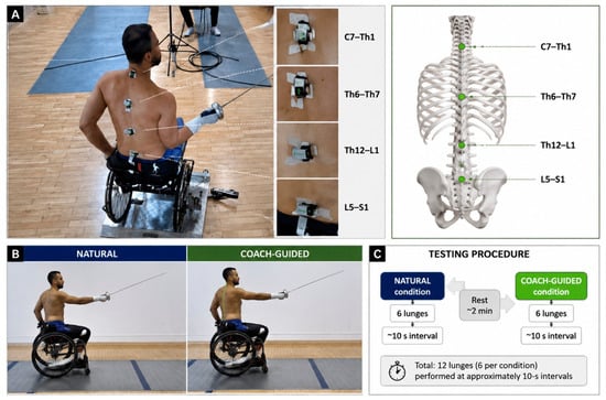
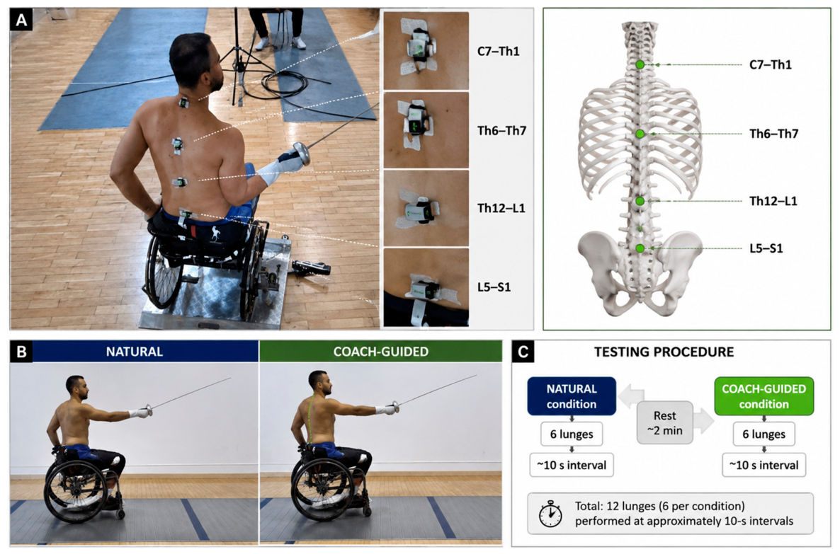
Four IMUs were positioned along the spine at the following anatomical levels: C7–Th1, Th6–Th7, Th12–L1, and L5–S1 (Figure 1). All anatomical landmarks were identified and palpated by the same examiner, a physiotherapist with 8 years of clinical experience, to ensure consistent sensor placement across participants. The sensors were positioned with a consistent anatomical orientation at each measurement site.
Figure 1.
Experimental setup, spinal IMU placement, testing conditions, and experimental procedure. (A) Representative experimental setup showing the participant performing the fencing task with four IMUs positioned along the spine at C7–Th1, Th6–Th7, Th12–L1, and L5–S1; the corresponding sensor placements are illustrated alongside the photograph. (B) Representative fencing lunge performed under the Natural and Coach-guided conditions. (C) Schematic overview of the testing procedure, with six fencing lunges performed under each condition at approximately 10-s intervals.
Participants performed six fencing lunges under each of two experimental conditions: Natural and Coach-guided. In the Natural condition, participants performed the fencing lunges using their habitual technique. In the Coach-guided condition, they performed the same task while following the standardized coach instructions concerning trunk and scapular positioning. Consecutive lunges were performed at approximately 10-s intervals.
The IMU data were recorded continuously to characterize segmental spinal movement. Acceleration and angular velocity were analyzed in the mediolateral (ML; x), proximodistal (PD; y), and anteroposterior (AP; z) directions, together with their respective resultant values. These variables were treated as measures of segmental movement dynamics; they were not interpreted as direct measures of absolute spinal posture, given the potential effects of soft-tissue movement and skin-mounted sensor displacement.
2.3. Experimental Procedure and Data Analysis
Before data collection, each participant completed a familiarization trial under coach supervision and then performed an individualized warm-up routine. The experimental conditions were presented in a fixed order for all participants: the Natural condition was always performed first (Trial 1), followed by the Coach-guided condition (Trial 2). The order was therefore not randomized or counterbalanced. Six lunges without weapon contact with a target were recorded in each condition. The final analytical dataset contained 83 lunges: 42 Natural and 41 Coach-guided, corresponding to 332 lunge-by-segment observations across the four spinal segments. One Coach-guided lunge was not represented in the final analytical file, resulting in four missing segment observations. This fixed order creates a potential confound between Condition and trial order, including learning, habituation, or fatigue.
In Trial 1, participants performed the fencing lunge as quickly as possible, including rapid forward trunk movement followed by immediate return to the initial fencing position. In Trial 2, participants were instructed to consciously retract and stabilize the scapulae before movement initiation and to maintain scapular control throughout the movement. Before each repetition in Trial 2, the coach provided standardized verbal instructions and movement corrections (e.g., “retract your scapulae, prepare for stance”). Prior experience with this specific scapular-control cue was not formally assessed, and no objective verification of scapular position or muscle activation was performed. The Natural condition represented the participant’s self-selected scapular and trunk strategy, whereas the Coach-guided condition represented the standardized coach-guided instruction.
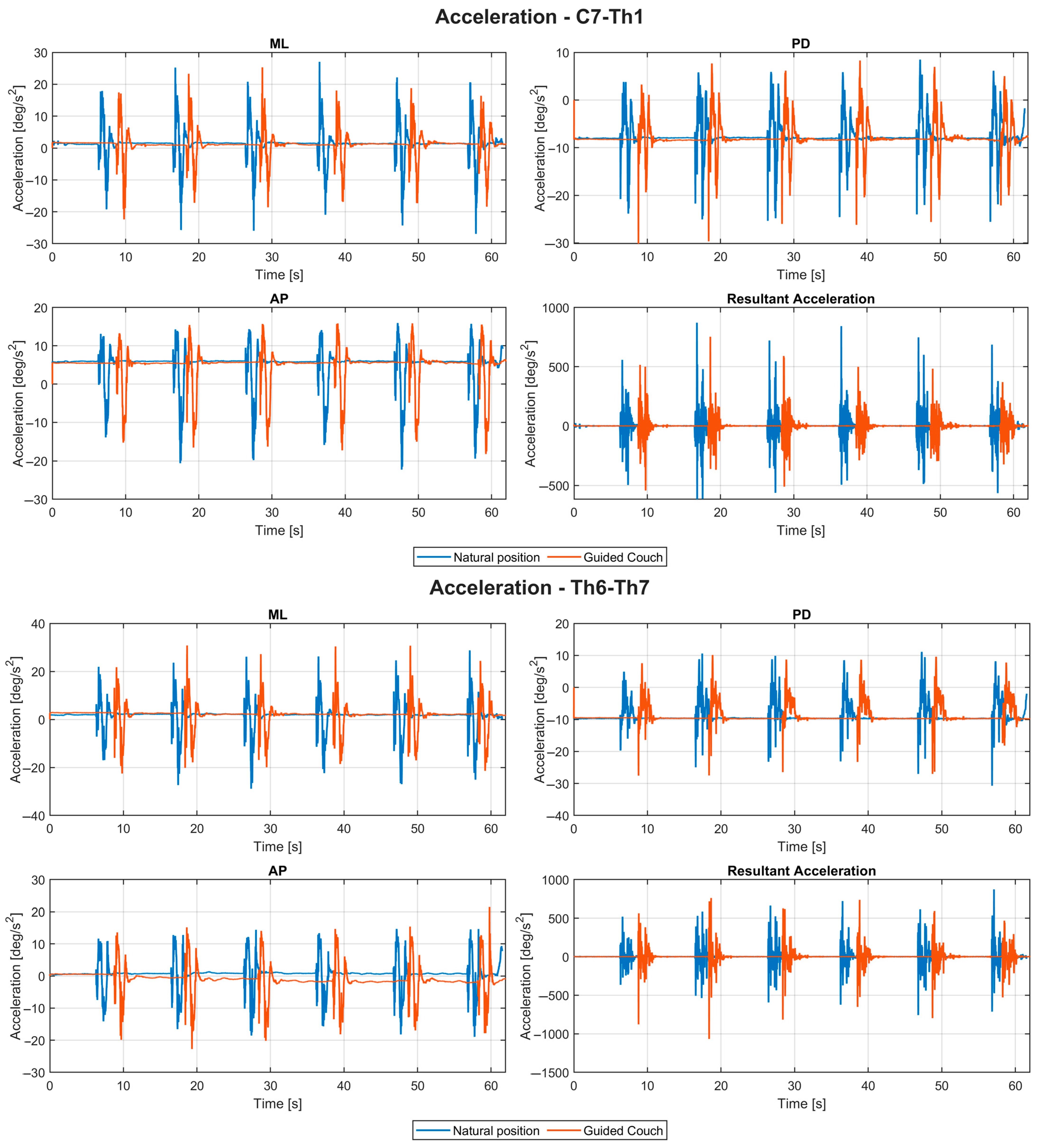
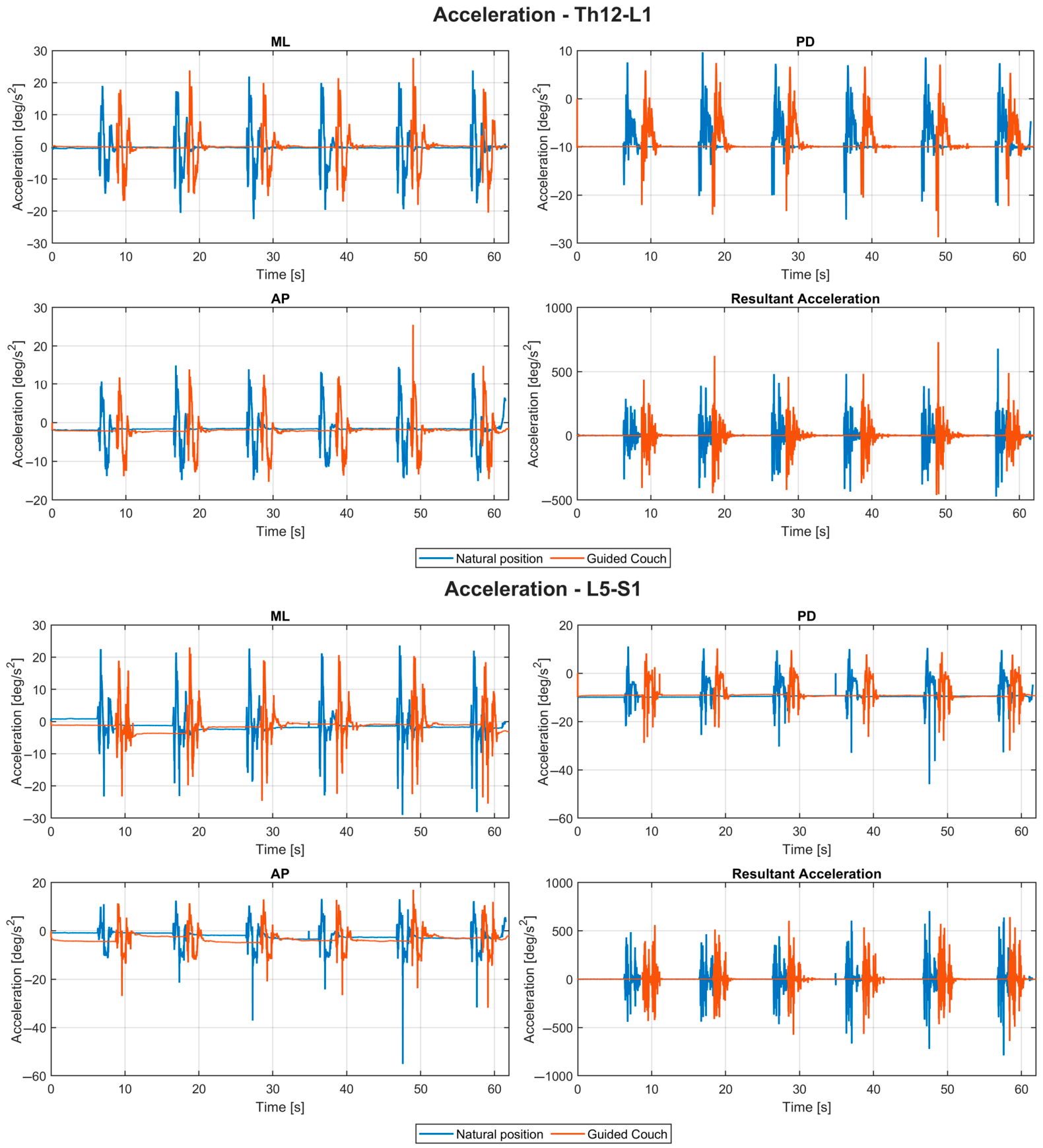
Angular velocity signals obtained from the gyroscope component of the IMU sensors were recorded continuously during all trials. Angular velocity signals were subsequently used to calculate resultant angular velocity, angular acceleration, and angular jerk for each spinal segment. Angular acceleration was computed as the first derivative of angular velocity over time, whereas angular jerk was calculated as the first derivative of angular acceleration. All rotational variables were expressed, respectively, in degrees per second (deg/s), degrees per second squared (deg/s2), and degrees per second cubed (deg/s3). Raw IMU signals were exported and processed in MatLab R2021a (MathWorks Inc., Natick, MA, USA). All motion signals were low-pass filtered using a fourth-order zero-phase Butterworth filter with a cutoff frequency of 10 Hz in order to reduce high-frequency noise and movement artifacts while preserving biomechanically relevant movement components. Filtered three-dimensional rotational kinematic signals derived from IMU measurements were subsequently used for further analyses. Representative raw IMU recordings from a single participant performing six fencing lunges are provided in Appendix Figure A1 and Figure A2, illustrating the measured angular velocity and acceleration, respectively, across the four spinal segments (C7–Th1, Th6–Th7, Th12–L1, and L5–S1) in the mediolateral (ML; x), anteroposterior (AP; z), and proximodistal (PD; y) directions, together with the corresponding resultant values, for the Natural and Coach-guided conditions.
2.4. Kinematic Variables and Lunge Detection
For each spinal segment, resultant angular velocity and angular acceleration were calculated as the Euclidean norm of the three orthogonal axes according to the following equations:
Individual fencing lunges were identified automatically using a peak-based movement-detection algorithm applied to the resultant angular velocity signal obtained from the C7–Th1 segment. The reference signal was additionally smoothed using a Gaussian smoothing procedure to reduce transient oscillations and postural micro-corrections. Major movement peaks corresponding to fencing lunges were detected using predefined criteria, including a minimum peak height equal to 35% of the maximal signal amplitude, a minimum peak prominence equal to 20% of the maximal signal amplitude, and a minimum inter-peak distance of 5 s. When more than six valid movement peaks were detected, the six peaks with the greatest amplitudes were retained for further analysis. This procedure was designed to identify the six targeted fencing lunges while minimizing false-positive detections associated with postural oscillations, recovery movements, and non-task-related trunk activity.
For each detected lunge, an analysis window extending from 1 s before the movement peak to 2 s after the movement peak was extracted. This standardized window captured the main phases of the lunge, including movement initiation, acceleration, peak propulsion, and recovery.
For each detected lunge and spinal segment, the following biomechanical variables were calculated: peak angular velocity, peak angular acceleration, RMS angular velocity, RMS angular acceleration, movement duration, time to peak angular velocity, time to peak angular acceleration, kinematic energy index, and peak angular jerk.
Peak angular velocity and peak angular acceleration were defined as the maximum resultant angular velocity and angular acceleration values observed within the segmented lunge window. Movement duration was defined as the interval between automatically detected movement onset and movement termination within the segmented lunge window. Time-to-peak variables were calculated as the elapsed time between movement onset and the occurrence of the respective peak angular velocity or peak angular acceleration.
Angular jerk was used to characterize the temporal dynamics of angular acceleration. Angular jerk was calculated as the first derivative of resultant angular acceleration with respect to time:
For each lunge, peak angular jerk was defined as the maximum absolute value of angular jerk observed within the segmented lunge window. Thus, peak angular jerk quantified the largest rate of change in resultant angular acceleration during the movement.
The kinematic energy index was calculated as the time integral of squared resultant angular velocity:
where represents resultant angular velocity and and represent the beginning and end of the segmented lunge window, respectively. The kinematic energy index was interpreted as a relative measure of movement-related angular velocity magnitude rather than as mechanical energy, because mass and inertial parameters were not incorporated into its calculation.
2.5. Statistical Analysis
All statistical analyses were performed in MatLab R2021a (MathWorks, Natick, MA, USA). The final analytical dataset comprised seven participants and 332 lunge-by-segment observations derived from 83 retained lunges performed under two experimental conditions and recorded at four spinal segments. Because multiple lunges were performed by the same participant, individual lunges were not treated as independent observations in the primary inferential analysis. Repeated lunges were therefore averaged within each participant, condition, and spinal segment before fitting the final mixed-effects models, resulting in 56 participant-by-condition-by-segment observations (7 participants × 2 conditions × 4 spinal segments).
Eight dependent variables were included in the final analysis: peak angular velocity, peak angular acceleration, peak angular jerk, RMS angular velocity, RMS angular acceleration, kinematic energy index, time to peak angular velocity, and time to peak angular acceleration.
Prior to statistical modelling, the distributions of the kinematic variables were examined. Because several variables showed right-skewed distributions, natural logarithmic transformation was applied to peak angular velocity, peak angular acceleration, peak angular jerk, RMS angular velocity, RMS angular acceleration, and the kinematic energy index. All variables subjected to logarithmic transformation were strictly positive in the final dataset; therefore, no constant shift was required before transformation. Time to peak angular velocity and time to peak angular acceleration were analyzed without logarithmic transformation. Descriptive statistics were calculated using the original, untransformed values and are reported as medians with interquartile ranges (Q1–Q3).
Linear mixed-effects models (LMMs) were fitted using the MatLab fitlme function. Condition (Natural vs. Coach-guided), Segment (C7–Th1, Th6–Th7, Th12–L1, and L5–S1), and their interaction were specified as fixed effects, while participant identity was included as a random intercept. The basic model specification was:
where Y represents the respective kinematic outcome.
Y ∼ Condition × Segment + (1∣Participant)
Model diagnostics included inspection of residual-versus-fitted plots, normal quantile–quantile plots, and residual distributions. Potentially extreme observations were additionally screened using a robust z-score based on the median absolute deviation (MAD). An absolute robust z-score >6 was used as the predefined threshold for this quality-control procedure. This screening was used to identify potentially extreme observations and was considered alongside the model diagnostics.
Effect sizes for omnibus fixed effects were expressed as partial eta-squared (η2p) values. Partial eta-squared values were interpreted according to conventional thresholds: small (η2p ≥ 0.01), moderate (η2p ≥ 0.06), and large (η2p ≥ 0.14) [16].
To account for multiple testing, the Benjamini–Hochberg false-discovery-rate (FDR) procedure was applied across the omnibus tests. Eight dependent variables and three fixed effects (Condition, Segment, and Condition × Segment) resulted in 24 omnibus tests. Statistical significance was defined as an FDR-adjusted q-value < 0.05.
When a significant omnibus Segment effect was identified, six pairwise contrasts among the four spinal segments were evaluated using the fitted linear mixed-effects model. Pairwise contrasts were specified to estimate segment differences averaged across the two experimental conditions. The contrasts were tested using the coefTest function in MATLAB R2021a (MathWorks, Natick, MA, USA), with the same random-intercept structure for participant as in the corresponding omnibus model. The resulting p-values were adjusted for multiple comparisons using the Holm procedure. Pairwise comparisons were interpreted in the context of the corresponding omnibus Segment effect.
Because the Natural condition was always performed before the Coach-guided condition, the experimental condition was potentially confounded with trial order. Therefore, Condition effects were interpreted as exploratory associations rather than evidence of a causal effect of the coaching intervention. Potential order-related influences, including familiarization, habituation, and fatigue, were considered when interpreting the results.
3. Results
The final analysis comprised 56 participant-by-condition-by-segment observations derived from seven participants, two conditions, and four spinal segments. Descriptive values are presented as median (Q1–Q3) on the original measurement scale, whereas inferential analyses were performed on the participant-level aggregated data using the transformed variables specified in Section 2.5. Eight kinematic outcomes were included in the final analysis.
Following Benjamini–Hochberg false-discovery-rate (FDR) correction across the 24 omnibus tests, three effects remained statistically significant: the main effect of Condition for RMS angular acceleration (F(1,42) = 17.00, p < 0.001, q = 0.004, η2p = 0.288), the main effect of Segment for peak angular jerk (F(3,42) = 5.00, p = 0.005, q = 0.038, η2p = 0.263), and the main effect of Segment for time to peak angular velocity (F(3,42) = 5.33, p = 0.003, q = 0.038, η2p = 0.276). No Condition × Segment interaction survived FDR correction (Table 1).
3.1. Angular Velocity- and Timing-Related Parameters
Peak angular velocity showed no significant main effect of Condition (F(1,42) = 1.01, p = 0.322, q = 0.594, η2p = 0.023) or Segment (F(3,42) = 1.52, p = 0.223, q = 0.486, η2p = 0.098). The Condition × Segment interaction was also not significant (F(3,42) = 0.005, p = 0.999, q = 0.999, η2p < 0.001) (Table 1).
RMS angular velocity showed a nominal Condition effect (F(1,42) = 4.50, p = 0.040, η2p = 0.097), but this effect did not survive FDR correction (q = 0.154). Neither Segment (F(3,42) = 0.73, p = 0.541, q = 0.721, η2p = 0.049) nor the Condition × Segment interaction (F(3,42) = 0.026, p = 0.994, q = 0.999, η2p = 0.002) was significant.
Time to peak angular velocity showed no significant main effect of Condition (F(1,42) = 0.62, p = 0.437, q = 0.674, η2p = 0.014). In contrast, Segment had a significant effect (F(3,42) = 5.33, p = 0.003, q = 0.038, η2p = 0.276). The Condition × Segment interaction was not significant (F(3,42) = 1.70, p = 0.182, q = 0.436, η2p = 0.108).
Holm-adjusted pairwise comparisons showed that the L5–S1 segment differed significantly from Th6–Th7 for time to peak angular velocity (F(1,48) = 13.27, p < 0.001, pHolm = 0.004). The remaining five segment comparisons were not significant after Holm correction. The C7–Th1 versus L5–S1 comparison was nominally significant before correction (p = 0.012) but did not remain significant after Holm adjustment (pHolm = 0.060) (Table 2).
Table 1.
Angular velocity- and timing-related parameters. Values are presented as median (Q1–Q3). Statistical values are raw p-values followed by Benjamini–Hochberg FDR-adjusted q-values. Partial eta-squared (η2p) is reported for the corresponding omnibus effect where available. Significant effects are marked with an asterisk (p, q < 0.05).
Table 2.
Holm-adjusted pairwise comparisons for significant Segment effects from Table 1. Pairwise comparisons were performed following significant omnibus Segment effects. pHolm, Holm-adjusted p-value. Holm correction was applied separately to the six pairwise comparisons for each outcome. * indicates statistical significance after Holm correction (pHolm < 0.05).
3.2. Angular Acceleration-Related Parameters
Peak angular acceleration showed no significant main effect of Condition (F(1,42) = 4.67, p = 0.036, q = 0.154, η2p = 0.100), Segment (F(3,42) = 0.99, p = 0.406, q = 0.674, η2p = 0.066), or Condition × Segment interaction (F(3,42) = 0.84, p = 0.478, q = 0.674, η2p = 0.057) after FDR correction (Table 3).
Table 3.
Angular acceleration- and timing-related parameters. Values are presented as median (Q1–Q3). Statistical values are raw p-values followed by Benjamini–Hochberg FDR-adjusted q-values. Partial eta-squared (η2p) is reported for the corresponding omnibus effect where available. Significant effects are marked with an asterisk (p, q < 0.05).
RMS angular acceleration showed the strongest Condition effect in the analysis and remained significant after FDR correction (F(1,42) = 17.00, p < 0.001, q = 0.004, η2p = 0.288). This large effect indicates a substantial condition-associated difference in RMS angular acceleration. The Segment effect was nominally significant (F(3,42) = 2.92, p = 0.045, q = 0.154, η2p = 0.173) but did not survive FDR correction. The Condition × Segment interaction was not significant (F(3,42) = 1.39, p = 0.258, q = 0.516, η2p = 0.091).
Time to peak angular acceleration showed no significant effect of Condition (F(1,48) = 0.03, p = 0.866, q = 0.999, η2p < 0.001), Segment (F(3,48) = 0.11, p = 0.954, q = 0.999, η2p = 0.007), or Condition × Segment interaction (F(3,48) = 0.46, p = 0.714, q = 0.902, η2p = 0.028).
3.3. Peak Angular Jerk and Kinematic Energy Index
Peak angular jerk showed no significant main effect of Condition (F(1,42) = 2.37, p = 0.132, q = 0.351, η2p = 0.053), but Segment had a significant effect (F(3,42) = 5.00, p = 0.005, q = 0.038, η2p = 0.263). The Condition × Segment interaction was not significant (F(3,42) = 2.08, p = 0.117, q = 0.351, η2p = 0.129) (Table 4).
Table 4.
Peak angular jerk and kinematic energy index. Values are presented as median (Q1–Q3). Statistical values are raw p-values followed by Benjamini–Hochberg FDR-adjusted q-values. Partial eta-squared (η2p) is reported for the corresponding omnibus effect where available. Significant effects are marked with an asterisk (p, q < 0.05).
Holm-adjusted pairwise comparisons showed a significant difference between L5–S1 and Th6–Th7 for peak angular jerk (F(1,48) = 11.16, p = 0.002, pHolm = 0.010). The estimated difference was −0.148 on the log-transformed scale, indicating lower peak angular jerk at L5–S1 than at Th6–Th7. None of the remaining five pairwise comparisons remained significant after Holm correction (Table 5).
Table 5.
Holm-adjusted pairwise comparisons for significant Segment effects from Table 4. Pairwise comparisons were performed following significant omnibus Segment effects. pHolm, Holm-adjusted p-value. Holm correction was applied separately to the six pairwise comparisons for each outcome. * indicates statistical significance after Holm correction (pHolm < 0.05).
The kinematic energy index showed a nominal Condition effect (F(1,42) = 4.50, p = 0.040, q = 0.154, η2p = 0.097), but this did not survive FDR correction. Segment was not significant (F(3,42) = 0.88, p = 0.458, q = 0.674, η2p = 0.059), and the Condition × Segment interaction was not significant (F(3,42) = 0.027, p = 0.994, q = 0.999, η2p = 0.002).
3.4. Overall Pattern of Findings
Across the eight outcomes, the strongest evidence concerned RMS angular acceleration as a function of Condition and segment-related differences in peak angular jerk and time to peak angular velocity. After FDR correction, only these three omnibus effects remained significant. Thus, the data support condition-associated differences in RMS angular acceleration and segment-related differences in peak angular jerk and the timing of peak angular velocity.
The largest omnibus effect was observed for the Condition effect on RMS angular acceleration (η2p = 0.288), followed by the Segment effects for time to peak angular velocity (η2p = 0.276) and peak angular jerk (η2p = 0.263). These effect sizes are large according to the thresholds specified in Section 2.5.
Importantly, none of the Condition × Segment interactions survived FDR correction. Therefore, the present analysis does not provide statistical evidence that the association between experimental Condition and the kinematic outcomes differed systematically across the four spinal segments.
Because the final inferential sample comprised only seven participants, the statistically significant effects should be interpreted as exploratory. The fixed order of conditions also prevents a causal interpretation of the observed Condition effects, because familiarization, habituation, fatigue, or other order-related influences cannot be excluded.
Because the Natural condition was always performed before the Coach-guided condition, the observed Condition effects are interpreted as condition-associated differences rather than evidence of a causal coaching effect. The results therefore indicate differences associated with the experimental condition, but they do not establish that the coach-guided instruction itself caused the observed changes.
4. Discussion
The aim of the present study was to evaluate whether coach-guided scapular-control instruction was associated with differences in spinal kinematics during wheelchair fencing lunges using an IMU-based motion capture system. It was hypothesized that the instruction would be associated with changes in spinal movement patterns, particularly angular acceleration- and jerk-related parameters, with smaller effects on angular velocity and timing. The findings partially supported this hypothesis. After correction for multiple comparisons, the only significant Condition effect was observed for RMS angular acceleration, whereas Segment effects were identified for peak angular jerk and time to peak angular velocity. No Condition × Segment interaction survived FDR correction. Thus, the findings indicate condition-associated differences in acceleration magnitude and segment-related differences in jerk and movement timing, rather than evidence of a segment-specific response to the experimental condition. This pattern is broadly consistent with biomechanical observations emphasizing the role of angular acceleration and neuromuscular coordination in sport-specific movement organization [17].
The magnitude of the significant effects was relatively large, with η2p = 0.288 for the Condition effect on RMS angular acceleration, η2p = 0.276 for the Segment effect on time to peak angular velocity, and η2p = 0.263 for the Segment effect on peak angular jerk. These findings suggest that the principal differences detected in the present dataset concerned the magnitude and temporal organization of trunk motion rather than a generalized change in rotational speed. Importantly, the absence of significant Condition × Segment interactions indicates that the present data do not support a systematic redistribution of the condition-associated effect across the four spinal segments. The significant segment effects were instead attributable to differences among spinal levels, with the post hoc analyses identifying L5–S1 versus Th6–Th7 as the only significant pairwise contrast for both peak angular jerk and time to peak angular velocity.
The observed segment-related differences are consistent with the functional role of the trunk in wheelchair fencing. Previous studies have demonstrated that wheelchair fencing performance depends strongly on trunk stability, postural coordination, and upper-body control because athletes cannot generate propulsion using the lower limbs [1,4]. In particular, Borysiuk, et al. [4] emphasized the contribution of postural muscle activation and trunk stabilizers to movement organization and technical execution. Similarly, Fung, et al. [1] demonstrated that trunk kinematics differ according to functional classification and may influence movement effectiveness. The present results extend this context by showing that peak angular jerk and the timing of peak angular velocity vary across spinal segments, including a significant difference between L5–S1 and Th6–Th7. These findings are compatible with the view that different spinal regions contribute differently to force transmission and postural regulation [18]. However, because scapular kinematics and muscle activation were not directly assessed, the present findings cannot establish that scapular stabilization or proximal stabilization was responsible for the observed spinal differences.
The inclusion of athletes from two functional wheelchair fencing classifications should also be considered when interpreting the findings. Trunk control and postural stability may differ between categories A and B, and pooling both groups may have increased inter-individual variability and obscured classification-specific movement patterns [1,3]. The small sample prevented meaningful classification-specific analyses and also means that individual movement strategies may have contributed substantially to the observed variability. Accordingly, the present findings should be regarded as exploratory, and future studies with larger, classification-specific samples should determine whether the observed segmental patterns are consistent across functional groups.
The practical relevance of the present findings lies primarily in the organization of trunk movement rather than in increasing movement velocity. The significant Condition effect for RMS angular acceleration indicates that the experimental conditions were associated with different overall levels of angular acceleration, whereas peak angular jerk did not show a significant Condition effect. At the same time, the significant Segment effects for peak angular jerk and time to peak angular velocity indicate that movement dynamics and timing differed between spinal levels. From a coaching perspective, these findings may be relevant to understanding how technical instructions are associated with the distribution and temporal organization of trunk motion during explosive fencing actions. However, the present data do not establish that one condition represents superior movement control or provides a performance advantage.
Jerk-related variables are commonly interpreted as indicators of movement smoothness and temporal organization, with higher jerk values generally reflecting more abrupt changes in movement. In the present study, the significant Segment effect for peak angular jerk was driven by the difference between L5–S1 and Th6–Th7 (pHolm = 0.010). This result indicates that the two spinal regions exhibited different peak jerk characteristics, but it does not demonstrate that the coach-guided condition reduced jerk at either segment. Likewise, the significant Segment effect for time to peak angular velocity was driven by the same L5–S1 versus Th6–Th7 comparison (pHolm = 0.004). These findings suggest segment-specific differences in the temporal and dynamic organization of trunk motion, while the absence of a significant Condition × Segment interaction argues against interpreting them as a condition-specific redistribution of movement across the spine. Similar relationships between movement coordination, muscle activation, and technical execution have been described in dynamic sport-specific tasks [19]. Previous research has also linked insufficient trunk control with compensatory upper-body movement strategies and increased mechanical loading within the shoulder complex [5]. These observations provide biomechanical context, but the present study did not directly assess loading or injury risk.
The thoracic and lumbosacral regions remain biomechanically relevant in the context of wheelchair fencing because the trunk forms a mechanical link between the shoulder girdle and the lower trunk during rapid upper-limb actions. Previous work by Raud, et al. [6] suggested that scapular positioning and clavicular motion may influence upper-body coordination during wheelchair fencing, while comparable relationships between movement coordination and technical efficiency have been reported in other ballistic and striking sports [20]. In the present study, however, the statistically significant segment-related effects should not be interpreted as direct evidence of altered scapular function. Rather, they indicate that the kinematic characteristics measured at different spinal levels were not equivalent during the fencing lunge. Direct assessment of scapular motion and muscle activation would be required to determine whether the observed spinal patterns are related to the intended scapular-control strategy.
The findings also indicate that not all kinematic variables were equally sensitive to the experimental condition. Peak angular velocity and RMS angular velocity did not show significant Condition effects after FDR correction, and the kinematic energy index likewise did not survive correction. In contrast, RMS angular acceleration showed the strongest Condition effect, while peak angular jerk and time to peak angular velocity showed significant Segment effects. This pattern suggests that acceleration magnitude, movement jerk, and segment-specific timing may provide more sensitive descriptions of trunk movement organization than global measures of rotational speed in this exploratory dataset. The findings therefore support the potential usefulness of IMU-derived acceleration- and jerk-based variables for characterizing spinal coordination during wheelchair fencing, while emphasizing that their interpretation should remain descriptive and hypothesis-generating in the present sample.
The present findings are also consistent with previous work examining postural muscle coordination during fencing-specific tasks [4]. However, the current analysis provides a more conservative interpretation because the inferential results were based on participant-level aggregation and corrected for multiple comparisons. The significant omnibus effects were limited to three of the 24 tests, and the post hoc analyses identified only one significant segment contrast for each of the two significant Segment effects. Accordingly, the results should be interpreted as evidence of selected differences in movement organization rather than as evidence of broad or pervasive biomechanical changes induced by the coach-guided condition. In elite sport biomechanics, such segmental differences may nevertheless be useful for generating hypotheses about movement organization and repeatability, particularly when examined in larger and more controlled samples.
The use of IMU-based motion analysis represented an important methodological aspect of the present study. Wearable inertial sensors enable biomechanical measurements during sport-specific tasks performed outside traditional laboratory environments. Previous investigations confirmed the validity and reliability of IMU systems for assessing trunk kinematics during dynamic athletic activities [9,10]. Brouwer et al. [9] demonstrated accurate three-dimensional trunk orientation measurements during dynamic sports movements, whereas Chia et al. [10] confirmed the validity of IMU systems for evaluating trunk kinematics during rapid athletic tasks. Recent reviews have further emphasized the growing importance of wearable sensor technologies in sports biomechanics and Paralympic sport assessment [11]. The present study demonstrates the feasibility of applying IMU-derived acceleration- and jerk-based measures to wheelchair fencing-specific tasks. The methodological contribution therefore lies less in the use of IMUs themselves and more in the application of segmental acceleration, jerk, and timing measures to examine spinal movement organization under sport-specific coaching conditions. This approach may support future field-based studies of movement coordination, provided that the findings are validated against direct biomechanical and neuromuscular measurements.
Several limitations should be considered when interpreting the present findings. First, the fixed order of the experimental conditions is an important limitation: the Natural condition was always performed before the Coach-guided condition. Consequently, learning, familiarization, habituation, fatigue, or other order effects cannot be excluded, and the observed Condition effect for RMS angular acceleration cannot be attributed solely to the coach-guided instruction. Second, the final inferential sample comprised only seven participants, reflecting the limited availability of elite wheelchair fencers. Athletes from functional categories A and B were analyzed together, despite potential differences in trunk control and postural stability between classifications [1,3]. This pooling may have increased variability and reduced the ability to detect classification-specific effects. Third, scapular control was not directly measured: scapular kinematics, shoulder-girdle motion, and muscle activation were not quantified. Therefore, the observed spinal kinematic differences cannot be interpreted as direct evidence that the intended scapular stabilization was achieved. Future studies should combine IMU-based spinal motion analysis with direct scapular kinematics and electromyography. Fourth, the study assessed acute responses during a controlled task and did not evaluate long-term training adaptations, competitive performance, hit accuracy, injury risk, pain, fatigue, or shoulder or spinal loading. The findings therefore cannot be interpreted as evidence of improved performance, injury prevention, or long-term training benefits. Finally, repeated lunges were averaged within participant, condition, and segment before the final modelling; consequently, trial-level variability was not explicitly modelled as a separate random effect. Although this aggregation was consistent with the predefined participant-level analysis, it limits inference about within-participant trial-to-trial variability. In addition, the final linear mixed-effects models included a participant-specific random intercept but did not explicitly model participant-specific random slopes for Condition or Segment. Given the small sample of seven participants, more complex random-effects structures were not pursued because they could result in unstable model estimation. Consequently, potential heterogeneity in the effects of Condition or Segment across participants cannot be fully characterized. Benjamini–Hochberg FDR correction and Holm-adjusted post hoc comparisons were applied to control multiple testing, but the small sample and exploratory design still warrant cautious interpretation. Taken together, the results should be considered preliminary, hypothesis-generating observations rather than definitive evidence of a causal effect of scapular-control instruction. Future studies should use randomized or counterbalanced condition orders, larger classification-specific samples, direct scapular and muscle-activation measurements, and longitudinal designs to determine whether the observed movement patterns are reproducible and relevant to wheelchair fencing performance or training.
5. Conclusions
The final analysis identified a condition-associated difference in RMS angular acceleration and segment-related differences in peak angular jerk and time to peak angular velocity. Post hoc analyses showed that the significant segment differences were between L5–S1 and Th6–Th7 for both peak angular jerk (pHolm = 0.010) and time to peak angular velocity (pHolm = 0.004). No Condition × Segment interaction survived FDR correction; therefore, the present findings do not provide evidence that the effect associated with experimental Condition differed systematically across spinal segments. Given the small sample of seven participants and the fixed order of conditions, these findings should be considered exploratory and cannot be interpreted as evidence that coach-guided scapular-control instruction caused the observed changes. Furthermore, scapular kinematics and muscle activation were not directly measured, so the observed spinal kinematic differences cannot be interpreted as direct evidence of improved scapular stabilization. Future studies should use larger samples, counterbalanced or randomized condition orders, and direct measures of scapular motion and muscle activation to determine whether the observed kinematic patterns are reproducible and whether they have meaningful implications for wheelchair fencing performance.
Author Contributions
Conceptualization, M.B., J.S. and J.W.; methodology, M.B.; software, J.S. and M.S.; validation, M.B., J.S., M.S. and J.W.; formal analysis, M.B.; investigation, J.S.; resources, J.S.; data curation, J.S.; writing—original draft preparation, M.B., J.W.; writing—review and editing, M.B., J.S., M.S. and J.W.; visualization, M.B.; supervision, J.W.; project administration, J.S. and M.S.; funding acquisition, J.S. and M.S. All authors have read and agreed to the published version of the manuscript.
Funding
This research was funded by the Ministry of Science and Higher Education, under the Józef Piłsudski University of Physical Education research project “Morphological and physiological adaptations to physical activity in wheelchair users”, number UPB_Nr_15_2025.
Institutional Review Board Statement
The study was conducted in accordance with the Declaration of Helsinki and approved by the Ethics Committee of Józef Piłsudski University of Physical Education in Warsaw (protocol code SKE.0030.41.2025, date of approval 13 May 2025).
Informed Consent Statement
Informed consent was obtained from all subjects involved in the study.
Data Availability Statement
The data presented in this study are available upon request from the corresponding author due to the privacy of the participants involved.
Conflicts of Interest
The authors declare no conflicts of interest.
Appendix A. Supplementary IMU Kinematic Data
The following figures present representative raw IMU recordings obtained from a single participant performing six fencing lunges under the Natural and Coach-guided conditions. The recordings show angular velocity (Figure A1) and acceleration (Figure A2) across four spinal segments (C7–Th1, Th6–Th7, Th12–L1, and L5–S1) and provide additional visualization of the movement patterns described in the main text.
Figure A1.
Representative raw IMU angular velocity recordings from a single participant performing six fencing lunges across four spinal segments (C7–Th1, Th6–Th7, Th12–L1, and L5–S1), shown in the mediolateral (ML), anteroposterior (AP), and proximodistal (PD) directions, together with the resultant angular velocity. Blue lines indicate the Natural condition, whereas red lines indicate the Coach-guided condition.
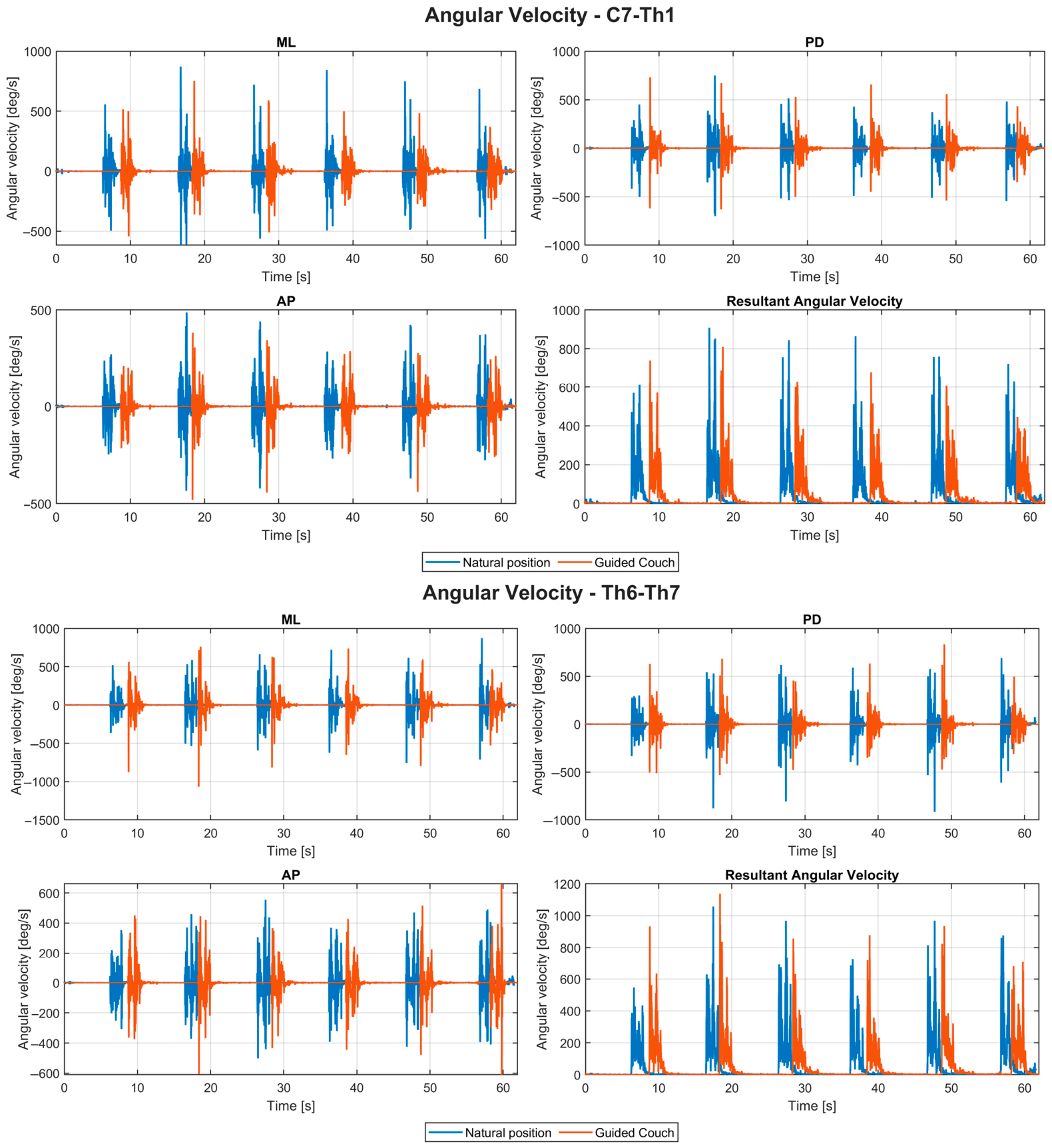
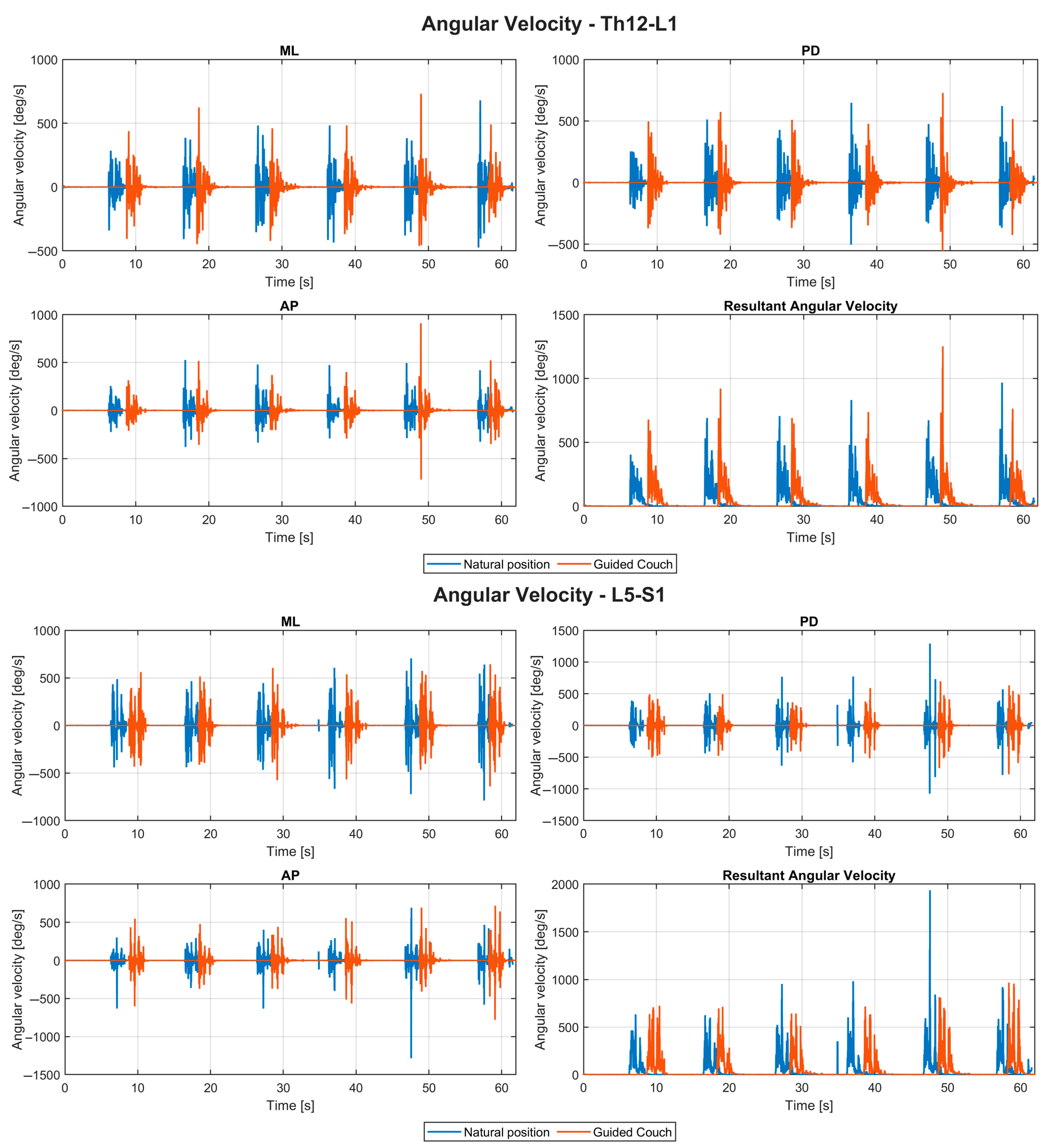
Figure A2.
Representative raw IMU acceleration recordings from a single participant performing six fencing lunges across four spinal segments (C7–Th1, Th6–Th7, Th12–L1, and L5–S1), shown in the mediolateral (ML), anteroposterior (AP), and proximodistal (PD) directions, together with the resultant acceleration. Blue lines indicate the Natural condition, whereas red lines indicate the Coach-guided condition.
References
- Fung, Y.-K.; Chow, B.-C.; Fong, D.T.-P.; Chan, K.-M. A kinematic analysis of trunk ability in wheelchair fencing: A pilot study. In Proceedings of the XXVIII International Conference on Biomechanics in Sports, Marquette, MI, USA, 19–23 July 2010; pp. 126–129. [Google Scholar]
- Blazkiewicz, M.; Borysiuk, Z.; Gzik, M. Determination of loading in the lower limb joints during step-forward lunge in fencing. Acta Bioeng. Biomech. 2018, 20, 3–8. [Google Scholar] [CrossRef] [Scilit]
- Chung, W.M. Kinematic and Electromyographic Analysis of Wheelchair Fencing. Ph.D. Thesis, The Hong Kong Polytechnic University, Hong Kong, China, 2015. [Google Scholar]
- Borysiuk, Z.; Blaszczyszyn, M.; Piechota, K.; Konieczny, M.; Cynarski, W.J. Correlations between the EMG Structure of Movement Patterns and Activity of Postural Muscles in Able-Bodied and Wheelchair Fencers. Sensors 2023, 23, 135. [Google Scholar] [CrossRef] [Scilit] [PubMed]
- Yildirim, N.U.; Comert, E.; Ozengin, N. Shoulder pain: A comparison of wheelchair basketball players with trunk control and without trunk control. J. Back Musculoskelet. Rehabil. 2010, 23, 55–61. [Google Scholar] [CrossRef] [Scilit] [PubMed]
- Raud, A.; Poulet, Y.; Aude, L.; Bourgain, M.; Sauret, C.; Pillet, H. Scapula and clavicle kinematics during wheelchair fencing: A preliminary study. Multidiscip. Biomech. J. 2024, 1, 455–457. [Google Scholar] [CrossRef] [Scilit]
- Li, Y.; Zhou, Z. Angular Velocity Estimation in Asymmetric IMU Array. In Proceedings of the 2023 2nd International Conference on Automation, Robotics and Computer Engineering (ICARCE), Wuhan, China, 14–16 December 2023; IEEE: Piscataway, NJ, USA, 2023; pp. 1–5. [Google Scholar]
- Yang, P.; Chen, Y.; Chen, Y.; Dang, F. Gyro-Free Inertial Measurement Unit With Unfettered Accelerometer Array Distribution and for the Object With Position Change in Center of Gravity. IEEE Sens. J. 2021, 21, 9423–9435. [Google Scholar] [CrossRef] [Scilit]
- Brouwer, N.P.; Yeung, T.; Bobbert, M.F.; Besier, T.F. 3D trunk orientation measured using inertial measurement units during anatomical and dynamic sports motions. Scand. J. Med. Sci. Sports 2021, 31, 358–370. [Google Scholar] [CrossRef] [Scilit] [PubMed]
- Chia, L.; Andersen, J.T.; McKay, M.J.; Sullivan, J.; Megalaa, T.; Pappas, E. Evaluating the validity and reliability of inertial measurement units for determining knee and trunk kinematics during athletic landing and cutting movements. J. Electromyogr. Kinesiol. 2021, 60, 102589. [Google Scholar] [CrossRef] [Scilit] [PubMed]
- Zhu, J.; Ye, Z.; Liu, R.; Liu, J. Inertial measurement units (IMUs) for biomechanical analysis in sport: A review of applications, challenges and future directions. Sens. Rev. 2025, 46, 88–104. [Google Scholar] [CrossRef] [Scilit]
- Mosler, D.; Góra, T.; Kaczmarski, J.; Błaszczyszyn, M.; Chociaj, M.; Borysiuk, Z. Target kinematic effect in Kyokushin karate front kicks: An analysis of velocity, acceleration, and muscle activation patterns. Phys. Act. Rev. 2025, 13, 156–166. [Google Scholar] [CrossRef] [Scilit]
- Heneghan, N.R.; Lokhaug, S.M.; Tyros, I.; Longvastøl, S.; Rushton, A. Clinical reasoning framework for thoracic spine exercise prescription in sport: A systematic review and narrative synthesis. BMJ Open Sport Exerc. Med. 2020, 6, e000713. [Google Scholar] [CrossRef] [Scilit] [PubMed]
- Marszałek, J.; Molik, B. Reliability of measurement of active trunk movement in wheelchair basketball players. PLoS ONE 2019, 14, e0225515. [Google Scholar] [CrossRef] [Scilit] [PubMed]
- Chikh, S.; Garnier, C.; Faupin, A.; Pinti, A.; Boudet, S.; Azaiez, F.; Watelain, E. Arm-trunk coordination in wheelchair initiation displacement: A study of anticipatory and compensatory postural adjustments during different speeds and directions of propulsion. J. Electromyogr. Kinesiol. 2018, 40, 16–22. [Google Scholar] [CrossRef] [Scilit] [PubMed]
- Cohen, J. Statistical Power Analysis for the Behavioral Sciences; Routledge: London, UK, 1988. [Google Scholar]
- Shahidi, S. Lactate Kinetics and Fatigue Dynamics in Elite Taekwondo Athletes during a 30-Second Continuous Jump Protocol. Phys. Act. Rev. 2025, 13, 12. [Google Scholar] [CrossRef] [Scilit]
- Bartosz, M.; Latocha, A.; Motowidło, J.; Krzysztofik, M.; Zajac, A. The relationship between countermovement jump performance and sprinting speed in elite sprinters. Phys. Act. Rev. 2024, 12, 29. [Google Scholar] [CrossRef] [Scilit]
- Čuj, J.; Gajdoš, M.; Nechvátal, P.; Grus, C.; Macej, M.; Kendrová, L. Changes in muscle activity of selected muscles during walking in high-heeled shoes on a treadmill. Phys. Act. Rev. 2025, 13, 14–25. [Google Scholar] [CrossRef] [Scilit]
- Petrů, J.; Vencúrik, T. The effect of jump topspin serve speed on reception quality in men’s volleyball. Phys. Act. Rev. 2024, 12, 11–19. [Google Scholar] [CrossRef] [Scilit]

Disclaimer/Publisher’s Note: The statements, opinions and data contained in all publications are solely those of the individual author(s) and contributor(s) and not of MDPI and/or the editor(s). MDPI and/or the editor(s) disclaim responsibility for any injury to people or property resulting from any ideas, methods, instructions or products referred to in the content. |
© 2026 by the authors. Licensee MDPI, Basel, Switzerland. This article is an open access article distributed under the terms and conditions of the Creative Commons Attribution (CC BY) license.