


Abstract
Background/Objectives: Defecatory straining requires coordinated abdominal-wall and lumbopelvic muscle activity to generate propulsive pressure. This study investigated whether sitting posture alters ultrasound-derived transversus abdominis (TrA) contractile behavior and trunk surface electromyographic (EMG) activity during simulated defecatory straining. Methods: Twenty healthy young adults (10 females and 10 males) performed quiet sitting and a maximal Valsalva maneuver in four standardized postures: neutral posture (NP), anterior trunk lean with anterior pelvic tilt (AT), posterior trunk lean/posterior pelvic tilt (PT), and collapsed posture (CO). TrA thickness was measured with B-mode ultrasonography, and the TrA contraction ratio was calculated as Valsalva thickness divided by resting thickness. Surface EMG root mean square amplitudes of the superficial multifidus (sMF), erector spinae (ES), external oblique (EO), and internal oblique (IO) were analyzed at rest, during Valsalva, and as an activation ratio (Valsalva/rest). Repeated-measures, one-way ANOVA with Bonferroni-adjusted comparisons was used. Results: Ultrasonographic measurements demonstrated excellent intra-rater reliability (ICC = 0.984–0.999). Resting TrA thickness did not differ among postures. During Valsalva, AT showed greater absolute and relative TrA thickness change than NP. EMG analyses showed higher absolute sMF and ES activity in AT both at rest and during Valsalva, whereas EO and IO activity during Valsalva did not differ among postures. The ES activation ratio was highest in NP among postures. Conclusions: Anterior trunk lean with anterior pelvic tilt was characterized by a distinct pattern of TrA behavior and posterior trunk muscle activity during a Valsalva-based simulated straining task. However, whether this posture-dependent muscle response increases intra-abdominal or rectal propulsive pressure or improves defecatory function remains unknown. The accompanying EMG pattern suggests that this advantage is associated with greater resting postural activation of the posterior trunk wall rather than generalized abdominal effort.
1. Introduction
Chronic constipation is a prevalent disorder that impairs quality of life, with particularly high prevalence in older adults [1,2,3,4,5]. Primary chronic constipation includes slow-transit constipation and functional defecation disorders, such as dyssynergic defecation [1,2]. Although colonic transit has been studied extensively, evacuation is also a biomechanical process requiring coordinated abdominal-wall expansion, diaphragmatic descent, rectal propulsion, and pelvic-floor/anal relaxation [6]. Inadequate rectal pressurization is therefore recognized as an important pathophysiological feature of impaired rectal evacuation [1,2,6]. Crucially, this required rectal pressurization may be mechanically driven by the increase in intra-abdominal pressure (IAP) generated by the trunk musculature [6,7].
The ability to generate intra-abdominal pressure (IAP) may be particularly relevant in older adults because sarcopenia has been associated with both the onset and severity of constipation [8,9]. Among the abdominal-wall muscles, the transversus abdominis (TrA) is considered a key regulator of IAP because of its corset-like architecture and its capacity to increase abdominal pressure with limited sagittal trunk torque [10,11,12,13]. In addition, IAP has been shown to correlate with objective measures of abdominal-wall tension during clinical evaluation tasks [13], supporting the mechanistic relevance of abdominal-wall muscle behavior during straining.
Postural alignment can substantially alter trunk-muscle recruitment. Compared with slouched sitting, more erect sitting has been reported to increase TrA thickness and lateral abdominal-wall activation [14,15]. Sagittal pelvic orientation also modifies the coordination of the abdominal muscles, erector spinae, and multifidus [16,17]. A forward trunk lean combined with anterior pelvic tilt may increase the demand on the lumbar extensors to resist the gravitational flexion moment, thereby producing a bracing-like state in which abdominal and paraspinal muscles co-activate to increase trunk stiffness [18,19]. In contrast, flexed or collapsed postures may reduce active lumbar extensor activity through the flexion-relaxation phenomenon and increase reliance on passive structures such as the thoracolumbar fascia [11,12,20,21].
Direct measurement of IAP or anorectal pressure with catheter-based systems remains important but is invasive and may alter natural recruitment strategies. B-mode ultrasonography provides a non-invasive method to quantify abdominal muscle thickness, and changes in TrA thickness have been shown to relate to muscle activation, particularly at low-to-moderate contraction levels [22,23]. Nevertheless, ultrasound alone cannot determine whether posture-related TrA thickening reflects generalized abdominal effort or a broader trunk-stabilization strategy. Adding surface EMG of the abdominal and paraspinal muscles can therefore clarify the neuromuscular pattern accompanying posture-dependent TrA thickening.
The present study investigated the immediate effects of four seated postures—neutral posture (NP), anterior trunk lean with anterior pelvic tilt (AT), posterior trunk lean/posterior pelvic tilt (PT), and collapsed posture (CO)—on TrA thickness and trunk muscle activity at rest and during a standardized Valsalva maneuver used as a laboratory-based simulated straining task. This maneuver was not intended to reproduce all physiological components of defecation. A healthy young adult cohort was used to isolate biomechanical postural effects without confounding from age-related sarcopenia, altered rectal sensation, pelvic floor dyssynergia, or other clinical factors [6,24,25].
We hypothesized that AT would enhance TrA thickening during the Valsalva-based simulated straining task and that EMG would reveal a posture-specific stabilization pattern rather than a simple increase in global abdominal effort.
2. Materials and Methods
2.1. Participants
Twenty healthy young adults (10 females and 10 males) voluntarily participated in this study. Their mean ± standard deviation (SD) age, height, and body mass were 26.3 ± 2.6 years, 171.3 ± 8.4 cm, and 62.5 ± 13.3 kg, respectively. Participants were excluded if they had a history of orthopedic or neurological disorders affecting the trunk or pelvic regions, abdominal surgery, or chronic constipation. All participants provided written informed consent before the experiment. The study was conducted in a controlled laboratory environment at Tohoku University between August and September 2024. The protocol was approved by the institutional research ethics committee and was conducted in accordance with the Declaration of Helsinki.
2.2. Experimental Protocol and Postural Conditions
Participants sat on a height-adjustable stool without a backrest, with the hips and knees flexed to approximately 90 degrees and both feet flat on the floor. The position of the feet was kept constant across all four postural conditions; however, bilateral weight distribution was not quantitatively assessed. Measurements were performed under four standardized postural conditions. Trunk flexion and extension angles were measured using a handheld goniometer according to the range-of-motion measurement method established by the Japanese Orthopedic Association and the Japanese Association of Rehabilitation Medicine [26]. The basic arm was aligned with the posterior surface of the sacrum, and the moving arm was aligned with the line connecting the spinous processes of T1 and L5. Two trained examiners performed the measurements from the lateral side while the participants were seated. Pelvic orientation was standardized separately using visual observation and standardized verbal instructions. Participants were instructed to maintain an anterior pelvic tilt in AT and a posterior pelvic tilt in PT and CO. The examiners checked the posture before each trial and provided corrective verbal feedback when necessary. Pelvic tilt and lumbar lordosis were not quantitatively measured.
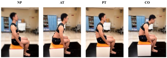
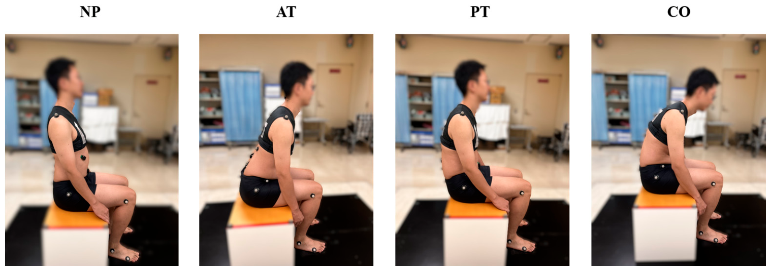
The four postures (Figure 1) were defined as follows: (1) neutral posture (NP), an upright sitting position with a trunk angle of approximately 0 degrees; (2) anterior trunk lean with anterior pelvic tilt (AT), a forward-leaning posture with a trunk angle of approximately 40 degrees and an anteriorly tilted pelvis; (3) posterior trunk lean/posterior pelvic tilt (PT), a posture characterized by posterior pelvic rotation and posterior trunk orientation; and (4) collapsed posture (CO), a slumped posture with forward trunk flexion of approximately 40 degrees combined with posterior pelvic tilt. A goniometer was used to standardize the target trunk angle across participants.
Figure 1.
Experimental Postures: Neutral Posture (NP), Anterior Trunk Lean Posture (AT), Posterior Trunk Lean Posture (PT), and Collapsed Posture (CO).
In each posture, measurements were obtained during quiet rest and during a maximal Valsalva maneuver. During rest, participants were instructed to breathe quietly and relax the abdominal muscles. During the Valsalva maneuver, participants were instructed to perform sustained forced expiration against a closed glottis for approximately 5 s. This standardized Valsalva maneuver was used as a laboratory-based model of voluntary straining and was not intended to reproduce physiological defecation. Four trials were performed for each condition, separated by at least 1 min of rest. The first trial was used for familiarization. For the ultrasound measurements, the second and third trials were used for analysis. For the EMG measurements, the subsequent three trials (second, third, and fourth) were recorded.
2.3. Ultrasonographic Measurements
A B-mode ultrasonography system (ARIETTA Prologue LE; Hitachi, Tokyo, Japan) equipped with a linear-array transducer (18.5 MHz; 38 mm width) was used to measure TrA thickness. The protocol was based on previously validated procedures for abdominal-muscle ultrasound imaging [27,28,29,30,31,32,33].
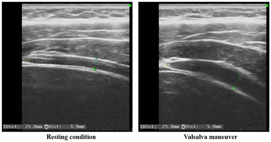
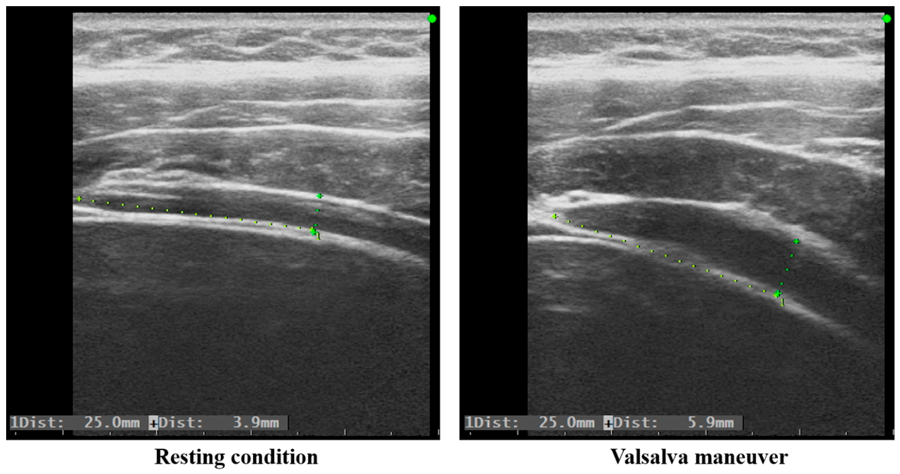
The transducer was positioned on the right side of the abdomen along the midaxillary line, midway between the 11th rib and the iliac crest, parallel to the muscle fibers of the TrA. The examiner adjusted the transducer orientation to ensure that the fascial borders of the external oblique, internal oblique, and TrA appeared parallel on the monitor. To standardize the imaging field, the anterior edge of the TrA was aligned with the left border of the ultrasound screen (Figure 2).
Figure 2.
Representative B-mode ultrasonographic images of the transversus abdominis (TrA) muscle at rest and during the Valsalva maneuver. Representative ultrasound images illustrating the measurement of the TrA muscle thickness. The anterior edge of the TrA was aligned with the left border of the ultrasound screen. The vertical distance between the superficial and deep fascial layers (indicated by dotted lines) represents the muscle thickness, measured approximately 2.5 cm posterior to the anterior edge of the TrA. (Left): resting condition (TrA thickness = 3.9 mm). (Right): during the Valsalva maneuver (TrA thickness = 5.9 mm).
During image acquisition, participants were instructed to breathe quietly, and the image was frozen at the end of a normal expiration to minimize the influence of respiratory movement on muscle thickness. For the Valsalva maneuver, the image was captured during the sustained straining effort (approximately 5 s) against a closed glottis, ensuring consistent intra-abdominal pressure generation across trials. The thickness of the TrA was defined as the perpendicular distance between the superficial and deep fascial layers. To ensure measurement consistency, this distance was measured at a standardized point 2.5 cm lateral to the insertion of the TrA into the rectus sheath on the ultrasound image. This approach has demonstrated good to excellent intra-rater reliability in previous studies [27,32,33]. The TrA thickness ratio was calculated by dividing TrA thickness during the Valsalva maneuver by TrA thickness at rest. Additionally, the change in TrA thickness was calculated as TrA thickness during the Valsalva maneuver minus TrA thickness at rest.
2.4. Surface Electromyographic Measurements
Surface EMG was recorded from the right superficial lumbar multifidus at L5 (sMF), erector spinae (ES), external oblique (EO), internal oblique (IO), and rectus abdominis (RA) using a wireless EMG system (Trigno Wireless EMG system, Delsys Inc., Natick, MA, USA). Skin preparation and electrode placement were performed according to SENIAM recommendations and abdominal-muscle electrode-orientation literature, with electrode orientation aligned as closely as possible to the underlying muscle-fiber direction [17,21,34,35]. Specifically, to minimize crosstalk and ensure signal validity, surface EMG electrodes were placed parallel to the muscle fiber directions over the muscle bellies, following previously validated anatomical guidelines and recommendations:
- -
- External Oblique (EO): Placed midway between the costal margin of the ribs and the iliac crest, oriented at approximately 45° to the horizontal line along the muscle fiber orientation [17,35].
- -
- Internal Oblique (IO): Placed 1 cm medial to the anterior superior iliac spine (ASIS), oriented horizontally below the level of the ASIS to isolate the transverse/internal oblique fibers and minimize crosstalk from the overlying external oblique [34,35].
- -
- Erector Spinae (ES): Placed 3 cm lateral to the L3 spinous process, oriented vertically along the muscle belly [17,21].
- -
- Rectus Abdominis (RA): Placed bilaterally 1 cm above the umbilicus and 2 cm lateral to the midline, oriented vertically along the muscle belly [34,35].
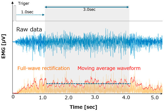
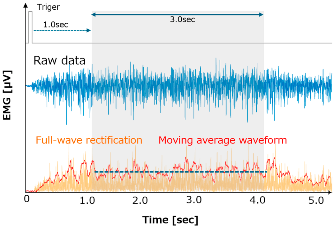
EMG signals were sampled at 1000 Hz and analyzed using a custom MATLAB program (R2024a, MathWorks, Natick, MA, USA). Raw signals were full-wave rectified and smoothed using a centered simple moving average filter with a 100 ms window (corresponding to 100 data points) to obtain the linear envelope, consistent with the standard surface EMG processing principle.
A digital trigger signal was synchronized with the onset of the verbal instruction for both resting and Valsalva conditions. To minimize initial movement artifacts and capture a stable contractile phase, a 3 s analysis epoch beginning 1 s after the trigger was extracted. For each condition, three trials were selected, and the mean amplitude of the moving average over this 3 s epoch was calculated for each trial and then averaged across the three trials. Three EMG outcomes were derived for each muscle: absolute activity at rest, absolute activity during the Valsalva maneuver, and the EMG activation ratio, calculated as the Valsalva amplitude divided by the resting amplitude within the same posture (Figure 3).
Figure 3.
Representative surface electromyography (EMG) signal processing and quantification workflow. The top channel displays the digital trigger signal and the raw EMG data. The bottom channel shows the full-wave rectified signal (orange) and the smoothed moving average waveform (red, 100 ms centered window). The gray shaded area denotes the 3 s analysis epoch, which begins 1 s after the trigger onset to capture a stable contractile phase. The horizontal blue dashed line represents the mean amplitude calculated over this 3 s epoch.
Because muscle-specific MVIC measurements were not included in the original experimental protocol, retrospective MVIC normalization was not possible. Although MVIC normalization is generally recommended for surface EMG, no single standardized MVIC protocol has been established that consistently elicits maximal activation across all trunk muscles, and multiple muscle-specific MVC tasks may be required for appropriate normalization [36,37]. In addition, non-normalized surface EMG amplitudes can be influenced by electrode placement, electrode–skin impedance, subcutaneous tissue thickness, and individual morphology [36]. Therefore, the absolute EMG values in the present study were interpreted cautiously and were used only for within-participant and within-muscle comparisons across postures during the same measurement session. The electrodes remained in place throughout all postural conditions, and absolute EMG amplitudes were not compared across muscles or participants. The EMG activation ratio was used as a complementary measure of the relative change in muscle activity from rest to the Valsalva maneuver.
2.5. Reliability and Statistical Analysis
For ultrasonographic reliability, the two analyzed trials for each postural condition were used to calculate the intraclass correlation coefficient (ICC) based on a one-way random-effects model for absolute agreement (ICC(1,2)). Absolute reliability was quantified using the standard error of measurement (SEM) and the minimal detectable change at the 95% confidence level (MDC95).
The mean value of the two analyzed trials was used for statistical analysis. Ultrasound outcomes were TrA thickness at rest, TrA thickness during the Valsalva maneuver, and the TrA contraction ratio (Valsalva thickness/resting thickness). EMG outcomes were absolute RMS amplitude at rest, absolute RMS amplitude during Valsalva, and the activation ratio (Valsalva RMS/resting RMS) for each muscle. For the interpretation of the EMG findings, the activation ratio was used as the principal indicator of the task-related change in EMG activity from rest to the Valsalva maneuver. Absolute EMG amplitudes at rest and during the Valsalva maneuver were retained as complementary outcomes to characterize posture-dependent baseline and task-related activity.
A one-way repeated-measures ANOVA was conducted separately for each outcome to assess the main effect of posture. When the sphericity assumption was violated, the Greenhouse–Geisser correction was applied. When a significant main effect was observed, Bonferroni-adjusted post hoc comparisons were conducted. Partial eta-squared was reported as an effect-size index. Statistical analyses were performed using SPSS Statistics for Windows, Version 29.0 (IBM Corp., Armonk, NY, USA), with the significance level set at p < 0.05.
3. Results
3.1. Reliability of Ultrasonographic Measurements
Ultrasonographic measurements of TrA thickness demonstrated excellent intra-rater reliability across postural and task conditions (Table 1). ICC(1,2) values ranged from 0.984 to 0.999. SEM values ranged from 0.038 to 0.195 mm, and MDC95 values ranged from 0.104 to 0.540 mm, confirming high absolute reliability of the measurement protocol.
Table 1.
Intra-rater reliability of TrA thickness measurements under each condition (rest and straining).
3.2. Ultrasound-Derived TrA Thickness Ratio
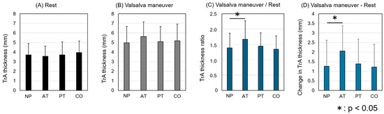
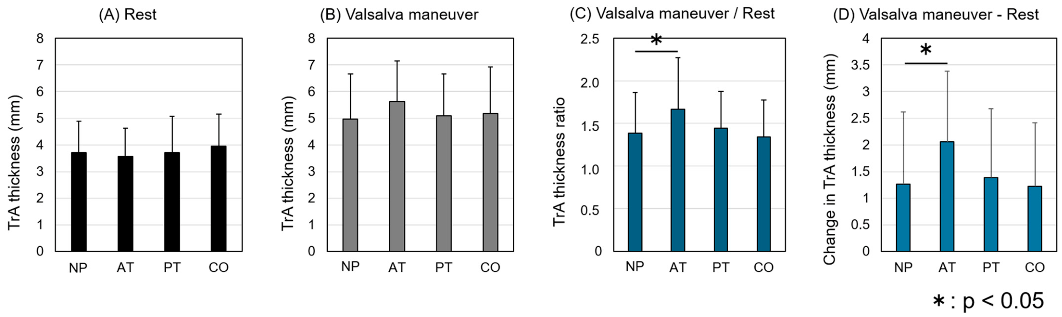
The TrA thickness at rest and during the Valsalva maneuver, the TrA contraction ratio, and the change in TrA thickness across the four postures are shown in Figure 4.
Figure 4.
Transversus abdominis (TrA) muscle thickness and contraction ratio. (A) Absolute TrA thickness (mm) at rest, (B) absolute TrA thickness (mm) during the Valsalva maneuver, (C) the thickness ratio calculated as the thickness during the Valsalva maneuver relative to the resting condition, and (D) change in TrA thickness, calculated as TrA thickness during the Valsalva maneuver minus TrA thickness at rest. Bars represent the mean values, and error bars denote the standard deviations. NP: Neutral Posture; AT: Anterior Trunk Lean Posture; PT: Posterior Trunk Lean Posture; CO: Collapsed Posture. Asterisk (*) indicates a statistically significant difference between the two postures (p < 0.05).
For TrA thickness at rest, repeated-measures ANOVA with Greenhouse–Geisser correction showed no significant main effect of posture, F(2.2, 41.1) = 1.41, p = 0.255, partial η2 = 0.069.
For TrA thickness during the Valsalva maneuver, the main effect of posture did not reach statistical significance, F(3, 57) = 1.80, p = 0.157, partial η2 = 0.087, indicating that the data did not provide sufficient evidence of an overall posture effect on absolute TrA thickness.
For the TrA contraction ratio, repeated-measures ANOVA with Greenhouse–Geisser correction showed a significant main effect of posture, F(2.2, 41.5) = 3.60, p = 0.033, partial η2 = 0.159. Bonferroni-adjusted post hoc comparisons indicated that the contraction ratio was higher in AT (1.66 ± 0.61) than in NP (1.39 ± 0.48), p = 0.001. No significant differences were observed among NP, PT, and CO.
For the change in TrA thickness, repeated-measures ANOVA with Greenhouse–Geisser correction showed a significant main effect of posture, F(1.9, 36.3) = 3.82, p = 0.033, partial η2 = 0.167. Bonferroni-adjusted post hoc comparisons indicated that the change in TrA thickness was greater in AT (2.06 ± 1.33 mm) than in NP (1.26 ± 1.36 mm), p < 0.001. No significant differences were observed among NP, PT, and CO.
3.3. Muscle Activity
3.3.1. EMG Activation Ratio
Table 2 shows the EMG activation ratios across the four postures. For the EMG activation ratio, a significant main effect of posture was observed for ES, F(1.991, 37.828) = 5.665, p = 0.007, partial η2 = 0.230. Bonferroni-adjusted post hoc analysis indicated that the ES activation ratio was highest in NP compared with the other postures (all p < 0.05). No significant main effect of posture was observed for the sMF or EO activation ratio.
Table 2.
Comparison of EMG Activity Ratio across Four Postures.
3.3.2. Absolute Muscle Activity at Rest
The absolute muscle activities at rest across the four postures are presented in Table 3. For absolute muscle activity at rest, one-way repeated-measures ANOVA revealed significant main effects of posture for sMF, F(1.684, 32.004) = 15.581, p < 0.001, partial η2 = 0.451; ES, F(2.232, 42.413) = 56.240, p < 0.001, partial η2 = 0.747; and EO, F(1.646, 31.277) = 4.585, p = 0.024, partial η2 = 0.194. Bonferroni-adjusted post hoc comparisons showed that resting sMF and ES activities were greater in AT than in NP, PT, and CO (all p < 0.05). Although the EO omnibus test reached significance, Bonferroni-adjusted pairwise comparisons did not identify significant differences among the four postures.
Table 3.
Comparison of Absolute Muscle Activity at Rest across four postures.
3.3.3. Absolute Muscle Activity During the Valsalva Maneuver
The absolute muscle activities during the Valsalva maneuver across the four postures are presented in Table 4. During maximal Valsalva, one-way repeated-measures ANOVA revealed significant main effects of posture for sMF, F(1.588, 30.172) = 12.084, p < 0.001, partial η2 = 0.389, and ES, F(2.156, 40.969) = 82.312, p < 0.001, partial η2 = 0.812. Consistent with the resting condition, AT elicited the highest absolute sMF and ES activity during straining compared with the other postures (all p < 0.05). In contrast, posture did not significantly affect absolute EO activity, F(1.601, 30.418) = 0.884, p = 0.402, partial η2 = 0.044, IO activity, F(1.246, 23.680) = 2.691, p = 0.108, partial η2 = 0.124, and RA activity, F(1.668, 31.683) = 1.631, p = 0.214, partial η2 = 0.079, during Valsalva.
Table 4.
Comparison of Absolute Muscle Activity During the Valsalva Maneuver across four postures.
4. Discussion
4.1. Principal Findings
The present study examined how sitting posture modifies ultrasound-derived TrA behavior and trunk muscle EMG activity during a Valsalva-based simulated straining task. The main finding was that AT maximized the TrA thickness ratio compared with NP, despite similar resting TrA thickness across postures. The EMG results provide an important mechanistic interpretation: the postural advantage of AT was accompanied by higher absolute sMF and ES activity at rest and during Valsalva, whereas EO and IO activity during Valsalva did not increase across postures. Thus, the AT posture did not simply induce greater global abdominal effort. Rather, it appeared to create a lumbopelvic condition characterized by elevated posterior-trunk-wall activation before and during straining. In addition, NP showed the highest ES activation ratio and relatively low TrA thickness. These findings provide novel evidence that the biomechanical configuration of sitting posture—particularly the combination of anterior trunk lean and anterior pelvic tilt—was associated with greater absolute and relative TrA thickness change than NP during the simulated straining task by promoting posterior trunk-wall activation leading to mechanical stabilization.
4.2. Biomechanical Interpretation
A controlled anterior trunk lean with anterior pelvic tilt shifts the lumbopelvic complex toward a more extended alignment and increases the external flexion moment acting on the trunk. To maintain this posture, the posterior trunk muscles must generate stabilizing activity before the Valsalva maneuver begins. The present EMG results support this interpretation: resting sMF and ES activity were significantly higher in AT than in all other postures. We interpret this elevated baseline activity as greater resting postural activation, meaning a preparatory stabilization state induced by posture rather than a response to straining alone.
During the Valsalva maneuver, the AT posture maintained the highest sMF and ES activity while also producing the greatest TrA thickness ratio and change. This pattern is consistent with the concept that abdominal-wall and paraspinal co-activation contributes to trunk stiffness and load sharing [7,18,19]. A stable posterior trunk wall may provide a mechanically advantageous base against which the TrA can generate circumferential abdominal-wall tension. The absence of significant posture effects for EO and IO activity during Valsalva further suggests that the TrA thickening advantage in AT was not merely the result of increased lateral abdominal-wall effort. Instead, AT may optimize deep abdominal behavior by combining anterior pelvic orientation, spinal extensor pre-activation, and enhanced lumbopelvic stability.
This interpretation is also consistent with the anatomical and biomechanical role of the thoracolumbar fascia (TLF). The TLF is not simply a passive fascial envelope; rather, it forms a multilayered aponeurotic system that mechanically links the paraspinal muscles with the deep abdominal muscles, including the TrA and internal oblique [12]. Experimental work has shown that simulated paraspinal muscle contraction and deep abdominal tension interact through the middle and posterior layers of the TLF, suggesting a co-dependent tension-sharing mechanism between the posterior and anterolateral trunk muscles [38]. In the present study, AT was characterized by greater absolute ES and sMF activity and a greater TrA thickness ratio than NP during the Valsalva-based simulated straining task. Although the TLF was not directly assessed, the concurrent pattern of posterior trunk muscle activity and TrA behavior in AT may be compatible with a literature-based hypothesis of posture-dependent load sharing between the posterior and anterolateral trunk structures. This proposed mechanism remains speculative and requires direct investigation.
The PT and CO postures did not enhance TrA thickening. Both postures involve posterior pelvic rotation and a more flexed lumbopelvic alignment. Such alignments may reduce the contribution of active spinal extensor stabilization and increase reliance on passive posterior structures, including the posterior ligamentous tissues and the TLF, particularly when the trunk is positioned closer to flexion [11,12,20,21]. This interpretation is supported by elastography evidence showing that TLF stiffness is posture-dependent and increases with flexion-related loading [11]. However, it is important to distinguish passive fascial tension from active muscle-derived tension. In a flexed sitting posture, the TLF should not be interpreted as completely slack or completely tensioned; rather, its mechanical state depends on the balance between passive elongation and active muscle inputs. These facts suggest that passive posterior tension alone may be insufficient to facilitate coordinated TrA thickening during voluntary straining.
In the NP, the present data showed relatively low TrA thickness together with increased ES activity. Because the spine was not positioned in marked flexion, passive elongation of the TLF due to flexion-related loading was probably limited compared with more flexed postures. Nevertheless, the mechanical state of the TLF cannot be interpreted simply as slack. As previously mentioned, the TLF functions as an integrated fascial-aponeurotic complex that mechanically couples the paraspinal muscles with the deep abdominal wall, including the TrA and internal oblique [12,38]. In addition, tensile loading studies have demonstrated that forces generated through muscular attachments, including the erector spinae and abdominal aponeuroses, can be transmitted through the posterior and middle layers of the lumbar fascia [39,40]. Thus, the present findings may indicate a shift in TLF-mediated tension sharing: TrA-related fascial tension input was relatively reduced, whereas ES-related posterior fascial tension input was increased. Such a reciprocal or compensatory pattern may represent a posture-dependent stabilization strategy in which posterior muscle activation contributes to lumbopelvic control when deep abdominal contribution is limited.
4.3. Clinical Implication
These findings provide a physiological rationale for reconsidering simple toileting posture instructions. The results do not support the assumption that strictly upright sitting is necessarily the most favorable posture for generating an abdominal straining strategy. Instead, a controlled forward lean with anterior pelvic orientation may create a more advantageous lumbopelvic condition for TrA thickening, supported by increased sMF and ES activity. This concept is broadly consistent with prior evidence that toileting posture can influence perceived straining and defecation time, although previous studies mainly examined squatting or increased hip flexion rather than the specific trunk-pelvic configurations tested here [41].
Clinical translation requires caution. This study did not directly measure rectal pressure, IAP, pelvic-floor relaxation, stool evacuation, or patient-reported symptoms. Therefore, the findings should be interpreted as evidence of posture-dependent neuromuscular and structural behavior during simulated straining, not as direct proof that AT improves defecatory function. Future studies should combine ultrasound, surface or intramuscular EMG, and anorectal manometry in older adults and patients with defecatory disorders to test whether the AT posture improves rectal propulsive pressure and evacuation efficiency.
4.4. Limitations
Several limitations should be acknowledged. First, healthy young adults were studied to isolate postural biomechanics; consequently, generalizability to older adults, individuals with sarcopenia, and patients with functional defecation disorders is limited. These clinical populations may present with altered rectal sensation, pelvic-floor dyssynergia, reduced muscle mass, and altered visceral compliance, which may interact with posture differently.
Second, IAP and anorectal pressure were not directly measured. Although TrA thickness changes and abdominal-wall tension are relevant non-invasive indicators of muscle behavior and pressure-related mechanics [23], ultrasound-derived thickening cannot be equated directly with force, IAP, or rectal propulsion. Simultaneous high-resolution anorectal manometry would be necessary to confirm whether AT increases propulsive pressure during evacuation.
Third, the standardized Valsalva maneuver used in this study did not reproduce all physiological components of defecation. Specifically, respiratory coordination, diaphragmatic descent, relaxation of the external anal sphincter and puborectalis, pelvic-floor coordination, anorectal pressure gradients, and actual evacuation were not assessed. Therefore, the observed changes in TrA thickness and trunk muscle activity should be interpreted as responses to a laboratory-based simulated straining task rather than as direct measures of defecatory mechanics or function.
Fourth, surface EMG is susceptible to cross-talk and cannot directly record the deep TrA. EMG amplitudes were not normalized to maximal voluntary contractions; therefore, absolute EMG results should be interpreted as within-muscle, within-participant postural comparisons rather than as cross-muscle activation levels [36,42].
Fifth, although trunk flexion and extension angles were standardized using a handheld goniometer according to the measurement method established by the Japanese Orthopedic Association and the Japanese Association of Rehabilitation Medicine, pelvic tilt and lumbar lordosis were not quantitatively measured. Pelvic orientation was standardized using visual observation and standardized verbal instructions, and a trained examiner checked the posture before each trial. Nevertheless, residual interparticipant and intertrial variations in lumbopelvic alignment could not be excluded. In addition, bilateral weight distribution between the feet was not instrumentally assessed. Therefore, the present findings should be interpreted as the effects of composite seated-posture conditions, and the independent contributions of trunk inclination, pelvic orientation, and lumbar curvature cannot be determined.
Finally, the exact contribution of breathing pattern, glottal closure consistency, and trial order could not be fully separated from the postural effects.
5. Conclusions
In healthy young adults, AT was associated with a greater TrA thickness ratio and change than NP during a Valsalva-based simulated straining task. AT was also characterized by greater absolute sMF and ES amplitudes at rest and during the maneuver in within-participant and within-muscle comparisons. These findings indicate posture-dependent differences in trunk muscle behavior. However, they do not establish that AT increases intra-abdominal pressure, rectal propulsive pressure, evacuation efficiency, or defecatory function. Direct physiological and clinical measurements are required before recommendations regarding constipation management or toileting posture can be made.
Supplementary Materials
The following supporting information can be downloaded at https://www.mdpi.com/article/10.3390/biomechanics6030079/s1. Dataset S1: De-identified participant-level TrA thickness data across the four postural conditions (.csv).
Author Contributions
Conceptualization, Y.S., T.K. and K.H.; methodology, Y.S., H.I., S.Y., T.K. and K.H.; formal analysis, Y.S. and H.I.; investigation, Y.S., H.I., S.Y., T.K., R.S., T.S. and W.Z.; resources, T.K., K.H., S.E. and H.K.; data curation, Y.S.; writing—original draft preparation, Y.S.; writing—review and editing, H.I., S.Y., T.K., K.H., S.E., R.S., W.Z., T.S. and H.K.; visualization, Y.S.; supervision, S.E. and H.K.; project administration, Y.S.; funding acquisition, T.K. All authors have read and agreed to the published version of the manuscript.
Funding
This research was funded by TOTO Ltd. The study was conducted as part of the project “Preliminary Study on Excretory Function Based on Medical and Physiological Knowledge: Exploring the Factors that Enable Effortless Urination and Defecation without Straining.”.
Institutional Review Board Statement
The study was conducted in accordance with the Declaration of Helsinki and was approved by the institutional research Ethics Committee Tohoku University Graduate School of Dentistry.
Informed Consent Statement
Informed consent was obtained from all subjects involved in the study. Written informed consent has been obtained from the patients to publish this paper.
Data Availability Statement
De-identified participant-level TrA thickness data used in the additional analyses are provided in Supplementary Dataset S1. The remaining data are not publicly available due to privacy and ethical restrictions but may be available from the corresponding author upon reasonable request, subject to applicable ethical and institutional approval.
Acknowledgments
The authors would like to thank the staff of the Department of Rehabilitation, Tohoku University Hospital, for their assistance with this study. During the preparation of this manuscript/study, the authors used ChatGPT 5.5 and Gemini 3.5 for the purposes of drafting and refining text, and generating ideas for manuscript structure. The authors have reviewed and edited the output and take full responsibility for the content of this publication.
Conflicts of Interest
TOTO Ltd. provided financial support for this study. T.K., an employee of TOTO Ltd., participated in the conceptualization and methodological development of the study and contributed to manuscript review. Data analysis was performed by Y.S. and H.I. The academic authors were responsible for the interpretation of the results and the final decision to submit the manuscript. No specific commercial product was evaluated in this study. The remaining authors declare no conflicts of interest.
Abbreviations
The following abbreviations are used in this manuscript:
| TrA | transversus abdominis |
| sMF | superficial lumbar multifidus |
| ES | erector spinae |
| EO | external oblique |
| IO | internal oblique |
| NP | neutral posture |
| AT | anterior trunk lean with anterior pelvic tilt |
| PT | posterior trunk lean/posterior pelvic tilt |
| CO | collapsed posture |
| TLF | thoracolumbar fascia |
References
- Rao, S.S.; Patcharatrakul, T. Diagnosis and Treatment of Dyssynergic Defecation. J. Neurogastroenterol. Motil. 2016, 22, 423–435. [Google Scholar] [CrossRef] [Scilit] [PubMed]
- Rao, S.S.C.; Rattanakovit, K.; Patcharatrakul, T. Diagnosis and management of chronic constipation in adults. Nat. Rev. Gastroenterol. Hepatol. 2016, 13, 295–305. [Google Scholar] [CrossRef] [Scilit] [PubMed]
- Bharucha, A.E.; Pemberton, J.H.; Locke, G.R. American Gastroenterological Association Technical Review on Constipation. Gastroenterology 2013, 144, 218–238. [Google Scholar] [CrossRef] [Scilit] [PubMed]
- Suares, N.C.; Ford, A.C. Prevalence of, and risk factors for, chronic idiopathic constipation in the community: Systematic review and meta-analysis. Am. J. Gastroenterol. 2011, 106, 1582–1591. [Google Scholar] [CrossRef] [Scilit] [PubMed]
- Salari, N.; Ghasemianrad, M.; Ammari-Allahyari, M.; Rasoulpoor, S.; Shohaimi, S.; Mohammadi, M. Global prevalence of constipation in older adults: A systematic review and meta-analysis. Wien. Klin. Wochenschr. 2023, 135, 389–398. [Google Scholar] [CrossRef] [Scilit] [PubMed]
- Deb, B.; Sharma, M.; Fletcher, J.G.; Srinivasan, S.G.; Chronopoulou, A.; Chen, J.; Bailey, K.R.; Feuerhak, K.J.; Bharucha, A.E. Inadequate Rectal Pressure and Insufficient Relaxation and Abdominopelvic Coordination in Defecatory Disorders. Gastroenterology 2022, 162, 1111–1122.E2. [Google Scholar] [CrossRef] [Scilit] [PubMed]
- Cholewicki, J.; Juluru, K.; McGill, S.M. Intra-abdominal pressure mechanism for stabilizing the lumbar spine. J. Biomech. 1999, 32, 13–17. [Google Scholar] [CrossRef] [Scilit] [PubMed]
- Nakagawa, H.; Takeshima, T.; Ozaka, A.; Sasaki, S.; Hamaguchi, S.; Fukuhara, S. The Association between Sarcopenia and Constipation Onset in Community-dwelling Older Adults in Japan. Intern. Med. 2025, 64, 1965–1970. [Google Scholar] [CrossRef] [Scilit] [PubMed]
- Asaoka, D.; Takeda, T.; Inami, Y.; Abe, D.; Shimada, Y.; Matsumoto, K.; Ueyama, H.; Matsumoto, K.; Komori, H.; Akazawa, Y.; et al. Association between the severity of constipation and sarcopenia in elderly adults: A single-center university hospital-based, cross-sectional study. Biomed. Rep. 2021, 14, 2. [Google Scholar] [CrossRef] [Scilit] [PubMed]
- Owen, P.J.; Rantalainen, T.; Scheuring, R.A.; Belavy, D.L. Axial loading and posture cues in contraction of transversus abdominis and multifidus with exercise. Sci. Rep. 2020, 10, 11218. [Google Scholar] [CrossRef] [Scilit] [PubMed]
- Chen, B.; Liu, C.; Lin, M.; Deng, W.; Zhang, Z. Effects of body postures on the shear modulus of thoracolumbar fascia: A shear wave elastography study. Med. Biol. Eng. Comput. 2021, 59, 383–390. [Google Scholar] [CrossRef] [Scilit] [PubMed]
- Willard, F.H.; Vleeming, A.; Schuenke, M.D.; Danneels, L.; Schleip, R. The thoracolumbar fascia: Anatomy, function and clinical considerations. J. Anat. 2012, 221, 507–536. [Google Scholar] [CrossRef] [Scilit] [PubMed]
- Novak, J.; Jacisko, J.; Busch, A.; Cerny, P.; Stribrny, M.; Kovari, M.; Podskalska, P.; Kolar, P.; Kobesova, A. Intra-abdominal pressure correlates with abdominal wall tension during clinical evaluation tests. Clin. Biomech. 2021, 88, 105426. [Google Scholar] [CrossRef] [Scilit] [PubMed]
- Waongenngarm, P.; Rajaratnam, B.S.; Janwantanakul, P. Perceived body discomfort and trunk muscle activity in three prolonged sitting postures. J. Phys. Ther. Sci. 2015, 27, 2183–2187. [Google Scholar] [CrossRef] [Scilit] [PubMed]
- Reeve, A.; Dilley, A. Effects of posture on the thickness of transversus abdominis in pain-free subjects. Man. Ther. 2009, 14, 679–684. [Google Scholar] [CrossRef] [Scilit] [PubMed]
- Hemming, R.; du Rose, A.; Sheeran, L.; van Deursen, R.; Sparkes, V. Relationships between trunk muscle activation and thoraco-lumbar kinematics in non-specific chronic low back pain subgroups during a forward bending task. Gait Posture 2024, 107, 96–103. [Google Scholar] [CrossRef] [Scilit] [PubMed]
- Takaki, S.; Kaneoka, K.; Okubo, Y.; Otsuka, S.; Tatsumura, M.; Shiina, I.; Miyakawa, S. Analysis of muscle activity during active pelvic tilting in sagittal plane. Phys. Ther. Res. 2016, 19, 50–57. [Google Scholar] [CrossRef] [Scilit] [PubMed]
- Matthijs, O.C.; Dedrick, G.S.; James, C.R.; Brismée, J.M.; Hooper, T.L.; McGalliard, M.K.; Sizer, P.S., Jr. Co-contractive activation of the superficial multifidus during volitional preemptive abdominal contraction. PM R J. Inj. Funct. Rehabil. 2014, 6, 13–21. [Google Scholar] [CrossRef] [Scilit] [PubMed]
- Vera-Garcia, F.J.; Elvira, J.L.L.; Brown, S.H.M.; McGill, S.M. Effects of abdominal stabilization maneuvers on the control of spine motion and stability against sudden trunk perturbations. J. Electromyogr. Kinesiol. 2007, 17, 556–567. [Google Scholar] [CrossRef] [Scilit] [PubMed]
- Floyd, W.F.; Silver, P.H. The function of the erectores spinae muscles in certain movements and postures in man. J. Physiol. 1955, 129, 184–203. [Google Scholar] [CrossRef] [Scilit] [PubMed]
- Solomonow, M.; Baratta, R.V.; Banks, A.; Freudenberger, C.; Zhou, B.H. Flexion–relaxation response to static lumbar flexion in males and females. Clin. Biomech. 2003, 18, 273–279. [Google Scholar] [CrossRef] [Scilit] [PubMed]
- McMeeken, J.M.; Beith, I.D.; Newham, D.J.; Milligan, P.; Critchley, D.J. The relationship between EMG and change in thickness of transversus abdominis. Clin. Biomech. 2004, 19, 337–342. [Google Scholar] [CrossRef] [Scilit] [PubMed]
- Hodges, P.W.; Pengel, L.H.; Herbert, R.D.; Gandevia, S.C. Measurement of muscle contraction with ultrasound imaging. Muscle Nerve 2003, 27, 682–692. [Google Scholar] [CrossRef] [Scilit] [PubMed]
- Noelting, J.; Ratuapli, S.K.; Bharucha, A.E.; Harvey, D.M.; Ravi, K.; Zinsmeister, A.R. Normal Values for High-Resolution Anorectal Manometry in Healthy Women: Effects of Age and Significance of Rectoanal Gradient. Off. J. Am. Coll. Gastroenterol. ACG 2012, 107, 1530–1536. [Google Scholar] [CrossRef] [Scilit] [PubMed]
- Bharucha, A.E.; Wald, A.; Enck, P.; Rao, S. Functional Anorectal Disorders. Gastroenterology 2006, 130, 1510–1518. [Google Scholar] [CrossRef] [Scilit] [PubMed]
- Japanese Society of Rehabilitation Medicine. Revision of the range of motion measurement and notation method (April 2022 revision). Jpn. J. Rehabil. Med. 2021, 58, 1188–1200. [Google Scholar] [CrossRef] [Scilit]
- Linek, P.; Saulicz, E.; Wolny, T.; Myśliwiec, A. Intra-rater reliability of B-mode ultrasound imaging of the abdominal muscles in healthy adolescents during the active straight leg raise test. PM R J. Inj. Funct. Rehabil. 2015, 7, 53–59. [Google Scholar] [CrossRef] [Scilit] [PubMed]
- Gong, W. Correlations between Transversus Abdominis Thickness, Lumbar Stability, and Balance of Female University Students. J. Phys. Ther. Sci. 2013, 25, 681–683. [Google Scholar] [CrossRef] [Scilit] [PubMed][Green Version]
- Nabavi, N.; Mosallanezhad, Z.; Haghighatkhah, H.R.; Mohseni Bandpeid, M.A. Reliability of rehabilitative ultrasonography to measure transverse abdominis and multifidus muscle dimensions. Iran. J. Radiol. 2014, 11, e21008. [Google Scholar] [CrossRef] [Scilit] [PubMed]
- Prentice, C.L.S.; Milanese, S.; Massy-Westropp, N.; Maranna, S. The reliability of rehabilitative ultrasound to measure lateral abdominal muscle thickness: A systematic review and meta-analysis. Musculoskelet. Sci. Pract. 2021, 53, 102357. [Google Scholar] [CrossRef] [Scilit] [PubMed]
- Kellis, E.; Ellinoudis, A.; Intziegianni, K.; Kofotolis, N. Muscle Thickness During Core Stability Exercises in Children and Adults. J. Hum. Kinet. 2020, 71, 131–144. [Google Scholar] [CrossRef] [Scilit] [PubMed]
- Puentedura, E.J.; Buckingham, S.J.; Morton, D.; Montoya, C.; Fernandez de las Penas, C. Immediate Changes in Resting and Contracted Thickness of Transversus Abdominis After Dry Needling of Lumbar Multifidus in Healthy Participants: A Randomized Controlled Crossover Trial. J. Manip. Physiol. Ther. 2017, 40, 615–623. [Google Scholar] [CrossRef] [Scilit] [PubMed]
- Mangum, L.C.; Sutherlin, M.A.; Saliba, S.A.; Hart, J.M. Reliability of Ultrasound Imaging Measures of Transverse Abdominis and Lumbar Multifidus in Various Positions. PM R 2016, 8, 340–347. [Google Scholar] [CrossRef] [Scilit] [PubMed]
- Hermens, H.J.; Freriks, B.; Disselhorst-Klug, C.; Rau, G. Development of recommendations for SEMG sensors and sensor placement procedures. J. Electromyogr. Kinesiol. 2000, 10, 361–374. [Google Scholar] [CrossRef] [Scilit] [PubMed]
- Ng, J.K.; Kippers, V.; Richardson, C.A. Muscle fibre orientation of abdominal muscles and suggested surface EMG electrode positions. Electromyogr. Clin. Neurophysiol. 1998, 38, 51–58. [Google Scholar] [PubMed]
- Burden, A. How should we normalize electromyograms obtained from healthy participants? What we have learned from over 25years of research. J. Electromyogr. Kinesiol. 2010, 20, 1023–1035. [Google Scholar] [CrossRef] [Scilit] [PubMed]
- Vera-Garcia, F.J.; Moreside, J.M.; McGill, S.M. MVC techniques to normalize trunk muscle EMG in healthy women. J. Electromyogr. Kinesiol. 2010, 20, 10–16. [Google Scholar] [CrossRef] [Scilit] [PubMed]
- Vleeming, A.; Schuenke, M.D.; Danneels, L.; Willard, F.H. The functional coupling of the deep abdominal and paraspinal muscles: The effects of simulated paraspinal muscle contraction on force transfer to the middle and posterior layer of the thoracolumbar fascia. J. Anat. 2014, 225, 447–462. [Google Scholar] [CrossRef] [Scilit] [PubMed]
- Vleeming, A.; Pool-Goudzwaard, A.L.; Stoeckart, R.; van Wingerden, J.P.; Snijders, C.J. The posterior layer of the thoracolumbar fascia. Its function in load transfer from spine to legs. Spine 1995, 20, 753–758. [Google Scholar] [CrossRef] [Scilit]
- Barker, P.J.; Briggs, C.A.; Bogeski, G. Tensile transmission across the lumbar fasciae in unembalmed cadavers: Effects of tension to various muscular attachments. Spine 2004, 29, 129–138. [Google Scholar] [CrossRef] [Scilit] [PubMed]
- Sikirov, D. Comparison of straining during defecation in three positions: Results and implications for human health. Dig. Dis. Sci. 2003, 48, 1201–1205. [Google Scholar] [CrossRef] [Scilit] [PubMed]
- De Luca, C.J. The Use of Surface Electromyography in Biomechanics. J. Appl. Biomech. 1997, 13, 135–163. [Google Scholar] [CrossRef] [Scilit]

Disclaimer/Publisher’s Note: The statements, opinions and data contained in all publications are solely those of the individual author(s) and contributor(s) and not of MDPI and/or the editor(s). MDPI and/or the editor(s) disclaim responsibility for any injury to people or property resulting from any ideas, methods, instructions or products referred to in the content. |
© 2026 by the authors. Licensee MDPI, Basel, Switzerland. This article is an open access article distributed under the terms and conditions of the Creative Commons Attribution (CC BY) license.