


Abstract
In this study, the ultrastructural characteristics of the mature spermatozoon of the cestode Gerbillitaenia psammomi, a parasite of the fat sand rat (Psammomys obesus) in the south of Tunisia, were examined by means of transmission electron microscopy. The spermatozoon is of Levron et al.’s type VI. This type of sperm cell is mainly characterized by the presence of a single axoneme of the 9 + ‘1’ pattern typical of the trepaxonematan Platyhelminthes, crest-like bodies, a periaxonemal sheath, twisted cortical microtubules, and a spiraled nucleus. The spermatozoon of G. psammomi possesses two crest-like bodies of different length. The results obtained in the present study suggest certain similarities with the remaining analyzed catenotaeniids; namely, Catenotaenia pusilla, Skrjabinotaenia (Meggittina) gerbilli, and Spasskijela lobata. The results are compared and discussed according to several characteristics found in the catenotaeniids and other studied cestodes, particularly those of the order Cyclophyllidea.
1. Introduction
The genus Gerbillitaenia Haukisalmi & Elmahy, 2025 (Cyclophyllidea: Catenotaeniidae) (http://zoobank.org/urn:lsid:zoobank.org:pub:E881B558-FAD7-4C9D-ABEC-A65BEC89DCF0, accessed on 2 February 2026) has recently been created, with Gerbillitaenia psammomi (Mikhail & Fahmy, 1968) as its type and sole species [1]. This species was originally described as Skrjabinotaenia psammomi Mikhail & Fahmy, 1968, parasitizing the fat sand rat Psammomys obesus Cretzschmar, 1828 (Rodentia: Muridae: Gerbillinae) in Egypt [2]. Additionally, some records of Skrjabinotaenia oranensis Joyeux & Foley, 1930, parasitizing Meriones shawi (Rozet, 1833) (Rodentia: Muridae) in Morocco [3] and P. obesus in Egypt [4,5,6] have been synonymized with G. psammomi by Haukisalmi and Elmahy [1]. Finally, it is worth mentioning our recent report of G. psammomi parasitizing P. obesus for the first time in Tunisia [7]. The latter work [7] also provided the first molecular data on the species and described negative density-dependent growth (a crowding effect). In the present study, additional data regarding sperm characteristics and their organization in the spermatozoon are presented to expand the literature on G. psammomi.
Over the past few decades, ultrastructural studies of sperm have proven useful for analyzing phylogenetic relationships in various groups of parasitic flatworms such as monogeneans, digeneans, and cestodes [8,9,10,11,12,13,14]. In the case of eucestodes, there have been numerous studies on species belonging to different orders; namely, Bothriocephalidea, Caryophyllidea, Cyclophyllidea, Diphyllidea, Diphyllobothriidea, Haplobothriidea, Lecanicephalidea, Nippotaeniidae, Onchoproteocephalidea, Spathebothriidea, Tetrabothriidea, Tetraphyllidea, and Trypanorhyncha [8,10,11,12,15,16,17,18,19,20,21,22,23,24]. In reference to the cyclophyllidean family Catenotaeniidae, the ultrastructural organization of sperm has been studied in three species; namely, Catenotaenia pusilla (Goeze, 1782), Spasskijela lobata (Baer, 1925)—published as Skrjabinotaenia lobata (Baer, 1925)—and Skrjabinotaenia (Meggittina) gerbilli (Wertheim, 1954)—published as Meggittina gerbilli Wertheim, 1954 [25,26,27]. In the present study, we describe the ultrastructural characteristics of the spermatozoon in a fourth species belonging to the genus Gerbillitaenia—namely, G. psammomi, a parasite of the fat sand rat (P. obesus) in Tunisia—for the first time.
2. Materials and Methods
2.1. Specimens
A total of 1332 specimens of G. psammomi were recovered from the intestines of 72 fat sand rats, P. obesus, trapped in different localities of Southern Tunisia (El Fejij, El Menzela, Ksar El Hallouf, and Tamaghza) during several field missions conducted in 2023 and 2024. The studied gerbillines were captured using snap traps, sacrificed by means of cervical dislocation and, finally, quickly analyzed for intestinal parasites under a Heerbrugg Wild M3Z stereomicroscope (Leica Microsystems, Wetzlar, Germany).
2.2. Light Microscopy Study and Identification
The recovered living cestodes were fixed in 70% ethanol during the fieldwork. Later, in the laboratory, the specimens were stained with Semichon’s acetic carmine, differentiated in 50% acid–alcohol solution, dehydrated in an ethanol series and with 2-propanol, cleared in clove oil and, finally, mounted on slides using Canada balsam. Finally, they were identified under a Leitz Laborlux K microscope (Leica Microsystems) according to the pertinent literature [1,2,3]. A complete re-description of G. psammomi was recently published [7]. Some stained and mounted specimens were deposited in the “Museu de Ciències Naturals de Barcelona”, Spain (accession numbers: MZB 2024-3090, MZB 2024-3091, MZB 2024-3092, and MZB 2024-3093). Additionally, sequences of partial 28S rDNA were submitted to the GenBank database (accession numbers: PP291541, PP291542, and PP291543) (see Boubakri et al. [7] for more detailed information).
2.3. Transmission Electron Microscopy
For the present ultrastructural study, 19 living specimens recovered from four fat sand rats were rinsed with a 0.9% NaCl solution immediately upon their removal from the intestine. Then, a total of 27 portions of mature proglottids from these specimens were cut and fixed in 2.5% glutaraldehyde buffered with 0.1 M sodium cacodylate buffer at pH 7.4 for 3 h at 4 °C. After glutaraldehyde fixation, the samples were rinsed in 0.1 M sodium cacodylate buffer at pH 7.4 (three 15 min washes) and postfixed in 1% osmium tetroxide with 0.9% potassium ferricyanide in the same buffer for 1 h at 4 °C. After rinsing in Milli-Q water (Millipore Gradient A10, Millipore Co., Merck KGaA, Darmstadt, Germany) (a quick wash plus three 15 min washes), the mature proglottids were dehydrated using an ethanol series (70%, 80%, 90%, and 96% ethanol, 1 time each for 15 min; absolute ethanol, 2 times for 30 min) and propylene oxide (1 h). Finally, the 27 portions of proglottids were embedded in Spurr’s epoxy resin as follows: 1:3 Spurr and propylene oxide (overnight), 1:1 Spurr and propylene oxide (6 h), 3:1 Spurr and propylene oxide (overnight), pure Spurr (6 h), pure Spurr (overnight), and polymerization at 60 °C for 96 h. Semithin sections (500 nm thick) of four blocks (one for each of the four gerbils) were obtained using a Leica Reichert-Jung Ultracut E ultramicrotome (Leica Microsystems), placed on slides, and stained with a 1% methylene blue–1% borax mixture. These sections were used to locate the vas deferens. Ultrathin sections (60–70 nm thick) were obtained using a Leica Reichert-Jung Ultracut E ultramicrotome (Leica Microsystems), placed on 200-mesh copper grids, and double-stained with uranyl acetate and lead citrate [28]. A total of eight grids containing 5–10 ultrathin sections per grid were prepared for each of the four studied blocks. Finally, the stained grids were observed under a JEOL 1010 transmission electron microscope (JEOL Ltd., Tokyo, Japan) operated at an accelerating voltage of 80 kV at the “Centres Científics i Tecnològics de la Universitat de Barcelona (CCiTUB)”. TEM micrographs were obtained by means of the connected camera using Gatan’s DigitalMicrograph software (version 2.11) (Pleasanton, CA, USA).
3. Results
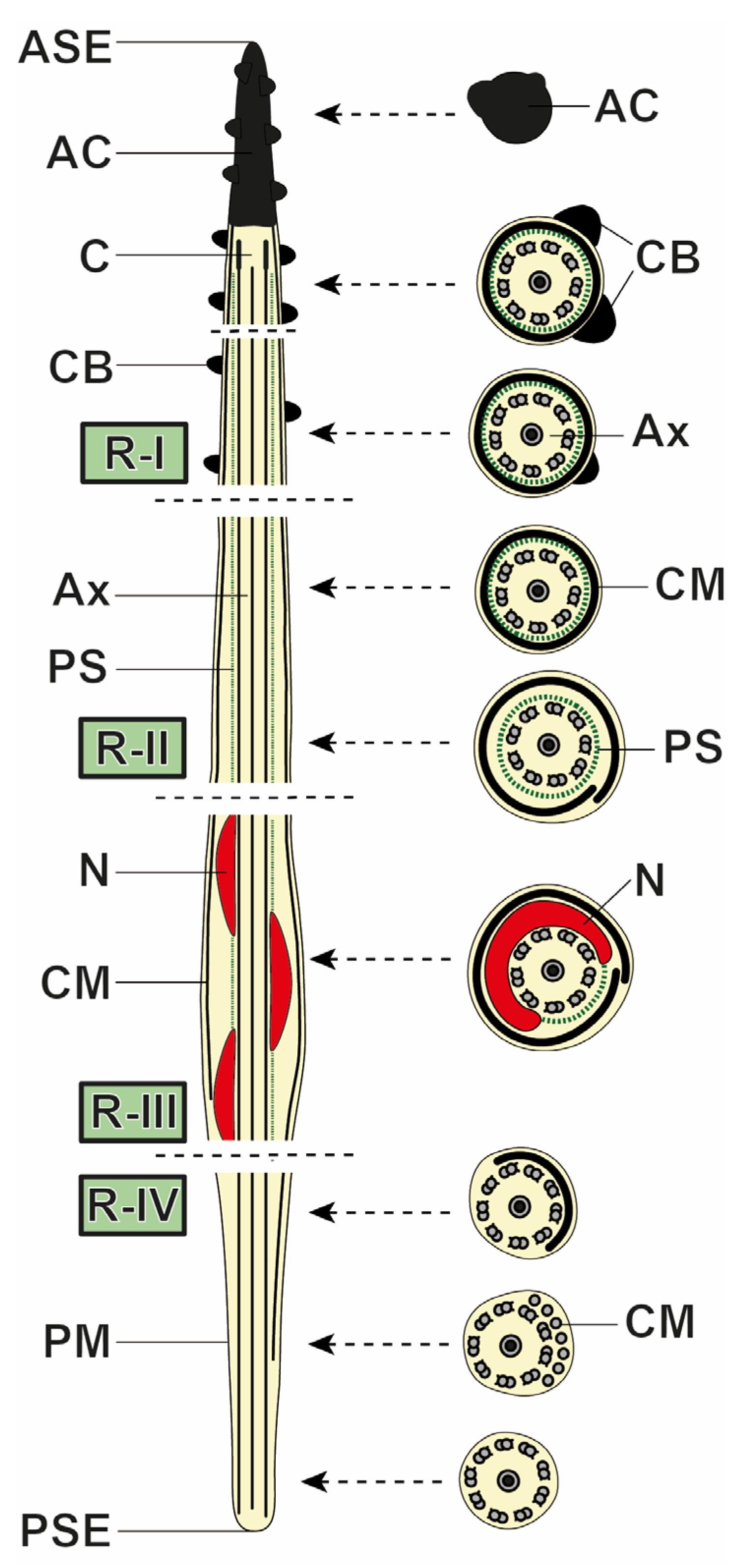
The mature spermatozoon of G. psammomi is a filiform cell tapered at both extremities. It has a single axoneme of the 9 + ‘1’ pattern of trepaxonematan Platyhelminthes surrounded by a periaxonemal sheath, two crest-like bodies of different lengths, a submembranous layer of twisted cortical microtubules, and a spiraled nucleus. The ultrastructural organization of these characteristics based on observations made from four blocks of cestodes recovered from four different gerbils—considering the large number of sections observed for each feature and, therefore, each feature’s reproducibility—enables us to consider four different regions from the anterior to the posterior sperm extremities (I to IV) (Figure 1, Figure 2 and Figure 3).
3.1. Region I (Anterior Extremity)
Region I corresponds to the anterior spermatozoon extremity and is particularly characterized by the presence of the crest-like bodies (Figure 1a–h and Figure 3). The spermatozoon is capped by an electron-dense apical cone more than 1200 nm long and 225 nm wide (Figure 1a,b,d and Figure 3). The total length of the apical cone cannot be provided due to the difficulty in observing this structure in its entirety in longitudinal sections. At the level of the apical cone, the two electron-dense and helicoidally placed crest-like bodies are already present (Figure 1b,d and Figure 3). These two crest-like bodies are of different lengths, and their maximum thickness of 130 nm (95 to 130 nm, n = 22) is observed at the level of the centriole (Figure 1e,f). In the posterior part of region I, one of the crest-like bodies disappears, and later, the stop in the helical course of the second crest-like body marks the transition between regions I and II (Figure 1b,h and Figure 3). The 9 + ‘1’ axoneme appears at the base of the apical cone (Figure 1e–g and Figure 3) and is surrounded by a periaxonemal sheath (Figure 1g,h and Figure 3). Finally, a submembranous layer of twisted cortical microtubules is also present (Figure 1b,c,g,h and Figure 3). The twisting angle of the cortical microtubules with respect to the hypothetical long axis of the sperm cell is between 30° and 40° (n = 45).
Figure 1.
Spermatozoa of Gerbillitaenia psammomi, region I. (a,b) Longitudinal sections showing the anterior spermatozoon extremity, apical cone, and presence of two crest-like bodies. (c) A longitudinal section of the transition between regions I and II marked by the stopping of the helical crest-like bodies. (d) A cross-section at the level of the apical cone. Crest-like bodies are already present. (e,f) Cross-sections showing the progressive appearance of the centriole. (g,h) Cross-sections showing two crest-like bodies (anterior part of region I) and one crest-like body (posterior part of region I). AC, apical cone; ASE, anterior spermatozoon extremity; Ax, axoneme; CB, crest-like body; CM, cortical microtubule; PS, periaxonemal sheath; R-I, region I; R-II, region II. Scale bars = 300 nm.
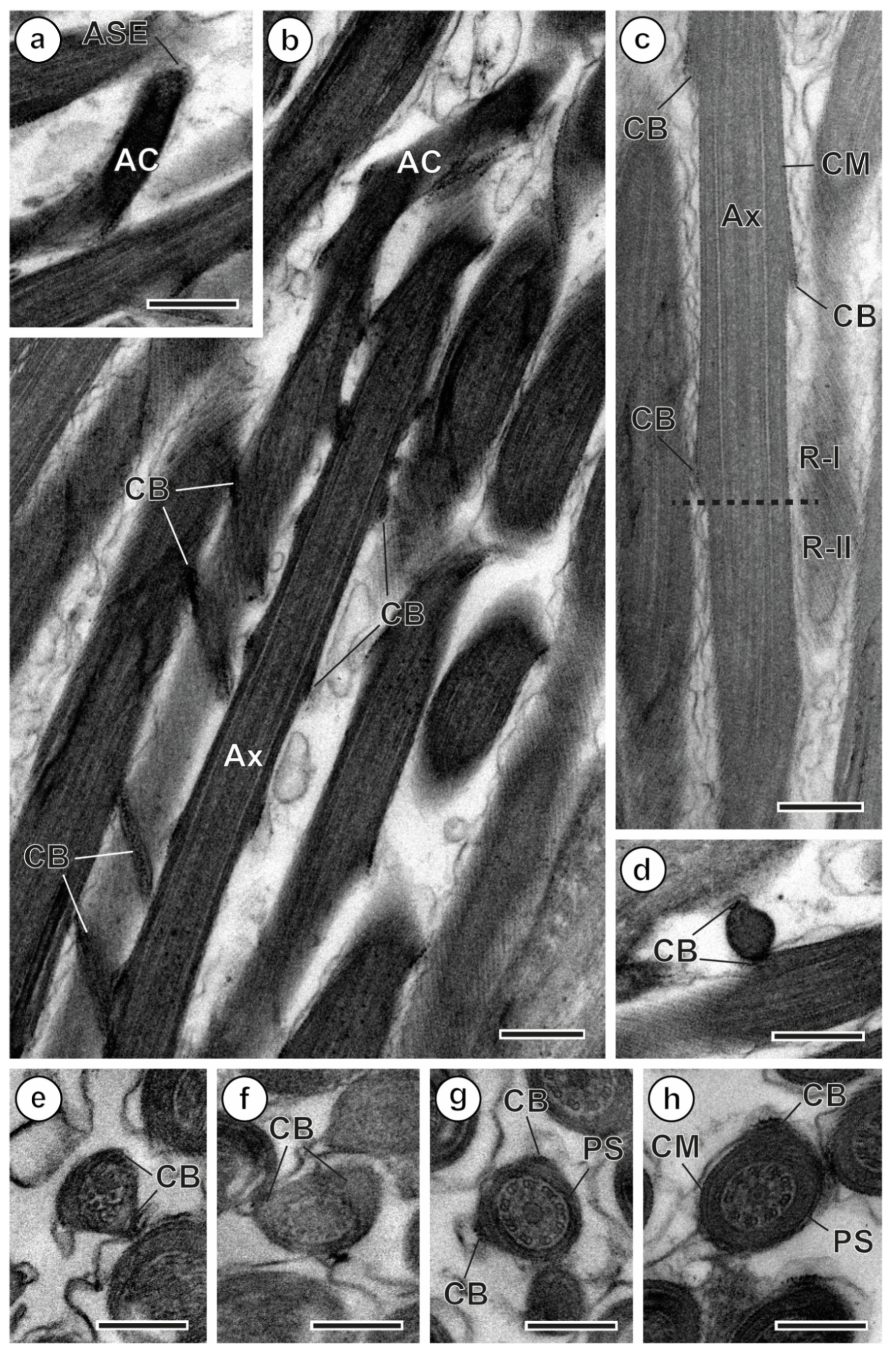
Figure 2.
Spermatozoa of Gerbillitaenia psammomi, regions II–IV. (a,b) Cross-sections of regions II and III showing the periaxonemal sheath, nucleus, and spiraled cortical microtubules. (c) A longitudinal section of the nuclear region (region III) that shows the periaxonemal sheath and the spiraled nucleus and cortical microtubules. (d–f) Cross-sections of region IV showing the progressive reduction in cortical microtubules and their disappearance. Note that the cortical microtubules become parallel. (g) A cross-section showing the beginning of the axonemal disorganization. Ax, axoneme; CM, cortical microtubule; N, nucleus; PS, periaxonemal sheath; R-II, region II; R-III, region III. Scale bars = 300 nm.
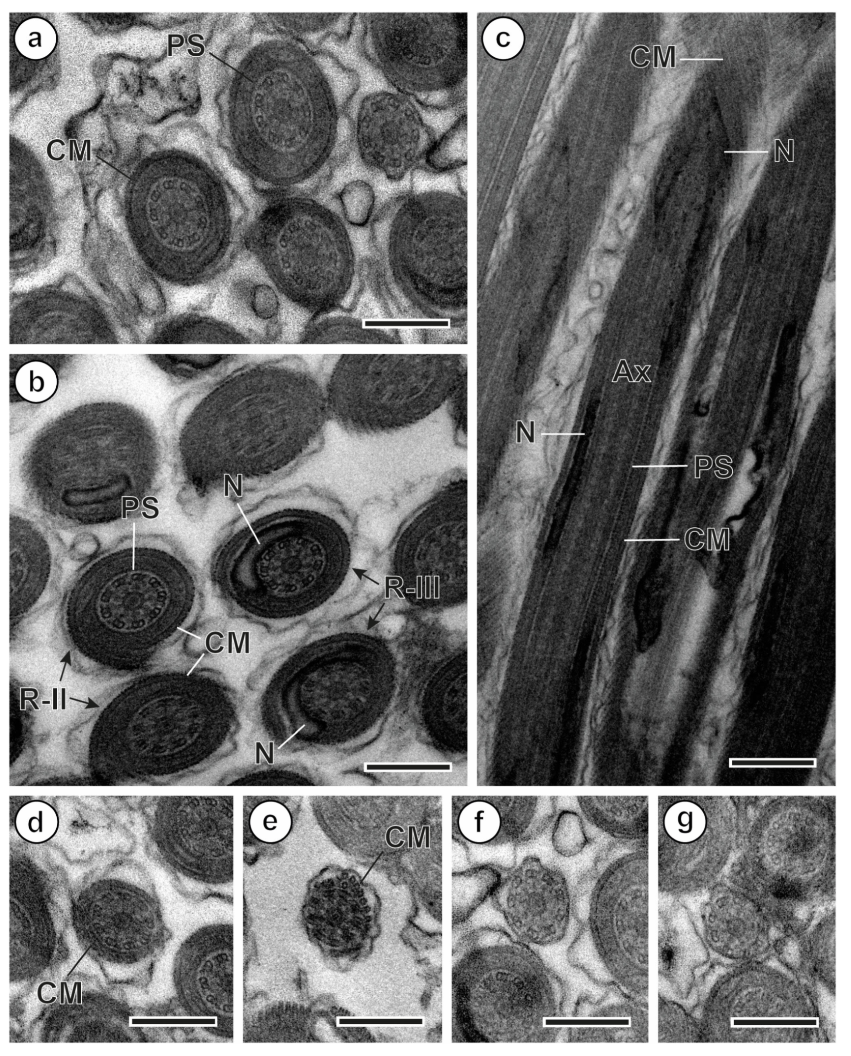
Figure 3.
A schematic drawing of the mature spermatozoon of Gerbillitaenia psammomi. AC, apical cone; ASE, anterior spermatozoon extremity; Ax, axoneme; C, centriole; CB, crest-like body; CM, cortical microtubule; N, nucleus; PM, plasma membrane; PS, periaxonemal sheath; PSE, posterior spermatozoon extremity; R-I, R-II, R-III, R-IV: regions I, II, III, IV.
3.2. Region II
Region II is the intermediate area of the sperm cell located between the anterior extremity and the nuclear region. Thus, its main characteristic is the absence of crest-like bodies. It has the axoneme with its periaxonemal sheath and is surrounded by the submembranous layer of twisted cortical microtubules (Figure 2a,b and Figure 3). In the anterior part of region II, just after the disappearance of crest-like bodies, the cortical microtubules form a continuous layer (Figure 2a); meanwhile, in the posterior part, the layer of cortical microtubules is discontinuous (Figure 2a,b).
3.3. Region III (Nuclear Region)
Region III is the nuclear region of the male gamete and is characterized by the presence of the nucleus as a cordon spiraled around the axoneme (Figure 2b,c and Figure 3). In cross-sections, the nucleus shows a horseshoe shape (Figure 2b). Moreover, a discontinuous layer of twisted cortical microtubules is still present under the plasma membrane. The periaxonemal sheath begins its disappearance in this part of the spermatozoon occupied by the nucleus (Figure 2b).
3.4. Region IV (Posterior Extremity)
Region IV corresponds to the posterior extremity of the spermatozoon. This region is characterized by the presence of the axoneme and cortical microtubules that progressively disappear (Figure 2d–g and Figure 3). In the anterior part of this region, the number of cortical microtubules decreases (Figure 2d); the microtubules become parallel to the hypothetical long axis of the sperm cell (Figure 2e) and finally disappear (Figure 2f). Thus, the posterior tip of the male gamete has only the axoneme (Figure 2f,g).
4. Discussion
The mature spermatozoa of G. psammomi display some characteristics that are present in the sperm cells of most cestodes. However, analyses of their organization along the male gamete and, for some of them, their absence are crucial to establishing the type of spermatozoon according to Levron et al. [12].
4.1. Axoneme and Periaxonemal Sheath
The 9 + ‘1’ type of axoneme is characterized by the presence of nine peripheral doublets and a central core. It was postulated by Ehlers [29,30] as the general and constant pattern for the axonemes in the spermatozoa of trepaxonematan Platyhelminthes, including the parasitic flatworms (Monogenea, Digenea, and Cestoda), whereas the non-trepaxonematan Platyhelminthes have the classical type 9 + 2. However, concerning this axonemal pattern, there are some exceptions, such as schistosomes and didymozoids (Digenea), which have particular patterns: the aberrant 9 + ‘1’ type and the 9 + 0 type, respectively [31,32,33].
The spermatozoa of eucestodes have one or two axonemes. Two axonemes are present in the sperm cells of Bothriocephalidea, Diphyllidea, Diphyllobothriidea, Haplobothriidea, Onchoproteocephalidea, Spathebothriidea, Tetraphyllidea, and Trypanorhyncha [12,18,21,22,23,24]. Contrarily, the sperm cell of G. psammomi, like those of the remaining studied members of Cyclophyllidea and those of Caryophyllidea, Lecanicephalidea, Nippotaeniidea, Phyllobothriidea, Rhinebothriidea, and Tetrabothriidea, has a single axoneme [12,17,19,20,25,26,27,34,35,36,37].
The presence of a periaxonemal sheath surrounding the axoneme has been considered an important characteristic to establish the different models of spermatozoa in Eucestoda [12]. This structural characteristic is present in tetrabothiideans and also in most cyclophyllideans, except for the representatives of the families Mesocestoididae, Hymenolepididae, and Nematotaeniidae and some Anoplocephalidae. Thus, the presence of a periaxonemal sheath, among other characteristics, characterizes the spermatozoan types VI and VII [12]. Considering the family Catenotaeniidae, both G. psammomi and the other studied species have a periaxonemal sheath in their male gametes [25,26,27].
4.2. Anterior Region (Crest-like Bodies and Apical Cone)
Crest-like bodies consist of one or more helical cordons located in the anterior extremity of the spermatozoa of numerous eucestodes. Only species from Caryophyllidea, Diphyllobothriidea, Haplobothriidea, Spathebothriidea, and Trypanorhyncha lack these structures [12]. The number of crest-like bodies varies between one and 12; one or two is the most frequent number, but in several species, the number is higher. This is the case for Hymenolepididae, where this number is between six in Cladogynia serrata (Fuhrmann, 1906) and Rodentolepis microstoma (Dujardin, 1845) [38,39,40] and 12 in Rodentolepis nana (Siebold, 1852) [38,41]. Other cyclophyllideans—namely, Aporina delafondi (Railliet, 1892) and Sudarikovina taterae Hunkeler, 1972 (Anoplocephalidae)—have five and seven crest-like bodies, respectively [42,43]. Among the catenotaeniids, all the studied species have two crest-like bodies of different lengths, with a maximum thickness of around 60–80 nm for C. pusilla, S. lobata, and S. (M.) gerbilli and 130 nm for G. psammomi [25,26,27].
An electron-dense apical cone has been described in numerous species of cyclophyllideans [44,45]. The length of the apical cone is highly variable. There are species with a reduced apical cone (i.e., 300 nm long in A. delafondi) while, in other species, it can reach a size of 2000 nm, such as in Joyeuxiella echinorhynchoides (Sonsino, 1889); Joyeuxiella pasqualei (Diamare, 1893); Paroniella reynoldsae Meggitt, 1926; Raillietina baeri Meggitt & Subramanian, 1927; and S. lobata or Taenia hydatigena Pallas, 1766 [26,46,47,48,49]. In G. psammomi, as in the remaining studied catenotaeniids, the length of the apical cone is always more than 1000 nm long [25,26,27].
4.3. Cortical Microtubules
In the cestodes, cortical microtubules may be parallel or twisted. Cestodes of the orders Tetrabothriidea and Cyclophyllidea have sperm cells with twisted cortical microtubules. This condition was postulated as a synapomorphy for these two orders [8,10,11]. However, in the cyclophyllidean species of the family Mesocestoididae—namely, Mesocestoides lineatus (Goeze, 1782) and Mesocestoides litteratus (Batsch, 1786)—the cortical microtubules have a parallel disposition along the spermatozoon. These two species also exhibit other primitive conditions or characteristics in the spermiogenesis process [50,51,52]. Thus, over time, the systematic position of this family among the Eucestoda has been a subject of ongoing discussion [11,50,53].
In Cyclophyllidea, the angle that describes the arrangement of cortical microtubules along the spermatozoon with respect to the sperm’s hypothetical long axis varies in a range between 15° in A. delafondi and R. nana and 60° in Raillietina tunetensis [41,42,54]. However, the most common value for this angle of spiralization is around 40° [45,49] as occurs in the Catenotaeniidae species including G. psammomi [25,26,27]. It is remarkable that, in general, the species with a reduced cortical microtubule angle are those with the highest numbers of crest-like bodies. Indeed, in the family Hymenolepididae, the numbers of crest-like bodies (CBs) and the angles of cortical microtubules are as follows: 12CBs and 10–15° in R. nana, 7CBs and 20–30° in R. microstoma, 6CBs and 25° in R. serrata, and 8CBs and 30° in Rodentolepis straminea (Goeze, 1782) [39,40,41,55].
4.4. Nucleus and Mitochondrion
The region of the spermatozoon with the nucleus is considered the posterior extremity. A study of the fertilization process in a species of the digenean genus Gonapodasmius Ishii, 1935 (Didymozoidae), showed that the sperm cell coils around the oocyte. A fusion of membranes is produced, and the nuclear part of the spermatozoon is the last to be incorporated into the oocyte [56]. In this sense, several structures present in the anterior extremity of the spermatozoa of digeneans and cestodes, such as external ornamentations, spine-like bodies, the apical cone, and crested bodies, could play an important role in the fertilization process [49,56,57,58].
The nucleus in most orders of Eucestoda has a parallel disposition along the sperm cell [12]. Contrarily, it has been described as being spiraled around the axoneme in all Cyclophyllidea and in other orders (Tetrabothriidea, Lecanicephalidea, Nippotaeniidea, and Diphyllidea) [12,17,19,20,21,59].
Like all eucestodes, G. psammomi lacks mitochondria in its spermatozoa [12]. In fact, the absence of mitochondria in the sperm cells was postulated as a synapomorphy for Eucestoda [8,11]. To the contrary, the gycocotylidean and amphylinidean cestodes have a mitochondrion in their spermatozoa [16,60,61], as do other parasitic platyhelminths such as monogeneans and digeneans [8,9,13,14].
4.5. Spermatozoon Type
For Eucestoda, Levron et al. [12] established seven types of spermatozoa (I to VII). Types I and II spermatozoa are mainly characterized by having two axonemes, and they can be differentiated by the presence (in type I) or absence (in type II) of crest-like bodies. The remaining models of sperm cells (types III to VII) have a single axoneme; they can be distinguished by considering the presence or absence of some characteristics such as crest-like bodies, periaxonemal sheathes, and intracytoplasmic walls, and also by the parallel or spiraled pattern of the cortical microtubules and nucleus.
Our results in G. psammomi are coincident with those observed in the previously studied catenotaeniids: C. pusilla, S. (M.) gerbilli, and S. lobata [25,26,27] (see Table 1). The species of the order Cyclophyllidea have male gametes of type V, VI, or VII, with the exception of representatives of the family Mesocestoididae, which have type IV [50,52]. The spermatozoa of both M. lineatus and M. litteratus, as mentioned above, have parallel cortical microtubules as their main feature differentiating them from types V, VI, and VII, which have twisted cortical microtubules. In fact, the complicated position of mesocestoidids within the order Cyclophyllidea has been discussed by several authors [11,50,53]. The type VI spermatozoa observed in G. psammomi and in the remaining studied catenotaeniids have also been cited for species of other cyclophyllidean families; namely, Anoplocephalidae, Dilepididae, Dipylidiidae, and Gryporhynchidae. Concerning anoplocephalids, type VI has been described for the spermatozoa of only two of the studied species: Mathevotaenia herpestis (Kofend, 1917) and Stilesia globipunctata (Rivolta, 1874) [62,63]. Other anoplocephalids exhibit type V or VII [12]. In the family Dilepididae, the sperm type VI has been described in Angularella beema (Clerc, 1906), Anomotaenia quelea, and Molluscotaenia crassiscolex (von Linstow, 1890) [44,64,65]. Among dipylidiids, Dipylidium caninum (Linnaeus, 1758), J. echinorhynchoides, and J. pasqualei also exhibit this type [46,66,67]. Finally, in Gryporhynchidae, the sperm type VI has been reported in the sole studied species, Valipora mutabilis Linton, 1927 [68].
Table 1.
Main ultrastructural characteristics of the spermatozoa in Catenotaeniidae species.
4.6. Phylogenetic Implications
In our previous study on G. psammomi, the obtained 28S rDNA phylogenetic tree (see Figure 5 in Boubakri et al. [7]) showed two different clades corresponding to the subfamilies Skrjabinotaeniinae and Catenotaeniinae. G. psammomi was clearly grouped in the clade of Skrjabinotaeniinae together with species of the genera Skrjabinotaenia and Meggittina, recently considered a subgenus of Skrjabinotaenia [1]. Haukisalmi et al. [69,70] also obtained genetic results that clearly showed the monophyly of the subfamily Skrjabinotaeniinae. Conversely, after analyzing species from the genera Catenotaenia, Catenotaenioides, Hemicatenotaenia, and Pseudocatenotaenia, the same authors [69,70] found that the subfamily Catenotaeniinae is not monophyletic. Furthermore, they demonstrated that C. pusilla shows closer phylogenetic relationships with the skrajabinotaeniines than with the other catenotaeniinae.
Despite limited data, observations to date of the sperm ultrastructure in Catenotaeniidae have been remarkably homogeneous (see Table 1). However, to establish a more robust pattern for the family and its two subfamilies, it is necessary to study other genera and species belonging to the subfamily Catenotaeniinae. This is particularly important given the molecular results showing the close relationship between C. pusilla and Skrjabinotaeniinae.
5. Conclusions
Despite the small number of species studied within the family Catenotaeniidae, current data on the sperm of G. psammomi and other studied catenotaenids show similarity in the sperm characteristics and their organization along the sperm cell. All the catenotaeniids studied to date exhibit spermatozoa with a single 9 + ‘1’ axoneme with a periaxonemal sheath, two crest-like bodies of different lengths, twisted cortical microtubules, and a spiraled nucleus. Moreover, dense granules and transverse cytoplasmic walls are absent. Although only a single species was newly examined ultrastructurally in the present study, the male gamete of G. psammomi was found to be similar to that of the other analyzed catenotaeniids based on the presence or absence of the above-mentioned characteristics and seems to be consistent with type VI established by Levron et al. [12].
Actual sampling is taxonomically and quantitatively limited and, consequently, future ultrastructural studies are needed. They should focus not only on increasing the number of species in the family, particularly from different genera, in order to determine the sperm type more robustly but also on addressing the spermiogenesis process to analyze whether this process shows a similar pattern in the family Catenotaeniidae.
Author Contributions
Conceptualization, J.M. and H.K.; Methodology, A.B., J.M. and H.K.; Investigation, A.B., J.M. and H.K.; Resources, J.M.; Writing—Original Draft Preparation, A.B., J.M. and H.K.; Writing—Review and Editing, J.M. and H.K.; Supervision, J.M. and H.K.; Project Administration, J.M. and H.K. All authors have read and agreed to the published version of the manuscript.
Funding
This research received no external funding.
Institutional Review Board Statement
Gerbils were trapped under the permission of the “Direction Générale des Forêts (Ministère de l’Agriculture, des Resources Hydrauliques et de la Pêche Maritime)” of Tunisia, references 1822/2023 (2 October 2023) and 1728/2024 (30 September 2024). Animal work was conducted in accordance with the European Directive 2010/63/UE and the Spanish Government Laws 42/2007 and RD 630/2013.
Informed Consent Statement
Not applicable.
Data Availability Statement
RAW TEM files are available at https://ubarcelona-my.sharepoint.com/my?id=%2Fpersonal%2Fjordimiquel%5Fub%5Fedu%2FDocuments%2FGerbillitaenia%20psammomi%20Sperm&viewid=2f4a0134%2D43c5%2D4efe%2D832b%2Da52540518f9a (accessed on 2 February 2026) upon reasonable academic request to the corresponding author.
Acknowledgments
The authors wish to thank Hédi Boubakri for his valuable help during fieldwork. The authors also thank the staff of the “Centres Científics i Tècnics de la Universitat de Barcelona” (CCiTUB) for their support in the preparation of TEM samples. We are grateful to the “Direction Générale des Forêts (Ministère de l’Agriculture, des Ressources Hydrauliques et de la Pêche Maritime)” for granting permission to trap fat sand rats in Southern Tunisia. A.B. received an alternating scholarship from the University of Gabes, granted by the Ministry of Higher Education and Scientific Research of Tunisia. J.M. is a member of the 2021-SGR-00359 research group.
Conflicts of Interest
The authors declare that they have no conflicts of interest.
References
- Haukisalmi, V.; Elmahy, R.A. Taxonomic review of Skrjabinotaenia Akhumyan, Meggittina Lynsdale and Gerbillitaenia gen. nov. (Cestoda: Catenotaeniidae), parasites of rodents. Zootaxa 2025, 5590, 185–208. [Google Scholar] [CrossRef] [Scilit]
- Mikhail, J.W.; Fahmy, M.A.M. Study on some members of genus Skrjabinotaenia with a description of a new species and a review of the subfamily Catenotaeniinae Spasski, 1946. Zool. Ann. 1968, 181, 439–450. [Google Scholar]
- Joyeux, C.; Baer, J.G.; Gaud, J. Recherches helminthologiques marocaines. Cestodes (deuxième note). Arch. Inst. Pasteur Maroc 1951, 4, 93–102. [Google Scholar]
- Wolfgang, R.W. Helminth parasites of reptiles, birds, and mammals in Egypt. II. Catenotaenia aegyptica sp. nov. from myomorph rodents, with additional notes on the genus. Can. J. Zool. 1956, 34, 6–20. [Google Scholar] [CrossRef] [Scilit]
- Elmahy, R.A. Flatworms of Some wild Mammals with Special Reference to Hymenolepidids from Egypt. Ph.D. Thesis, Tanta University, Tanta, Egypt, 2012. [Google Scholar]
- Elmahy, R.A. A scanning electron microscopic study on Meggittina gerbilli Wertheim, 1954 and Skrjabinotaenia oranensis Joyeux and Foley, 1930 (Cyclophyllidea: Catenotaeniidae) from rodents in Egypt. Delta J. Sci. 2022, 45, 115–124. [Google Scholar] [CrossRef] [Scilit]
- Boubakri, A.; Kacem, H.; Martín-Carrillo, N.; Foronda, P.; Selmi, S.; Miquel, J. New data on the catenotaeniid cestode Gerbillitaenia psammomi (Mikhail and Fahmy, 1968) parasitizing Fat sand rats, Psammomys obesus (Muridae: Gerbillinae), in Tunisia. J. Helminthol. 2025, 99, e62. [Google Scholar] [CrossRef] [Scilit]
- Justine, J.-L. Phylogeny of parasitic Platyhelminthes: A critical study of synapomorphies proposed on the basis of the ultrastructure of spermiogenesis and spermatozoa. Can. J. Zool. 1991, 69, 1421–1440. [Google Scholar] [CrossRef] [Scilit]
- Justine, J.-L. Cladistic study in the Monogenea (Platyhelminthes), based upon a parsimony analysis of spermiogenetic and spermatozoal ultrastructural characters. Int. J. Parasitol. 1991, 21, 821–838. [Google Scholar] [CrossRef] [Scilit]
- Justine, J.-L. Spermatozoa as phylogenetic characters for the Eucestoda. J. Parasitol. 1998, 84, 385–408. [Google Scholar] [CrossRef] [Scilit]
- Justine, J.-L. Spermatozoa as phylogenetic characters for the Platyhelminthes. In Interrelationships of Platyhelminthes; Littlewood, D.T.J., Bray, R.A., Eds.; Taylor & Francis: London, UK, 2001; pp. 231–238. [Google Scholar]
- Levron, C.; Miquel, J.; Oros, M.; Scholz, T. Spermatozoa of tapeworms (Platyhelminthes, Eucestoda): Advances in ultrastructural and phylogenetic studies. Biol. Rev. 2010, 85, 523–543. [Google Scholar] [CrossRef] [Scilit]
- Bakhoum, A.J.S.; Miquel, J.; Ndiaye, P.I.; Justine, J.-L.; Falchi, A.; Bâ, C.T.; Marchand, B.; Quilichini, Y. Advances in spermatological characters in the Digenea: Review and proposal of spermatozoa models and their phylogenetic importance. Adv. Parasitol. 2017, 98, 111–165. [Google Scholar] [CrossRef] [Scilit] [PubMed]
- Justine, J.-L.; Poddubnaya, L.G. Spermiogenesis and spermatozoon ultrastructure in basal polyopisthocotylean monogeneans, Hexabothriidae and Chimaericolidae, and their significance for the phylogeny of the Monogenea. Parasite 2018, 25, 7. [Google Scholar] [CrossRef] [Scilit]
- Bruňanská, M.; Matey, V.; Nebesářová, J. Ultrastructure of the spermatozoon of the diphyllobothriidean cestode Cephalochlamys namaquensis (Cohn, 1906). Parasitol. Res. 2012, 111, 1037–1043. [Google Scholar] [CrossRef] [Scilit] [PubMed]
- Bruňanská, M.; Poddubnaya, L.G.; Xylander, W.E.R. Spermatozoon cytoarchitecture of Amphilina foliacea (Platyhelminthes, Amphilinidea). Parasitol. Res. 2012, 111, 2063–2069. [Google Scholar] [CrossRef] [Scilit] [PubMed]
- Bruňanská, M.; Bílý, T.; Nebesářová, J. Nippotaenia mogurndae Yamaguti et Myiata, 1940 (Cestoda, Nippotaeniidea): First data on spermiogenesis and sperm ultrastructure. Parasitol. Res. 2015, 114, 1443–1453. [Google Scholar] [CrossRef] [Scilit]
- Bruňanská, M.; Matoušková, M.; Jasinská, R.; Nebesářová, J.; Poddubnaya, L.G. Heteromorphism of sperm axonemes in a parasitic flatworm, progenetic Diplocotyle olrikii Krabbe, 1874 (Cestoda, Spathebothriidea). Parasitol. Res. 2020, 119, 177–187. [Google Scholar] [CrossRef] [Scilit]
- Cielocha, J.J.; Yoneva, A.; Cantino, M.E.; Daniels, S.; Jensen, K. Ultrastructure of the sperm of Adelobothrium sp. (Cestoda: Lecanicephalidea). Invertebr. Biol. 2013, 132, 315–325. [Google Scholar] [CrossRef] [Scilit]
- Cielocha, J.J.; Martinez, E.; Jackson, A.; Yoneva, A. Characterization of spermatozoon ultrastructure in Tetragonocephalum sp. (Cestoda: Lecanicephalidea: Tetragonocephalidae) from the whipray, Urogymnus asperrimus (Dasyatidae: Urogymninae). J. Parasitol. 2022, 108, 500–510. [Google Scholar] [CrossRef] [Scilit]
- Marigo, A.M.; Eira, C.; Bâ, C.T.; Miquel, J. Spermiogenesis and spermatozoon ultrastructure of the diphyllidean cestode Echinobothrium euterpes (Neifar, Tyler and Euzet 2001) Tyler 2006, a parasite of the common guitarfish Rhinobatos rhinobatos. Parasitol. Res. 2011, 109, 809–821. [Google Scholar] [CrossRef] [Scilit]
- Marigo, A.M.; Świderski, Z.; Bâ, C.T.; Miquel, J. Spermiogenesis and ultrastructure of the spermatozoon of the trypanorhynch cestode Aporhynchus menezesi (Aporhynchidae), a parasite of the velvet belly lanternshark Etmopterus spinax (Elasmobranchii: Etmopteridae). Folia Parasitol. 2011, 58, 69–78. [Google Scholar] [CrossRef] [Scilit]
- Marigo, A.M.; Delgado, E.; Torres, J.; Bâ, C.T.; Miquel, J. Spermiogenesis and spermatozoon ultrastructure of the bothriocephalidean cestode Clestobothrium crassiceps (Rudolphi, 1819), a parasite of the teleost fish Merluccius merluccius (Gadiformes: Merlucciidae). Parasitol. Res. 2012, 110, 19–30. [Google Scholar] [CrossRef] [Scilit] [PubMed]
- Marigo, A.M.; Levron, C.; Bâ, C.T.; Miquel, J. Ultrastructural study of spermiogenesis and the spermatozoon of the proteocephalidean cestode Barsonella lafoni de Chambrier et al., 2009, a parasite of the catfish Clarias gariepinus (Burchell, 1822) (Siluriformes, Clariidae). Zool. Anz. 2012, 251, 147–159. [Google Scholar] [CrossRef] [Scilit]
- Hidalgo, C.; Miquel, J.; Torres, J.; Marchand, B. Ultrastructural study of spermiogenesis and the spermatozoon in Catenotaenia pusilla, an intestinal parasite of Mus musculus. J. Helminthol. 2000, 74, 73–81. [Google Scholar] [CrossRef] [Scilit] [PubMed]
- Miquel, J.; Bâ, C.T.; Marchand, B. Ultrastructure of the spermatozoon of Skrjabinotaenia lobata (Cyclophyllidea, Catenotaeniidae), intestinal parasite of Apodemus sylvaticus (Rodentia, Muridae). J. Submicrosc. Cytol. Pathol. 1997, 29, 521–526. [Google Scholar]
- Aouina, F.; Kacem, H.; Martín-Carrillo, N.; Foronda, P.; Miquel, J. Spermatological characterization of the cestode Meggittina gerbilli (Cyclophyllidea: Catenotaeniidae), a parasite of gerbils, Gerbillus gerbillus and Gerbillus campestris (Rodentia: Muridae) in Tunisia. Animals 2024, 14, 12. [Google Scholar] [CrossRef] [Scilit]
- Reynolds, E.S. The use of lead citrate at high pH as an electronopaque stain in electronmicroscopy. J. Cell Biol. 1963, 17, 208–212. [Google Scholar] [CrossRef] [Scilit]
- Ehlers, U. Phylogenetisches System der Plathelminthes. Verh. Naturwiss. Ver. Hamb. (NF) 1984, 27, 291–294. [Google Scholar]
- Ehlers, U. Phylogenetic relationships within the Platyhelminthes. In The Origins and Relationships of Lower Invertebrates; Conway Morris, S., George, J.D., Gibson, R., Platt, H.M., Eds.; Oxford University Press: Oxford, UK, 1985; pp. 143–158. [Google Scholar]
- Jamieson, B.G.M.; Justine, J.-L. Spermatozoa, Spermatogenesis and Fertilization in Schistosoma. In Schistosoma: Biology, Pathology and Control; Jamieson, B.G.M., Ed.; CRC Press: Boca Raton, FL, USA, 2017; pp. 300–319. [Google Scholar]
- Justine, J.-L.; Mattei, X. Étude ultrastructurale de la spermiogenèse et du spermatozoïde d’un Plathelminthe: Gonapodasmius (Trematoda: Didymozoidae). J. Ultrastruct. Res. 1982, 79, 350–365. [Google Scholar] [CrossRef] [Scilit]
- Justine, J.-L.; Mattei, X. A spermatozoon with two 9 + 0 axonemes in a parasitic flatworm, Didymozoon (Digenea: Didymozoidae). J. Submicrosc. Cytol. 1983, 15, 1101–1105. [Google Scholar]
- Matoušková, M.; Bílý, T.; Bruňanská, M.; Mackiewicz, J.S.; Nebesářová, J. Ultrastructure, cytochemistry and electron tomography analysis of Caryophyllaeides fennica (Schneider, 1902) (Cestoda: Lytocestidae) reveals novel spermatology characteristics in the Eucestoda. Parasitol. Res. 2018, 117, 3091–3102. [Google Scholar] [CrossRef] [Scilit]
- Matoušková, M.; Bílý, T.; Bruňanská, M.; Oros, M.; Kostič, B.; Nebesářová, J. New data on spermiogenesis and trepaxonematan axoneme in basal tapeworms (Cestoda, Caryophyllidea, Lytocestidae) parasitizing cyprinid fishes. Sci. Rep. 2019, 9, 12881. [Google Scholar] [CrossRef] [Scilit] [PubMed]
- Matoušková, M.; Bruňanská, M.; Nebesářová, J.; Poddubnaya, L.G. Ultrastructure and cytochemistry of the mature spermatozoon of Khawia armeniaca (Cholodkovsky, 1915) (Caryophyllidea: Lytocestidae) a parasite of Capoeta capoeta sevangi (De Filippi, 1865) (Teleostei, Cyprinidae). Helminthologia 2020, 57, 353–360. [Google Scholar] [CrossRef] [Scilit] [PubMed]
- Yoneva, A.; Levron, C.; Oros, M.; Orosová, M.; Scholz, T. Ultrastructure of spermiogenesis and mature spermatozoon of Breviscolex orientalis (Cestoda: Caryophyllidea). Parasitol. Res. 2011, 108, 997–1005. [Google Scholar] [CrossRef] [Scilit] [PubMed]
- Miquel, J.; Ndiaye, P.I.; Feliu, C. Crest-like bodies in the spermatozoon of Hymenolepididae cestodes from Iberian rodents. Rev. Ibér. Parasitol. 2007, 67, 27–33. [Google Scholar]
- Bâ, C.T.; Marchand, B. Ultrastructure of the Retinometra serrata spermatozoon (Cestoda) intestinal parasite of turtle-doves in Senegal. J. Submicrosc. Cytol. Pathol. 1993, 25, 233–238. [Google Scholar]
- Bâ, C.T.; Marchand, B. Ultrastructure of spermiogenesis and the spermatozoon of Vampirolepis microstoma (Cestoda, Hymenolepididae), intestinal parasite of Rattus rattus. Microsc. Res. Tech. 1998, 42, 218–225. [Google Scholar] [CrossRef] [Scilit]
- Bâ, C.T.; Marchand, B. Reinvestigation of the ultrastructure of spermiogenesis and the spermatozoon of Hymenolepis nana (Cestoda, Cyclophyllidea), parasite of the small intestine of Rattus rattus. Mol. Reprod. Dev. 1992, 33, 39–45. [Google Scholar] [CrossRef] [Scilit]
- Bâ, C.T.; Marchand, B. Ultrastructure of spermiogenesis and the spermatozoon of Aporina delafondi (Cyclophyllidea, Anoplocephalidae) intestinal parasite of turtle doves in Senegal. Int. J. Parasitol. 1994, 24, 225–235. [Google Scholar] [CrossRef] [Scilit]
- Bâ, A.; Bâ, C.T.; Marchand, B. Ultrastructure of spermiogenesis and the spermatozoon of Sudarikovina taterae (Cestoda, Cyclophyllidea, Anoplocephalidae) intestinal parasite of Tatera gambiana (Rodentia, Gerbillidae). J. Submicrosc. Cytol. Pathol. 2000, 32, 137–144. [Google Scholar]
- Marigo, A.M.; Bâ, C.T.; Miquel, J. Spermiogenesis and spermatozoon ultrastructure of the dilepidid cestode Molluscotaenia crassiscolex (von Linstow, 1890), an intestinal parasite of the common shrew Sorex araneus. Acta Zool. 2011, 92, 116–125. [Google Scholar] [CrossRef] [Scilit]
- Miquel, J.; Świderski, Z.; Feliu, C. Spermatozoon ultrastructure of Thysanotaenia congolensis (Cyclophyllidea, Anoplocephalidae, Inermicapsiferinae): Phylogenetic implications. Parasitol. Res. 2016, 115, 3083–3091. [Google Scholar] [CrossRef] [Scilit]
- Ndiaye, P.I.; Agostini, S.; Miquel, J.; Marchand, B. Ultrastructure of spermiogenesis and the spermatozoon in the genus Joyeuxiella Fuhrmann, 1935 (Cestoda, Cyclophyllidea, Dipylidiidae): Comparative analysis of J. echinorhynchoides (Sonsino, 1889) and J. pasqualei (Diamare, 1893). Parasitol. Res. 2003, 91, 175–186. [Google Scholar] [CrossRef] [Scilit] [PubMed]
- Bâ, C.T.; Bâ, A.; Marchand, B. Ultrastructure of the spermatozoon of Paroniella reynoldsae (Cyclophyllidea, Davaineidae) an intestinal parasite of Corvus albus (Aves, Corvidae). Acta Parasitol. 2005, 50, 208–214. [Google Scholar]
- Bâ, C.T.; Bâ, A.; Marchand, B. Ultrastructure of the spermatozoon of Raillietina (Raillietina) baeri (Cyclophyllidea, Davaineidae) an intestinal parasite of the multimammate rat, Mastomys huberti (Rodentia, Muridae). Parasitol. Res. 2005, 97, 173–178. [Google Scholar] [CrossRef] [Scilit] [PubMed]
- Miquel, J.; Khallaayoune, K.; Azzouz-Maache, S.; Pétavy, A.-F. Spermatological characteristics of the genus Taenia inferred from the ultrastructural study on Taenia hydatigena. Parasitol. Res. 2015, 114, 201–208. [Google Scholar] [CrossRef] [Scilit]
- Miquel, J.; Feliu, C.; Marchand, B. Ultrastructure of spermiogenesis and the spermatozoon of Mesocestoides litteratus (Cestoda, Mesocestoididae). Int. J. Parasitol. 1999, 29, 499–510. [Google Scholar] [CrossRef] [Scilit]
- Conn, D.B. Early spermatogenesis, sperm ultrastructure and spermatoferic duct cytoarchitecture in Mesocestoides lineatus (Platyhelminthes: Cestoda). In Proceedings of the 9th International Congress on Invertebrate Reproduction and Development, Grahamstown, South Africa, 15–20 July 2001; p. 71. [Google Scholar]
- Miquel, J.; Eira, C.; Świderski, Z.; Conn, D.B. Mesocestoides lineatus (Goeze, 1782) (Mesocestoididae): New data on sperm ultrastructure. J. Parasitol. 2007, 93, 545–552. [Google Scholar] [CrossRef] [Scilit]
- Mariaux, J. A molecular phylogeny of the Eucestoda. J. Parasitol. 1998, 84, 114–124. [Google Scholar] [CrossRef] [Scilit]
- Bâ, C.T.; Marchand, B. Ultrastructure of spermiogenesis and the spermatozoon of Raillietina (Raillietina) tunetensis (Cyclophyllidea, Davaineidae) intestinal parasite of turtle doves in Senegal. Int. J. Parasitol. 1994, 24, 237–248. [Google Scholar] [CrossRef] [Scilit]
- Bâ, C.T.; Marchand, B. Ultrastructure of the spermatozoon of Hymenolepis straminea (Cyclophyllidea, Hymenolepididae) intestinal parasite of Arvicanthis niloticus in Senegal. Invertebr. Reprod. Dev. 1996, 29, 243–247. [Google Scholar] [CrossRef] [Scilit]
- Justine, J.-L.; Mattei, X. Ultrastructural observations on the spermatozoon, ovocyte and fertilization process in Gonapodasmius, a gonochoristic Trematode (Trematoda: Digenea: Didymozoidae). Acta Zool. 1984, 65, 171–177. [Google Scholar] [CrossRef] [Scilit]
- Świderski, Z.; Conn, D.B.; Miquel, J.; Mlocicki, D. Fertilization in the cestode Gallegoides arfaai (Mobedi et Ghadirian, 1977) Tenora et Mas-Coma, 1978 (Cyclophyllidea, Anoplocephalidae). Acta Parasitol. 2004, 49, 108–115. [Google Scholar]
- Justine, J.-L.; Mattei, X. Réinvestigation de l’ultrastructure du spermatozoïde d’Haematoloechus (Trematoda: Haematoloechidae). J. Ultrastr. Res. 1982, 81, 322–332. [Google Scholar] [CrossRef] [Scilit] [PubMed]
- Stoitsova, S.R.; Georgiev, B.B.; Dacheva, R.B. Ultrastructure of spermiogenesis and the mature spermatozoon of Tetrabothrius erostris Loennberg, 1896 (Cestoda, Tetrabothriidae). Int. J. Parasitol. 1995, 25, 1427–1436. [Google Scholar] [CrossRef] [Scilit] [PubMed]
- Rohde, K.; Watson, N. Ultrastructure of spermatogenesis and sperm of Austramphilina elongata (Platyhelminthes, Amphilinidea). J. Submicrosc. Cytol. 1986, 18, 361–374. [Google Scholar]
- Xylander, W.E.R. Ultrastructural studies on the reproductive system of Gyrocotylidea and Amphilinidea (Cestoda): Spermatogenesis, spermatozoa, testes and vas deferens of Gyrocotyle. Int. J. Parasitol. 1989, 19, 897–905. [Google Scholar] [CrossRef] [Scilit]
- Bâ, C.T.; Marchand, B. Ultrastructural particularities of the spermatozoon of Stilesia globipunctata (Cestoda) parasite of the small intestine of sheep and goats in Senegal. J. Submicrosc. Cytol. Pathol. 1992, 24, 29–34. [Google Scholar]
- Bâ, C.T.; Marchand, B. Ultrastructure of spermiogenesis and the spermatozoon of Mathevotaenia herpestis (Cestoda), intestinal parasite of Atelerix albiventrix in Sénégal. Acta Zool. 1994, 75, 167–175. [Google Scholar] [CrossRef] [Scilit]
- Bâ, A.; Ndiaye, P.I.; Bâ, C.T.; Miquel, J. Ultrastructure of the spermatozoon of Anomotaenia quelea (Mettrick, 1961) (Cestoda, Cyclophyllidea, Dilepididae), an intestinal parasite of Quelea quelea (Aves, Ploceidae) in Senegal. Zool. Anz. 2013, 253, 119–125. [Google Scholar] [CrossRef] [Scilit]
- Yoneva, A.; Miquel, J.; Świderski, Z.; Georgieva, K.; Mizinska, Y.; Georgiev, B.B. Ultrastructure of spermiogenesis and mature spermatozoon of Angularella beema (Clerc, 1906) (Cestoda, Cyclophyllidea, Dilepididae). Acta Parasitol. 2006, 51, 264–272. [Google Scholar] [CrossRef] [Scilit]
- Miquel, J.; Marchand, B. Ultrastructure of the spermatozoon of Dipylidium caninum (Cestoda, Cyclophyllidea, Dilepididae), an intestinal parasite of Canis familiaris. Parasitol. Res. 1997, 83, 349–355. [Google Scholar] [CrossRef] [Scilit]
- Miquel, J.; Świderski, Z.; Marchand, B. Spermatological characters in the Dipylidiidae Stiles, 1896 (Cestoda, Cyclophyllidea). Acta Parasitol. 2005, 50, 65–73. [Google Scholar]
- Yoneva, A.; Świderski, Z.; Georgieva, K.; Nikolov, P.N.; Mizinska, Y.; Georgiev, B.B. Spermiogenesis and sperm ultrastructure of Valipora mutabilis Linton, 1927 (Cestoda, Cyclophyllidea, Gryporhynchidae). Parasitol. Res. 2008, 103, 1397–1405. [Google Scholar] [CrossRef] [Scilit]
- Haukisalmi, V.; Hardman, L.M.; Henttonen, H. Taxonomic review of cestodes of the genus Catenotaenia Janicki, 1904 in Eurasia and molecular phylogeny of the Catenotaeniidae (Cyclophyllidea). Zootaxa 2010, 2489, 1–33. [Google Scholar] [CrossRef] [Scilit]
- Haukisalmi, V.; Ribas, A.; Junker, K.; Spickett, A.; Matthee, S.; Henttonen, H.; Jrijer, J.; Halajian, A.; Anders, J.L.; Nakao, M. Molecular systematics and evolutionary history of catenotaeniid cestodes (Cyclophyllidea). Zool. Scr. 2018, 47, 221–230. [Google Scholar] [CrossRef] [Scilit]

Disclaimer/Publisher’s Note: The statements, opinions and data contained in all publications are solely those of the individual author(s) and contributor(s) and not of MDPI and/or the editor(s). MDPI and/or the editor(s) disclaim responsibility for any injury to people or property resulting from any ideas, methods, instructions or products referred to in the content. |
© 2026 by the authors. Licensee MDPI, Basel, Switzerland. This article is an open access article distributed under the terms and conditions of the Creative Commons Attribution (CC BY) license.