


Abstract
Recent lunar missions have generated a growing demand for automated and reliable processing of lunar regolith images. However, imaging degradations, limited annotations, and the lack of paired clean–degraded reference data still hinder the robustness and transferability of existing methods. Unlike previous studies focusing primarily on high-level geological interpretation, this review emphasizes the foundational role of low-level restoration and reconstruction in lunar regolith analysis. We organize recent progress into a full-pipeline framework spanning image reconstruction, morphology extraction, and geological interpretation, while clarifying the evidential roles of regolith-specific, lunar-surface-transferable, and general computer-vision references. Specifically, we review physically guided restoration, particle segmentation, and three-dimensional morphological quantification for irregular regolith grains, with particular attention to dense packing, occlusion, boundary ambiguity, and limited global-context reasoning in CNN-based segmentation. We further discuss downstream applications including mineralogical inversion, space-weathering characterization, multimodal fusion, and cross-modal collaborative representation linking microscopic regolith characterization with macroscopic orbital and in situ observations. The review also identifies degradation-induced error propagation across the pipeline, where unresolved low-level degradations may bias boundary delineation, morphological statistics, and downstream compositional and geological interpretation. We conclude that physically constrained benchmarks, joint restoration-analysis models, and lightweight transferable vision models are critical for improving scientific fidelity in data-limited and resource-constrained lunar exploration.
1. Introduction
Since the mid-20th century, lunar exploration missions have accumulated a substantial body of scientific data. Early missions, including the U.S. Apollo program and the Soviet Luna missions, provided the first in situ lunar surface images and returned samples, thereby laying the foundation for lunar surface geological studies [1]. Since the beginning of the 21st century, lunar exploration has entered a new phase characterized by higher spatial resolution, multi-scale observation, and increasing attention to complex polar environments. China’s Chang’e-5 (CE-5) mission returned the youngest known lunar basalt samples [2], while Chang’e-6 (CE-6) achieved the first sample return from the lunar far side [3]. Meanwhile, NASA’s Artemis program and the Commercial Lunar Payload Services (CLPS) initiative are advancing crewed exploration and in situ resource utilization (ISRU), particularly in the lunar south polar region, where polar ice and cold-trapped volatiles are key scientific and resource targets [4,5]. These missions have generated a rapidly expanding volume of lunar image data; however, the scientific utilization of such data remains constrained by persistent image degradation and limited automated interpretation capability.
With the advancement of current deep-space missions and the planning of future lunar base concepts, such as the International Lunar Research Station (ILRS) and Artemis Base Camp, lunar surface and regolith image data increasingly involve multiple modalities, cross-scale coverage, and high acquisition rates. At the orbital scale, multispectral and hyperspectral imagers produce large-scale spectral image cubes for compositional and geological mapping. At the in situ exploration scale, landers and rovers acquire surface images under complex illumination conditions, especially near the lunar poles and within permanently shadowed regions (PSRs), where low solar incidence angles and extreme contrast pose severe imaging challenges. At the laboratory microscopic scale, scanning electron microscopy (SEM), transmission electron microscopy (TEM), and X-ray micro- and nano-computed tomography (X-ray CT) provide complementary observations of returned regolith samples, ranging from particle surface textures and nanoscale microstructures to non-destructive three-dimensional internal structures [6].
Faced with this data expansion from macroscopic geological contexts to microscopic particle and crystal structures, traditional approaches based on manual interpretation or handcrafted image-processing methods, such as homomorphic filtering, contrast-limited adaptive histogram equalization (CLAHE), and threshold segmentation, have shown clear limitations. Manual interpretation is labor-intensive and difficult to scale to large, heterogeneous datasets, while handcrafted methods often lack robustness under complex degradation conditions. In lunar regolith imaging, such degradations may arise from extreme illumination, sensor noise, radiation-induced artifacts, limited depth of field, particle overlap, and modality-specific imaging mechanisms. These factors can substantially reduce the reliability of conventional feature extraction and morphological analysis [7]. Therefore, computer vision methods, particularly deep-learning-based approaches, are increasingly needed to support automated, quantitative, and high-throughput analysis of multi-source lunar regolith data. Although deep learning provides the primary methodological lens of this review, established computer-vision techniques, including thresholding, watershed segmentation, and handcrafted enhancement methods, are also discussed when they serve as baselines, historical context, or complementary components in multimodal analysis pipelines.
In recent years, a number of reviews have summarized the applications of artificial intelligence in planetary science, lunar remote sensing, and extraterrestrial geological analysis. For macroscopic morphological exploration, recent surveys have reviewed deep-learning-based automatic lunar crater detection [8]. For mineralogical analysis, previous studies have discussed lunar mineral composition inversion and spectral unmixing using data from instruments such as the Moon Mineralogy Mapper [9]. In the broader field of extraterrestrial geological target detection, existing reviews have also summarized the challenges of applying deep learning models across different planetary bodies and exploration scenarios [10]. These studies provide important references for planetary remote sensing and intelligent interpretation.
To define the scope of this structured narrative review, we selected peer-reviewed studies and representative preprints according to their relevance to one of three evidence categories: direct image-based analysis of lunar regolith or returned lunar samples, lunar-surface vision tasks that may be transferable to in situ regolith imaging, and general computer-vision or materials-imaging methods with clear methodological relevance to reconstruction, segmentation, or interpretation. Studies focusing only on general planetary science, purely geological interpretation without image-processing methodology, or computer-vision algorithms without a plausible transfer path to lunar regolith imaging were excluded from detailed discussion.
In this review, lunar regolith image processing is considered across three observational scales: orbital observations of regolith-dominated lunar surfaces, in situ surface imaging at centimeter-to-submillimeter resolution, and laboratory micro- and nanoscale imaging of returned regolith samples. Selected lunar-surface vision tasks with clear methodological relevance are also included. Crater detection, rock segmentation, and terrain perception are therefore treated as supporting or transferable evidence rather than as direct regolith-specific evidence.
Rather than treating all included studies as equivalent evidence, we grouped the reviewed literature into three categories, as summarized in Table 1. This categorization clarifies why selected lunar-surface vision studies and general computer-vision methods are discussed in this review: they are included as transferable technical references whose assumptions must be re-examined under lunar regolith imaging conditions, not as direct substitutes for lunar-regolith-specific validation.
Table 1.
Scope and evidential roles of studies reviewed in this article.
First, many existing surveys focus primarily on macroscopic remote sensing observations and high-level semantic applications, whereas in situ surface imagery and laboratory microscopic imagery of regolith particles have received comparatively limited attention. Second, the physical and imaging characteristics of lunar regolith introduce specific degradation effects, such as extreme illumination in polar surface observations and beam-hardening artifacts in X-ray CT imaging. These degradations affect not only low-level image quality but also downstream segmentation, morphological quantification, and geological interpretation. Third, current studies often evaluate individual algorithms or end-task performance in isolation, with insufficient attention to the coupling among image reconstruction, morphology extraction, and semantic interpretation, as well as the propagation of uncertainty across the analysis pipeline.
To address these gaps, this review complements existing macroscopic remote-sensing-oriented surveys by focusing on the distinctive physical, morphological, and imaging properties of lunar regolith. We adopt a bottom-up technical perspective that covers low-level image reconstruction, mid-level particle segmentation and morphological quantification, and high-level geological information interpretation. This organization aims to establish a full-pipeline framework that better reflects the practical workflow of computer vision in multi-source lunar regolith analysis. As illustrated in Figure 1, the reviewed studies are organized into three hierarchical levels (L1–L3), while the framework also emphasizes how degradation and uncertainty introduced at early stages may propagate through the processing chain and ultimately affect the reliability of downstream scientific interpretation.
Figure 1.
Three-level deep learning framework for multi-source lunar regolith analysis. The framework organizes deep-learning-based lunar regolith analysis into low-level image reconstruction, mid-level morphology extraction, and high-level geological interpretation, while highlighting the cross-level propagation of uncertainty from image degradation to downstream scientific inference.
2. Multi-Scale Imaging and Coupled Degradation Mechanisms of Lunar Regolith
Before reviewing deep-learning algorithms for quantitative lunar regolith analysis, it is necessary to characterize the multi-scale imaging modalities from which lunar regolith data are acquired and to clarify the degradation mechanisms that affect these images. Across orbital, in situ, and laboratory scales, lunar regolith imagery is influenced not only by sensor limitations but also by extreme illumination, radiation exposure, and modality-specific imaging physics. In this review, we refer to the nonlinear interaction of these factors as coupled degradation, which constitutes a central challenge for applying computer vision methods to lunar science.
2.1. Overview of Multi-Scale and Multimodal Imaging Systems
Current visual data acquisition for the lunar surface and regolith has established a comprehensive multimodal observation framework spanning orbital, in situ, and laboratory scales, as summarized in Figure 2. These complementary sensing modalities provide geological information ranging from global compositional context to particle-scale morphology and internal microstructure.
Figure 2.
Multi-scale observation framework for lunar surface and regolith analysis. Multi-source visual data from orbital, in situ, and laboratory observations jointly characterize lunar regolith from global composition and distribution to local morphology, physical properties, and micro-/nano-scale structural and compositional features. White boxes in SEM/TEM images indicate selected regions of interest for subsequent magnified observations and micro-/nano-scale analyses.
- Orbital remote sensing: Multispectral and hyperspectral imagers aboard lunar-orbiting satellites (e.g., NASA’s Moon Mineralogy Mapper (M3) aboard India’s Chandrayaan-1 mission, China’s Chang’e-1 Interference Imaging Spectrometer, IIM) are the core instruments for acquiring macroscopic geological context of the lunar surface. These sensors collect continuous spectra spanning visible to near-infrared wavelengths, generating spectral image cubes rich in both spatial and spectral dimensions. They have been widely used to map the global spatial distribution of lunar surface minerals (e.g., pyroxene, olivine, plagioclase) and water/hydroxyl [11]. Beyond multispectral and hyperspectral reflectance data, recent lunar studies also use derived optical-maturity parameters, such as OMAT, radar-polarimetric products, such as circular-polarization ratio, and boulder-distribution maps to characterize surface maturity, roughness, block abundance, and landing-site hazards [12,13].
- In situ surface exploration: Lunar landers and rovers (e.g., the Yutu rovers of Chang’e-3/4 and future polar exploration rovers of the Artemis program) are equipped with panoramic cameras (PCAMs), descent cameras (LCAMs), and in situ spectrometers (VNISs). These instruments can acquire authentic geometric textures and in situ photometric characteristics with sub-millimeter to centimeter spatial resolution under conditions free from Earth’s atmospheric interference, providing direct visual evidence for macroscopic physical and mechanical property assessment of lunar regolith and rover obstacle-avoidance navigation [14].
- Laboratory micro- and nano-scale investigation: For authentic lunar regolith samples returned by the Apollo program and Chang’e-5/6 missions, ground-based laboratories have introduced ultra-high-resolution physical observation methods. Scanning/transmission electron microscopy (SEM/TEM) enables nanoscale morphological and lattice observation; X-ray micro- and nano-computed tomography (X-ray CT) breaks through the limitations of two-dimensional surfaces, enabling non-destructive three-dimensional voxel-level internal structure mapping of individual regolith particles or rock fragments, thereby enabling quantitative characterization of internal porosity and three-dimensional morphology [6].
2.2. Coupled Degradation Mechanisms in Lunar Surface and Microscopic Environments
Although multimodal imaging technologies provide vast amounts of data, image quality during actual observations is severely constrained by extreme deep-space environments and the detection limits of microscopic instruments. These interfering factors are rarely isolated; instead, they interact across sensors, illumination conditions, radiation environments, and modality-specific imaging physics, leading to coupled degradation across orbital, in situ, and laboratory scales, as illustrated in Figure 3.
Figure 3.
Coupled multi-scale degradation mechanisms in lunar exploration imaging. Illumination and radiation effects, beam-hardening and mineral-related artifacts, and diffraction- or registration-induced distortions jointly degrade lunar regolith images across orbital, in situ, and laboratory scales, resulting in uncertainties in feature extraction, segmentation, elemental mapping, and geological interpretation.
2.2.1. Extreme Illumination and Radiation-Induced Noise in Macroscopic and in Situ Imaging
In in situ lunar exploration, the absence of atmospheric scattering leads to highly directional illumination and extreme contrast on the lunar surface. During polar exploration, especially in and around permanently shadowed regions (PSRs), low solar elevation angles and the absence of direct illumination in shadowed areas produce sharp illumination transitions and high-dynamic-range scenes, in which topographic details in dark regions may be severely underexposed or obscured [15]. In addition, cosmic rays and solar energetic particles, which are not attenuated by an atmosphere, can induce impulse-like noise, hot pixels, and transient bright spots on CCD/CMOS sensors. The coupling of non-uniform illumination and radiation-induced sensor noise increases image complexity and reduces the robustness of contrast enhancement, shadow removal, feature extraction, and terrain interpretation.
2.2.2. Beam Hardening and High-Density Mineral Artifacts in X-Ray CT Imaging
During X-ray micro- and nano-CT scanning of lunar regolith particles, high-atomic-number or high-density mineral phases, such as ilmenite and Fe-bearing phases including nanophase metallic iron, preferentially attenuate low-energy X-ray photons, leading to a pronounced beam-hardening effect [16]. This effect can introduce cupping artifacts and radial streak artifacts in reconstructed three-dimensional voxel volumes. Such artifacts distort local gray-level distributions, blur or fragment particle boundaries, and reduce the continuity of internal structures. As a result, voxel-based threshold segmentation becomes highly susceptible to over-segmentation, boundary fragmentation, and biased estimation of porosity, particle morphology, and other three-dimensional descriptors.
2.2.3. Resolution Limits and Cross-Modal Misregistration in Nanoscale TEM–EDS Imaging
At the nanoscale, electron microscopy is constrained by finite spatial resolution, electron scattering, sample thickness effects, and Poisson counting noise, particularly in elemental mapping. Meanwhile, structural TEM images and elemental EDS maps are generated through different imaging mechanisms and acquisition processes, making them vulnerable to spatial misregistration caused by sample drift, beam instability, and geometric or scanning distortions. The coupling of resolution limitations and cross-modal misregistration weakens the pixel- or region-level correspondence between morphology and composition, thereby reducing the reliability of mineral phase identification, nanophase iron characterization, and space-weathering interpretation.
2.3. Implications for Computer Vision Tasks and Dataset Construction
The degradation mechanisms of the above multi-scale imagery directly constitute the core computational bottlenecks impeding the automation of lunar regolith geological analysis. The three major scientific tasks currently facing lunar regolith visual image processing are: (1) quantification of 3D morphological and mechanical characteristics of complex degraded particles; (2) robust geological mapping and mineral inversion under extreme illumination terrain; and (3) automatic identification and extraction of nanoscale space weathering features.
Against this backdrop, one of the major obstacles to overcoming the above bottlenecks using deep learning models lies in the extreme scarcity of authentic annotated datasets and the prevalence of domain shift, that is, distributional differences between training data such as simulants, synthetic images, or physics-rendered data and real lunar exploration imagery. On the one hand, although terrestrial lunar regolith simulants (e.g., JSC-1A, LHS-1, LMS-1) increasingly approximate authentic samples in macroscopic physical and chemical parameters such as chemical composition, particle size distribution, and mechanical properties, quantitative X-ray micro-CT analysis has confirmed significant statistical distribution differences in microscopic 3D particle morphology compared with authentic Apollo lunar regolith—traditional simulants have engineering-designed particle size distributions and mineralogical characteristics, but particle shape has not been systematically engineered [17]. This discrepancy stems from both the inability of terrestrial mechanical crushing processes to replicate the unique fracture surfaces created by sustained micrometeoroid impacts and the lack of microscopic surface textures and spectral features imparted by authentic space weathering processes in simulants. Furthermore, models that perform well on synthetic data still face a significant cross-domain generalization gap when transferred to real lunar imagery [18]. On the other hand, during actual lunar surface exploration, the non-repeatability of extreme illumination and imaging conditions makes obtaining strictly paired, clean reference images for supervised training nearly impossible. Therefore, establishing standardized lunar-regolith image benchmarks that encompass multiple modalities and incorporate physically constrained degradation models, together with developing blind restoration and self-supervised learning paradigms, represents an important direction for future research. Such benchmarks should evaluate not only conventional computer-vision performance but also whether the resulting images, representations, and predictions preserve information required for subsequent scientific analysis.
In this review, scientific fidelity refers to the extent to which an image-processing or learning-based result preserves or supports the reliable estimation of task-relevant physical and geological information, rather than merely improving visual appearance. Its operational assessment is modality- and task-dependent. Relevant criteria may include intensity and structural consistency after reconstruction; preservation of particle boundaries and topology; stability of particle-size, shape, surface-roughness, and porosity descriptors; agreement of mineral-phase classifications or abundance estimates with reference measurements, expert annotations, or independent analytical modalities; stability of downstream geological inferences; and calibration of predictive uncertainty where probabilistic outputs are available. Not every criterion applies to every imaging modality; each benchmark should therefore specify the physical quantities and downstream measurements that must remain stable, within predefined tolerances, for its intended scientific use.
3. Low-Level Vision Reconstruction Methods
Low-level restoration and reconstruction can form an early stage of the lunar regolith image analysis pipeline when imaging degradations are sufficiently strong to obscure scientifically relevant structures or bias downstream analysis. It should be emphasized that reconstruction is not advocated as a universal preprocessing step for all lunar regolith images; for well-controlled laboratory or close-range images with adequate illumination and resolution, direct segmentation or quantitative analysis may be appropriate. As discussed previously, unresolved or residual degradations may propagate into particle segmentation, morphological measurement, and mineralogical interpretation through the cross-level uncertainty cascade shown in Figure 1. Accordingly, when reconstruction is required, current low-level reconstruction methods are advancing along two complementary technical pathways.
The first pathway relies on supervised learning with physically simulated degradation. Synthetic paired datasets can be constructed through ray tracing, optical simulation, or instrument-aware degradation modeling, enabling networks to learn restoration mappings under controlled conditions. However, distribution discrepancies between synthetic and real lunar imagery remain unavoidable, and models trained only on simulated data may generate blurred details or spurious geological textures when transferred to real deep-space observations.
The second pathway explores unsupervised, zero-shot, and self-supervised learning, which directly addresses the lack of paired clean references in lunar regolith imaging. For low-light PSR images, zero-shot and self-guided enhancement methods can optimize enhancement functions without paired training data. For microscopic image denoising, self-supervised methods exploit noise statistics as internal learning signals, allowing reconstruction without clean reference images.
These two pathways are complementary rather than mutually exclusive. Here, “physically guided restoration” denotes reconstruction methods that use sensor calibration, illumination geometry, radiative-transfer constraints, or degradation models to constrain training or inference. When degradation mechanisms can be reasonably modeled, physics-simulation-driven supervised training can incorporate prior knowledge of imaging geometry and instrument response. When paired references are unavailable or physically impossible to obtain, unsupervised and self-supervised paradigms provide viable alternatives. Following this perspective, this section reviews three representative reconstruction scenarios: extreme illumination restoration, microscopic artifact suppression, and task-driven super-resolution.
3.1. Extreme Illumination Restoration and Enhancement
This subsection mainly concerns orbital and in situ lunar surface imagery under natural illumination, especially PSR observations; laboratory SEM/TEM and X-ray CT regolith imaging are discussed separately in Section 3.2, where detector noise, beam hardening, resolution limits, and sample-preparation effects dominate. Although modern detectors are highly sensitive, PSR imaging remains challenging because extremely low photon flux, hard shadow boundaries, and environment-dependent sensor noise can jointly reduce the signal-to-noise ratio and obscure weak topographic details [19]. The absence of atmospheric scattering on the Moon further produces abrupt transitions between directly illuminated and shadowed regions, resulting in high-dynamic-range scenes and severe loss of topographic information in dark regions [20]. Although traditional methods such as histogram equalization and homomorphic filtering have low computational cost, they readily trigger noise amplification when brightening extremely low-SNR regions, making it difficult to meet the texture-fidelity requirements for subsequent geological analysis.
Along the supervised physics-simulation pathway, early studies mostly employed high-precision digital elevation models (DEMs) combined with ray-tracing rendering engines (e.g., the PANGU simulator) to rigorously simulate solar elevation angles and terrain occlusion, batch-generating paired datasets of “shadow-degraded images–globally uniform illumination images” for training deep networks [21]. More recently, the MoonAnything benchmark, based on real lunar terrain data, adopted a physics-based rendering engine to generate large-scale supervised datasets, providing large-scale geometric and photometric supervision under diverse illumination conditions [22]. However, relying solely on geometric rendering cannot perfectly reproduce real inter-terrain secondary reflections on the lunar surface or the complex electronic noise induced by cosmic rays. Studies have shown that foundation models fine-tuned on synthetic data still face a significant cross-domain generalization gap when transferred to real lunar imagery [18]. When processing real in-orbit imagery, such supervised models often produce detail blurring or artifacts due to domain shift.
To address the dilemma of missing paired references in real data, recent studies have increasingly explored unsupervised and zero-shot learning pathways. For example, ZSPL-PSR employs a parameter learning network and a parameter weight learning structure to separately estimate mid-tone brightness, shadow brightness, and contrast parameters, directly optimizing PSR images without any paired datasets [23]. IllumiCurveNet adopts an encoder–decoder architecture combined with spatial attention and adaptive gamma correction, using a self-guided loss function as the training signal to achieve competitive enhancement performance on PSR images without paired training data [24].
Furthermore, the HORUS framework provides a representative example of integrating physics-based simulation with self-learning strategies. By combining CCD calibration measurements, LROC NAC environmental metadata, and 3D ray tracing, HORUS constructs a forward physical noise model and incorporates physical priors (e.g., instrument response, illumination geometry, and noise statistics) into the restoration process, enhancing the visibility of weak PSR topographic structures, such as subtle crater rims, boulder-like obstacles, shadowed relief patterns, and other traverse-relevant surface features [19]. This work suggests that incorporating physical imaging mechanisms can improve the reliability and interpretability of extreme-illumination restoration compared with purely data-driven pixel-level fitting. A structured summary of representative methods for lunar extreme illumination restoration is provided in Table 2.
Table 2.
Representative studies on extreme illumination restoration in lunar surface imagery.
3.2. Microscopic Artifact Suppression and Edge-Preserving Reconstruction
In laboratory environments, the image quality of acquired lunar regolith microstructures is constrained by physical detection limits, and obtaining real paired reference images faces fundamental obstacles.
In X-ray CT imaging, high-density lunar regolith particles rich in ilmenite readily cause preferential absorption of low-energy X-rays, leaving severe cupping artifacts and radial streak artifacts (beam hardening) in reconstructed voxels. Hanna and Ketcham’s planetary materials CT review systematically discusses the causes and mitigation strategies for such artifacts [16]. In CT imaging more broadly, supervised deep learning metal artifact reduction (DL-MAR) has become a widely explored strategy for repairing damaged sinograms (projection domain), primarily performing artifact correction from three domains: sinogram domain, image domain, and dual domain [25]. The standard approach artificially synthesizes masks of high-density minerals into clean conventional rock CT projections to construct training pairs. However, the breccias and agglutinitic glass in authentic returned lunar regolith samples exhibit extremely irregular morphologies, far more complex than those in medical imaging or terrestrial standard rocks. Supervised models relying solely on such synthetic regular priors tend to smooth out real physical angularity when generalizing to authentic lunar regolith.
In SEM/TEM imaging, preserving nanoscale boundaries and textures is particularly important. Because prolonged or high-dose electron-beam exposure may alter beam-sensitive space-weathering rims and other fine structures, low-dose acquisition is often preferred. However, the resulting reduction in electron counts lowers the signal-to-noise ratio and increases the relative contribution of Poisson shot noise. For precious returned samples, repeated high-dose acquisition may be physically impractical or scientifically undesirable, making clean-reference-free denoising approaches an important option.
These approaches nevertheless differ in their training-data requirements. Noise2Noise learns from paired, statistically independent noisy observations of the same underlying signal and therefore eliminates the need for clean targets, but still requires paired noisy observations under appropriate assumptions about the noise distribution [26]. In contrast, Noise2Void adopts a blind-spot self-supervised training strategy and can learn from individual noisy images without either clean targets or corresponding noisy-image pairs [27]. In this review, these two paradigms are therefore distinguished as clean-target-free paired-noisy training and blind-spot self-supervised training, respectively.
Clean-reference-free denoising has been increasingly explored in materials-science electron microscopy. For example, the SHINE framework applies self-supervised learning to low-dose TEM imaging of diverse material systems, including catalysts and nanoparticles, demonstrating its potential for high-throughput and minimally invasive electron microscopy [28]. Noise2Void-based denoising has also been evaluated on atomic-resolution STEM images, where it can suppress noise while preserving sharp interfacial structures [29]. However, these studies primarily concern terrestrial materials, and specialized clean-reference-free denoising studies explicitly targeting lunar regolith SEM/TEM images remain limited. Given the distinctive mineral composition, irregular particle morphology, and space-weathering products of lunar regolith, direct transfer from terrestrial materials imaging remains nontrivial. In particular, nanophase metallic iron and amorphous rims may be observed under extremely low-SNR conditions, in which excessive denoising could suppress scientifically meaningful boundaries and fine textures. Adapting clean-reference-free denoising methods to lunar regolith SEM/TEM imagery and systematically evaluating their boundary-, texture-, and feature-preservation performance therefore represent important directions for future research.
3.3. Degradation-Aware and Task-Driven Super-Resolution Reconstruction
The effective spatial resolution of lunar scientific imagery is constrained by modality-specific acquisition and transmission factors. For spaceborne observations, these factors include optical aperture, detector sampling, platform motion, onboard processing, and downlink compression, whereas laboratory micro-CT imaging is additionally affected by focal-spot size, detector resolution, scanning geometry, noise, and reconstruction algorithms. Consequently, lunar surface and regolith images may contain mixed and partially unknown degradations, including optical blur, undersampling, motion blur, detector noise, reconstruction artifacts, and compression artifacts.
Supervised super-resolution (SR) commonly relies on predefined degradation models, particularly bicubic downsampling, to construct paired low- and high-resolution images. Such training is effective when the assumed degradation adequately represents the actual image-formation process. However, a mismatch between synthetic training degradation and real acquisition conditions may introduce biased or unsupported high-frequency structures. This risk is particularly important in scientific imaging when perceptual or adversarial objectives prioritize visually plausible textures over reference-relative fidelity. Therefore, lunar-image SR should be treated not merely as a perceptual enhancement problem, but as a reconstruction problem in which geological and morphological fidelity must be explicitly preserved.
3.3.1. Degradation-Aware and Task-Driven Strategies
Existing lunar-image SR studies can be broadly organized into two complementary directions. The first direction is degradation-aware and multi-observation reconstruction. Physically motivated training degradations may incorporate the optical point-spread function (PSF), detector sampling, platform motion, noise statistics, and onboard compression. When these parameters are only partially known, such models can be combined with blind restoration or adaptive kernel-estimation strategies. Lunar HighRes-net represents a related multi-observation approach: it recursively fuses multiple low-resolution observations of the same lunar region and was trained and evaluated using data derived from the NASA Lunar Reconnaissance Orbiter image database and a synthetic virtual-Moon dataset [30]. This study demonstrates the value of complementary information across multiple observations, although it does not explicitly model the complete physical degradation process of the imaging system.
The second direction is task-driven reconstruction, in which model optimization and evaluation are linked to information required for downstream geoscientific interpretation rather than being restricted to pixel-wise reconstruction losses. Zhong et al. combined lunar digital elevation model SR with an optimized Faster R-CNN framework to improve small-crater detection [31]. The study additionally reported manually validated detections of previously uncatalogued sub-kilometer crater candidates, illustrating how an SR method may be evaluated through its contribution to a downstream geological task rather than through visual sharpness alone.
For lunar regolith particle imagery without paired high-resolution ground truth, the SOLO framework provides a representative blind enhancement strategy based on low-resolution observations. It incorporates particle-morphology information through segmentation priors and optimal-transport-based constraints and introduces correlation-consistency objectives intended to preserve semantically relevant structures [32]. This design is intended to reduce unsupported structural changes while enhancing fine-scale image information. However, apparent fine structures that emerge only after reconstruction and cannot be reliably traced to information in the low-resolution input may represent digital processing artifacts rather than recovered physical features. Reliable distinction between such artifacts and real physical structures requires validation against independent high-resolution measurements. Collectively, these studies indicate that the scientific value of lunar-image SR depends on both degradation realism and the preservation of task-relevant geological information.
3.3.2. Representative Illustrative Case
SwinIR, an image-restoration model based on the Swin Transformer, uses shifted-window self-attention for image restoration and super-resolution [33]. Although SwinIR was not developed specifically for lunar imagery, it provides a representative transformer-based architecture for these tasks. To provide a concrete before-and-after illustration of the reconstruction effects discussed above, Figure 4 presents a controlled within-volume case study using micro-CT reconstruction slices of a lunar regolith specimen. This example is intended to visualize representative reconstruction behavior rather than to establish a comprehensive benchmark or demonstrate generalization across specimens and acquisition systems.
Figure 4.
Representative before-and-after super-resolution of micro-CT reconstruction slices from a lunar regolith specimen. (a) Acquired high-resolution reconstruction used as a pseudo-reference; (b) synthetically degraded low-resolution input; (c,d) bicubic and SwinIR reconstructions. The yellow boxes in panels (a–d) indicate the selected regions of interest (ROIs) used for the enlarged visual comparisons in panels (e–g), highlighting representative microstructural details for qualitative evaluation. (e–g) corresponding enlarged regions; (h,i) absolute-error maps displayed using a shared normalized scale of 0–0.035; and (j) foreground PSNR, specimen-bounding-box SSIM, foreground MAE, and output-to-reference intensity slope over held-out slices 100–120 (, descriptive slice-wise mean ± SD). All grayscale panels use a common linear intensity window defined by the 1st and 99.8th percentiles of the foreground pseudo-reference intensities. The native low-resolution input was enlarged by nearest-neighbor interpolation for display only; this enlarged display image was not used for reconstruction or metric calculation. Errors outside the specimen were masked.
The acquired reconstruction slices had a nominal in-plane pixel spacing of 1.0274 µm and were treated as high-resolution pseudo-references. Their 16-bit intensities were normalized to [0, 1] using a fixed scale of 65,535. Synthetic low-resolution observations were generated using a Gaussian PSF approximation with high-resolution pixel and reflect padding, followed by antialiased bicubic downsampling and additive Gaussian noise with on the normalized intensity scale.
A single-channel SwinIR implementation was randomly initialized and trained from scratch; no pretrained weights were used. Training employed high-resolution patches and corresponding low-resolution inputs, with a batch size of 4. The model was optimized using Adam for 100 epochs with 200 mini-batch steps per epoch, an initial learning rate of , and cosine-annealing decay. The objective combined a foreground-weighted Charbonnier reconstruction loss with a Sobel-gradient consistency term weighted by 0.05. Neither perceptual nor adversarial loss was applied.
Slices 11–69 and 131–150 were used for training, slices 80–89 for validation, and slices 100–120 for held-out evaluation. Frames 70–79, 90–99, and 121–130 were excluded as 10-slice axial buffers to reduce direct leakage between spatially correlated subsets. PSNR and MAE were calculated over foreground pixels, operationally defined by 65,535, whereas SSIM was calculated within the specimen bounding box. An output-to-reference intensity slope was additionally obtained for each slice by ordinary least-squares fitting, , over the foreground pixels. The coefficient a was used as a simple measure of global intensity fidelity, with an ideal value of unity. All metrics were calculated separately for each test slice and summarized using the descriptive slice-wise mean and sample standard deviation.
Under the predefined synthetic degradation, the SwinIR reconstruction showed higher reference-relative foreground PSNR than bicubic interpolation ( versus dB), higher specimen-bounding-box SSIM ( versus ), and lower foreground MAE ( versus ). Its output-to-reference intensity slope was also closer to unity ( versus ). The shared-scale absolute-error maps in Figure 4 provide a corresponding visual comparison of the reconstruction errors.
These results are descriptive and apply only to the predefined synthetic degradation and the examined micro-CT volume. All training, validation, and test slices were obtained from the same volume, and the consecutive test slices are spatially correlated rather than independent specimens. Moreover, the acquired reconstruction serves only as a pseudo-reference. The comparison therefore does not establish generalization across specimens, scanners, or real acquisition conditions, nor does it demonstrate an improvement in physical scanner resolution or confirm the recovery of structures absent from the low-resolution observations. Downstream phase discrimination, segmentation accuracy, and morphological measurement stability were not evaluated in this illustrative case.
More generally, lunar surface and regolith super-resolution should be evaluated using both conventional image-quality metrics and hallucination-sensitive, task-relevant measurements. In accordance with the definition of scientific fidelity introduced in Section 2.3, relevant criteria may include intensity consistency, particle-boundary deviation, stability of morphology descriptors, preservation of pore structures, surface-roughness error, mineral-phase consistency, and downstream segmentation or detection accuracy. These measures should be reported alongside PSNR, SSIM, and perceptual-quality scores to determine whether a reconstruction preserves information required for scientific analysis rather than merely producing a sharper-looking image.
4. Instance Segmentation and 3D Quantification of Particle Morphology
If low-level restoration and reconstruction aim to recover physically faithful image representations, mid-level morphology extraction converts pixel- and voxel-level signals into particle instances and geometric descriptors with geomechanical significance. This section reviews computer vision methods for lunar regolith particle segmentation and morphology quantification, with emphasis on dense 2D imagery, boundary-preserving instance segmentation, and 3D voxel-based spatial measurement.
4.1. Scientific and Engineering Significance of Morphology Extraction
Before introducing specific visual segmentation algorithms, it is essential to clarify the driving forces behind extracting the microscopic morphology of lunar regolith particles. Unlike terrestrial sand and gravel, which tend toward roundness due to prolonged weathering and water transport, the evolution of lunar regolith is strongly shaped by space-weathering processes, including hypervelocity micrometeoroid impacts, solar-wind implantation, and galactic cosmic-ray irradiation. These processes contribute to the angular, irregular, and compositionally heterogeneous morphologies commonly observed in lunar regolith particles [1].
In the scientific dimension, morphology serves as a physical yardstick for assessing the evolutionary maturity of lunar regolith. Micrometeoroid impacts simultaneously exert two types of morphological modification: on the one hand, comminution fractures pristine rock into breccias with sharp edges; on the other hand, the instantaneous high temperature of impact fuses fine particles into agglutinates with extremely complex morphologies and micron-scale pores. Therefore, precisely extracting the morphological distribution of particles (e.g., aspect ratio, roughness) can provide complementary constraints on regolith maturation and impact-related evolution when interpreted together with particle size, composition, maturity indicators, and geological context [34].
In the engineering dimension, quantitative characterization of particle morphology provides an important basis for evaluating regolith handling and construction technologies for future International Lunar Research Station (ILRS) and ISRU activities. The pronounced angularity and surface irregularity of lunar regolith can enhance interparticle interlocking and friction, thereby influencing bulk shear behavior, rover traction and sinkage, and the processability and mechanical performance of regolith-based construction materials. These engineering responses are not controlled by particle morphology alone, but also depend on particle-size distribution, packing state, composition, cohesion, loading conditions, lunar gravity, and the relevant wheel or manufacturing parameters. Because terrestrial crushing and processing may not fully reproduce the three-dimensional morphology distributions of returned lunar particles, quantitative comparison between regolith simulants and authentic samples is important for assessing simulant fidelity. Recent three-dimensional characterization studies have enabled the particle-shape and size distributions of multiple lunar regolith simulants to be compared with those of Apollo samples [17]. Automated analysis of micro-CT and other three-dimensional imaging data can provide morphology-informed geometric inputs for constructing particle models and conducting subsequent discrete element method simulations.
4.2. 2D Dense Segmentation
In 2D optical or SEM imagery, lunar regolith particles typically exhibit dense packing, mutual occlusion, and particle contact, where adjacent grains touch or overlap in projection, making the boundaries between them difficult to identify and separate. Watershed segmentation is a traditional image-processing method that treats a grayscale or gradient image as a topographic surface and separates touching objects along inferred watershed lines, analogous to ridges between drainage basins. However, complex internal textures may be mistaken for particle boundaries, causing a single particle to be split into multiple regions, a problem known as over-segmentation.
With the development of deep learning, particle extraction has increasingly adopted encoder–decoder semantic segmentation architectures, such as U-Net [35], and instance segmentation frameworks, such as Mask R-CNN [36]. U-Net-style models perform pixel-wise foreground–background or multiclass segmentation, whereas Mask R-CNN detects individual particles and predicts an instance-specific mask for each region of interest. Semantic segmentation is therefore suitable for delineating the overall particle phase, while instance segmentation is more appropriate when individual particles need to be distinguished.
Compared with conventional thresholding and watershed-based pipelines, learning-based methods can be more robust to variations in particle morphology, texture, and image contrast, although their performance depends strongly on the quantity and quality of annotated training data. CNN-based models are effective at extracting local edge and texture features and remain computationally practical for particle segmentation. However, their local inductive bias and limited effective modeling of long-range spatial relationships can become restrictive in densely packed, mutually occluded, or contacting particle fields. Under these conditions, maintaining boundary continuity and distinguishing adjacent instances remain challenging, potentially resulting in merged instances or missed particles. These limitations have motivated the introduction of attention mechanisms and Transformer-based architectures to enhance global-context modeling.
Representative 2D particle-segmentation approaches are summarized in Table 3. The table provides a comparative overview of conventional, CNN-based, Transformer-based, and frequency-aware approaches, while the following discussion further examines their implications for boundary preservation and quantitative morphology analysis.
Table 3.
Representative 2D approaches for lunar regolith particle segmentation.
Transformer-based architectures, such as Mask2Former [38], formulate instance segmentation as a query-based mask classification problem. By modeling long-range dependencies through self-attention and cross-attention, these methods can better exploit global context and improve mask completeness under dense packing, particle contact, and occlusion. Nevertheless, their performance in lunar regolith imagery still depends on the availability of reliable annotations, domain-representative training data, and boundary-aware optimization strategies.
Although Transformer-based architectures can improve instance-level detection, accurate particle separation alone is insufficient for lunar regolith morphology analysis. Scientifically important features, such as sharp fracture edges, angular contours, and rough surface textures, are often concentrated at high-frequency boundaries. Neural networks trained with conventional region-based losses may favor smooth low-frequency structures. This tendency is related to spectral bias, i.e., the tendency of neural networks to learn smooth low-frequency structures more easily than sharp high-frequency boundaries, as well as boundary-class imbalance and limited image resolution [39]. Consequently, predicted masks may become over-smoothed or rounded, which is inconsistent with the angular and fractured morphology of lunar regolith particles and may bias subsequent estimates of angularity, roughness, aspect ratio, and other geomechanical descriptors.
To address this problem, a recent study proposed a frequency-aware parameter-efficient adaptation framework for high-fidelity lunar regolith particle segmentation [37]. The framework introduces high-frequency initialized LoRA (HiFi-LoRA), which initializes the low-rank adaptation matrices using a low-rank approximation of a high-pass filter, thereby injecting a strong preference for high-frequency boundary information into the adaptation process. It further incorporates a wavelet energy modulation (WEM) regularizer that models the relationship between contour complexity and mask area and adaptively constrains the generation of boundary details at different object scales. Evaluations on the expert-annotated Lunar Regolith Segmentation Dataset (LRSD) reported improvements in regional metrics, including mIoU and DSC, together with a marked reduction in HD95 relative to the evaluated baselines. This study illustrates how frequency-aware parameter initialization and geometric regularization can mitigate boundary over-smoothing without explicitly introducing a separate edge-prediction branch.
Taken together, these approaches indicate that morphology-oriented segmentation should be evaluated not only by region-level accuracy, but also by the preservation of angular contours, thin boundaries, and other features that affect subsequent quantitative analysis.
4.3. 3D Voxel Segmentation and Spatial Quantification
Although 2D segmentation provides efficient access to particle outlines and projected size distributions, many geomechanically important descriptors of lunar regolith are inherently three-dimensional. A single 2D projection or surface image cannot unambiguously distinguish surface openings, through-connected voids, closed internal pores, and deeply re-entrant external contours. True particle volume, surface area, and sphericity likewise cannot be reliably inferred from a single viewing direction.
Representative examples of morphologically complex agglutinate-like composite particles are shown in Figure 5. Their spatially varying surface textures, angular attached fragments, rounded openings, and re-entrant outer contours illustrate several potential difficulties for AI-based segmentation. Dark openings may be confused with the exterior background, attached fragments may be incorrectly separated as independent particles, and deep contour concavities may be bridged or smoothed by the segmentation algorithm. These examples therefore demonstrate why mask accuracy alone is insufficient for morphology-oriented analysis and why preserving local boundaries and topological features is essential.
Figure 5.
Representative SEM morphologies of two agglutinate-like composite particles and observable features potentially relevant to AI-based segmentation. (a,c) Whole-particle views, with the dashed rectangles indicating the regions enlarged in (b,d), respectively. In (b), the annotations indicate (1) a rounded surface opening or cavity, (2) a relatively smooth continuous region, (3) an angular surface fragment, and (4) an irregular external boundary. In (d), the annotations indicate (1) a rounded surface opening, (2) a heterogeneous rough surface, (3) a surface-attached angular fragment, and (4) a deep re-entrant notch in the outer contour. These features may cause confusion between particle openings and the surrounding background, excessive fragmentation of a single composite particle, and uncertainty in boundary delineation. The annotations describe SEM-observable morphology only and do not constitute independent mineralogical or compositional identification. Scale bars: 100 µm in (a,c), 20 µm in (b), and 50 µm in (d).
Because SEM reveals only the exposed surface, Figure 5 cannot determine whether the apparent openings are shallow depressions, interconnected pores, or closed internal cavities. With the increasing adoption of high-resolution X-ray CT, lunar regolith morphology analysis has therefore extended from 2D projection and surface imagery to non-destructive 3D voxel volumes. CT reduces the topological ambiguity associated with 2D observations and provides access to internal pore systems and complete particle geometry.
The core challenges of CT data processing include the high computational cost of gigabyte- to terabyte-scale voxel volumes, together with gray-level ambiguity and beam-hardening artifacts associated with high-density mineral phases such as ilmenite. Direct evidence from returned lunar samples remains limited. In a Chang’e-5 micro-CT study, a voxel-wise random forest classifier using intensity, boundary, and texture features was applied to the volumetric data, enabling the extraction and subsequent characterization of more than 0.7 million particles [6]. A more recent Chang’e-6 study combined high-resolution micro-CT imaging with semi-supervised machine learning for large-scale particle reconstruction and morphological analysis [40]. These studies provide direct evidence for machine-learning-assisted particle extraction from returned-sample CT volumes, but they should not be characterized as applications of StarDist-based instance separation.
General volumetric segmentation architectures, such as 3D U-Net [41] and V-Net [42], provide transferable methodological references for voxel-level classification. For instance separation in densely packed volumes, general computer-vision methods may regress distance transforms, affinity maps, or radial boundary representations. StarDist-3D, for example, represents object geometry using star-convex polyhedra defined by radial distances from candidate centers to object boundaries [43]. Although this representation can separate densely packed objects in other imaging domains, its applicability to lunar regolith remains to be systematically validated. Strongly re-entrant contours such as those illustrated in Figure 5 may violate the star-convex assumption and lead to inaccurate reconstruction of highly irregular agglutinates.
At the post-segmentation characterization stage, a large-scale study established three-dimensional shape and size data for particles from 17 lunar regolith simulants and compared their morphology with available Apollo 11 and Apollo 14 regolith data [17]. This work provides an important resource for particle-shape comparison and simulant assessment, but it does not constitute a lunar regolith instance-segmentation method. After successful extraction of 3D instance masks, the algorithmic pipeline enters the spatial quantification stage. Based on discrete 3D voxel arrays, the algorithm can automatically compute individual particle surface area, equivalent spherical diameter, 3D aspect ratio, including flatness and elongation, and 3D sphericity. For agglutinate particles, these methods can further estimate external contour volume and internal closed-cavity volume, thereby supporting quantitative computation of internal porosity.
In this way, visual algorithms provide a pathway from image pixels and voxels to engineering parameters. The reliability of these parameters depends strongly on reconstruction quality, segmentation strategy, and the preservation of particle boundaries, internal pores, and irregular 3D geometry. High-fidelity 3D geometric descriptors can be exported as mesh models or particle descriptors for discrete element method simulations, providing data support for analyzing the macroscopic mechanical response of lunar regolith during excavation, wheel–soil interaction, or lander plume impingement [40].
Representative approaches for 3D particle segmentation and morphology quantification are summarized in Table 4. The table distinguishes direct evidence from returned lunar samples from general volumetric methods that remain transferable methodological references.
Table 4.
Representative 3D approaches for lunar regolith particle segmentation and morphology quantification.
Taken together, Table 3 and Table 4 show that morphology extraction for lunar regolith is not merely a segmentation problem. At this stage of the analysis pipeline, scientific fidelity requires the preservation of angular contours, concavities, internal pores, particle topology, and derived three-dimensional shape descriptors. Future studies should therefore report not only IoU- and Dice-based segmentation metrics, but also boundary displacement, topological error, angularity deviation, porosity error, and the stability of downstream morphology descriptors.
5. Geological Semantic Interpretation and Multimodal Data Fusion
If the preceding sections focus on image reconstruction and morphology extraction, high-level geological semantic interpretation aims to convert reconstructed images, particle masks, and quantitative descriptors into scientifically meaningful information about mineral composition, space-weathering state, and regolith evolutionary history. This section reviews computer-vision and machine-learning methods for lunar regolith geological interpretation across three levels: mineral phase identification and classification, identification and quantification of space-weathering features, and multimodal data fusion.
5.1. Spectroscopy- and Image-Driven Mineral Phase Unmixing and Classification
Accurate identification of lunar regolith mineral phases relies on two complementary data sources: orbital hyperspectral imagers (e.g., the Moon Mineralogy Mapper M3) provide continuous spectra covering visible to near-infrared wavelengths, identifying rock-forming minerals such as olivine, pyroxene, and plagioclase through diagnostic absorption features; laboratory back-scattered electron (BSE) images, especially when combined with energy-dispersive X-ray spectroscopy (EDS), distinguish mineral phases through grayscale contrast and elemental composition, while providing sub-micron to micron-scale spatial information. However, both data sources have their own limitations—hyperspectral data are constrained by spatial resolution, with mixed-pixel problems inevitably present in image pixels; BSE images alone, while capable of resolving micron-scale mineral grains, lack precise spectral diagnostic capability.
To overcome the limitations of single data sources, the integration of spectral and image-analysis methods has become an important direction. On the spectral side, physics-prior-guided deep learning methods improve mixed-pixel unmixing by embedding mineral spectral formation mechanisms into network training. For example, PGU-Net, a Hapke-physics-guided deep autoencoder for nonlinear blind unmixing of lunar hyperspectral data, integrates the Hapke radiative transfer model into a deep autoencoder and constrains abundance inversion through a physics-consistency loss [44]. The study reported lower endmember spectral angle distances and abundance root-mean-square errors than representative baselines on synthetic lunar regolith datasets. When applied to M3 observations near the Chang’e-5 and Chang’e-6 landing regions, the model produced physically plausible mineral spatial distributions that were broadly consistent with independent mineral products and returned-sample constraints.
On the image side, automated mineral analysis systems enable high-throughput quantitative characterization of laboratory samples through the joint analysis of BSE images and EDS data. For example, a study using the TESCAN TIMA system analyzed Chang’e-5 fine-grained lunar regolith and identified nearly 80,000 particles larger than 2 µm within 45 h. By combining BSE grayscale contrast with EDS information, the system automatically classified mineral phases, and the obtained mineral types and abundance proportions were highly consistent with X-ray diffraction and Raman spectroscopy results [45]. At a finer scale, for EDS spectral data of lunar basalts, a study designed a big-data algorithm leveraging machine learning to construct an automatic mineral classifier, capable of accurately identifying major rock-forming minerals after validation against a lunar mineral database, and successfully applied to whole-rock composition analysis of Chang’e-5 basalts [46].
Taken together, these studies demonstrate that spectral analysis and image-based mineral characterization provide complementary compositional and morphological information for lunar regolith mineral identification.
5.2. Identification and Quantification of Space Weathering Features
At the microscopic scale, the surface exposure and maturation history of lunar regolith is primarily recorded by space-weathering microstructures, whose identification and quantification provide critical constraints on the degree of surface exposure. As illustrated in Figure 6, lunar regolith particles are continuously modified by micrometeoroid bombardment, solar-wind ion implantation, galactic cosmic-ray irradiation, and impact-induced localized melting. These processes commonly produce amorphous rims ranging from tens to hundreds of nanometers in thickness, together with nanophase metallic iron particles, commonly referred to as npFe0. These npFe0 particles may be spherical, ellipsoidal, or irregular in shape and are heterogeneously distributed within amorphous rims, glassy materials, and along grain boundaries. Such microstructures serve not only as key indicators of particle exposure history and the extent of space weathering, but also as a major physical cause of the reddening and darkening of lunar surface reflectance spectra [47].
Figure 6.
Space-weathering microstructures in a lunar regolith particle. Space-weathering processes, including micrometeoroid bombardment, solar-wind implantation, galactic cosmic-ray irradiation, and localized melting, contribute to the formation and evolution of amorphous rims and Fe0-rich nanophase particles (npFe0) on lunar regolith particles.
TEM is the primary means of identifying these nanoscale weathering features. However, in TEM fields of view, manual identification of low-contrast amorphous rims and npFe0 particles requires substantial expert effort and may introduce statistical bias due to subjective interpretation. Automated image analysis, particularly deep-learning-based segmentation, therefore represents a promising direction for quantifying weathering rims and nanophase iron particles. In principle, segmentation networks can be trained to extract rim thickness, rim continuity, npFe0 particle size distribution, and spatial density, thereby providing quantitative indicators for inferring particle exposure history and space-weathering degree. In practice, fully automated deep-learning-based quantification of space-weathering features in authentic lunar regolith TEM images remains at an early stage. Current evidence is still dominated by expert interpretation and semi-automated measurement. Future models should therefore be benchmarked against expert-annotated rim boundaries, npFe0 particle statistics, and cross-modal compositional constraints, rather than relying on image-level segmentation accuracy alone.
At the SEM scale, automated mineral analysis can provide particle-scale morphological and mineralogical context relevant to space-weathering interpretation, although it does not by itself constitute direct automated identification of space-weathering features. The aforementioned automated mineral analysis system, while obtaining mineral classification results, can simultaneously characterize particle-scale morphology and mineralogical associations on regolith grain surfaces [45]. The complementary analysis strategy combining SEM morphology and EDS elemental information can jointly constrain the identification of weathering products from both morphological variation and chemical compositional changes.
5.3. Morphology–Composition–Structure Multimodal Fusion Interpretation
Single modalities in lunar regolith analysis have inherent limitations: SEM morphology images lack precise mineral lattice information, EDS elemental maps provide composition but limited three-dimensional context, Raman spectroscopy provides molecular vibrational signatures but limited spatial coverage, and CT volumes provide internal porosity but lack direct compositional information. Therefore, multimodal fusion aims not only to combine complementary measurements, but also to align morphology, composition, and structure across different spatial resolutions and physical sensing mechanisms. Depending on the task, fusion can be performed at the data level, feature level, or decision level.
At the laboratory analysis level, a study proposed a LIBS-Raman multimodal architecture, fusing two complementary analytical techniques—Raman spectroscopy and laser-induced breakdown spectroscopy—at the feature level through machine learning models to improve the accuracy and automation of lunar regolith mineral identification [48]. SEM-EDS-based automated mineral identification methods have also been validated through systematic comparison with other microbeam analytical techniques, such as electron probe microanalysis, Raman spectroscopy, and TEM, thereby assessing their mineral identification accuracy and reliability [49]. These works represent important steps in lunar regolith characterization moving from single-modality analysis to multimodal synergy.
The above methods collectively indicate that lunar science image analysis is progressively moving from isolated processing of individual modalities toward cross-modal collaborative representation. Such collaboration not only compensates for the information deficiencies of single modalities, but also provides a methodological foundation for constructing a full-chain lunar regolith characterization system spanning microscopic samples, in situ observations, and macroscopic orbital data. In this context, uncertainty-aware fusion is particularly important, because errors in reconstruction, segmentation, and mineral classification may propagate across modalities and ultimately influence geological interpretation.
Representative approaches for high-level geological interpretation are summarized in Table 5, with emphasis on mineral unmixing, mineral classification, and expert and semi-automated characterization of space-weathering features.
Table 5.
Representative approaches for geological interpretation in lunar regolith analysis.
Beyond these task-specific interpretation methods, lunar regolith analysis increasingly requires the integration of spectroscopic, compositional, morphological, and microstructural evidence. Representative multimodal fusion and cross-modal validation strategies are summarized in Table 6.
Table 6.
Representative approaches for multimodal fusion and cross-modal validation in lunar regolith analysis.
Taken together, Table 5 and Table 6 show that these modalities are complementary but not interchangeable, because they differ in spatial scale, physical measurement mechanism, and uncertainty source. High-level geological interpretation therefore depends not only on the complementarity of multiple data sources, but also on cross-modal consistency and the explicit treatment of uncertainty.
At the interpretation level, scientific fidelity should therefore be assessed through agreement with reference measurements, expert annotations, returned-sample constraints, or independent analytical modalities, together with the stability of geological inferences under changes in reconstruction, segmentation, and data-fusion settings.
6. Core Challenges and Future Directions
Despite significant progress in computer vision technologies for lunar regolith image reconstruction, morphology extraction, and geological semantic interpretation, current research is still mainly evaluated in laboratory settings or on task-specific datasets. Facing future high-frequency and long-duration lunar exploration missions, existing algorithmic pipelines must further improve their robustness, generalization capability, computational efficiency, and deployability. This section summarizes the core challenges and future development trends of computer-vision-based lunar regolith analysis.
6.1. Cross-Domain Adaptation
As discussed previously, the scarcity of authentic lunar regolith samples results in annotated data volumes far from sufficient to meet the massive data demands of deep learning models. At present, training on terrestrial lunar regolith simulants, synthetic imagery, or physics-rendered data provides a practical alternative, but significant domain shifts remain between these data sources and real lunar exploration imagery.
To bridge this simulation-to-real (Sim2Real) gap, unsupervised domain adaptation techniques provide a promising direction. Related research has utilized domain adaptation techniques to transfer deep learning models from the lunar surface to the Martian surface, achieving automatic detection of geomorphological landmarks such as rockfalls and craters in optical imagery and elevation maps, demonstrating the potential feasibility of interplanetary surface domain adaptation [50]. The LuMon benchmark further shows that foundation models fine-tuned on synthetic data can achieve improved in-domain performance while exhibiting limited cross-domain generalization on real lunar imagery [18]. On remote sensing image domain adaptation and domain generalization segmentation tasks, Earth-Adapter attempts to separate artifact information from semantic signals through discrete Fourier transform-based frequency-domain decoupling and adapter mixing, reporting a 9.0% mIoU improvement on domain adaptation tasks [51]. Such frequency-domain-decoupling-based domain adaptation methods provide methodological references for aligning simulated, laboratory, and real lunar regolith imagery, although their effectiveness still requires systematic validation on lunar-regolith-specific benchmarks.
6.2. Vision Foundation Models
Current models for lunar regolith segmentation or classification are mostly “task-specific” small models, which not only require long development cycles but are also difficult to reuse across tasks. With the rise of Vision Foundation Models (VFMs) represented by the Segment Anything Model (SAM) [52], models pre-trained on large-scale general-purpose data are increasingly being explored for remote-sensing and Earth-science applications.
SAM has demonstrated promising zero-shot transferability in lunar vision tasks. A SAM-based general crater-detection framework successfully recognized crater-like targets across different data types and viewing geometries without additional training or fine-tuning [53], illustrating the potential of large-scale pre-training for transferable lunar-surface perception.
Domain-specific adaptation has further extended SAM to lunar-surface rock extraction. LunarRockSAM fine-tunes the SAM mask decoder on a manually annotated lunar-rock dataset and introduces a bright-spot prompting strategy, in which high-reflectance candidate regions are automatically identified and converted into point prompts. A conditional screening mechanism is additionally used to filter unreliable masks. The method reported an average precision of 90% and a recall of 86% for pixel-level rock segmentation in the Chang’e-3 and Chang’e-5 landing areas [54]. These studies demonstrate the potential of both zero-shot prompting and domain-specific adaptation for lunar imagery. For microscopic regolith-particle segmentation, however, differences in image contrast, particle contact, surface texture, and boundary complexity motivate more specialized adaptation strategies.
For lunar regolith particle segmentation, a recent study introduced high-frequency initialized LoRA (HiFi-LoRA), a frequency-aware extension of low-rank adaptation [55], together with a wavelet energy modulation regularizer [37]. HiFi-LoRA initializes the adaptation matrices using a low-rank approximation of a high-pass filter, whereas the WEM regularizer constrains the relationship between contour complexity and object scale. On the expert-annotated Lunar Regolith Segmentation Dataset, the combined framework reported improvements in both region-based and boundary-oriented metrics. These results suggest that parameter-efficient adaptation can be made more suitable for lunar-regolith-specific segmentation by incorporating explicit frequency and geometric priors. Nevertheless, its transferability across returned-sample batches, scanners, resolutions, and other microscopic imaging modalities remains to be established.
6.3. Toward Joint and End-to-End Optimization
As discussed in Section 3, Section 4 and Section 5, lunar regolith image analysis is commonly implemented as a sequence of independently optimized stages, including restoration, segmentation, morphological quantification, and geological interpretation. Although such modular pipelines are straightforward to construct and evaluate, errors introduced at an early stage may propagate downstream. For example, denoising or illumination enhancement may suppress weak but scientifically relevant textures, whereas segmentation errors may bias particle-size distributions, shape descriptors, and subsequent compositional or geological interpretation.
A promising research direction is therefore to move toward task-aware restoration and jointly optimized analysis pipelines. In such frameworks, the reconstruction module should be constrained not only by pixel-level image-quality objectives but also by downstream scientific-fidelity objectives, such as boundary and topology preservation, stability of morphological descriptors, mineral-abundance accuracy, cross-modal consistency, and uncertainty calibration. Joint optimization may reduce the risk that visually plausible enhancement introduces scientifically misleading structures or biases downstream measurements. Uncertainty estimates should likewise be propagated across stages rather than reported only for the final prediction.
Existing physics-guided methods provide important foundations for this direction, although they do not yet constitute fully joint restoration–interpretation systems. In lunar hyperspectral analysis, PGU-Net embeds the Hapke radiative-transfer model into a deep autoencoder and constrains abundance inversion through a physics-consistency loss, enabling physics-guided nonlinear blind unmixing [44]. For lunar permanently shadowed region (PSR) imagery, HORUS combines an instrument-informed physical noise model with learning-based denoising to restore extremely low-light observations, using physically generated degradation for training rather than directly acquired pairs of low-light and clean reference images from the same PSR scenes [19]. These studies demonstrate how physical priors can constrain individual reconstruction or inversion tasks. Building on them, future work should investigate differentiable pipelines that jointly optimize restoration and downstream analysis, supported by task-level fidelity metrics and explicit uncertainty propagation.
6.4. Toward Resource-Constrained Onboard Intelligence
Most lunar image-analysis studies reviewed in this article evaluate their methods using ground-based computing environments. However, future in situ exploration may benefit from performing selected perception and data-screening tasks onboard, particularly when communication bandwidth, transmission opportunities, latency, power consumption, and computing resources are constrained. Onboard processing is not intended to replace comprehensive ground-based scientific analysis; rather, it may support time-sensitive operations such as image-quality assessment, navigation, hazard detection, observation prioritization, and selective data downlink.
At the hardware level, field-programmable gate arrays (FPGAs) are potential platforms for onboard neural-network inference because of their reconfigurability, relatively low power consumption, and compatibility with fault-tolerant design. Monopoli et al. developed a fault-tolerant system-on-chip architecture based on the XQRKU060 FPGA and evaluated its suitability for reliable onboard AI under radiation-related hardware constraints [56]. Although this study was not designed specifically for lunar regolith image analysis, it provides a transferable engineering reference for future deployment studies.
At the algorithmic level, quantization, pruning, knowledge distillation, and parameter-efficient adaptation may reduce the memory, energy, and computational requirements of restoration and interpretation models. Their suitability for lunar applications should be evaluated using not only inference speed and model size but also scientific-fidelity criteria, including boundary preservation, uncertainty calibration, and robustness to illumination and sensor degradation. Future research should therefore explore hardware–software co-design for a limited set of well-defined onboard tasks, together with fail-safe mechanisms that defer uncertain or scientifically sensitive cases to ground-based analysis. Such systems may enable more efficient observation prioritization and selective downlink while preserving the role of ground-based experts in high-precision geological interpretation.
6.5. Standardized Benchmarks and Scientific-Fidelity Evaluation
In addition to improving adaptation algorithms, future research should establish standardized benchmarks for lunar regolith images covering orbital, in situ, SEM/TEM, and X-ray CT modalities. Following the definition introduced in Section 2.3, scientific-fidelity evaluation should be tailored to the imaging modality and downstream scientific task. For reconstruction, relevant criteria may include intensity and structural consistency, artifact suppression, edge preservation, and hallucination-sensitive evaluation. For particle segmentation and morphology analysis, benchmarks should assess boundary and topological accuracy, together with the stability of particle-size, shape, roughness, and porosity descriptors. For mineralogical and geological interpretation, evaluation should include agreement with reference measurements or independent analytical modalities, mineral-abundance error, cross-modal consistency, and the stability of downstream geological inferences. Uncertainty calibration and cross-domain robustness should be assessed wherever probabilistic predictions and multiple acquisition domains are available.
The principal challenges, research priorities, and validation focuses discussed above are summarized in Table 7.
Table 7.
Core challenges, priorities, and validation focuses for deep-learning-based lunar regolith image analysis.
Across these directions, future progress should be assessed not only by conventional computer-vision accuracy, but also by physical consistency, uncertainty calibration, and the stability of downstream scientific measurements.
6.6. Limitations of This Review
Several methodological limitations should be acknowledged when interpreting the findings of this review. Direct deep-learning applications to authentic lunar regolith images remain scarce; consequently, a substantial portion of the methods discussed here are drawn from adjacent fields, and their practical applicability to returned samples and in situ regolith imagery requires domain-specific validation. The reviewed imaging modalities span vastly different spatial scales and physical sensing mechanisms, which precludes direct quantitative benchmarking across studies. Some recent benchmarks and algorithms are currently available only as preprints or conference proceedings, whose long-term impact remains to be assessed. Given the absence of standardized lunar regolith benchmark datasets, this review intentionally emphasizes methodological trends, physical degradation mechanisms, and scientific risks rather than providing a formal meta-analysis of algorithmic performance.
7. Conclusions
This structured review has organized recent advances in deep learning for lunar regolith image processing into a full-pipeline computer-vision framework spanning “low-level image reconstruction—mid-level morphology extraction—high-level semantic interpretation” (Figure 1).
At the low-level reconstruction stage, zero-shot and physics-guided methods have shown promise for extreme-illumination restoration, whereas lunar-regolith-specific validation for learning-based CT beam-hardening correction remains limited. At the mid-level morphology stage, the reviewed approaches span watershed segmentation, CNNs, Transformers, frequency-aware boundary-preserving methods, and CT-based learning for large-scale 3D morphology analysis. At the high-level interpretation stage, physics-guided spectral unmixing and multimodal analysis offer promising routes toward quantitative mineralogical and space-weathering characterization, although validation of fully automated methods on returned lunar samples remains limited.
Viewing the entire pipeline, a recurring risk is the progressive propagation of uncertainty across reconstruction, morphology extraction, and geological interpretation. This is not a new theoretical contribution, but rather an application of a general principle of error propagation in multi-stage computer-vision and signal-processing pipelines to the specific context of lunar regolith analysis [57,58,59]. In this setting, unresolved low-level degradations—such as extreme-illumination artifacts or beam-hardening effects—may be amplified by subsequent processing stages, biasing particle boundaries, morphological descriptors, and ultimately mineralogical or geological conclusions. Representative works such as PGU-Net and HORUS illustrate how embedding radiative-transfer models or physical noise models into learning-based pipelines can improve physical consistency and scientific interpretability [19,44]. More broadly, these studies support the principle that scientific fidelity should take precedence over visual appearance in lunar image analysis.
Looking ahead, lunar regolith image analysis is expected to move from task-specific, ground-based offline processing toward more generalizable, uncertainty-aware, and deployable intelligent systems. The integration of Vision Foundation Models, cross-domain adaptation, self-supervised learning, physics-informed modeling, standardized benchmarks, and resource-constrained onboard AI inference may enable more autonomous in situ analysis during future lunar exploration missions, providing technical support for sample analysis, rover navigation, resource assessment, and long-term lunar research infrastructure.
Author Contributions
Conceptualization, S.G. and Y.W.; methodology, S.G., L.Z. and L.W.; investigation, S.G., L.Z., L.W., K.S. and S.J.; writing—original draft preparation, S.G.; writing—review and editing, S.G., L.Z., L.W., K.S., S.J., Y.B. and Y.W.; supervision, Y.B. and Y.W. All authors have read and agreed to the published version of the manuscript.
Funding
This research received no external funding.
Institutional Review Board Statement
Not applicable.
Informed Consent Statement
Not applicable.
Data Availability Statement
No new public dataset was generated in this review. The illustrative experiment presented in Figure 4 used an existing micro-CT image volume of lunar regolith available to the authors for research purposes. The representative SEM images shown in Figure 5 were also obtained from existing image data available to the authors. The underlying micro-CT and SEM data are not publicly available. The processed images and descriptive results supporting these illustrative examples are included in the article.
Acknowledgments
The authors would like to thank their respective institutions for providing the research environment and literature resources that supported this work.
Conflicts of Interest
The authors declare no conflicts of interest.
Abbreviations
The following abbreviations are used in this manuscript:
| AI | Artificial Intelligence |
| BSE | Backscattered Electron |
| CCD | Charge-Coupled Device |
| CMOS | Complementary Metal-Oxide-Semiconductor |
| CNN | Convolutional Neural Network |
| CT | Computed Tomography |
| DEM | Digital Elevation Model |
| DL-MAR | Deep Learning Metal Artifact Reduction |
| EDS | Energy-Dispersive X-ray Spectroscopy |
| FPGA | Field-Programmable Gate Array |
| IIM | Interference Imaging Spectrometer |
| ILRS | International Lunar Research Station |
| ISRU | In Situ Resource Utilization |
| LCAM | Descent Camera |
| LIBS | Laser-Induced Breakdown Spectroscopy |
| LoRA | Low-Rank Adaptation |
| LRO | Lunar Reconnaissance Orbiter |
| LROC | Lunar Reconnaissance Orbiter Camera |
| M3 | Moon Mineralogy Mapper |
| NAC | Narrow Angle Camera |
| npFe0 | Nanophase Metallic Iron |
| PCAM | Panoramic Camera |
| PGU-Net | Physics-Guided Unmixing Network |
| PSF | Point Spread Function |
| PSNR | Peak Signal-to-Noise Ratio |
| PSR | Permanently Shadowed Region |
| RoI | Region of Interest |
| SAM | Segment Anything Model |
| SEM | Scanning Electron Microscopy |
| SNR | Signal-to-Noise Ratio |
| SoC | System-on-Chip |
| SR | Super-Resolution |
| SSIM | Structural Similarity Index Measure |
| STEM | Scanning Transmission Electron Microscopy |
| TEM | Transmission Electron Microscopy |
| TIMA | TESCAN Integrated Mineral Analyzer |
| VFM | Vision Foundation Model |
| VNIS | Visible and Near-Infrared Imaging Spectrometer |
| X-ray CT | X-ray Computed Tomography |
| YOLO | You Only Look Once |
References
- Heiken, G.H.; Vaniman, D.T.; French, B.M. Lunar Sourcebook, A User’s Guide to the Moon; Cambridge University Press: Cambridge, UK, 1991. [Google Scholar]
- Li, Q.L.; Zhou, Q.; Liu, Y.; Xiao, Z.; Lin, Y.; Li, J.H.; Ma, H.X.; Tang, G.Q.; Guo, S.; Tang, X.; et al. Two-Billion-Year-Old Volcanism on the Moon from Chang’e-5 Basalts. Nature 2021, 600, 54–58. [Google Scholar] [CrossRef] [Scilit] [PubMed]
- Li, C.; Hu, H.; Yang, M.F.; Liu, J.; Zhou, Q.; Ren, X.; Liu, B.; Liu, D.; Zeng, X.; Zuo, W.; et al. Nature of the Lunar Far-Side Samples Returned by the Chang’E-6 Mission. Natl. Sci. Rev. 2024, 11, nwae328. [Google Scholar] [CrossRef] [Scilit] [PubMed]
- National Aeronautics and Space Administration. Artemis III Science Definition Team Report; Technical Report NASA/SP-20205009602; NASA: Washington, DC, USA, 2020.
- Kereszturi, A. Polar Ice on the Moon. In Encyclopedia of Lunar Science; Cudnik, B., Ed.; Springer International Publishing: Cham, Switzerland, 2023; pp. 971–980. [Google Scholar] [CrossRef] [Scilit]
- Wu, H.; Zou, Y.; Zhang, C.; Yang, W.; Wu, B.; Yung, K.L.; Zhao, Q. Micro-CT Characterization of the Chang’e-5 Lunar Regolith Samples. J. Geophys. Res. Planets 2025, 130, e2024JE008787. [Google Scholar] [CrossRef] [Scilit]
- Bickel, V.T.; Moseley, B.; Lopez-Francos, I.; Shirley, M. Peering into Lunar Permanently Shadowed Regions with Deep Learning. Nat. Commun. 2021, 12, 5607. [Google Scholar] [CrossRef] [Scilit] [PubMed]
- Chaini, C.; Jha, V.K. A Review on Deep Learning-Based Automated Lunar Crater Detection. Earth Sci. Inform. 2024, 17, 3863–3898. [Google Scholar] [CrossRef] [Scilit]
- Yang, C.; Zhang, X.; Bruzzone, L.; Liu, B.; Liu, D.; Ren, X.; Benediktsson, J.A.; Liang, Y.; Yang, B.; Yin, M.; et al. Comprehensive Mapping of Lunar Surface Chemistry by Adding Chang’e-5 Samples with Deep Learning. Nat. Commun. 2023, 14, 7554. [Google Scholar] [CrossRef] [Scilit] [PubMed]
- Di, K.; Ye, L.; Wang, R.; Wang, Y. Advances in Planetary Target Detection and Classification Using Remote Sensing Data. Natl. Remote Sens. Bull. 2021, 25, 365–380. [Google Scholar] [CrossRef] [Scilit]
- Pieters, C.M.; Boardman, J.; Buratti, B.; Chatterjee, A.; Clark, R.; Glavich, T.; Green, R.; Head, J.; Isaacson, P.; Malaret, E.; et al. The Moon Mineralogy Mapper (M3) on Chandrayaan-1. Curr. Sci. 2009, 96, 500–505. [Google Scholar]
- Kereszturi, A.; Novak, R.; Tomka, R.; Steinmann, V. Crater Maturity Differences Connected to Optical Morphology at Former Lunar Landing Sites. Acta Astronaut. 2025, 234, 520–535. [Google Scholar] [CrossRef] [Scilit]
- Tomka, R.; Boazman, S.; Bradak, B.; Heather, D.J.; Kereszturi, A.; Pal, B.D.; Steinmann, V. Boulder Distribution, Circular Polarization, and Optical Maturity: A Survey of Example Lunar Polar Terrains for Future Landing Sites. Adv. Space Res. 2024, 73, 2243–2260. [Google Scholar] [CrossRef] [Scilit]
- Di, K.; Liu, Z.; Wan, W.; Peng, M.; Liu, B.; Wang, Y.; Gou, S.; Yue, Z. Geospatial Technologies for Chang’e-3 and Chang’e-4 Lunar Rover Missions. Geo-Spat. Inf. Sci. 2020, 23, 87–97. [Google Scholar] [CrossRef] [Scilit]
- Jia, R.; Wu, B.; Mahanti, P. Shape-from-Shading with Secondary Illumination for 3D Reconstruction of Permanently Shadowed Regions at the Lunar South Pole. Acta Astronaut. 2026, 238, 889–903. [Google Scholar] [CrossRef] [Scilit]
- Hanna, R.D.; Ketcham, R.A. X-Ray Computed Tomography of Planetary Materials: A Primer and Review of Recent Studies. Geochemistry 2017, 77, 547–572. [Google Scholar] [CrossRef] [Scilit]
- Kafka, O.L.; Moser, N.H.; Chiaramonti, A.N.; Garboczi, E.J.; Wilkerson, R.P.; Rickman, D.L. Measurement of the Three-Dimensional Shape and Size Distribution of 17 Lunar Regolith Simulants: Simulant Shape and Size Inter-Comparison and Simulant Shape Comparison with Apollo 11 and Apollo 14 Lunar Regolith. Icarus 2025, 434, 116542. [Google Scholar] [CrossRef] [Scilit]
- Sekmen, A.; Gunes, F.; Horoz, F.; Isik, U.; Ozaydin, A.; Topaloglu, A.; Ustundas, U.; Yeni, A.; Soken, E.; Sahin, E.; et al. LuMon: A Comprehensive Benchmark and Development Suite with Novel Datasets for Lunar Monocular Depth Estimation. In Proceedings of the IEEE/CVF Conference on Computer Vision and Pattern Recognition (CVPR) Workshops, Denver, CO, USA, 3–7 June 2026; pp. 10368–10378. [Google Scholar]
- Moseley, B.; Bickel, V.; López-Francos, I.G.; Rana, L. Extreme Low-Light Environment-Driven Image Denoising over Permanently Shadowed Lunar Regions with a Physical Noise Model. In Proceedings of the 2021 IEEE/CVF Conference on Computer Vision and Pattern Recognition (CVPR), Nashville, TN, USA, 20–25 June 2021; pp. 6313–6323. [Google Scholar] [CrossRef] [Scilit]
- Robinson, M.S.; Brylow, S.M.; Tschimmel, M.; Humm, D.; Lawrence, S.J.; Thomas, P.C.; Denevi, B.W.; Bowman-Cisneros, E.; Zerr, J.; Ravine, M.A.; et al. Lunar Reconnaissance Orbiter Camera (LROC) Instrument Overview. Space Sci. Rev. 2010, 150, 81–124. [Google Scholar] [CrossRef] [Scilit]
- Dunstan, M.; Martin, I.; Parkes, S.; Gestido, M.S. PANGU v4: A Software Tool for Testing Vision-Based Guidance and Navigation Systems for In-Orbit, Entry, Descent and Landing, and Surface Mobility Operations. In Proceedings of the Data Systems in Aerospace Conference, DASIA 2018, Oxford, UK, 29–31 May 2018; pp. 1–7. [Google Scholar]
- Grethen, C.; Shi, Y.; Gasparini, S.; Morin, G. MoonAnything: A Vision Benchmark with Large-Scale Lunar Supervised Data. In Proceedings of the ACM Multimedia Systems Conference 2026, MMSys ’26, New York, NY, USA, 4–8 April 2026; pp. 457–463. [Google Scholar] [CrossRef] [Scilit]
- Zhang, F.; Tu, Z.; Hao, W.; Chen, Y.; Li, F.; Ye, M. Zero-Shot Parameter Learning Network for Low-Light Image Enhancement in Permanently Shadowed Regions. IEEE Trans. Geosci. Remote Sens. 2024, 62, 1–16. [Google Scholar] [CrossRef] [Scilit]
- Jain, S.; Jain, S.; Prajapati, A.; Singh, G.; Kumar Vishwakarma, D. IllumiCurveNet: Low-Light Image Enhancement of Lunar Permanently Shadowed Regions Using a Self-Guided Loss Framework. In Proceedings of the 2025 International Joint Conference on Neural Networks (IJCNN), Rome, Italy, 30 June–5 July 2025; pp. 1–8. [Google Scholar] [CrossRef] [Scilit]
- Kleber, C.E.J.; Karius, R.; Naessens, L.E.; van Toledo, C.O.; van Osch, J.A.C.; Boomsma, M.F.; Heemskerk, J.W.T.; van der Molen, A.J. Advancements in supervised deep learning for metal artifact reduction in computed tomography: A systematic review. Eur. J. Radiol. 2024, 181, 111732. [Google Scholar] [CrossRef] [Scilit] [PubMed]
- Lehtinen, J.; Munkberg, J.; Hasselgren, J.; Laine, S.; Karras, T.; Aittala, M.; Aila, T. Noise2Noise: Learning Image Restoration without Clean Data. In Proceedings of the 35th International Conference on Machine Learning, ICML 2018, Stockholm, Sweden, 10–15 July 2018; pp. 2965–2974. [Google Scholar]
- Krull, A.; Buchholz, T.O.; Jug, F. Noise2Void-Learning Denoising from Single Noisy Images. In Proceedings of the 2019 IEEE/CVF Conference on Computer Vision and Pattern Recognition (CVPR); IEEE: Long Beach, CA, USA, 2019; pp. 2129–2137. [Google Scholar] [CrossRef] [Scilit]
- Kim, J.; Rhee, J.; Kang, S.; Jung, M.; Kim, J.; Jeon, M.; Park, J.; Ham, J.; Kim, B.H.; Lee, W.C.; et al. Self-Supervised Machine Learning Framework for High-Throughput Electron Microscopy. Sci. Adv. 2025, 11, eads5552. [Google Scholar] [CrossRef] [Scilit] [PubMed]
- Thornley, W.; Sullivan-Allsop, S.; Cai, R.; Clark, N.; Gorbachev, R.; Haigh, S.J. Noise2Void for Denoising Atomic Resolution Scanning Transmission Electron Microscopy Images. npj Comput. Mater. 2026, 12, 68. [Google Scholar] [CrossRef] [Scilit]
- Delgado-Centeno, J.I.; Sanchez-Cuevas, P.J.; Martinez, C.; Olivares-Mendez, M. Enhancing Lunar Reconnaissance Orbiter Images via Multi-Frame Super Resolution for Future Robotic Space Missions. IEEE Robot. Autom. Lett. 2021, 6, 7721–7727. [Google Scholar] [CrossRef] [Scilit]
- Zhong, Z.; Lai, J.; Zhong, Y.; Xu, Y.; Cui, F. Enhancing Lunar DEM Data Using Super-Resolution Techniques and Optimizing the Faster R-CNN Network for Sub-Kilometer Crater Detection. Icarus 2025, 430, 116483. [Google Scholar] [CrossRef] [Scilit]
- Wang, Y.; Wang, L.; Shang, K.; Zhang, L.; Li, Y.; Xu, C.; Zhang, W.; Tang, J.; Zheng, Y.; Pang, Y.; et al. Blind Lunar Soil Image Enhancement. In Proceedings of the ICASSP 2026—2026 IEEE International Conference on Acoustics, Speech and Signal Processing (ICASSP), Barcelona, Spain, 3–8 May 2026; pp. 11067–11071. [Google Scholar] [CrossRef] [Scilit]
- Liang, J.; Cao, J.; Sun, G.; Zhang, K.; Van Gool, L.; Timofte, R. SwinIR: Image Restoration Using Swin Transformer. In Proceedings of the IEEE/CVF International Conference on Computer Vision Workshops, Montreal, QC, Canada, 11–17 October 2021; pp. 1833–1844. [Google Scholar] [CrossRef] [Scilit]
- McKay, D.S.; Heiken, G.; Basu, A.; Blanford, G.; Simon, S.; Reedy, R.; French, B.M.; Papike, J. The Lunar Regolith. In Lunar Sourcebook: A User’s Guide to the Moon; Heiken, G.H., Vaniman, D.T., French, B.M., Eds.; Cambridge University Press: Cambridge, UK, 1991; pp. 285–356. [Google Scholar]
- Ronneberger, O.; Fischer, P.; Brox, T. U-Net: Convolutional Networks for Biomedical Image Segmentation. In Proceedings of the Medical Image Computing and Computer-Assisted Intervention—MICCAI 2015; Navab, N., Hornegger, J., Wells, W.M., Frangi, A.F., Eds.; Springer: Cham, Switzerland, 2015; pp. 234–241. [Google Scholar] [CrossRef] [Scilit]
- He, K.; Gkioxari, G.; Dollár, P.; Girshick, R. Mask R-CNN. In Proceedings of the 2017 IEEE International Conference on Computer Vision (ICCV), Venice, Italy, 22–29 October 2017; pp. 2980–2988. [Google Scholar] [CrossRef] [Scilit]
- Wang, Y.; Wang, L.; Zhang, L.; Li, Y.; Xu, C.; Zhang, W.; Tang, J.; Zheng, Y.; Pang, Y.; Jiang, S.; et al. Deeply Seeking Boundary for Lunar Regolith Segmentation. Proc. AAAI Conf. Artif. Intell. 2026, 40, 10252–10260. [Google Scholar] [CrossRef] [Scilit]
- Cheng, B.; Misra, I.; Schwing, A.G.; Kirillov, A.; Girdhar, R. Masked-Attention Mask Transformer for Universal Image Segmentation. In Proceedings of the 2022 IEEE/CVF Conference on Computer Vision and Pattern Recognition (CVPR), New Orleans, LA, USA, 18–24 June 2022; pp. 1280–1289. [Google Scholar] [CrossRef] [Scilit]
- Rahaman, N.; Baratin, A.; Arpit, D.; Draxler, F.; Lin, M.; Hamprecht, F.; Bengio, Y.; Courville, A. On the Spectral Bias of Neural Networks. In Proceedings of the 36th International Conference on Machine Learning, PMLR, Long Beach, CA, USA, 9–15 June 2019; pp. 5301–5310. [Google Scholar]
- Wang, H.; Zhou, S.; Zhang, X.; Zhou, Q.; Jiang, Y.; Deng, Y.; Liu, J.; Lin, Z.; Li, F.; Zhang, C.; et al. Particle Morphology Controls the Bulk Mechanical Behavior of Far-Side Lunar Regolith from Chang’e-6 Samples and Deep Learning. Research 2026, 9, 1064. [Google Scholar] [CrossRef] [Scilit] [PubMed]
- Çiçek, Ö.; Abdulkadir, A.; Lienkamp, S.S.; Brox, T.; Ronneberger, O. 3D U-Net: Learning Dense Volumetric Segmentation from Sparse Annotation. In Proceedings of the Medical Image Computing and Computer-Assisted Intervention–MICCAI 2016: 19th International Conference, Athens, Greece, 17–21 October 2016; pp. 424–432. [Google Scholar] [CrossRef] [Scilit]
- Milletari, F.; Navab, N.; Ahmadi, S.A. V-Net: Fully Convolutional Neural Networks for Volumetric Medical Image Segmentation. In Proceedings of the 2016 Fourth International Conference on 3D Vision (3DV), Stanford, CA, USA, 25–28 October 2016; pp. 565–571. [Google Scholar] [CrossRef] [Scilit]
- Weigert, M.; Schmidt, U.; Haase, R.; Sugawara, K.; Myers, G. Star-Convex Polyhedra for 3D Object Detection and Segmentation in Microscopy. In Proceedings of the 2020 IEEE Winter Conference on Applications of Computer Vision (WACV), Snowmass, CO, USA, 1–5 March 2020; pp. 3655–3662. [Google Scholar] [CrossRef] [Scilit]
- Lin, Q.; Liu, C.; Han, D.; Liu, W.; Bo, Z.; Zhang, P. A Hapke Physics-Guided Deep Autoencoder for Lunar Hyperspectral Unmixing. Remote Sens. 2026, 18, 1123. [Google Scholar] [CrossRef] [Scilit]
- Chen, Q.; Song, W.; Wang, Z.; Chen, H.; Guo, Z.; Li, Y.; Xu, D.; Wang, X.; Huang, K. Automated Fast and Quantitative Mineralogical Characterization of Chang’e-5 Lunar Soils. At. Spectrosc. 2024, 45, 381–390. [Google Scholar] [CrossRef] [Scilit]
- Yuan, J.; Huang, H.; Chen, Y.; Yang, W.; Tian, H.; Zhang, D.; Zhang, H. Automatic Bulk Composition Analysis of Lunar Basalts: Novel Big-Data Algorithm for Energy-Dispersive X-ray Spectroscopy. ACS Earth Space Chem. 2023, 7, 370–378. [Google Scholar] [CrossRef] [Scilit]
- Denevi, B.W.; Noble, S.K.; Christoffersen, R.; Thompson, M.S.; Glotch, T.D.; Blewett, D.T.; Garrick-Bethell, I.; Gillis-Davis, J.J.; Greenhagen, B.T.; Hendrix, A.R.; et al. Space Weathering at the Moon. Rev. Mineral. Geochem. 2023, 89, 611–650. [Google Scholar] [CrossRef] [Scilit]
- Pigeon, J.; Khomh, F.; Boudreault, R.; Ashraf, A.; Maghoul, P. LIBS-Raman Multimodal Archit. for Automated Lunar Prospecting. In Proceedings of the Earth and Space 2024, Miami, FL, USA, 15–18 April 2024; pp. 209–219. [Google Scholar] [CrossRef] [Scilit]
- Tang, X.; Gu, L.; Zhang, D.; Li, X.; Jia, L.; Wang, L.; Tian, H.; Cai, S.; Yang, W.; Li, Q.; et al. Optimized Multi-Microbeam Analytical Techniques for Rapid and Accurate Identification of Lunar Minerals: Insights from Chang’e-5 Basaltic Clasts. J. Anal. At. Spectrom. 2026, 41, 88–100. [Google Scholar] [CrossRef] [Scilit]
- Alshehhi, R.; Gebhardt, C. Automated Geological Landmarks Detection on Mars Using Deep Domain Adaptation From Lunar High-Resolution Satellite Images. IEEE J. Sel. Top. Appl. Earth Obs. Remote Sens. 2022, 15, 2274–2283. [Google Scholar] [CrossRef] [Scilit]
- Hu, X.; Gong, Z.; Wang, Y.; Jia, Y.; Lin, F.; Gao, D.; An, K.; Han, J.; Sun, Z.; Luo, G.; et al. Earth-Adapter: Bridge the Geospatial Domain Gaps with a Frequency-Guided Mixture of Adapters. Proc. AAAI Conf. Artif. Intell. 2026, 40, 4941–4949. [Google Scholar] [CrossRef] [Scilit]
- Kirillov, A.; Mintun, E.; Ravi, N.; Mao, H.; Rolland, C.; Gustafson, L.; Xiao, T.; Whitehead, S.; Berg, A.C.; Lo, W.Y.; et al. Segment Anything. In Proceedings of the 2023 IEEE/CVF International Conference on Computer Vision (ICCV), Paris, France, 1–6 October 2023; pp. 3992–4003. [Google Scholar] [CrossRef] [Scilit]
- Giannakis, I.; Bhardwaj, A.; Sam, L.; Leontidis, G. A Flexible Deep Learning Crater Detection Scheme Using Segment Anything Model (SAM). Icarus 2024, 408, 115797. [Google Scholar] [CrossRef] [Scilit]
- Wang, Y.; Ye, H.; Fa, W. LunarRockSAM: A Domain-Adapted SAM with Bright-Spots Prompting and Conditional Screening for Lunar Rock Extraction. IEEE J. Sel. Top. Appl. Earth Obs. Remote Sens. 2026, 19, 11427–11444. [Google Scholar] [CrossRef] [Scilit]
- Hu, E.J.; Shen, Y.; Wallis, P.; Allen-Zhu, Z.; Li, Y.; Wang, S.; Wang, L.; Chen, W. LoRA: Low-Rank Adaptation of Large Language Models. In Proceedings of the International Conference on Learning Representations, Virtual, 25–29 April 2022. [Google Scholar]
- Monopoli, M.; Biondi, M.; Moranti, S.; Nannipieri, P.; Fanucci, L. Toward Reliable Onboard AI in Space: A Fault-Tolerant Soft GPU-Based System-on-Chip. IEEE Trans. Very Large Scale Integr. (VLSI) Syst. 2025, 33, 3478–3491. [Google Scholar] [CrossRef] [Scilit]
- Ku, H.H. Notes on the Use of Propagation of Error Formulas. J. Res. Natl. Bur. Stand. Sect. C Eng. Instrum. 1966, 70C, 263–273. [Google Scholar] [CrossRef] [Scilit]
- Joint Committee for Guides in Metrology. Evaluation of Measurement Data—Guide to the Expression of Uncertainty in Measurement; JCGM 100:2008, GUM 1995 with minor corrections; JCGM: Sèvres, France, 2008. [Google Scholar] [CrossRef] [Scilit]
- Kendall, A.; Gal, Y. What Uncertainties Do We Need in Bayesian Deep Learning for Computer Vision? In Proceedings of the 31st International Conference on Neural Information Processing Systems, Long Beach, CA, USA, 4–9 December 2017; pp. 5574–5584. [Google Scholar]
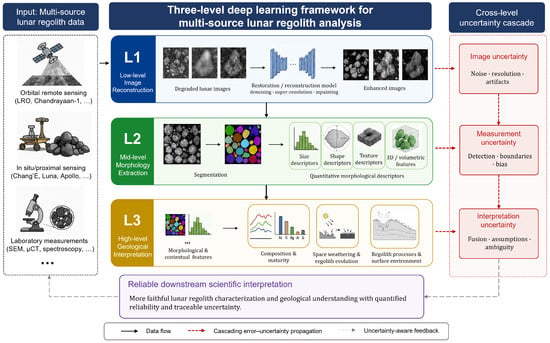
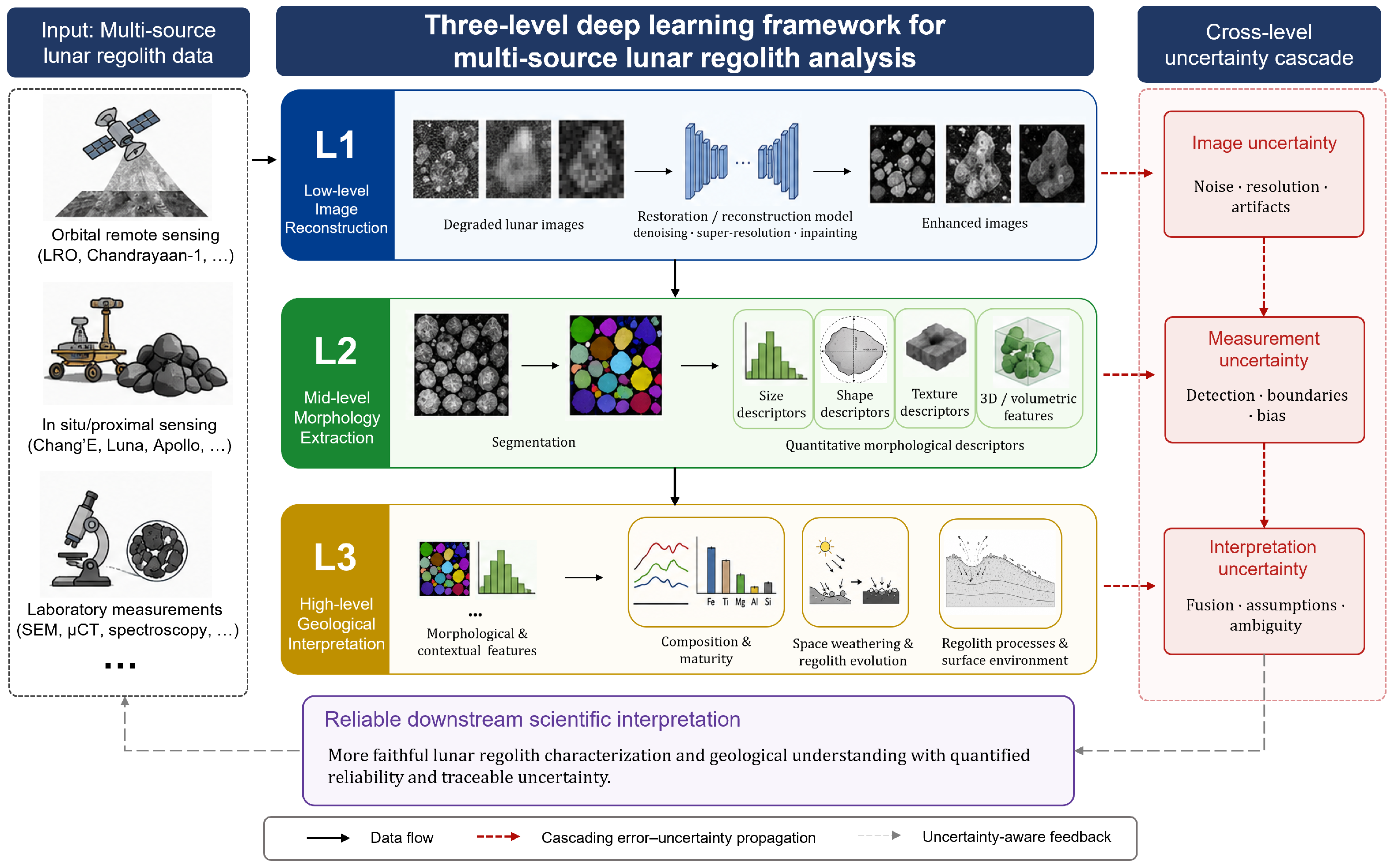
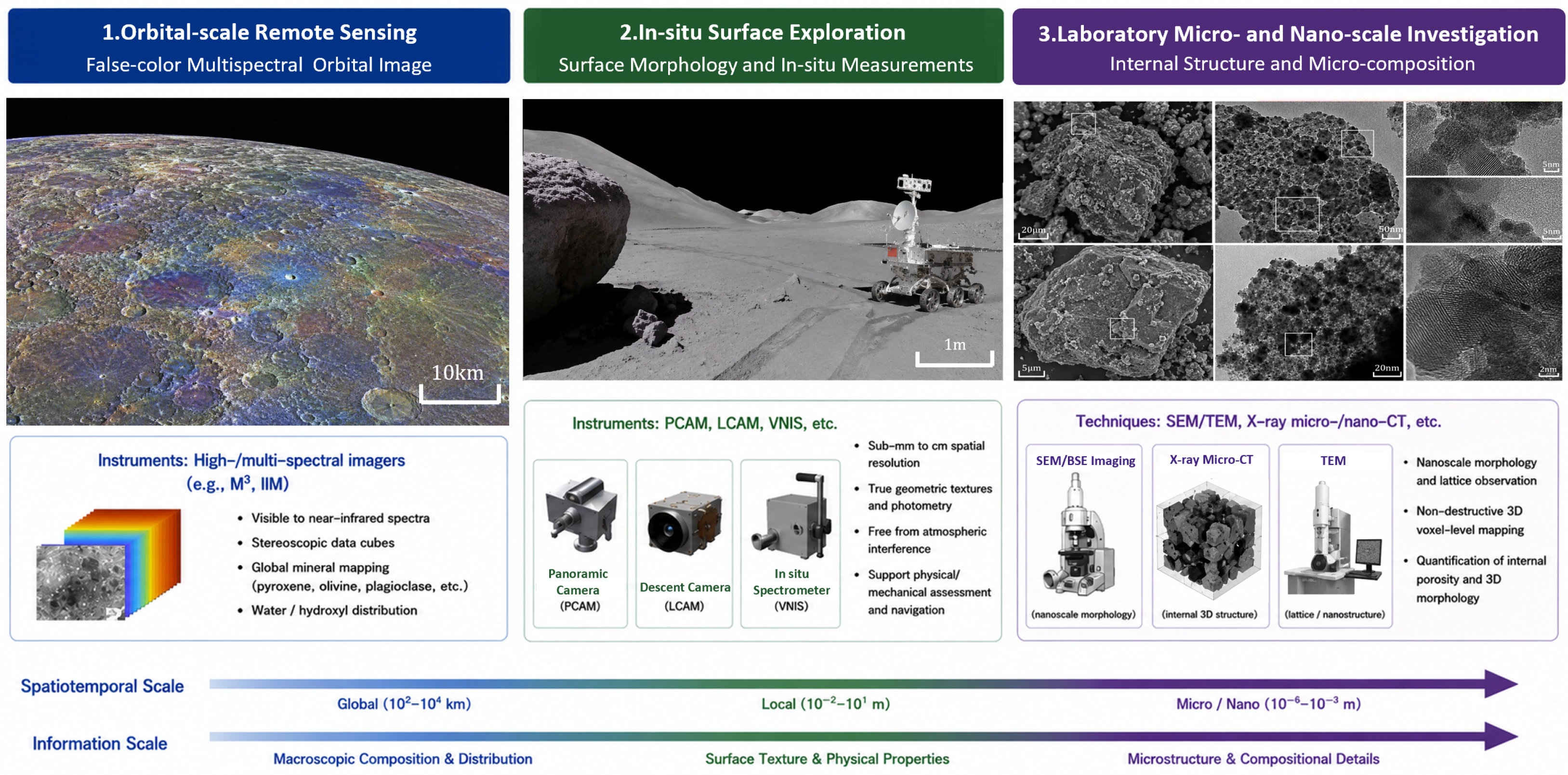
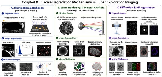
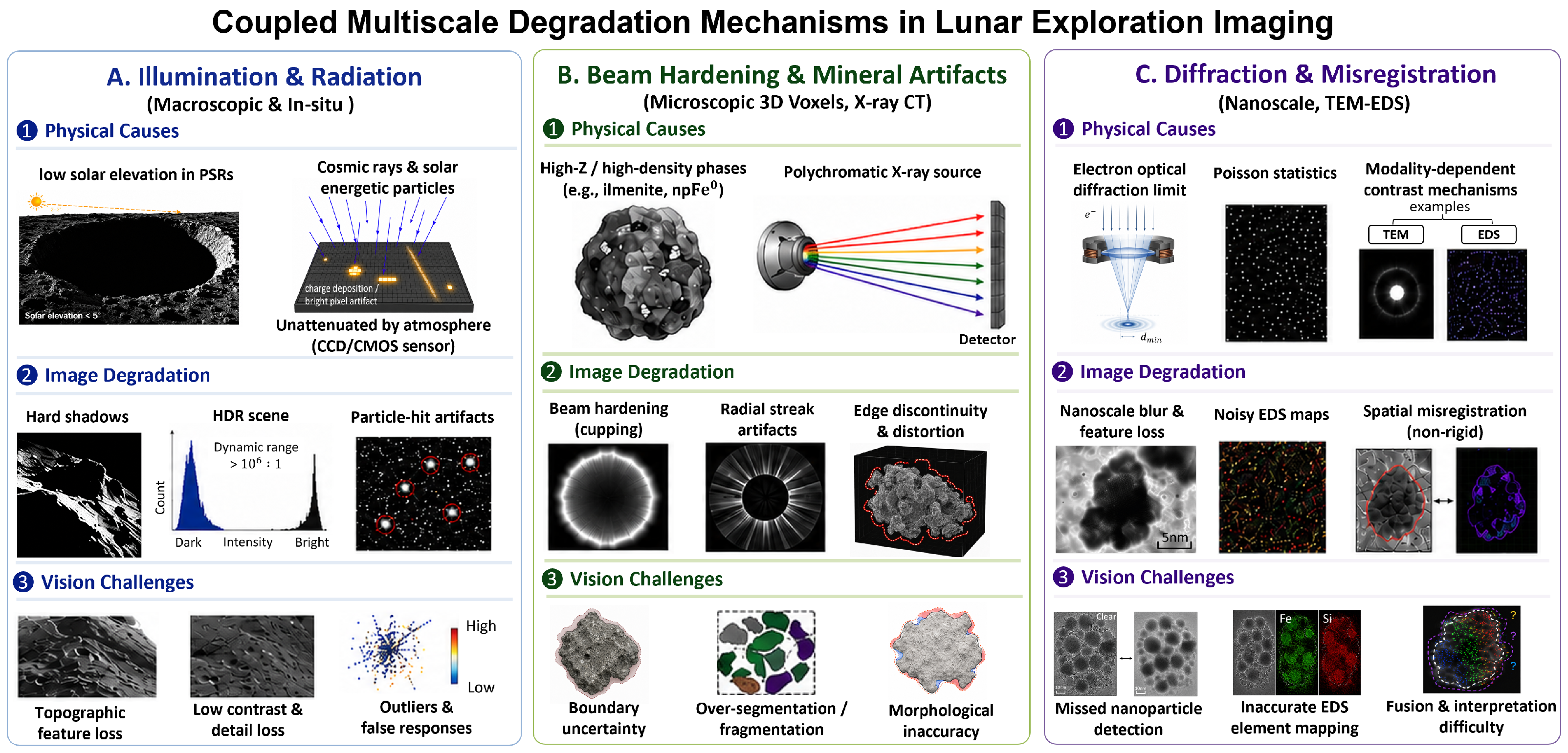
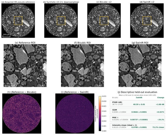
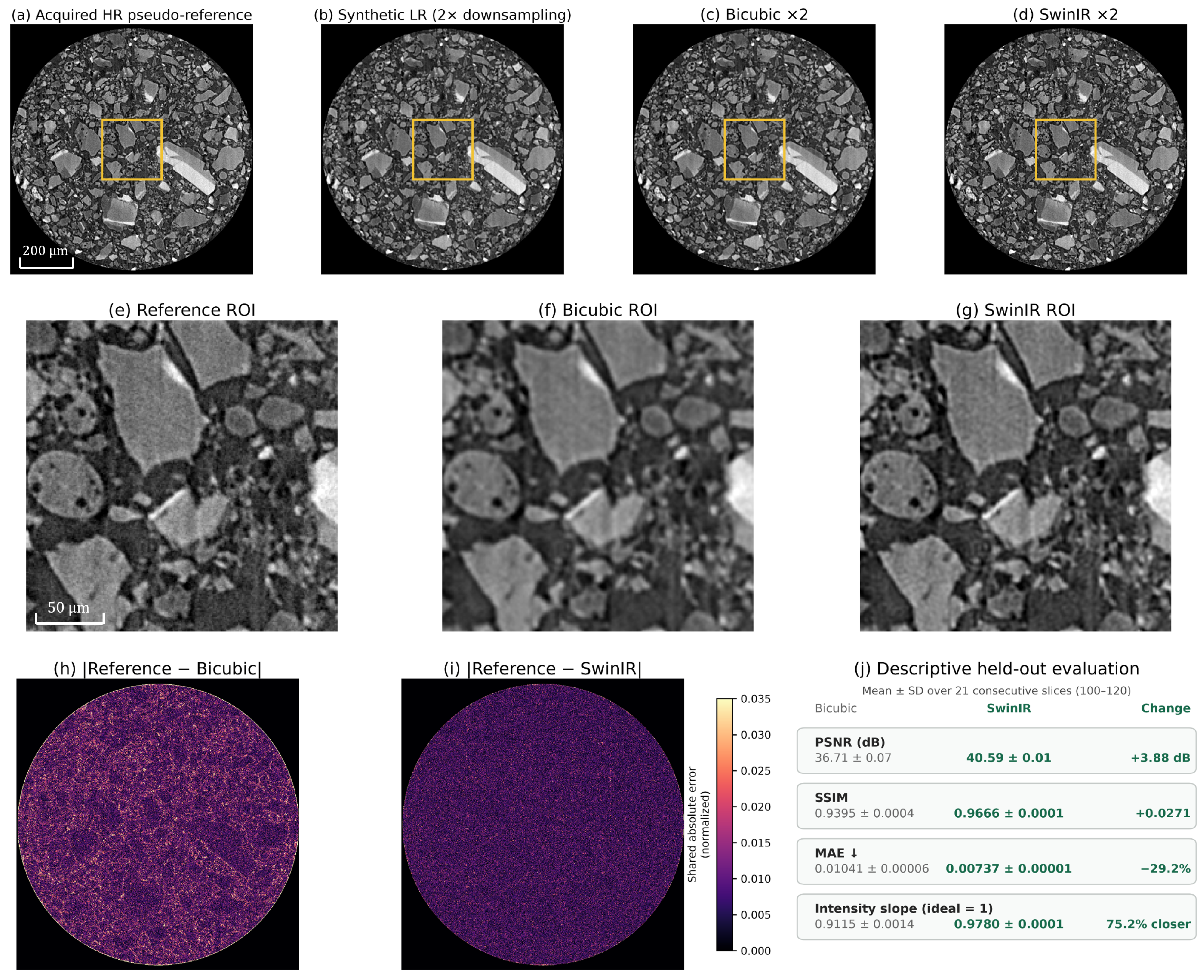
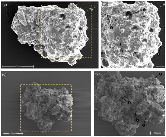
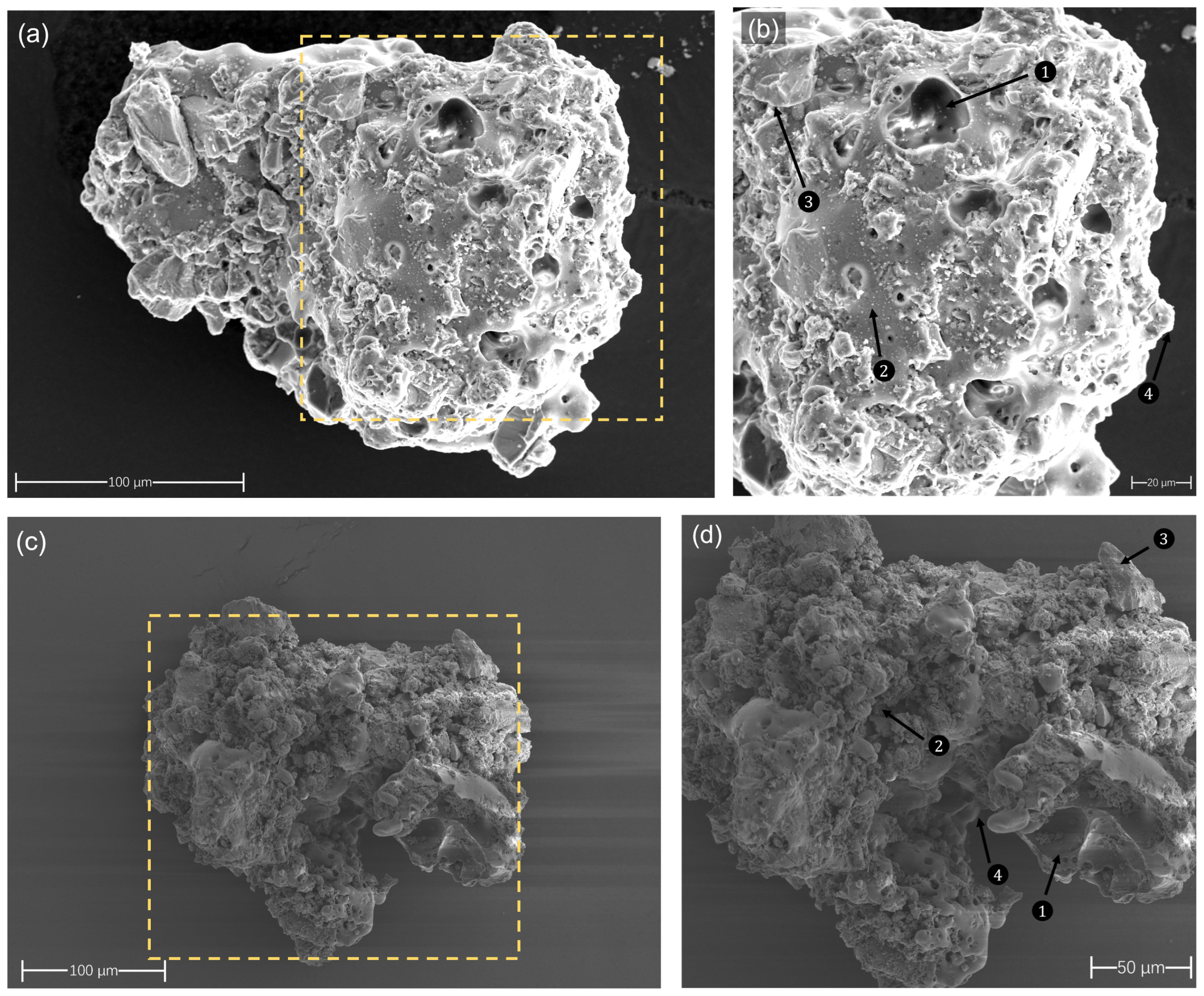
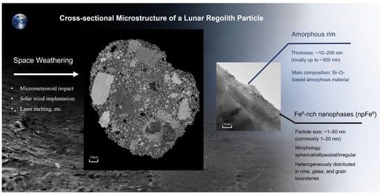
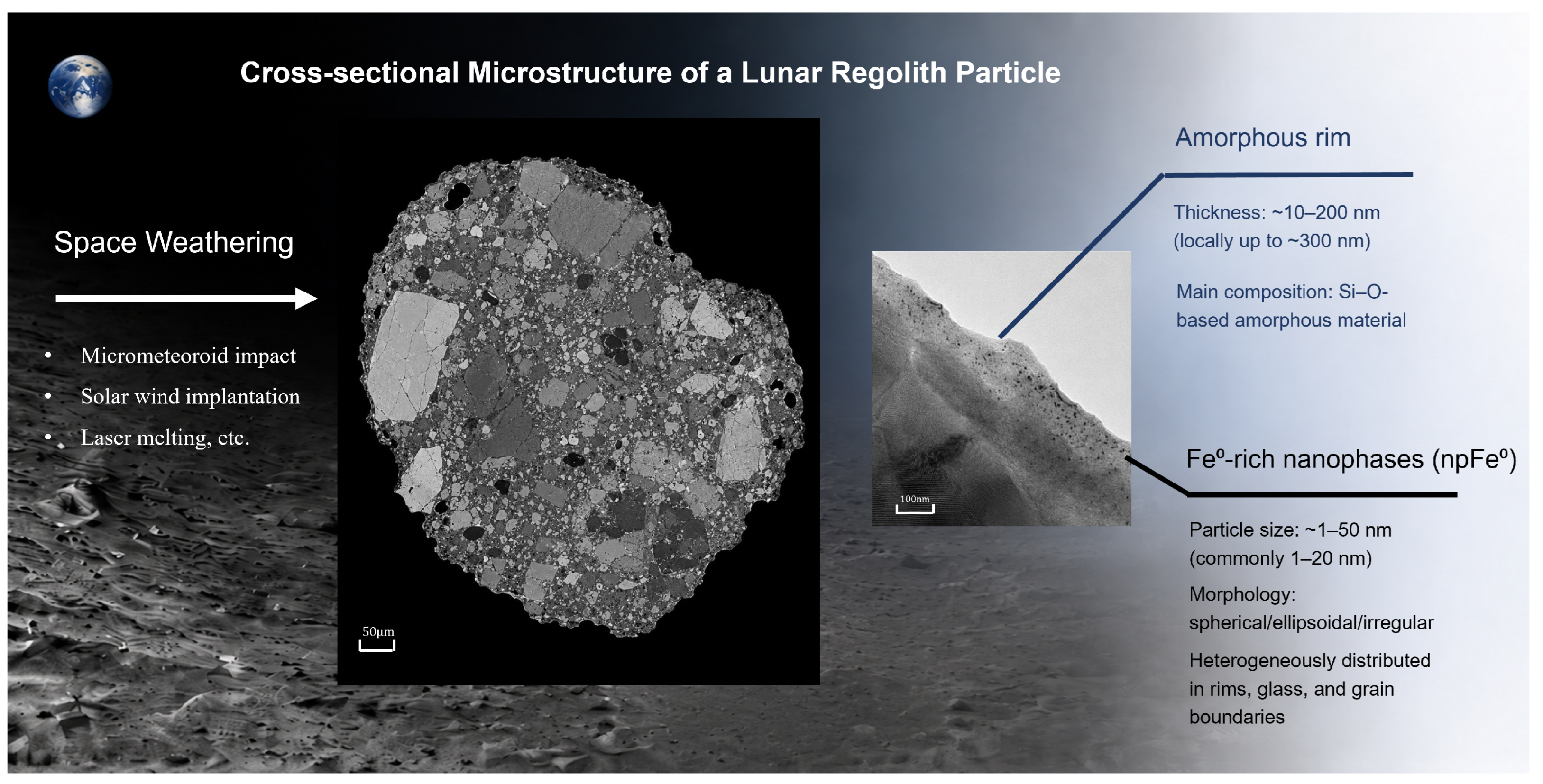

Disclaimer/Publisher’s Note: The statements, opinions and data contained in all publications are solely those of the individual author(s) and contributor(s) and not of MDPI and/or the editor(s). MDPI and/or the editor(s) disclaim responsibility for any injury to people or property resulting from any ideas, methods, instructions or products referred to in the content. |
© 2026 by the authors. Licensee MDPI, Basel, Switzerland. This article is an open access article distributed under the terms and conditions of the Creative Commons Attribution (CC BY) license.