


Abstract
Background: Tandem leucine-rich repeats (LRRs) are typically classified into eleven types; however, several variant motifs have also been reported. Here, we identified new LRR variants that exhibit dual characteristics of two distinct types. We investigated how the dual characteristics influence the structure and function of LRRs. Methods: We conducted sequence similarity searches using the protein database and analyzed sequence features. We also characterized the structural features of these LRR variant motifs using solved structures and AlphaFold models and investigated their potential biological functions through domain analysis. Results: Of the identified 3222 proteins, approximately 60% originate from the bacterial PVC superphylum. The variants were classified into two groups: one defined by the consensus sequence LxxLxLxx(C/T)xzI TDxxLxx(L/F)xx(L/C)xx, and the other by LxxLxLxxCxxI TDxxLxxLxxLP (where “z” denotes a deletion). The LRRs highly similar to the variants are occasionally observed in solved structures and comprise three types of super-secondary structures (SSSs): β-strand–α-helix adjoining a 3(10)-helix–β-strand, β-strand–3(10)-helix–β-strand, and β-strand–3(10)-helix adjoining an α-helix–β-strand. The AlphaFold models adopt these SSSs and, in addition, include the SSS of the β–α–β motif. Functional annotation identified kinase and F-box domains in a subset of these LRR proteins. Conclusions: The coexistence of these four SSSs and the high frequency of the first SSS appear to reflect the dual characteristics of the LRR variants. The LRR variant-containing proteins suggest potential roles in bacterial immunity and ubiquitination. The present findings expand the structural diversity of LRR proteins and provide new insights into their functional roles.
1. Introduction
Leucine-rich repeats (LRRs), which occur in tandem, are widely distributed in proteins across all domains of life, including viruses [1]. LRR-containing proteins are involved in a wide range of biological processes, including immune responses, signal transduction, cell adhesion, DNA repair, recombination, transcription, RNA processing, disease resistance, and apoptosis [2,3,4,5,6,7]. In addition, some LRR-containing proteins participate in ubiquitination pathways, contributing to the regulation of cellular processes such as protein degradation, signaling, and pathogen sensing [8,9]. The repeating unit length (RUL), which generally consists of 20–30 amino acids, is divided into a highly conserved segment (HCS) and a variable segment (VS). For ease of explanation, we introduce the following abbreviations. The HCS, consisting of eleven residues, is denoted as HCS = 11 [10,11,12], and correspondingly, the VS with eleven residues is denoted as VS = 11. The same notation is used hereafter.
In most LRR types, the HCS is represented by the consensus sequences LxxLxLxxNxL or LxxLxLxxCxxL, with HCS = 11 or 12, where “L” denotes Leu, Val, or Ile; “N” denotes Asn or Thr; “C” denotes Cys or Asn; and “x” denotes any amino acid. The occurrence of Phe, Ala, Cys or Met at the “L” position, of Ser, Thr or Arg at the “N” position, and of Asn at the “C” position occurs less frequently. This canonical class is classified into nine types, including RI-like, cysteine-containing (CC), Gala, SDS22-like, plant-specific (PS), Leptospira-like, bacterial, IRREKO, and typical [13,14,15,16]. Additional consensus sequences, LxxVxIPxxV and LxxLxFGxxF with HCS = 10 (referred to here as the atypical canonical class), have also been identified in TpLRR and FNIP, respectively [17,18,19].
For all eleven types, three-dimensional structures have been determined. In LRRs, positions 3–5 in the HCS form short β-strands, whereas the VS adopt various secondary structures such as α-helix, 3(10)-helix and polyproline II helix. The parallel stacking of these β-strands produces a superhelical arrangement (referred to as a solenoid structure). The overall shape resembles a horseshoe (arc), a prism, or a circle (Baumkuchen ring-like shape) [18]. The solenoid structure consists of four parts: a concave surface, ascending loops, a convex surface, and descending loops [3]. The hydrophobic core of the solenoid structure is formed by conserved hydrophobic residues in the consensus sequence [2,3,4]. The Asn side chain at the “Asn” position (at position 9 in the HCS) forms a continuous hydrogen-bond network known as the asparagine ladder [2,20]. A conserved phenylalanine forms a buried spine, representing a unique hydrophobic packing arrangement. This spine has been observed in the Nogo receptor, TLR3, LGI1, and Crov588 [17,21,22,23].
Super-secondary structures (SSSs), which consist of several adjacent secondary-structure elements, are commonly observed in proteins. SSSs are compact and recurrent folding motifs within polypeptide chains [24]. Common examples of SSSs include the helix–turn–helix, helix–loop–helix, coiled coil, β-hairpin, Greek key, β-barrel, β–α–β motif, and zinc finger. In this context, “helix” and “α” refer to an α-helix, whereas “β” and “beta” refer to a β-strand [25]. A composite helix, made up of contiguous α- and 3(10)-helices (α/3(10) composite), is certainly present in protein structures [26].
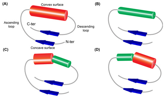
The VS parts of CC, RI-like, and GALA LRRs [12] adopt α-helices characterized by consecutive (i → i + 4) hydrogen bonds [27], whereas those of SDS22-like, PS, and Leptospira-like LRRs [11] adopt 3(10)-helices characterized by consecutive (i → i + 3) hydrogen bonds [27,28]. The LRR domains of RI-like, CC, and GALA types form a repeating β-strand–ascending loop–α-helix–descending loop–β-strand (β–α–β) motif (Figure 1A) [12]. In contrast, the LRR domains of SDS22-like, Leptospira-like, and PS types were previously described as adopting an SSS consisting of a 3(10)-helix and a β-turn [11]. However, because the β-turn forms part of the descending loop in LRR solenoid structures, we re-examined this secondary structure assignment. We found that these three types of LRR domains are instead represented by a distinct SSS composed of a β-strand–ascending loop–3(10)-helix–descending loop–β-strand (β–3(10)–β) motif (Figure 1B).
Figure 1.
Four super-secondary structures in CC/SDS22-like, short CC/SDS22-like, and short SDS22-like LRRs. (A) β–α–β motif. (B) β–3(10)–β motif. (C) β–α/3(10)–β motif. (D) β–3(10)/α–β motif. “β” indicated by dark blue arrows is β–strand. “α” indicated by red tube is α-helix. “3(10)” indicated by a green tube shows a 3(10)-helix. “α/3(10)” shows a composite helix made up of contiguous α- and 3(10)-helices. “3(10)/α” shows a composite helix made up of contiguous 3(10)- and α-helices. The α-helix tube is thicker than the 3(10)-helix tube. The composite helices have a kink between the two helices.
SDS22-like LRRs are represented by the consensus sequence LxxLxLxxNxI xxIxxLxxLxx (RUL = 22; HCS = 11 and VS = 11), whereas PS LRRs by LxxLxLxxNxL SGxIPxxLxxLxx (RUL = 24; HCS = 11 and VS = 13). A recent sequence analysis identified a novel LRR type present in more than 280 proteins from microorganisms, including protists, fungi, and bacteria [29]. Its consensus sequence is LxxLDLxxTxV SGxLxxLxxLxx (HCS = 11 and VS = 12). The four-residue SGxL motif in the N-terminal region of the VS is highly similar to the SGxI motif found in PS LRRs. In addition, the nine-residue LxxLxxLxx motif in the C-terminal region of the VS is identical to that of SDS22-like LRRs. This novel LRR type exhibits dual characteristics of SDS22-like and PS LRRs and was therefore designated as PS/SDS22-like LRR [29]. Chameleon sequences are identical amino acid fragments that adopt distinct conformations [30,31]. PS/SDS22-like LRRs exhibit four patterns of secondary structure in their VS parts, suggesting that these sequences may possess chameleon-like properties.
We hypothesized that another novel LRR type with dual characteristics might exist, analogous to PS/SDS22-like LRRs. A viral protein (UniProtKB: A0A6C0H0T7) comprising 351 residues contains ten tandem LRRs [32]. Sequence alignment showed that most of these repeats (8/10) conform well to the consensus sequence LxxLxLxxCxxI TDxxLxxLxxLxx (RUL = 25; HCS = 12 and VS = 13) (Table 1). The consensus sequence of CC LRRs is LxxLxLxxCxxL TDxGLxxLAxxCxx (RUL = 26; HCS = 12 and VS = 14) or LxxLxLxxNxL TDxGLxxLAxxCxx (Table 1) [12]. In this novel LRR type, the eight-residue TDxxLxxL motif in the N-terminal region of the VS is identical to that of CC LRRs. In addition, the nine-residue LxxLxxLxx motif in the C-terminal region of the VS is identical to that of SDS22-like LRRs. Thus, this novel LRR type also exhibits the dual characteristics of CC and SDS22-like LRRs, and we designate it as CC/SDS22-like LRR.
Table 1.
Consensus sequences of CC/SDS22-like and short CC/SDS22-like leucine-rich repeats (LRRs).
In the present study, we addressed the following questions: (a) Are proteins containing CC/SDS22-like LRRs unique, or are they widespread among organisms? (b) If they are widespread, do CC/SDS22-like LRRs possess sequence features in addition to the dual characteristics? (c) If they are widespread, what are their structural features? Is the duality of CC/SDS22-like LRRs reflected in their secondary and three-dimensional structures? What kinds of secondary structures (SSSs) do CC/SDS22-like LRRs adopt? (d) If they are widespread, are they involved in various biological processes, as are other LRR-containing proteins? We performed sequence similarity searches against protein databases and analyzed the sequences. We then characterized the structural features of CC/SDS22-like LRR motifs using solved structures and AlphaFold models [33,34], and investigated their potential biological functions through domain analysis.
We identified CC/SDS22-like LRRs in 3222 proteins, approximately 60% of which originate from the bacterial PVC superphylum (Table S1). These newly identified LRR motifs can be classified into two groups. The first group (CC/SDS22-like LRR) is characterized by the consensus sequences such as LxxLxLxxCxxI TDxxLxxLxxLxx, LxxLxLxxTxV TDxxLxxLxxLxx, or LxxLxLxxTxV TDxGLxHFxxCxx, with VS = 13. The second group (termed short CC/SDS22-like LRR) has the consensus sequence, LxxLxLxxCxxI TDxxLxxLxxLP, with VS = 12. The LRRs highly similar to the CC/SDS22-like type are occasionally observed in solved structures and comprise three types of super-secondary structures (SSSs): β-strand–α-helix adjoining a 3(10)-helix–β-strand (β—α/3(10)-β), β-strand–3(10)-helix–β-strand (β-3(10)-β), and β-strand–3(10)-helix adjoining an α-helix–β-strand (β-3(10)/α–β),. The AlphaFold models adopt these SSSs and, in addition, include the SSS of the β–α–β motif. The coexistence of these four SSSs and the high frequency of the β–α/3(10)–β motif appear to reflect the dual characteristics of CC/SDS22-like LRRs. These proteins are frequently associated with F-box, kinase, or TIR domains, suggesting potential roles in ubiquitination and bacterial immunity. The present findings expand the known structural diversity of LRR proteins and provide new insights into their potential functional roles.
2. Materials and Methods
We searched for proteins containing CC/SDS22-like LRRs using the sequence of a CC/SDS22-like LRR domain (UniProtKB: A0A6C0H0T7) as a query. Sequence similarity searches were first performed using BLAST and FASTA at the Bioinformatics Center, Institute for Chemical Research, Kyoto University (http://www.genome.jp) on 2 March 2024, and candidate proteins were identified. The amino acid sequences of these candidates were retrieved from the UniProt database [1]. LRR domains, including CC/SDS22-like LRRs, in the candidate proteins were then assigned using LRRpred [35]. Subsequently, sequence similarity searches were iteratively performed using the identified LRR domain sequences. In addition, we identified a related type in four proteins. This type was designated as short CC/SDS22-like LRR, as its VS part consists of twelve residues, one residue shorter than that of CC/SDS22-like LRR (VS = 13). In total, 3222 proteins were identified and analyzed, as described below. The consensus sequences of these novel LRR types were determined based on the occurrence probability of amino acid residues at each position using WebLogo 3 (https://weblogo.berkeley.edu/logo.cgi (accessed on 2 March 2024) [36]. Bold uppercase letters indicate more than 70% occurrence of a residue at a given position, uppercase letters indicate 40–70%, and lowercase letters indicate 20–40%.
For the structural analysis of CC/SDS22-like LRRs, two approaches were employed. First, structural data on CC/SDS22-like LRRs were collected, including both experimentally solved and computationally predicted structures. Six solved structures—coupling factor (PDB ID: 3E4G), FBXL17 (6W66), LRRC8 (6FNW), InlK (4L3F), lmo2027 (5KZS), and LMOf2365_1397 (4EZG)—were identified (Table 2) [37,38,39,40,41]. Predicted structures were obtained using AlphaFold, a deep learning–based method [33,34].
Table 2.
Consensus sequences of CC/SDS22-like and short CC/SDS22-like leucine-rich repeats (LRRs).
Second, secondary-structure assignments of the obtained structures were performed using the DSSP-PPII program [42,43]. The reasons are as follows: DSSP-PPII, STRIDE [44], and XTLSSTR [45] differ in their assignments of α-helices, 3(10)-helices, and β-turns [46]. DSSP-PPII, which identifies precise hydrogen-bonding patterns corresponding to regular secondary structures, defines eight types of secondary structures [42]. We applied rule 6.3 as described in previous studies [11,12]. In this notation, “-GGG-” indicates a five-residue 3(10)-helix. HELFIT analysis, which determines helix parameters such as helix axis, helix pitch, helix radius, number of residues per turn, and handedness, indicates that DSSP-PPII and rule 6.3 provide the most appropriate assignment [11,12,28]. β-Turn types were identified using the PROMOTIF program [47]. The PROMOTIF program defines residue i of a β-turn as the residue immediately preceding the “TT” or “SS” assignment. The “-TT-TT-” pattern represents two consecutive β-turns. Further details are provided in the previous studies [11,12].
3. Results
We identified and analyzed 3222 proteins (Table S1). The LRRs within 2369 of these proteins were categorized into two groups: CC/SDS22-like LRRs (in 2365 proteins) and short CC/SDS22-like LRRs (in 4 proteins) (Table S1). Of the 2365 proteins, 1867 originate from the bacterial PVC superphylum, which comprises the phyla Planctomycetes, Verrucomicrobia, Chlamydiae, Lentisphaerae, and Kiritimatiellaeota [48]. In addition, 60 and 90 proteins are derived from the SAR and Discoba lineages, respectively, and 344 proteins originate from other bacteria, one from archaea, and three from metagenomes. Among the 3222 proteins, the remaining 853 are annotated as F-box/LRR-repeat protein 14 (FBXL14) [49], originating from the phyla Viridiplantae (777 proteins) and SAR (66 proteins). Ten proteins are derived from other groups, including Discoba, Amoebozoa, Rhodophyta, Cryptophyceae, and fungi.
3.1. Consensus Sequences of the New Types of LRR
3.1.1. Three Subtypes of CC/SDS22-like LRR with HCS = 11 or 12 and VS = 13
CC/SDS22-like LRRs can be classified into three subtypes (Table 1). The first subtype was identified in 246 proteins. Among these, 96 proteins—encoding LRR domains composed primarily of CC/SDS22-like LRRs with RUL = 25 (HCS = 12 and VS = 13)—are found in the bacterial PVC superphylum, including Chlamydiae (81 proteins), Planctomycetes (14), and Verrucomicrobia (1). In addition, two proteins from the Oxalobacteraceae bacterium (phylum Proteobacteria) and three proteins from metagenomic datasets contain CC/SDS22-like LRR domains.
The genome sequence of Protochlamydia amoebophila UWE25 (phylum Chlamydiae) has been reported previously [50,51], and 73 proteins from this organism contain CC/SDS22-like LRR domains. This first subtype accounts for 91% of the total repeat number (976/1073) (Table 1), with the consensus sequence LQHL(n/d)LSxCNxL TDAGLAHLxPLxA. In the phylum Planctomycetes, 14 proteins from four species contain a total of 195 LRRs, of which approximately four-fifths (154/195 = 0.79) are CC/SDS22-like LRRs with the consensus sequence LqxLdLxgCxxI TDAGLAHLxxLxx (Table 1).
A total of 140 proteins containing LRR domains composed mainly of the first subtype were identified in two eukaryotic groups: Stramenopiles and Discoba (specifically Bodo saltans). In Stramenopiles, 50 proteins from 30 species contain 967 LRRs, 44 of which are homologous. More than three-quarters of these repeats (752/967 = 0.78) are CC/SDS22-like LRRs (Table 1), with the consensus sequence LtsL(n/d)LxgCnx(i/lv)TDAGLAHLxxLxx. In Bodo saltans, three-quarters of the total LRRs (641/857 = 0.75) across 90 proteins belong to the CC/SDS22-like type, with the consensus sequence LQxLdLxgCxxI TDAGLxx(i/v/l)AxLxq (Table 1).
In the class Chlorophyta (green algae), 11 species encode 52 CC/SDS22-like LRR proteins. Among them, 32 proteins from Ostreobium quekettii contain CC/SDS22-like LRRs with RUL = 25, accounting for approximately four-fifths of the repeats (423/531 = 0.80) (Table 1), with the consensus sequence LtxLdLsgCxx(I/v) TDxGLxx(l/v)gxLtx. In addition, 12 proteins from Tetraselmis sp. GSL018 contain CC/SDS22-like LRRs (RUL = 25), with the consensus sequence LtxLdLxgCxx(v/i/l)TDxGLxxLxxLxx, representing 57% of the repeats (104/181) (Table 1).
The second subtype of CC/SDS22-like LRRs (RUL = 24; HCS = 11 and VS = 13), characterized by strong occupancy of Thr at position 8 in the HCS, was identified in 2079 proteins (Table S1). Bacteria, particularly those in the PVC superphylum, encode proteins containing LRR domains composed predominantly of this subtype with RUL = 24 (HCS = 11 and VS = 13). Of these proteins, 74% (1735/2079) originate from the bacterial PVC superphylum. In the phylum Planctomycetes, 26 proteins from Symmachiella dynata are annotated as internalin-A (InlA_1–InlA_26). Most LRRs (246/265 = 0.93) belong to this subtype (Table 1), with the consensus sequence LxxLxLxxTqx(V/i)(s/t)DaGLehLrgLtn. Similarly, Blastopirellula cremea encodes 82 CC/SDS22-like LRR proteins, in which the majority of repeats (804/933 = 0.86) belong to this subtype, with the consensus sequence L(e/k)xLxLxgTqx(I/v)TDaGLehLkgLts (Table 1).
Members of candidate bacterial phyla, including Candidatus Melainabacteria, Hydrogenedentes, Poribacteria, and Stahlbacteria, also encode proteins containing this subtype. In addition, Candidatus Obscuribacter phosphatis, Candidatus Obscuribacter sp., and two unclassified bacteria encode eight homologous proteins. Among 298 repeats, 73% (208/298) correspond to the second subtype, with the consensus sequence L(r/k)eLxL(d/n)txxI(t/s)DxGLxxLxxLxx (Table 1). One archaeal species from the phylum Euryarchaeota encodes a CC/SDS22-like LRR protein containing six repeats, with the consensus sequence LxxLxLxxTxITDxGLxEVxxLxx. In addition, marine metagenomic datasets contain proteins encoding this subtype.
The third subtype of CC/SDS22-like LRRs (RUL = 24; HCS = 11 and VS = 13) was identified in 28 proteins from eight species in the phylum Planctomycetes (Table 1). Most repeats (204/239 = 0.85) belong to this subtype, with the consensus sequence LTxLxLxxTxV TDAGLAHFKdCKn (Table 1). The first repeat unit in these LRR domains is often characterized by HCS = 12 rather than HCS = 11, with the consensus sequence LT(a/e)(V/L)dLsgNpqVTDAGLAhFKdCKn. The feature is a strong preference for Phe at position 8 in the VS.
3.1.2. F-Box/LRR-Repeat Protein 14 (FBXL14)
Homologs of F-box/LRR-repeat protein 14 (FBXL14) were identified across many species. Most individual LRR domains in FBXL14 homologs contain both the first and second subtypes of CC/SDS22-like LRRs, with the second subtype being more prevalent. To determine the consensus sequences, we analyzed representative FBXL14 proteins from fifty-one plant species. The consensus sequence of the second subtype (663/969 = 0.68) is LksLnLsg(c/s/t)x(I/v) TDaGLxhLkgLxn, in which position 9 accommodates not only Thr but also Cys and Ser. The consensus sequence of the first subtype (202/969 = 0.21) is L(t/e)SLN(L/f)(n/s)x(C/N)(n/d)x(I/l) TDxG(L/M)(e/k)x(I/L)SGLtN, in which position 9 preferentially contains Cys and Asn.
3.1.3. Short CC SDS22-like LRR with HCS = 12 and VS = 12
This subtype, characterized by short VS (=12), was identified in three bacterial proteins. The consensus sequence is LqxLDLSGCxGI TDaGLAHLkx(L/m)P (34/58 = 0.59) (Table 1). A short CC/SDS22-like LRR with HCS = 11 was also identified in a single protein from a Bdellovibrionales bacterium, in which all six LRRs conform to the consensus sequence LRxxLxxTxV TDxGLxxLKGLP.
3.2. Structures of CC/SDS22-like LRRs
3.2.1. CC/SDS22-like LRRs Occasionally Observed in Solved Structures
In solved structures, ten LRRs highly similar to CC/SDS22-like LRRs were occasionally identified (Table 2). The VS parts of these LRRs exhibit the same hydrophobic residue pattern as those of CC/SDS22-like LRRs (Table 1). Secondary structure assignment revealed that the VS parts can be classified into three types. The first type is represented by patterns such as “-HHHHHHGGG-TT” (e.g., the third repeat in bovine mitochondrial factor B). Following rule 6.3, this pattern consists of an eight-residue α-helix followed by a five-residue 3(10)-helix and a type I β-turn. This helical region corresponds to a composite helix composed of contiguous α- and 3(10)-helices (an α/3(10) composite) [26]. A similar pattern, “-HHHHGGGGG-TT” (the seventh repeat in FBXL17), consists of a six-residue α-helix followed by a seven-residue 3(10)-helix and a type I β-turn (Table 2). In both cases, the β-turn forms part of the descending loop. These CC/SDS22-like LRRs are characterized by an SSS consisting of a β-strand–ascending loop–α/3(10) composite–descending loop–β-strand (β–α/3(10)–β) motif (Figure 1A–D). The α/3(10) and 3(10)/α composite helices introduce a kink between the two helical segments, as observed in solved structures [26].
The second type was observed in seven LRRs (including the first repeat of bovine mitochondrial factor B; the third and fourth repeats of InlK; the fifth repeats of lmo2027 and lmo2470; the third repeat of LMOf2365_1397, and the seventh repeat of LRRC8) (Table 2). In this type, the VS part consists of a 3(10)-helix of five to nine residues and two tandem β-turns or one β-turn forming part of the descending loop. These LRRs exhibit an SSS consisting of a β-strand–ascending loop–3(10)-helix–descending loop–β-strand (β–3(10)–β) motif, similar to those observed in SDS22-like, PS, and Leptospira-like LRRs [11].
The third type was observed in a single LRR (the third repeat of LMOf2365_1397). The VS part consists of a five-residue 3(10)-helix followed by a six-residue α-helix and two tandem β-turns (Table 2). This type is characterized by an SSS consisting of a β-strand–ascending loop–3(10)-helix/α-helix–descending loop–β-strand (β–3(10)/α–β) motif. Consequently, three types of SSSs of β–α/3(10)–β, β–3(10)–β, and β–3(10)/α–β occur in the ten CC/SDS22-like LRRs seen in the known structures. (Table 2).
3.2.2. AlphaFold Models of CC/SDS22-like LRRs
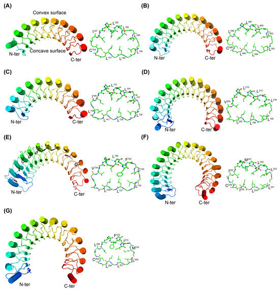
We further analyzed AlphaFold models of CC/SDS22-like and short SDS/22-like LRR domains in eighteen representative proteins. Their secondary structures are shown in Table 3 and Table S2, and the three-dimensional structures are shown in Figure 2A–G. The AlphaFold models of the CC/SDS22-like LRR domains exhibited high average pLDDT scores (>90 and/or >70) and low predicted aligned error (PAE) scores. Thus, the predicted structures appear to be reliable. In all cases, parallel β-strand stacking within the HCS parts forms a solenoid structure, and all conserved hydrophobic residues contribute to the formation of the hydrophobic core. The three SSS types described above in solved structures were also observed in the AlphaFold models. The SSSs of the β–α/3(10)–β motif, the β–3(10)–β motif, and the β–3(10)/α–β motif occur 76, 26, and 6 times, respectively. In addition, the SSS of the β–α–β motif occurs 90 times. Short CC/SDS22-like LRR domains also adopt these four types of SSSs in the AlphaFold models. Among the four SSSs, the β–α/3(10)–β and β–α–β motifs occur more frequently.
Table 3.
Secondary structures of CC/SDS22-like and short CC/SDS22-like LRRs in structures of seven representative proteins predicted by AlphaFold.
Figure 2.
Structures of CC/SDS22-like LRR domains (A–F) and a short CC/SDS22-like LRR domain (G) in seven representative proteins. (A) Disease resistance R13L4/SHOC-2 like LRR domain-containing protein from viral metagenome (UniProtKB: A0A6C0H0T7). (B) BTB domain-containing protein from Protochlamydia amoebophila (strain UWE25) (Q6MEE0). (C) (A0A355G5V6_9PLAN). (D) Alanyl-tRNA synthetase protein from Fimbriiglobus ruber (A0A225EG43). (E) A non-specific serine/threonine protein kinase from Pirellulaceae bacterium (A0A925DAG5). (F) A leucine-rich repeat family protein from Arabidopsis thaliana (A0A178WET7). (G) EBS67_14515 from bacterium (A0A9E5EXX2). The left side shows the overall structure of the LRR domains. The right side shows a single LRR unit, highlighting that conserved hydrophobic residues contribute to the hydrophobic core. α-Helices or 3(10)-helices are shown as thick tubes, β-sheets as arrows. On the left side of panel (E), the side chains of conserved phenylalanine residues are shown in black. The sequence alignments of LRRs in these proteins are shown in Table 2 and Table S2.
4. Discussion
4.1. Extensive Presence of CC/SDS22-like LRR-Containing Proteins
The present study identified 3222 proteins containing CC/SDS22-like and short CC/SDS22-like LRRs (Table S1). Of these, 2211 were identified in the kingdom Bacteria, including 1867 from the PVC superphylum, while 1003 were identified in the kingdom Eukaryota and one in Archaea. These results indicate that CC/SDS22-like LRR-containing proteins are widely distributed. Interestingly, approximately 60% of the 3222 proteins originate from the bacterial PVC superphylum.
4.2. Sequence Features of CC/SDS22-like LRRs
CC/SDS22-like LRRs, which exhibit dual characteristics, possess additional sequence features. Based on these features, they can be divided into three subtypes (Table 1). The first subtype CC/SDS22-like LRRs are characterized by a conserved His at position 7 in the VS. Conserved His is also observed in CC-LRR variants in viral proteins [32,52]. Multiple histidine imidazole rings may form hydrogen bonds on the convex surface of the solenoid structure. The second subtype is characterized by a conserved Thr at position 10, instead of an Asn at position 11 in the HCS (HCS = 11). This substitution has also been observed in SDS22-like LRRs and their variants in viral proteins, as well as in PS/SDS22-like LRRs in proteins from the eukaryotic SAR group [29,32,52]. The Thr residue may form hydrogen bonds via its polar hydroxyl side chain, thereby contributing to stabilization of the solenoid structure. The third subtype is characterized by a conserved Phe at position 8 in the VS, which forms a phenylalanine spine (Figure 2E), as observed in LRR proteins such as the Nogo receptor, TLR3, LGI1, and Crov588 [17,21,22,23].
4.3. Simultaneous Occurrence of Four SSSs in CC/SDS22-like LRRs
The LRRs highly similar to the CC/SDS22-like type are occasionally observed in solved structures. These LRRs comprise three types of SSSs: β–α/3(10)–β, β–3(10)–β, and β–3(10)/α–β. The AlphaFold models adopt these SSSs and, in addition, include the SSS of the β–α–β motif (Figure 1A–D). The two SSSs, β–α/3(10)–β and β–α–β, are predominant (Table 3 and Table S2). The coexistence of these four SSSs and the high frequency of the β–α/3(10)–β motif appear to reflect the dual characteristics of CC/SDS22-like LRRs.
Previous studies suggested that PS/SDS22-like LRRs with dual characteristics exhibit chameleon-like sequence properties [29]. A similar phenomenon was also observed in CC/SDS22-like LRRs. A representative example is a BTB domain-containing protein from Protochlamydia amoebophila (UniProtKB: Q6MEE0), which contains thirteen CC/SDS22-like LRRs (Table S2). The VS sequences of LRR11 (TDAGLAHLTPLIN) and LRR12 (TNAGLAHLTPLVA) are highly similar; however, their secondary structures differ. LRR11 adopts a 3(10)-helix, whereas LRR12 adopts an α-helix (Table S2). This observation suggests that CC/SDS22-like LRRs exhibit chameleon-like behavior.
Taken together, the dual structural characteristics inherited from CC and SDS22-like LRRs, combined with the intrinsic chameleon-like nature of their sequences, may enable CC/SDS22-like LRRs to adopt multiple SSSs. This structural flexibility provides a plausible explanation for the simultaneous occurrence of four distinct SSS types within even a single LRR domain. The structural plasticity of chameleon-like sequences drives a wide array of immune functions, host–pathogen interactions, and cellular behaviors [53,54,55,56].
We acknowledge the limitations of AI-based structure prediction. AlphaFold predicts protein structures as static, single-state snapshots. Consequently, its primary limitation in capturing structural flexibility is its tendency to favor a single dominant conformation—often the most represented state in the training data—while failing to account for alternative conformational states [57].
4.4. Functional Implications of CC/SDS22-like LRR-Containing Proteins
So far, little is known about the functions of proteins containing CC/SDS22-like LRRs. However, these proteins are frequently associated with a wide variety of domains (Table S1), suggesting their involvement in diverse biological processes.
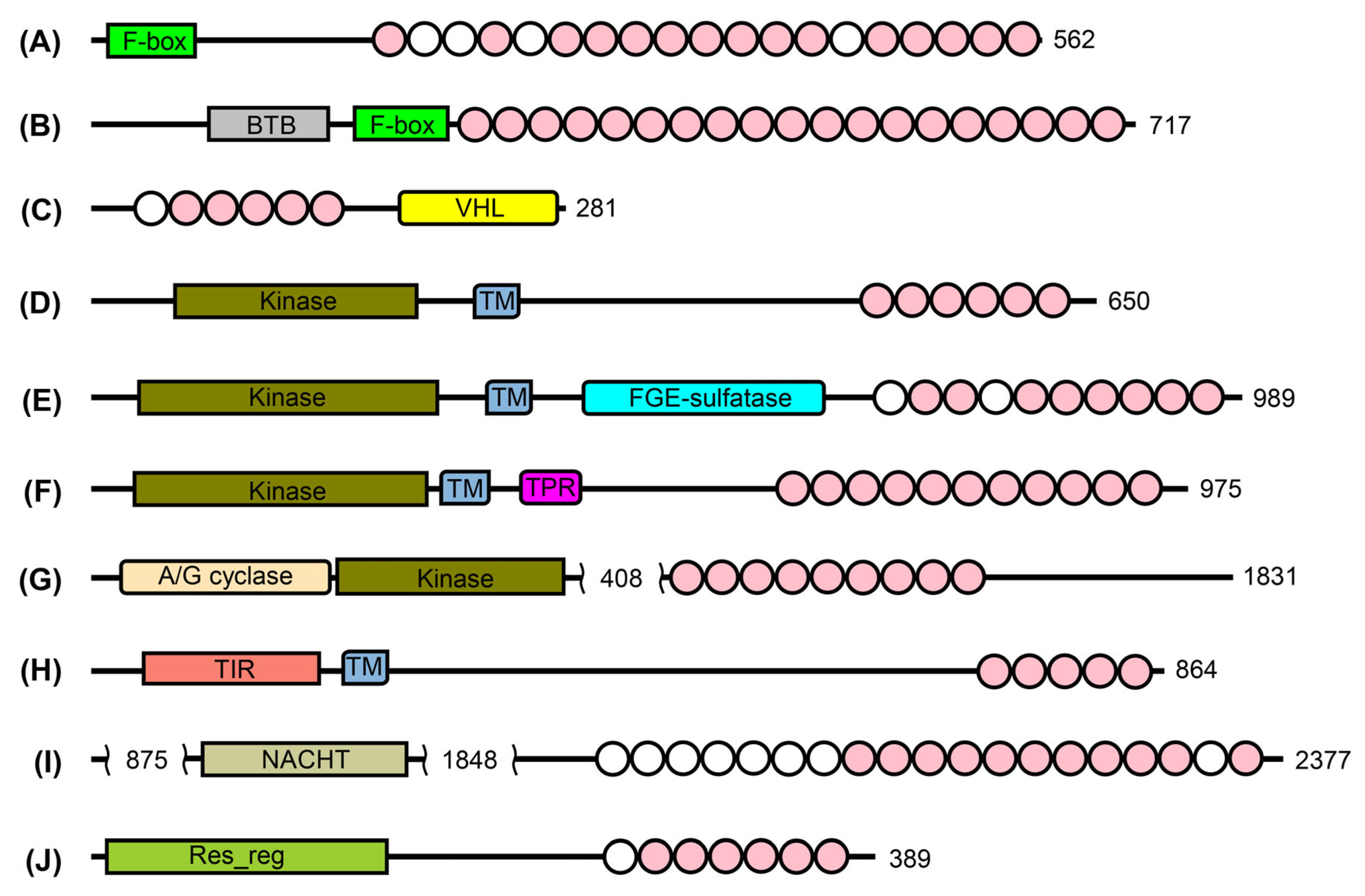
One prominent example is the F-box domain. F-box/LRR proteins (FBXLs) are widely distributed across eukaryotes, prokaryotes, and viruses [9], where they function as components of the SCF ubiquitin ligase complex, mediating protein ubiquitination and degradation. Domain analysis indicates that many CC/SDS22-like LRR proteins belong to the FBXL family, suggesting their involvement in ubiquitination pathways (Figure 3A). Structural studies of five human FBXL proteins (FBXL1, 2, 3, 5, and 17) have shown that their LRR domains are of the CC type or closely related variants, in which the VS part predominantly adopts an α-helical conformation [38,58,59,60,61]. In contrast, Mimiviridae (NCLDVs) encode many FBXL proteins in which the LRRs are short CC types with RUL = 22 [52]. These LRR types differ from CC/SDS22-like LRRs found in bacterial FBXLs, suggesting that variation in LRR subtype may contribute to species-specific functional adaptation. Sixty-four CC/SDS22-like LRR proteins from the phylum Chlamydiae, Candidatus Protochlamydia, that are FBXLs contain a BTB domain at the N-terminus of the F-box domain (Figure 3B). Structural studies have shown that the BTB domain in complex with FBXL17 (an LRR-containing protein) is involved in the recognition of degradation signals (“degrons”) in human cells [38,62]. Therefore, CC/SDS22-like LRR domains may contribute to substrate recognition in ubiquitination pathways, although this remains to be experimentally verified. CC/SDS22-like LRR proteins from Planctomycetes, Verrucomicrobiaceae bacteria, and Woeseia sp. contain a VHL domain (Figure 3C), which acts as a substrate recognition component of an E3 ubiquitin ligase complex [63]. This observation appears to support a role for these LRR proteins in ubiquitination-related processes.
Figure 3.
Domain architectures of ten proteins containing CC/SDS22-like LRRs. (A) PHPALM_29341 from phytophthora palmivora var. palmivora (UniProtKB: A0A2P4X7T7). (B) fbxL14_3 from Candidatus Protochlamydia amoebophila (A0A0C1H8F1). (C) CMO38_05550 from Verrucomicrobiaceae bacterium (A0A2E4AB07). (D) GCJUQL4_47850 from Gemmataceae bacterium (A0A4V0I9Z6). (E) PX52LOC_03625 from Limnoglobus roseus (A0A5C1AHQ6). (F) prkC_4 from the Planctomycetes bacterium ADurb.Bin12 (A0A1V6D218). (G) DB346_24525 from Verrucomicrobia bacterium (A0A2T6CPM7). (H) ENR53_04930 from Gemmataceae bacterium (A0A7C4Y7X7). (I) BSAL_51985 from Bodo saltans (A0A0S4IL11). (J) D6739_03220 from Nitrospirae bacterium (A0A3M1R2E1). Abbreviations: Transmembrane region (TM), von Hippel–Lindau disease tumour suppressor β domain (VHL), Sulfatase-modifying factor enzyme (FGE-sulfatase), Tetratricopeptide repeats (TPR), Guanylate cyclase domain (A/G cyclase), Toll/interleukin-1 receptor domain (TIR), Response regulator receiver domain (Res_reg). Circles indicate LRR units: pink circles denote CC/SDS22-like units, and white circles denote irregular units.
Many CC/SDS22-like LRR proteins are associated with protein kinase domains that mediate protein phosphorylation (Figure 3D–G). Their domain architecture, with the exception of the DB346_24525 protein, closely resembles that of plant LRR receptor kinases, which play critical roles in plant growth, development, defense, reproduction, and symbiosis [7]. Accordingly, these bacterial proteins may function as LRR-containing kinases involved in signal transduction, potentially contributing to environmental sensing and cellular responses. The LRRs in plant LRR receptor kinases are the PS type, which further supports the idea of species-specific functional adaptation. Two transmembrane CC/SDS22-like LRR proteins contain Toll/interleukin-1 receptor (TIR) domains (Figure 3H), and their domain architecture resembles that of vertebrate Toll-like receptors (TLRs) [6,64]. In addition, eight CC/SDS22-like LRR proteins from the phylum Discoba, including Bodo saltans, contain NACHT domains (Figure 3I), which are known to be involved in innate immune responses. Bacterial NACHT-containing proteins with LRR domains provide immunity against both DNA and RNA phages [65]. Thus, CC/SDS22-like LRR proteins may function as components of bacterial immune systems that protect against bacteriophages.
Three CC/SDS22-like LRR proteins from Nitrospirae and Pseudomonadota bacteria (Figure 3J) contain response regulator receiver domains, which receive signals from sensor partners in bacterial two-component systems [66]. In these proteins, the LRR domain may influence signal transduction. Notably, one homologous protein in Viridiplantae (A0A8S9KMR5) contains PS LRRs rather than CC/SDS22-like LRRs, again suggesting species-specific functional adaptation.
In summary, these observations indicate that proteins containing the new LRR types may be involved in key biological processes, particularly ubiquitination, immune responses, and signal transduction. Furthermore, variation in LRR subtypes across different taxa suggests that these domains may contribute to species-specific functional adaptation.
5. Conclusions
We identified 3222 proteins containing CC/SDS22-like and short CC/SDS22-like LRRs through sequence similarity searches, approximately 60% of which originate from the bacterial PVC superphylum. The LRRs highly similar to the CC/SDS22-like type are occasionally observed in solved structures and comprise three types of super-secondary structures (SSSs): β—α/3(10)-β, β-3(10)-β, and β-3(10)/α–β. The AlphaFold models adopt these SSSs and, in addition, include the SSS of the β–α–β motif. The coexistence of these four SSSs and the high frequency of the β–α/3(10)–β motif appear to reflect the dual characteristics of CC/SDS22-like LRRs. Functional annotation identified kinase and F-box domains within some of these LRR proteins, suggesting potential roles in bacterial immunity or ubiquitination.
These findings expand the structural diversity of LRR proteins and provide new insights into their functional roles. The present findings are hypothetical, and further experimental research is required to provide conclusive evidence.
Supplementary Materials
The following supporting information can be downloaded at https://www.mdpi.com/article/10.3390/biochem6030015/s1: Figure S1. Predicted Aligned Error (PAE) of the AlphaFold structures shown in Figure 2 and Table 3. Table S1. CC/SDS22-like and short CC/SDS22-like LRR -containing proteins identified here; Table S2. Secondary structures of CC/SDS22-like and short CC/SDS22-like LRRs in structures of eighteen representative proteins predicted by AlphaFold.
Author Contributions
Conceptualization, N.M.; methodology, N.M. and D.B.; software, D.B. and P.E.; validation, N.M., D.B. and P.E.; formal analysis, N.M. and D.B.; investigation, N.M. and D.B.; data curation, N.M. and D.B.; writing—original draft preparation, N.M.; writing—review and editing, N.M. and D.B.; visualization, N.M.; supervision, N.M.; project administration, N.M.; funding acquisition, D.B. All authors have read and agreed to the published version of the manuscript.
Funding
D.B. was supported by Grant-in-Aid for Postdoctoral FELLOWSHIP from Mongolian National University of Education (Funder ID: 100020678, Grant No. MNUE2024F002).
Data Availability Statement
The sequence data that support the findings of this study are available from the corresponding author. All data associated with this study are present in the paper.
Conflicts of Interest
The authors declare no conflicts of interest.
Abbreviations
The following abbreviations are used in this manuscript:
| LRR | Leucine-rich repeat |
| RUL | Repeating unit length of LRR |
| HCS | Highly conserved segment of the LRR repeating unit |
| VS | Variable segment of the LRR repeating unit |
| CC | Cysteine-containing |
| PS | Plant specific |
| SSS | Super-secondary structure |
| FBXL | F-box/LRR-repeat protein |
| PAE | Predicted aligned error |
References
- UniProt Consortium. UniProt: The universal protein knowledgebase in 2023. Nucleic Acids Res. 2023, 51, D523–D531. [Google Scholar] [CrossRef] [Scilit] [PubMed]
- Kobe, B.; Deisenhofer, J. The leucine-rich repeat: A versatile binding motif. Trends Biochem. Sci. 1994, 19, 415–421. [Google Scholar] [CrossRef] [Scilit] [PubMed]
- Bella, J.; Hindle, K.L.; McEwan, P.A.; Lovell, S.C. The leucine-rich repeat structure. Cell. Mol. Life Sci. 2008, 65, 2307–2333. [Google Scholar] [CrossRef] [Scilit] [PubMed]
- Enkhbayar, P.; Kamiya, M.; Osaki, M.; Matsumoto, T.; Matsushima, N. Structural principles of leucine-rich repeat (LRR) proteins. Proteins 2004, 54, 394–403. [Google Scholar] [CrossRef] [Scilit] [PubMed]
- Matsushima, N.; Kretsinger, R.H. Leucine Rich Repeats: Sequences, Structures, Ligand-Interactions, and Evolution; Lambert Academic Publishing: Saarbrücken, Germany, 2016; pp. 1–201. [Google Scholar]
- Asami, J.; Shimizu, T. Structural and functional understanding of the toll-like receptors. Protein Sci. 2021, 30, 761–772. [Google Scholar] [CrossRef] [Scilit] [PubMed]
- Chakraborty, S.; Nguyen, B.; Wasti, S.D.; Xu, G. Plant leucine-rich repeat receptor kinase (LRR-RK): Structure, ligand perception, and activation mechanism. Molecules 2019, 24, 3081. [Google Scholar] [CrossRef] [Scilit] [PubMed]
- Ti, Y.; Li, C.X. Ubiquitination in NB-LRR-mediated immunity. Curr. Opin. Plant Biol. 2012, 15, 392–399. [Google Scholar] [CrossRef] [Scilit] [PubMed]
- Dai, M.; Chen, S.; Wang, Y.; Fan, J.; Pan, X.; Sang, C.; Liu, Y.; Hu, M.; Ma, L.; Wang, S. F-box proteins at the crossroads of ubiquitination and tumor immunity: Regulatory networks and immunotherapy strategies. Front. Immunol. 2025, 16, 1596344. [Google Scholar] [CrossRef] [Scilit] [PubMed]
- Batkhishig, D.; Bilguun, K.; Enkhbayar, P.; Miyashita, H.; Kretsinger, R.H.; Matsushima, N. Super secondary structure consisting of a polyproline II helix and a β-turn in leucine rich repeats in bacterial type III secretion system effectors. Protein J. 2018, 37, 223–236. [Google Scholar] [CrossRef] [Scilit] [PubMed]
- Batkhishig, D.; Enkhbayar, P.; Kretsinger, R.H.; Matsushima, N. A strong correlation between consensus sequences and unique super secondary structures in leucine rich repeats. Proteins 2020, 88, 840–852. [Google Scholar] [CrossRef] [Scilit] [PubMed]
- Batkhishig, D.; Enkhbayar, P.; Kretsinger, R.H.; Matsushima, N. A crucial residue in the hydrophobic core of the solenoid structure of leucine rich repeats. Biochim. Biophys. Acta Proteins Proteom. 2021, 1869, 140631. [Google Scholar] [CrossRef] [Scilit] [PubMed]
- Kobe, B.; Kajava, A.V. The leucine-rich repeat as a protein recognition motif. Curr. Opin. Struct. Biol. 2001, 11, 725–732. [Google Scholar] [CrossRef] [Scilit] [PubMed]
- Kajava, A.V.; Anisimova, M.; Peeters, N. Origin and evolution of GALA-LRR, a new member of the CC-LRR subfamily: From plants to bacteria? PLoS ONE 2008, 3, e1694. [Google Scholar] [CrossRef] [Scilit] [PubMed]
- Matsushima, N.; Miyashita, H.; Mikami, T.; Kuroki, Y. A nested leucine rich repeat (LRR) domain: The precursor of LRRs is a ten or eleven residue motif. BMC Microbiol. 2010, 10, 235. [Google Scholar] [CrossRef] [Scilit] [PubMed]
- Matsushima, N.; Takatsuka, S.; Miyashita, H.; Kretsinger, R.H. Leucine rich repeat proteins: Sequences, mutations, structures and diseases. Protein Pept. Lett. 2019, 26, 108–131. [Google Scholar] [CrossRef] [Scilit] [PubMed]
- Huyton, T.; Jaiswal, M.; Taxer, W.; Fischer, M.; Görlich, D. Crystal structures of FNIP/FGxxFN motif-containing leucine-rich repeat proteins. Sci. Rep. 2022, 12, 16430. [Google Scholar] [CrossRef] [Scilit] [PubMed]
- Enkhbayar, P.; Miyashita, H.; Kretsinger, R.H.; Matsushima, N. Helical parameters and correlations of tandem leucine rich repeats in proteins. J. Proteom. Bioinform. 2014, 7, 139–150. [Google Scholar] [CrossRef]
- Takkouche, A.; Qiu, X.; Sedova, M.; Jaroszewski, L.; Godzik, A. Unusual Structural and Functional Features of TpLRR/BspA-like LRR Proteins. J. Struct. Biol. 2023, 215, 108011. [Google Scholar] [CrossRef] [Scilit] [PubMed] [PubMed Central]
- Klein, S.A.; Majumdar, A.; Barrick, D. A second backbone: The contribution of a buried asparagine ladder to the global and local stability of a leucine-rich repeat protein. Biochemistry 2019, 58, 3480–3493. [Google Scholar] [CrossRef] [Scilit] [PubMed]
- Choe, J.; Kelker, M.S.; Wilson, I.A. Crystal structure of human Toll-like receptor 3 (TLR3) ectodomain. Science 2005, 309, 581–585. [Google Scholar] [CrossRef] [Scilit] [PubMed]
- He, X.L.L.; Bazan, J.F.; McDermott, G.; Park, J.B.; Wang, K.; Tessier-Lavigne, M.; He, Z.; Garcia, K. Structure of the Nogo Receptor Ectodomain: A Recognition module implicated in myelin inhibition. Neuron 2003, 38, 177–185. [Google Scholar] [CrossRef] [Scilit] [PubMed]
- Yamagata, A.; Goto-Ito, S.; Sato, Y.; Shiroshima, T.; Maeda, A.; Watanabe, M.; Saitoh, T.; Maenaka, K.; Terada, T.; Yoshida, T.; et al. Structural insights into modulation and selectivity of transsynaptic neurexin LRRTM interaction. Nat. Commun. 2018, 9, 3964. [Google Scholar] [CrossRef] [Scilit] [PubMed]
- Rudnev, V.R.; Kulikova, L.I.; Nikolsky, K.S.; Malsagova, K.A.; Kopylov, A.T.; Kaysheva, A.L. Current approaches in super-secondary structures investigation. Int. J. Mol. Sci. 2021, 22, 11879. [Google Scholar] [CrossRef] [Scilit] [PubMed] [PubMed Central]
- Super Secondary Structure. Ditki (Smart Biology). Available online: https://ditki.com/course/biochemistry/proteins/protein-structure/936/super-secondary-structure/notes (accessed on 24 January 2026).
- Pal, L.; Dasgupta, B.; Chakrabarti, P. 310-helix adjoining alpha-helix and beta-strand: Sequence and structural features and their conservation. Biopolymers 2005, 78, 147–162. [Google Scholar] [CrossRef] [Scilit] [PubMed]
- Donohue, J. Hydrogen bonded helical configurations of the polypeptide chain. Proc. Natl. Acad. Sci. USA 1953, 39, 470–478. [Google Scholar] [CrossRef] [Scilit] [PubMed]
- Enkhbayar, P.; Hikichi, K.; Osaki, M.; Kretsinger, R.H.; Matsushima, N. 310-helices in proteins are parahelices. Proteins 2006, 64, 691–699. [Google Scholar] [CrossRef] [Scilit] [PubMed]
- Matsushima, N.; Batkhishig, D.; Enkhbayar, P.; Kretsinger, R.H. A dual leucine-rich repeat in proteins from the eukaryotic SAR group. Protein Pept. Lett. 2023, 30, 574–586. [Google Scholar] [CrossRef] [Scilit] [PubMed]
- Ghozlane, A.; Joseph, A.P.; Bornot, A.; de Brevern, A.G. Analysis of protein chameleon sequence characteristics. Bioinformation 2009, 3, 367–369. [Google Scholar] [CrossRef] [Scilit] [PubMed]
- Roterman, I.; Slupina, M.; Stapor, K.; Konieczny, L.; Gądek, K.; Nowakowski, P. Chameleon sequences—Structural effects in proteins characterized by hydrophobicity disorder. ACS Omega 2024, 9, 38506–38522. [Google Scholar] [CrossRef] [Scilit] [PubMed]
- Matsushima, N.; Kretsinger, R.H. Numerous variants of leucine rich repeats in proteins from nucleo-cytoplasmic large DNA viruses. Gene 2022, 817, 146156. [Google Scholar] [CrossRef] [Scilit] [PubMed]
- Jumper, J.; Evans, R.; Pritzel, A.; Green, T.; Figurnov, M.; Ronneberger, O.; Tunyasuvunakool, K.; Bates, R.; Žídek, A.; Potapenko, A.; et al. Highly accurate protein structure prediction with AlphaFold. Nature 2021, 596, 583–589. [Google Scholar] [CrossRef] [Scilit] [PubMed]
- Varadi, M.; Bertoni, D.; Magana, P.; Paramval, U.; Pidruchna, I.; Radhakrishnan, M.; Tsenkov, M.; Nair, S.; Mirdita, M.; Yeo, J.; et al. AlphaFold protein structure database in 2024: Providing structure coverage for over 214 million protein sequences. Nucleic Acids Res. 2024, 52, D368–D375. [Google Scholar] [CrossRef] [Scilit] [PubMed]
- Matsushima, N.; Miyashita, H.; Mikami, T.; Kuroki, Y. A new method for the identification of leucine-rich repeats by incorporating protein second structure prediction. In Bioinformatics: Genome Bioinformatics and Computational Biology; Nova Science Publishers: Hauppauge, NY, USA, 2011; pp. 61–88. [Google Scholar]
- Crooks, G.E.; Hon, G.; Chandonia, J.-M.; Brenner, S.E. WebLogo: A sequence logo generator. Genome Res. 2004, 14, 1188–1190. [Google Scholar] [CrossRef] [Scilit] [PubMed]
- Lee, J.K.; Belogrudov, G.I.; Stroud, R.M. Crystal structure of bovine mitochondrial factor B at 0.96-Å resolution. Proc. Natl. Acad. Sci. USA 2008, 105, 13379–13384. [Google Scholar] [CrossRef] [Scilit] [PubMed]
- Mena, E.L.; Jevtić, P.; Greber, B.J.; Gee, C.L.; Lew, B.G.; Akopian, D.; Nogales, E.; Kuriyan, J.; Rape, M. Structural basis for dimerization quality control. Nature 2020, 586, 452–456. [Google Scholar] [CrossRef] [Scilit] [PubMed]
- Deneka, D.; Sawicka, M.; Lam, A.K.; Paulino, C.; Dutzler, R. Structure of a volume-regulated anion channel of the LRRC8 family. Nature 2018, 558, 254–259. [Google Scholar] [CrossRef] [Scilit] [PubMed]
- Neves, D.; Job, V.; Dortet, L.; Cossart, P.; Dessen, A. Structure of internalin InlK from the human pathogen Listeria monocytogenes. J. Mol. Biol. 2013, 425, 4520–4529. [Google Scholar] [CrossRef] [Scilit] [PubMed]
- Faralla, C.; Bastounis, E.E.; Ortega, F.E.; Light, S.H.; Rizzuto, G.; Gao, L.; Marciano, D.K.; Nocadello, S.; Anderson, W.F.; Robbins, J.R. Listeria monocytogenes InlP interacts with afadin and facilitates basement membrane crossing. PLoS Pathog. 2018, 14, e1007094. [Google Scholar] [CrossRef] [Scilit] [PubMed]
- Kabsch, W.; Sander, C. Dictionary of protein secondary structure: Pattern recognition of hydrogen-bonded and geometrical features. Biopolymers 1983, 22, 2577–2637. [Google Scholar] [CrossRef] [Scilit] [PubMed]
- Touw, W.G.; Baakman, C.; Black, J.; Tee Beek, T.A.; Krieger, E.; Joosten, R.; Vriend, G. A Series of PDB-related Databanks for Everyday Needs. Nucleic Acids Res. 2015, 43, D364–D368. [Google Scholar] [CrossRef] [Scilit] [PubMed] [PubMed Central]
- Frishman, D.; Argos, P. Knowledge-based protein secondary structure assignment. Proteins 1995, 23, 566–579. [Google Scholar] [CrossRef] [Scilit] [PubMed]
- King, S.M.; Johnson, W.C. Assigning secondary structure from protein coordinate data. Proteins 1999, 35, 313–320. [Google Scholar] [CrossRef]
- Mansiaux, Y.; Joseph, A.P.; Gelly, J.C.; de Brevern, A.G. Assignment of PolyProline II conformation and analysis of sequence--structure relationship. PLoS ONE 2011, 6, e18401. [Google Scholar] [CrossRef] [Scilit] [PubMed] [PubMed Central]
- Hutchinson, E.G.; Thornton, J.M. PROMOTIF—A program to identify and analyze structural motifs in proteins. Protein Sci. 1996, 5, 212–220. [Google Scholar] [CrossRef] [Scilit] [PubMed]
- Rivas-Marin, E.; Canosa, I.; Devos, D.P. Evolutionary cell biology of division mode in the bacterial Planctomycetes-Verrucomicrobia-Chlamydiae superphylum. Front. Microbiol. 2016, 7, 1964. [Google Scholar] [CrossRef] [Scilit] [PubMed]
- GeneCards. FBXL14. Available online: https://www.genecards.org/cgi-bin/carddisp.pl?gene=FBXL14 (accessed on 28 April 2026).
- Horn, M.; Collingro, A.; Schmitz-Esser, S.; Beier, C.L.; Purkhold, U.; Fartmann, B.; Brandt, P.; Nyakatura, G.J.; Droege, M.; Frishman, D.; et al. Illuminating the evolutionary history of chlamydiae. Science 2004, 304, 728–730. [Google Scholar] [CrossRef] [Scilit] [PubMed]
- Domman, D.; Collingro, A.; Lagkouvardos, I.; Gehre, L.; Weinmaier, T.; Rattei, T.; Subtil, A.; Horn, M. Massive expansion of ubiquitination-related gene families within the Chlamydiae. Mol. Biol. Evol. 2014, 31, 2890–2904. [Google Scholar] [CrossRef] [Scilit] [PubMed]
- Matsushima, N.; Miyashita, H.; Tamaki, S.; Kretsinger, R.H. Shrinking of repeating unit length in leucine-rich repeats from double-stranded DNA viruses. Arch. Virol. 2021, 166, 43–64. [Google Scholar] [CrossRef] [Scilit] [PubMed]
- Shoemaker, R.L.; Larsen, R.J.; Larsen, P.A. Single-domain antibodies and aptamers drive new opportunities for neurodegenerative disease research. Front. Immunol. 2024, 15, 1426656. [Google Scholar] [CrossRef] [Scilit] [PubMed]
- Zhang, Y.; Tang, Y.; Wang, L.; Zheng, J. Antimicrobial peptides as cross-seeding modulators at the neurodegenerative–infectious interface. Research 2026, 9, 1149. [Google Scholar] [CrossRef] [Scilit] [PubMed]
- Sannigrahi, A.; Nandi, I.; Chall, S.; Jawed, J.J.; Halder, A.; Majumdar, S.; Karmakar, S.; Chattopadhyay, K. Conformational switch driven membrane pore formation by mycobacterium secretory protein MPT63 induces macrophage cell death. ACS Chem. Biol. 2019, 14, 1601–1610. [Google Scholar] [CrossRef] [Scilit] [PubMed]
- Xia, S.; Chen, Z.; Shen, C.; Fu, T.M. Higher-order assemblies in immune signaling: Supramolecular complexes and phase separation. Protein Cell 2021, 12, 680–694. [Google Scholar] [CrossRef] [Scilit] [PubMed]
- Ma, P.; Li, D.-W.; Brüschweiler, R. Predicting protein flexibility with AlphaFold. Proteins 2023, 91, 847–855. [Google Scholar] [CrossRef] [Scilit] [PubMed]
- Hao, B.; Zheng, N.; Schulman, B.A.; Wu, G.; Miller, J.J.; Pagano, M.; Pavletich, N.P. Structural basis of the Cks1-dependent recognition of p27Kip1 by the SCFSkp2 ubiquitin ligase. Mol. Cell 2005, 20, 9–19. [Google Scholar] [CrossRef] [Scilit] [PubMed]
- Kuchay, S.; Wang, H.; Marzio, A.; Jain, K.; Homer, H.; Fehrenbacher, N.; Philips, M.R.; Zheng, N.; Pagano, M. GGTase3 is a newly identified geranylgeranyltransferase targeting a ubiquitin ligase. Nat. Struct. Mol. Biol. 2019, 26, 628–636. [Google Scholar] [CrossRef] [Scilit] [PubMed]
- Xing, W.; Busino, L.; Hinds, T.R.; Marionni, S.T.; Saifee, N.H.; Bush, M.F.; Pagano, M.; Zheng, N. SCFFBXL3 ubiquitin ligase targets cryptochromes at their cofactor pocket. Nature 2013, 496, 64–68. [Google Scholar] [CrossRef] [Scilit] [PubMed]
- Wang, H.; Shi, H.; Rajan, M.; Canarie, E.R.; Hong, S.; Simoneschi, D.; Pagano, M.; Bush, M.F.; Stoll, S.; Leibold, E.A. FBXL5 regulates IRP2 stability in iron homeostasis via an oxygen-responsive [2Fe-2S] cluster. Mol. Cell 2020, 78, 31–41. [Google Scholar] [CrossRef] [Scilit] [PubMed]
- Cao, S.; Garcia, S.F.; Shi, H.; James, E.I.; Kito, Y.; Shi, H.; Mao, H.; Kaisari, S.; Rona, G.; Deng, S.; et al. Recognition of BACH1 quaternary structure degrons by two F-box proteins under oxidative stress. Cell 2024, 187, 7568–7584. [Google Scholar] [CrossRef] [Scilit] [PubMed]
- Buckley, D.L.; Van Molle, I.; Gareiss, P.C.; Tae, H.S.; Michel, J.; Noblin, D.J.; Jorgensen, W.L.; Ciulli, A.; Crews, C.M. Targeting the von Hippel–Lindau E3 ubiquitin ligase using small molecules to disrupt the VHL/HIF-1α interaction. J. Am. Chem. Soc. 2012, 134, 4465–4468. [Google Scholar] [CrossRef] [Scilit] [PubMed]
- Matsushima, N.; Miyashita, H.; Enkhbayar, P.; Kretsinger, R.H. Comparative geometrical analysis of leucine-rich repeat structures in the nod-like and toll-like receptors in vertebrate innate immunity. Biomolecules 2015, 5, 1955–1978. [Google Scholar] [CrossRef] [Scilit] [PubMed]
- Kibby, E.M.; Conte, A.N.; Burroughs, A.M.; Nagy, T.A.; Vargas, J.A.; Whalen, L.A.; Aravind, L.; Whiteley, A.T. Bacterial NLR-related proteins protect against phage. Cell 2023, 186, 2410–2424. [Google Scholar] [CrossRef] [Scilit] [PubMed]
- Wolanin, P.M.; Webre, D.J.; Stock, J.B. Mechanism of phosphatase activity in the chemotaxis response regulator CheY. Biochemistry 2003, 42, 14075–14082. [Google Scholar] [CrossRef] [Scilit] [PubMed]

Disclaimer/Publisher’s Note: The statements, opinions and data contained in all publications are solely those of the individual author(s) and contributor(s) and not of MDPI and/or the editor(s). MDPI and/or the editor(s) disclaim responsibility for any injury to people or property resulting from any ideas, methods, instructions or products referred to in the content. |
© 2026 by the authors. Licensee MDPI, Basel, Switzerland. This article is an open access article distributed under the terms and conditions of the Creative Commons Attribution (CC BY) license.