


Abstract
Mandibular osteoma is a benign bone tumour that is frequently discovered incidentally during a routine radiographic examination. Surgical management may be challenging when the lesion is located close to the inferior alveolar nerve, due to the potential risk of neurological complications. This case report describes the management of an intraosseous osteoma of the right posterior mandible in a 47-year-old woman. Given the intimate relationship between the lesion and the inferior alveolar nerve, a fully digital workflow was adopted to optimize surgical planning and enhance intraoperative safety. A patient-specific 3D-printed surgical guide was designed to define the osteotomy using piezoelectric surgery accurately and to facilitate precise bone cutting, minimizing the risk of injury to adjacent neurovascular structures. Histopathological examination confirmed the diagnosis of osteoma. This case highlights the advantages of combining digital guided surgery and piezoelectric bone surgery to increase surgical precision and minimize the risk of nerve injury. In this case, the combined use of digital guided surgery and piezoelectric bone surgery facilitated precise surgical access while supporting preservation of the adjacent neurovascular structures.
1. Introduction
Osteoma is an uncommon benign osteogenic tumour composed of mature compact or cancellous bone, characterized by slow growth and a predominantly asymptomatic clinical course. Among the jaws, the mandible is the most affected site. They are frequently detected incidentally during routine radiographic examinations and are classified according to their site of origin as central (endosteal), peripheral (periosteal), or extraskeletal lesions [1]. Because of their indolent behaviour, asymptomatic osteomas are generally managed with periodic clinical and radiological follow-up. Surgical excision is indicated when the lesion demonstrates progressive growth, causes pain, functional impairment, facial asymmetry, diagnostic uncertainty, or poses a risk to adjacent vital anatomical structures [2].
The surgical management of central mandibular osteomas becomes considerably more challenging when the lesion is in proximity to the inferior alveolar nerve (IAN). In these cases, complete lesion removal must be carefully balanced with preservation of neural integrity to minimize the risk of postoperative neurosensory complications. Achieving an accurate osteotomy is therefore essential to ensure complete excision while limiting unnecessary bone removal and reducing the likelihood of iatrogenic nerve injury [2].
In recent years, advances in digital technologies and computer-guided surgery have significantly improved the precision, predictability, and safety of oral and maxillofacial surgical procedures. Virtual surgical planning combined with patient-specific three-dimensional (3D)-printed surgical guides enables accurate transfer of the preoperative plan to the operative field, facilitating precise osteotomies and improving the preservation of adjacent anatomical structures [3]. When integrated with piezoelectric surgery, this approach may further enhance surgical control by enabling selective bone cutting while reducing the risk of damage to surrounding soft tissues and neurovascular structures.
Although computer-guided surgery is now well established in implant dentistry and has been increasingly adopted in other areas of oral and maxillofacial surgery, its application to the management of benign intraosseous mandibular lesions remains limited. Evidence regarding its use for lesions located adjacent to the inferior alveolar nerve is still scarce [2,4].
The present case report describes the surgical management of a central mandibular osteoma located near the right inferior alveolar nerve using a fully digital workflow based on virtual surgical planning, a patient-specific 3D-printed surgical guide, and piezoelectric surgery. This approach enabled accurate osteotomy planning and execution while supporting preservation of the inferior alveolar nerve. The case highlights the potential of combining digital guided surgery and piezoelectric surgery to enhance surgical safety and precision in anatomically complex mandibular procedures.
2. Case Presentation
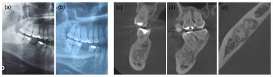
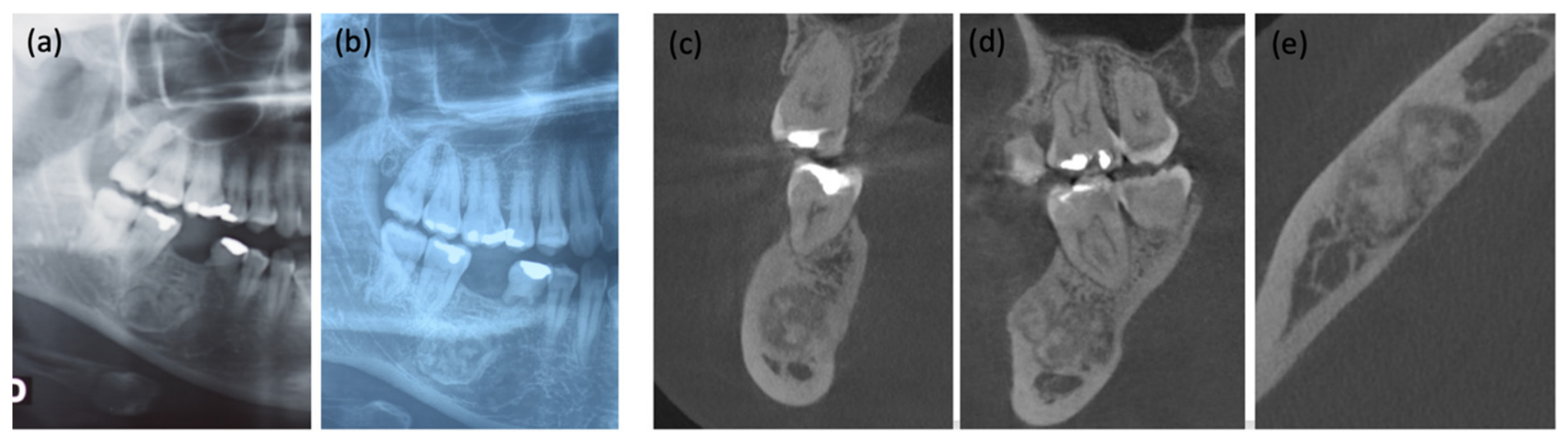
A 47-year-old female patient was referred by her dentist to our Unit of Oral Medicine and Special Care Dentistry, AOUP “P. Giaccone”, Palermo, Italy, in 2023 for the evaluation of a mandibular bone lesion. The patient reported a slow progressive increase in lesion size over the previous years, which was radiographically documented by comparison of X-rays of dental arches obtained in 2018 and 2023 (Figure 1a,b).
Figure 1.
Radiological progression over a 5-year period. (a) Initial orthopantomogram (OPT) taken in 2018. (b) Follow-up OPT taken in 2023. (c–e) Cone Beam Computed Tomography (CBCT) scan taken in 2024.
Her past medical history was significant for breast carcinoma, diagnosed in 2018, for which the patient had undergone surgical resection. At the time of first oral examination, she was receiving Tamoxifen (20 mg daily) and reported no other systemic conditions.
Clinical and radiographic examination revealed persistence of the deciduous tooth 8.5 associated with agenesis of its permanent successor, tooth 4.5. The most recent X-ray of dental arches showed a well-defined radiopaque lesion in the right posterior mandible, corresponding to the region of tooth 4.6, near the inferior alveolar canal.
To further evaluate the lesion, a CBCT scan was performed. CBCT revealed a well-defined, predominantly radiopaque but internally heterogeneous lesion in the right posterior mandible, with irregular margins and a thin peripheral radiolucent rim. The lesion measured approximately 1 × 1.7 × 1 cm. Its mineralized appearance was not completely homogeneous, and the lesion caused an impression on the roof of the mandibular canal, which was displaced caudally (Figure 1c–e).
Following the oncologist’s recommendation, the patient underwent magnetic resonance imaging (MRI) of the maxillofacial region, performed with and without contrast medium. The MRI confirmed the size of the oval lesion and the proximity to the mandibular canal, with mild expansion of both buccal and lingual cortical plates. The lesion appeared hypointense on T1-, T2-, and STIR-weighted sequences, showed no contrast enhancement, and did not display MRI features suggestive of an aggressive process.
Although only benign odontogenic lesions were considered in the differential diagnosis, surgery was planned because of the lesion’s progressive increase in size and its proximity to the inferior alveolar nerve, to establish a definitive diagnosis, to prevent potential neural involvement, and to allow future implant-prosthetic rehabilitation.
A conservative approach was adopted to minimise the risk of neurological complications, due to the close relationship between the lesion and the inferior alveolar nerve.
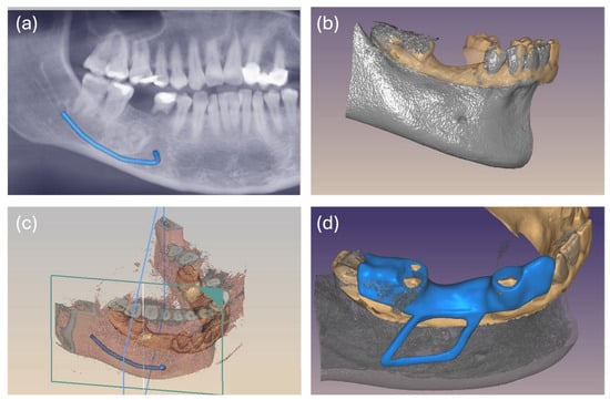
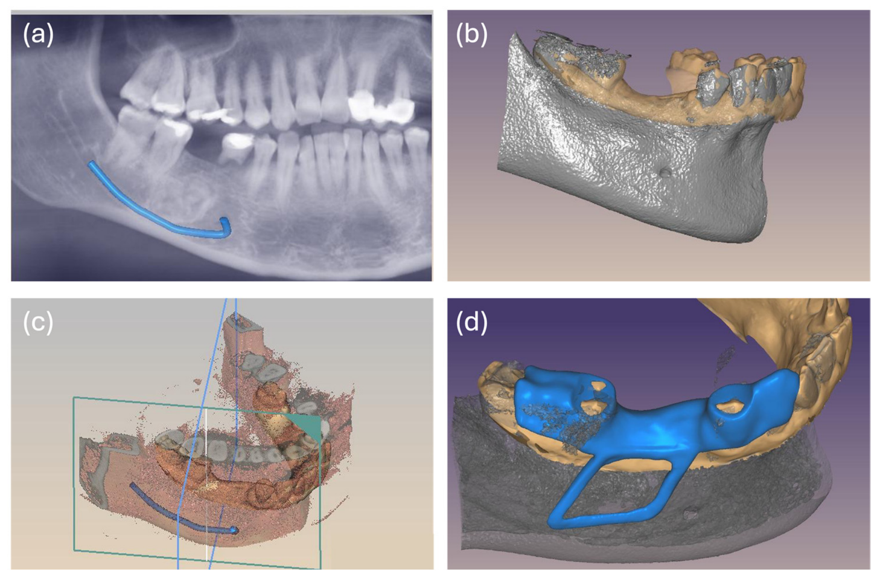
Preoperative digital planning was based on the integration of CBCT and intraoral surface-scan data (Figure 2). The CBCT DICOM dataset was processed to obtain a three-dimensional reconstruction of the mandible and the relevant anatomical structures. Attention was paid to the identification and tracking of the inferior alveolar canal throughout the region of interest to assess its three-dimensional relationship with the lesion. The CBCT-derived model was subsequently registered with the STL dataset obtained from an intraoral scan acquired using a TRIOS 3 scanner (3Shape, Copenhagen, Denmark). Following dataset alignment and verification, virtual surgical planning and patient-specific guide design were performed using exocad software (exocad DentalCAD 3.2 Elefsina, Exocad GmbH, Darmstadt, Germany). According to the surgeon’s indications, a tooth-supported guide was designed incorporating an open surgical window corresponding to the planned cortical access and extending to the inferior aspect of the lesion. The window was intended to transfer the planned location and spatial extent of the osteotomy to the operative field while maintaining direct visualization of the underlying bone and surgical site. Osteotomy depth was not mechanically constrained by the guide.
Figure 2.
Digital workflow for inferior alveolar canal tracing, virtual surgical planning, and design of the patient-specific surgical guide. (a) CBCT-based tracing of the inferior alveolar canal. (b) Registration of the CBCT-derived mandibular model with the intraoral surface scan. (c) Virtual surgical planning showing the three-dimensional relationship among the lesion, the inferior alveolar canal, and the planned surgical access. (d) Final design of the tooth-supported patient-specific surgical guide with an open surgical window corresponding to the planned cortical access.
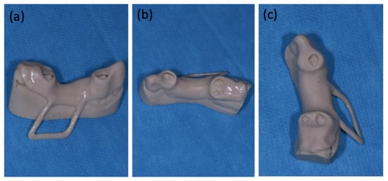
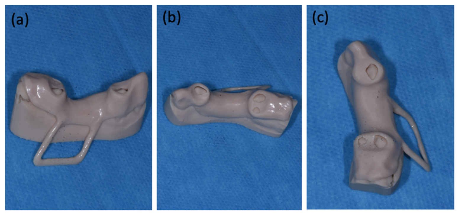
The final guide design was exported as an STL file and additively manufactured using an Asiga 3D printer (Asiga, Sydney, Australia). After printing, the guide underwent support removal, finishing, cleaning, and cold sterilization according to the material protocol (Figure 3). Before surgery, intraoral seating was verified to confirm complete tooth-supported fit, stability without rocking, and correspondence of the surgical window with the planned operative site.
Figure 3.
(a–c) Different views of the final 3D-printed surgical guide.
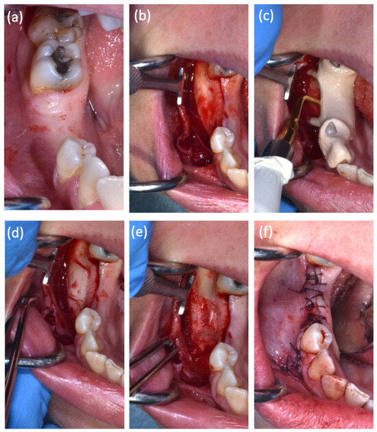
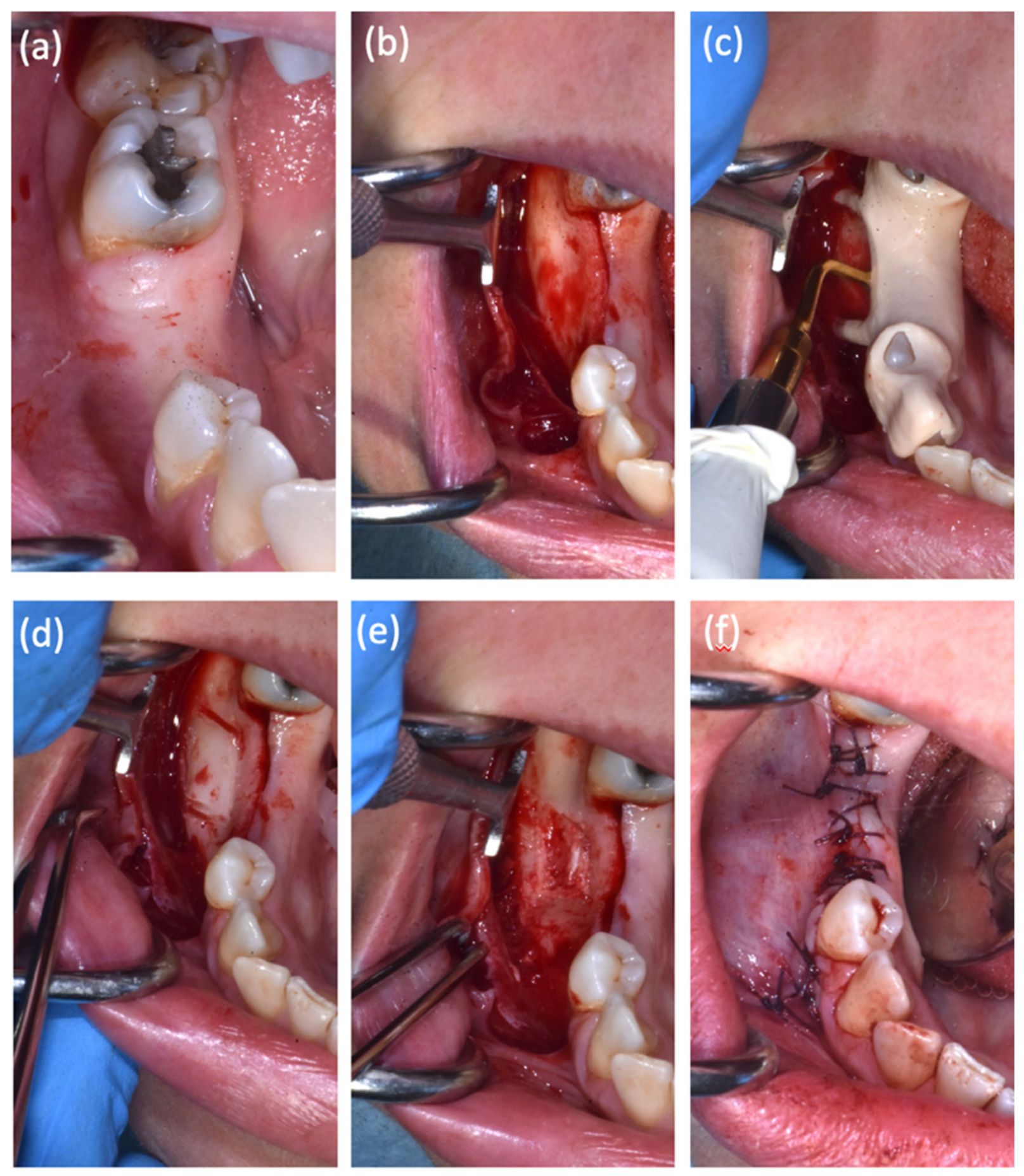
The surgical procedure was performed under local anaesthesia, including inferior alveolar nerve block and buccal infiltration anaesthesia. A full thickness mucoperiosteal flap was raised through a crestal incision associated with mesial and distal vertical releasing incisions, allowing adequate exposure of the underlying mandibular bone (Figure 4a,b).
Figure 4.
Step-by-step surgical management of the lesion. (a) Pre-operative clinical presentation; (b) exposure of the underlying mandibular bone following the elevation of a full thickness mucoperiosteal flap; (c) Patient-specific tooth-supported surgical guide in position, with the open surgical window transferring the planned osteotomy location to the operative field; (d) the resulting conservative cortical bone window obtained using piezoelectric surgery; (e) complete removal of the lesion, with careful preservation of the surrounding anatomical structures; (f) post-operative view showing the surgical site sutured for primary closure.
After flap elevation, the tooth-supported surgical guide was positioned and its complete seating and stability were reconfirmed. The open surgical window identified the planned cortical access and guided the location and trajectory of the piezoelectric osteotomy. Osteotomy depth was controlled intraoperatively through direct visualization and careful advancement of the piezoelectric insert in accordance with the preoperative CBCT-based plan (Figure 4c,d). The lesion was carefully removed, preserving the surrounding anatomical structures, particularly the inferior alveolar nerve. After complete lesion removal, the residual bone defect was left to heal spontaneously without grafting or additional reconstructive procedures, given its limited size and the preservation of the surrounding mandibular bone and contour.
The surgical site was irrigated with sterile saline solution; the flap was repositioned and sutured with interrupted sutures to achieve primary closure (Figure 4e,f).
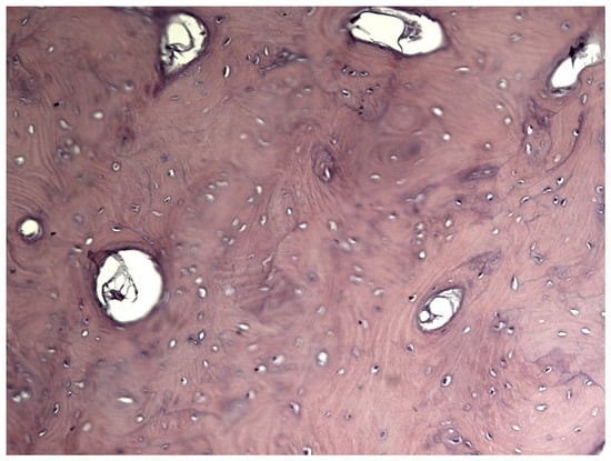
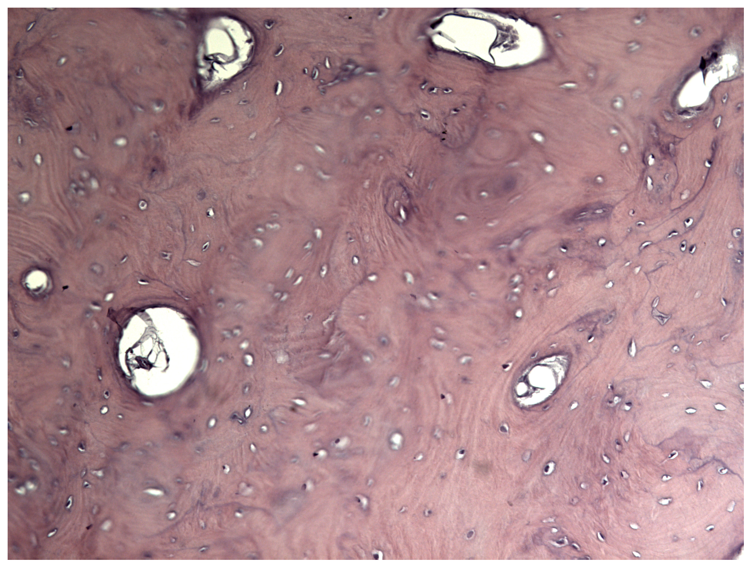
The excised specimen was submitted for histopathological examination, which confirmed the diagnosis of osteoma (Figure 5).
Figure 5.
Histopathological examination of the excisional biopsy showing mature lamellar bone with well-defined osteons and Haversian canals (Hematoxylin and eosin (H&E) staining, Original magnification ×20).
The postoperative course was uneventful; the patient did not complain of any neurosensory disturbances or other complications. A standardized neurosensory assessment was performed by comparing the operated and contralateral sides within the distribution of the mental nerve. Light-touch sensation, pin-prick discrimination, and two-point discrimination were assessed at predefined points of the lower lip and chin, and any area of altered sensation was mapped and recorded during follow-up.
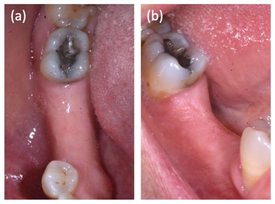
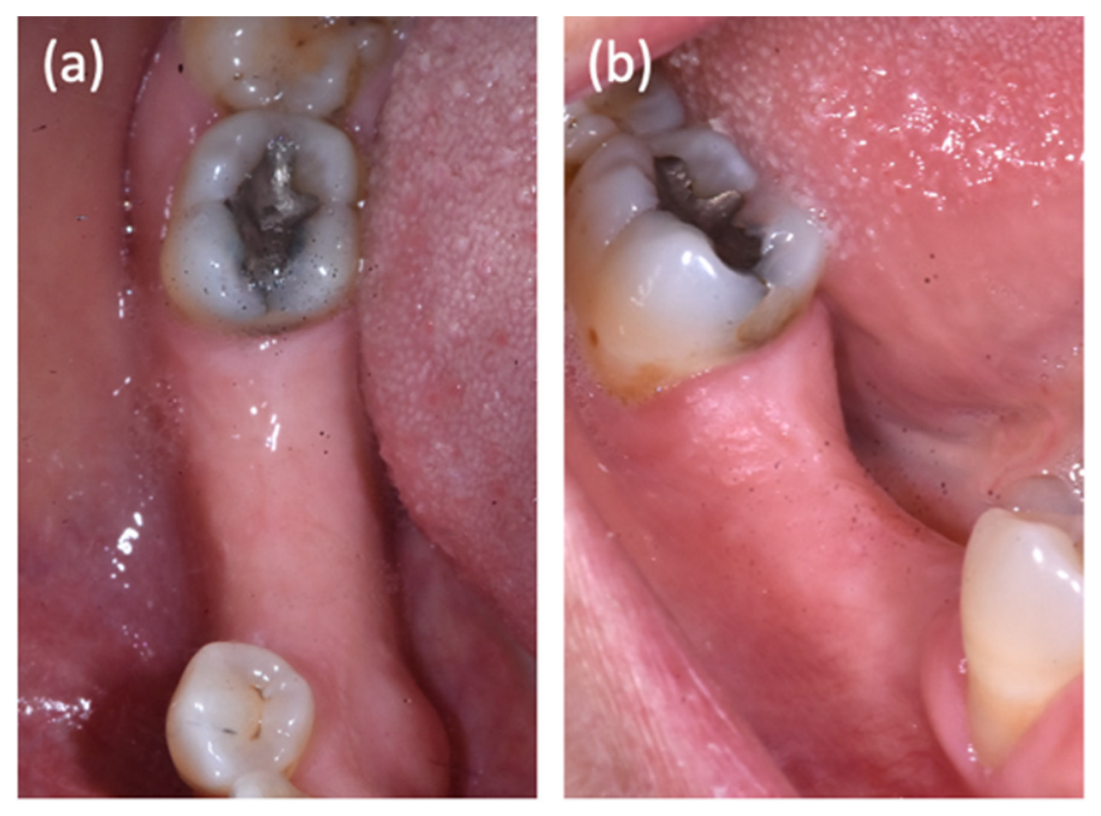
At the latest follow-up at 24 months, complete clinical healing was observed, with no neurosensory impairment. The patient also reported that she had decided not to proceed with implant-prosthetic rehabilitation. Although postoperative radiographic assessment was recommended, the patient declined further imaging because of the absence of postoperative complications and her decision not to pursue implant-prosthetic treatment (Figure 6a,b).
Figure 6.
Follow up two years after the surgery. (a) Occlusal and (b) buccal views of the surgical site.
3. Discussion
Osteomas generally exhibit slow but continuous growth and are often managed conservatively. Surgical excision is reserved for lesions showing progressive enlargement, causing functional or aesthetic impairment, presenting diagnostic uncertainty, or threatening adjacent vital anatomical structures [4,5]. In the present case, progressive growth together with the intimate relationship between the lesion and the inferior alveolar nerve justified surgical management despite the benign appearance of the lesion.
Intraosseous forms are particularly rare and are often asymptomatic, being incidentally detected during radiographic examinations [2,5,6].
In the present case, the radiological appearance was not entirely typical of a densely and homogeneously radiopaque osteoma. The lesion showed heterogeneous mineralization, a peripheral radiolucent rim, and mild cortical expansion, features that broadened the radiological differential diagnosis. In particular, ossifying fibroma was considered because it may present as a well-demarcated expansile lesion with variable internal mineralization and a peripheral radiolucent component. Osteoblastoma was also considered because of its potential heterogeneous mineralized appearance and expansile behavior. However, the slow progression documented over several years, the limited cortical expansion, and the absence of imaging features suggestive of aggressive behavior supported an indolent benign process [3]. As these findings were not pathognomonic, definitive diagnosis required histopathological examination.
When a mandibular osteoma is located adjacent to the inferior alveolar nerve, accurate osteotomy becomes the key determinant of safe surgical management. Precise surgical planning is essential to ensure complete lesion removal while preserving neural integrity and minimizing the risk of postoperative neurosensory complications.
Recent advances in digital technologies have significantly improved the management of complex oral-maxillofacial procedures. The integration of DICOM radiographic data with STL surface scans allows accurate three-dimensional planning and the fabrication of patient-specific 3D-printed surgical guides. However, the application of such guides in the management of intraosseous mandibular lesions remains limited and not yet standardised in the literature [4,7].
Although computer-guided surgery has become a standard approach in implant dentistry and is increasingly adopted in orthognathic and reconstructive surgery, its application for the removal of benign intraosseous mandibular lesions remains limited. Current evidence mainly consists of isolated case reports and small case series, with no standardized surgical approach available.
In the present case, the patient-specific tooth-supported surgical guide served not merely as a positioning aid but as a risk-mitigation tool. The guide allowed accurate transfer of the virtual surgical plan to the operative field, defining the exact location and extension of the cortical osteotomy while minimizing unnecessary bone removal. This was particularly relevant considering the intimate relationship between the osteoma and the inferior alveolar nerve, where even minor deviations could have increased the risk of nerve injury [8,9,10,11].
The guided approach also contributed to maintaining a conservative surgical access, improving intraoperative orientation and reducing the need for repeated verification of the osteotomy site.
Dynamic computer-assisted navigation represents an alternative approach that offers real-time intraoperative guidance and the possibility of modifying the surgical trajectory during the procedure; however, its clinical superiority over static guidance has not yet been established for the management of benign intraosseous mandibular lesions [12,13].
The advantages provided by piezosurgery should therefore be interpreted in conjunction with the guided approach. While piezoelectric inserts selectively cut mineralized tissues, the patient-specific guide ensures that osteotomy is performed exactly where planned, making these two technologies highly complementary when operating close to critical neurovascular structures [14].
The available literature consistently reports several advantages associated with patient-specific surgical guides in the management of benign jaw lesions [2,4,15]. Beyond improving surgical accuracy, 3D-printed guides enhance visualization of complex anatomical relationships, facilitate minimally invasive osteotomies, reduce operative time, decrease the need for additional bone removal, and may reduce postoperative morbidity. Moreover, virtual planning allows the surgeon to anticipate potential intraoperative risks and optimize the surgical strategy before entering the operative field.
Among the few reports describing guided removal of mandibular benign lesions adjacent to the inferior alveolar nerve, the authors employed a CAD/CAM surgical guide to facilitate localization of the lesion and preserve the mandibular canal [4,16,17]. However, unlike our case, their guide was primarily designed to assist lesion localization rather than to define a complete patient-specific osteotomy. Furthermore, the integration of a fully digital workflow with piezosurgery in our case further enhanced surgical control while minimizing the risk of neural injury.
This report also has inherent limitations. As a single case report, the clinical outcomes cannot be generalized, and no comparison with conventional freehand surgery was possible. A further limitation is the absence of postoperative radiographic documentation, as the patient declined additional imaging during follow-up. Moreover, dynamic computer-assisted navigation was not available at our institution at the time of treatment. Although dynamic navigation could have provided real-time intraoperative guidance and greater procedural flexibility, current evidence is insufficient to establish its superiority over static guidance for the surgical management of intraosseous mandibular lesions such as the present case [12,13]. Therefore, although the present case demonstrates the technical feasibility and potential clinical advantages of this approach, prospective comparative studies are required to determine whether computer-guided surgery significantly improves surgical safety, operative time, postoperative neurosensory outcomes, and patient-reported outcomes compared with conventional techniques [4,15,17].
Within the limitations of a single case report, this experience suggests that patient-specific computer-guided surgery should not be considered merely a technological adjunct but rather a valuable strategy to improve surgical safety during the management of mandibular lesions located in proximity to the inferior alveolar nerve. When combined with piezosurgery, this fully digital workflow allows accurate transfer of virtual planning to the surgical field while minimizing unnecessary bone removal and potentially reducing the risk of iatrogenic neural injury [11,18,19,20].
4. Conclusions
This case illustrates the feasibility of combining virtual surgical planning, a patient-specific 3D-printed surgical guide, and piezoelectric surgery for the management of a mandibular lesion located adjacent to the inferior alveolar nerve. In this patient, the digital workflow facilitated accurate transfer of the preoperative plan to the surgical field and supported a conservative osteotomy. Further studies are needed to determine whether this approach provides measurable advantages in terms of surgical safety and neurosensory outcomes.
Author Contributions
Conceptualization, M.E.M., R.M. and G.C.; methodology, V.R. and M.M.; validation, R.M. and G.C.; investigation, M.E.M., M.C., R.S. and R.M.; data curation, M.E.M., M.C., R.S. and R.M.; writing—original draft preparation, M.E.M., M.C., R.S. and R.M.; writing—review and editing, R.M. and G.C.; visualization, R.M. and G.C.; project administration, R.M. All authors have read and agreed to the published version of the manuscript.
Funding
This research received no external funding.
Institutional Review Board Statement
The study was conducted according to the guidelines of the Declaration of Helsinki. Ethical review and approval were waived for this study, since the study is based on common clinical practices.
Informed Consent Statement
Informed consent was obtained from all subjects involved in the study.
Data Availability Statement
Data are contained within the article.
Conflicts of Interest
The authors declare no conflicts of interest.
References
- Molina, T.; Gonzalez, M.W.; Lee, H.; Rivera Virella, B.; Guerrero Rodriguez, L.M.; Sedghizadeh, P.P.; Salib, N. Mandibular Osteomas: Report of Two Cases with Surgical Management. Clin. Case Rep. 2025, 13, e71437. [Google Scholar] [CrossRef] [Scilit]
- Degerliyurt, K.; Akar, V.; Denizci, S.; Yucel, E. Bone lid technique with piezosurgery to preserve inferior alveolar nerve. Oral Surg. Oral Med. Oral Pathol. Oral Radiol. Endodontology 2009, 108, e1–e5. [Google Scholar] [CrossRef] [Scilit] [PubMed]
- Tarsitano, A.; Ricotta, F.; Spinnato, P.; Chiesa, A.M.; Di Carlo, M.; Parmeggiani, A.; Miceli, M.; Facchini, G. Craniofacial osteomas: From diagnosis to therapy. J. Clin. Med. 2021, 10, 5584. [Google Scholar] [CrossRef] [Scilit]
- De Ketele, A.; Meeus, J.; Shaheen, E.; Verstraete, L.; Politis, C. The usefulness of cutting guides for resection or biopsy of mandibular lesions: A technical note and case report. J. Stomatol. Oral Maxillofac. Surg. 2023, 124, 101272. [Google Scholar] [CrossRef] [Scilit] [PubMed]
- Gernandt, S.; Tomasella, O.; Scolozzi, P.; Fenelon, M. Contribution of 3D printing for the surgical management of jaws cysts and benign tumors: A systematic review of the literature. J. Stomatol. Oral Maxillofac. Surg. 2023, 124, 101433. [Google Scholar] [CrossRef] [Scilit]
- Kashwani, R.; Kumari, P.; Sawhney, H.; Singh, M.; Gahlot, J. Solitary benign peripheral osteoma of angle of mandible diagnosed using cone beam computed tomography: A case report. Arch. Dent. Res. 2023, 13, 49–52. [Google Scholar] [CrossRef] [Scilit]
- Wolf-Grotto, I.; Nogueira, L.M.; Milani, B.; Marchiori, E.C. Management of giant osteoma in the mandible associated with minor trauma: A case report. J. Med. Case Rep. 2022, 16, 8. [Google Scholar] [CrossRef] [Scilit] [PubMed]
- Ramadorai, A.; Tay, A.B.G.; Vasanthakumar, G.; Lye, W.K. Nerve Injury After Surgical Excision of Mandibular Third Molars Under Local Anesthesia: An Audit. J. Maxillofac. Oral Surg. 2019, 18, 307–313. [Google Scholar] [CrossRef] [Scilit]
- Omara, M.; Gouda, A.; Ali, S. Computer-guided buccal cortical plate separation for removal of calcified benign odontogenic tumors affecting the mandibular angle region. Maxillofac. Plast. Reconstr. Surg. 2022, 44, 30. [Google Scholar] [CrossRef] [Scilit]
- Hernando, J.; Aguilar-Salvatierra, A.; Leizaola-Cardesa, I.O.; Seijas, P.M.; Aguirre-Echebarria, P.; Martín, J.; Gómez-Moreno, G. Piezosurgery for treatment of large recurrent mandibular osteoma. J. Craniofacial Surg. 2018, 29, e219–e221. [Google Scholar] [CrossRef] [Scilit] [PubMed]
- Delarue, M.; Perez, C.; Lucidarme, Q.; Bornert, F. Management of a solitary bone cyst using a custom-made surgical guide for a minimally invasive approach: Technical note and case report. BMC Oral Health 2024, 24, 560. [Google Scholar] [CrossRef] [Scilit] [PubMed]
- Requena-Gatell, A.; Moya-Martínez, T.; Sánchez-Torres, A.; Valmaseda-Castellón, E.; Figueiredo, R.; Delgado-Molina, E. Dynamic Computer-Assisted Surgery in Oral Surgery: A Systematic Review. J. Clin. Med. 2026, 15, 886. [Google Scholar] [CrossRef] [Scilit]
- Ahmadi, S.; Liang, X.; Xu, F.; Wang, C.; Xie, L.; Yu, M.; Tu, J.; Na, S. Dynamic navigation systems in dento-alveolar surgery: Advancements and clinical applications. Saudi Dent. J. 2025, 37, 60. [Google Scholar] [CrossRef] [Scilit]
- Sheta, M.S.; Ghouraba, R.F.; Ibrahim, M.T. Bone lid technique versus standard technique for treatment of mandibular lesions. BMC Oral Health 2024, 24, 875. [Google Scholar] [CrossRef] [Scilit]
- Rappelli, G.; Franco, R.; Di, G.M.; Franceschini, C.; Rastelli, S.; Capogreco, M.; D’amario, M. The Comparative Efficacy of Burs Versus Piezoelectric Techniques in Third Molar Surgery: A Systematic Review Following the PRISMA Guidelines. Medicina 2024, 60, 2049. [Google Scholar] [CrossRef] [Scilit]
- Manera, C.; Tessari, M.L.; Boccuto, M.; Bacci, C. Clinical Effectiveness of Surgical Marginal Resection with Piezoelectric Device on Bisphosphonate-Related Osteonecrosis of the Jaws: A Retrospective Study. J. Clin. Med. 2025, 14, 3792. [Google Scholar] [CrossRef] [Scilit]
- Werny, J.G.; Frank, K.; Fan, S.; Sagheb, K.; Al-Nawas, B.; Narh, C.T.; Schiegnitz, E. Freehand vs. computer-aided implant surgery: A systematic review and meta-analysis-part 1: Accuracy of planned and placed implant position. Int. J. Implant Dent. 2025, 11, 35. [Google Scholar] [CrossRef] [Scilit]
- Shi, Y.; Wang, J.; Ma, C.; Shen, J.; Dong, X.; Lin, D. A systematic review of the accuracy of digital surgical guides for dental implantation. Int. J. Implant Dent. 2023, 9, 38. [Google Scholar] [CrossRef] [Scilit]
- Sivolella, S.; Brunello, G.; Panda, S.; Schiavon, L.; Khoury, F.; Del Fabbro, M. The Bone Lid Technique in Oral and Maxillofacial Surgery: A Scoping Review. J. Clin. Med. 2022, 11, 3667. [Google Scholar] [CrossRef] [Scilit] [PubMed]
- Block, M.S.; Emery, R.W. Static or Dynamic Navigation for Implant Placement—Choosing the Method of Guidance. J. Oral Maxillofac. Surg. 2016, 74, 269–277. [Google Scholar] [CrossRef] [Scilit]
Disclaimer/Publisher’s Note: The statements, opinions and data contained in all publications are solely those of the individual author(s) and contributor(s) and not of MDPI and/or the editor(s). MDPI and/or the editor(s) disclaim responsibility for any injury to people or property resulting from any ideas, methods, instructions or products referred to in the content. |
© 2026 by the authors. Licensee MDPI, Basel, Switzerland. This article is an open access article distributed under the terms and conditions of the Creative Commons Attribution (CC BY) license.