Next Issue
Volume 5, June
Previous Issue
Volume 4, December


Oral, Volume 5, Issue 1 (March 2025) – 21 articles

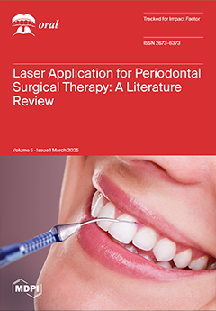
Cover Story (view full-size image): We aim to examine the effectiveness and capabilities of Diode, Er:YAG, CO2, and Nd:YAG lasers in periodontal surgical therapy by analyzing the existing literature that focuses on their laser use alone or as a supplement to the already existing periodontal surgical techniques, comparing it to conventional periodontal surgical therapy. Of the final 18 selected studies, 5 of them show statistically better PD reduction in the laser group. CAL was statistically higher in the laser group in six articles, while one study indicated better PD and CAL results in the control group. Improved GR reduction with a laser was noted in two articles, while one article reported a negative influence from a laser in GR. BOP was significantly better with a laser in one study, while the remaining two studies reported the same results as the control group. View this paper
  • Issues are regarded as officially published after their release is announced to the table of contents alert mailing list.
  • You may sign up for e-mail alerts to receive table of contents of newly released issues.
  • PDF is the official format for papers published in both, html and pdf forms. To view the papers in pdf format, click on the "PDF Full-text" link, and use the free Adobe Reader to open them.
Order results
Result details
Select all
Export citation of selected articles as:
24 pages, 1955 KiB  
Review
The Role of Tissue Engineering in Orthodontic and Orthognathic Treatment: A Narrative Review
by Rosana Farjaminejad, Samira Farjaminejad, Melika Hasani, Franklin Garcia-Godoy, Babak Sayahpour, Anand Marya and Abdolreza Jamilian
Oral 2025, 5(1), 21; https://doi.org/10.3390/oral5010021 - 20 Mar 2025
Viewed by 567
Abstract
Background: Orthodontics and orthognathic surgery present challenges such as extended treatment durations, patient discomfort, and complications like root resorption. Recent advancements in tissue engineering and nanotechnology offer promising solutions by improving bone regeneration, periodontal repair, and biomaterial integration. Objectives: This review explores the [...] Read more.
Background: Orthodontics and orthognathic surgery present challenges such as extended treatment durations, patient discomfort, and complications like root resorption. Recent advancements in tissue engineering and nanotechnology offer promising solutions by improving bone regeneration, periodontal repair, and biomaterial integration. Objectives: This review explores the integration of scaffold-based tissue engineering and nanotechnology in orthodontics, focusing on their roles in accelerating bone regeneration, reducing treatment times, and minimizing adverse effects to enhance the predictability and success of orthodontic interventions. Methods: Relevant literature was selected from PubMed, Scopus, and Web of Science, focusing on studies related to scaffold technology, biomaterials, and nanotechnology in orthodontics. Keywords included “tissue engineering”, “orthodontics”, “biomaterials”, “scaffolds”, “nanotechnology”, and “bone regeneration”. Priority was given to peer-reviewed original studies, systematic reviews, and meta-analyses addressing innovative treatment approaches and clinical outcomes. Results: Findings indicate that scaffolds enhance bone regeneration and periodontal repair, while nanoparticles improve biomaterial integration and drug delivery efficiency. These advancements contribute to faster, more predictable orthodontic treatments with reduced complications. However, challenges such as high costs, regulatory hurdles, and the need for long-term clinical validation remain barriers to widespread adoption. Conclusions: Tissue engineering and nanotechnology offer minimally invasive, biologically driven solutions for orthodontic treatment. While significant progress has been made, further clinical studies, cost-effective strategies, and regulatory approvals are needed to integrate these innovations into routine practice. Full article
Show Figures

Figure 1

9 pages, 2601 KiB  
Case Report
A Technique to Integrate a Simultaneous Tooth- and Bone-Supported Surgical Guide for Lateral Sinus Lift and Precision Corticotomy
by Andrew B. Cameron, Ranu Acharya, Lavanya Ajay Sharma, Tyng-Tyng Lee, Peng Shao and Ajay Sharma
Oral 2025, 5(1), 20; https://doi.org/10.3390/oral5010020 - 14 Mar 2025
Viewed by 409
Abstract
Background/Objectives: Surgical guides have been used in a variety of dental procedures, such as implant placement to improve clinical accuracy and reduce post-operative complications. This report presents a novel and versatile workflow for the design and fabrication of a “multi-purpose” fully-guided tooth- and [...] Read more.
Background/Objectives: Surgical guides have been used in a variety of dental procedures, such as implant placement to improve clinical accuracy and reduce post-operative complications. This report presents a novel and versatile workflow for the design and fabrication of a “multi-purpose” fully-guided tooth- and bone-supported one-piece surgical guide. Methods: Briefly, intraoral and perioral anatomical features were captured by an intraoral scan and a cone-beam computed tomography scan. The data were segmented and aligned with analysis software to enable the digital design of surgical guides. The versatility of this method was demonstrated through its application in the two cases presented: the first involved a lateral sinus lift with simultaneous implant placement, and the second involved the removal of a foreign object from the alveolar bone prior to implant placement. Results: Positive clinical outcomes were confirmed at follow-up visits for up to 12 months. Conclusions: This method may be applied to a range of challenging clinical scenarios, such as apicectomy, the extraction of supernumerary or unerupted teeth, corticotomy to facilitate orthodontic movement, the precise reduction of bony spurs or exostoses, and the conservative surgical removal of pathologies. Full article
Show Figures

Figure 1

12 pages, 2129 KiB  
Article
Comparative Analysis of Gene Expression in Periodontal Ligament Stem Cells Exposed to Biodentine and Bio-C Repair: Implications for Cementogenesis—An In Vitro Study
by Mahmoud M. Bakr, Mahmoud Al Ankily, Mohammed Meer and Mohamed Shamel
Oral 2025, 5(1), 19; https://doi.org/10.3390/oral5010019 - 13 Mar 2025
Viewed by 533
Abstract
Background/Objectives: Bioactive materials are gaining increased popularity as materials of choice for pulpal regeneration. A similar trend is emerging with root repair materials; however, there is a significant gap in the literature about cementogenic ability of bioceramic repair materials on the periodontal [...] Read more.
Background/Objectives: Bioactive materials are gaining increased popularity as materials of choice for pulpal regeneration. A similar trend is emerging with root repair materials; however, there is a significant gap in the literature about cementogenic ability of bioceramic repair materials on the periodontal ligament cells. The aim of the present study was to investigate the effect of bioceramic materials (Biodentine and Bio-C Repair) on the cementogenesis potential of the periodontal ligament stem cells (PDLSCs). Methods: PDLSCs were isolated using the enzymatic digestion approach from sound extracted teeth. Material extracts were prepared on rubber discs and immersed in fresh growth medium for 24 h at 37 °C. Reverse transcription–quantitative polymerase chain reaction (RT-qPCR) was used to detect the mRNA expression levels of cementogenic markers cementum protein 1 (CEMP1), Cementum attachment protein (CAP), pathway markers transforming growth factor β1(TGF-β1), bone morphogenic protein 2 (BMP2), and inflammatory marker IL-6. Results: Both materials (Biodentine and Bio-C Repair) showed significantly higher gene expressions when compared to the control groups. The gene expression with Bio-C Repair significantly increased when compared with Biodentine, except for TGF-β1 expression, where both materials exhibited similar results. Conclusions: Bio-C Repair demonstrated increased gene expression of cementogenic markers compared to Biodentine under the tested conditions. Further in vivo studies are deemed necessary to translate the findings from this study into clinical practice. Full article
(This article belongs to the Special Issue Advanced Dental Materials for Oral Rehabilitation)
Show Figures

Graphical abstract

17 pages, 1767 KiB  
Article
Internet of Things and Smart Technologies in Oral Health: Trends, Impacts, and Challenges
by Susana J. Calderon, Stephen Mujeye Sr and Melissa I. Calvillo
Oral 2025, 5(1), 18; https://doi.org/10.3390/oral5010018 - 12 Mar 2025
Viewed by 760
Abstract
Objective: This study aims to discover the most current trends, impacts, and challenges of using IoT devices and smart technologies in oral health. Method: A modified systematic mapping method was used to generate and answer five research questions. Twelve databases were queried to [...] Read more.
Objective: This study aims to discover the most current trends, impacts, and challenges of using IoT devices and smart technologies in oral health. Method: A modified systematic mapping method was used to generate and answer five research questions. Twelve databases were queried to identify published literature from 2017 to 2023. Abstract screening and full-text review were conducted to identify studies meeting inclusion criteria. The Pandas library in Python Version 3.9.19 and a Fibonacci series were used to identify keyword trends in abstracts. Full-text analysis was conducted to synthesize findings relevant to the impacts and challenges of IoDT. Results: A total of 958 unduplicated articles were identified from the literature databases. After review, 33 articles were included. Publications related to IoDT are rapidly increasing over the last 7 years and keywords relating to toothbrushing were the most common. The most common research strategy was design and creation, followed by experimental methods. Design and creation of smart technologies in oral health are in a phase of measurement optimization using IoT which is being used for prevention, early detection, monitoring, and treatment of dental disease as well as silent communication devices. Challenges in IoDT continue to include measurement accuracy and user acceptability. Conclusions: Research in IoDT is predicted to continue to advance rapidly. Dental providers and public health agencies can look to this research to develop best practices. However, more research on how IoDT can facilitate desirable outcomes in a cost-effective and user-friendly way is needed. Full article
Show Figures

Figure 1

18 pages, 2077 KiB  
Review
Odontogenic Infections Resistant to Empiric Therapy, Opportunistic Prevotella, and Metronidazole: A Clinical Case Series and Review of the Literature
by Ned Leonard Nix, Nicholas T. Zusman and Nejat Düzgüneş
Oral 2025, 5(1), 17; https://doi.org/10.3390/oral5010017 - 11 Mar 2025
Viewed by 580
Abstract
Background/Objectives: Treating odontogenic infections of the head, neck, and oral cavity requires surgical and pharmacotherapeutic interventions of single and multi-space infections. Prevotella species can become virulent during post-extraction wound healing at an extraction site, under conditions of excessive plaque, nearby gingivitis, or periodontitis, [...] Read more.
Background/Objectives: Treating odontogenic infections of the head, neck, and oral cavity requires surgical and pharmacotherapeutic interventions of single and multi-space infections. Prevotella species can become virulent during post-extraction wound healing at an extraction site, under conditions of excessive plaque, nearby gingivitis, or periodontitis, and in immunocompromised patients. The aims of this narrative review are to describe the pathogenesis of Prevotella species, particularly those resistant to surgery and empiric antibiotic therapy, and to emphasize the need for the identification of specific antibiotic-resistant Prevotella species. Methods: Three clinical cases from the lead author’s private practice were described, in which Prevotella species were significant opportunistic pathogens in odontogenic infections. The PubMed database was searched for “Prevotella and Odontogenic Infection and (or Amoxicillin or Clindamycin or Metronidazole or Flagyl®)” from 1996 to 2024. Thirty additional references were used to develop the section on Prevotella pathogenesis. Results: Metronidazole administration was deemed necessary in the cases described here. This intervention mitigated or resolved these odontogenic infections that were refractory to conservative surgical therapy that included source control. Prevotella buccae was found to be prevalent in odontogenic infections and pericoronitis, and to be resistant to commonly used antibiotics. Prevotella species were highly susceptible to Metronidazole (Flagyl®). The antibiotic was effective in mitigating the disease when basic principles of the treatment of odontogenic infections were employed. Conclusions: The clinical cases and the review of the literature indicate that Metronidazole is strongly indicated for the treatment of odontogenic opportunistic infections with Prevotella species. Full article
(This article belongs to the Special Issue Current Issues in Oral Health)
Show Figures

Figure 1

20 pages, 1013 KiB  
Systematic Review
Port-Wine Stains’ Orodental Manifestations and Complications: A Systematic Review
by Andreea Kui, Marius Negucioiu, Smaranda Buduru, Ana-Maria Condor, Daria Chira, Daniela Cornelia Condor and Ondine Patricia Lucaciu
Oral 2025, 5(1), 16; https://doi.org/10.3390/oral5010016 - 7 Mar 2025
Viewed by 739
Abstract
Background/Objectives: Port-wine stains (PWSs), also known as naevus flammeus or capillary malformations, are congenital cutaneous lesions, typically located in the head/neck area. The manifestations of PWSs include aesthetic and functional impairments due to soft and hard tissue modifications. Sturge–Weber syndrome is characterised by [...] Read more.
Background/Objectives: Port-wine stains (PWSs), also known as naevus flammeus or capillary malformations, are congenital cutaneous lesions, typically located in the head/neck area. The manifestations of PWSs include aesthetic and functional impairments due to soft and hard tissue modifications. Sturge–Weber syndrome is characterised by additional neuro-ocular manifestations. This systematic review aimed to identify and overview the orodental manifestations of PWSs. Methods: Several databases (PubMed, Web of Science, Scopus, Embase, Cochrane Library) were searched using keywords for PWSs and oral and dental manifestations. The results were centralised, deduplicated, and selected in a two-step sequence. Data were extracted using pre-defined extraction forms and represented graphically and tabulated. The Newcastle–Ottawa and Joanna Briggs Institute scales were used for quality assessment. Results: Out of 884 results, 43 studies were selected for inclusion. Eleven studies investigated patients diagnosed with PWSs, while thirty-two studies investigated SWS patients. Regarding study designs, eight studies were retrospective, one was cross-sectional, two were case series, and thirty-two were case reports. The most frequently mentioned manifestations were gingival hypertrophy, lip hypertrophy, mucosal or gingival staining, malocclusion, gingivitis or gingival bleeding, and facial asymmetry. Conclusions: Due to the rarity of this pathology, the orodental manifestations of PWSs are not widely known to dentists. Soft tissue hypertrophy, malocclusion, and bony hypertrophy are significant concerns that need to be addressed during treatment. Full article
Show Figures

Figure 1

15 pages, 5156 KiB  
Article
Finite Element Analysis of Stress Distribution in Monolithic High-Translucency Zirconia Dental Prostheses
by Fernando Araújo da Costa Ward, Luciano Pessanha Moreira, Pedro Araújo da Costa Ward, Paula Cipriano da Silva, Manuel Fellipe Rodrigues Pais Alves and Claudinei Santos
Oral 2025, 5(1), 15; https://doi.org/10.3390/oral5010015 - 3 Mar 2025
Viewed by 491
Abstract
Background/Objectives: High-translucency zirconia is a dental ceramic offering excellent aesthetic results but with mechanical limitations restricting its applications. This study aimed to simulate the mechanical behavior of anatomical dental prostheses made from high-translucency zirconia using the finite element method (FEM) to assess [...] Read more.
Background/Objectives: High-translucency zirconia is a dental ceramic offering excellent aesthetic results but with mechanical limitations restricting its applications. This study aimed to simulate the mechanical behavior of anatomical dental prostheses made from high-translucency zirconia using the finite element method (FEM) to assess the material’s reliability. Methods: Samples of high-translucency zirconia were compacted, sintered, and characterized for relative density. Structural and microstructural analyses were performed using X-ray diffraction (XRD) and scanning electron microscopy (SEM). Its mechanical properties, including hardness, fracture toughness, and flexural strength, were measured. Additionally, elastic parameters such as Young’s modulus and Poisson’s ratio were determined using the impulse excitation technique and subsequently employed in numerical simulations under various masticatory loads (50 to 500 N). These simulations modeled an anatomical molar (tooth 26) using the HyperMesh and ABAQUS codes, applying loads from three directions: vertical, angular (45°), and horizontal, at different points on the prosthesis. Results: The sintered zirconia ceramics exhibited excellent densification and a microstructure composed of cubic and tetragonal grains (c-ZrO2 and t-ZrO2). The measured properties included a hardness of 1315 ± 48 HV, fracture toughness of 3.7 ± 0.2 MPam1/2, and flexural strength of 434 ± 67 MPa. Elastic parameters were determined as a Young’s modulus of 192.2 ± 4.8 GPa and a Poisson’s ratio of 0.31. Numerical simulations demonstrated that vertically applied loads of 500 N resulted in a maximum stress of approximately 299.2 MPa, horizontal stress reached 320.8 MPa at a 200 N load, and angular stress peaked at 447.3 MPa under a 350 N load. These findings indicate that the material can safely withstand these conditions without failure. Conclusions: Within the limits of this investigation, the methodology proved to be an effective tool for predicting the mechanical behavior of new dental ceramics. For high-translucency zirconia, the material demonstrated high reliability under masticatory vertical loads up to 500 N, angular loads up to 350 N, and horizontal loads up to 200 N. Full article
Show Figures

Figure 1

14 pages, 6864 KiB  
Case Report
Orthodontic and Surgical Treatment of Delayed Eruption of Mandibular Central Incisors and Canines: A Case Report
by Ioanna Pouliezou, Eirini Vasoglou, Zoi Papafilippopoulou and Michail Vasoglou
Oral 2025, 5(1), 14; https://doi.org/10.3390/oral5010014 - 3 Mar 2025
Viewed by 574
Abstract
Background/Objectives: Delayed tooth eruption (DTE) is a very challenging clinical situation, and the pathogenesis has been associated with local, systemic, and genetic factors. The aim of this presentation is to describe the management of such a case with delayed eruption of mandibular central [...] Read more.
Background/Objectives: Delayed tooth eruption (DTE) is a very challenging clinical situation, and the pathogenesis has been associated with local, systemic, and genetic factors. The aim of this presentation is to describe the management of such a case with delayed eruption of mandibular central incisors and canines. Methods: An 11-year-old female patient presented with DTE involving lower incisors (32, 42) and canines, seeking orthodontic treatment. Furthermore, lower permanent central incisors were congenitally missing while lower deciduous incisors were still in place. After a year-and-a-half of just monitoring the case and having suggested the extraction of deciduous first molars and canines, orthodontic treatment started with fixed appliances in the upper and lower jaw. First and second premolars gradually erupted after the precursor’s extraction. Lower canines and the two existing incisors were, after several months, surgically exposed and a gold chain apparatus was bonded on each one, while the closed eruption technique was chosen. A 0.17 × 0.25 stainless steel (SS) lingual wire, leaning on the buccal tubes of the lower molar bands and properly manipulated around them, was fitted in conjunction with the labial brackets and wires in order to facilitate the traction of the impacted teeth, which was carried out for one after the other using either an elastic chain or a second super-elastic wire. Results: All impacted teeth were finally restored to their proper place. The root of 71 did not present resorption, so the tooth was maintained in the lower arch while inter-proximal reduction was performed in the upper arch in the anterior region, in order to anticipate the lower missing incisor. Finally, a stable occlusion with proper contacts was achieved. Conclusions: Proper diagnosis, on-time extractions of deciduous teeth, and a well-designed treatment plan regarding space management and orthodontic traction of impacted teeth contributed to successful treatment outcomes in this DTE case. Full article
Show Figures

Figure 1

11 pages, 491 KiB  
Review
Adjunctive Effects of Diode Laser in Surgical Periodontal Therapy: A Narrative Review of the Literature
by Giuseppe D’Albis, Marta Forte, Maria Chiara Fioriello, Alexandra Artin, Anna Montaruli, Adriano Di Grigoli, Rada Kazakova, Mariya Dimitrova and Saverio Capodiferro
Oral 2025, 5(1), 13; https://doi.org/10.3390/oral5010013 - 27 Feb 2025
Viewed by 697
Abstract
The surgical procedures in the treatment of periodontitis generally aim to reduce pocket depth, improve teeth maintenance, and enhance long-term stability. Several approaches have been proposed over the years including different techniques, drugs, and medical devices, with the main purpose to minimalize the [...] Read more.
The surgical procedures in the treatment of periodontitis generally aim to reduce pocket depth, improve teeth maintenance, and enhance long-term stability. Several approaches have been proposed over the years including different techniques, drugs, and medical devices, with the main purpose to minimalize the surgical procedure and improve both the outcomes and patient compliance. Among all the medical devices proposed in surgical and non-surgical periodontology, different laser wavelengths (e.g., Erbium: YAG, Erbium: CR, KTP, Diode) have been studied worldwide but generally with inconsistent results. Nowadays, the diode laser is one of the most used lasers among general dentists, also promoted as an adjunctive therapy in periodontology, mainly in non-surgical procedures. This study aims to review data emerging from the literature on the use of diode lasers and adjunctive therapy to the conventional periodontal surgery. This research was conducted using PubMed and Scopus search engines with a combination of relevant keywords, including “Surgical Periodontal Therapy”, “Periodontal Surgery”, “Periodontal Regenerative Surgery”, and “Surgical Treatment for Periodontal Disease” in combination with “Diode Laser”, “Diode-Driven Laser”, and “Diode Emission Laser”. Selected articles were carefully reviewed to identify studies reporting data on the effectiveness of diode lasers in periodontal surgery. Results of the current review suggest potential clinical benefits of diode laser-assisted periodontal surgery, as studies reported improvements in key parameters such as clinical attachment level (CAL), bleeding on probing (BOP), and plaque index (PI) postoperatively. Additionally, VAS scores, indicative of post-surgical discomfort, were lower for laser-treated sites, and a short-term reduction in periodontal pathogens was observed. However, the clinical outcomes significantly differ across the studies, and the evidence does not uniformly support a superior effectiveness of diode lasers as an adjunctive tool in surgical periodontology. While the data indicate that diode laser use may contribute to enhanced periodontal health and possibly accelerate healing in some cases, these findings should be interpreted cautiously, as further research, possibly multicentric and in large numbers but mandatory with well-defined protocols (stage of periodontitis, preliminary non-surgical procedures and results, laser wavelength and protocol of use, post-operative maintenance, follow-up clinical and radiological criteria) are surely needed to possibly validate the observations emerging from the current review and eventually to standardize clinical protocols in the future. Such limitations have been well addressed in this paper and are clearly discussed and essentially related to the focus on the total uncertainty of the literature and general caution. Full article
(This article belongs to the Special Issue Lasers in Oral Sciences)
Show Figures

Figure 1

15 pages, 649 KiB  
Review
The Integration of Salivary pH Meters and Artificial Intelligence in the Early Diagnosis and Management of Dental Caries in Pediatric Dentistry: A Scoping Review
by Eliza Denisa Sgiea, Corina Marilena Cristache, Tamara Mihut, Sergiu Drafta and Irina Adriana Beuran
Oral 2025, 5(1), 12; https://doi.org/10.3390/oral5010012 - 10 Feb 2025
Viewed by 1330
Abstract
Dental caries is one of the most prevalent chronic conditions among children globally. Salivary pH monitoring, an essential diagnostic parameter, plays a critical role in understanding caries risk and oral health. This scoping review aims to evaluate the application of digital salivary pH [...] Read more.
Dental caries is one of the most prevalent chronic conditions among children globally. Salivary pH monitoring, an essential diagnostic parameter, plays a critical role in understanding caries risk and oral health. This scoping review aims to evaluate the application of digital salivary pH meters in pediatric dentistry, particularly in caries diagnosis and prevention, while exploring the potential integration of artificial intelligence (AI) in this domain. Methods: A literature search was conducted across the PubMed, Web of Science, and Scopus databases for studies published between 2014 and 2024. The inclusion criteria focused on clinical studies involving children aged 1 to 18 years and the use of digital salivary pH meters. Studies that utilized AI in conjunction with salivary pH monitoring were also reviewed. Data were extracted and analyzed to assess the effectiveness of pH meters in caries detection and their role in broader oral health applications. Results: Out of 549 articles screened, 11 met the inclusion criteria. The review highlighted the utility of digital pH meters for assessing caries risk, monitoring dietary impacts, and evaluating the effectiveness of preventive treatments. However, none of the studies combined salivary pH monitoring with AI. Emerging technologies, such as smartphone-based pH sensors, have demonstrated promising applications for real-time, non-invasive diagnostics. Conclusions: Digital salivary pH meters provide precise and reproducible measurements, significantly enhancing caries risk assessment and preventive strategies in pediatric dentistry. While AI integration remains unexplored in this context, its potential to refine risk prediction models and personalize treatments underscores the need for future research in this area. These advancements could improve caries prevention and management, enhancing pediatric oral health outcomes. Full article
(This article belongs to the Special Issue Artificial Intelligence in Oral Medicine: Advancements and Challenges)
Show Figures

Figure 1

20 pages, 565 KiB  
Review
Laser Application for Periodontal Surgical Therapy: A Literature Review
by Stefanos Zisis, Vasileios Zisis and Andreas Braun
Oral 2025, 5(1), 11; https://doi.org/10.3390/oral5010011 - 8 Feb 2025
Viewed by 1133
Abstract
Objectives: The aim of this article is to examine the effectiveness and capabilities of laser use in periodontal surgical therapy by analyzing the existing literature that focuses on laser use alone or as a supplement to the already existing periodontal surgical techniques, comparing [...] Read more.
Objectives: The aim of this article is to examine the effectiveness and capabilities of laser use in periodontal surgical therapy by analyzing the existing literature that focuses on laser use alone or as a supplement to the already existing periodontal surgical techniques, comparing it to conventional periodontal surgical therapy, with the intent to reach a better understanding of the efficiency and therapeutic potential of lasers in periodontal surgery. Methods: An electronic search of the PubMed, Embase, and Cochrane databases was performed between October 2023 and December 2023 to identify all of the articles published in the last 15 years and investigate information about the application of Diode, Erbium:yttrium–aluminum–garnet (Er:YAG), Carbon Dioxide (CO2), and Neodymium yttrium–aluminum–garnet (Nd:YAG) lasers to surgical periodontal therapy in human trials. Results: The database search yielded 18 studies. All of the databases showed a clinical improvement in pocket depth (PD), clinical attachment level (CAL), gingival recession (GR), and bleeding on probing (BOP) in both the test and control groups, with results from five articles showing statistically better PD reduction in the laser group compared to the control group. CAL gain was statistically higher in the laser group in six articles, while one study indicated better PD and CAL results in the control group. Improved GR reduction with a laser was noted in two articles, while one article reported a negative influence from a laser in GR. BOP was significantly better with laser in one study, while the remaining two studies reported the same results as the control group. Conclusions: Laser application in resective surgery exhibits additional benefits to the already established techniques, while in regenerative surgery, more investigation is needed. Diode laser use in periodontal surgery is already widespread and shows clinical efficacy, while low-level laser therapy (LLLT) has an exceptional potential for a variety of applications, promoting better wound healing and less post-surgical complications. However, more trials and studies are needed to further examine the maximum efficiency of each laser type in periodontal surgical therapy. Full article
(This article belongs to the Special Issue Lasers in Oral Sciences)
Show Figures

Figure 1

12 pages, 530 KiB  
Article
Assessing the Oral-Health-Related Quality of Life in Patients with Dental Prosthetics: A Cross-Sectional Study from Eastern Croatia
by Ingrid Kovačević, Ivana Barać, Katarina Major Poljak, Slavko Čandrlić and Marija Čandrlić
Oral 2025, 5(1), 10; https://doi.org/10.3390/oral5010010 - 7 Feb 2025
Viewed by 1017
Abstract
Objectives: This cross-sectional study aimed to evaluate the oral-health-related quality of life (OHRQoL) among patients with fixed, removable, and combined dental prosthetic restorations in Osijek, Croatia, focusing on potential variations by gender, age, and prosthetic type. Methods: This study included 313 patients treated [...] Read more.
Objectives: This cross-sectional study aimed to evaluate the oral-health-related quality of life (OHRQoL) among patients with fixed, removable, and combined dental prosthetic restorations in Osijek, Croatia, focusing on potential variations by gender, age, and prosthetic type. Methods: This study included 313 patients treated at dental clinics within the Osijek-Baranja County Health Center. The standardized Oral Health Impact Profile-14 (OHIP-14) questionnaire, validated in Croatian (OHIP-CRO14), was used to assess the participants’ OHRQoL. Statistical analyses were conducted to explore associations between demographic factors, prosthetic types, and OHRQoL domains. Results: Psychological discomfort emerged as the domain with the lowest reported OHRQoL, whereas social disability was the least affected. Gender showed no significant association with OHRQoL outcomes. However, age influenced OHRQoL scores. Patients with fixed prosthetic restorations reported better OHRQoL in functional limitation, physical pain, and physical disability compared to those with removable or combined prosthetics. Conclusions: Patients with fixed prosthetic restorations reported better OHRQoL, particularly in functional limitation, physical pain, and physical disability domains. While no significant gender differences were observed, age significantly influenced physical disability scores, with younger participants reporting lower scores and older participants higher scores. These results emphasize the importance of prosthetic type in improving OHRQoL and guiding clinical decision-making in dental practice. Full article
Show Figures

Figure 1

21 pages, 294 KiB  
Article
Protocols and Technologies Used by Italian Dental Professionals to Maintain Good Oral Health in Orthodontic Patients Before, During and After Treatment: A Survey Study
by Silvia Sabatini, Matteo Castaldi, Adriana Assunta De Stefano, Gabriella Galluccio, Roberta Grassi and Gianna Maria Nardi
Oral 2025, 5(1), 9; https://doi.org/10.3390/oral5010009 - 6 Feb 2025
Viewed by 799
Abstract
Background: Orthodontic appliances increase biofilm accumulation by expanding plaque retention sites. Enamel demineralization and periodontal inflammation are considered to be the most prevalent consequences of biofilm formation in orthodontic patients, with reported prevalence rates of up to 50%. To date, there are different [...] Read more.
Background: Orthodontic appliances increase biofilm accumulation by expanding plaque retention sites. Enamel demineralization and periodontal inflammation are considered to be the most prevalent consequences of biofilm formation in orthodontic patients, with reported prevalence rates of up to 50%. To date, there are different procedures and indications that have been used for the treatment of these conditions. Therefore, professionals may use different technologies and protocols to control bacterial biofilm. The aim of this study was to investigate the protocols and technologies used by Italian dental professionals to maintain good oral health in orthodontic patients before, during and after treatment. Methods: A total of 155 dental professionals, dentists, dental hygienist students and dental hygienists, attending the Italian Academy of Advanced Technologies in Oral Hygiene Sciences (A.T.A.S.I.O.) congress, completed a questionnaire to identify prevention technologies and protocols before, during and after orthodontic treatment. Results: The results show equivalent responses between dentists and dental hygienists, with most dental hygienists more likely to personalize treatment. Remineralization technologies and protocols are selected based on patient characteristics. During professional oral hygiene sessions, the most commonly used technologies before, during and after therapy are ultrasonic scaler and airflow with powders. Mouthwashes and toothpastes are customized and shared with the patient, with remineralizing technologies predominant in toothpastes. After treatment, aesthetics is evaluated and the whitening protocol is selected based on the patient’s characteristics. Conclusions: There is not a specific prevention program associated with each orthodontic therapy in the literature, so it is necessary to tailor the treatment to best manage the risks of orthodontic therapy and maintain healthy tissues. Full article
22 pages, 1142 KiB  
Article
A Mixed-Methods Study of the Challenges of Nurses in a Sri Lankan National Hospital in the Provision of Clinical Oral Hygiene Care
by Sherine Loyidcy Mariyanayagam, Sachith Piyumal Abeysundara, Kehinde Kazeem Kanmodi, Ruwan Duminda Jayasinghe and Rasika Manori Jayasinghe
Oral 2025, 5(1), 8; https://doi.org/10.3390/oral5010008 - 5 Feb 2025
Viewed by 1180
Abstract
Background: There are multiple challenges associated with the provision of quality oral hygiene care by nurses. This study aimed to identify the challenges experienced by nurses in the provision of oral hygiene care at the National Hospital, Kandy, Sri Lanka. Methods: This mixed-method [...] Read more.
Background: There are multiple challenges associated with the provision of quality oral hygiene care by nurses. This study aimed to identify the challenges experienced by nurses in the provision of oral hygiene care at the National Hospital, Kandy, Sri Lanka. Methods: This mixed-method study was conducted among nurses at the National Hospital, Kandy, Sri Lanka, in medical, surgical and intensive care units. Self-administered questionnaires and an interviewer-administered question guide were used to collect quantitative and qualitative data from 228 and 30 participants, respectively. The collected data were analyzed using the SPSS version 26.0 software (quantitative analysis) and Braun and Clarke’s thematic analysis framework (qualitative analysis). Results: Over threequarters (77.61%) of the participants experienced difficulties in providing oral hygiene care for their patients. The majority (62.18%, n = 97) of them reported that patients’ behaviors and difficulties in movements (physical disability) were the most common challenges they had experienced, while a lack of standardized guidelines, insufficient access to oral hygiene equipment and materials, inadequate supervisory support, overwhelming workloads, staffing shortages, time limitations, and difficulties in securing consistent patient cooperation were lesser challenges. A significant association was found between the participant’s clinical departments and their perception of associated challenges concerning oral hygiene care provision (p = 0.018). There were no significant associations between the educational level (p = 0.720), work experience (p = 0.739), or gender (p = 0.734) of the participants and their perception of challenges associated with performing oral hygiene care. Conclusions: The nurses faced patient-, healthcare professional-, and hospital administration-related challenges, which restricted them from providing effective oral hygiene care. In-service training, awareness programs, standardized protocols, and oral hygiene care instruments and materials to promote the oral health of patients are recommended. Full article
Show Figures

Figure 1

16 pages, 2022 KiB  
Article
Survival of Endodontically Treated Teeth with Direct and Indirect Posts: A 5-Year Retrospective Evaluation at an Australian University Dental Clinic
by Tu Man Chau, Lavanya Ajay Sharma, Lan Xiao, Zhiyuan Li and Menaka Abuzar
Oral 2025, 5(1), 7; https://doi.org/10.3390/oral5010007 - 3 Feb 2025
Viewed by 1162
Abstract
Restoring endodontically treated teeth has always been a challenge in dentistry. Dental posts are used when further retention is required for the final restoration, indicating that the tooth is already structurally compromised. Varying survival rates have been reported by previous studies, and there [...] Read more.
Restoring endodontically treated teeth has always been a challenge in dentistry. Dental posts are used when further retention is required for the final restoration, indicating that the tooth is already structurally compromised. Varying survival rates have been reported by previous studies, and there does not seem to be consensus on what factors are favorable regarding the survival of teeth treated with dental posts. Background/Objectives: This study investigated the survival of teeth treated with direct and indirect posts placed by dentists and dental students at an Australian university over a five-year period. Methods: This retrospective study included 684 direct and indirect posts placed from January 2018 to December 2022 at Griffith University dental clinic. The survival of the teeth treated with dental posts was assessed using Kaplan–Meier analysis. The potential effect of patient, tooth and operator factors was assessed using Cox regression analysis. Results: The posts had a five-year survival rate of 94% and survival time of 46.16 months. No statistically significant difference in survival was noted between the different groups in this study. Conclusions: Teeth treated with dental posts have been shown to have good short-term survival; however, long-term survival could not be commented on. Within the limitations of this study, survival was not associated with the location of the tooth, type of tooth, operator expertise, post material, cement, restoration or opposing dentition. Full article
Show Figures

Figure 1

7 pages, 2706 KiB  
Case Report
Low-Grade Myofibroblastic Sarcoma in the Oral Cavity: Case Report and Clinical Insights
by Mafalda Azevedo, Paulo de Araújo, Ricardo Faria-Almeida and Francisco Correia
Oral 2025, 5(1), 6; https://doi.org/10.3390/oral5010006 - 26 Jan 2025
Viewed by 580
Abstract
Background/Objectives: Low-grade myofibroblastic sarcoma is a rare malignant myofibroblastic tumor, first described by Gabbiani et al. in 1971. It predominantly affects adult males and is uncommon in children. The tumor can occur anywhere in the body, with a preference for the limbs [...] Read more.
Background/Objectives: Low-grade myofibroblastic sarcoma is a rare malignant myofibroblastic tumor, first described by Gabbiani et al. in 1971. It predominantly affects adult males and is uncommon in children. The tumor can occur anywhere in the body, with a preference for the limbs and head and neck regions, including the oral cavity. Methods: A case report of a 22-year-old male patient with no systemic conditions who presented with a painless, red, firm swelling between teeth 34 and 35 was examined. Panoramic radiography was unremarkable. The lesion measured 19 × 8 mm. Histological analysis revealed a spindle cell proliferation with atypia. Immunohistochemical staining was positive for SMA and showed a low Ki-67 proliferative index, while negative for Desmin, CD34, and beta-catenin. The final diagnosis was low-grade myofibroblastic sarcoma. Results: After surgical excision, the lesion recurred within one month. The patient was referred to the Portuguese Institute of Oncology for further treatment. Conclusions: Low-grade myofibroblastic sarcoma is a rare tumor with a tendency for local recurrence and metastasis. Accurate diagnosis, which includes histological examination and immunohistochemistry, is essential to differentiate it from other benign and malignant lesions. This case highlights the importance of an incisional biopsy to confirm diagnosis prior to surgical excision, thereby reducing the risk of relapse and enabling appropriate treatment planning. Full article
Show Figures

Figure 1

20 pages, 987 KiB  
Article
A Mixed Methods Comparison of Oral Hygiene Behaviors by Gender Among Mexican-Origin Young Adults in California
by Melissa Yu, Guadalupe X. Ayala, Melody K. Schiaffino, Kristin S. Hoeft, Vanessa Malcarne and Tracy L. Finlayson
Oral 2025, 5(1), 5; https://doi.org/10.3390/oral5010005 - 20 Jan 2025
Viewed by 887
Abstract
Objective: This mixed methods study explores gender differences in, and reasons for, toothbrushing and flossing among Mexican-origin adults. Methods: Interviews and surveys about oral hygiene behaviors were collected from 72 adults (ages 21–40) living on the California–Mexico border. Interviews were audio-recorded, transcribed in [...] Read more.
Objective: This mixed methods study explores gender differences in, and reasons for, toothbrushing and flossing among Mexican-origin adults. Methods: Interviews and surveys about oral hygiene behaviors were collected from 72 adults (ages 21–40) living on the California–Mexico border. Interviews were audio-recorded, transcribed in their original language (English/Spanish), then coded. Survey responses were linked to coded transcripts in Dedoose. Qualitative reports were thematically analyzed for each behavior, stratified into four groups by gender and whether or not participants met American Dental Association (ADA) weekly guidelines (brushing ≥ 14/week; flossing ≥ 7/week). Self-reported weekly frequencies of brushing and flossing were collected continuously, and then dichotomized as meeting guidelines or not. Kruskal–Wallis and chi-square tests assessed differences in hygiene behavior frequency by gender. Negative binomial and logistic regressions were performed, accounting for socio-demographic characteristics. Results: Overall, 76% and 40% of adults met ADA guidelines for brushing and flossing, respectively. There were no differences in meeting ADA guidelines by gender. When brushing was examined continuously, women brushed 1.24 (1.05–1.47; p = 0.0099) times the rate of weekly brushing than men in the full model; flossing frequency differences were not found. Men and women, whether ADA guidelines were met or not, identified similar brushing and flossing facilitators (health concerns, aesthetics), and barriers (lack of time, not being home). Some women (mothers) were motivated to brush to be role models for their children. Self-efficacy, or confidence in ability to brush or floss, was described differently by adults who met ADA guidelines (high self-efficacy) compared to the adults not meeting guidelines (low self-efficacy). Conclusions: Integrating quantitative and qualitative data obtained from Mexican-origin adults identified few differences in both oral hygiene behaviors and the factors that influence their enactment. Full article
Show Figures

Figure 1

24 pages, 39013 KiB  
Article
Computational Assessment of Biocompatibility and Toxicity of Graphene and Its Derivatives for Dental Adhesives
by Ravinder Saini
Oral 2025, 5(1), 4; https://doi.org/10.3390/oral5010004 - 14 Jan 2025
Viewed by 853
Abstract
Background/Objectives: Graphene and its derivatives have garnered attention for their unique properties that could enhance dental biomaterials. Understanding their interactions with biological systems is crucial for optimizing their application in dentistry. This study aimed to comprehensively evaluate the biocompatibility, molecular interactions, and toxicity [...] Read more.
Background/Objectives: Graphene and its derivatives have garnered attention for their unique properties that could enhance dental biomaterials. Understanding their interactions with biological systems is crucial for optimizing their application in dentistry. This study aimed to comprehensively evaluate the biocompatibility, molecular interactions, and toxicity profiles of graphene and its derivatives for potential dental applications using in silico approaches. Methods: The study employed molecular-docking simulations, 100 ns molecular dynamics (MD) simulations, pharmacophore modeling, and in silico toxicity assessments. Key bone-related proteins and receptors were selected to assess the potential of graphene-based materials in dental restorative and regenerative therapies. Results: Molecular-docking simulations revealed strong interactions of Graphene Quantum Dots (GQDs) and sulfur-doped graphene with critical bone-related receptors, suggesting their potential for reinforcing dentin and promoting bone regeneration. MD simulations demonstrated stable complex formations, with occasional fluctuations indicating areas for material optimization. In silico toxicity assessments indicated favorable profiles for high-purity graphene and selected doped graphenes (nitrogen-, fluorine-, and sulfur-doped), while graphene oxide (GO) exhibited concerning toxicity levels, highlighting the importance of mitigating strategies. Conclusions: Graphene and its derivatives exhibit promising biocompatibility and molecular interaction profiles relevant to dental applications. Challenges such as GO’s toxicity and occasional instability in simulations suggest the need for further research into surface modifications and material refinement. These findings pave the way for advancing graphene-based dental materials toward clinical implementation, potentially revolutionizing dental prosthetics and treatments. Full article
Show Figures

Graphical abstract

26 pages, 5178 KiB  
Article
Estimating Age and Sex from Dental Panoramic Radiographs Using Neural Networks and Vision–Language Models
by Salem Shamsul Alam, Nabila Rashid, Tasfia Azrin Faiza, Saif Ahmed, Rifat Ahmed Hassan, James Dudley and Taseef Hasan Farook
Oral 2025, 5(1), 3; https://doi.org/10.3390/oral5010003 - 8 Jan 2025
Cited by 1 | Viewed by 1855
Abstract
Purpose: The purpose of this study was to compare multiple deep learning models for estimating age and sex using dental panoramic radiographs and identify the most successful deep learning models for the specified tasks. Methods: The dataset of 437 panoramic radiographs was divided [...] Read more.
Purpose: The purpose of this study was to compare multiple deep learning models for estimating age and sex using dental panoramic radiographs and identify the most successful deep learning models for the specified tasks. Methods: The dataset of 437 panoramic radiographs was divided into training, validation, and testing sets. Random oversampling was used to balance the class distributions in the training data and address the class imbalance in sex and age. The models studied were neural network models (CNN, VGG16, VGG19, ResNet50, ResNet101, ResNet152, MobileNet, DenseNet121, DenseNet169) and vision–language models (Vision Transformer and Moondream2). Binary classification models were built for sex classification, while regression models were developed for age estimations. Sex classification was evaluated using precision, recall, F1 score, accuracy, area under the curve (AUC), and a confusion matrix. For age regression, performance was evaluated using mean squared error (MSE), mean absolute error (MAE), root mean squared error (RMSE), R2, and mean absolute percentage error (MAPE). Results: In sex classification, neural networks achieved accuracies of 85% and an AUC of 0.85, while Moondream2 had much lower accuracy (49%) and AUC (0.48). DenseNet169 performed better than other models for age regression, with an R2 of 0.57 and an MAE of 7.07. Among sex classes, the CNN model achieved the highest precision, recall, and F1 score for both males and females. Vision Transformers that specialised in identifying objects from images demonstrated weaker performance in dental panoramic radiographs, with an inference time of 4.5 s per image. Conclusions: The CNN and DenseNet169 were the most effective models for classifying sex and age regression, performing better than other models for estimating age and sex from dental panoramic radiographs. Full article
(This article belongs to the Special Issue Artificial Intelligence in Oral Medicine: Advancements and Challenges)
Show Figures

Figure 1

9 pages, 199 KiB  
Review
Current AI Applications and Challenges in Oral Pathology
by Zaizhen Xu, Alice Lin and Xiaoyuan Han
Oral 2025, 5(1), 2; https://doi.org/10.3390/oral5010002 - 6 Jan 2025
Viewed by 1483
Abstract
Artificial intelligence (AI), particularly through machine learning (ML) and deep learning (DL) techniques such as convolutional neural networks (CNNs) and natural language processing (NLP), has shown remarkable promise in image analysis and clinical documentation in oral pathology. In order to explore the transformative [...] Read more.
Artificial intelligence (AI), particularly through machine learning (ML) and deep learning (DL) techniques such as convolutional neural networks (CNNs) and natural language processing (NLP), has shown remarkable promise in image analysis and clinical documentation in oral pathology. In order to explore the transformative potential of artificial intelligence (AI) in oral pathology, this review highlights key studies demonstrating current AI’s improvement in oral pathology, such as detecting oral diseases accurately and streamlining diagnostic processes. However, several limitations, such as data quality, generalizability, legal and ethical considerations, financial constraints, and the need for paradigm shifts in practice, are critically examined. Addressing these challenges through collaborative efforts, robust validation, and strategic integration can pave the way for AI to revolutionize oral pathology, ultimately improving patient outcomes and advancing the field. Full article
(This article belongs to the Special Issue Artificial Intelligence in Oral Medicine: Advancements and Challenges)
14 pages, 237 KiB  
Article
Assessing Knowledge Gaps and Referral Practices in Pediatric Malocclusion Etiology: A Cross-Sectional E-Survey of Pediatricians and Family Physicians in Croatia
by Karmela Dzaja and Antonija Tadin
Oral 2025, 5(1), 1; https://doi.org/10.3390/oral5010001 - 6 Jan 2025
Viewed by 888
Abstract
Objective: Malocclusion affects oral health and quality of life, emphasizing the critical need to assess the knowledge and practices of healthcare providers in the prevention and treatment of malocclusion. The aim of this cross-sectional study was to investigate the knowledge and practice of [...] Read more.
Objective: Malocclusion affects oral health and quality of life, emphasizing the critical need to assess the knowledge and practices of healthcare providers in the prevention and treatment of malocclusion. The aim of this cross-sectional study was to investigate the knowledge and practice of Croatian pediatricians and family physicians regarding malocclusion, its causes and its impact on children’s health. Methods: An online self-structured questionnaire with 34 closed-ended questions was distributed to pediatricians and general practitioners throughout Croatia. The questionnaire addressed demographic data, knowledge of the causes of malocclusion, impact on children’s health and referral practices. Data were summarized in descriptive statistics, and a generalized linear model (GLM) examined the relationships between knowledge, referral practices and sociodemographic factors (p < 0.05). Results: Of 446 participants, including 364 general practitioners and 82 pediatricians (77.8% women), the mean knowledge score was 12.77 ± 3.41 out of 18, with 54.9% scoring at or above the median (Md = 13.00). Pediatricians had a higher mean score (13.77 ± 1.19) than family physicians (12.54 ± 3.60, p = 0.042). A higher level of knowledge was found among physicians working in community health centers (p ≤ 0.001) and among those recommending orthodontic examinations (p = 0.042). Over 90% of pediatricians and family physicians recognized hereditary factors, pacifier use, thumb and finger sucking, bottle feeding, trauma and accidents as causes of malocclusion. In addition, over 80% of respondents reported informing parents about harmful habits that can lead to misaligned teeth, with 100% of pediatricians giving such advice. Conclusions: Pediatricians have better knowledge about malocclusion compared to family physicians, and physicians in community health centers have the highest level of knowledge. It is recommended to improve the training of general practitioners and to promote early orthodontic examinations to improve preventive care. Full article
Previous Issue
Next Issue
Back to TopTop