Previous Issue
Volume 4, September


Oral, Volume 4, Issue 4 (December 2024) – 13 articles

  • Issues are regarded as officially published after their release is announced to the table of contents alert mailing list.
  • You may sign up for e-mail alerts to receive table of contents of newly released issues.
  • PDF is the official format for papers published in both, html and pdf forms. To view the papers in pdf format, click on the "PDF Full-text" link, and use the free Adobe Reader to open them.
Order results
Result details
Select all
Export citation of selected articles as:
16 pages, 2205 KiB  
Systematic Review
Prevalence of Dental Caries Among 6–11-Year-Olds in Nigeria: A Systematic Review and Meta-Analysis
by Adebola Oluyemisi Ehizele, Ezekiel Taiwo Adebayo, Ifeoluwa Eunice Adewole, Olunike Rebecca Abodunrin, Folahanmi Tomiwa Akinsolu, Francisca Nwaokorie, Omolola Titilayo Alade, Abideen Olurotimi Salako, Joanne Lusher, Oliver Chukwujekwe Ezechi and Morẹ́nikẹ́ Oluwátóyìn Foláyan
Oral 2024, 4(4), 600-615; https://doi.org/10.3390/oral4040047 - 6 Dec 2024
Abstract
Objective: This systematic review and meta-analysis aimed to assess the prevalence of dental caries among 6–11-year-old children in Nigeria and its distribution by sex, socioeconomic status, and geopolitical zones. Method: A systematic search was conducted in MEDLINE (PubMed), Google Scholar, and Cochrane Library [...] Read more.
Objective: This systematic review and meta-analysis aimed to assess the prevalence of dental caries among 6–11-year-old children in Nigeria and its distribution by sex, socioeconomic status, and geopolitical zones. Method: A systematic search was conducted in MEDLINE (PubMed), Google Scholar, and Cochrane Library (CENTRAL) to identify relevant studies published between 2012 and 2022. Cohort, cross-sectional, and observational studies reporting dental caries prevalence data among 6–11-year-olds in Nigeria were included. Using the random effects model, prevalence was analysed based on sex, socioeconomic status, and the country’s six geopolitical zones. The primary outcome was the pooled prevalence of dental caries. Meta-analysis was performed using the random effects model, and study quality was assessed using the Joanna Briggs Institute Critical Appraisal Checklist. The systematic review was registered with PROSPERO (CRD42023396948). Results: Data were pooled from 8 of 1237 screened articles. The pooled prevalence of dental caries was 1.5% (95% CI: −1.2–4.1). Prevalence was lower among males (1.5%, 95% CI: −1.4–4.3) compared to females (1.4%, 95% CI: −1.6–4.4); 0.1% (95% CI: −1.0–1.1) among those with high socioeconomic status, 0.2% (95% CI: −1.6 to 1.9) among those with middle socioeconomic status, and 0.6% (95% CI: −2.8 to 3.9) among those with low socioeconomic status. The prevalence also ranged from 1.1% (95% CI: −1.8 to 4.1) in southwest, 1.1% (95% CI: −5.4 to 7.6) south-south, 2.3% (95% CI: −7.1 to 11.6) in southeast, and 2.3% (95% CI: −7.1 to 11.7) in northwest. Conclusions: Nigeria. Caries intervention may need to be tailored to address the observed sex, socioeconomic status, and geopolitical zone disparities. Full article
(This article belongs to the Special Issue Oral Health in the Global South)
Show Figures

Figure 1

11 pages, 612 KiB  
Article
A Pre-Adoption Assessment of a Collaborative Care Approach to Dental-Fear Treatment
by Kelly A. Daly, Richard E. Heyman, Alison L. Drew, Amy M. Smith Slep, Rochelle Bubis, Jahyung (Jai) Lee, Victoria V. V. Pearce, Racquel Jones, Marissa Ruggiero and Mark S. Wolff
Oral 2024, 4(4), 589-599; https://doi.org/10.3390/oral4040046 - 6 Dec 2024
Viewed by 152
Abstract
Background: With an estimated global prevalence rate of over 30%, dental fear is a ubiquitous public health phenomenon. Dental fear’s adverse effects on patient oral health and quality of life are well established; the stresses and financial repercussions it can pose to providers [...] Read more.
Background: With an estimated global prevalence rate of over 30%, dental fear is a ubiquitous public health phenomenon. Dental fear’s adverse effects on patient oral health and quality of life are well established; the stresses and financial repercussions it can pose to providers are increasingly recognized. Although dental fear is highly treatable, a disseminable intervention that satisfies the needs of patients and dental care providers has yet to be realized. Objectives: We sought to understand allied dental professionals’ experiences treating patients with dental fear and their impressions of, and receptivity to, a stepped-care approach, including perceived barriers and facilitators to its adoption. Methods: Thirteen semi-structured focus groups comprising students in (a) current dental hygiene programs and (b) other dental programs, who had prior experience working as licensed dental hygienists or allied professionals (i.e., dental assistants, dental therapists, dental treatment coordinators; N = 49) were conducted. Focus group discussions were recorded, transcribed, and thematically analyzed. Results: Participant responses supported the need for innovation in managing dental fear in practices, and supported hygienists as likely facilitators of any new behavioral approach. Overall, participants were receptive to the idea of a stepped-care treatment approach but identified several factors (e.g., accessibility, costs, treatment credibility) that would need to be addressed for its adoption by patients and dental professionals. Conclusion: A stepped-care approach to dental fear treatment has promise to meet the needs of patients and dental professionals. Potential barriers and facilitators to adoption provide a roadmap for future intervention refinement and efficacy testing. Full article
(This article belongs to the Special Issue Current Issues in Oral Health)
Show Figures

Figure 1

11 pages, 1057 KiB  
Article
Beyond Fluoride: Exploring Silicon’s Potential for Dental Repair and Caries Prevention
by Andressa Feitosa Bezerra de Oliveira, Bianne Maria de Melo Costa, Anderson Gomes Forte, Elizabeth Barreto Galvão de Sousa, Vitória Régia Rolim Nunes, Arthur Felipe de Brito Andrade, Juliellen Luiz da Cunha, Nayanna Lana Soares Fernandes, Ana Maria Barros Chaves Pereira, Fabiano Vieira Vilhena, Paulo Henrique Perlatti D’Alpino and Fábio Correia Sampaio
Oral 2024, 4(4), 578-588; https://doi.org/10.3390/oral4040045 - 5 Dec 2024
Viewed by 209
Abstract
This in vitro study evaluated the protective effect of an Si-containing toothpaste associated with different fluoride concentrations by reducing the depth of the demineralized lesions in dentin after cariogenic pH cycling. Methods: Sixty bovine dentin blocks (4 × 4 × 2 mm) were [...] Read more.
This in vitro study evaluated the protective effect of an Si-containing toothpaste associated with different fluoride concentrations by reducing the depth of the demineralized lesions in dentin after cariogenic pH cycling. Methods: Sixty bovine dentin blocks (4 × 4 × 2 mm) were randomly assigned into five groups (n = twelve/group): RGS1 (Si toothpaste with 1450 ppm F), RGS2 (Si toothpaste with 1100 ppm F), RGS3 (fluoride-free Si toothpaste), negative control (NC) (fluoride-free toothpaste), and positive control (PC) (1450 ppm F toothpaste). The specimens underwent a three-day pH cycling model simulating the oral environment with twice-daily brushing. The mineral loss (ΔF) and lesion depth (ΔFmax) were assessed using quantitative light-induced fluorescence (QLF), and scanning electron microscopy (SEM) provided a morphological analysis. Elemental analyses were performed with an energy-dispersive X-ray spectrometer (EDS). ANOVA and Tukey’s test were used for the data analysis (α = 5%). Results: EDS confirmed the Si in the toothpaste formulations. The groups treated with fluoride silicon toothpastes (RGS1, RGS2) had significantly reduced mineral loss and lesion depth compared to the PC group. The NC group exhibited the greatest mineral loss and lesion depth. SEM images revealed occluded superficial dentin tubules and a protective mineral layer in the groups treated with Si toothpaste, whereas no deposition (NC group) or limited mineral deposition (PC group) was observed in the control groups. Conclusions: The findings suggest that the Si toothpastes favored protection against dentin demineralization, even at a lower concentration or free of fluoride, with dentin tubule occlusion, thus representing a promising oral care product over traditional fluoride toothpastes. Full article
Show Figures

Figure 1

11 pages, 908 KiB  
Review
Efficacy of Triphala and Chlorhexidine Mouthwashes on Gingival Inflammation and Dental Plaque in Children: A Systematic Review
by Anuja Singaraju, Sivakumar Nuvvula, Venkata Ratna Kumar Rudravaram, Karthik Anchala, Kanamarlapudi Venkata Saikiran and Sreekanth Kumar Mallineni
Oral 2024, 4(4), 567-577; https://doi.org/10.3390/oral4040044 - 18 Nov 2024
Viewed by 520
Abstract
Aim: The aim of this study was to evaluate and compare the efficacy of Triphala and chlorhexidine mouthwashes in reducing gingivitis and dental plaque in children. Methodology: A literature search was confined to the English language using MeSH terms conferring to PICO format [...] Read more.
Aim: The aim of this study was to evaluate and compare the efficacy of Triphala and chlorhexidine mouthwashes in reducing gingivitis and dental plaque in children. Methodology: A literature search was confined to the English language using MeSH terms conferring to PICO format in PubMed, Cochrane Library, and Ovid (SP), covering the period from January 1960 to August 2022. A search in Google Scholar and the grey literature and a hand search of references was performed to find additional data. Suitable studies were selected based on the predefined inclusion and exclusion criteria. Quality analysis of the selected studies was performed using the Cochrane Risk of Bias Tool for Randomized Controlled Trials. Results: Seven hundred and forty-seven articles were retrieved from three databases (PubMed, Cochrane Library, Ovid (SP), and other sources). Results: A total of 747 studies were retrieved from electronic databases and hand searches. After removing duplications, 519 were available; among them, 495 irrelevant citations were excluded with inclusion and exclusion criteria. Twenty-four citations were eligible for abstract screening, and fourteen citations were excluded including invitational studies, narrative reviews, animal studies, and studies that involved adults. Finally, studies for full texts were screened for eligibility for the research question, and then only five studies were available upon full-text phase analysis. The five studies involved 1740 children to evaluate the efficacy of Triphala and chlorhexidine mouthwashes in reducing gingivitis and dental plaque in children. Among them, one article showed low risk, three articles showed moderate risk, and one article showed high risk. Conclusion: While Triphala and chlorhexidine both reduce gingivitis, reports indicate that Triphala is less effective than chlorhexidine in improving plaque index scores. Further controlled studies are needed to confirm the effectiveness of Triphala mouthwash in children. Full article
Show Figures

Figure 1

10 pages, 842 KiB  
Article
Detection of Elementary White Mucosal Lesions by an AI System: A Pilot Study
by Gaetano La Mantia, Federico Kiswarday, Giuseppe Pizzo, Giovanna Giuliana, Giacomo Oteri, Mario G. C. A. Cimino, Olga Di Fede and Giuseppina Campisi
Oral 2024, 4(4), 557-566; https://doi.org/10.3390/oral4040043 - 14 Nov 2024
Viewed by 413
Abstract
Aim: Accurately identifying primary lesions in oral medicine, particularly elementary white lesions, is a significant challenge, especially for trainee dentists. This study aimed to develop and evaluate a deep learning (DL) model for the detection and classification of elementary white mucosal lesions (EWMLs) [...] Read more.
Aim: Accurately identifying primary lesions in oral medicine, particularly elementary white lesions, is a significant challenge, especially for trainee dentists. This study aimed to develop and evaluate a deep learning (DL) model for the detection and classification of elementary white mucosal lesions (EWMLs) using clinical images. Materials and Methods: A dataset was created by collecting photographs of various oral lesions, including oral leukoplakia, OLP plaque-like and reticular forms, OLL, oral candidiasis, and hyperkeratotic lesions from the Unit of Oral Medicine. The SentiSight.AI (Neurotechnology Co.®, Vilnius, Lithuania) AI platform was used for image labeling and model training. The dataset comprised 221 photos, divided into training (n = 179) and validation (n = 42) sets. Results: The model achieved an overall precision of 77.2%, sensitivity of 76.0%, F1 score of 74.4%, and mAP of 82.3%. Specific classes, such as condyloma and papilloma, demonstrated high performance, while others like leucoplakia showed room for improvement. Conclusions: The DL model showed promising results in detecting and classifying EWMLs, with significant potential for educational tools and clinical applications. Expanding the dataset and incorporating diverse image sources are essential for improving model accuracy and generalizability. Full article
(This article belongs to the Special Issue Current Issues in Oral Health)
Show Figures

Figure 1

19 pages, 3350 KiB  
Review
Association Between Hashimoto’s Thyroiditis and Periodontal Disease: A Narrative Review
by Aiman Ahsan, Zobia Rafiq, Mahnoor Fatima, Marium Naeem, Jaweria Niamat, Syed Jawad Ali Bukhari and Muhammad Saad Shaikh
Oral 2024, 4(4), 538-556; https://doi.org/10.3390/oral4040042 - 14 Nov 2024
Viewed by 658
Abstract
Objective: This review aims to elucidate the link between Hashimoto’s thyroiditis (HT) and periodontal disease (PD) and to substantiate whether the autoimmune mechanisms involved in the pathogenesis of HT influence the integrity of oral tissues, eventually inducing the development of PD. Methods: The [...] Read more.
Objective: This review aims to elucidate the link between Hashimoto’s thyroiditis (HT) and periodontal disease (PD) and to substantiate whether the autoimmune mechanisms involved in the pathogenesis of HT influence the integrity of oral tissues, eventually inducing the development of PD. Methods: The present article is a narrative review that has been composed conforming to the Scale for the Assessment of Narrative Review Articles (SANRA) guidelines on the topic ‘Association between HT and PD’. Results: Eight studies, including four case–control studies, one cross-sectional study, two case reports, and one bidirectional Mendelian randomization study, were cited. These studies were filtered by language (all in English) and relevance to the topic and were sourced from the Google Scholar and PubMed databases. The results suggest a potential link between HT and PD, indicating that HT may have a direct impact on oral tissues. Conclusion: Existing research shows limited but probable evidence associating HT with PD; nevertheless, further large-scale studies with refined methodologies are required to assess this hypothesis and elucidate the precise mechanisms by which HT may contribute to PD pathogenesis. Full article
Show Figures

Figure 1

13 pages, 3413 KiB  
Article
Clinical Evaluation of Buccal Infiltration with Articaine for Endodontic Anesthesia in Mandibular Molars with Irreversible Pulpitis
by Maria Rachel Monteiro, Carlos Fernando Mourão, Maria Cristina Volpato, Francisco Carlos Groppo, Francisco Haiter-Neto, Roberto Sacco and José Flávio Affonso de Almeida
Oral 2024, 4(4), 525-537; https://doi.org/10.3390/oral4040041 - 6 Nov 2024
Viewed by 718
Abstract
Background and Objectives: This study investigates the efficacy of 4% articaine buccal infiltration in patients with mandibular molar irreversible pulpitis. The goal is to understand the anatomical factors contributing to articaine’s success in mandibular infiltrations. Materials and Methods: A randomized controlled trial was [...] Read more.
Background and Objectives: This study investigates the efficacy of 4% articaine buccal infiltration in patients with mandibular molar irreversible pulpitis. The goal is to understand the anatomical factors contributing to articaine’s success in mandibular infiltrations. Materials and Methods: A randomized controlled trial was conducted with 50 patients diagnosed with symptomatic irreversible pulpitis in mandibular molars. Patients were divided into two groups: 4% articaine buccal infiltration (n = 30) and 2% lidocaine nerve block (n = 20). Pain was assessed using visual analog scales (VASs) before diagnosis, after thermal pulp sensitivity testing, and 5 and 10 min post-anesthetic application. Cone beam computed tomography (CBCT) was used to measure mandibular bone parameters. Results: The success rate for articaine buccal infiltration was 36.55%, with no significant difference between the efficacy in the first and second molars. CBCT measurements indicated no significant influence of buccal cortical bone thickness or distance from the mental foramen on anesthetic efficacy. However, articaine administration achieved anesthesia in some cases where lidocaine did not. Conclusions: Anatomical structures such as cortical bone thickness and distance from the mental foramen do not significantly influence the anesthetic efficacy of articaine in mandibular posterior infiltration for irreversible pulpitis treatment. These findings suggest that factors other than anatomical variations contribute to articaine’s superior performance in some clinical scenarios. Full article
Show Figures

Graphical abstract

20 pages, 2345 KiB  
Article
Nonlinear Finite Element Analysis of Bone–Implant Contact in Three Short Dental Implant Models with Varying Osseointegration Percentages
by Dawit Bogale Alemayehu, Masahiro Todoh and Song-Jeng Huang
Oral 2024, 4(4), 505-524; https://doi.org/10.3390/oral4040040 - 22 Oct 2024
Viewed by 964
Abstract
Objectives: Dental implants have become a cornerstone of restorative dentistry, providing a long-lasting method for tooth replacement. The degree of osseointegration has a significant effect on biomechanical stability at the bone–implant contact (BIC), determining the continued efficacy of these implants. However, the [...] Read more.
Objectives: Dental implants have become a cornerstone of restorative dentistry, providing a long-lasting method for tooth replacement. The degree of osseointegration has a significant effect on biomechanical stability at the bone–implant contact (BIC), determining the continued efficacy of these implants. However, the exact consequences of changing osseointegration levels on different implant designs, especially in bones with variable densities, are not well known. Methods: This study used 3D finite element analysis (FEA) to look at the biomechanical performance of three short dental implants: BioMet 3iT3, Straumann® Standard Plus Short-Regular Neck (SPS-RN), and Straumann® Standard Plus Short-Wide Neck (SPS-WN). This paper tested the implants at four stages of osseointegration: 25%, 50%, 75%, and 100% in both high-density (bone type III) and low-density (bone type IV) cancellous bone. It also created and examined realistic CAD models under static occlusal loading conditions to assess stress distribution and major strains at the bone–implant contact. Results: The study discovered that as osseointegration increases, von Mises stress and principal strains go down significantly for all implant types. The SPS-WN implant had the lowest strain values, especially for bone with low density. These reductions demonstrate increased mechanical stability as the bone–implant interface becomes more capable of dispersing mechanical stresses, minimizing the potential for localized deformation and bone resorption. Conclusions: The results highlight the importance of achieving optimum osseointegration to reduce mechanical stress and increase the lifespan of dental implants. The SPS-WN type implant performed better in biomechanical tests than the others, especially when bone conditions were not ideal. This makes it a great choice for clinical applications that need long-term implant success. Full article
Show Figures

Figure 1

18 pages, 4034 KiB  
Article
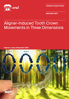
Determining Aligner-Induced Tooth Movements in Three Dimensions Using Clinical Data of Two Patients
by Ignacio Filippon, Christine Tanner, Jeannette A. von Jackowski, Georg Schulz, Tino Töpper and Bert Müller
Oral 2024, 4(4), 487-504; https://doi.org/10.3390/oral4040039 - 18 Oct 2024
Viewed by 561
Abstract
The effectiveness of a series of optically transparent aligners for orthodontic treatments depends on the anchoring of each tooth. In contrast with the roots, the crowns’ positions and orientations are measurable with intraoral scans, thus avoiding any X-ray dose. Exemplified by two patients, [...] Read more.
The effectiveness of a series of optically transparent aligners for orthodontic treatments depends on the anchoring of each tooth. In contrast with the roots, the crowns’ positions and orientations are measurable with intraoral scans, thus avoiding any X-ray dose. Exemplified by two patients, we demonstrate that three-dimensional crown movements could be determined with micrometer precision by registering weekly intraoral scans. The data show the movement and orientation changes in the individual crowns of the upper and lower jaws as a result of the forces generated by the series of aligners. During the first weeks, the canines and incisors were more affected than the premolars and molars. We detected overall tooth movement of up to about 1 mm during a nine-week active treatment. The data on these orthodontic treatments indicate the extent to which actual tooth movement lags behind the treatment plan, as represented by the aligner shapes. The proposed procedure can not only be used to quantify the clinical outcome of the therapy, but also to improve future planning of orthodontic treatments for each specific patient. This study should be treated with caution because only two cases were investigated, and the approach should be applied to a reasonably large cohort to reach strong conclusions regarding the efficiency and efficacy of this therapeutic approach. Full article
Show Figures

Graphical abstract

20 pages, 519 KiB  
Review
Silicon-, Silica-, and Silicate-Toothpastes for Remineralization and Repair of Teeth: A Scoping Review
by Fabio Correia Sampaio, Andressa Feitosa Bezerra de Oliveira, Nayanna Lana Soares Fernandes, Ana Carolina Cheron Gentile, Giovanna Bueno Marinho, Marcelo José Strazzeri Bönecker, Marco Aurelio Benini Paschoal, Paulo Henrique Perlatti D’Alpino and Fabiano Vieira Vilhena
Oral 2024, 4(4), 467-486; https://doi.org/10.3390/oral4040038 - 15 Oct 2024
Viewed by 1378
Abstract
Objective: The purpose of this scoping review was to identify gaps in the literature and summarize findings from studies examining the use of silicon-, silica-, and silicate-based toothpastes for the remineralization and repair of mineralized tooth tissues. Methods: A 10-year literature search [...] Read more.
Objective: The purpose of this scoping review was to identify gaps in the literature and summarize findings from studies examining the use of silicon-, silica-, and silicate-based toothpastes for the remineralization and repair of mineralized tooth tissues. Methods: A 10-year literature search was conducted using PubMed and Scopus, adhering to PRISMA 2020 guidelines. A total of 331 studies were initially identified, with 56 full-text review articles. After selecting the manuscripts, 27 studies were qualitatively analyzed by four reviewers, focusing on the results of both in vivo and in vitro methods. Results: The findings suggest that toothpastes containing silicon, silica, and silicate demonstrate promising results for remineralization and enamel repair, with evidence of mineral layer formation and/or deep enamel surface remineralization under various conditions. Additionally, the use of these toothpastes can lead to the obliteration of dentinal tubules within a few days. The results collectively support the efficacy of these toothpastes in enamel repair. Most of the clinical studies focused on dentine hypersensitivity, followed by white spot lesions. Conclusions: Silicon-, silica-, and silicate-based toothpastes (bioactive Si-toothpastes) can be considered effective based mostly on laboratory studies. There remains a need for more in vivo research studies on enamel and dentin mineral repair. Existing studies provide strong evidence that these technologies can reduce dentin hypersensitivity and promote enamel–dentin repair. Full article
Show Figures

Figure 1

8 pages, 1053 KiB  
Article
Influence of Root Canal Size and Curvature on Insertion Depth of Three Different Endodontic Irrigation Needles
by Michael Pinker, Wilhelm Frank, Karl-Thomas Wrbas and Jörg Philipp Tchorz
Oral 2024, 4(4), 459-466; https://doi.org/10.3390/oral4040037 - 12 Oct 2024
Viewed by 645
Abstract
The aim of this study was to evaluate the influence of root canal size and curvature on the insertion depth of three different endodontic irrigation needles. Four root canals with curvatures ranging from 0° to 69.72° were first enlarged to size .04/25. The [...] Read more.
The aim of this study was to evaluate the influence of root canal size and curvature on the insertion depth of three different endodontic irrigation needles. Four root canals with curvatures ranging from 0° to 69.72° were first enlarged to size .04/25. The insertion depths of a standard open-ended irrigation needle (SI), a single side-vented needle (SV), and a TruNatomy irrigation needle (TN) were then measured repeatedly in the chosen root canals and recorded as distances short of the working length. In curved canals, the SI and SV were tested with and without pre-bending. After enlargement to a greater taper (.06/25), these measurements were repeated. For a descriptive analysis, means and standard deviations were computed, and comparison was performed using the Wilcoxon test and formulated using a Monte-Carlo approximation with the level of significance set to 5%. Due to its flexible material, the best overall results in terms of insertion depth close to the working length were observed for the TN. The SI and SV could be inserted deeper in straight root canals and after enlargement to a greater taper (p < 0.05). In curved root canals, pre-bending of the SI and SV resulted in statistically significantly higher insertion depths (p < 0.05). In conclusion, cannula material properties, preparation size, and insertion depth mainly depend on each other. In curved root canals, a more flexible cannula like the TN should be favored to achieve better insertion depths. If using stainless-steel cannulas, they should be pre-bent to facilitate an insertion closer to the working length. Full article
Show Figures

Figure 1

10 pages, 908 KiB  
Article
Mandibular Advancement after Pubertal Peak with Acrylic Splint Herbst Appliance Anchored to Four Miniscrews: A Retrospective Controlled Study
by Antonio Manni, Andrea Boggio, Fabio Castellana, Giorgio Gastaldi and Mauro Cozzani
Oral 2024, 4(4), 449-458; https://doi.org/10.3390/oral4040036 - 10 Oct 2024
Viewed by 582
Abstract
Objectives: The treatment of class II patients with mandibular retrusion often involves the use of a Herbst appliance, due to its efficiency and reduced need for compliance. Despite skeletal benefits, undesired dental effects, caused by anchorage loss, could reduce mandibular advancement, especially after [...] Read more.
Objectives: The treatment of class II patients with mandibular retrusion often involves the use of a Herbst appliance, due to its efficiency and reduced need for compliance. Despite skeletal benefits, undesired dental effects, caused by anchorage loss, could reduce mandibular advancement, especially after the pubertal peak. Although the period between cervical vertebral maturation (CVM) 3 and CMV4 is considered the best choice to maximize the orthopedic outcome, clinicians cannot always treat patients during this ideal time window, as they present at their first visit later. The goal of this study is to evaluate whether the combination of an acrylic splint Herbst appliance with skeletal anchorage and elastic chains in both the upper and lower arch (Skeletal Therapy Manni Telescopic Herbst 4: STM4) could improve the efficiency of functional treatment also in CVM 5–6 patients. Methods: Lateral cephalograms of 10 consecutively treated patients taken at the beginning and at the end of the Herbst phase were analyzed and compared with those of 10 untreated patients of the same sex, age, malocclusion, and skeletal maturation. Results: According to the Pancherz analysis, the treated group of patients showed the significant advancement of the pogonion (4.75 mm), with a reduction in ANB (−3.3°) and Wits (−4.15 mm). The combination of miniscrews and elastic ligatures allowed the labial tipping of the upper incisors (+6.65°) and limited the labial flaring of the lower ones (+3.05°), maintaining the overjet needed for mandibular advancement. Conclusions: The STM4 technique might be an effective protocol for skeletal class II correction after the pubertal peak since it significantly reduces unfavorable dental compensations, increasing the skeletal effects and the esthetic outcome of the orthopedic treatment. Full article
Show Figures

Graphical abstract

8 pages, 1206 KiB  
Article
Advancing Management of Oral Lesion Patients with Epidermolysis Bullosa: In Vivo Evaluation with Optical Coherence Tomography of Ultrastructural Changes after Application of Cord Blood Platelet Gel and Laser Photobiomodulation
by Alessio Gambino, Ezio Sindici, Simona Astesano, Lucia Basiglio, Valeria Vallone and Paolo Giacomo Arduino
Oral 2024, 4(4), 441-448; https://doi.org/10.3390/oral4040035 - 27 Sep 2024
Viewed by 577
Abstract
Background: Inherited epidermolysis bullosa (EB) is a group of genetic disorders with skin fragility and blistering. The use of Cord Blood Platelet Gel (CBPG) in combination with laser photobiomodulation (PBM) leads to a reduction in lesions. The aim of this study is to [...] Read more.
Background: Inherited epidermolysis bullosa (EB) is a group of genetic disorders with skin fragility and blistering. The use of Cord Blood Platelet Gel (CBPG) in combination with laser photobiomodulation (PBM) leads to a reduction in lesions. The aim of this study is to evaluate clinical and morphometric changes with Optical Coherence Tomography (OCT) during GPC-PBM therapy. Methods: OCT scanning before the first session (T0), with relative measurement of the thicknesses of the epithelium (EP) and lamina propria (LP), and three consecutive sessions (once daily for 3 days) of CBPG and PBM applications were performed. A new OCT scan at the end of the three sessions (T1) and a week after (T2) were performed. All OCT scans were compared with the values of healthy reference tissues of the same site. Results: A statistically confirmed increase in EP thickness and a decrease in LP thickness with a progressive reduction in inflammatory content were highlighted. This case series did not have recurrences in the treated sites, or adverse reactions to therapy. Conclusions: This study shows the advantages of OCT monitoring in evaluating the effects of therapy at an ultrastructural level with a possibility of obtaining objective, precise, and repeatable measurements with an atraumatic device. Full article
Show Figures

Figure 1

Previous Issue
Back to TopTop