Article

Comparative Analysis of Gene Expression in Periodontal Ligament Stem Cells Exposed to Biodentine and Bio-C Repair: Implications for Cementogenesis—An In Vitro Study

1
School of Medicine and Dentistry, Griffith University, Gold Coast, QLD 4222, Australia
2
Oral Biology Department, Faculty of Dentistry, The British University in Egypt, El Sherouk City, Cairo 11837, Egypt
*
Author to whom correspondence should be addressed.
Submission received: 1 February 2025 / Revised: 1 March 2025 / Accepted: 5 March 2025 / Published: 13 March 2025
(This article belongs to the Special Issue Advanced Dental Materials for Oral Rehabilitation)

Abstract

:
Background/Objectives: Bioactive materials are gaining increased popularity as materials of choice for pulpal regeneration. A similar trend is emerging with root repair materials; however, there is a significant gap in the literature about cementogenic ability of bioceramic repair materials on the periodontal ligament cells. The aim of the present study was to investigate the effect of bioceramic materials (Biodentine and Bio-C Repair) on the cementogenesis potential of the periodontal ligament stem cells (PDLSCs). Methods: PDLSCs were isolated using the enzymatic digestion approach from sound extracted teeth. Material extracts were prepared on rubber discs and immersed in fresh growth medium for 24 h at 37 °C. Reverse transcription–quantitative polymerase chain reaction (RT-qPCR) was used to detect the mRNA expression levels of cementogenic markers cementum protein 1 (CEMP1), Cementum attachment protein (CAP), pathway markers transforming growth factor β1(TGF-β1), bone morphogenic protein 2 (BMP2), and inflammatory marker IL-6. Results: Both materials (Biodentine and Bio-C Repair) showed significantly higher gene expressions when compared to the control groups. The gene expression with Bio-C Repair significantly increased when compared with Biodentine, except for TGF-β1 expression, where both materials exhibited similar results. Conclusions: Bio-C Repair demonstrated increased gene expression of cementogenic markers compared to Biodentine under the tested conditions. Further in vivo studies are deemed necessary to translate the findings from this study into clinical practice.

Graphical Abstract

1. Introduction

Therapeutic approaches for managing teeth with pulp exposure due to extensive caries focus on preserving and maintaining pulpal vitality [1]. A wide range of materials and methodologies have been proposed to improve outcomes in vital pulp therapy. Calcium hydroxide has been widely used in such therapies due to its antibacterial properties and ability to stimulate dentin bridge formation. However, its poor mechanical properties and limited longevity often make it a provisional solution rather than a definitive treatment.
The development of mineral trioxide aggregate (MTA) marked a significant advancement as a root repair material and pulp-capping agent, offering superior sealing ability, biocompatibility, and bioactivity compared to calcium hydroxide [2].
Recently, bioactive materials for vital pulp therapy, root canal sealers, and root repairs have been introduced to alleviate the disadvantages of MTA, such as long setting time and discoloration potential [3,4,5]. The physicochemical and biological properties were remarkably improved with the calcium silicate-based bioactive materials. These materials have been developed and used as root repair materials and sealers for root canals with excellent biocompatibility and bioactivity [6,7]. These bioactive materials introduce a novel approach to root filling by releasing calcium ions and calcium hydroxide and creating a layer between the cement and dentinal wall. These components frequently have an influence on the tissues surrounding the apex of a tooth. They can potentially facilitate the formation of cementum, which creates a biological barrier and promotes the process of healing [8].
The root repair bioactive materials based on calcium silicate demonstrate distinctive biological mechanisms that render them exceptionally successful in dental applications, especially in vital pulp therapy, root restoration, and endodontic sealing. The mechanisms encompass ion release and mineralization, biocompatibility and cytocompatibility, facilitation of tissue regeneration, anti-inflammatory and antimicrobial properties, hydroxyapatite formation and sealing characteristics, stimulation of growth factors, and molecular pathways (Table 1) [9,10].
These materials interact both directly and indirectly with surrounding tissues [11]. Direct interactions occur when the material contacts the tissues, potentially causing inflammatory responses, cytotoxicity, or tissue irritation. These interactions are influenced by the material‘s composition, properties, application method, and the root canal system’s anatomy [12]. Indirect interactions involve the release of chemicals or ions from the material, such as calcium and hydroxyl ions, which can aid in tissue healing and regeneration. Bioceramic materials, in particular, release calcium and phosphate ions that promote the formation of new mineralized tissue [13,14]. Ex vivo studies have shown that bioceramic materials can stimulate the formation of mineralized tissues when periodontal ligament stem cells (PDLSCs) are cultured in contact with these materials [15,16,17].
Periapical tissues are significant because they contain different types of mesenchymal stem cells (MSCs), most significantly the PDLSCs [18]. These cells demonstrate strong anti-inflammatory and immunomodulatory properties. They also have the capacity to differentiate into specialized cell lineages such as osteoblasts, cementoblasts, adipocytes, and chondrocytes [19]. This transformation can be achieved using specific culture media that induce the desired differentiation. PDLSCs have been shown to have the capacity to generate alveolar bone, cementum, gingiva, periodontal ligaments, peripheral nerves, and blood vessels in living organisms [20,21,22]. Previous research has utilized PDLSCs found in periapical tissues to investigate the bioactivity of several endodontic sealers in vitro [17,23].
Biomineralization and cementogenesis are two crucial processes facilitated by bioactive materials in regenerative dentistry. Biomineralization involves the release of calcium and phosphate ions, which aggregate to create hydroxyapatite, thereby emulating the natural composition of tooth tissues and improving the occlusion of defects. These materials also regulate pH to establish an antibacterial environment and activate signaling pathways such as BMP and TGF-β, which promote mineralized tissue development. Cementogenesis requires the differentiation of PDLSCs into cementoblasts, stimulated by bioactive factors, resulting in cementum deposition. The process is augmented by the elevation of cementogenic markers (e.g., CEMP1, BMP-2, TGF-β1) and the attenuation of inflammation, leading to a biological seal and facilitating periodontal and periapical repair. Collectively, these systems rehabilitate the structural integrity and functionality of compromised oral tissues [24].
Bio-C Repair is a cytocompatible, ready-to-use bioceramic repair material with a biocompatibility and biomineralization capacity. However, specific information on this material’s cementogenic potential on PDLSCs during the repair of the periodontal ligament and surrounding tissues is lacking. Additionally, the molecular mechanisms underlying the cementogenesis induced by Bio-C Repair remain unclear. The aim of the present study was to investigate the effect of bioceramic materials, Biodentine (Septodont, Lancaster, PA, USA) and Bio-C Repair (Angelus Odontologia, Londrina, Paraná, Brazil) on the cementogenesis potential of the periodontal ligament stem cells (PDLSCs). The following null hypothesis was adopted: these materials are expected to exhibit no differences in gene expression compared with other root repair materials.

2. Materials and Methods

2.1. PDLSCs Isolation and Culture

Seven sound teeth were collected from adult patients (aged 18–25) who had given their consent. The extractions were performed as part of their orthodontic treatment plan at The British University in Egypt. Inclusion and exclusion criteria for the teeth are summarized in Table 2. The study was approved by the institutional review board (IRB) under the reference (FD BUE REC 24-037).
PDLSCs were isolated using the enzymatic digestion approach, as previously described by multiple studies. The periodontal tissue of the extracted teeth was minced into fragments with a diameter of roughly 2 mm. The experiment was conducted in a Petri dish using a phosphate buffer solution (PBS) with a pH of 7.4, supplemented with antibiotics. Enzymatic digestion of the tissues was performed using a solution containing 3 mg/mL of collagenase type I and 4 mg/mL of dispase (Sigma, Cream Ridge, NJ, USA). The cells were cultured in a 75 cm2 flask using Dulbecco’s Modified Eagle Medium (DMEM) supplemented with 10% fetal bovine serum (FBS). The culture medium was changed on a daily basis. The cells utilized in this study were derived from passages 3 to 5.

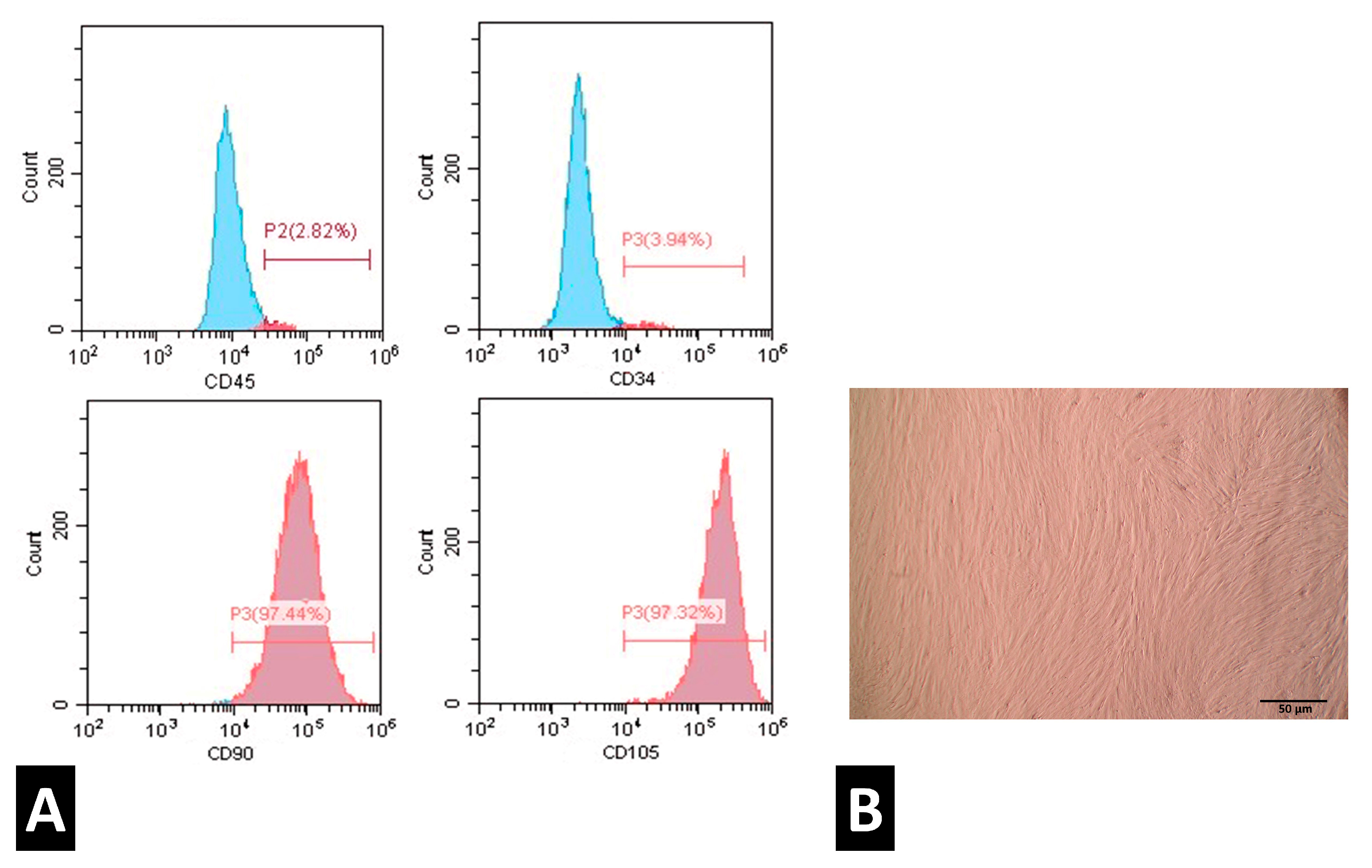
2.2. Characterization of PDLSCs

An inverted phase microscope was used to examine the morphology of the cultured cells. Flow cytometric analysis was used to identify PDLSCs based on their immunophenotype. The monoclonal antibodies employed for targeting CD34, CD45, CD90, and CD105 were obtained from Biosciences, CA, USA. In summary, a 100 μL cell suspension sample was combined with 10 μL of the monoclonal antibody and left to incubate for 30 min in a dark environment at 37 °C. Subsequently, the cells were rinsed with a phosphate-buffered saline (PBS) solution that included 2% bovine serum albumin. PDLSCs were suspended in PBS and examined using a Facial Action Coding System (FACS) Calibre Flow Cytometer (BD Biosciences) [19].

2.3. Material Extracts and Preparation

Root repair materials, Bio-C Repair and Biodentine, were assessed in this study (Table 3). The sample size calculation was performed in G*Power software version 3.1.9.2 (Heinrich Heine University, Dusseldorf, Germany) at a significance level of p ≤ 0.05 for evaluation of osteogenic differentiation. The calculated sample number was 20. Cylindrical rubber molds were prepared, and the materials were prepared according to the manufacturer’s recommendations, disinfected by ultraviolet exposure on both sides for 30 min, and left to set for 48 hours. Each disc was placed in a separate well of a 24-well plate and then put in fresh medium for 24 h at 37 °C. The extraction process was carried out per ISO 10993-12 standards [25], where the material surface area/medium volume ratio was 3 mm2/mL [26].

2.4. Reverse Transcription–Quantitative Polymerase Chain Reaction (RT-qPCR)

The mRNA expression levels of cementogenic markers cementum protein 1 (CEMP1) and cementum attachment protein (CAP), pathway markers transforming growth factor β1(TGF-β1) and and bone morphogenic protein 2 (BMP2), and inflammatory marker IL-6 were detected by RT-qPCR after 7 days of exposure of the materials to osteogenic medium. The control group consisted of PDLSCs in normal medium. PDLSCs cultured in osteogenic medium only without materials served as the positive control. Total RNA extraction was performed using TRIzol (Zymo Research Corp. Irvine, CA, USA). cDNA synthesis was performed using the miScript Reverse Transcription Kit. RT-qPCR was performed using Maxima Syber Green qPCR Master Mix. mRNA expression levels were normalized to β-actin. Gene expression was determined using the 2−ΔΔCt formula [27]. Primer sequences used in the current study are listed in Table 4.

2.5. Statistical Analysis

A one-way analysis of variance (ANOVA) statistical analysis was performed, followed by Tukey’s multiple comparison tests. Data on the relative fold change of the tested genes are expressed as means and standard deviations. A p-value of less than 0.05 was considered significant. Experiments were performed in triplicate.

3. Results

3.1. PDLSCs Characterization

Flow cytometric analysis results for PDLSCs characterization showed a high expression of MSC surface markers CD90 and CD105 and a negative expression of hematopoietic markers CD34 and CD45 (Figure 1A), thus confirming the stemness of the isolated PDLSCs. On the other hand, inverted phase microscopic examination showed spindle-shaped cells that were confluent (Figure 1B).

3.2. Gene Expression Analysis

The RT-qPCR analysis of CEMP1, CAP, TGF-β1, BMP2, and IL-6 revealed that Bio-C Repair consistently showed the highest relative fold change across all genes tested, indicating a strong upregulation of these genes compared to the control and positive control groups. Biodentine also showed significant upregulation of all genes, but generally to a lesser extent than Bio-C Repair. Comparing Bio-C Repair and Biodentine, Bio-C Repair showed a significantly higher expression of all genes tested (CEMP1 (p = 0.002), CAP (p = 0.01), BMP2 (p = 0.01), and IL-6 (p = 0.003)), with the exception of TGF-β1 (p = 0.07), where no significant difference was found between the two groups (Figure 2).

4. Discussion

Bioceramic root materials have recently been introduced as root canal filling and root repair materials and show superior biocompatibility and bioactivity properties [28]. They demonstrate enduring bioactivity through the diffusion of molecules during and after solidification. These molecules can have an effect on the periapical tissues directly or indirectly through diffusion from the root canal system [29]. Several studies have focused mainly on studying three materials. However, not enough information is available on these materials’ cementogenic, anti-inflammatory, and regenerative properties. This study aimed to evaluate the effect of bioceramic materials, Biodentine, and Bio-C Repair on the cementogenic, inflammatory, and regenerative gene expression of periodontal ligament stem cells (PDLSCs).
PDLSCs have shown promising results in the cementogenic and osteogenic process and, therefore, in the regeneration of periodontal tissues [30]. Bioceramic root repair materials, when used in the treatment of root defects, come into contact with the PDLSCs and periapical tissues. Therefore, evaluating the bioactivity and regenerative properties of these materials on PDLSCs is crucial for the repair of these tissues.
In the current study, the null hypothesis that the tested materials would exhibit no differences in gene expression compared with other root repair materials was rejected. Biodentine and Bio-C Repair groups showed an upregulation of the cementogenic proteins CEMP1 and CAP, indicating that both materials promoted the cementogenic process in PDLSCs. These genes play a role in the differentiation of stem cells into osteoblasts and cementoblasts, which in turn contribute to the healing of the tooth periapical region [31,32]. Moreover, CAP facilitates the maturation and deposition of the mineralized extracellular matrix [33]. Bio-C Repair showed a relatively significantly higher expression of the cementogenic genes than Biodentine, suggesting that it could be more potent in periapical healing and regeneration of the periodontium. The increased release of calcium ions from Biodentine and Bio-C Repair plays a significant role in its bioactivity. This ion release contributes to the neutral pH, which supports the mineralization process. Moreover, the higher expression of the cementogenic genes in Bio-C Repair might be related to the higher Zirconium oxide content, which is found as a radiopacifier that enhances cytocompatibility. The current results are in agreement with several studies showing that bioceramics have the potential to promote cementogenesis due to the release of calcium ions and other bioactive substances [29,34].
The upregulation of TGF-β1 and BMP2 in the Biodentine and Bio-C Repair suggests that both materials activate essential signaling pathways involved in osteogenic and cementogenic differentiation. TGF-β1 is a versatile cytokine that has a crucial function in controlling cell proliferation, differentiation, and the formation of the extracellular matrix [35]. TGF-β1 is vital in promoting the differentiation of PDLSCs into cementoblasts and osteoblasts [36]. This pathway encompasses Smad proteins, which migrate to the nucleus and govern the expression of genes implicated in matrix formation and mineralization. On the other hand, BMP2 plays a crucial role in the process of bone growth and cementogenesis [37]. Moreover, BMP2 is recognized for its capacity to stimulate the differentiation of mesenchymal stem cells into osteoblasts. BMP2 attaches to specific receptors on the cell membrane, resulting in the phosphorylation of Smad proteins and initiating other signaling pathways like MAPK [38]. As a consequence, the genes required for the synthesis of bone and cementum matrix proteins are transcribed.
The significantly higher expression of TGF-β1 and BMP2 in the Bio-C Repair group than in Biodentine might be related to the increased calcium ion release from Bio-C Repair, which can enhance the activation of cellular signaling pathways like TGF-β and BMP signaling.
The inflammatory gene IL-6 is commonly acknowledged as a cytokine that promotes inflammation, and it is frequently increased in reaction to tissue damage or infection. Within the framework of periodontal disease, IL-6 is commonly linked to persistent inflammation, which plays a role in the advancement of tissue damage [39]. IL-6 also plays a role in regeneration by regulating the equilibrium between inflammation and repair. It can aid in the shift from the period of inflammation to the phase of tissue regeneration by stimulating the attraction and stimulation of cells that are engaged in the process of repairing tissues, such as stem cells [40]. Our gene expression results showed a similar trend across all markers due to their roles in inflammatory responses, immune regulation, healing and repair, as well as periodontal tissue remodeling and bone formation. Performing a gene expression analysis at a later stage of healing would show different results, including downregulation of IL-6, which is mainly involved in the early inflammatory responses but is reduced to promote repair and avoid prolonged inflammation that may result in tissue damage and delayed healing [41,42]. In a similar way, the levels of TGF-β1 are decreased after the initial repair phase to prevent excessive fibrosis [43,44]. Furthermore, our results could be confirmed by not only testing the same genes at different timelines of the repair process, but also by testing other genes that are downregulated during periodontal tissue repair, including Matrix Metalloproteinases (MMPs) [45,46], Cyclooxygenase-2 (COX-2) [47], and Receptor Activator of Nuclear Factor kappa-Β Ligand (RANKL) [48].
Similar studies confirm the findings revealed in the present experiment; for example, it has been shown that Biodentine is highly biocompatible with hPDLSCs, resulting in a positive effect on biological processes [49]. Along the same lines, another study evaluated different endodontic cements and concluded that Biodentine, Bio-C Repair, and MTA Repair HP were able to induce hPDLSC proliferation when compared to Cimmo HD and White MTA, which were cytotoxic to hPDLSCs [50]. Recent studies in the literature show that Biodentine promotes significantly higher mineralization and osteo/cementogenic marker expression on hPDLSCs when evaluated against new materials like Ceraputty [51]. Our choice to investigate calcium silicate and bioceramic materials was validated by review articles that indicated that these materials remain valid and reliable options for stem cell therapy as well as endodontic/periodontic regenerative procedures [52,53]. Furthermore, in alignment with our results, Bio-C Repair showed the best results in comparison to Biodentine and MTA with regards to osteoclastic inhibition in teeth with replacement root resorption [54]. In addition to the above, Bio-C Repair was reported to successfully manage lateral root perforations [55]. This indicates that Bio-C Repair could potentially be a superior option in the repair of different tissues of the periodontium (periodontal ligament, cementum, and alveolar bone).
The increased gene expression levels identified with Bio-C Repair indicate that it may be more successful in clinical situations that necessitate improved regenerative results, such as in the management of periodontal abnormalities or root repairs where strong bone and cementum creation is crucial. However, the conditions in the current study are limited to in vitro scenarios and need to be confirmed by in vivo studies to ensure a more comprehensive understanding of their therapeutic potential, which would account for the dynamic nature of the oral cavity with multiple factors that can affect the regenerative potential of the bioceramic materials, including oral hygiene, oral microbiome, operator skills, concurrent infections, and the body’s immune response.
Furthermore, future studies will aim to include comprehensive physicochemical analyses of the tested dental materials using advanced techniques such as scanning electron microscopy (SEM) and energy-dispersive X-ray spectroscopy (EDX) to better understand how these properties influence cellular responses and regenerative outcomes. This additional data would enhance the overall understanding of the material–cell interactions.
Additionally, although our gene expression analysis indicated Bio-C Repair’s superior performance, particularly in upregulating TGF-β1 and BMP2, we recognize the need to explore the underlying molecular pathways driving these differences. These pathways play a pivotal role in tissue repair and regeneration, and their detailed exploration could provide valuable insights into the mechanisms of action. We consider this experiment to be a pilot study that paves the way for future research focusing on investigating these signaling cascades in greater depth, employing techniques such as protein expression analysis and pathway-specific inhibitors to better elucidate the molecular basis of the observed effects.

5. Conclusions

In summary, Bio-C Repair demonstrated increased gene expression of cementogenic markers compared to Biodentine under the tested conditions. These findings suggest that Bio-C Repair may have enhanced bioactivity and interaction with cellular signaling pathways; however, further studies are needed to confirm these observations and establish its regenerative potential in more clinically relevant scenarios.

Author Contributions

Conceptualisation, M.M.B. and M.S.; methodology, M.A.A. and M.S.; software, M.A.A. and M.S.; investigation, M.M.B., M.A.A., M.M. and M.S.; resources, M.M.B. and M.M.; data curation, M.A.A. and M.S.; writing—original draft preparation, M.M.B. and M.S.; writing—review and editing, M.M.B., M.A.A., M.M. and M.S.; supervision, M.M.B. and M.M. All authors have read and agreed to the published version of the manuscript.

Funding

This research received no external funding.

Institutional Review Board Statement

This study was conducted in accordance with the Declaration of Helsinki and approved by the institutional review board (IRB) of The British University in Egypt (under the reference [FD BUE REC 24-037], date: 20 April 2024).

Informed Consent Statement

Informed consent for participation was obtained from all subjects involved in the study.

Data Availability Statement

The work includes all the original contributions made, and any additional questions can be forwarded to the corresponding author.

Conflicts of Interest

The authors declare no conflicts of interest.

References

  1. Duncan, H.F. Present status and future directions—Vital pulp treatment and pulp preservation strategies. Int. Endod. J. 2022, 55, 497–511. [Google Scholar] [CrossRef] [PubMed]
  2. Hanna, S.N.; Perez Alfayate, R.; Prichard, J. Vital Pulp Therapy an Insight Over the Available Literature and Future Expecta-tions. Eur. Endod. J. 2020, 5, 46–53. [Google Scholar] [PubMed]
  3. Wuersching, S.N.; Diegritz, C.; Hickel, R.; Huth, K.C.; Kollmuss, M. A comprehensive in vitro comparison of the biological and physicochemical properties of bioactive root canal sealers. Clin. Oral Investig. 2022, 26, 6209–6222. [Google Scholar] [CrossRef] [PubMed]
  4. Jo, S.B.; Kim, H.K.; Lee, H.N.; Kim, Y.-J.; Patel, K.D.; Knowles, J.C.; Lee, J.-H.; Song, M. Physical Properties and Biofunctionalities of Bioactive Root Canal Sealers In Vitro. Nanomaterials 2020, 10, 1750. [Google Scholar] [CrossRef]
  5. Washio, A.; Morotomi, T.; Yoshii, S.; Kitamura, C. Bioactive Glass-Based Endodontic Sealer as a Promising Root Canal Filling Material without Semisolid Core Materials. Materials 2019, 12, 3967. [Google Scholar] [CrossRef]
  6. Inada, R.N.H.; Silva, E.C.A.; Lopes, C.S.; Queiroz, M.B.; Torres, F.F.E.; da Silva, G.F.; Cerri, P.S.; Guerreiro-Tanomaru, J.M.; Tanomaru-Filho, M. Biocompatibility, bioactivity, porosity, and sealer/dentin interface of bioceramic ready-to-use sealers using a dentin-tube model. Sci. Rep. 2024, 14, 16768. [Google Scholar] [CrossRef]
  7. Estivalet, M.S.; de Araújo, L.P.; Immich, F.; da Silva, A.F.; Ferreira, N.d.S.; da Rosa, W.L.O.; Piva, E. Bioactivity Potential of Bioceramic-Based Root Canal Sealers: A Scoping Review. Life 2022, 12, 1853. [Google Scholar] [CrossRef]
  8. de Sousa Reis, M.; Scarparo, R.K.; Steier, L.; de Figueiredo, J.A.P. Periradicular inflammatory response, bone resorption, and ce-mentum repair after sealing of furcation perforation with mineral trioxide aggregate (MTA Angelus) or Biodentine. Clin. Oral Investig. 2019, 23, 4019–4027. [Google Scholar] [CrossRef]
  9. Estrela, C.; Cintra, L.T.A.; Duarte, M.A.H.; Rossi-Fedele, G.; Gavini, G.; Sousa-Neto, M.D. Mechanism of action of Bioactive Endodontic Materials. Braz. Dent. J. 2023, 34, 1–11. [Google Scholar] [CrossRef]
  10. Wang, X.; Xiao, Y.; Song, W.; Ye, L.; Yang, C.; Xing, Y.; Yuan, Z. Clinical application of calcium silicate-based bioceramics in endodontics. J. Transl. Med. 2023, 21, 853. [Google Scholar] [CrossRef]
  11. Jung, M.-K.; Park, S.-C.; Kim, Y.-J.; Park, J.-T.; Knowles, J.C.; Park, J.-H.; Dashnyam, K.; Jun, S.-K.; Lee, H.-H.; Lee, J.-H. Premixed Calcium Silicate-Based Root Canal Sealer Reinforced with Bioactive Glass Nanoparticles to Improve Biological Properties. Pharmaceutics 2022, 14, 1903. [Google Scholar] [CrossRef] [PubMed]
  12. Jin, H.; Li, Y.; Wang, Q.; Dong, M.; Yang, M.; Chen, W.; Wang, S.; Zhang, H.; Zheng, S.; Cao, C.Y.; et al. A strontium and amorphous calcium phosphate dipped premixed injectable calcium silicate-based ceramic for dental root canal sealing. Ceram. Int. 2021, 47, 33738–33750. [Google Scholar] [CrossRef]
  13. Tanvir, A.H.; Khaleque, A.; Kim, G.-H.; Yoo, W.-Y.; Kim, Y.-Y. The Role of Bioceramics for Bone Regeneration: History, Mechanisms, and Future Perspectives. Biomimetics 2024, 9, 230. [Google Scholar] [CrossRef] [PubMed]
  14. Aminoshariae, A.; Primus, C.; Kulild, J.C. Tricalcium silicate cement sealers: Do the potential benefits of bioactivity justify the drawbacks? J. Am. Dent. Assoc. 2022, 153, 750–760. [Google Scholar] [CrossRef]
  15. Alchawoosh, A.; Hashimoto, K.; Kawashima, N.; Noda, S.; Nozaki, K.; Okiji, T. Hydraulic calcium silicate-based root canal sealers mitigate proinflammatory cytokine synthesis and promote osteogenesis in vitro. J. Dent. Sci. 2023, 18, 1731–1739. [Google Scholar] [CrossRef]
  16. Zhao, Z.; Liu, J.; Weir, M.D.; Schneider, A.; Ma, T.; Oates, T.W.; Xu, H.H.K.; Zhang, K.; Bai, Y. Periodontal ligament stem cell-based bioactive constructs for bone tissue engineering. Front. Bioeng. Biotechnol. 2022, 10, 1071472. [Google Scholar] [CrossRef]
  17. López-García, S.; Lozano, A.; García-Bernal, D.; Forner, L.; Llena, C.; Guerrero-Gironés, J.; Moraleda, J.M.; Murcia, L.; Rodríguez-Lozano, F.J. Biological Effects of New Hydraulic Materials on Human Periodontal Ligament Stem Cells. J. Clin. Med. 2019, 8, 1216. [Google Scholar] [CrossRef]
  18. Di Vito, A.; Bria, J.; Antonelli, A.; Mesuraca, M.; Barni, T.; Giudice, A.; Chiarella, E. A Review of Novel Strategies for Human Periodontal Ligament Stem Cell Ex Vivo Expansion: Are They an Evidence-Based Promise for Regenerative Periodontal Therapy? Int. J. Mol. Sci. 2023, 24, 7798. [Google Scholar] [CrossRef]
  19. Queiroz, A.; Albuquerque-Souza, E.; Gasparoni, L.M.; de França, B.N.; Pelissari, C.; Trierveiler, M.; Holzhausen, M. Therapeutic potential of periodontal ligament stem cells. World J. Stem Cells 2021, 13, 605–618. [Google Scholar] [CrossRef]
  20. Mohebichamkhorami, F.; Fattahi, R.; Niknam, Z.; Aliashrafi, M.; Naeimi, S.K.; Gilanchi, S.; Zali, H. Periodontal ligament stem cells as a promising therapeutic target for neural damage. Stem Cell Res. Ther. 2022, 13, 273. [Google Scholar] [CrossRef]
  21. Tomokiyo, A.; Wada, N.; Maeda, H. Periodontal Ligament Stem Cells: Regenerative Potency in Periodontium. Stem Cells Dev. 2019, 28, 974–985. [Google Scholar] [CrossRef] [PubMed]
  22. Wang, M.; Xie, J.; Wang, C.; Zhong, D.; Xie, L.; Fang, H. Immunomodulatory Properties of Stem Cells in Periodontitis: Current Status and Future Prospective. Stem Cells Int. 2020, 2020, 9836518. [Google Scholar] [CrossRef] [PubMed]
  23. Sanz, J.L.; Guerrero-Gironés, J.; Pecci-Lloret, M.R.; Melo, M. Biological interactions between calcium silicate-based endodontic biomaterials and periodontal ligament stem cells: A systematic review of in vitro studies. Int. Endod. J. 2021, 54, 2025–2043. [Google Scholar] [CrossRef]
  24. Souza, T.A.; Bezerra, M.M.; Silva, P.G.B.; Costa, J.J.N.; Carneiro, R.F.L.A.; Barcelos, J.O.F.; Vasconcelos, B.C.; Chaves, H.V. Bone morphogenetic proteins in biomineralization of two endodontic restorative cements. J. Biomed. Mater. Res. Part B Appl. Biomater. 2021, 109, 348–357. [Google Scholar] [CrossRef] [PubMed]
  25. ISO 10993-12; Biological Evaluation of Medical Devices—Part 12: Sample Preparation and Reference Materials. International Organization for Standardization: Geneva, Switzerland, 2021. Available online: www.iso.org/standard/75769.html (accessed on 4 March 2025).
  26. Saber, S.M.; Gomaa, S.M.; Elashiry, M.M.; El-Banna, A.; Schäfer, E. Comparative biological properties of resin-free and resin-based calcium silicate-based endodontic repair materials on human periodontal ligament stem cells. Clin. Oral Investig. 2023, 27, 6757–6768. [Google Scholar] [CrossRef]
  27. Alhazmi, Y.A.; Aljabri, M.Y.; Raafat, S.N.; Gomaa, S.M.; Shamel, M. Exploring the Effects of Low-Level Laser Therapy on the Cyto-compatibility and Osteo/Odontogenic Potential of Gingival-Derived Mesenchymal Stem Cells: Preliminary Report. Appl. Sci. 2023, 13, 8490. [Google Scholar] [CrossRef]
  28. Raghavendra, S.S.; Jadhav, G.R.; Gathani, K.M.; Kotadia, P. Bioceramics in endodontics—A review. J. Istanb. Univ. Fac. Dent. 2017, 51, 128–137. [Google Scholar] [CrossRef]
  29. Dong, X.; Xu, X. Bioceramics in Endodontics: Updates and Future Perspectives. Bioengineering 2023, 10, 354. [Google Scholar] [CrossRef]
  30. Liu, J.; Ruan, J.; Weir, M.D.; Ren, K.; Schneider, A.; Wang, P.; Oates, T.W.; Chang, X.; Xu, H.H.K. Periodontal Bone-Ligament-Cementum Regeneration via Scaffolds and Stem Cells. Cells 2019, 8, 537. [Google Scholar] [CrossRef]
  31. Arroyo, R.; López, S.; Romo, E.; Montoya, G.; Hoz, L.; Pedraza, C.; Garfias, Y.; Arzate, H. Carboxy-Terminal Cementum Protein 1-Derived Peptide 4 (cemp1-p4) Promotes Mineralization through wnt/β-catenin Signaling in Human Oral Mucosa Stem Cells. Int. J. Mol. Sci. 2020, 21, 1307. [Google Scholar] [CrossRef]
  32. Gauthier, P.; Yu, Z.; Tran, Q.T.; Bhatti, F.-U.; Zhu, X.; Huang, G.T.-J. Cementogenic genes in human periodontal ligament stem cells are downregulated in response to osteogenic stimulation while upregulated by vitamin C treatment. Cell Tissue Res. 2017, 368, 227. [Google Scholar] [CrossRef] [PubMed]
  33. Yan, X.; Zhang, Q.; Ma, X.; Zhong, Y.; Tang, H.; Mai, S. The mechanism of biomineralization: Progress in mineralization from in-tracellular generation to extracellular deposition. Jpn. Dent. Sci. Rev. 2023, 59, 181–190. [Google Scholar] [CrossRef] [PubMed]
  34. López-García, S.; Sánchez-Bautista, S.; García-Bernal, D.; Lozano, A.; Forner, L.; Sanz, J.L.; Murcia, L.; Rodríguez-Lozano, F.J.; Oñate-Sánchez, R.E. Premixed calcium silicate-based ceramic sealers promote osteo-genic/cementogenic differentiation of human periodontal ligament stem cells: A microscopy study. Microsc. Res. Tech. 2024, 87, 1584–1597. [Google Scholar] [CrossRef] [PubMed]
  35. Deng, Z.; Fan, T.; Xiao, C.; Tian, H.; Zheng, Y.; Li, C.; He, J. TGF-β signaling in health, disease, and therapeutics. Signal Transduct. Target. Ther. 2024, 9, 61. [Google Scholar]
  36. Fan, C.; Ji, Q.; Zhang, C.; Xu, S.; Sun, H.; Li, Z. TGF-β induces periodontal ligament stem cell senescence through increase of ROS production. Mol. Med. Rep. 2019, 20, 3123–3130. [Google Scholar] [CrossRef]
  37. Pitaru, S.; Pritzki, A.; Bar-Kana, I.; Grosskopf, A.; Savion, N.; Narayanan, A.S. Bone morphogenetic protein 2 induces the expression of cementum attachment protein in human periodontal ligament clones. Connect. Tissue Res. 2002, 43, 257–264. [Google Scholar] [CrossRef]
  38. Rahman, S.; Akhtar, N.; Jamil, H.M.; Banik, R.S.; Asaduzzaman, S.M. TGF-β/BMP signaling and other molecular events: Regulation of osteoblastogenesis and bone formation. Bone Res. 2015, 3, 15005. [Google Scholar] [CrossRef]
  39. Mazurek-Mochol, M.; Bonsmann, T.; Mochol, M.; Poniewierska-Baran, A.; Pawlik, A. The Role of Interleukin 6 in Periodontitis and Its Complications. Int. J. Mol. Sci. 2024, 25, 2146. [Google Scholar] [CrossRef]
  40. Kerkis, I.; da Silva, Á.P.; Araldi, R.P. The impact of interleukin-6 (IL-6) and mesenchymal stem cell-derived IL-6 on neurological conditions. Front. Immunol. 2024, 15, 1400533. [Google Scholar] [CrossRef]
  41. Ancuța, C.; Chirieac, R.; Ancuța, E.; Țănculescu, O.; Solomon, S.M.; Fătu, A.M.; Doloca, A.; Iordache, C. Exploring the Role of Inter-leukin-6 Receptor Inhibitor Tocilizumab in Patients with Active Rheumatoid Arthritis and Periodontal Disease. J. Clin. Med. 2021, 10, 878. [Google Scholar] [CrossRef]
  42. Huang, J.; Cai, X.; Ou, Y.; Zhou, Y.; Wang, Y. Resolution of inflammation in periodontitis: A review. Int. J. Clin. Exp. Pathol. 2018, 11, 4283–4295. [Google Scholar] [PubMed]
  43. Kaigler, D.; A Cirelli, J.; Giannobile, W.V. Growth factor delivery for oral and periodontal tissue engineering. Expert Opin. Drug Deliv. 2006, 3, 647–662. [Google Scholar] [CrossRef] [PubMed]
  44. Ramirez, H.; Patel, S.B.; Pastar, I. The Role of TGFβ Signaling in Wound Epithelialization. Adv. Wound Care 2014, 3, 482–491. [Google Scholar] [CrossRef] [PubMed]
  45. Franco, C.; Patricia, H.-R.; Timo, S.; Claudia, B.; Marcela, H. Matrix Metalloproteinases as Regulators of Periodontal Inflammation. Int. J. Mol. Sci. 2017, 18, 440. [Google Scholar] [CrossRef]
  46. Radzki, D.; Negri, A.; Kusiak, A.; Obuchowski, M. Matrix Metalloproteinases in the Periodontium—Vital in Tissue Turnover and Unfortunate in Periodontitis. Int. J. Mol. Sci. 2024, 25, 2763. [Google Scholar] [CrossRef]
  47. Bezerra, M.M.; de Lima, V.; Alencar, V.B.; Vieira, I.B.; Brito, G.A.C.; Ribeiro, R.A.; Rocha, F.A.C. Selective cyclooxygenase-2 inhibition prevents alveolar bone loss in experimental periodontitis in rats. J. Periodontol. 2000, 71, 1009–1014. [Google Scholar] [CrossRef]
  48. Qasim, S.S.B.; Al-Otaibi, D.; Al-Jasser, R.; Gul, S.S.; Zafar, M.S. An Evidence-Based Update on the Molecular Mechanisms Underlying Periodontal Diseases. Int. J. Mol. Sci. 2020, 21, 3829. [Google Scholar] [CrossRef]
  49. Abuarqoub, D.; Aslam, N.; Jafar, H.; Abu Harfil, Z.; Awidi, A. Biocompatibility of Biodentine™ ® with Periodontal Ligament Stem Cells: In Vitro Study. Dent. J. 2020, 8, 17. [Google Scholar] [CrossRef]
  50. Pedrosa, M.d.S.; Alves, T.; Rahhal, J.G.; Nogueira, F.N.; Sipert, C.R. Cytotoxicity of reparative endodontic cements on human periodontal ligament stem cells. Pesqui. Bras. Odontopediatr. Clin. Integr. 2022, 22, e210114. [Google Scholar] [CrossRef]
  51. López-García, S.; Rodríguez-Lozano, F.J.; Sanz, J.L.; Forner, L.; Pecci-Lloret, M.P.; Lozano, A.; Murcia, L.; Sánchez-Bautista, S.; Oñate-Sánchez, R.E. Biological properties of Ceraputty as a retrograde filling material: An in vitro study on hPDLSCs. Clin. Oral Investig. 2023, 27, 4233–4243. [Google Scholar] [CrossRef]
  52. Vaiani, L.; Boccaccio, A.; Uva, A.E.; Palumbo, G.; Piccininni, A.; Guglielmi, P.; Cantore, S.; Santacroce, L.; Charitos, I.A.; Ballini, A. Ceramic Materials for Biomedical Applications: An Overview on Properties and Fabrication Processes. J. Funct. Biomater. 2023, 14, 146. [Google Scholar] [CrossRef] [PubMed]
  53. Alshehri, M.M.; Alhawsawi, B.F.; Alghamdi, A.; Aldobaikhi, S.O.; Alanazi, M.H.; Alahmad, F.A. The Management of Root Perforation: A Review of the Literature. Cureus 2024, 16, e72296. [Google Scholar] [CrossRef] [PubMed]
  54. de Souza, G.L.; Freitas, G.A.N.; Ribeiro, M.T.H.; Lemus, N.X.A.; Soares, C.J.; Moura, C.C.G. Effects of different calcium-silicate based ma-terials on fracture resistance of immature permanent teeth with replacement root resorption and osteoclastogenesis. Restor. Dent. Endod. 2023, 48, e21. [Google Scholar] [CrossRef] [PubMed]
  55. de Toubes, K.S.; Tonelli, S.Q.; Girelli, C.F.M.; Azevedo, C.G.d.S.; Thompson, A.C.T.; Nunes, E.; Silveira, F.F. Bio-C Repair—A New Bioceramic Material for Root Perforation Management: Two Case Reports. Braz. Dent. J. 2021, 32, 104–110. [Google Scholar] [CrossRef]
Figure 1. (A) Flow cytometric analysis for PDLSCs characterization. (B) Morphological appearance of PDLSCs under an inverted phase microscope.
Oral 05 00019 g001
Figure 2. RT-qPCR analysis of CEMP1, CAP, TGF-β1, BMP-2, and IL-6 expression and comparative statistics between groups. Different letters indicate significance * = p < 0.05, ** = p < 0.01, *** = p < 0.001, **** = p < 0.0001.
Oral 05 00019 g002
Table 1. Summary of the most common bioactive materials.
Bioactive MaterialIntroductionMode of ApplicationBenefits/Uses
Calcium HydroxideIntroduced in the 1930s; widely used in pulp therapy and endodontics.Applied as a pulp-capping material or intracanal medicament.Antibacterial properties, stimulates reparative dentin formation, promotes healing.
Mineral Trioxide Aggregate (MTA)Developed in the 1990s as a root repair and pulp-capping material.Used for pulp capping, apexification, perforation repair, and root-end filling.Superior sealing ability, biocompatibility, promotes hydroxyapatite formation, stimulates tissue healing.
BiodentineA calcium silicate-based material introduced as a dentin substitute in 2010.Used in direct/indirect pulp capping, root repair, and dentin replacement.Shorter setting time than MTA, promotes dentin bridge formation, excellent biocompatibility.
Bio-C RepairA newer ready-to-use bioceramic material developed for root repair.Applied as a repair material for root perforations, apexification, and retrograde fillings.Cytocompatibility, biomineralization, cementogenic potential, ease of application.
Calcium Silicate-Based SealersEmerging class of root canal sealers in the 2010s.Used as sealers in root canal treatment.Release of calcium ions, hydroxyapatite formation, excellent sealing ability, bioactivity.
TheraCal LCLight-cured resin-modified calcium silicate material introduced in the 2010s.Applied for direct/indirect pulp capping.Controlled calcium release, quick setting, ease of handling, promotes dentin bridge formation.
Bioceramic PuttyIntroduced as a premixed calcium silicate-based material for endodontic repairs.Used in perforation repair, root-end filling, and apexification procedures.Antibacterial, high bioactivity, promotes tissue regeneration, convenient premixed form.
Table 2. Inclusion and exclusion criteria for the teeth.
Inclusion CriteriaExclusion Criteria
Fully erupted permanent teeth
Teeth with caries
Teeth with intact root structure
Teeth with restorations
Non-carious teeth
Teeth with fractures
Donor age (e.g., 18–30 years)
Teeth from donors with systemic conditions
Orthodontic reason for extraction
Teeth with incomplete root development
Table 3. Root repair materials used in the study.
MaterialCompositionCompanyForm
BiodentinePowder: Tricalcium silicate, zirconium oxide, calcium carbonate, iron oxide. The liquid is made up of water with some calcium chloride and plasticizer additions.Septodont, Saint Maur-des-Fosses, France.Powder and liquid
Bio-C RepairTricalcium silicate, calcium oxide, zirconium oxide, iron oxide, silicon dioxide, dispersing agent.Angelus, Londrina, PR, Brazil.Ready for use syringe.
Table 4. Primer sequences of genes used in RT-qPCR.
GeneForward SequenceReverse Sequence
CEMP1GGGCACATCAAGCACTGACAGCCCTTAGGAAGTGGCTGTCCAG
CAPTTTTTCTGGTCGCGTGGACTTCACCAGCAACTCCAACAGG
TGF-β1GGATACCAACTATTGCTTCAGCTAGGCTCCAAATGTAGGGGCAGGG
BMP2TGTATCGCAGGCACTCAGGTCACCACTCGTTTCTGGTAGTTCTTC
β-actinTCCGTCGCCGGTCCACACCCTCACCAACTGGGACGATATG
Disclaimer/Publisher’s Note: The statements, opinions and data contained in all publications are solely those of the individual author(s) and contributor(s) and not of MDPI and/or the editor(s). MDPI and/or the editor(s) disclaim responsibility for any injury to people or property resulting from any ideas, methods, instructions or products referred to in the content.

