


Abstract
Survivors of primary gastric lymphoma (PGL) face a significantly elevated and persistent risk of developing second primary malignancies (SPMs), with gastric adenocarcinoma representing the most frequent SPM and standardized incidence ratios reaching up to 16-fold above the general population. This excess risk persists for decades after initial treatment and is associated with increased cause-specific mortality compared to matched primary cancers. Among patients with PGL, approximately 5% develop gastric cancer (with two-thirds being metachronous), and nearly 15% harbor precancerous lesions including atrophic gastritis, intestinal metaplasia, and dysplasia. Beyond gastric malignancies, survivors also experience elevated rates of extra-gastric SPMs, particularly digestive system tumors (43%), respiratory cancers (21%), and urinary tract malignancies (13%). Key risk factors include treatment with immunochemotherapy or radiotherapy, advanced age, male sex, advanced stage at diagnosis, ulcerative-type lymphoma morphology, and persistent Helicobacter pylori (HP) infection. Patients receiving combined chemoradiotherapy demonstrate the highest SPM risk, particularly for gastric and pancreatic cancers. These findings underscore the critical importance of lifelong, risk-adapted surveillance strategies integrating both hematology and gastroenterology follow-up. Annual endoscopic surveillance is recommended for high-risk patients, with intervals adjusted according to lymphoma histology, HP status, and the presence of precancerous gastric lesions. Mandatory HP eradication with confirmation of response is essential for reducing gastric cancer risk. Future research priorities include prospective, standardized studies to better quantify SPM risk, validation of molecular and microbiological biomarkers for individualized risk stratification, and development of predictive models to enable personalized surveillance protocols and improve long-term outcomes in this vulnerable population.
1. Introduction
1.1. Overview of Primary Gastric Lymphomas
Primary gastric lymphomas (PGLs) are the most common extranodal non-Hodgkin lymphomas, accounting for approximately 5% of all gastric malignancies and 30–40% of all extranodal lymphomas. The stomach is the most frequently involved site in the gastrointestinal tract, with a median age at diagnosis of 50–60 years and a slight male predominance [1,2,3,4,5,6].
The main histological subtypes are as follows:
- Diffuse large B-cell lymphoma (DLBCL): This is the most common subtype, representing 50–60% of PGLs. DLBCL is aggressive and may arise de novo or through transformation from a lower-grade lymphoma [2,3,4,5,7,8].
- Extranodal marginal zone B-cell lymphoma of mucosa-associated lymphoid tissue (MALT lymphoma): This accounts for 30–40% of cases. Mucosa-associated lymphoid tissue (MALT) lymphoma is typically indolent and strongly associated with Helicobacter pylori (HP) infection, which is considered a key driver of lymphomagenesis in this subtype [1,4,9,10,11].
- Other, less common histological subtypes include mantle cell lymphoma, follicular lymphoma, and, rarely, T-cell lymphomas, but these are much less frequently encountered in the stomach [2,12,13].
The pathogenesis of MALT lymphoma is closely linked to chronic antigenic stimulation from HP, and eradication of the infection can induce remission in many cases. In contrast, the role of HP in DLBCL is less clear [1,4,10,11]. The clinical course and prognosis are highly dependent on histological subtype and stage at diagnosis, with MALT lymphomas generally having a more favorable prognosis than DLBCL [3,14].
Gastric lymphoma, specifically gastric MALT lymphoma, arises from acquired lymphoid tissue that forms in the gastric mucosa in response to chronic antigenic stimulation, most commonly driven by Helicobacter pylori infection (~90% of cases). The normal stomach lacks organized lymphoid tissue; MALT is acquired only after chronic inflammation triggers lymphoid infiltration [15,16,17].
The acquired MALT in the stomach recapitulates the structure of organized lymphoid tissue found in other mucosal sites (e.g., Peyer’s patches) and includes the following [18,19,20]:
- Marginal zone B cells, i.e., the cell of origin for MALT lymphoma: These are CD20+, CD5−, CD10−, cyclin D1−B cells residing in the marginal zone surrounding lymphoid follicles [18,19].
- Reactive lymphoid follicles with germinal centers containing follicular dendritic cells (CD21/CD23+) and follicular B cells.
- Intratumoral T helper cells—H. pylori-specific CD4+ T cells that provide critical costimulatory signals (via CD40–CD40L interaction) and Th2-type cytokines (including BAFF) to sustain B-cell proliferation.
- FOXP3+ regulatory T cells—present within the tumor microenvironment, modulating the immune response.
- Neutrophils and macrophages—recruited by the innate immune response to H. pylori, releasing oxygen free radicals that contribute to DNA damage.
- Plasma cells—differentiated from the neoplastic B-cell clone, producing IgM antibodies directed against autoantigens.
The pathogenetic process leading to MALT lymphoma evolves through a well-defined multistep sequence, progressing from chronic antigenic stimulation and acquired lymphoid tissue formation to clonal B-cell expansion and, ultimately, to antigen-independent malignant transformation driven by recurrent genetic alteration [15,17,19,20,21,22]:
- Step 1—Chronic inflammation and MALT acquisition. H. pylori colonizes the gastric mucosa, triggering chronic gastritis with lymphoid infiltration and formation of organized MALT. This creates the substrate from which lymphoma can arise.
- Step 2—Antigen-driven B-cell expansion. The bacterial infection sustains B-cell proliferation through two mechanisms: (a) indirect stimulation via H. pylori-specific T helper cells that provide costimulatory signals (CD40/CD40L) and cytokines to autoreactive B cells, and (b) direct stimulation through the H. pylori CagA oncoprotein, which is translocated into B cells and activates SHP-2, ERK, p38 MAPK, and upregulates anti-apoptotic Bcl-2 and Bcl-xL. Importantly, the neoplastic B cells respond to autoantigens rather than directly to H. pylori antigens.
- Step 3—Clonal selection and early lymphoma (antigen-dependent). Polyclonal B-cell expansion transitions to oligoclonal and then monoclonal proliferation. At this stage, the lymphoma clone remains critically dependent on H. pylori-mediated immune stimulation, and eradication of H. pylori can induce complete remission in 75–80% of early-stage cases.
- Step 4—Acquisition of genetic autonomy (antigen-independent). Reactive oxygen species from the neutrophilic response promote genetic damage. Key genetic lesions include the following [11,15,16,19]:
- t(11;18)(q21;q21)—BIRC3/MALT1 fusion (~25% of cases), which constitutively activates NF-κB and renders the lymphoma resistant to H. pylori eradication (<5% response).
- t(1;14)(p22;q32)—BCL10/IGH translocation, also activating NF-κB.
- t(14;18)(q32;q21)—IGH/MALT1 translocation.
- TNFAIP3 (A20) inactivation—loss of a negative regulator of NF-κB.
- Additional abnormalities such as trisomy 3 and microsatellite instability.
- Step 5—High-grade transformation. Further accumulation of genetic hits, including inactivation of tumor suppressor genes p53 and p16, can lead to transformation into diffuse large B-cell lymphoma (DLBCL), which requires treatment as DLBCL.
The convergence of these genetic lesions on the NF-κB and NOTCH signaling pathways, the same pathways normally activated by the inflammatory microenvironment, explains the transition from antigen-dependent to antigen-independent growth and provides the biological rationale for both antimicrobial therapy in early disease and targeted approaches in refractory cases [19].
Figure 1 resumes the relevant steps in lymphomagenesis of PGL.
Figure 1.
Multistep pathogenesis of gastric MALT lymphoma.
1.2. Improvements in Treatment and Long-Term Survival
Recent improvements in treatment and long-term survival for patients with PGLs are driven by a shift toward organ-preserving, risk-adapted strategies based on histological subtype, stage, and HP status. For gastric MALT lymphoma, first-line therapy for localized, HP-positive disease is antibiotic eradication, which achieves complete remission rates of 75–80% and excellent long-term outcomes, with a “watch-and-wait” approach for delayed responders [19,23,24,25,26,27]. Antibiotic regimens for HP eradication in gastric MALT lymphoma follow standard HP treatment protocols with the choice of regimen based on regional antibiotic resistance patterns, prior antibiotic exposure, and patient allergies. The NCCN guidelines recommend “currently accepted antibiotic therapy for HP as initial treatment for early-stage gastric MALT lymphoma” [11,18,19,22]. In clinical practice, eradication is achieved in the vast majority of cases (75–95%), although approximately one-fifth of patients require a second-line antibiotic regimen to obtain clearance of the infection [22,28]. First-line treatment relies on several antibiotic regimens that differ in efficacy, tolerability, and feasibility. Bismuth-based quadruple therapy (BQT), consisting of a proton pump inhibitor (PPI), bismuth, tetracycline, and metronidazole administered for 14 days, is currently recommended as the preferred first-line approach by major guidelines, with eradication rates ranging from 75% to 90% [18,22]. Its main strength lies in its efficacy irrespective of clarithromycin resistance, which exceeds 15–20% in many geographic regions; however, its complexity, requiring multiple daily administrations of four agents, and frequent gastrointestinal side effects may negatively impact adherence. Rifabutin-based triple therapy, consisting of omeprazole, amoxicillin, and rifabutin, represents a valid alternative, particularly when BQT is not feasible, achieving eradication rates of approximately 83.8% in per-protocol analyses [18,29]. This regimen offers the advantage of simplified dosing and activity against clarithromycin- and metronidazole-resistant strains, although concerns remain regarding potential myelotoxicity, particularly neutropenia, and the theoretical risk of selecting rifamycin-resistant organisms. More recently, potassium-competitive acid blocker (PCAB)-based regimens incorporating vonoprazan, either as dual therapy with amoxicillin or as triple therapy with amoxicillin and clarithromycin, have emerged as highly effective options, with eradication rates approaching 80–90% [22]. The superior and more stable acid suppression achieved with vonoprazan, independent of CYP2C19 polymorphisms, likely accounts for their efficacy even in the presence of antibiotic resistance, although long-term real-world data remain limited and costs are higher. In contrast, classic PPI-based triple therapy, consisting of PPI, clarithromycin, and amoxicillin, while historically widely used and simple to administer, now achieves eradication rates of 75–83% and should be restricted to settings with low clarithromycin resistance, less than 15%, and in patients without prior macrolide exposure, given its declining effectiveness worldwide [22]. Despite the availability of effective first-line options, approximately 20% of patients require a second course of therapy to achieve eradication [19]. In gastric MALT lymphoma, treatment failure has particularly relevant clinical implications, as persistent infection may delay lymphoma regression and necessitate escalation to oncologic therapies such as radiotherapy or anti-CD20-based treatment. Among second-line options, levofloxacin-based triple therapy, consisting of PPI, levofloxacin, and amoxicillin, achieves eradication rates of 77–84%, although its efficacy is increasingly compromised by rising fluoroquinolone resistance, which exceeds 25–30% in some regions [18]. More intensive approaches, such as tetracycline–levofloxacin quadruple therapy, consisting of PPI, bismuth, tetracycline, and levofloxacin, have demonstrated excellent results, with eradication rates approaching 98% in selected series, and may represent a valuable strategy in refractory cases. Alternatively, high-dose dual therapy with a high-dose PPI and amoxicillin offers eradication rates of approximately 75.6–83.8% and is particularly attractive from an antibiotic stewardship perspective, as it relies on a single agent with low resistance rates [18].
Following successful eradication, lymphoma regression is typically gradual rather than immediate, with complete histologic remission often requiring several months and, in some cases, more than one year. In terms of response assessment, restaging is generally performed at approximately 6 months by means of endoscopy with multiple biopsies, allowing evaluation of both H. pylori eradication and lymphoma regression. This delayed response underscores the importance of a watch-and-wait approach after therapy, provided there is no evidence of disease progression. It is essential to distinguish between bacterial eradication success (>95%) and lymphoma regression (75%). Even after successful H. pylori eradication, approximately 25% of patients do not achieve complete lymphoma remission. The main predictive factors for non-response include the following:
- t(11;18)(q21;q21) translocation—present in ~25% of cases, predicts a <5% response to antibiotic therapy.
- Submucosal invasion or beyond—depth of tumor infiltration inversely correlates with response probability.
- Regional lymph node involvement—less likely to respond to antibiotic therapy alone.
- Proximal third location or multifocal lesions—associated with inferior response to eradication.
A relevant exception is represented by patients harboring the t(11;18)(q21;q21) translocation, which is present in roughly 25% of cases. This genetic alteration is associated with resistance to antibiotic therapy, with response rates below 5%, reflecting a shift toward antigen-independent tumor growth. In this setting, alternative upfront strategies, such as involved-site radiotherapy or anti-CD20-based therapy (e.g., rituximab), should be considered even when HP infection is documented.
For HP-negative or eradication-refractory localized MALT lymphoma, involved-site radiotherapy (typically 20–30 Gy) provides near-universal complete remission and long-term disease control, with relapses in only 5–10% of cases [24,30,31]. Systemic therapy (rituximab alone or with chlorambucil/bendamustine) is reserved for advanced or symptomatic disease [11,24]. The European Society for Medical Oncology recommends rituximab plus lenalidomide for relapsed/refractory MALT lymphoma [11].
For primary gastric DLBCL, anthracycline-based chemoimmunotherapy (R-CHOP) is the standard, yielding 4-year progression-free and overall survival rates of 88% and 91%, respectively, in localized disease [7,32]. Consolidative radiotherapy and HP eradication may be considered on a case-by-case basis, but surgery is now rarely indicated [32,33]. Similarly to other lymphoma types [34,35], interim PET imaging is increasingly used to guide therapy response [32].
The relationship between gastric MALT lymphoma and DLBCL follows two main biological pathways:
- Secondary DLBCL (transformation): Arises from pre-existing MALT lymphoma through clonal evolution, often with residual low-grade components and shared molecular features (e.g., NF-κB pathway activation).
- De novo DLBCL: Develops as a primary aggressive lymphoma without a preceding MALT phase, typically lacking MALT features and resembling nodal DLBCL biology.
Overall, these advances have led to marked improvements in long-term survival and quality of life, with 5-year overall survival rates approaching 80–90% for localized disease, and a significant reduction in the need for gastrectomy [24,32,33,36].
Treatment is now individualized, with therapy selection based on lymphoma type, stage, and HP status [24,37].
1.3. Emergence of Second Primary Malignancies (SPMs) as a Relevant Clinical Issue
The emergence of second primary malignancies (SPMs) has become a clinically relevant issue in patients with PGLs due to improved long-term survival and the multifactorial risk profile associated with both the disease and its treatment. Patients with gastric MALT lymphoma and DLBCL have a significantly increased risk of developing SPMs, particularly gastric adenocarcinoma and other solid tumors, compared to the general population [38,39,40,41,42,43,44,45,46,47,48,49,50,51,52,53]. Table 1 summarizes the main population-based studies focusing on the occurrence of SPMs among PGLs.
This risk is especially pronounced in those with persistent HP infection, gastric atrophy, or intestinal metaplasia, and is further elevated in patients who receive chemotherapy or immunochemotherapy rather than HP eradication alone [39,40,41,42,43].
SPMs may occur synchronously or metachronously, and their development is associated with older age, male sex, and prior exposure to cytotoxic therapy [41,42,43]. The prognosis of SPMs, especially digestive tract and lung cancers, is generally poorer than that of matched primary tumors [42,43].
Given these findings, long-term endoscopic surveillance and risk-adapted follow-up are now considered essential components of post-remission care for PGL patients, particularly those with precancerous gastric lesions or who have received systemic therapy [38,40,54,55,56,57]. Early detection of SPMs can improve outcomes, but the overall burden of SPMs underscores the need for individualized survivorship plans and judicious use of cytotoxic therapies.
This necessitates lifelong, risk-adapted surveillance and follow-up, including regular endoscopic assessment for early detection of SPMs, especially in patients with persistent gastric mucosal abnormalities or those treated with immunochemotherapy or radiotherapy [39,40,54,58]. Early identification and management of SPMs can improve outcomes, but the overall burden underscores the importance of individualized survivorship care and judicious use of cytotoxic therapies.
The aim of this review is to provide a comprehensive overview of SPMs occurring after PGLs, with particular emphasis on their incidence, temporal patterns, and anatomical distribution.
Table 1.
Population-based studies on the impact of SPM in PGL.
2. Incidence and Spectrum of Second Primary Malignancies
The overall incidence of SPMs in patients with PGLs is elevated compared with the general population, with standardized incidence ratios (SIRs) typically ranging from 1.2 to 3.4 across cohorts and subtypes [39,40,55,62]. In a large population-based cohort of primary gastrointestinal non-Hodgkin lymphoma survivors (including gastric cases), the SIR for all SPMs was 1.20 (95% CI 1.14–1.27), with higher SIRs for hematolymphoid malignancies (1.82) and solid tumors (1.16) [55]. For gastric MALT lymphoma specifically, SIRs for all SPMs range from 1.7 to 3.4, and the SIR for gastric adenocarcinoma is particularly high, reported as 16.1 (95% CI 5.19–37.56) [39,40,62]. Cumulative incidence varies by follow-up duration and population. In a prospective cohort of gastric MALT lymphoma, the cumulative incidence of SPMs was approximately 15% at 6 years, while in a multi-center study of PGLs (DLBCL and MALT) [40], 5.1% developed gastric cancer (metachronous 3.2%, synchronous 1.9%) over a median follow-up of 5 years [38]. Compared to the general population, the risk of SPMs is consistently higher, especially for gastric adenocarcinoma and other digestive tract cancers [38,39,40,55,56,62]. The risk is most pronounced in the first 5–10 years but persists and may even increase with longer follow-up, with SIRs for certain cancers (e.g., gastric, pancreatic, leukemia) remaining elevated beyond 20 years [38,40,55]. Temporal trends show that SPM risk persists for decades after lymphoma diagnosis, with excess risk for both solid and hematologic malignancies remaining significant even after 20 years of follow-up. The risk is higher in younger patients at lymphoma diagnosis and in those treated with combined chemoradiotherapy [40,55].
Compared with other NHL subtypes, the excess risk of second primary malignancies observed in PGL appears broadly similar for extra-gastric tumors, but substantially higher for gastric adenocarcinoma. In a SEER-based analysis, the cumulative incidence of digestive tract SPMs among lymphoma survivors was 1.92% at 20 years [42]. Similarly, a meta-analysis by Pirani et al. reported a pooled relative risk of 1.88 for any secondary malignancy and 1.32 for solid tumors among NHL survivors [51]. In another multinational registry study, Brennan et al. reported an overall SIR of 1.47 for SPMs, whereas other SEER-based analyses showed lower overall SIRs around 1.14 [63].
Further evidence derives from the large Swedish population-based study by Joelsson et al., including 32,100 NHL patients, which demonstrated a 40% increased risk of solid tumors (HR 1.4) and a 5-fold increased risk of MDS/AML (HR 5.2) compared with the general population. Importantly, the excess risk remained largely stable over time despite the introduction of rituximab-based regimens. Among solid tumors, increased risks were observed for gastrointestinal cancers (HR 1.27), lung cancers (HR 1.5), urinary tract cancers (HR 1.3), melanoma (HR 1.7), and non-melanoma skin cancers (HR 3.6). The authors further highlighted that the pooled relative risk for solid tumors reported across NHL cohorts generally ranges between 1.32 and 1.40, substantially lower than the gastric cancer-specific risk observed in PGL, where SIRs for gastric adenocarcinoma may reach up to 16-fold above the general population.
Collectively, these findings suggest that the incidence of extra-gastric SPMs in PGLs does not substantially exceed that reported in NHL survivors overall, particularly among DLBCL patients receiving R-CHOP-like regimens. In contrast, the risk of gastric adenocarcinoma appears uniquely elevated in PGL, likely reflecting the contribution of chronic Helicobacter pylori-driven inflammation, field cancerization, and persistent gastric mucosal injury.
2.1. Gastric Second Primary Malignancies
Gastric adenocarcinoma is the most frequent SPM in patients with PGLs, with a cumulative incidence of 5.1% (3.2% metachronous, 1.9% synchronous) in large multi-center cohorts [38,64,65]. The SIR for gastric cancer in this population is markedly elevated, reaching 16.1 compared to the general population, and the risk persists for years after lymphoma diagnosis and treatment [39,66,67]. Synchronous gastric cancers are diagnosed concurrently or within six months of the lymphoma, often presenting as early-stage disease and sometimes as collision tumors, while metachronous gastric cancers develop after a latency period (median 5–7 years) [38,56,66,68].
The clinical significance is substantial: gastric adenocarcinoma in this setting is associated with poorer survival than lymphoma alone, and prognosis is determined primarily by the adenocarcinoma stage rather than the lymphoma [68]. Early detection is more common in lymphoma survivors due to surveillance, but advanced cases still occur and contribute to mortality [38,68]. Precancerous gastric lesions, atrophy, intestinal metaplasia, and low-grade intraepithelial neoplasia are present in 14.6% of PGL patients and are strongly associated with the subsequent development of gastric adenocarcinoma [38,69,70,71]. These lesions are frequently localized to the area of prior lymphoma and do not regress after lymphoma remission, with their prevalence and severity increasing over time [70]. Chronic HP infection is a key driver of both lymphoma and adenocarcinoma, and nearly all cases of second primary gastric cancer occur in HP-infected patients [38,68,72,73]. Both gastric adenocarcinoma and gastric MALT lymphoma share HP infection as the common initiating event, but they diverge fundamentally in the target cell (epithelial vs. B lymphocyte), the molecular pathways hijacked, and the pattern of genetic progression [74,75,76,77,78,79,80]. Gastric adenocarcinoma arises from epithelial cells and typically develops through the multistep Correa cascade, progressing from chronic gastritis to atrophy, intestinal metaplasia, dysplasia, and ultimately carcinoma over a period of decades. This process is driven by a combination of direct bacterial oncogenic effects and inflammation-mediated damage. In particular, the CagA oncoprotein, delivered into epithelial cells via the type IV secretion system, undergoes tyrosine phosphorylation and activates SHP-2 signaling, promoting cellular proliferation and genomic instability. In parallel, VacA toxin and reactive oxygen species generated by inflammatory cells induce DNA damage, while epigenetic alterations, including promoter methylation of genes such as MLH1, CDKN2A, and CDH1, contribute to tumor suppressor silencing. Additional genetic events, including TP53 mutations and receptor tyrosine kinase amplifications, further sustain malignant transformation. In summary, the two malignancies can be viewed as divergent consequences of the same infectious trigger acting on different cellular compartments within the gastric mucosa. The shared HP-driven inflammatory milieu simultaneously creates conditions for both epithelial and lymphoid transformation, which explains why patients with MALT lymphoma carry an increased risk of developing gastric adenocarcinoma, particularly when precancerous epithelial lesions (atrophy, intestinal metaplasia) coexist.
Field cancerization describes the phenomenon where chronic inflammation and mucosal injury (often due to HP) create a gastric microenvironment with widespread genetic and epigenetic alterations, predisposing to both lymphoma and adenocarcinoma in the same or adjacent regions [73,81]. This underlies the high rate of synchronous and metachronous malignancies and supports the need for long-term endoscopic surveillance in this population [54,58,82,83].
2.2. Extra-Gastric Second Primary Malignancies
The incidence of extra-gastric SPMs in patients with a history of PGL is significantly elevated compared to the general population. The most robust population-based data show an SIR of 1.16 for solid tumors and 1.82 for hematolymphoid malignancies in primary gastrointestinal non-Hodgkin lymphoma survivors, which includes gastric lymphoma [55]. Among solid tumors, colorectal, lung, breast, and prostate cancers are the most frequently observed extra-gastric SPMs, with digestive system tumors (including colorectal) accounting for 42.86%, respiratory system tumors (including lung) for 20.63%, and urinary system tumors (including prostate) for 12.70% of SPMs after lymphomas [42,43,84]. Hematological SPMs, such as leukemia, also show a markedly increased risk (SIR up to 2.57) [40,55]. Clinical significance is substantial: the development of extra-gastric SPMs is associated with increased long-term mortality, and the prognosis of SPMs, especially lung and digestive tract cancers, is generally poorer than that of matched primary tumors [42,43,85]. More than half of patients with two incident cancers die from their second malignancy, underscoring the importance of vigilant surveillance and risk-adapted follow-up [85].
Shared risk factors play a key role in the development of extra-gastric SPMs. These include persistent lifestyle exposures (e.g., smoking, alcohol), genetic susceptibility, chronic inflammation, and prior exposure to cytotoxic therapies (chemotherapy, radiotherapy), which are independent risk factors for subsequent SPMs [55,86]. Chemotherapy and combined chemoradiotherapy are associated with the highest SPM risk, particularly in younger patients [40,42,55]. Surveillance bias may contribute to higher detection rates of SPMs in lymphoma survivors, as these patients undergo more frequent and thorough follow-up, leading to earlier diagnosis of asymptomatic or early-stage extra-gastric cancers [38,87]. However, the excess risk persists even after accounting for surveillance, indicating a true biological increase in SPM incidence. Overall, patients with PGL have a 1.2- to 1.8-fold increased risk of extra-gastric SPMs, including colorectal, lung, breast, prostate, and hematological malignancies, with significant clinical impact and multifactorial etiology involving shared risk factors and surveillance effects.
3. Risk Factors for Second Primary Malignancies
3.1. Patient-Related Factors
Younger age at the time of PGL diagnosis is associated with a higher relative risk of developing SPMs, although the absolute number of SPMs is greater in older survivors due to longer exposure and increased comorbidity burden [40,55,88]. Male sex is an independent risk factor for SPMs, with men experiencing higher incidence and mortality from SPMs compared to women [43,88,89].
Comorbidities such as chronic obstructive pulmonary disease, liver cirrhosis, hepatitis-C infection, diabetes, and cardiovascular disease significantly increase SPM risk, likely due to shared carcinogenic pathways and immune dysregulation [88,89]. Lifestyle factors, including smoking and alcohol use, are established contributors to SPM risk, particularly for lung, head and neck, and digestive tract cancers; physical inactivity and lower socioeconomic status are also associated with worse outcomes [89].
Genetic susceptibility, including germline alterations and family history of cancer, predisposes to both lymphoma and SPMs, and genome-wide association studies have identified loci which may also influence SPM risk [41,90,91]. Recent evidence supports a relevant contribution of inherited defects in DNA damage response pathways to the development of lymphoid malignancies and their association with second primary cancers. In a large case–control study, germline pathogenic variants in genes such as ATM, CHEK2, and TP53 were significantly enriched in patients with lymphoid malignancies, conferring a moderate to high increase in disease risk and showing subtype-specific associations, thereby highlighting the biological heterogeneity of these neoplasms [91]. Complementary data from smaller clinical series indicate that lymphoma patients who develop synchronous or metachronous solid tumors frequently harbor germline alterations in DNA repair pathways, particularly involving genes of the Fanconi anemia complementation group, suggesting an underlying predisposition to genomic instability [90]. Notably, molecular analyses demonstrate that coexisting malignancies typically arise as independent events, with distinct somatic mutational profiles, although occasional overlap in affected pathways may be observed. Collectively, these findings support a model in which impaired DNA repair mechanisms, rather than treatment-related factors alone, constitute a shared etiologic substrate linking lymphoid neoplasms and second primary solid tumors, reinforcing the possible importance of genetic susceptibility in shaping cancer risk across different tissues.
Immune status is critical. Immunosuppression from underlying immune disorders, HIV infection, or therapy significantly elevates the risk of SPMs, including both solid and hematologic malignancies [41,92].
3.2. Disease-Related Factors
Histological subtype is a disease-related risk factor: both DLBCL and MALT lymphoma of the stomach are associated with an increased risk of developing SPMs, particularly gastric adenocarcinoma. The risk appears to be higher in DLBCL, especially after treatment, but is also significant in MALT lymphoma, with SIRs for gastric cancer up to 16-fold higher than the general population [38,39,40,56,93].
Stage at diagnosis is relevant: patients with more advanced disease (Lugano stage IIE or IV) have a higher risk of developing gastric cancer or precancerous conditions compared to those with localized disease [38,94].
HP infection and chronic gastric inflammation are central risk factors. HP infection is present in the majority of patients who develop gastric cancer or precancerous lesions after gastric lymphoma, and persistent infection drives chronic inflammation, which promotes both lymphomagenesis and gastric carcinogenesis [38,53,56,62,95,96,97,98,99]. Nearly all cases of second primary gastric cancer and most precancerous lesions occur in HP-infected individuals with a history of gastric lymphoma [38]. The presence of atrophy, intestinal metaplasia, or dysplasia in the gastric mucosa is strongly associated with the development of second primary gastric adenocarcinoma. These precancerous lesions are common in gastric lymphoma patients (atrophy 14.6%, intestinal metaplasia 8.9%, low-grade intraepithelial neoplasia 1.9%) and are more frequent in those who subsequently develop gastric cancer [38,81]. The risk is further increased when these lesions persist after lymphoma remission, reflecting a field cancerization effect [38,58,81].
3.3. Treatment-Related Factors
HP eradication therapy reduces the risk of developing second primary gastric adenocarcinoma in patients with a history of PGL. This effect is most clearly demonstrated in HP-positive gastric MALT lymphoma, where eradication leads to high rates of lymphoma remission and a significant reduction in subsequent gastric cancer risk compared to non-eradicated patients, particularly when eradication occurs before the development of advanced atrophy or intestinal metaplasia [62,100,101]. The pooled relative risk of gastric cancer after successful eradication is approximately 0.46, indicating a more than 50% reduction in risk [100].
For early-stage HP-positive gastric DLBCL, eradication therapy can induce remission in a subset of patients, but the impact on SPM risk is less well established and likely less pronounced than in MALT lymphoma [102,103]. Nevertheless, eradication is recommended as first-line therapy in HP-positive gastric MALT lymphoma and could be considered with caution in selected early-stage DLBCL cases [102].
The evidence indicates that HP eradication does not confer a clear or consistent protective effect against SPMs outside the stomach. Large cohort studies demonstrate that HP infection is associated with a modestly increased risk of overall cancer, including extra-gastric sites, but the reduction in cancer risk following eradication is primarily limited to gastric adenocarcinoma, with no statistically significant decrease in the incidence of extra-gastric malignancies overall [104]. Subgroup analyses suggest a possible reduction in the risk of specific extra-gastric cancers, such as bronchus and lung cancer (hazard ratio 0.60), following eradication, but these findings are inconsistent across studies and may be influenced by confounding factors and surveillance bias [104]. For other extra-gastric malignancies, including colorectal, pancreatic, and hematological cancers, the available data do not support a significant protective effect of HP eradication [104,105].
R-CHOP-based immunochemotherapy, including rituximab, cyclophosphamide, doxorubicin, vincristine, and prednisone, has not been established as an independent risk factor for the development of gastric adenocarcinoma, and the observed associations are best interpreted within a multifactorial framework. Long-term follow-up studies in lymphoma survivors consistently report a modest increase in the overall incidence of secondary malignancies, with standardized incidence ratios around 1.5, including a comparable excess for gastrointestinal cancers and a higher risk for lung cancer [64,106,107]. This signal appears to be at least partially dose-dependent, as higher cumulative exposure to cyclophosphamide and doxorubicin has been associated with an increased risk of solid tumors, whereas rituximab itself does not seem to contribute to carcinogenesis and may even exert a protective effect on subdiaphragmatic malignancies. Notably, data from prospective and retrospective cohorts indicate that gastric cancer is among the most frequently observed secondary solid tumors in patients treated with rituximab-containing regimens, with cumulative incidences approaching 8% at 10 years in selected series. However, when focusing specifically on gastric adenocarcinoma, the available evidence supports a different interpretation, whereby the excess risk is more plausibly driven by the underlying gastric microenvironment rather than by chemotherapy exposure [56,72,108]. This is underscored by studies in primary gastric DLBCL showing a non-negligible incidence of metachronous gastric adenocarcinoma, in which HP-related inflammation, rather than treatment variables, emerged as the main determinant of risk [108]. Consistently, case-based observations have identified persistent infection as a central driver even after immunochemotherapy, and cohort analyses in gastric MALT lymphoma have failed to demonstrate an independent contribution of chemotherapy to secondary cancer development after adjustment for clinical covariates [72]. Taken together, these data suggest that, although R-CHOP may be associated with a modest increase in the overall burden of secondary malignancies, the risk of gastric adenocarcinoma in this context is largely attributable to the pre-existing inflammatory and infectious milieu rather than to the immunochemotherapy regimen itself.
3.4. Radiotherapy: Dose, Field, and Late Effects
Ionizing radiation induces a distinctive spectrum of DNA damage that differs fundamentally from endogenous processes, with double-strand breaks (DSBs) representing the most cytotoxic lesion and clustered DNA damage constituting the hallmark of radiation exposure [109,110]. DNA injury occurs through both direct ionization of the DNA molecule and indirect mechanisms mediated by reactive oxygen species generated during water radiolysis, particularly hydroxyl radicals [111]. This results in a heterogeneous array of lesions, including DSBs, single-strand breaks, oxidative base damage, and DNA–protein crosslinks, the latter being favored under hypoxic conditions [112,113]. A defining feature of radiation-induced damage is the formation of clustered lesions, namely multiple types of DNA damage occurring within a few base pairs, which are significantly more frequent than isolated DSBs and are inherently difficult to repair [114,115,116]. DSBs are primarily resolved through non-homologous end joining, a rapid but error-prone pathway active throughout the cell cycle, or homologous recombination, a high-fidelity mechanism restricted to the S and G2 phases. However, clustered and complex DSBs often exhibit delayed or incomplete repair, leading to misrepair events such as insertions, deletions, chromosomal rearrangements, and translocations, thereby promoting genomic instability [114,116,117,118]. Consistently, whole-genome sequencing studies of radiation-associated tumors have identified characteristic mutational signatures, including an excess of small deletions with microhomology at junctions, increased balanced inversions, and complex structural variants, which differ from spontaneous mutational patterns [119,120,121,122,123]. Importantly, radiation can induce persistent genomic instability that extends across multiple cell generations, manifesting as chromosomal aberrations, increased mutation rates, and micronuclei formation, and may also affect non-irradiated neighboring cells through bystander effects [121,122,123]. Within this context, residual DNA damage in long-lived tissue stem cells represents a critical substrate for carcinogenesis, with the risk of secondary malignancies increasing in a stochastic manner with radiation dose, without a clear threshold [124,125]. Collectively, these mechanisms support a model in which radiation-induced carcinogenesis is driven by complex, poorly reparable DNA lesions and sustained genomic instability, ultimately leading to the accumulation of oncogenic alterations along distinct mutational trajectories.
The recommended radiotherapy dose for PGL is 24–30 Gy delivered to the entire stomach using involved-site radiation therapy (ISRT) techniques. For HP-independent or refractory stage IE gastric MALT lymphoma, prospective data support 24–25.5 Gy as highly effective, with complete remission rates exceeding 95% and minimal acute toxicity [30,126,127]. For early-stage gastric DLBCL, consolidation radiotherapy after chemotherapy is typically delivered at 30–40 Gy, but 24–30 Gy is increasingly used with modern techniques [128,129]. Field specifications have evolved from extended-field approaches to ISRT, which limits the target volume to the stomach and, if indicated, involved adjacent nodes, minimizing exposure to surrounding organs at risk (OARs) such as the liver, kidneys, pancreas, and heart [30,127,130,131]. Advanced planning (3D-conformal or IMRT) is standard to further reduce the OAR dose [126,127,129]. Potential late effects include increased risk of SPMs, particularly gastric adenocarcinoma (6–10 times higher than the general population), as well as dose-dependent risks of diabetes (related to pancreatic exposure), liver and kidney dysfunction, lymphopenia, and infection-related mortality (with higher splenic doses) [130,131,132,133,134]. The risk of late toxicities and SPMs is dose- and field-dependent; thus, dose- and field-reduction strategies are emphasized to mitigate these risks [30,130,131]. Ultra-low-dose (4 Gy) regimens are under investigation to further minimize late effects, but 24 Gy remains the standard for durable local control [131]. Long-term surveillance for SPMs and organ dysfunction is recommended due to these late risks [107,135].
3.5. Immunochemotherapy and Cumulative Treatment Exposure
Immunochemotherapy and cumulative treatment exposure can increase the risk of developing SPMs in PGL patients. This risk is most pronounced in those receiving combined chemoradiotherapy or multi-agent chemotherapy, including rituximab-containing protocols such as R-CHOP. The SIR for second primary cancers is significantly higher in patients treated with immunochemotherapy or chemoradiotherapy than in those managed with HP eradication alone, with SIRs for all SPMs ranging from 1.32 to 3.98 across cohorts and analytic methods [39,40,55,136,137,138,139].
The risk is cumulative: greater exposure to cytotoxic agents, especially alkylating agents and topoisomerase II inhibitors, and higher cumulative doses are associated with increased rates of both solid tumors (e.g., gastric, pancreatic, and lung cancers) and hematologic malignancies (such as leukemia and myelodysplastic/myeloproliferative syndromes) [40,52,55,140,141,142,143,144]. The risk persists and may even increase beyond 10–20 years after initial therapy, highlighting the importance of long-term surveillance [42,55,145].
In contrast, patients treated with HP eradication alone have a lower risk of SPMs, emphasizing the role of treatment selection in modifying long-term outcomes [39,40]. The cumulative incidence of SPMs rises steadily with time and is not significantly influenced by prior or synchronous cancer history, highlighting the independent effect of lymphoma-directed therapy.
Given the increased risk of SPMs after immunochemotherapy and radiotherapy, treatment selection should prioritize regimens with lower cumulative toxicity and minimize exposure to alkylating agents and radiation when feasible [11,19,30,42,146].
3.6. Immunological Impairment, Gastric Lymphoma, and Second Primary Malignancies
Immune dysregulation in gastric malignancies, including both gastric adenocarcinoma and MALT lymphoma, arises from a complex interplay between adaptive and innate immune components within a microenvironment largely shaped by chronic HP-driven inflammation. In gastric adenocarcinoma, regulatory T cells (Tregs) represent a central axis of immunosuppression, promoting CD8+ T-cell exhaustion through adenosine production mediated by CD39/CD73 and through the activity of specialized subsets, including TNFR2+, CCR8+, and LAG3+ Tregs, which are enriched within the tumor microenvironment and sustain inhibitory signaling via cytokines such as TGF-β [147,148,149]. These mechanisms are further amplified by hypoxia-induced HIF-1α signaling, which enhances Treg differentiation and function [150]. In parallel, immune checkpoint pathways, particularly PD-1/PD-L1, play a dominant role in tumor immune evasion, with PD-L1 expression driven by oncogenic signaling, inflammatory cytokines such as IFN-γ, and specific molecular contexts including EBV infection and microsatellite instability [151,152]. Additional inhibitory axes, such as Tim-3/Galectin-9, CTLA-4, TIGIT, and B7-H3, further contribute to T-cell dysfunction and resistance to immune checkpoint blockade [153,154,155,156,157]. The immunosuppressive landscape is reinforced by myeloid-derived suppressor cells and tumor-associated macrophages, particularly M2-polarized subsets, which are expanded in response to HP infection and cytokines such as IL-35, and are associated with impaired antigen presentation, enhanced angiogenesis, and T-cell inhibition [158,159,160]. In contrast, gastric MALT lymphoma is characterized by a distinct immunobiological profile centered on constitutive activation of the NF-κB pathway, driven by recurrent chromosomal translocations, TNFAIP3 inactivation, and chronic antigenic stimulation, ultimately promoting B-cell survival and immune escape independently of classical checkpoint dominance [161,162,163,164,165]. Radiotherapy introduces an additional layer of immune modulation, exerting both immunostimulatory and immunosuppressive effects [156,166,167,168,169]. On one hand, radiation can enhance antitumor immunity through cGAS–STING activation, immunogenic cell death, increased antigen presentation, and recruitment of cytotoxic T cells. On the other, it may promote immune evasion by inducing PD-L1 expression, expanding Tregs, recruiting suppressive myeloid populations, and increasing extracellular adenosine levels.
Immunological impairment, which represents a hallmark of lymphoproliferative diseases [170,171,172,173,174,175,176,177], is a key driver of increased risk for developing SPMs in patients with a history of gastric lymphoma. Chronic immune dysregulation in lymphoma survivors may facilitate oncogenesis by impairing tumor surveillance and promoting the persistence of oncogenic infections [178,179,180,181,182]. Population-level data demonstrate that the risk of SPMs in lymphoma survivors cannot be explained solely by prior therapy; rather, immune dysfunction, whether disease-related, therapy-induced, or both, plays a central role. Bidirectional analyses show that non-Hodgkin lymphoma and several solid tumors (e.g., skin squamous cell carcinoma, melanoma) are mutually associated, and these cancers are highly responsive to immune suppression, supporting the concept that immunological impairment underlies the elevated SPM risk [183,184]. Patients treated with immunochemotherapy or cytotoxic agents, which further compromise immune surveillance, have a significantly higher incidence of SPMs compared to those managed with HP eradication alone [39,40] and, indeed, chemotherapy for gastric MALT lymphoma is an independent risk factor for SPMs, with an age- and sex-adjusted hazard ratio of 3.98 [40]. The increased risk is observed for both solid tumors and hematologic malignancies, and is particularly pronounced in older patients and those with persistent immunosuppression [43]. The link between PGL and SPM is driven by a multilayered impairment of immune surveillance, largely orchestrated by chronic HP-induced inflammation [110,119,120,185]. At the local level, the expansion of FOXP3+ regulatory T cells and accumulation of myeloid-derived suppressor cells create a profoundly immunosuppressive microenvironment, mediated by adenosine production, TGF-β and IL-10 secretion, and checkpoint upregulation, which together inhibit cytotoxic T-cell activity and facilitate the parallel survival of neoplastic B cells and transformation of epithelial cells [186,187]. This process is further amplified by HP virulence factors, such as CagA, which enhance PD-L1 expression, and by constitutive NF-κB activation in lymphoma cells, sustaining chronic inflammation and oxidative DNA damage within the gastric mucosa [157,188,189,190,191,192,193]. Beyond the local environment, lymphoma itself induces a persistent systemic immune dysfunction, characterized by the expansion of suppressive myeloid populations, T-cell exhaustion, and chronic inflammatory signaling, which may persist long after treatment and contribute to impaired antitumor immunity [15,194,195,196,197,198,199]. Treatment-related factors further exacerbate this condition, as radiotherapy and chemotherapy promote checkpoint expression, Treg expansion, and additional immunosuppressive signaling [55,185,191,200]. Collectively, these mechanisms establish a state of chronic immune escape that favors the development of SPMs, supporting the need for long-term surveillance in patients with PGLs.
Another emerging factor is the importance of clonal hematopoiesis in the occurrence of SPMs, particularly myeloid neoplasms [201,202,203,204,205]. Patients with lymphoma who harbor clonal hematopoesis (CH) prior to cytotoxic therapy have a significantly higher risk of subsequent therapy-related myeloid neoplasms, with hazard ratios exceeding 13 in case–control studies [204]. The cumulative incidence of therapy-related myeloid neoplasms at 5–10 years is markedly higher in those with CH compared to those without, and this risk is independent of lymphoma subtype or treatment regimen [203,204,206,207,208,209].
Features of CH that confer the highest risk include mutations in TP53 or spliceosome genes, high variant allele fractions, and the presence of multiple mutations. Hematopoietic stressors, such as chemotherapy, radiotherapy, and chronic inflammation, further promote clonal expansion and transformation to overt malignancy [203,210,211,212,213,214,215].
4. Follow-Up Intensity
4.1. Surveillance Intervals and Modalities
Annual upper endoscopy is recommended indefinitely for early detection of gastric adenocarcinoma, particularly in patients with prior gastric lymphoma or treated with immunochemotherapy. The American Gastroenterological Association supports continued endoscopic surveillance in individuals at increased risk for gastric cancer, including those with previous gastric neoplasia, irrespective of time since remission, unless limited by comorbidities or patient preference [216,217]. Surveillance intervals may be tailored based on histology and risk factors, but annual endoscopy is generally sufficient and avoids excessive detection bias.
For extra-gastric SPMs such as lung and colorectal cancer, standard population-based screening protocols should be followed. This includes colonoscopy every 10 years (or more frequently if indicated) for colorectal cancer, as recommended for the general population [42,218]. No lymphoma-specific modifications to these intervals are supported by current evidence. Routine imaging (CT or PET) for asymptomatic patients is discouraged outside of clinical trial settings, as the false-positive rate is high and does not improve SPM detection or outcomes. The Lugano Classification, published by the American Society of Clinical Oncology and collaborating societies, recommends that follow-up scans be prompted by clinical indications rather than routine intervals [219].
Laboratory monitoring may be performed at regular follow-up visits to screen for hematologic SPMs, but there is no consensus on interval or modality beyond standard post-lymphoma care [219].
Moreover, lymphoma relapse and the development of SPMs in patients with a history of PGL are generally independent events, with relapse not directly increasing the risk or accelerating the timing of SPMs, as shown in a retrospective cohort of patients treated with immunochemotherapy and radiotherapy [136]. While relapse of gastric lymphoma may prompt more intensive follow-up, this does not appear to increase the detection or alter the timing of SPMs, which are primarily driven by cumulative treatment exposure (chemotherapy, radiotherapy) and patient-related risk factors [39,40,42,136].
4.2. Latency Periods and Risk over Time
The latency periods for SPMs in patients with a history of PGL, particularly following immunochemotherapy, typically range from several years to over two decades, with risk persisting and sometimes increasing with longer follow-up [55,66,136]. The median latency for metachronous gastric adenocarcinoma is approximately five years, but cases have been documented up to 17 years post-lymphoma diagnosis [58,66,204]. For solid tumors such as pancreatic and oral cavity cancers, as well as hematological malignancies like leukemia, the SIRs remain elevated beyond 20 years, indicating a sustained long-term risk trajectory [55,220]. The cumulative incidence of SPMs increases progressively over time without reaching a clear plateau and appears independent of prior or synchronous cancer history [136]. Early SPMs (within 5 years) are more likely to be solid tumors, while hematologic SPMs (e.g., leukemia, myelodysplastic syndrome) tend to occur later, often 10–20 years after initial lymphoma therapy [55,220]. SPMs, particularly gastric adenocarcinoma and lung cancer, are associated with worse prognosis than matched primary tumors, with excess mortality from SPMs and non-cancer causes persisting for decades [40,43,55].
5. Clinical Implications
5.1. Risk-Adapted Endoscopic Follow-Up
As aforementioned, annual endoscopic surveillance is recommended indefinitely, beginning within the first year after completion of lymphoma therapy [93,216]. Second-look endoscopy with image-enhanced techniques may be considered within 6–12 months if initial surveillance identifies nonvisible dysplasia or indeterminate lesions, with subsequent intervals adjusted based on histopathology and eradication of HP. Endoscopic surveillance should be intensified only if high-grade dysplasia or early gastric cancer is detected, with shorter intervals (3–6 months) until curative resection is confirmed [216].
Figure 2 delineates a hypothetical follow-up strategy to be applied in PGL survivors.
Figure 2.
Proposed hypothetical follow-up strategy for survivors of primary gastric lymphoma (PGL).
5.2. Identification of High-Risk Subgroups
High-risk subgroups for developing SPMs in patients with a history of PGL, particularly following immunochemotherapy and radiotherapy, include the following:
- Patients treated with immunochemotherapy or radiotherapy: Exposure to these modalities, especially multi-agent chemotherapy and combined chemoradiotherapy, is independently associated with a significantly increased risk of SPMs, including both solid tumors and hematologic malignancies [39,40,43,136].
- Older age (≥60 years): Advanced age at lymphoma diagnosis is a strong risk factor for SPMs, with higher cumulative incidence and mortality in this group [43].
- Male sex: Men have a higher incidence of SPMs and worse outcomes compared to women [43].
- Patients with advanced stage at diagnosis (Lugano IIE/IV or Ann Arbor III/IV): Advanced disease stage is associated with increased risk of gastric cancer and precancerous conditions [28,38].
- Ulcerative-type gastric lymphoma: This morphological subtype is linked to a higher risk of synchronous or metachronous gastric cancer and precancerous lesions [38].
- Persistent HP infection and chronic gastric inflammation: Ongoing HP infection and associated mucosal changes (atrophy, intestinal metaplasia, dysplasia) markedly increase the risk of gastric adenocarcinoma as an SPM [38].
- Presence of precancerous gastric lesions: Patients with atrophy, intestinal metaplasia, or low-grade intraepithelial neoplasia are at elevated risk for subsequent gastric cancer [38].
- History of prior or synchronous malignancy: While not directly increasing SPM risk, these patients require careful surveillance due to cumulative risk [136].
5.3. Importance of Eradication and Confirmation of Response
HP eradication is essential in this context, particularly by preventing the progression of precancerous gastric lesions and reducing the risk of gastric adenocarcinoma [19,22,23,221,222]. The American Gastroenterological Association further emphasizes that HP eradication is a critical adjunct to endoscopic surveillance for both primary and secondary prevention of gastric cancer, with meta-analyses demonstrating a 46% reduction in gastric cancer incidence and a 39% reduction in mortality among those successfully treated [100,216]. Confirmation of response to eradication is crucial, as persistent infection or incomplete eradication is associated with ongoing risk of chronic gastritis, atrophy, intestinal metaplasia, and dysplasia [22,62,216]. Systematic follow-up with endoscopic biopsies is required to document both HP clearance and histologic remission of lymphoma, and to monitor for the development or progression of precancerous changes [22,23,216].
5.4. Monitoring of Atrophic Gastritis and Intestinal Metaplasia
Building on the risk stratification approach, recent multi-center cohort studies and meta-analyses have clarified that patients with PGL have a substantially increased prevalence of atrophic gastritis and intestinal metaplasia, with 14.6% and 8.9% respectively, and these lesions confer a higher risk of metachronous gastric adenocarcinoma [38,216,223,224]. Surveillance endoscopy in this population enables early detection of neoplastic progression, with most cases of gastric cancer and precancerous lesions occurring in those with persistent HP infection or incomplete eradication [38].
For risk-adapted monitoring, systematic biopsy protocols, such as the updated Sydney System, should be used at each surveillance endoscopy to map the extent and severity of atrophy and metaplasia, and to stage intestinal metaplasia using the OLGIM system [225]. Patients with extensive or corpus-involving lesions, or those with high OLGIM stage (III–IV), are at greatest risk and benefit most from continued surveillance. The adjunctive use of serum pepsinogen and gastrin-17 levels may further refine risk stratification and help identify those with more extensive atrophic changes [225].
The natural history of these lesions is variable: while some cases regress or persist without progression, the annual risk of progression to gastric cancer in patients with intestinal metaplasia is approximately 3.4 per 1000 person-years, and for atrophic gastritis is 1.2 per 1000 person-years [223].
Surveillance programs in low-incidence regions have demonstrated that regular endoscopic follow-up detects early, curable gastric cancer and that the overall risk of neoplastic progression is low but clinically significant [225].
5.5. Adherence to Standard Cancer Screening Programs
Long-term survivors of PGL, especially those treated with immunochemotherapy and radiotherapy, should adhere to all age- and sex-appropriate national cancer screening programs (such as colorectal, breast, and cervical cancer screening) as recommended for the general population, unless specific treatment exposures or individual risk factors warrant modification. This approach is supported by the American Society of Clinical Oncology, the National Comprehensive Cancer Network, and the American Cancer Society, which emphasize that routine cancer screening should not be omitted in cancer survivors and may require adaptation based on prior therapies and cumulative risk [123,226,227,228].
Integration with risk-adapted endoscopic surveillance is essential. All survivors should undergo risk stratification for gastric cancer based on histology, HP status, and the presence of atrophic gastritis or intestinal metaplasia. Multidisciplinary and coordinated follow-up is critical. Survivorship care should be shared between hematology/oncology, gastroenterology, and primary care, with clear survivorship care plans and communication to ensure comprehensive monitoring for recurrence, SPMs, and late effects of therapy. The Institute of Medicine and the American Academy of Pediatrics recommend that all survivors have a written survivorship care plan summarizing diagnosis, treatment, and follow-up recommendations, and that care be coordinated to address both cancer-specific and general health needs. Monitoring for late effects of immunochemotherapy and radiotherapy, including secondary malignancies, organ-specific toxicities, and psychosocial issues, should be incorporated into routine follow-up. At each visit, symptoms should be reviewed, targeted clinical examination performed, and guideline-recommended surveillance tests ordered [123,219,226,227,228,229,230,231,232,233].
There are gaps in the evidence regarding optimal surveillance intervals for gastric cancer, and further research is needed to refine risk-adapted protocols [123,216]. Until more data are available, the consensus is to combine adherence to standard cancer screening programs with individualized, risk-adapted endoscopic surveillance and multidisciplinary follow-up to optimize long-term outcomes in this high-risk population.
5.6. Importance of Pharmacovigilance Programs
Pharmacovigilance is critically important in the context of SPMs and PGLs because it enables early detection, assessment, and prevention of drug-related adverse events, including the development of SPMs, associated with cancer therapies. Oncology drugs, especially immunochemotherapy and radiotherapy, carry a substantial risk of late toxicities and secondary cancers due to their intrinsic biologic toxicity and narrow therapeutic windows [234,235,236,237].
Active pharmacovigilance systems facilitate systematic monitoring of adverse events and long-term outcomes in real-world settings, allowing clinicians to identify patterns of SPMs that may be linked to specific regimens or cumulative exposures. This is particularly relevant for gastric lymphoma survivors, who have a significantly increased risk of SPMs (above all gastric adenocarcinoma) after immunochemotherapy or radiotherapy, as demonstrated by elevated SIRs and poorer survival compared to matched primary cancers [39,42,136].
Pharmacovigilance also supports risk stratification and tailored follow-up, ensuring that high-risk patients (e.g., those with prior cytotoxic therapy, older age, or persistent comorbidities) receive appropriate surveillance and preventive interventions [42,218,238]. Furthermore, robust pharmacovigilance data inform regulatory actions, post-marketing safety updates, and clinical guidelines, ultimately improving patient safety and quality of life [234,239,240,241,242,243,244].
6. Conclusions
Survival in PGL has improved substantially, but SPMs remain a significant long-term complication. Gastric adenocarcinoma is the most frequent SPM, while extra-gastric solid and hematologic tumors also contribute to excess mortality. Risk increases over time and reflects chronic inflammation, immune dysregulation, and cumulative treatment exposure. Lifelong, risk-adapted endoscopic surveillance with confirmed HP eradication, together with adherence to standard cancer screening programs, is essential to optimize long-term outcomes in this population.
Author Contributions
Writing—original draft preparation, F.E.P., C.V., L.G., D.G.C., P.D.L. and A.D.; writing—review and editing, C.V. and A.D. All authors have read and agreed to the published version of the manuscript.
Funding
This research received no external funding.
Institutional Review Board Statement
Not applicable.
Informed Consent Statement
Not applicable.
Data Availability Statement
No new data were created or analyzed in this study.
Conflicts of Interest
The authors declare no conflicts of interest.
References
- Psyrri, A.; Papageorgiou, S.; Economopoulos, T. Primary Extranodal Lymphomas of Stomach: Clinical Presentation, Diagnostic Pitfalls and Management. Ann. Oncol. 2008, 19, 1992–1999. [Google Scholar] [CrossRef] [Scilit]
- Alvarez-Lesmes, J.; Chapman, J.R.; Cassidy, D.; Zhou, Y.; Garcia-Buitrago, M.; Montgomery, E.A.; Lossos, I.S.; Sussman, D.; Poveda, J. Gastrointestinal Tract Lymphomas a Review of the Most Commonly Encountered Lymphomas. Arch. Pathol. Lab. Med. 2021, 145, 1585–1596. [Google Scholar] [CrossRef] [Scilit]
- Wang, Y.-G.; Zhao, L.-Y.; Liu, C.-Q.; Pan, S.-C.; Chen, X.-L.; Liu, K.; Zhang, W.-H.; Yang, K.; Chen, X.-Z.; Zhang, B.; et al. Clinical Characteristics and Prognostic Factors of Primary Gastric Lymphoma: A Retrospective Study with 165 Cases. Medicine 2016, 95, e4250. [Google Scholar] [CrossRef] [Scilit]
- Ferrucci, P.F.; Zucca, E. Primary Gastric Lymphoma Pathogenesis and Treatment: What Has Changed over the Past 10 Years? Br. J. Haematol. 2007, 136, 521–538. [Google Scholar] [CrossRef] [Scilit]
- Bai, Z.; Zhou, Y. A Systematic Review of Primary Gastric Diffuse Large B-Cell Lymphoma: Clinical Diagnosis, Staging, Treatment and Prognostic Factors. Leuk. Res. 2021, 111, 106716. [Google Scholar] [CrossRef] [Scilit]
- Vetro, C.; Bonanno, G.; Giulietti, G.; Romano, A.; Conticello, C.; Chiarenza, A.; Spina, P.; Coppolino, F.; Cunsolo, R.; Di Raimondo, F. Rare Gastrointestinal Lymphomas: The Endoscopic Investigation. World J. Gastrointest. Endosc. 2015, 7, 928–949. [Google Scholar] [CrossRef] [Scilit]
- Diamantidis, M.D.; Papaioannou, M.; Hatjiharissi, E. Primary Gastric Non-Hodgkin Lymphomas: Recent Advances Regarding Disease Pathogenesis and Treatment. World J. Gastroenterol. 2021, 27, 5932–5945. [Google Scholar] [CrossRef] [Scilit] [PubMed]
- Duminuco, A.; Scarso, S.; Del Fabro, V.; Caruso, L.A.; Stanzione, G.; Di Raimondo, F.; Palumbo, G.A.; Vetro, C. Diffuse Large B-Cell Lymphoma in the New Era: Prognostic Tools for Mapping Risk. Ann. Hematol. 2025, 104, 4897–4911. [Google Scholar] [CrossRef] [Scilit] [PubMed]
- Fischbach, W.; Wilms, K. Primäre Magenlymphome vom MALT-Typ. Gegenwärtiger Wissensstand zu Atiologie, Therapie und Prognose. Strahlenther. Onkol. 1995, 171, 549–553. [Google Scholar] [PubMed]
- Isaacson, P.G. Recent Developments in Our Understanding of Gastric Lymphomas. Am. J. Surg. Pathol. 1996, 20, S1–S7. [Google Scholar] [CrossRef] [Scilit]
- Silkenstedt, E.; Salles, G.; Campo, E.; Dreyling, M. B-Cell Non-Hodgkin Lymphomas. Lancet 2024, 403, 1791–1807. [Google Scholar] [CrossRef] [Scilit]
- Small, S.; Barnea Slonim, L.; Williams, C.; Karmali, R. B Cell Lymphomas of the GI Tract. Curr. Gastroenterol. Rep. 2021, 23, 9. [Google Scholar] [CrossRef] [Scilit]
- Duminuco, A.; Romano, A.; Ferrarini, I.; Santuccio, G.; Chiarenza, A.; Figuera, A.; Caruso, L.A.; Motta, G.; Palumbo, G.A.; Mogno, C.; et al. Monocyte-to-Platelets Ratio (MPR) at Diagnosis Is Associated with Inferior Progression-Free Survival in Patients with Mantle Cell Lymphoma: A Multi-Center Real-Life Survey. Ann. Hematol. 2024, 103, 3043–3052. [Google Scholar] [CrossRef] [Scilit]
- Cogliatti, S.B.; Schmid, U.; Schumacher, U.; Eckert, F.; Hansmann, M.-L.; Hedderich, J.; Takahashi, H.; Lennert, K. Primary B-Cell Gastric Lymphoma: A Clinicopathological Study of 145 Patients. Gastroenterology 1991, 101, 1159–1170. [Google Scholar] [CrossRef] [Scilit] [PubMed]
- Du, M.-Q.; Isaccson, P.G. Gastric MALT Lymphoma: From Aetiology to Treatment. Lancet Oncol. 2002, 3, 97–104. [Google Scholar] [CrossRef] [Scilit]
- Nakamura, S.; Matsumoto, T. Helicobacter Pylori and Gastric Mucosa-Associated Lymphoid Tissue Lymphoma: Recent Progress in Pathogenesis and Management. World J. Gastroenterol. 2013, 19, 8181–8187. [Google Scholar] [CrossRef] [Scilit]
- Pereira, M.-I.; Medeiros, J.A. Role of Helicobacter Pylori in Gastric Mucosa-Associated Lymphoid Tissue Lymphomas. World J. Gastroenterol. 2014, 20, 684–698. [Google Scholar] [CrossRef] [Scilit]
- National Comprehensive Cancer Network. NCCN Clinical Practice Guidelines in Oncology: B-Cell Lymphomas; Version 3.2026; National Comprehensive Cancer Network (NCCN): Plymouth Meeting, PA, USA, 2026. [Google Scholar]
- Rossi, D.; Bertoni, F.; Zucca, E. Marginal-Zone Lymphomas. N. Engl. J. Med. 2022, 386, 568–581. [Google Scholar] [CrossRef] [Scilit] [PubMed]
- Parsonnet, J.; Isaacson, P.G. Bacterial Infection and MALT Lymphoma. N. Engl. J. Med. 2026, 350, 213–215. [Google Scholar] [CrossRef] [Scilit] [PubMed]
- Zucca, E.; Bertoni, F.; Vannata, B.; Cavalli, F. Emerging Role of Infectious Etiologies in the Pathogenesis of Marginal Zone B-Cell Lymphomas. Clin. Cancer Res. 2014, 20, 5207–5216. [Google Scholar] [CrossRef] [Scilit]
- Chey, W.D.; Howden, C.W.; Moss, S.F.; Morgan, D.R.; Greer, K.B.; Grover, S.; Shah, S.C. ACG Clinical Guideline: Treatment of Helicobacter Pylori Infection. Off. J. Am. Coll. Gastroenterol. ACG 2024, 119, 1730–1753. [Google Scholar] [CrossRef] [Scilit] [PubMed]
- Min, G.-J.; Kang, D.; Lee, H.H.; Kim, S.-J.; Kim, T.Y.; Jeon, Y.-W.; O, J.H.; Choi, B.-O.; Park, G.; Cho, S.-G. Long-Term Clinical Outcomes of Gastric Mucosa-Associated Lymphoid Tissue Lymphoma in Real-World Experience. Ann. Hematol. 2023, 102, 877–888. [Google Scholar] [CrossRef] [Scilit]
- Fischbach, W. Long-Term Follow-up of Gastric Lymphoma after Stomach Conserving Treatment. Best Pract. Res. Clin. Gastroenterol. 2010, 24, 71–77. [Google Scholar] [CrossRef] [Scilit]
- Pirosa, M.C.; Stathis, A.; Rossi, D.; Zucca, E. SOHO State of the Art Updates and Next Questions: Treatment Options for Marginal Zone Lymphoma. Clin. Lymphoma Myeloma Leuk. 2025, 25, 476–483. [Google Scholar] [CrossRef] [Scilit]
- Vetro, C.; Chiarenza, A.; Romano, A.; Amico, I.; Calafiore, V.; Di Raimondo, C.; Coppolino, F.; Di Raimondo, F. Prognostic Assessment and Treatment of Primary Gastric Lymphomas: How Endoscopic Ultrasonography Can Help in Tailoring Patient Management. Clin. Lymphoma Myeloma Leuk. 2014, 14, 179–185. [Google Scholar] [CrossRef] [Scilit]
- Bayerdörffer, E.; Neubauer, A.; Rudolph, B.; Thiede, C.; Lehn, N.; Eidt, S.; Stolte, M. Regression of Primary Gastric Lymphoma of Mucosa-Associated Lymphoid Tissue Type after Cure of Helicobacter Pylori Infection. Lancet 1995, 345, 1591–1594. [Google Scholar] [CrossRef] [Scilit]
- Lemos, F.F.B.; de Castro, C.T.; Calmon, M.S.; Luz, M.S.; Pinheiro, S.L.R.; dos Santos, C.F.S.M.; Santos, G.L.C.; Marques, H.S.; Delgado, H.A.; Teixeira, K.N.; et al. Effectiveness of Helicobacter Pylori Eradication in the Treatment of Early-Stage Gastric Mucosa-Associated Lymphoid Tissue Lymphoma: An up-to-Date Meta-Analysis. World J. Gastroenterol. 2023, 29, 2202–2221. [Google Scholar] [CrossRef] [Scilit] [PubMed]
- Matthews, H.C.; Alexander, J.T. Treatment of Helicobacter Pylori Infection. JAMA 2026, 335, 1628. [Google Scholar] [CrossRef] [Scilit] [PubMed]
- Rolf, D.; Reinartz, G.; Rehn, S.; Kittel, C.; Eich, H.T. Development of Organ-Preserving Radiation Therapy in Gastric Marginal Zone Lymphoma. Cancers 2022, 14, 873. [Google Scholar] [CrossRef] [Scilit]
- Wirth, A.; Gospodarowicz, M.; Aleman, B.M.P.; Bressel, M.; Ng, A.; Chao, M.; Hoppe, R.T.; Thieblemont, C.; Tsang, R.; Moser, L.; et al. Long-Term Outcome for Gastric Marginal Zone Lymphoma Treated with Radiotherapy: A Retrospective, Multi-Centre, International Extranodal Lymphoma Study Group Study. Ann. Oncol. 2013, 24, 1344–1351. [Google Scholar] [CrossRef] [Scilit]
- Lewis, C.S.; Joy, G.; Jensen, P.; Barraclough, A.; Franco, N.; Talaulikar, D.; Hawkes, E.A.; El-Galaly, T.C.; Villa, D.; Dickinson, M.; et al. Primary Gastric Diffuse Large B-Cell Lymphoma: A Multicentre Retrospective Study. Br. J. Haematol. 2024, 205, 534–541. [Google Scholar] [CrossRef] [Scilit] [PubMed]
- Yoon, S.S.; Coit, D.G.; Portlock, C.S.; Karpeh, M.S. The Diminishing Role of Surgery in the Treatment of Gastric Lymphoma. Ann. Surg. 2004, 240, 28–37. [Google Scholar] [CrossRef] [Scilit] [PubMed]
- Romano, A.; Vetro, C.; Donnarumma, D.; Forte, S.; Ippolito, M.; di Raimondo, F. Early Interim 2-(1)Fluoro-2-Deoxy-D-Glucose Positron Emission Tomography Is Prognostically Superior to Peripheral Blood Lymphocyte/Monocyte Ratio at Diagnosis in Classical Hodgkin’s Lymphoma. Haematologica 2012, 97, e21–e23. [Google Scholar] [CrossRef] [Scilit]
- Romano, A.; Parrinello, N.L.; Vetro, C.; Chiarenza, A.; Cerchione, C.; Ippolito, M.; Palumbo, G.A.; Di Raimondo, F. Prognostic Meaning of Neutrophil to Lymphocyte Ratio (NLR) and Lymphocyte to Monocyte Ration (LMR) in Newly Diagnosed Hodgkin Lymphoma Patients Treated Upfront with a PET-2 Based Strategy. Ann. Hematol. 2018, 97, 1009–1018. [Google Scholar] [CrossRef] [Scilit]
- Min, G.J.; Kim, T.Y.; Jeon, Y.W.; Joo Hyun, O.; Choi, B.O.; Park, G.; Park, Y.H.; Cho, S.G. Diagnosis, Treatment, and Prognosis of Primary Intraocular Lymphoma: Single-Center Real-World Clinical Experience. Cancer Med. 2023, 12, 7911–7922. [Google Scholar] [CrossRef] [Scilit]
- Cuccurullo, R.; Govi, S.; Ferreri, A.J.M. De-Escalating Therapy in Gastric Aggressive Lymphoma. World J. Gastroenterol. 2014, 20, 8993–8997. [Google Scholar]
- Feng, Y.; Duan, T.-J.; Huang, Q.; Li, Z.-Y.; Liu, Y.-P.; Luo, M.-S.; Lu, G.-F.; Shi, W.; Zhang, Z.-Y.; Li, H.-X. The Clinicopathological Characteristics of Gastric Cancer and Precancerous Conditions in Gastric DLBCL and MALT Lymphoma Patients: A Multi-Center Retrospective Study. Ann. Med. 2023, 55, 1393–1406. [Google Scholar] [CrossRef] [Scilit]
- Amiot, A.; Jooste, V.; Gagniere, C.; Lévy, M.; Copie-Bergman, C.; Dupuis, J.; Le Baleur, Y.; Belhadj, K.; Sobhani, I.; Haioun, C.; et al. Second Primary Malignancies in Patients Treated for Gastric Mucosa-Associated Lymphoid Tissue Lymphoma. Leuk. Lymphoma 2017, 58, 2057–2064. [Google Scholar] [CrossRef] [Scilit]
- Tajika, M.; Matsuo, K.; Ito, H.; Chihara, D.; Bhatia, V.; Kondo, S.; Tanaka, T.; Mizuno, N.; Hara, K.; Hijioka, S.; et al. Risk of Second Malignancies in Patients with Gastric Marginal Zone Lymphomas of Mucosa Associate Lymphoid Tissue (MALT). J. Gastroenterol. 2014, 49, 843–852. [Google Scholar] [CrossRef] [Scilit]
- Zhan, Z.; Guo, W.; Wan, X.; Bai, O. Second Primary Malignancies in Non-Hodgkin Lymphoma: Epidemiology and Risk Factors. Ann. Hematol. 2023, 102, 249–259. [Google Scholar] [CrossRef] [Scilit] [PubMed]
- Liu, L.; Zhang, Q.; Chen, B. Correlation between Lymphoma and Second Primary Malignant Tumor. Medicine 2023, 102, e33712. [Google Scholar] [CrossRef] [Scilit] [PubMed]
- Liu, Y.; Chu, Y.; Liu, J.; Ge, X.; Ding, M.; Li, P.; Liu, F.; Zhou, X.; Wang, X. Incidence and Mortality of Second Primary Malignancies after Lymphoma: A Population-Based Analysis. Ann. Med. 2023, 55, 2282652. [Google Scholar] [CrossRef] [Scilit] [PubMed]
- Shani, A.; Schutt, A.J.; Weiland, L.H. Primary Gastric Malignant Lymphoma Followed by Gastric Adenocarcinoma: Report of 4 Cases and Review of the Literature. Cancer 1978, 42, 2039–2044. [Google Scholar] [CrossRef] [Scilit] [PubMed]
- Yang, Y.; Yang, Y.; Yan, S. Risk and Survival of Second Primary Malignancies Following Diagnosis of Gastric Mucosa-Associated Lymphoid Tissue Lymphomas: A Population-Based Study. Curr. Probl. Cancer 2021, 45, 100735. [Google Scholar] [CrossRef] [Scilit]
- Zucca, E.; Pinotti, G.; Roggero, E.; Comi, M.A.; Pascarella, A.; Capella, C.; Pedrinis, E.; Cavalli, E. High Incidence of Other Neoplasms in Patients with Low-Grade Gastric MALT Lymphoma. Ann. Oncol. 1995, 6, 726–728. [Google Scholar] [CrossRef] [Scilit]
- Travis, L.B.; Curtis, R.E.; Glimelius, B.; Holowaty, E.; Van Leeuwen, F.E.; Lynch, C.F.; Adami, J.; Gospodarowicz, M.; Wacholder, S.; Inskip, P.; et al. Second Cancers Among Long-Term Survivors of Non-Hodgkin’s Lymphoma. JNCI J. Natl. Cancer Inst. 1993, 85, 1932–1937. [Google Scholar] [CrossRef] [Scilit]
- Ng, A.K.; Kenney, L.B.; Gilbert, E.S.; Travis, L.B. Secondary Malignancies Across the Age Spectrum. Semin. Radiat. Oncol. 2010, 20, 67–78. [Google Scholar] [CrossRef] [Scilit]
- Parsons, M.W.; Rock, C.; Chipman, J.J.; Shah, H.R.; Hu, B.; Stephens, D.M.; Tao, R.; Tward, J.D.; Gaffney, D.K. Secondary Malignancies in Non-Hodgkin Lymphoma Survivors: 40 Years of Follow-up Assessed by Treatment Modality. Cancer Med. 2023, 12, 2624–2636. [Google Scholar] [CrossRef] [Scilit]
- Morton, L.M.; Curtis, R.E.; Linet, M.S.; Bluhm, E.C.; Tucker, M.A.; Caporaso, N.; Ries, L.A.G.; Fraumeni, J.F. Second Malignancy Risks After Non-Hodgkin’s Lymphoma and Chronic Lymphocytic Leukemia: Differences by Lymphoma Subtype. J. Clin. Oncol. 2010, 28, 4935–4944. [Google Scholar] [CrossRef] [Scilit]
- Pirani, M.; Marcheselli, R.; Marcheselli, L.; Bari, A.; Federico, M.; Sacchi, S. Risk for Second Malignancies in Non-Hodgkin’s Lymphoma Survivors: A Meta-Analysis. Ann. Oncol. 2011, 22, 1845–1858. [Google Scholar] [CrossRef] [Scilit]
- Mudie, N.Y.; Swerdlow, A.J.; Higgins, C.D.; Smith, P.; Qiao, Z.; Hancock, B.W.; Hoskin, P.J.; Linch, D.C. Risk of Second Malignancy After Non-Hodgkin’s Lymphoma: A British Cohort Study. J. Clin. Oncol. 2026, 24, 1568–1574. [Google Scholar] [CrossRef] [Scilit]
- Fox, J.G.; Wang, T.C. Inflammation, Atrophy, and Gastric Cancer. J. Clin. Investig. 2007, 117, 60–69. [Google Scholar] [CrossRef] [Scilit]
- Matysiak-Budnik, T.; Priadko, K.; Bossard, C.; Chapelle, N.; Ruskoné-Fourmestraux, A. Clinical Management of Patients with Gastric MALT Lymphoma: A Gastroenterologist’s Point of View. Cancers 2023, 15, 3811. [Google Scholar] [CrossRef] [Scilit] [PubMed]
- Wu, J.; Yang, J.; Ding, Y.; Wang, J.; Cheng, X.; You, L.; Zhao, F. Second Cancer Incidence and Cause-Specific Mortality in Primary Gastrointestinal Non-Hodgkin Lymphoma Survivors: A Population-Based Cohort Study. Cancer Med. 2025, 14, e71405. [Google Scholar] [CrossRef] [Scilit]
- Inaba, K.; Kushima, R.; Murakami, N.; Kuroda, Y.; Harada, K.; Kitaguchi, M.; Yoshio, K.; Sekii, S.; Takahashi, K.; Morota, M.; et al. Increased Risk of Gastric Adenocarcinoma after Treatment of Primary Gastric Diffuse Large B-Cell Lymphoma. BMC Cancer 2013, 13, 499. [Google Scholar] [CrossRef] [Scilit]
- Morgner, A.; Miehlke, S.; Stolte, M.; Neubauer, A.; Alpen, B.; Thiede, C.; Klann, H.; Hierlmeier, F.X.; Ell, C.; Ehninger, G.; et al. Development of Early Gastric Cancer 4 and 5 Years after Complete Remission of Helicobacter Pylori Associated Gastric Low Grade Marginal Zone B Cell Lymphoma of MALT Type. World J. Gastroenterol. 2001, 7, 248–253. [Google Scholar] [CrossRef] [Scilit] [PubMed]
- Copie-Bergman, C.; Locher, C.; Levy, M.; Chaumette, M.T.; Haioun, C.; Delfau-Larue, M.H.; Leroy, K.; Gaulard, P.; Delchier, J.C. Metachronous Gastric MALT Lymphoma and Early Gastric Cancer: Is Residual Lymphoma a Risk Factor for the Development of Gastric Carcinoma? Ann. Oncol. 2005, 16, 1232–1236. [Google Scholar] [CrossRef] [Scilit]
- Matysiak-Budnik, T.; Jamet, P.; Ruskoné-Fourmestraux, A.; de Mascarel, A.; Velten, M.; Maynadié, M.; Woronoff, A.; Trétarre, B.; Marrer, E.; Delafosse, P.; et al. Gastric MALT Lymphoma in a Population-Based Study in France: Clinical Features, Treatments and Survival. Aliment. Pharmacol. Ther. 2019, 50, 654–663. [Google Scholar] [CrossRef] [Scilit] [PubMed]
- Koch, P.; Grothaus-Pinke, B.; Hiddemann, W.; Willich, N.; Reers, B.; del Valle, F.; Bodenstein, H.; Pfreundschuh, M.; Möller, E.; Kocik, J.; et al. Primary Lymphoma of the Stomach: Three-Year Results of a Prospective Multicenter Study. Ann. Oncol. 1997, 8, 85–88. [Google Scholar] [CrossRef] [Scilit]
- Dizengof, V.; Levi, I.; Etzion, O.; Fich, A.; Blum, A.; Chertok, I.R.A.; Ben-Yakov, G.; Rouvio, O.; Greenbaum, U. Incidence Rates and Clinical Characteristics of Primary Gastrointestinal Non-Hodgkin Lymphoma: A Population Study. Eur. J. Gastroenterol. Hepatol. 2020, 32, 569–574. [Google Scholar] [CrossRef] [Scilit]
- Wündisch, T.; Dieckhoff, P.; Greene, B.; Thiede, C.; Wilhelm, C.; Stolte, M.; Neubauer, A. Second Cancers and Residual Disease in Patients Treated for Gastric Mucosa-Associated Lymphoid Tissue Lymphoma by Helicobacter Pylori Eradication and Followed for 10 Years. Gastroenterology 2012, 143, 936–942. [Google Scholar] [CrossRef] [Scilit] [PubMed]
- Brennan, P.; Coates, M.; Armstrong, B.; Colin, D.; Boffetta, P. Second Primary Neoplasms Following Non-Hodgkin’s Lymphoma in New South Wales, Australia. Br. J. Cancer 2000, 82, 1344–1347. [Google Scholar] [CrossRef] [Scilit]
- Montalbàn, C.; Castrillo, J.M.; López-Abente, G.; Abraira, V.; Serrano, M.; Bellas, C.; Piris, M.A.; Carrion, R.; Cruz, M.A.; García-LarañTa, J.; et al. Other Cancers in Patients with Gastric Malt Lymphoma. Leuk. Lymphoma 1999, 33, 161–168. [Google Scholar] [CrossRef] [Scilit]
- Palmela, C.; Fonseca, C.; Faria, R.; Baptista, R.B.; Ribeiro, S.; Ferreira, A.O. Increased Risk for Metachronous Gastric Adenocarcinoma Following Gastric MALT Lymphoma—A US Population-Based Study. United Eur. Gastroenterol. J. 2017, 5, 473–478. [Google Scholar] [CrossRef] [Scilit]
- Zorlu, A.F.; Atahan, I.L.; Gedikoglu, G.; Ruacan, S.; Sayek, I.; Tekuzman, G. Does Gastric Adenocarcinoma Develop after the Treatment of Gastric Lymphoma? J. Surg. Oncol. 1993, 54, 126–131. [Google Scholar] [CrossRef] [Scilit]
- Nakamura, S.; Aoyagi, K.; Iwanaga, S.; Yao, T.; Tsuneyoshi, M.; Fujishima, M. Synchronous and Metachronous Primary Gastric Lymphoma and Adenocarcinoma: A Clinicopathological Study of 12 Patients. Cancer 1997, 79, 1077–1085. [Google Scholar] [CrossRef]
- Capelle, L.G.; den Hoed, C.M.; de Vries, A.C.; Biermann, K.; Casparie, M.K.; Meijer, G.A.; Kuipers, E.J. Premalignant Gastric Lesions in Patients with Gastric Mucosa-Associated Lymphoid Tissue Lymphoma and Metachronous Gastric Adenocarcinoma: A Case–Control Study. Eur. J. Gastroenterol. Hepatol. 2012, 24, 42–47. [Google Scholar] [CrossRef] [Scilit]
- Rentien, A.-L.; Lévy, M.; Copie-Bergman, C.; Gagniere, C.; Dupuis, J.; Le Baleur, Y.; Belhadj, K.; Sobhani, I.; Haioun, C.; Delchier, J.-C.; et al. Long-Term Course of Precancerous Lesions Arising in Patients with Gastric MALT Lymphoma. Dig. Liver Dis. 2018, 50, 181–188. [Google Scholar] [CrossRef] [Scilit]
- Pimentel-Nunes, P.; Libânio, D.; Marcos-Pinto, R.; Areia, M.; Leja, M.; Esposito, G.; Garrido, M.; Kikuste, I.; Megraud, F.; Matysiak-Budnik, T.; et al. Management of Epithelial Precancerous Conditions and Lesions in the Stomach (MAPS II): European Society of Gastrointestinal Endoscopy (ESGE), European Helicobacter and Microbiota Study Group (EHMSG), European Society of Pathology (ESP), and Sociedade Portuguesa de Endoscopia Digestiva (SPED) Guideline Update 2019. Endoscopy 2019, 51, 365–388. [Google Scholar] [CrossRef] [Scilit] [PubMed]
- Sakr, R.; Massoud, M.; Aftimos, G.; Chahine, G. Gastric Adenocarcinoma Secondary to Primary Gastric Diffuse Large B-Cell Lymphoma. J. Gastric Cancer 2017, 17, 180–185. [Google Scholar] [CrossRef] [Scilit] [PubMed]
- Goteri, G.; Ranaldi, R.; Rezai, B.; Baccarini, M.G.; Bearzi, I. Synchronous Mucosa-Associated Lymphoid Tissue Lymphoma and Adenocarcinoma of the Stomach. Am. J. Surg. Pathol. 1997, 21, 505–509. [Google Scholar] [CrossRef] [Scilit] [PubMed]
- Sagaert, X.; Van Cutsem, E.; De Hertogh, G.; Geboes, K.; Tousseyn, T. Gastric MALT Lymphoma: A Model of Chronic Inflammation-Induced Tumor Development. Nat. Rev. Gastroenterol. Hepatol. 2010, 7, 336–346. [Google Scholar] [CrossRef] [Scilit]
- Hatakeyama, M. Malignant Helicobacter Pylori-Associated Diseases: Gastric Cancer and MALT Lymphoma. In Helicobacter pylori in Human Diseases: Advances in Microbiology, Infectious Diseases and Public Health Volume 11; Kamiya, S., Backert, S., Eds.; Springer International Publishing: Cham, Switzerland, 2019; pp. 135–149. [Google Scholar]
- Müller, A.; He, J. A Double Whammy on Gastric Cancer Risk. N. Engl. J. Med. 2023, 388, 1225–1229. [Google Scholar] [CrossRef] [Scilit]
- Bass, A.J.; Thorsson, V.; Shmulevich, I.; Reynolds, S.M.; Miller, M.; Bernard, B.; Hinoue, T.; Laird, P.W.; Curtis, C.; Shen, H.; et al. Comprehensive Molecular Characterization of Gastric Adenocarcinoma. Nature 2014, 513, 202–209. [Google Scholar] [CrossRef] [Scilit]
- Li, Y.; Xu, Q.; Chen, Z.; Chen, M.; Han, K.; Zhang, Z.; Shen, A.; Fu, X. A Comprehensive Review of Genetic Mutations Occurring in the Development and Progression of Gastric Cancer. Dig. Dis. 2025, 43, 630–640. [Google Scholar] [CrossRef] [Scilit] [PubMed]
- Zhou, J.; Li, J.; Chen, J.; Lan, X.; Ai, Y.; Liu, P.; Peng, J.; Pan, X.; Zhang, Y.; Zhang, H.; et al. Decoding Inflammatory Mediators in the Correa’s Cascade: From Chronic Gastritis to Carcinogenesis and Targeted Therapies. Int. Immunopharmacol. 2025, 162, 115191. [Google Scholar] [CrossRef] [Scilit]
- Nishizawa, T.; Suzuki, H. Gastric Carcinogenesis and Underlying Molecular Mechanisms: Helicobacter Pylori and Novel Targeted Therapy. Biomed Res. Int. 2015, 2015, 794378. [Google Scholar] [CrossRef] [Scilit]
- Arista-Nasr, J.; Jim’enez-Rosas, F.; Uribe-Uribe, N.; Herrera-Goepfert, R.; Lazos-Ochoa, M. Pathological Disorders of the Gastric Mucosa Surrounding Carcinomas and Primary Lymphomas. Am. J. Gastroenterol. 2001, 96, 1746–1750. [Google Scholar] [CrossRef]
- Navsaria, L.; Badillo, A.; Wang, M. Second Cancers in a Patient with Gastric MALT Lymphoma. Case Rep. Med. 2020, 2020, 1213596. [Google Scholar] [CrossRef] [Scilit]
- Travis, L.B. The Epidemiology of Second Primary Cancers. Cancer Epidemiol. Biomark. Prev. 2006, 15, 2020–2026. [Google Scholar] [CrossRef] [Scilit] [PubMed]
- Ławniczak, M.; Gawin, A.; Jaroszewicz-Heigelmann, H.; Rogoza-Mateja, W.; Raszeja-Wyszomirska, J.; Białek, A.; Karpińska-Kaczmarczyk, K.; Starzyńska, T. Synchronous and Metachronous Neoplasms in Gastric Cancer Patients: A 23-Year Study. World J. Gastroenterol. 2014, 20, 7480–7487. [Google Scholar] [CrossRef] [Scilit]
- Donin, N.; Filson, C.; Drakaki, A.; Tan, H.-J.; Castillo, A.; Kwan, L.; Litwin, M.; Chamie, K. Risk of Second Primary Malignancies among Cancer Survivors in the United States, 1992 through 2008. Cancer 2016, 122, 3075–3086. [Google Scholar] [CrossRef] [Scilit] [PubMed]
- Kjaer, T.K.; Andersen, E.A.W.; Ursin, G.; Larsen, S.B.; Bidstrup, P.E.; Winther, J.F.; Borre, M.; Johansen, C.; Dalton, S.O. Cumulative Incidence of Second Primary Cancers in a Large Nationwide Cohort of Danish Cancer Survivors: A Population-Based Retrospective Cohort Study. Lancet Oncol. 2024, 25, 126–136. [Google Scholar] [CrossRef] [Scilit]
- Sung, J.J.; Ahn, A.R.; Park, H.S.; Jang, K.Y.; Moon, W.S.; Lee, J.-H.; Kim, K.M.; Chung, M.J. Incidence and Pattern of Second Primary Cancer in Patients Diagnosed with Primary Cancer. Oncol. Lett. 2024, 28, 535. [Google Scholar] [CrossRef] [Scilit]
- Chien, S.-H.; Liu, C.-J.; Hong, Y.-C.; Teng, C.-J.; Hu, Y.-W.; Ku, F.-C.; Yeh, C.-M.; Chiou, T.-J.; Gau, J.-P.; Tzeng, C.-H. Development of Second Primary Malignancy in Patients with Non-Hodgkin Lymphoma: A Nationwide Population-Based Study. J. Cancer Res. Clin. Oncol. 2015, 141, 1995–2004. [Google Scholar] [CrossRef] [Scilit]
- Nam, S.Y.; Jo, J. Prognostic Outcome of Extranodal Marginal Zone B-Cell Lymphoma: A Nationwide Cohort. Sci. Rep. 2025, 16, 2425. [Google Scholar] [CrossRef] [Scilit]
- Zhou, D.; Han, L.; Jin, C.; Bi, L. Clinical and Genetic Characteristics in Lymphoma Patients with a Second Solid Malignancy. Front. Oncol. 2023, 13, 1152290. [Google Scholar] [CrossRef] [Scilit]
- Boddicker, N.J.; Mwangi, R.; Robinson, D.P.; Allmer, C.; Rosenthal, A.C.; Habermann, T.M.; Feldman, A.L.; Rimsza, L.M.; King, R.L.; Larson, M.C.; et al. Risk of Lymphoid Malignancy Associated with Cancer Predisposition Genes. Blood Cancer J. 2025, 15, 71. [Google Scholar] [CrossRef] [Scilit]
- Duminuco, A.; Nardo, A.; Palumbo, G.A. Occurrence of Lymphoproliferative Disorders during Ruxolitinib Treatment: May Fedratinib Be the Turning Point? Hematol. Oncol. 2024, 42, e3259. [Google Scholar] [CrossRef] [Scilit] [PubMed]
- Jiang, S.; Zhen, H.; Jiang, H. Second Primary Malignancy in Diffuse Large B-Cell Lymphoma Patients: A SEER Database Analysis. Curr. Probl. Cancer 2020, 44, 100502. [Google Scholar] [CrossRef] [Scilit] [PubMed]
- Major, A.; Smith, D.E.; Ghosh, D.; Rabinovitch, R.; Kamdar, M. Risk and Subtypes of Secondary Primary Malignancies in Diffuse Large B-Cell Lymphoma Survivors Change over Time Based on Stage at Diagnosis. Cancer 2020, 126, 189–201. [Google Scholar] [CrossRef] [Scilit]
- Kim, S.S.; Ruiz, V.E.; Carroll, J.D.; Moss, S.F. Helicobacter Pylori in the Pathogenesis of Gastric Cancer and Gastric Lymphoma. Cancer Lett. 2011, 305, 228–238. [Google Scholar] [CrossRef] [Scilit]
- Park, J.; Koo, J. Helicobacter Pylori Infection in Gastric Mucosa-Associated Lymphoid Tissue Lymphoma. World J. Gastroenterol. 2014, 20, 2751–2759. [Google Scholar] [CrossRef] [Scilit]
- Polk, D.B.; Peek, R.M. Helicobacter Pylori: Gastric Cancer and Beyond. Nat. Rev. Cancer 2010, 10, 403–414. [Google Scholar] [CrossRef] [Scilit] [PubMed]
- Peek, R.M.; Blaser, M.J. Helicobacter Pylori and Gastrointestinal Tract Adenocarcinomas. Nat. Rev. Cancer 2002, 2, 28–37. [Google Scholar] [CrossRef] [Scilit]
- Uemura, N.; Okamoto, S.; Yamamoto, S.; Matsumura, N.; Yamaguchi, S.; Yamakido, M.; Taniyama, K.; Sasaki, N.; Schlemper, R.J. Helicobacter Pylori Infection and the Development of Gastric Cancer. N. Engl. J. Med. 2026, 345, 784–789. [Google Scholar] [CrossRef] [Scilit]
- Doorakkers, E.; Lagergren, J.; Engstrand, L.; Brusselaers, N. Eradication of Helicobacter Pylori and Gastric Cancer: A Systematic Review and Meta-Analysis of Cohort Studies. JNCI J. Natl. Cancer Inst. 2016, 108, djw132. [Google Scholar] [CrossRef] [Scilit] [PubMed]
- Ford, A.C.; Forman, D.; Hunt, R.H.; Yuan, Y.; Moayyedi, P. Helicobacter Pylori Eradication Therapy to Prevent Gastric Cancer in Healthy Asymptomatic Infected Individuals: Systematic Review and Meta-Analysis of Randomised Controlled Trials. BMJ Br. Med. J. 2014, 348, g3174. [Google Scholar] [CrossRef] [Scilit]
- Ferreri, A.J.M.; Govi, S.; Ponzoni, M. The Role of Helicobacter Pylori Eradication in the Treatment of Diffuse Large B-Cell and Marginal Zone Lymphomas of the Stomach. Curr. Opin. Oncol. 2013, 25, 470–479. [Google Scholar] [CrossRef] [Scilit] [PubMed]
- Kuo, S.-H.; Yeh, K.-H.; Wu, M.-S.; Lin, C.-W.; Hsu, P.-N.; Wang, H.-P.; Chen, L.-T.; Cheng, A.-L. Helicobacter Pylori Eradication Therapy Is Effective in the Treatment of Early-Stage H Pylori–Positive Gastric Diffuse Large B-Cell Lymphomas. Blood 2012, 119, 4838–4844. [Google Scholar] [CrossRef] [Scilit]
- Loosen, S.H.; Mertens, A.; Klein, I.; Leyh, C.; Krieg, S.; Kandler, J.; Luedde, T.; Roderburg, C.; Kostev, K. Association between Helicobacter Pylori and Its Eradication and the Development of Cancer. BMJ Open Gastroenterol. 2024, 11, e001377. [Google Scholar] [CrossRef] [Scilit]
- Kuo, Y.-C.; Ko, H.-J.; Yu, L.-Y.; Shih, S.-C.; Wang, H.-Y.; Lin, Y.-C.; Hu, K.-C. Kill Two Birds with One Stone? The Effect of Helicobacter Pylori Eradication in Decreased Prevalence of Gastric Cancer and Colorectal Cancer. Cancers 2024, 16, 3881. [Google Scholar] [CrossRef] [Scilit] [PubMed]
- Geurts, Y.M.; Neppelenbroek, S.I.M.; Aleman, B.M.P.; Janus, C.P.M.; Krol, A.D.G.; van Spronsen, D.J.; Plattel, W.J.; Roesink, J.M.; Verschueren, K.M.S.; Zijlstra, J.M.; et al. Treatment-Specific Risk of Subsequent Malignant Neoplasms in Five-Year Survivors of Diffuse Large B-Cell Lymphoma☆. ESMO Open 2024, 9, 102248. [Google Scholar] [CrossRef] [Scilit]
- Watanabe, T.; Tobinai, K.; Wakabayashi, M.; Morishima, Y.; Kobayashi, H.; Kinoshita, T.; Suzuki, T.; Yamaguchi, M.; Ando, K.; Ogura, M.; et al. Outcomes after R-CHOP in Patients with Newly Diagnosed Advanced Follicular Lymphoma: A 10-Year Follow-up Analysis of the JCOG0203 Trial. Lancet Haematol. 2018, 5, e520–e531. [Google Scholar] [CrossRef] [Scilit]
- Chinen, Y.; Tanba, K.; Takagi, R.; Uchiyama, H.; Uoshima, N.; Shimura, K.; Fuchida, S.; Kiyota, M.; Nakao, M.; Tsukamoto, T.; et al. Second Primary Malignancy after Rituximab-Containing Immunochemotherapy for Diffuse Large B Cell Lymphoma. Leuk. Lymphoma 2020, 61, 3378–3386. [Google Scholar] [CrossRef] [Scilit] [PubMed]
- Citrin, D.E.; Timmerman, R.D. Effects of Radiotherapy in Normal Tissue. N. Engl. J. Med. 2026, 394, 996–1009. [Google Scholar] [CrossRef] [Scilit]
- Hirshfeld, J.W.; Ferrari, V.A.; Bengel, F.M.; Bergersen, L.; Chambers, C.E.; Einstein, A.J.; Eisenberg, M.J.; Fogel, M.A.; Gerber, T.C.; Haines, D.E.; et al. 2018 ACC/HRS/NASCI/SCAI/SCCT Expert Consensus Document on Optimal Use of Ionizing Radiation in Cardiovascular Imaging: Best Practices for Safety and Effectiveness. JACC 2018, 71, e283–e351. [Google Scholar] [CrossRef] [Scilit]
- Sage, E.; Shikazono, N. Radiation-Induced Clustered DNA Lesions: Repair and Mutagenesis. Free Radic. Biol. Med. 2017, 107, 125–135. [Google Scholar] [CrossRef] [Scilit]
- Nakano, T.; Xu, X.; Salem, A.M.H.; Shoulkamy, M.I.; Ide, H. Radiation-Induced DNA–Protein Cross-Links: Mechanisms and Biological Significance. Free Radic. Biol. Med. 2017, 107, 136–145. [Google Scholar] [CrossRef] [Scilit] [PubMed]
- Prise, K.M.; Schettino, G.; Folkard, M.; Held, K.D. New Insights on Cell Death from Radiation Exposure. Lancet Oncol. 2005, 6, 520–528. [Google Scholar] [CrossRef] [Scilit]
- Mahaney, B.L.; Meek, K.; Lees-Miller, S.P. Repair of Ionizing Radiation-Induced DNA Double-Strand Breaks by Non-Homologous End-Joining. Biochem. J. 2009, 417, 639–650. [Google Scholar] [CrossRef] [Scilit]
- Santivasi, W.L.; Xia, F. Ionizing Radiation-Induced DNA Damage, Response, and Repair. Antioxid. Redox Signal. 2014, 21, 251–259. [Google Scholar] [CrossRef] [Scilit]
- Nickoloff, J.A.; Sharma, N.; Allen, C.P.; Taylor, L.; Allen, S.J.; Jaiswal, A.S.; Hromas, R. Roles of Homologous Recombination in Response to Ionizing Radiation-Induced DNA Damage. Int. J. Radiat. Biol. 2023, 99, 903–914. [Google Scholar] [CrossRef] [Scilit]
- Jeggo, P.A.; Geuting, V.; Löbrich, M. The Role of Homologous Recombination in Radiation-Induced Double-Strand Break Repair. Radiother. Oncol. 2011, 101, 7–12. [Google Scholar] [CrossRef] [Scilit]
- Mavragani, I.V.; Nikitaki, Z.; Souli, M.P.; Aziz, A.; Nowsheen, S.; Aziz, K.; Rogakou, E.; Georgakilas, A.G. Complex DNA Damage: A Route to Radiation-Induced Genomic Instability and Carcinogenesis. Cancers 2017, 9, 91. [Google Scholar] [CrossRef] [Scilit]
- Behjati, S.; Gundem, G.; Wedge, D.C.; Roberts, N.D.; Tarpey, P.S.; Cooke, S.L.; Van Loo, P.; Alexandrov, L.B.; Ramakrishna, M.; Davies, H.; et al. Mutational Signatures of Ionizing Radiation in Second Malignancies. Nat. Commun. 2016, 7, 12605. [Google Scholar] [CrossRef] [Scilit]
- Matsuda, Y.; Tanabe, O. Genomic Landscape of Radiation-Induced Somatic Mutations in a Normal Cell. Carcinogenesis 2025, 46, bgaf044. [Google Scholar] [CrossRef] [Scilit]
- Little, J.B. Radiation Carcinogenesis. Carcinogenesis 2000, 21, 397–404. [Google Scholar] [CrossRef] [Scilit]
- Suzuki, K.; Ojima, M.; Kodama, S.; Watanabe, M. Radiation-Induced DNA Damage and Delayed Induced Genomic Instability. Oncogene 2003, 22, 6988–6993. [Google Scholar] [CrossRef] [Scilit]
- Goldberg, Z. Clinical Implications of Radiation-Induced Genomic Instability. Oncogene 2003, 22, 7011–7017. [Google Scholar] [CrossRef] [Scilit]
- Verginadis, I.I.; Citrin, D.E.; Ky, B.; Feigenberg, S.J.; Georgakilas, A.G.; Hill-Kayser, C.E.; Koumenis, C.; Maity, A.; Bradley, J.D.; Lin, A. Radiotherapy Toxicities: Mechanisms, Management, and Future Directions. Lancet 2025, 405, 338–352. [Google Scholar] [CrossRef] [Scilit]
- Omer, D.M.; Shah, F.; Luthra, A.; Chen, C.T.; Lee, C.I.; Williams, H.; Walch, H.; Verheij, F.S.; Rosen, R.; Alvarez, J.; et al. Clinical and Genomic Characterization of Secondary Rectal Cancer After Radiotherapy for Prostate Cancer. JAMA Netw. Open 2025, 8, e251039. [Google Scholar] [CrossRef] [Scilit]
- Jeon, S.H.; Chang, J.H.; Kim, I.H.; Yoon, H.I.; Eom, K.-Y. Reduced-Dose Radiation Therapy for Stage IE Gastric Mucosa-Associated Lymphoid Tissue Lymphoma: A Multi-Institutional Prospective Study (KROG 16-18). Int. J. Radiat. Oncol. Biol. Phys. 2025, 121, 1000–1005. [Google Scholar] [CrossRef] [Scilit]
- Pinnix, C.C.; Gunther, J.R.; Milgrom, S.A.; Cruz Chamorro, R.J.; Medeiros, L.J.; Khoury, J.D.; Amini, B.; Neelapu, S.; Lee, H.J.; Westin, J.; et al. Outcomes After Reduced-Dose Intensity Modulated Radiation Therapy for Gastric Mucosa-Associated Lymphoid Tissue (MALT) Lymphoma. Int. J. Radiat. Oncol. Biol. Phys. 2019, 104, 447–455. [Google Scholar] [CrossRef] [Scilit]
- Aleman, B.M.P.; Haas, R.L.M.; van der Maazen, R.W.M. Role of Radiotherapy in the Treatment of Lymphomas of the Gastrointestinal Tract. Best Pract. Res. Clin. Gastroenterol. 2010, 24, 27–34. [Google Scholar] [CrossRef] [Scilit]
- Liu, X.; Fang, H.; Tian, Y.; Wang, W.-H.; Song, Y.-W.; Wang, S.-L.; Liu, Y.-P.; He, X.-H.; Dong, M.; Ren, H.; et al. Intensity Modulated Radiation Therapy for Early-Stage Primary Gastric Diffuse Large B-Cell Lymphoma: Dosimetric Analysis, Clinical Outcome, and Quality of Life. Int. J. Radiat. Oncol. Biol. Phys. 2016, 95, 712–720. [Google Scholar] [CrossRef] [Scilit]
- Reinartz, G.; Baehr, A.; Kittel, C.; Oertel, M.; Haverkamp, U.; Eich, H.T. Biophysical Analysis of Acute and Late Toxicity of Radiotherapy in Gastric Marginal Zone Lymphoma—Impact of Radiation Dose and Planning Target Volume. Cancers 2021, 13, 1390. [Google Scholar] [CrossRef] [Scilit]
- Gunther, J.R.; Xu, J.; Bhutani, M.S.; Strati, P.; Fang, P.Q.; Wu, S.Y.; Dabaja, B.S.; Dong, W.; Bhosale, P.R.; Flowers, C.R.; et al. Response-Adapted Ultra-Low-Dose 4 Gy Radiation as Definitive Therapy of Gastric MALT Lymphoma: A Single-Centre, Pilot Trial. Lancet Haematol. 2024, 11, e521–e529. [Google Scholar] [CrossRef] [Scilit]
- Travis, L.B.; Ng, A.K.; Allan, J.M.; Pui, C.-H.; Kennedy, A.R.; Xu, X.G.; Purdy, J.A.; Applegate, K.; Yahalom, J.; Constine, L.S.; et al. Second Malignant Neoplasms and Cardiovascular Disease Following Radiotherapy. JNCI J. Natl. Cancer Inst. 2012, 104, 357–370. [Google Scholar] [CrossRef] [Scilit]
- Schaapveld, M.; Aleman, B.M.P.; Van Eggermond, A.M.; Janus, C.P.M.; Krol, A.D.G.; Van Der Maazen, R.W.M.; Roesink, J.; Raemaekers, J.M.M.; De Boer, J.P.; Zijlstra, J.M.; et al. Second Cancer Risk Up to 40 Years after Treatment for Hodgkin’s Lymphoma. N. Engl. J. Med. 2025, 373, 2499–2511. [Google Scholar] [CrossRef] [Scilit]
- Berrington de Gonzalez, A.; Gilbert, E.; Curtis, R.; Inskip, P.; Kleinerman, R.; Morton, L.; Rajaraman, P.; Little, M.P. Second Solid Cancers After Radiation Therapy: A Systematic Review of the Epidemiologic Studies of the Radiation Dose-Response Relationship. Int. J. Radiat. Oncol. Biol. Phys. 2013, 86, 224–233. [Google Scholar] [CrossRef] [Scilit]
- Ohkubo, Y.; Saito, Y.; Ushijima, H.; Onishi, M.; Kazumoto, T.; Saitoh, J.; Kubota, N.; Kobayashi, H.; Maseki, N.; Nishimura, Y.; et al. Radiotherapy for Localized Gastric Mucosa–Associated Lymphoid Tissue Lymphoma: Long-Term Outcomes over 10 Years. J. Radiat. Res. 2017, 58, 537–542. [Google Scholar] [CrossRef] [Scilit]
- Jeong, J.U.; Lee, H.C.; Song, J.H.; Eom, K.Y.; Kim, J.H.; Kwak, Y.K.; Kim, W.C.; Lee, S.Y.; Choi, J.H.; Lee, K.K.; et al. Long-Term Clinical Efficacy of Radiotherapy for Patients with Stage I-II Gastric Extranodal Marginal Zone B-Cell Lymphoma of Mucosa-Associated Lymphoid Tissue: A Retrospective Multi-Institutional Study. Cancer Res. Treat. 2025, 57, 570–579. [Google Scholar] [CrossRef] [Scilit]
- Duminuco, A.; Novello, G.; Mauro, E.; Scalisi, E.; Del Fabro, V.; Sambataro, D.; Palumbo, G.; Di Raimondo, F.; Romeo, D. Chemotherapy Extravasation: Diagnosis, Prevention and Management. J. Chemother. 2025, 38, 125–137. [Google Scholar] [CrossRef] [Scilit]
- Rock, C.B.; Chipman, J.J.; Parsons, M.W.; Weil, C.R.; Hutten, R.J.; Tao, R.; Tward, J.D.; Shah, H.R.; Hu, B.; Stephens, D.M.; et al. Second Primary Malignancies in Diffuse Large B-Cell Lymphoma Survivors with 40 Years of Follow Up: Influence of Chemotherapy and Radiation Therapy. Adv. Radiat. Oncol. 2022, 7, 101035. [Google Scholar] [CrossRef] [Scilit]
- Tao, L.; Clarke, C.A.; Rosenberg, A.S.; Advani, R.H.; Jonas, B.A.; Flowers, C.R.; Keegan, T.H.M. Subsequent Primary Malignancies after Diffuse Large B-Cell Lymphoma in the Modern Treatment Era. Br. J. Haematol. 2017, 178, 72–80. [Google Scholar] [CrossRef] [Scilit]
- Trab, T.; Baech, J.; Jakobsen, L.H.; Husby, S.; Severinsen, M.T.; Eloranta, S.; Gørløv, J.S.; Jørgensen, J.M.; Gudbrandsdottir, S.; Larsen, T.S.; et al. Second Primary Malignancies in Patients with Lymphoma in Denmark after High-Dose Chemotherapy and Autologous Haematopoietic Stem-Cell Transplantation: A Population-Based, Retrospective Cohort Study. Lancet Haematol. 2023, 10, e838–e848. [Google Scholar] [CrossRef] [Scilit]
- Duminuco, A.; Nardo, A.; Garibaldi, B.; Vetro, C.; Longo, A.; Giallongo, C.; Di Raimondo, F.; Palumbo, G.A. Prediction of Survival and Prognosis Migration from Gold-Standard Scores in Myelofibrosis Patients Treated with Ruxolitinib Applying the RR6 Prognostic Model in a Monocentric Real-Life Setting. J. Clin. Med. 2022, 11, 7418. [Google Scholar] [CrossRef] [Scilit]
- Duminuco, A.; Nardo, A.; Giuffrida, G.; Leotta, S.; Markovic, U.; Giallongo, C.; Tibullo, D.; Romano, A.; Di Raimondo, F.; Palumbo, G.A. Myelofibrosis and Survival Prognostic Models: A Journey between Past and Future. J. Clin. Med. 2023, 12, 2188. [Google Scholar] [CrossRef] [Scilit]
- Duminuco, A.; Vaghela, R.; Virdee, S.; Woodley, C.; Asirvatham, S.; Curto-Garcia, N.; Sriskandarajah, P.; O’Sullivan, J.; de Lavallade, H.; Radia, D.; et al. QRISK3 Score Is Predictive of Thrombotic Risk in Patients with Myeloproliferative Neoplasms. Leukemia 2025, 39, 2384–2390. [Google Scholar] [CrossRef] [Scilit]
- Chattopadhyay, S.; Zheng, G.; Sud, A.; Sundquist, K.; Sundquist, J.; Försti, A.; Houlston, R.; Hemminki, A.; Hemminki, K. Second Primary Cancers in Non-Hodgkin Lymphoma: Family History and Survival. Int. J. Cancer 2020, 146, 970–976. [Google Scholar] [CrossRef] [Scilit]
- Krishnan, B.; Morgan, G.J. Non-Hodgkin Lymphoma Secondary to Cancer Chemotherapy. Cancer Epidemiol. Biomark. Prev. 2007, 16, 377–380. [Google Scholar] [CrossRef] [Scilit]
- Fabbri, N.; Mussetti, A.; Sureda, A. Second-Line Treatment of Diffuse Large B-cell Lymphoma: Evolution of Options. Semin. Hematol. 2023, 60, 305–312. [Google Scholar] [CrossRef] [Scilit]
- Shi, L.; Feng, M.; Du, S.; Wei, X.; Song, H.; Yixin, X.; Song, J.; Wenxian, G. Adenosine Generated by Regulatory T Cells Induces CD8+ T Cell Exhaustion in Gastric Cancer through A2aR Pathway. Biomed. Res. Int. 2019, 2019, 4093214. [Google Scholar] [CrossRef] [Scilit]
- Qu, Y.; Wang, X.; Bai, S.; Niu, L.; Zhao, G.; Yao, Y.; Li, B.; Li, H. The Effects of TNF-α/TNFR2 in Regulatory T Cells on the Microenvironment and Progression of Gastric Cancer. Int. J. Cancer 2022, 150, 1373–1391. [Google Scholar] [CrossRef] [Scilit]
- Jinushi, K.; Hayashi, Y.; Morishita, K.; Saito, T.; Kawashima, A.; Doki, Y.; Ueyama, A.; Wada, H. The Immunosuppressive Role of CCR8+ Tregs in Gastric Cancer. J. Cancer 2025, 16, 4390–4399. [Google Scholar] [CrossRef] [Scilit]
- Deng, B.; Zhu, J.M.; Wang, Y.; Liu, T.-T.; Ding, Y.-B.; Xiao, W.-M.; Lu, G.-T.; Bo, P.; Shen, X.-Z. Intratumor Hypoxia Promotes Immune Tolerance by Inducing Regulatory T Cells via TGF-Β1 in Gastric Cancer. PLoS ONE 2013, 8, e63777. [Google Scholar] [CrossRef] [Scilit]
- Boussiotis, V.A. Molecular and Biochemical Aspects of the PD-1 Checkpoint Pathway. N. Engl. J. Med. 2016, 375, 1767–1778. [Google Scholar] [CrossRef] [Scilit]
- Ma, C.; Patel, K.; Singhi, A.D.; Ren, B.; Zhu, B.; Shaikh, F.; Sun, W. Programmed Death-Ligand 1 Expression Is Common in Gastric Cancer Associated With Epstein-Barr Virus or Microsatellite Instability. Am. J. Surg. Pathol. 2016, 40, 1496–1506. [Google Scholar] [CrossRef] [Scilit]
- Yong, X.; Mu, D.; Ni, H.; Wang, X.; Zhang, T.; Chang, X.; He, S.; Zhou, D. Regulation of the CD8+ T Cell and PDL1/PD1 Axis in Gastric Cancer: Unraveling the Molecular Landscape. Crit. Rev. Oncol. Hematol. 2025, 212, 104750. [Google Scholar] [CrossRef] [Scilit]
- Aydın, E.M.; Demir, T.D.; Seymen, N.; Said, S.S.; Oktem-Okullu, S.; Tiftikci, A.; Cicek, B.; Tokat, F.; Tozun, N.; Ince, U.; et al. The Crosstalk between H. Pylori Virulence Factors and the PD1:PD-L1 Immune Checkpoint Inhibitors in Progression to Gastric Cancer. Immunol. Lett. 2021, 239, 1–11. [Google Scholar] [CrossRef] [Scilit]
- Hill, C.N.; Maita, G.; Cabrolier, C.; Aros, C.; Vega-Letter, A.M.; Gonzalez, P.; Kalergis, A.M.; Luz-Crawford, P.; Owen, G.I. Galectin-9 and Tim-3 in Gastric Cancer: A Checkpoint Axis Driving T Cell Exhaustion and Treg-Mediated Immunosuppression Independently of Anti-PD-1 Blockade. Front. Immunol. 2025, 16, 1600792. [Google Scholar] [CrossRef] [Scilit]
- Lynch, C.; Pitroda, S.P.; Weichselbaum, R.R. Radiotherapy, Immunity, and Immune Checkpoint Inhibitors. Lancet Oncol. 2024, 25, e352–e362. [Google Scholar] [CrossRef] [Scilit]
- Takayanagi, D.; Kitadani, J.; Katsuda, M.; Ojima, T.; Hayata, K.; Kawai, M.; Hashimoto, S.; Tagawa, K.; Sugino, T.; Wada, S.; et al. Impact of Helicobacter Pylori Infection on Molecular Alterations and Immune Dynamics in Gastric Cancer. J. Clin. Oncol. 2025, 43, 2626. [Google Scholar] [CrossRef] [Scilit]
- Zhang, X.; Zhang, G.; Sang, S.; Fei, Y.; Cao, X.; Song, W.; Liu, F.; Che, J.; Tao, H.; Wang, H.; et al. Single-Cell Dissection of Prognostic Architecture and Immunotherap Response in Helicobacter Pylori Infection-Associated Gastric Cancer. Elife 2025, 13, RP99337. [Google Scholar] [CrossRef]
- He, Z.; Dang, J.; Cui, X.; Li, B.; Bao, S.; Fan, J. The Distinct Role of IL-34 and IL-35 in Gastric Cancer. Front. Immunol. 2025, 16, 1559508. [Google Scholar] [CrossRef] [Scilit] [PubMed]
- Hu, W.; Chen, Z.M.; Wang, Y.; Yang, C.; Wu, Z.Y.; You, L.J.; Zhai, Z.Y.; Huang, Z.Y.; Zhou, P.; Huang, S.L.; et al. Single-Cell RNA Sequencing Dissects the Immunosuppressive Signatures in Helicobacter Pylori-Infected Human Gastric Ecosystem. Nat. Commun. 2025, 16, 3903. [Google Scholar] [CrossRef] [Scilit]
- Du, M.Q. MALT Lymphoma: A Paradigm of NF-ΚB Dysregulation. Semin. Cancer Biol. 2016, 39, 49–60. [Google Scholar] [CrossRef] [Scilit] [PubMed]
- Kuo, S.H.; Tsai, H.J.; Lin, C.W.; Yeh, K.-H.; Lee, H.-W.; Wei, M.-F.; Shun, C.-T.; Wu, M.-S.; Hsu, P.-N.; Chen, L.-T.; et al. The B-Cell-Activating Factor Signalling Pathway Is Associated with Helicobacter Pylori Independence in Gastric Mucosa-Associated Lymphoid Tissue Lymphoma without t(11;18)(Q21;Q21). J. Pathol. 2017, 241, 420–433. [Google Scholar] [CrossRef] [Scilit]
- Farinha, P.; Gascoyne, R.D. Molecular Pathogenesis of Mucosa-Associated Lymphoid Tissue Lymphoma. J. Clin. Oncol. 2005, 23, 6370–6378. [Google Scholar] [CrossRef] [Scilit] [PubMed]
- Anees, M.; Horak, P.; Schiefer, A.I.; Vaňhara, P.; El-Gazzar, A.; Perco, P.; Kiesewetter, B.; Müllauer, L.; Streubel, B.; Raderer, M.; et al. The Potential Evasion of Immune Surveillance in Mucosa Associated Lymphoid Tissue Lymphoma by DcR2-Mediated up-Regulation of Nuclear Factor-ΚB. Leuk. Lymphoma 2015, 56, 1440–1449. [Google Scholar] [CrossRef] [Scilit]
- Ohmae, T.; Hirata, Y.; Maeda, S.; Shibata, W.; Yanai, A.; Ogura, K.; Yoshida, H.; Kawabe, T.; Omata, M. Helicobacter Pylori Activates NF-KappaB via the Alternative Pathway in B Lymphocytes. J. Immunol. 2005, 175, 7162–7169. [Google Scholar] [CrossRef] [Scilit]
- Ochoa de Olza, M.; Navarro Rodrigo, B.; Zimmermann, S.; Coukos, G. Turning up the Heat on Non-Immunoreactive Tumours: Opportunities for Clinical Development. Lancet Oncol. 2020, 21, e419–e430. [Google Scholar] [CrossRef] [Scilit]
- Vetrugno, I.; Telarovic, I.; Sanchez-Fernandez, A.; Reichl, S.; Torelli, N.; Unkelbach, J.; Pruschy, M. Opposing Two-Fraction Regimens Combining Low and High Doses of Ionizing Radiation Elicit Differential Immune Responses. Int. J. Radiat. Oncol. Biol. Phys. 2026, in press. [Google Scholar] [CrossRef] [Scilit] [PubMed]
- Pitroda, S.P.; Chmura, S.J.; Weichselbaum, R.R. Integration of Radiotherapy and Immunotherapy for Treatment of Oligometastases. Lancet Oncol. 2019, 20, e434–e442. [Google Scholar] [CrossRef] [Scilit]
- Sia, J.; Hagekyriakou, J.; Chindris, I.; Albarakati, H.; Leong, T.; Schlenker, R.; Keam, S.P.; Williams, S.G.; Neeson, P.J.; Johnstone, R.W.; et al. Regulatory T Cells Shape the Differential Impact of Radiation Dose-Fractionation Schedules on Host Innate and Adaptive Antitumor Immune Defenses. Int. J. Radiat. Oncol. Biol. Phys. 2021, 111, 502–514. [Google Scholar] [CrossRef] [Scilit]
- Romano, A.; Conticello, C.; Cavalli, M.; Vetro, C.; La Fauci, A.; Parrinello, N.L.; Di Raimondo, F. Immunological Dysregulation in Multiple Myeloma Microenvironment. Biomed Res. Int. 2014, 2014, 198539. [Google Scholar] [CrossRef] [Scilit]
- Romano, A.; Vetro, C.; Caocci, G.; Greco, M.; Parrinello, N.L.; Di Raimondo, F.; La Nasa, G. Immunological Deregulation in Classic Hodgkin Lymphoma. Mediterr. J. Hematol. Infect. Dis. 2014, 6, e2014039. [Google Scholar] [PubMed]
- Vetro, C.; Romano, A.; Ancora, F.; Coppolino, F.; Brundo, M.V.; Raccuia, S.A.; Puglisi, F.; Tibullo, D.; La Cava, P.; Giallongo, C.; et al. Clinical Impact of the Immunome in Lymphoid Malignancies: The Role of Myeloid-Derived Suppressor Cells. Front. Oncol. 2015, 5, 104. [Google Scholar] [CrossRef] [Scilit]
- Romano, A.; Parrinello, N.L.; Vetro, C.; Tibullo, D.; Giallongo, C.; La Cava, P.; Chiarenza, A.; Motta, G.; Caruso, A.L.; Villari, L.; et al. The Prognostic Value of the Myeloid-Mediated Immunosuppression Marker Arginase-1 in Classic Hodgkin Lymphoma. Oncotarget 2016, 7, 67333–67346. [Google Scholar] [CrossRef] [Scilit] [PubMed]
- Duminuco, A.; Markovic, U.; Parrinello, N.L.; Lo Nigro, L.; Mauro, E.; Vetro, C.; Parisi, M.; Maugeri, C.; Fiumara, P.F.; Milone, G.; et al. Potential Clinical Impact of T-Cell Lymphocyte Kinetics Monitoring in Patients with B Cell Precursors Acute Lymphoblastic Leukemia Treated with Blinatumomab: A Single-Center Experience. Front. Immunol. 2023, 14, 1195734. [Google Scholar] [CrossRef] [Scilit] [PubMed]
- Dinnessen, M.A.W.; Visser, O.; Tonino, S.H.; Posthuma, E.F.M.; Blijlevens, N.M.A.; Kersten, M.J.; Lugtenburg, P.J.; Dinmohamed, A.G. Risk of Second Primary Malignancies in Patients with Follicular Lymphoma: A Population-Based Study in the Netherlands, 1989–2018. Blood Cancer J. 2021, 11, 179. [Google Scholar] [CrossRef] [Scilit]
- Kolijn, P.M.; Langerak, A.W. Immune Dysregulation as a Leading Principle for Lymphoma Development in Diverse Immunological Backgrounds. Immunol. Lett. 2023, 263, 46–59. [Google Scholar] [CrossRef] [Scilit]
- Ekström Smedby, K.; Vajdic, C.M.; Falster, M.; Engels, E.A.; Martínez-Maza, O.; Turner, J.; Hjalgrim, H.; Vineis, P.; Seniori Costantini, A.; Bracci, P.M.; et al. Autoimmune Disorders and Risk of Non-Hodgkin Lymphoma Subtypes: A Pooled Analysis within the InterLymph Consortium. Blood 2008, 111, 4029–4038. [Google Scholar] [CrossRef] [Scilit] [PubMed]
- Palumbo, G.A.; Cambria, D.; La Spina, E.; Duminuco, A.; Laneri, A.; Longo, A.; Vetro, C.; Giallongo, S.; Romano, A.; Di Raimondo, F.; et al. Ruxolitinib Treatment in Myelofibrosis and Polycythemia Vera Causes Suboptimal Humoral Immune Response Following Standard and Booster Vaccination with BNT162b2 MRNA COVID-19 Vaccine. Front. Oncol. 2023, 13, 518. [Google Scholar] [CrossRef] [Scilit]
- Giuffrida, G.; Markovic, U.; Condorelli, A.; Calagna, M.; Grasso, S.; Duminuco, A.; Riccobene, C.; Pelle, A.C.; Zanghi, G.; Di Raimondo, F. Relapse of Immune-Mediated Thrombotic Thrombocytopenic Purpura Following MRNA COVID-19 Vaccination: A Prospective Cohort Study. Haematologica 2022, 107, 2661–2666. [Google Scholar] [CrossRef] [Scilit]
- Duminuco, A.; Calagna, M.; Markovic, U.; Esposito, B.; Grasso, S.; Riccobene, C.; Di Raimondo, F.; Giuffrida, G. Acquired Hemophilia A Following COVID-19 Vaccination—The Importance of Prompt Diagnosis: A Case Report. Transfus. Apher. Sci. 2022, 62, 103577. [Google Scholar] [CrossRef] [Scilit]
- Duminuco, A.; Romano, A.; Leotta, D.; La Spina, E.; Cambria, D.; Bulla, A.; Del Fabro, V.; Tibullo, D.; Giallongo, C.; Palumbo, G.A.; et al. Clinical Outcome of SARS-CoV-2 Infections Occurring in Multiple Myeloma Patients after Vaccination and Prophylaxis with Tixagevimab/Cilgavimab. Front. Oncol. 2023, 13, 1157610. [Google Scholar] [CrossRef] [Scilit]
- Duminuco, A.; Nardo, A.; Orofino, A.; Giunta, G.; Conticello, C.; Del Fabro, V.; Chiarenza, A.; Parisi, M.S.; Figuera, A.; Leotta, S.; et al. Efficacy and Safety of Tixagevimab-Cilgavimab versus SARS-CoV-2 Breakthrough Infection in the Hematological Conditions. Cancer 2023, 130, 41–50. [Google Scholar] [CrossRef] [Scilit]
- Chattopadhyay, S.; Sud, A.; Zheng, G.; Yu, H.; Sundquist, K.; Sundquist, J.; Försti, A.; Houlston, R.; Hemminki, A.; Hemminki, K. Second Primary Cancers in Non-Hodgkin Lymphoma: Bidirectional Analyses Suggesting Role for Immune Dysfunction. Int. J. Cancer 2018, 143, 2449–2457. [Google Scholar] [CrossRef] [Scilit] [PubMed]
- Brennan, P.; Scélo, G.; Hemminki, K.; Mellemkjaer, L.; Tracey, E.; Andersen, A.; Brewster, D.H.; Pukkala, E.; McBride, M.L.; Kliewer, E.V.; et al. Second Primary Cancers among 109 000 Cases of Non-Hodgkin’s Lymphoma. Br. J. Cancer 2005, 93, 159–166. [Google Scholar] [CrossRef] [Scilit]
- Fan, J.; Zhu, J.; Xu, H. Strategies of Helicobacter Pylori in Evading Host Innate and Adaptive Immunity: Insights and Prospects for Therapeutic Targeting. Front. Cell. Infect. Microbiol. 2024, 14, 1342913. [Google Scholar] [CrossRef] [Scilit]
- Kuo, S.-H.; Wu, M.-S.; Yeh, K.-H.; Lin, C.-W.; Hsu, P.-N.; Chen, L.-T.; Cheng, A.-L. Novel Insights of Lymphomagenesis of Helicobacter Pylori-Dependent Gastric Mucosa-Associated Lymphoid Tissue Lymphoma. Cancers 2019, 11, 547. [Google Scholar] [CrossRef] [Scilit]
- Wherry, E.J.; Kurachi, M. Molecular and Cellular Insights into T Cell Exhaustion. Nat. Rev. Immunol. 2015, 15, 486–499. [Google Scholar] [CrossRef] [Scilit]
- Lin, W.C.; Tsai, H.F.; Kuo, S.H.; Wu, M.S.; Lin, C.W.; Hsu, P.I.; Cheng, A.L.; Hsu, P.N. Translocation of Helicobacter Pylori CagA into Human B Lymphocytes, the Origin of Mucosa-Associated Lymphoid Tissue Lymphoma. Cancer Res. 2010, 70, 5740–5748. [Google Scholar] [CrossRef] [Scilit]
- Reyes, V.E. Helicobacter Pylori and Its Role in Gastric Cancer. Microorganisms 2023, 11, 1312. [Google Scholar] [CrossRef] [Scilit]
- Apoorva, E.; Jacob, R.; Rao, D.N.; Kumar, S. Helicobacter Pylori Enhances HLA-C Expression in the Human Gastric Adenocarcinoma Cells AGS and Can Protect Them from the Cytotoxicity of Natural Killer Cells. Helicobacter 2024, 29, e13069. [Google Scholar] [CrossRef] [Scilit]
- Zhu, L.; Huang, Y.; Li, H.; Shao, S. Helicobacter Pylori Promotes Gastric Cancer Progression through the Tumor Microenvironment. Appl. Microbiol. Biotechnol. 2022, 106, 4375–4385. [Google Scholar] [CrossRef] [Scilit]
- Anthofer, M.; Windisch, M.; Haller, R.; Ehmann, S.; Wrighton, S.; Miller, M.; Schernthanner, L.; Kufferath, I.; Schauer, S.; Jelušić, B.; et al. Immune Evasion by Proteolytic Shedding of Natural Killer Group 2, Member D Ligands in Helicobacter Pylori Infection. Front. Immunol. 2024, 15, 1282680. [Google Scholar] [CrossRef] [Scilit]
- Xiong, C.; Chen, Z.; Wu, X.; Zhang, C.; Yuan, Z.; Wang, X.; Xiao, Z.; Chen, Y.; Deng, S.; Wu, X.; et al. The Impact of Multidimensional Interactions among Helicobacter Pylori Infection, Tumor Microenvironment, and Gut Microbiota on Gastric Cancer Immune Response. Eur. J. Pharmacol. 2026, 1011, 178401. [Google Scholar] [CrossRef] [Scilit]
- Parsonnet, J.; Hansen, S.; Rodriguez, L.; Gelb, A.B.; Warnke, R.A.; Jellum, E.; Orentreich, N.; Vogelman, J.H.; Friedman, G.D. Helicobacter Pylori Infection and Gastric Lymphoma. N. Engl. J. Med. 2026, 330, 1267–1271. [Google Scholar] [CrossRef] [Scilit]
- Du, M.Q. MALT Lymphoma: Genetic Abnormalities, Immunological Stimulation and Molecular Mechanism. Best Pract. Res. Clin. Haematol. 2017, 30, 13–23. [Google Scholar] [CrossRef] [Scilit]
- Cui, C.; Lan, P.; Fu, L. The Role of Myeloid-Derived Suppressor Cells in Gastrointestinal Cancer. Cancer Commun. 2021, 41, 442–471. [Google Scholar] [CrossRef] [Scilit] [PubMed]
- Dysthe, M.; Parihar, R. Myeloid-Derived Suppressor Cells in the Tumor Microenvironment. Adv. Exp. Med. Biol. 2020, 1224, 117–140. [Google Scholar] [CrossRef] [Scilit]
- Zhao, Y.; Lu, F.; Ye, J.; Ji, M.; Pang, Y.; Wang, Y.; Wang, L.; Li, G.; Sun, T.; Li, J.; et al. Myeloid-Derived Suppressor Cells and ΓδT17 Cells Contribute to the Development of Gastric MALT Lymphoma in H. Felis-Infected Mice. Front. Immunol. 2020, 10, 3104. [Google Scholar] [CrossRef] [Scilit] [PubMed]
- Wu, P.; Wu, D.; Ni, C.; Ye, J.; Chen, W.; Hu, G.; Wang, Z.; Wang, C.; Zhang, Z.; Xia, W.; et al. ΓδT17 Cells Promote the Accumulation and Expansion of Myeloid-Derived Suppressor Cells in Human Colorectal Cancer. Immunity 2014, 40, 785–800. [Google Scholar] [CrossRef] [Scilit]
- Deng, Y.; Liu, X.; Yang, X.; Wei, W.; Wang, J.; Yang, Z.; Sun, Y.; Hu, Y.; Zhang, H.; Wang, Y.; et al. IL-17-Producing Γδ T Cells in the Tumor Microenvironment Promote Radioresistance in Mice. J. Clin. Investig. 2025, 135, e193945. [Google Scholar] [CrossRef] [Scilit]
- Pohlkamp, C.; Vetro, C.; Dicker, F.; Meggendorfer, M.; Kern, W.; Haferlach, C.; Haferlach, T. Evidence of Clonality in Cases of Hypereosinophilia of Undetermined Significance. Leuk. Lymphoma 2019, 60, 2071–2074. [Google Scholar] [CrossRef] [Scilit]
- Hartmann, L.; Nadarajah, N.; Meggendorfer, M.; Höllein, A.; Vetro, C.; Kern, W.; Haferlach, T.; Haferlach, C.; Stengel, A. Molecular Characterization of a Second Myeloid Neoplasm Developing after Treatment for Acute Myeloid Leukemia. Leukemia 2020, 34, 811–820. [Google Scholar] [CrossRef] [Scilit]
- Lackraj, T.; Ben Barouch, S.; Medeiros, J.J.F.; Pedersen, S.; Danesh, A.; Bakhtiari, M.; Hong, M.; Tong, K.; Joynt, J.; Arruda, A.; et al. Clinical Significance of Clonal Hematopoiesis in the Setting of Autologous Stem Cell Transplantation for Lymphoma. Am. J. Hematol. 2022, 97, 1538–1547. [Google Scholar] [CrossRef] [Scilit] [PubMed]
- Takahashi, K.; Wang, F.; Kantarjian, H.; Doss, D.; Khanna, K.; Thompson, E.; Zhao, L.; Patel, K.; Neelapu, S.; Gumbs, C.; et al. Preleukaemic Clonal Haemopoiesis and Risk of Therapy-Related Myeloid Neoplasms: A Case-Control Study. Lancet Oncol. 2017, 18, 100–111. [Google Scholar] [CrossRef] [Scilit]
- Joelsson, J.; Wästerlid, T.; Rosenquist, R.; Jakobsen, L.H.; El-Galaly, T.C.; Smedby, K.E.; Eloranta, S. Incidence and Time Trends of Second Primary Malignancies after Non-Hodgkin Lymphoma: A Swedish Population-Based Study. Blood Adv. 2022, 6, 2657–2666. [Google Scholar] [CrossRef] [Scilit] [PubMed]
- Gillis, N.K.; Ball, M.; Zhang, Q.; Ma, Z.; Zhao, Y.; Yoder, S.J.; Balasis, M.E.; Mesa, T.E.; Sallman, D.A.; Lancet, J.E.; et al. Clonal Haemopoiesis and Therapy-Related Myeloid Malignancies in Elderly Patients: A Proof-of-Concept, Case-Control Study. Lancet Oncol. 2017, 18, 112–121. [Google Scholar] [CrossRef] [Scilit]
- Coombs, C.C.; Zehir, A.; Devlin, S.M.; Kishtagari, A.; Syed, A.; Jonsson, P.; Hyman, D.M.; Solit, D.B.; Robson, M.E.; Baselga, J.; et al. Therapy-Related Clonal Hematopoiesis in Patients with Non-Hematologic Cancers Is Common and Associated with Adverse Clinical Outcomes. Cell Stem Cell 2017, 21, 374–382.e4. [Google Scholar] [CrossRef] [Scilit]
- Bolton, K.L.; Ptashkin, R.N.; Gao, T.; Braunstein, L.; Devlin, S.M.; Kelly, D.; Patel, M.; Berthon, A.; Syed, A.; Yabe, M.; et al. Cancer Therapy Shapes the Fitness Landscape of Clonal Hematopoiesis. Nat. Genet. 2020, 52, 1219–1226. [Google Scholar] [CrossRef] [Scilit]
- Young, A.L.; Challen, G.A.; Birmann, B.M.; Druley, T.E. Clonal Haematopoiesis Harbouring AML-Associated Mutations Is Ubiquitous in Healthy Adults. Nat. Commun. 2016, 7, 12484. [Google Scholar] [CrossRef] [Scilit] [PubMed]
- Warren, J.T.; Link, D.C. Clonal Hematopoiesis and Risk for Hematologic Malignancy. Blood 2020, 136, 1599–1605. [Google Scholar] [CrossRef] [Scilit] [PubMed]
- Takahashi, K.; Nakada, D.; Goodell, M. Distinct Landscape and Clinical Implications of Therapy-Related Clonal Hematopoiesis. J. Clin. Investig. 2024, 134, e180069. [Google Scholar] [CrossRef] [Scilit]
- Yan, C.; Richard, M.A.; Gibson, C.J.; He, J.; Bosworth, A.; Crossman, D.K.; Singh, P.; Hageman, L.; Kalra, R.; Armenian, S.H.; et al. Clonal Hematopoiesis and Therapy-Related Myeloid Neoplasms After Autologous Transplant for Hodgkin Lymphoma. J. Clin. Oncol. 2024, 42, 2415–2424. [Google Scholar] [CrossRef] [Scilit] [PubMed]
- Gillis, N.; Ebied, A.; Thompson, Z.J.; Pidala, J.A. Clinical Impact of Clonal Hematopoiesis in Hematopoietic Cell Transplantation: A Review, Meta-Analysis, and Call to Action. Haematologica 2024, 109, 3952–3964. [Google Scholar] [CrossRef] [Scilit]
- Arends, C.M.; Kopp, K.; Hablesreiter, R.; Estrada, N.; Christen, F.; Moll, U.M.; Zeillinger, R.; Schmitt, W.D.; Sehouli, J.; Kulbe, H.; et al. Dynamics of Clonal Hematopoiesis under DNA-Damaging Treatment in Patients with Ovarian Cancer. Leukemia 2024, 38, 1378–1389. [Google Scholar] [CrossRef] [Scilit]
- Hagiwara, K.; Natarajan, S.; Wang, Z.; Zubair, H.; Mulder, H.L.; Dong, L.; Plyler, E.M.; Thimmaiah, P.; Ma, X.; Ness, K.K.; et al. Dynamics of Age- versus Therapy-Related Clonal Hematopoiesis in Long-Term Survivors of Pediatric Cancer. Cancer Discov. 2023, 13, 844–857. [Google Scholar] [CrossRef] [Scilit]
- Shah, S.C.; Wang, A.Y.; Wallace, M.B.; Hwang, J.H. AGA Clinical Practice Update on Screening and Surveillance in Individuals at Increased Risk for Gastric Cancer in the United States: Expert Review. Gastroenterology 2025, 168, 405–416.e1. [Google Scholar] [CrossRef] [Scilit] [PubMed]
- Vetro, C.; Romano, A.; Chiarenza, A.; Conticello, C.; Donnarumma, D.; Gorgone, A.; Coppolino, F.; Palumbo, G.A.; Bonanno, G.; Di Raimondo, F. Endoscopic Ultrasonography in Gastric Lymphomas: Appraisal on Reliability in Long-Term Follow-Up. Hematol. Oncol. 2012, 30, 180–185. [Google Scholar] [CrossRef] [Scilit]
- Nassi, L.; De Sanctis, V.; Loseto, G.; Gerardi, C.; Allocati, E.; Ciavarella, S.; Minoia, C.; Guarini, A.; Bari, A. Second Cancers in Classical Hodgkin Lymphoma and Diffuse Large B-Cell Lymphoma: A Systematic Review by the Fondazione Italiana Linfomi. Cancers 2022, 14, 519. [Google Scholar] [CrossRef] [Scilit]
- Cheson, B.D.; Fisher, R.I.; Barrington, S.F.; Cavalli, F.; Schwartz, L.H.; Zucca, E.; Lister, T.A. Recommendations for Initial Evaluation, Staging, and Response Assessment of Hodgkin and Non-Hodgkin Lymphoma: The Lugano Classification. J. Clin. Oncol. 2014, 32, 3059–3067. [Google Scholar] [CrossRef] [Scilit]
- Durnalı, A.G.; Tokluoğlu, S.; Alkış, N.; Ulaş, A.; Türköz, F.P. Primary Gastric Hodgkin Lymphoma and Metachronous Early Gastric Carcinoma: A Case Report. Med. Oncol. 2011, 28, 250–252. [Google Scholar] [CrossRef] [Scilit]
- Choe, Y.; Kim, B.-W.; Kim, T.H.; Chung, J.-W.; Kim, J.; Na, S.-Y.; Kim, J.S. The Optimal Interval of Surveillance Gastroscopy after Endoscopic Resection for Gastric Neoplasia: A Multicenter Cohort Study. Surg. Endosc. 2023, 37, 7556–7562. [Google Scholar] [CrossRef] [Scilit] [PubMed]
- Laoruangroj, C.; Habermann, T.M.; Wang, Y.; King, R.L.; Lester, S.C.; Thompson, C.A.; Witzig, T.E. Should All Patients With Stage IE Gastric Mucosa-Associated Lymphoid Tissue Lymphoma Receive Antibiotic Eradication Therapy for Helicobacter Pylori? JCO Oncol. Pract. 2024, 20, 1103–1108. [Google Scholar] [CrossRef] [Scilit] [PubMed]
- Akbari, M.; Tabrizi, R.; Kardeh, S.; Lankarani, K.B. Gastric Cancer in Patients with Gastric Atrophy and Intestinal Metaplasia: A Systematic Review and Meta-Analysis. PLoS ONE 2019, 14, e0219865. [Google Scholar] [CrossRef] [Scilit]
- Zullo, A. Gastric Low-Grade Mucosal-Associated Lymphoid Tissue-Lymphoma: Helicobacter Pylori and Beyond. World J. Gastrointest. Oncol. 2010, 2, 181. [Google Scholar] [CrossRef] [Scilit] [PubMed]
- den Hollander, W.J.; Holster, I.L.; den Hoed, C.M.; Capelle, L.G.; Tang, T.J.; Anten, M.-P.; Prytz-Berset, I.; Witteman, E.M.; ter Borg, F.; den Hartog, G.; et al. Surveillance of Premalignant Gastric Lesions: A Multicentre Prospective Cohort Study from Low Incidence Regions. Gut 2019, 68, 585. [Google Scholar] [CrossRef] [Scilit] [PubMed]
- Emery, J.; Butow, P.; Lai-Kwon, J.; Nekhlyudov, L.; Rynderman, M.; Jefford, M. Management of Common Clinical Problems Experienced by Survivors of Cancer. Lancet 2022, 399, 1537–1550. [Google Scholar] [CrossRef] [Scilit]
- Shapiro, C.L. Cancer Survivorship. N. Engl. J. Med. 2018, 379, 2438–2450. [Google Scholar] [CrossRef] [Scilit]
- Hudson, M.M.; Bhatia, S.; Casillas, J.; Landier, W.; Rogers, Z.R.; Allen, C.; Harper, J.; Hord, J.; Jain, J.; Warwick, A.; et al. Long-Term Follow-up Care for Childhood, Adolescent, and Young Adult Cancer Survivors. Pediatrics 2021, 148, e2021053127. [Google Scholar] [CrossRef] [Scilit] [PubMed]
- Egger-Heidrich, K.; Wolters, F.; Frick, M.; Halbsguth, T.; Müller, T.; Woopen, H.; Tausche, K.; Richter, D.; Gebauer, J. Long-Term Surveillance Recommendations for Young Adult Cancer Survivors. Cancer Treat. Rev. 2025, 139, 102992. [Google Scholar] [CrossRef] [Scilit]
- Hudson, M.M.; Ness, K.K.; Gurney, J.G.; Mulrooney, D.A.; Chemaitilly, W.; Krull, K.R.; Green, D.M.; Armstrong, G.T.; Nottage, K.A.; Jones, K.E.; et al. Clinical Ascertainment of Health Outcomes Among Adults Treated for Childhood Cancer. JAMA 2013, 309, 2371–2381. [Google Scholar] [CrossRef] [Scilit]
- Oeffinger, K.C.; Hudson, M.M.; Mertens, A.C.; Smith, S.M.; Mitby, P.A.; Eshelman-Kent, D.A.; Ford, J.S.; Jones, J.K.; Kamani, S.; Robison, L.L. Increasing Rates of Breast Cancer and Cardiac Surveillance among High-Risk Survivors of Childhood Hodgkin Lymphoma Following a Mailed, One-Page Survivorship Care Plan. Pediatr. Blood Cancer 2011, 56, 818–824. [Google Scholar] [CrossRef] [Scilit]
- Armenian, S.H.; Mertens, L.; Slorach, C.; Venkataraman, K.; Mascarenhas, K.; Nathwani, N.; Wong, F.L.; Forman, S.J.; Bhatia, S. Prevalence of Anthracycline-Related Cardiac Dysfunction in Long-Term Survivors of Adult-Onset Lymphoma. Cancer 2018, 124, 850–857. [Google Scholar] [CrossRef] [Scilit]
- Loya, A.; Ramachandran, V.; Ayaz, T.; Weng, C.Y. Second Primary Malignancies after Ocular Adnexal Lymphoma Diagnosis. BMC Ophthalmol. 2021, 21, 162. [Google Scholar] [CrossRef] [Scilit]
- Pitts, P.J.; Le Louet, H.; Moride, Y.; Conti, R.M. 21st Century Pharmacovigilance: Efforts, Roles, and Responsibilities. Lancet Oncol. 2016, 17, e486–e492. [Google Scholar] [CrossRef] [Scilit] [PubMed]
- Crestan, D.; Trojniak, M.P.; Francescon, S.; Fornasier, G.; Baldo, P. Pharmacovigilance of Anti-Cancer Medicines: Opportunities and Challenges. Expert Opin. Drug Saf. 2020, 19, 849–860. [Google Scholar] [CrossRef] [Scilit] [PubMed]
- Baldo, P.; Fornasier, G.; Ciolfi, L.; Sartor, I.; Francescon, S. Pharmacovigilance in Oncology. Int. J. Clin. Pharm. 2018, 40, 832–841. [Google Scholar] [CrossRef] [Scilit]
- Baldo, P.; De Paoli, P. Pharmacovigilance in Oncology: Evaluation of Current Practice and Future Perspectives. J. Eval. Clin. Pract. 2014, 20, 559–569. [Google Scholar] [CrossRef] [Scilit]
- Chen, S.-C.; Liu, C.-J.; Hu, Y.-W.; Yeh, C.-M.; Hu, L.-Y.; Wang, Y.-P.; Hung, Y.-P.; Tzeng, C.-H.; Chiou, T.-J.; Chen, T.-J.; et al. Second Primary Malignancy Risk among Patients with Gastric Cancer: A Nationwide Population-Based Study in Taiwan. Gastric Cancer 2016, 19, 490–497. [Google Scholar] [CrossRef] [Scilit]
- Palumbo, F.E.; Duminuco, A.; Longo, L.; Vitale, D.C.; Maugeri, C.; Brancati, S.; Parisi, M.S.; Palumbo, G.A.; Romano, G.L.; Drago, F.; et al. Length of Washout Period After Remission Does Not Influence Relapse Risk in Patients with Acute Myeloid Leukemia Treated with Hypomethylating Agents Combined with Venetoclax. J. Clin. Med. 2025, 14, 5007. [Google Scholar] [CrossRef] [Scilit]
- Longo, E.; Palumbo, F.E.; Duminuco, A.; Longo, L.; Vitale, D.C.; Brancati, S.; Maugeri, C.; Parisi, M.S.; Palumbo, G.A.; Romano, G.L.; et al. Does the Timing of Response Impact the Outcome of Relapsed/Refractory Acute Myeloid Leukemia Treated with Venetoclax in Combination with Hypomethylating Agents? A Proof of Concept from a Monocentric Observational Study. J. Clin. Med. 2025, 14, 5586. [Google Scholar] [CrossRef] [Scilit]
- Vetro, C.; Duminuco, A.; Gozzo, L.; Maugeri, C.; Parisi, M.; Brancati, S.; Longo, L.; Vitale, D.C.; Romano, G.L.; Ciuni, R.; et al. Pegylated Asparaginase-Induced Liver Injury: A Case-Based Review and Data From Pharmacovigilance. J. Clin. Pharmacol. 2022, 62, 1142–1150. [Google Scholar] [CrossRef] [Scilit]
- Gozzo, L.; Vetro, C.; Brancati, S.; Longo, L.; Vitale, D.C.; Romano, G.L.; Mauro, E.; Fiumara, P.F.; Maugeri, C.; Parisi, M.S.; et al. Off-Label Use of Venetoclax in Patients With Acute Myeloid Leukemia: Single Center Experience and Data From Pharmacovigilance Database. Front. Pharmacol. 2021, 12, 748766. [Google Scholar] [CrossRef] [Scilit]
- Palumbo, F.E.; Vetro, C.; Calafiore, V.; Duminuco, A.; Maugeri, C.; Parisi, M.; Palumbo, G.A.; Gozzo, L.; Longo, L.; Vitale, D.C.; et al. Extramedullary Acute Myeloid Leukemia: Evaluating Azacitidine—Venetoclax Combination Outcomes and the Role of Molecular Genetics and Bone Marrow Involvement. Eur. J. Haematol. 2025, 115, 505–509. [Google Scholar] [CrossRef] [Scilit] [PubMed]
- Gozzo, L.; Nardo, A.; Brancati, S.; Judica, A.; Duminuco, A.; Maugeri, C.; Parisi, M.; Longo, L.; Vitale, D.C.; Ruscica, R.; et al. Severe Gastrointestinal Toxicity Following the Use of Gilteritinib: A Case Series and Analysis of Postmarketing Surveillance Data. Healthcare 2023, 11, 1479. [Google Scholar] [CrossRef] [Scilit] [PubMed]
- Gozzo, L.; Leotta, S.; Romano, G.L.; Vetro, C.; Duminuco, A.; Milone, G.; Cupri, A.; Palumbo, F.E.; Brancati, S.; Ruscica, R.; et al. Early Access for Medicines in ITALY: The Case of Ruxolitinib for Patients with Graft-Versus-Host Disease. J. Clin. Med. 2024, 13, 4273. [Google Scholar] [CrossRef] [Scilit] [PubMed]
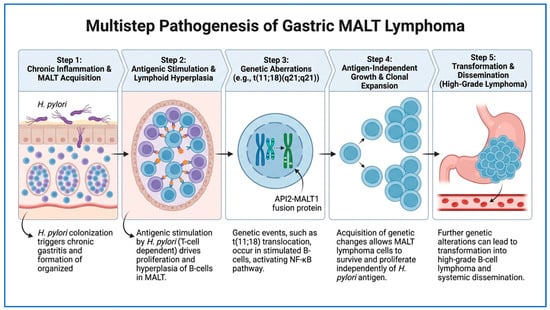
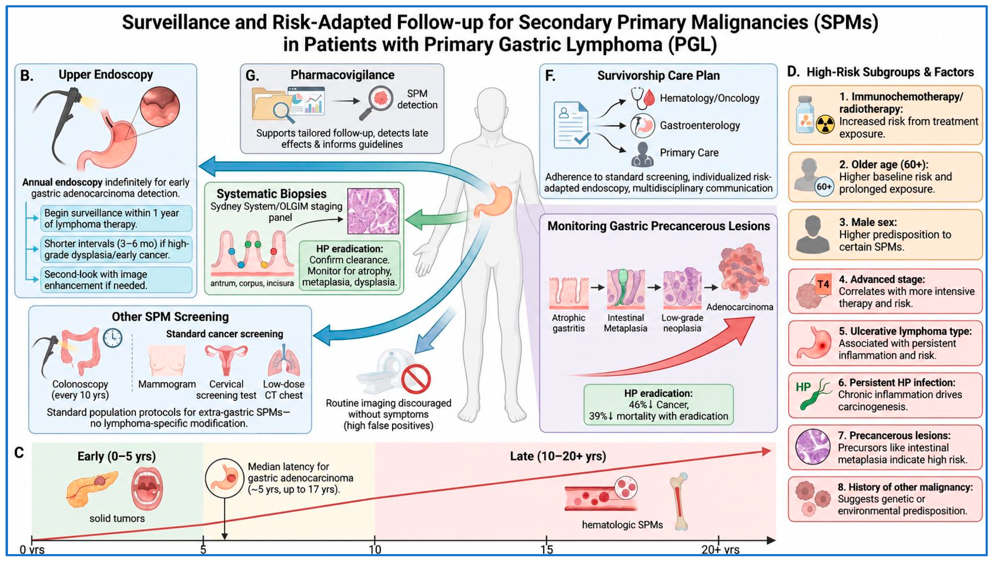
Disclaimer/Publisher’s Note: The statements, opinions and data contained in all publications are solely those of the individual author(s) and contributor(s) and not of MDPI and/or the editor(s). MDPI and/or the editor(s) disclaim responsibility for any injury to people or property resulting from any ideas, methods, instructions or products referred to in the content. |
© 2026 by the authors. Licensee MDPI, Basel, Switzerland. This article is an open access article distributed under the terms and conditions of the Creative Commons Attribution (CC BY) license.