


Abstract
Polymer-based dressings constitute an important class of macromolecular biomaterials enabling controlled drug delivery and enhanced wound healing performance. This review summarizes recent advances in the design, fabrication, and functionalization of polymer dressings, with emphasis on natural and synthetic polymer systems applied in biomedical topical and transdermal drug administration. Key material properties, including biocompatibility, mechanical stability, porosity, and degradation behavior, are discussed in relation to drug loading capacity and release kinetics. Current fabrication strategies, such as electrospinning, hydrogel formation, casting, and multilayer assembly, are critically evaluated with respect to structural control and scalability. Particular attention is given to antimicrobial and stimuli-responsive platforms capable of dynamic interaction with the wound microenvironment. Furthermore, challenges related to long-term stability, regulatory requirements, and clinical translation are addressed. By integrating recent experimental findings, this review highlights essential structure–function relationships governing polymer dressing performance and provides design guidelines for next-generation macromolecular topical and transdermal care systems with improved multifunctionality and clinical applicability.
1. Introduction
In recent years, drug delivery (DD) has shifted from conventional oral and injectable preparations towards more sophisticated systems that enhance compliance and therapeutic efficacy while minimizing side effects. Advanced transdermal and topical therapeutic systems have emerged as alternatives to conventional dosage forms, demonstrated in multiple reviews and clinical studies [1,2,3,4,5]. Early systems focused on small-molecule delivery (e.g., nicotine and hormones), whereas recent developments increasingly address biologics, vaccines, and wound healing [3,4,6,7,8]. These systems range from simple drug-loaded protective films to complex polymer patches, offering unique and compelling advantages over traditional dosage. They provide a non-invasive and painless delivery alternative to injections, enable the sustained release of active pharmaceutical ingredients (APIs) to target sites, allow for easy therapy termination by simply removing the patch, and offer great potential for improved patient compliance [9,10,11]. The pharmaceutical industry has broadly evolved from passive formulations like ointments and creams to polymer-based DD, prioritizing precision medical delivery and material innovation [12]. Therapeutic polymer patches comprise Transdermal Drug Delivery Systems (TDDSs), hydrogel wound dressings, dissolvable and coated adhesive systems, and smart biodegradable patches. They are multifunctional biomedical platforms that combine controlled release, antimicrobial functions, tissue support and even sensing abilities. Polymer patches were originally developed to deliver small molecules like nicotine and hormones, but have expanded rapidly to include localized treatment of pain, inflammation, wound healing, antimicrobial delivery, cancer therapy, insulin delivery, Gene therapy, cardiovascular applications, vaccination, etc. [4,13,14]. Despite proven success, cross-disciplinary gaps exist across polymer chemistry, materials engineering, pharmacokinetics, and clinical therapeutics, making comprehensive understanding challenging.
The skin, as the largest body organ and effective biological protective barrier, presents avenues and challenges for drug administration [15]. It provides accessible routes for local and systemic administration, whereas the inherent resistance of the stratum corneum to permeation of pharmacologically active molecules is driving innovation in developing polymeric systems that balance enhanced permeability and biocompatibility [16]. The efficacy of the delivery is not merely a function of the API but a complex interaction between the components and the epidermal layer. Many studies and innovations have focused on developing polymeric systems with tailored physicochemical characteristics like hydrophilicity, biocompatibility, mechanical strength, and adhesive properties to bypass etc. For instance, microneedle technology is advancing from concept to clinical applications for vaccines, biologics, and pain relief, as water-soluble polymeric technology that creates transient micro-channels to bypass molecular size and polarity barriers without full-needle penetration [17]. Hydrogel-based dressings are also increasingly being deployed in wound care as therapeutics to replace inert covering materials [18]. Modern hydrogel dressings are functionalized to maintain a hydrated microenvironment, enhance gas exchange, provide optimal adhesion, absorb fluids, incorporate antimicrobials, and serve as growth factors to accelerate healing and reduce scarring [19,20]. Recent studies highlight their expanded impact in hemostasis, angiogenesis, anti-inflammation, delivery of cells and biomolecules as well as their key limitations, including the high risk of microbial contamination, inconsistent mechanical strength, and difficulty in balancing sustained release with rheology and comfort [18,19].
A pressing, yet under-discussed aspect of polymer patches is the life cycle and environmental sustainability of material development. Traditional polymer patches made from fossil precursors are usually effective, with high stability, but the stability translates into persistence in the environment. Synthetic polymers deployed as adhesive matrices and backing layers may be durable but highly resistant to biodegradation, resulting in disposal concerns. This has increased demand for greener alternatives by regulatory and procurement bodies, herein focused on bioresorbable options with minimal environmental impact yet high safety and performance. Researchers have explored biobased precursors like PLA/PLGA, polyglycols, advanced biopolymers, etc., in the context of polymer patches, noting significant promise [21,22,23,24]. Nonetheless, challenges remain compatible with sterilization, variability in biobased materials, predictability of degradation profiles and alignment with strict pharmaceutical quality standards. Hence, concerted advances in materials science, polymer chemistry, process engineering and regulatory science are necessary to address sustainability.
This review therefore seeks to comprehensively explore polymeric patches, addressing the core components, trends and promising future trajectories. The study provides information on classification based on the mechanism of action (topical vs. transdermal) and design (matrix, reservoir, drug in adhesive). A detailed examination of natural and synthetic polymer formulation bases, penetrating enhancers, and plasticizers is presented, highlighting their roles and selection criteria. The paper further explores the essential manufacturing processes standardized through in vitro and in vivo performance evaluation methodologies. By investigating challenges and identifying recent developments, the review seeks to outline promising perspectives in the field, serving as a valuable resource for future innovation in the design and development of next-generation therapeutic polymer patches.
2. Pharmaceutical Products for Skin Application
Pharmaceuticals are defined as agents intended for mitigating, therapeutic and disease—preventive applications in humans or animals. They are characterized by their selective action in the treatment of common as well as rare diseases, which may affect entire organs or single cells. The process of discovery and development of medicinal substances derived from animal or plant sources is very demanding (products must not cause excessive and dangerous side effects). To enable accurate dosing, stability, patient acceptability, and efficient delivery to the site of action, APIs are formulated into specific dosage forms and drug delivery systems tailored to the route of administration [25]. Pharmaceutical dosage forms are commonly classified by physical state as solid (e.g., tablets, capsules, powders, granules), semi-solid (e.g., creams, ointments, gels, pastes), liquid (e.g., solutions, suspensions, emulsions, syrups), and gaseous or aerosol preparations (e.g., inhalations, sprays). Each class offers distinct advantages and limitations in terms of dose precision, stability, onset and duration of action, and patient acceptability, guiding selection for different clinical scenarios and patient groups [26,27]. Depending on the physicochemical properties of the active substance, the condition and age of the patient, the time and desired site of action, these agents may be administered topically or transdermally [28]. A diagram of drug formulations used in specific areas of the body is shown in Figure 1.
Figure 1.
Methods of drug administration. Adapted from [27], licensed under CC BY 4.0. Created in BioRender.com. https://BioRender.com/hqyk32y (accessed on 3 March 2026).
The transport of drugs through the skin is characterized by its ease of use and application, especially for patients who have difficulties with oral administration or who are unconscious [26]. The skin is an ideal place to deliver medicinal products both locally (which reduces the likelihood of side effects [26]) and systemically [29]. The most popular systems for use on the skin are semi-solid formulations, e.g., ointments, creams, lotions, gels and pastes. They consist of a simple or complex base (hydrophilic or hydrophobic), in which one or more active substances are usually dissolved or dispersed. Often, the composition of the base can have a major impact on the performance of the whole product [25]. Semi-solid dosage forms may have therapeutic, protective or cosmetic functions. It is a suitable dosage form for bitter drugs and is generally more stable compared to, e.g., liquid forms [26]. The composition may additionally contain auxiliary substances such as: emulsifiers, antioxidants, preservatives, plasticizers or permeation promoters [2,25]. In addition to commonly used products, novel gels, aerosol foams, bioadhesives and vesicular carriers are becoming increasingly well known. The disadvantages of local drug delivery systems are the possibility of allergic reactions, including irritation and dermatitis, as well as limited skin permeability of some active substances [29]. Therefore, when choosing a topical formulation, it is necessary to pay special attention to the appropriate formulation and functional characteristics of this type of product.
3. Medicinal Patches
Medicinal patches are solid or semi-solid preparations spread on a paper, fabric or plastic base [25]. Their purpose is to keep one or more active substances in close contact with the skin. They take the form of single-layer or multi-layer, thin and flexible materials of various shapes and sizes [25,30,31]. Patches have been used as a form of administration of the drug to the general circulation (transdermal drug delivery systems, TDDSs) or topically (to the skin or underlying muscle tissue) [32]. Active substances delivered transdermally or locally via medicinal patches reach the appropriate site in a painless, targeted and concentrated manner [5]. Multiple clinical and formulation studies report that transdermal delivery enables efficient, sustained administration with reduced adverse effects compared to oral systems. Studies have revealed that modern patches use polymer matrices, microreservoir structures, and drug-in-adhesive architectures to control release kinetics, enhance skin permeation, and improve mechanical flexibility and adhesion [33]. This is a significant advantage compared to oral drug delivery systems, which may have a negative effect on the gastrointestinal tract (patches avoid the first-pass metabolism) [32]. However, limitations related to dose loading, drug stability, solubility, skin permeation and skin irritation remain significant challenges [34].
Medicinal patches can be divided according to their mode of action as a topical or transdermal drug delivery system. They are generally classified as formulations intended for local or systemic use. Both approaches are common ways of delivering pharmaceutical drugs, including analgesics, that alleviate mild, moderate and even very severe pain conditions. Popular pharmaceuticals used in medical patches include: non-steroidal anti-inflammatory drugs, opioids, lidocaine, capsaicin, steroids, diclofenac sodium, metamizole, etc. When considering the use of patches in analgesic therapies, physicians must primarily consider the type of pain and which active substances are best suited to the target-specific pain receptors [35].
3.1. Topical Drug Delivery Systems
Topical preparations have been used for centuries, mainly for the treatment of local skin lesions. This therapy requires the active substance to penetrate the appropriate layers of the skin to treat any abnormalities (or relieve pain) [35]. Such drug delivery may be targeted to several sites such as the stratum corneum, viable epidermis, dermis, hair unit, subcutaneous tissue and deeper tissues [36]. The whole process should occur with little or no systemic accumulation (potentially reducing the possibility of side effects resulting from parenteral exposure) [35]. The effectiveness of therapy near the application of the material depends on the physicochemical properties of the active substance, the patient’s respect for the treatment regimen, as well as the ability of the system to remain in contact with the skin during treatment, promoting penetration of the medicinal substance through the skin barrier. Conventional formulations for topical and dermatological administration of drugs have some limitations. Several studies highlight poor permeability through the stratum corneum as a key limitation of conventional topical formulations, which is especially problematic for hydrophilic or high-molecular-weight formulations [37]. Wan & Huang [38] and Yu et al. [39] emphasized that even optimized semi-solid carriers often fail to achieve therapeutically relevant levels in deeper skin layers or systemic circulation for such molecules, making poor permeability a central limitation of classical topical therapy. To mitigate this, studies have explored excipient optimization (e.g., co-solvents, lipids, penetration enhancers) and bioadhesive polymer systems to increase residence time and improve partitioning into the skin, but these strategies generally provide only modest effects and are now viewed as adjuncts rather than definitive solutions, particularly when compared with more advanced approaches such as nanocarriers or innovative enhancement methods. For instance, Yi et al. [40] employed mitochondria-targeting delivery systems by using a DEA-modified HPMA copolymer-CPT conjugate, which facilitates the mitochondrial accumulation of the drug, thereby improving targeting effectiveness within cancer cells. Yu et al. [39] also report a combination of physical enhancers with nanocarriers, such as solid lipid nanoparticles and nanostructured lipid carriers, to further improve permeation and drug targeting efficiency across the skin. Li et al. [41] developed niosomal nanocarriers for dermal delivery of antioxidants like epigallocatechin gallate; this nano-carrier system allows for deeper skin penetration and sustained release, increasing antioxidant effects and cell protection. These strategies exemplify how targeting specificity, such as mitochondrial or cellular surface markers, and nanoencapsulation techniques can significantly improve drug permeation through challenging biological barriers.
The bioavailability of a pharmaceutical through the skin may be improved by the addition of appropriate excipients [36]. The preparation itself must remain on body tissues or wounds for a defined and effective period of time [37]. The main advantages of this route of delivery are: acceptance by the patient, due to its painless and non-invasive nature; good pharmacological and physiological response; and minimal systemic toxicity and exposure of the drug to undesirable sites [36]. Patches may provide local analgesia of the skin prior to injection of the solution or minor surgical procedures, as well as relieve minor muscle tension, sprains, bruises and some neuropathic pain syndromes. The most commonly used topical analgesics, which are available on the market, include lidocaine, lidocaine with tetracaine or prilocaine, capsaicin and diclofenac [35].
A very important issue concerning medical patches is dressings, which are responsible for the local delivery of drugs and the healing of wounds. These materials are primarily synthesized from hydrophilic, cross-linked polymers, either natural or synthetic, which exhibit swelling ability [42]. The current trend is to replace traditional dressings, e.g., cotton and gauze, which have no therapeutic effect, with technological and personalized forms. It is important that such patches, e.g., hydrogels, can be individually adjusted to maintain the patient’s anatomy and specific needs, which will ensure effectiveness and acceptable esthetic aspects [36]. In recent years, the formulation of the hydrogel-based patches has gained great importance due to their unique properties [43]. Hydrogels are characterized by high efficiency in retaining a significant amount of water (even up to 90% [43]) and the ability to reach a mass up to a thousand times greater than their dry mass after swelling. This significant amount of water present in the hydrogel structure plays an important role in the elasticity and hydration of the skin (helps maintain skin moisture) [43,44]. The moist environment under the dressing is particularly advantageous if the healing processes begin immediately after application of the product to mechanically damaged epidermis. Water first penetrates into the wound, which causes softening and moisturizing of the necrotic tissue, which promotes autolysis processes and clears the wound of exudate components (Figure 2) [42,45]. The hydrogel absorbs the wound exudate, effectively reducing bacterial growth, and its ability to swell moderately allows it to adapt closely to the wound surface (suitable for wounds with irregular shapes). Additionally, this swelling process can regulate the release of therapeutic agents and the hydrogel’s network structure, which prolongs treatment effects and promotes efficient wound healing [45].
Figure 2.
Mechanism of action of hydrogel-based dressings. Created in BioRender.com. https://BioRender.com/rzwep3l (accessed on 3 March 2026).
An additional advantage of hydrogels is their porosity, large specific surface area and appropriate degradation rate (occurring through hydrolysis) [46]. Currently, the skin wound-covering matrix is being integrated with typical types of drugs, bioactive agents, cells and/or specific genes to aid healing and accelerate regeneration [47]. Hydrogel patches may deliver a drug in a controlled manner over a limited period of time [43]. They are often found in the form of wound dressing used only to cover wounds, but they can also serve as drug delivery systems. The high water content of hydrogel-based products causes a cooling effect after their application, which is why they are particularly used for the treatment of burns. Some studies have also shown that they relieve pain and prevent scarring [42]. The ideal properties of the patches are determined by the kinetics of drug release (if he has one) and mechanical properties, e.g., transparent dressings are more beneficial due to the possibility of observing changes in the wound over time [42,43]. A key benefit of hydrogels lies in their ability to deliver bioactive compounds, such as antibiotics, anti-inflammatory agents and antioxidants, directly to the wound site [42]. Unfortunately, hydrogels are not able to eliminate pathogenic microorganisms on their own, which is why wound infections continue to be a major problem. New therapeutic development strategies focus on producing patches that provide a more favorable environment for wound and burn healing, and thus exhibit anti-microbial properties [46,48]. Such dressings can be divided into three categories: bioactive; passive or inert; and interactive. Bioactive dressings are created to gradually release bioactive molecules. On the other hand, inert and passive patches are common materials that act primarily as a physical barrier, protecting the wound from pathogens in the external environment (physical barrier function). Interactive dressing affects the surface of the wound, but also actively promotes healing process. Another classification of these materials is based on their complexity and divides them into traditional (e.g., gauze), modern (e.g., foams and films) and advanced (e.g., tissue engineering products) [36]. Table 1 outlines several notable examples, showcasing the innovative use of hydrogels in medicinal patches as effective drug delivery systems.
Table 1.
Representative examples of hydrogel-based medicinal patches for drug delivery.
3.2. Transdermal Drug Delivery Systems (TDDSs)
TDDSs facilitate the passage of therapeutic amounts of active drug substances through intact skin and into the general circulation to achieve systemic effects [25]. This method provides controlled and continuous administration of the drug [32]. One of the indicators supporting drug absorption is the measurable concentration of the active substance in blood and urine (in the form of its metabolites) [25]. In general, the administration of drugs through the skin is divided into three generations, based on their mechanism and complexity [60], as shown in Figure 3.
Figure 3.
Classification and distinguishing characteristics of Transdermal Drug Delivery Systems (TDDSs), categorized into first, second, and third generations. Created in BioRender. https://BioRender.com/vh4k98x (accessed on 3 March 2026).
TDDS products include nicotine, testosterone, hydrocortisone, scopolamine, estradiol, nitroglycerin, methylphenidate and insulin [2,31,61]. The advantages of this dosage form include discretion, reduced number of side effects, sustained (often long-term) drug delivery, and the elimination of first-pass metabolism [29], i.e., prevention of inactivation by digestive and hepatic enzymes [25]. In addition, transdermal delivery avoids serious risks associated with medical waste (disease transmission after needle reuse) and pain during subcutaneous injection, reducing patient discomfort [60]. Another important advantage is that the TDDS patches can be easily removed in case of side effects or when treatment is no longer required [62]. They are also easily and quickly identified in emergencies, e.g., fainting due to their physical presence, features and identifiers [25]. However, this method is not appropriate for controlling acute pain and clinical situations that require rapid drug dosing [26]. TDDS components may cause skin irritation and local swelling. Damage to the transdermal system may result in uncontrolled drug release. First-generation TDDSs are limited to low-molecular-weight, lipophilic drugs, a limitation extensively discussed in earlier reviews. Recent third-generation systems aim to overcome this barrier through microneedles, electroporation, thermal ablation, and targeted chemical disruptors to breach the stratum corneum barrier while sparing deeper tissues, enabling the delivery of macromolecules like proteins, peptides, and vaccines. For instance, Aziz et al. [63] demonstrated microneedle arrays delivering insulin with bioavailability comparable to subcutaneous injection in preclinical models, highlighting dose-sparing and reduced pain [63]. Kushwaha et al. [64] reported clinical trials of electroporation-enhanced patches for lidocaine, achieving a five-fold permeation increase without skin damage [64]. Crasta et al. [65] reviewed regulatory hurdles, noting FDA approvals for microneedle prototypes but emphasizing scalability issues in manufacturing uniform arrays and ensuring batch-to-batch reproducibility [65]. Therefore, only strong drugs are suitable candidates for transdermal administration due to the natural limitations of drug absorption imposed by the skin’s impermeability [25].
Patches can also be classified according to their construction. There are two main types: matrix and reservoir (membrane) [66]. The matrix system design is characterized by the presence of a semi-solid matrix, where drugs are evenly dispersed. The active ingredient layer is applied onto the adhesive layer, which is then supported by a release liner [4,30]. The structure of the matrix-type therapeutic system is shown in Figure 4 below.
Figure 4.
Types of patches: (a) matrix-type; (b) reservoir-type (membrane) patch; (c) single-layer drug in an adhesive patch; (d) multi-layer drug in an adhesive patch. Adapted from [67], licensed under CC BY 4.0. Created in BioRender.com. https://BioRender.com/46gfb7i (accessed on 3 March 2026).
In the case of a reservoir system, the patch design includes a reservoir containing a drug solution or suspension, which is separated from the release layer by a semi-permeable polymer membrane and adhesive [4,30]. The structure of the membrane system is shown in Figure 4. The simplest forms of these products in terms of construction are drug-in-adhesive patches. This single-layer structure is characterized by the incorporation of the active substance directly into the adhesive agent that contacts the skin [4]. The multi-layer version is very similar to the single-layer form, except that it includes a membrane between the two layers of the drug (Figure 4) [68,69].
The design and evaluation of therapeutic patches are guided by a set of ideal performance and patient-centric criteria, which also highlight their distinct advantages over conventional semi-solid formulations (e.g., creams, ointments, gels). An ideal patch should possess a shelf life of up to two years, a small application area (typically < 40 cm2), and a convenient dosing interval ranging from once daily to once weekly to maximize compliance. It must be visually acceptable (e.g., transparent or skin-toned), cause no unacceptable cutaneous reactions (allergy, irritation), and be easy to apply and remove without damaging the epidermis or leaving residue. Critically, it must maintain reliable adhesion and consistent biopharmaceutical activity throughout the wear period [70]. These requirements are met through a sophisticated formulation that, beyond the active pharmaceutical ingredient (API), includes a polymer matrix (natural or synthetic) as the foundational component, along with auxiliary agents such as stabilizers, permeation enhancers, solvents, and plasticizers [2,25,33].
The practical benefits stemming from this design are evident when compared to traditional topical forms. As summarized in Table 2, medicinal patches offer superior wearing comfort, application accuracy, and extended-release capabilities, while eliminating issues like greasiness and clothing contamination common with semi-solids. This unique profile has fueled their expansion beyond pure drug delivery into rehabilitative and cosmetic markets, including thermal/cooling patches and skincare patches [71].
Table 2.
Comparative analysis: conventional semi-solid formulations versus medicinal patches characteristics and ideal patch attributes [71].
3.3. Determinants of Skin Permeation
The delivery of active ingredients requires a comprehensive understanding of the skin’s structure, its physiology and associated diseases, as well as the properties of the drug and its carrier. There are certain internal skin factors that can affect the delivery of active ingredients, such as: the function of the skin barrier provided by the stratum corneum; hydration; anatomical location; pathological effects; age; ethnicity; and gender [36].
With age, drug absorption through the skin changes. Aged skin surface is much rougher and thinner, and there is a decrease in hydration and sebum production (so-called paper-thin skin) [28]. One of the main disadvantages in delivering active ingredients through the skin is the barrier function of the stratum corneum [28,36]. In order to diffuse effectively, molecules must meet several requirements, such as degree of ionization and small molecular size (the ideal drug molecular weight is 400 Da or less [25]). The drug should have a greater physicochemical affinity to the skin than to the carrier, so that the active substance leaves the matrix in favor of the epidermal surface. A certain solubility of the pharmaceutical in both lipids and water is considered important for effective transdermal absorption. Solubility in water determines the concentration at the site of absorption, and the division ratio affects the rate of transport. Several authors confirm that non-polar, lipophilic drugs selectively follow the transcellular route through corneocytes, whereas polar, hydrophilic compounds rely predominantly on the tortuous intercellular lipid pathway between keratinocytes. However, the quantitative contribution of the transfollicular route via pilosebaceous units remains debated, with recent studies estimating it at 0.1–13% of total flux depending on follicular density, drug size, and vehicle formulation [28,72]. The carrier containing the active substance and its properties (type of dosage form, e.g., patch, gel, emulsion; ingredients concentration and presence of permeation enhancers, pH, viscosity, and charge) can significantly affect the rate of drug delivery and its permeability [36]. Penetration of the patches is improved by an increase in temperature via an external heat source, as well as by substances that increase blood flow and by solvents, e.g., dimethyl sulfoxide (DMSO) [65]. In addition, occlusive preparations always improve the penetration of the active compound by increasing skin hydration. Therefore, the design of the appropriate carrier has a key impact on the quality of active agent, and thus the speed of the disease treated [36].
The therapeutic effect of locally applied preparations occurs in three stages. At first, the active substance is released from the formulation, then it is divided and distributed within the skin barrier, and finally, it penetrates or diffuses through the stratum corneum [36]. The passage of drugs into the skin involves two potential pathways: transepidermal (through the epidermis) and transappendageal (via the hair follicles, sebaceous and sweat glands) [65]. Hydrogels offer an effective platform for topical and local administration. Once loaded, drug release from hydrogels can occur via diffusion, swelling or chemically. In swelling-controlled release, the hydrogel absorbs biological fluids and expands, allowing the drug to diffuse as the polymer chains relax [42]. Patches designed to deliver the active substance to the general circulation have a similar onset of penetration as patches that only work in the area of the skin or muscle tissue. The exception is that this process involves several additional stages related to systemic absorption, including uptake of the drug through the network of capillaries in the papillary skin layer and further influence on the target organ [65]. Figure 5 shows the pattern of active substance penetration through the relevant skin layers for both topical and transdermal patches.
Figure 5.
Release profile for local and transdermal delivery systems. Created in BioRender.com. https://BioRender.com/6y94ihg (accessed on 3 March 2026).
3.4. Key Components of Patch Formulation
3.4.1. Polymer Bases
Polymer matrices serve as the foundational structural and functional component of therapeutic patches, controlling drug delivery, mechanical integrity, and biocompatibility. In multi-laminated transdermal drug delivery systems (TDDSs), polymers play distinct roles across different layers: as pressure-sensitive adhesives for skin contact, as rate-controlling membranes, as impermeable backing films, and as the primary drug-loaded matrix itself [30,32]. Each layer of the system requires specific characteristics provided by the appropriate type of polymer used. For example, polyurethanes provide flexibility, and polyvinyl alcohol (PVA) gives hydrophilicity and strength. On the other hand, poly(methyl methacrylate), or PMMA for short, is characterized by physical strength and ensures the transparency of the product. Polysiloxanes or silicones provide insulating properties, and the use of polyethylene, in addition to strength, also prevents swelling. A group of compounds, such as polyvinylpyrrolidone (PVP) allows the drug to be suspended in the matrix [70].
Polymer bases are the foundation of drug delivery systems. Hence, their selection is a critical step in the product manufacturing process. The nature of the substance, with such a high molecular weight, determines the release of the drug at the required therapeutic rate. Polymers have the ability to form films after being cross-linked with specific substances. Typically, they are dissolved in the appropriate solvent together with the drug molecules, and a thin patch is formed after solvent evaporation [27]. To obtain the desired properties, polymers can be used alone or as mixtures in various ratios [37]. Water-insoluble polymers form waterproof layers that are often brittle and inflexible, making them difficult to adhere to the skin. To increase their uniformity, plasticizers are often added, or mixtures of hydrophobic and polar polymers are formed [73]. The selection and design of the base polymer must be carefully considered when seeking to meet the various criteria for producing an effective patch. The main challenge is to design a polymer matrix and then optimize it for the drug load, not only in terms of its release properties, but also in terms of adhesion, balance, physicochemical properties, compatibility and stability with other system components as well as with the skin [32,74]. A polymer used in pharmaceuticals should meet several basic criteria. The base polymers should be stable, non-toxic, easily manufactured, and inexpensive. More importantly, their decomposition products must not be antagonistic to the patient. Properties such as swelling, biodegradation and mechanical properties such as stretchability can also be taken into account when choosing a polymer. The designed material must have sufficient bending resistance but also optimal tensile strength, to determine its ease of breaking and damage [27,32,75]. Overall, polymer selection depends strictly on the desired properties and the specific application for which it is intended [75].
Polymers are broadly classified by origin (natural vs. synthetic) and functionality, as detailed in Table 3. This selection includes common natural polymers like chitosan and alginate, known for their biocompatibility and hydrogel-forming ability, and synthetic ones like ethyl cellulose (EC) and various polyacrylates (Eudragit® series), prized for their predictable mechanical and release properties [73,76]. Synthetic polymers, while offering superior tunability and consistency, raise environmental concerns due to their persistence. Consequently, significant research is directed toward sustainable alternatives derived from natural, renewable sources (e.g., plant-based polymers, agro-industrial byproducts), aiming to combine performance with environmental responsibility [27].
Table 3.
Representative polymers used in therapeutic patch formulations, categorized by origin and key properties [73,76].
It is accepted that polymers such as crosslinked hydroxypropyl methylcellulose (HPMC), polyethylene glycol, chitosan, sodium alginate, ethyl cellulose and polyvinylpyrrolidone (PVP) are used as patch-forming matrices. Other compounds, such as silicone rubber and polyurethanes, are used to create membranes that control the rate of release [32]. The current base production route for polymer bases typically involves processes for obtaining synthetic patches. The advantage of this approach is the ability to confer polymer compounds with the desired mechanical and chemical properties. Unfortunately, these polymers are difficult to decompose naturally in biological processes, which negatively impacts the environment due to the generation of a large amount of waste. Researchers continue to work on more environmentally friendly production routes that provide sustainable alternatives to methods for obtaining synthetic materials. Natural polymers are obtained from inedible and easily available plants, as well as from by-products of the agricultural and food industries. Natural polymers, such as silk fibroin, keratin, alginate, hyaluronic acid, cellulose, collagen, and chitosan, are particularly useful when used in therapeutic patches due to their biocompatibility and ability to form strong, flexible bonds with the skin. They also have unique mechanical, chemical, and biological properties that can be customized for specific medical applications. The ability of natural polymers to form hydrogels increases the stability and protection of the drug through hydrophilic networks, further improving the stability and efficacy of the drug [27].
3.4.2. Penetration Enhancers
Penetration enhancers are compounds that temporarily and reversibly increase the permeability of the skin by altering the barrier to the flow of the desired substance (i.e., reducing its resistance to diffusion, e.g., drugs) and are considered an integral part of most transdermal preparations [70]. Barrier modifications involve applying the appropriate compound (chemical pathway) or changing the interaction of the preparation with the skin itself (physical pathway) [77]. Generally, permeation enhancers can work in three ways, e.g., they can interact with structural components and disturb the lipid structure in the stratum corneum of the epidermis (chemical enhancers) [65]. Penetration enhancers should be pharmacologically neutral, non-toxic, non-allergenic, and non-irritating. They usually have the ability to produce specific, reversible and predictable effects. They do not cause the loss of body fluids, electrolytes or other endogenous materials [70]. It is widely accepted that for penetration enhancers, increased potency is directly correlated with increased skin irritation. More than 300 substances have been shown to have the potential to increase skin permeability, and this number continues to grow [77]. They can be classified into two main categories: solvents and surfactants [70].
Solvents increase penetration, probably by swelling polar pathways in the skin and fluidizing lipids [2]. Propylene glycol and isopropyl myristate have the additional advantage of increasing penetration without evaporation (compared to, e.g., ethanol) [73].
Water is a well-recognized and strong natural penetration enhancer, and although its mechanism of action is not fully understood, it helps in the penetration of both hydrophilic and lipophilic molecules [78]. Water, as the primary solvent used in drug technology, must be appropriately purified of all impurities: chemical, e.g., chlorides, sulfates, calcium; and mechanical and biological, i.e., microorganisms. For this purpose, distillation, ion exchange or reverse osmosis are used, among other methods. Purified water must meet all the requirements, including that after evaporation to dryness, it must not leave more than 0.001% residue [25].
Another very useful group of solvents is alcohols, especially ethanol. This substance is widely used in pharmaceutical preparations due to its relatively high miscibility with water, as well as its ability to dissolve many flavoring, antibacterial and preservative agents. Manufacturers selling pharmaceuticals containing even trace amounts of alcohol are required to place warning labels, especially on products intended for children [25].
It is believed that surfactants facilitate polar transport, especially of hydrophilic drugs. Their ability to modify penetration depends on the polar “head” group and hydrocarbon chain length [2], i.e., their affinity to both environments (both polar and non-polar). Unfortunately, the addition of surfactants must be carefully balanced due to the high risk of skin irritation [70].
The latest techniques to improve drug penetration by physical methods include ionophoresis, electroporation, microneedles, skin puncture, needle-free injection, ultrasound and temperature manipulation [2,70]. Electroporation involves the application of short, high-voltage electrical pulses to the skin, while ionophoresis involves passing a few milliamperes of current to a few square centimeters of skin through an electrode placed in contact with the preparation [2]. Temperature changes and lasers use an external energy source (located in the disposable patch applicator) that penetrates the stratum corneum to form microchannels, without further damaging the living layers of the epidermis. Sonophoresis (ultrasound) increases skin permeability by using high (MHz range) or low (kHz range) frequencies [77]. Another technology currently under development uses microneedles located on the surface of a patch made of, for example, silicone [79]. Combinations of these techniques can potentially exhibit synergistic effects, e.g., electroporation disrupts the barrier structure of the epidermis, increasing its permeability to drugs, while ionophoresis provides an additional driving force for the diffusion of charged particles over passive diffusion [77].
3.4.3. Plasticizers
Plasticizers are generally used to change certain physical and mechanical properties of the material [80]. Typically, this includes imparting flexibility and/or improving strength [37], but can also involve reducing brittleness [80]. Reducing the stiffness of the polymer skeleton increases the diffusion properties of the drug (the higher the degree of crosslinking, the denser the matrix, and the slower the transport of active substance molecules) [70]. The plasticizer used should be compatible with the polymer matrix (but also with other polymers, e.g., adhesive) and have low skin permeability [37,80]. The high compatibility allows it to blend completely with the base polymer and remain permanently within it. In addition, these materials are expected to be durable and effective over a wide range of temperatures, health-friendly and safe, and relatively low in production cost. To demonstrate its effectiveness, the plasticizer should be able to diffuse from the solvent into the polymer base, where it disrupts the intermolecular interactions of the base. A flexible plasticizer molecule with a lower molecular weight (higher activity) or with long chains (CH2)n is more effective in increasing plasticity. In turn, low polarity and hydrogen bonds reduce the interactions in the polymer–plasticizer system. Forces acting on such a complex, in addition to hydrogen bonds, include dipole–dipole interactions and dispersion forces [80].
Plasticizers are most commonly non-volatile organic liquids or solids with a low melting point. When classifying these substances, compatibility, general structure (monomer or polymer), function, and chemical structure are taken into account. Phthalic acid esters are a frequently used group of these materials, which is why dioctyl phthalate accounts for 50% of the global consumption of plasticizers [80]. Sorbitol, glycerin, polyethylene glycol, propylene glycol, triethyl citrate, and others are also commonly used [37,70]. In addition, adipic, azelate and sebacate compounds are distinguished from other groups by their relatively low viscosity, providing flexibility to polymers used at low temperatures. Surfactants and preservatives can also influence the mechanical properties of the matrix [80]. Polyhydroxy alcohols found in therapeutic patches covered by patents are generally used for emollient purposes [80]. Glycerol is a clear, syrupy liquid with a sweet smell that is miscible with both water and ethanol. Propylene glycol has a lower viscosity than glycerol and is miscible with water in any ratio. It is a useful colorless, odorless liquid with a wide range of applications and often replaces glycerin in modern formulations. It has hygroscopic properties and promotes disinfectant effects (it is considered non-toxic, but has a minimally irritating effect—more than glycerol) [25]. Higher alcohols such as dodecanol, or mineral oil, isopropyl myristate and polyethylene glycol are also added to the patches. Well-known viscosity-enhancing agents used to form hydrogel dressings are high-molecular-weight derivatives of acrylic acid, cross-linked with sucrose or pentaerythritol, such as Carbomer or Carbopol. They are characterized by the ability to swell in water and high gelling properties, biodegradability and bioadhesiveness, which make them ideal for dermal delivery systems [25,81].
The addition of substances that affect the mechanical properties of the patch is intended to facilitate thermal treatment, modify drug release, or improve the appearance and/or surface quality of the dosage form [80]. In transdermal patches, the reduction in strength is less significant as long as it remains sufficient to withstand tearing during placement or change in position on the skin. The addition of a plasticizer is also important due to the adhesive properties of the base surface [82]. In the case of therapeutic patches, plasticizers are most commonly used in 5–20% proportions [80]. In addition, water can have a notable effect on mechanical behavior in polyvinyl alcohol because of his high moisture sensitivity. High water content can significantly decrease the mechanical strength and elastic modulus of PVA [83]. Table 4 presents conventional plasticizers and their approximate concentrations used in pharmaceutical formulations.
Table 4.
Key plasticizers used in patch formulations: properties, and typical use parameters [25,73,80,82].
3.4.4. Active Substances
A drug is defined as an agent intended for use in the diagnosis, mitigation, treatment (and cure) or prevention of diseases in humans or animals. One of its most important characteristics is its biological activity, and thus its effect on the body [25]. However, not all active substances are suitable for administration in the form of patches [27]. Therefore, the drug from which a therapeutic patch is designed should have well-defined physicochemical properties [80]. Whether the active substance is applied topically or systemically, it must penetrate the stratum corneum effectively. Pharmaceuticals with a predominantly lipophilic nature tend to pass through the skin better than their hydrophilic counterparts (because the epidermal matrix is hydrophobic) [73]. Molecular weight (less than 1000 Da, although ideally below 500 Da) plays a decisive role, as small molecules exhibit a higher diffusion rate [5,73,80]. The medicinal compound used must be effective even in small doses (<20 mg per day). An equally important issue to consider is the solubility in water, i.e., above 100 μg/mL [80]. In a situation where the medicinal compound is poorly absorbed by the body, the idea of a prodrug, i.e., a pharmacologically inactive substance, is used. The prodrug is much better absorbed, and once it is already in the body, it is metabolized (hydrolyzed) into the active form. This modification increases the bioavailability, and thus also the solubility, in water (and water/oil ratio) [84]. The list of the most commonly used active compounds in patches is given in Table 5.
Table 5.
Certain active substances used in therapeutic patches [4,31,35].
Several medicinal substances are currently available in TDDS patch form, including estradiol, clonidine, fentanyl, nicotine, scopolamine and nitroglycerin. The site of application varies depending on the category of the therapeutic active compound, e.g., estradiol is applied to the abdominal area and nitroglycerine to the chest [4]. On the other hand, topical patches usually contain a substance that acts directly with the epidermal layer, e.g., lidocaine, capsaicin, antibiotics and diclofenac [35,36].
3.4.5. Antimicrobial Substances
In addition to stabilizing pharmaceutical preparations against chemical and physical degradation caused by changing environmental conditions, selected products must also be protected against microbial contamination (i.e., appearance of bacteria, yeast or mold). Antibacterial preservatives are used to prevent the growth of microorganisms both during the manufacture, storage and use of a given product. These mainly include various organic compounds and plant essential oils. Some pharmaceutical materials, e.g., ophthalmic, are sterilized by physical methods. These processes include dry heating at 121 °C for one hour, or for 20 min under 1 bar pressure in an autoclave, or bacterial filtration [25].
Only a few microbial strains can grow below pH 3 or above pH 9, which is why most aqueous materials, unlike those containing sufficient amounts of ethyl alcohol, create an excellent growth environment for microbes. Studies show that as little as 15% (v/v) ethanol prevents bacterial growth in an acidic environment, and 18% (v/v) in an alkaline environment. On the other hand, a combination of paraben compounds is particularly effective against fungi [25]. Well-known preservatives such as ethanol, cresol and phenol can act as disinfectants, i.e., completely destroying microorganisms [85]. The choice of a preservative is based on several factors, including its solubility in water, pH, concentration (so that it does not cause discomfort to the patient, e.g., irritation, allergies), stability and compatibility with other ingredients [25].
Microorganisms exhibit different growth behaviors depending on the presence and effectiveness of preservatives. Without additional preservatives, they multiply rapidly, resulting in an uncontrolled population increase over time. When preservatives are insufficient, growth is slowed but not completely prevented, indicating partial inhibition. Under microbiostatic conditions, the microbial population remains constant, showing that growth is fully suppressed while existing organisms persist. Adequate preservative concentrations gradually reduce the number of microorganisms, demonstrating effective microbial control. In contrast, disinfection causes a rapid and nearly complete elimination of microorganisms, reflecting total microbial eradication. These behaviors illustrate the critical influence of preservative type and concentration on controlling microbial growth [85].
Preservatives interfere with the growth, multiplication and metabolism of microbes by, for example, modifying cell membrane permeability, causing irreversible coagulation, inhibiting cell metabolism, or oxidizing or hydrolyzing components. Only the undissociated fraction or molecular form of the preservative has protective properties because the ionized part is unable to penetrate the microorganism. As a result, it is very important that the preservative is properly prepared to function at the pH of the formulation. Acidic preservatives, such as benzoic acid, boric acid and sorbic acid, are more undissociated in the environment with a pH < 7. Conversely, basic antimicrobial preservatives are more effective at pH > 7 [25]. To maintain a specific pH, buffering substances are added to formulations.
Essential oils include volatile substances that are insoluble in water, possess a characteristic aroma, and usually have a complex composition. They can be obtained from plant material by dry or steam distillation, supercritical CO2 extraction or by appropriate mechanical processes that do not use heating (i.e., cold-pressed) [86]. Oils can be an ingredient of pharmaceutical products as a flavor and aroma enhancer, as preservatives (specifically due to certain ingredients, e.g., thymol) or because of their therapeutic effects, such as soothing, warming, and anti-inflammatory actions [65,86]. They can also promote wound healing [65]. Natural preservatives are rarely used in medical technology despite their advantages. This is mainly due to the low and partially unproven efficacy of essential oil components, including ballast compounds that can alter the therapeutic effect of drugs [85]. Essential oils are composed of many different chemical compounds, sometimes hundreds; therefore, further toxicological research on their interactions with medications and organs is needed [86].
3.4.6. Other Components
The backing layer prevents the drug loss and the migration of contaminants during storage [2]. This component must be characterized by flexibility, stretchability, impermeability to pharmaceuticals, chemical resistance, compatibility with excipients, and water resistance [2,80]. Occlusive materials help retain moisture in the skin and hydrate the application site, allowing for better drug penetration [25]. Generally, neutral materials such as polyethylene, vinyl acetate, polyester, polyethylene, polypropylene, polyvinylidene chloride, and polyurethane are used [68,80]. Sometimes this layer can be pigmented to enhance esthetic aspects [70]. Preferred materials are 2–3 mm thick and have a low water vapor penetration rate (less than about 20 g/m2 in a day) [25].
The adhesive should allow easy adhesion and close contact of the therapeutic patch with the skin without causing irritation [2,32]. This layer must be physiochemically and biologically compatible, including not changing the release profile of the drug. In transdermal systems, a pressure-sensitive adhesive is typically used. The adhesive must also allow easy removal without causing skin lesions. In some transdermal patches, this layer may contain an active substance [25]. It can be composed of silicone, rubber, polyvinyl acetate, polyisobutylene and also polyurethanes and hydrophilic polymers (because of their pressure-sensitive properties) [32,68].
The release liner is the part that protects the product from the environment and is removed before applying the patch to the skin [68,80]. Copolymers of ethylene and vinyl acetate, silicone polymers, aluminum foil or paper cloth are commonly used. The ideal liner should easily detach from the adhesive layer without affecting its structure [80].
The membrane mainly controls the release of the active substance. It is made of synthetic elastomers or other polymers (both natural and synthetic). Preferred material thickness ranges from 2 mm to 7 mm [68].
3.5. Fabrication and Manufacturing Processes
The production of patches involves many unit operations, each requiring appropriate testing and control during the process. This cycle includes mixing, coating, drying, laminating, die-cutting, and packaging [31,87]. The workflow of patch production is shown in Figure 6. Several recent reviews confirm that scaling TDDS manufacturing remains cost-intensive, primarily due to high solvent consumption in coating/drying stages, complex solvent recovery systems to meet environmental regulations, and stringent requirements for adhesion testing, permeation uniformity, and extractables/leachables profiling across large batches [65,88]. Ongoing innovations in 3D printing and solvent-free processes aim to address these hurdles for broader TDDS adoption [88,89].
Figure 6.
Diagram of the process for receiving therapeutic patches. Created in BioRender.com. https://BioRender.com/e3hvsdo (accessed on 3 March 2026).
The first step is to mix the drug with all the excipients (including the most important one—the polymer forming the base). During this phase, it is important to define the order of addition of the individual compounds and critical process parameters, such as mixing time, temperature and speed of mixing. Next, the homogeneity of the drug, the viscosity, and particle size distribution (if the drug is dispersed in the mixture) should be investigated. In addition, the risk of air trapping in the product should be minimized to ensure even coverage, e.g., with a backing layer. Finally, the holding times of the intermediate mixture are determined and verified during the process development to ensure the stability during coating [87]. Controlling the strength of the hydrogel is very important. Its higher value leads to difficult coating and cutting in the machine, causing damage to the material [90].
In the next step, the resulting high-viscosity mixture is coated with a carrier film (backing layer or a release liner [87]. The mixture is applied as a layer of defined and uniform thickness using coaters [31]. Solvent residues are removed in the drying ovens in order to meet regulatory and product performance requirements [87]. The dried matrix is closely bonded to the carrier film. The product can be laminated once or less frequently, twice after leaving the oven. Important parameters at this stage include drying temperature, line speed (including pump and fan settings), laminator pressure and ribbon tension. These factors directly affect product properties, such as active substance content or patch thickness (i.e., the amount of semi-liquid mixture applied per unit of film surface) [87].
The final stages of the production process are the cutting and packaging. Cutting involves separating the finished materials from the laminated rolls in a way that maximizes the available surface area and minimizes drug losses [87]. During the process, attention should be paid to the ribbon tension, pressure and temperature, as well as the time in the welding station. The most commonly used packaging for medical patches is pouches or sachets of various shapes and sizes. During production, it is recommended to carry out bag seal integrity tests to ensure that they have been properly sealed and have sterile conditions. A detailed description of the acceptance criteria and test procedures must be provided for product authorization [87].
4. Patch Research Methods
4.1. Tensile Test
In the study of the mechanical properties of patches, the main focus is the tensile test [80]. It is defined as the maximum stress that a material can withstand during static and short-term deformation [91]. This test allows you to study the stress that is needed to break the material. It provides information on the resistance to damage during storage and use [80]. Different types of samples can be used for elongation testing, i.e., paddles (shown in Figure 7), as well as strips or rings. Prepared samples are then fixed in the jaws of the testing apparatus (using their shoulder region) [91,92].
Figure 7.
Shape of paddle test.
The mechanism of sample stretching involves four steps. Initially, according to Hooke’s law, in the tested sample, stresses increase proportionally to the deformations:
where σ—stress; E—Young’s modulus; and ε—strain.
σ = Eε,
Young’s modulus (elastic modulus) is defined by the formula:
where F—maximum deformation force (N); l0—initial length (mm); A—initial cross-sectional area of the measuring section (m2); and Δl—absolute deformation (mm) [91,92].
The step corresponds to a straight line section on the σ = f (ε) graph [91,92,93], which is placed in Figure 8.
Figure 8.
Changes caused by the deformation of the sample during stretching. Adapted from [93], licensed under CC BY 4.0.
Young’s modulus (E) determined during the stretching test can inform about the relationship between the strain and the stress in the product. This modulus is determined for a double extension of the initial length of the measuring section, gage length (l0), at which point ε = 1. To the yield point, the material can show full elasticity and return to its original shape. In the second stage, permanent deformations appear, showing no possibility of returning to the previous shape (but the material is still in one piece). Here, a temporary tensile strength is formed, representing the maximum stress achieved over a short time. In the next stage, the so-called neck forms (growing until breaking). Further stretching leads to the formation of shear bands responsible for breaking the sample [91,92,93,94]. The changes in material shape corresponding to these stages are summarized in Figure 9.
Figure 9.
Shape changes: (1) prepared sample; (2) neck forming; (3) gage length elongation; (4) fracture. Adapted from [94], licensed under CC BY 4.0.
The stretching test allows the determination of the influence of the type and proportion of the plasticizer in the formulation. Tensile strength also varies depending on the type of polymer used to produce the base. Soft and weak polymers are associated with low tensile strength and low elongation values. In contrast, hard and brittle polymers are characterized by moderate tensile strength and low elongation values. The ideal matrix should be soft yet strong, and have high elongation values [80]. Test results are influenced by the method of preparation, shape and size of the sample, as well as the temperature and relative humidity of the environment (including air-conditioning conditions before measurements) and the type of apparatus [91].
The stress (σ; N/m2) can be calculated using the formula:
where F—maximum deformation force (n), and A—the initial cross-sectional area of the measuring section (m2).
σ = F/A
The strain (%) at the time of elongation can be calculated using the formula:
where l0—initial length (mm), and l—the length of the section at the time of tearing (mm) [92,95].
4.2. Microbiological Purity Test
Preparations must pass specific antimicrobial efficacy tests to ensure their ability to inhibit the growth of a wide range of microorganisms, including Gram-positive (Gram ((+))) and Gram-negative (Gram (−)) bacteria, yeast and mold. Preservatives added to pharmaceutical products are classified based on their action into microbiostatic agents (prevent growth, e.g., inhibiting reproduction) and microbicidal agents (irreversibly kill microorganisms). For example, benzyl alcohol demonstrates high efficacy against Gram-positive bacteria and moderate activity against Gram-negative strains, whereas bronopol and the formaldehyde-releasing agents imidazolidinyl urea and diazolidinyl urea are particularly effective against Gram-negative bacteria while maintaining moderate activity toward Gram-positive organisms. Paraben esters generally show moderate antibacterial activity, with somewhat lower effectiveness against Gram-negative bacteria. In contrast, sorbic acid, benzoic acid, and phenoxyethanol display relatively low antibacterial potency, especially against Gram-positive bacteria, whereas, regarding antifungal performance, sorbic acid exhibits pronounced activity against molds and moderate activity against yeasts, whereas benzoic acid and paraben esters show moderate efficacy against both yeasts and molds. Other preservatives, including benzyl alcohol, bronopol, imidazolidinyl urea, diazolidinyl urea, and phenoxyethanol, generally possess low-to-relatively-low antifungal activity [85]. These variations highlight the importance of selecting preservatives that are tailored to the specific microbial challenges anticipated in pharmaceutical formulations. However, supplementation of products alone is not enough; Good Manufacturing Practice (GMP) is also important. To test the purity of the material, the criteria outline in the applicable pharmacopeia should be used [96].
The European Pharmacopeia (Ph. Eur.) states that, as far as possible, antimicrobial efficacy testing should be performed in the final container. The preservation system is considered appropriate if there is a significant reduction or no increase in the number of microorganisms under the given conditions. Firstly, 105–106 CFU·mL−1 cells are added to the container at time zero. All relevant microbes are selected and listed in the pharmacopeias based on the likelihood of contaminating the medicinal product. Recommended: Pseudomonas aeruginosa (Gram-negative), Staphylococcus aureus (Gram-positive), Candida albicans (yeast), Aspergillus brasiliensis (mold). For inoculation, the container should be stored (incubated) at 20 °C ± 2 °C and protected from light. At certain time intervals, samples are taken to determine the number of living organisms. Recommended methods include plate counting or membrane filtration. The log10 reduction over time is then calculated. Acceptance criteria vary depending on the route of administration. Preparations for parenteral use and for the eyes are classified in group one. The second category includes products for the nose, skin and inhalation. On the other hand, the third category concerns the oral, rectal or mucosal membrane of the mouth. The European Pharmacopeia distinguishes two additional criteria. Criterion A represents the recommended effectiveness that the preservative should achieve. However, in some cases it cannot be achieved; for example, if the increased concentration of the preservative would harm the patient. In such cases, criterion B must be met [96]. Table 6 shows both criteria by route of administration.
Table 6.
Antimicrobial efficacy test. Reprinted from [96], licensed under CC BY 4.0.
4.3. Other Methods
The thickness of the patch is calculated by taking measurements at three to five locations using a digital micrometer or screw gauge. After the test, the average thickness and standard deviation of the measurement are determined to ensure that the obtained value is acceptable [33,61,70,97].
The uniformity of mass is determined by weighing ten single, randomly selected therapeutic patches. Then, the average mass with standard deviation is calculated. The mass of each individual patch may not differ significantly from the average value [61,70].
The flatness test is performed by cutting the patch into three longitudinal strips (one in the middle and two on each side of the base). The length of each strip is then measured [61]. Flatness is determined using the following equation:
where l1—the initial length of the strip, and l2—the final length of the strip.
The patch should remain smooth and show no physical deformation over time. A constriction value of 0% corresponds to 100% flatness of the product [70].
Moisture content is determined by carefully weighing the therapeutic patch and placing it in a desiccator with molten calcium chloride for 24 h at room temperature. After a set time has passed, the product is pulled out and weighed again, and then the appropriate calculations are made [33,61,97].
The adhesion test can be carried out using a probe, a thumb and a stainless steel ball [33,61]. It determines the ability of the base to adhere to the epidermis at low contact pressure [61,97]. The degree of adhesion depends on factors such as molecular weight of the polymer, chemical composition, and the type of adhesive used [61]. In the thumb test (traditional method), adhesion is expressed as the force required to detach the thumb from the surface of the patch [97]. Similarly, in the case of the probe test, it is necessary to detach the probe from the product [61,97]. In the rolling ball test, adhesion is related to the distance the ball travels across the adhesive surface. The lower the viscosity, the further it will move [33,97].
From a technological point of view, the rate of drug release is equally important. It determines the amount of active substance absorbed over a certain period of time. The articles describe three methods of testing: with a carrier disk, with an extraction chamber or with a rotating cylinder. As a result of the experiment, the repeatability of the production series can be demonstrated, but it does not show the conditions prevailing on the skin [97].
The drug content test is based on determining the area of the patch, which is then dissolved in the appropriate solvent of a defined volume. The resulting solution is then filtered and analyzed for drug content by UV or HPLC techniques. Each value represents the average of three samples [33,61,70].
5. Therapeutic Patches Used in Topical and Transdermal Action
Table 7 shows the compositions of therapeutic patches with both topical and transdermal action. The dominant technique for obtaining patches is the mixing and solvent evaporation method, but publications also include the freeze–thaw method, photo-crosslinking, 3D printing, and freeze-drying (lyophilization). When selecting the polymer forming the base, most authors prefer synthetic and hydrophilic compounds.
Table 7.
Examples and compositions of therapeutic patches with topical and transdermal action.
Polymer-based patches are a proven and constantly evolving drug delivery platform that combines better patient compliance with regulated drug release. In the pharmaceutical industry, these systems differ in their active pharmaceutical ingredients and auxiliary components, as Table 8 illustrates. They typically include topical systems with shorter, localized effects and transdermal patches with extended drug release (hours to several days).
Table 8.
The polymeric biomedical patch market demonstrated by representative commercially polymer-based patches, with their polymer composition, active pharmaceutical ingredients, and drug-delivery type (topical vs. transdermal).
6. Challenges and Future Perspectives
Despite significant advances in polymer-based wound dressings and drug delivery systems, translating laboratory findings into clinically successful systems remains challenging. The limitations span from material-related constraints to regulatory and scalability issues, which may have direct implications for clinical efficacy and commercial scale. Some synthetic polymers can cause local irritation, immune responses, or chronic inflammation, especially if their breakdown products build up in tissues. Even biodegradable materials must degrade at the right pace so they do not disrupt normal wound healing. Issues such as cytotoxicity and delayed hypersensitivity still occur with certain formulations, making rigorous biocompatibility testing essential before clinical use [34,118,119]. Variations in polymer makeup, crosslinking, drug–polymer interactions, and environmental factors all affect how a drug is released. In practice, adjusting these features to keep drug levels therapeutic at the wound site without causing a burst release or running out too early is still challenging, especially for advanced dressings that respond to physiological signals [120]. Natural polymers, although advantageous in biodegradability and cellular compatibility, frequently lack the mechanical robustness needed for sustained wear and physical protection. Combining polymers or employing blends can improve mechanics, but optimization without compromising biological performance is complex [121]. Complex polymer blending, precise solvent casting, and the addition of nanoparticles or bioactive agents are hard to reproduce consistently at large manufacturing scales. These difficulties can lead to batch-to-batch differences in drug loading, mechanical strength, and dissolution behavior, making quality control and regulatory approval more challenging. Antibiotic-loaded systems can lose effectiveness over time, and polymeric carriers must balance strong antimicrobial activity with safe host responses. Adding alternatives such as metal-based agents, antimicrobial peptides, photodynamic therapies, or stimuli-responsive release can improve performance, but these hybrid systems make design and evaluation more complex [122,123,124].
Key future perspectives are presented in Figure 10. Recent advancement projects suggest that the future of polymeric wound dressings and drug-delivery films lies in responsive, hybrid, biologically interactive, and personalized systems, driven by nanotechnology, advanced fabrication methods, and integrative biomaterials [125,126,127,128]. Following notable directions include the incorporation of graphene oxide (GO) and other nano-enhancements into polymer matrices. Utilization of moisture-responsive systems and sensor-integrated dressings capable of signaling changes in wound status is also included, which could significantly reduce the frequency of dressing changes and medical visits. Research is well advancing on polymers functionalized with cell-adhesion peptides, growth factors, or extracellular matrix components that aim to mimic the native wound environment, supporting angiogenesis and tissue regeneration [129]. Integration of bioelectronics and stimuli-responsive drug release systems also presents an exciting future perspective, such as electro-responsive polymer matrices. These systems use external electrical signals to trigger localized therapeutic release, enabling real-time control over dosing in response to clinical needs [130].
Figure 10.
Next-generation advancements in polymeric wound dressings and therapeutic delivery systems. Created in BioRender.com. https://BioRender.com/iorcth3 (accessed on 3 March 2026).
7. Conclusions
The present review consolidates the dispersed literature on polymer-based wound dressings into a coherent framework, enabling a critical comparison of material classes, fabrication strategies, and functional performance. By integrating data on polymer selection, surface modification, and controlled drug release mechanisms, this work highlights key structure–property relationships that govern therapeutic efficacy and clinical applicability. Importantly, the review identifies existing limitations, including insufficient long-term stability, limited scalability of fabrication techniques, and the lack of standardized evaluation protocols, which currently hinder broader clinical translation.
From a research perspective, the analysis indicates several promising future directions. These include the development of multifunctional dressings combining antimicrobial, anti-inflammatory, and regenerative functionalities, as well as the design of smart, stimuli-responsive systems capable of adapting to dynamic wound environments. Further investigation into biodegradable polymers with tunable degradation profiles and improved mechanical performance is also warranted. Additionally, greater emphasis should be placed on in vivo validation and clinically relevant testing models to bridge the gap between laboratory-scale studies and real-world medical applications.
Overall, this review provides a valuable reference point for researchers and clinicians by outlining current technological capabilities while clearly defining knowledge gaps. It thereby supports the rational development of next-generation polymer wound dressings and contributes to the advancement of personalized and more effective wound care strategies.
Author Contributions
Conceptualization, M.S. and K.L.; validation, M.S. and K.L.; formal analysis, K.L.; data curation, M.S., O.A. and K.L.; writing—original draft preparation, M.S. and O.A.; writing—review and editing, M.S., O.A. and K.L.; visualization, M.S. and O.A.; supervision, K.L.; project administration, K.L. All authors have read and agreed to the published version of the manuscript.
Funding
This research received no external funding.
Data Availability Statement
No new data were created or analyzed in this study.
Acknowledgments
This article was created as part of the authors’ statutory research activity at the Department of Micro, Nano and Bioprocess Engineering, Faculty of Chemistry, Wrocław University of Science and Technology (Poland). During the preparation of this manuscript, the authors used BioRender.com (https://www.biorender.com/) to create part of the figures. The authors have reviewed and edited the output and take full responsibility for the content of this publication.
Conflicts of Interest
The authors declare no conflicts of interest.
Abbreviations
The following abbreviations are used in this manuscript:
| AA | Acrylic Acid |
| ADHD | Attention-Deficit Hyperactivity Disorder |
| API | Active Pharmaceutical Ingredient |
| APS | Ammonium Persulfate |
| BHT | Butylated Hydroxytoluene |
| BIS | (N,N′-methylenebisacrylamide) |
| CHI-G | Gallic Acid-Conjugated Chitosan |
| CMC | Carboxymethyl Cellulose |
| CPT | Camptothecin |
| DADS | Diallyl Disulfide |
| DD | Drug Delivery |
| DEA | Diethylamine |
| DEX | Dexmedetomidine |
| DH | Dopamine-Modified Hyaluronic Acid |
| DMSO | Dimethyl Sulfoxide |
| EC | Ethyl Cellulose |
| EGF | Epidermal growth factors |
| EPG | Gallic Acid-Modified Polyamide ε-poly-l-lysine |
| EVA | Ethylene-Vinyl Acetate Copolymer |
| FDA | Food and Drug Administration |
| GelMA | Gelatin Methacryloyl |
| GelNB | Nitrobenzyl-Modified Gelatin |
| GMP | Good Manufacturing Practice |
| GO | Graphene Oxide |
| HA-PBA | Phenylboronic Acid-Modified Hyaluronic Acid |
| HDPE | High-Density Polyethylene |
| HPC | Hydroxypropyl Cellulose |
| HPCH | Hydroxypropyl Chitin |
| HPMA | N-(2-hydroxypropyl)methacrylamide |
| HPLC | High-Performance Liquid Chromatography |
| HPMC | Hydroxypropyl Methylcellulose |
| LAP | Lithium Phenyl-2,4,6-trimethylbenzoylphosphinate |
| MBA | N,N′-methylenebisacrylamide |
| MCD | Copper-Droped Carbon Dots |
| MS | Mungbean Starch |
| NI | No Increase |
| NR | No Recovery |
| PET | Polyethylene Terephthalate |
| PAGA | Poly(N-acryloyl glutamic acid) |
| PDMS | Polydimethylsiloxane |
| PIB | Polyisobutylene |
| PEG | Polyethylene Glycol |
| PEGDA | Poly(ethylene glycol) diacylate |
| PLA | Polylactic Acid |
| PLGA | Poly(lactic-co-glycolic) Acid |
| PMMA | Poly(methyl methacrylate) |
| PU | Polyurethane |
| PVA | Polyvinyl Alcohol |
| PVP | Polivinylpyrrolidone |
| SIS | Styrene-Isoprene-Styrene |
| SLS | Sodium Lauryl Sulfate |
| SSA | Sulfosuccinic Acid |
| TDDS | Transdermal Drug Delivery System |
| TEMED | N,N,N′,N′-tetramethylethylenediamine |
| UV | Ultraviolet Spectroscopy |
References
- Sivadasan, D.; Madkhali, O.A. The Design Features, Quality by Design Approach, Characterization, Therapeutic Applications, and Clinical Considerations of Transdermal Drug Delivery Systems—A Comprehensive Review. Pharmaceuticals 2024, 17, 1346. [Google Scholar] [CrossRef] [Scilit]
- Vaseem, R.S.; D’cruz, A.; Shetty, S.; Vardhan, A.; R, S.S.; Marques, S.M.; Kumar, L.; Verma, R. Transdermal Drug Delivery Systems: A Focused Review of the Physical Methods of Permeation Enhancement. Adv. Pharm. Bull. 2023, 4, 67–85. [Google Scholar] [CrossRef] [Scilit]
- Zhu, W.; Wei, T.; Xu, Y.; Jin, Q.; Chao, Y.; Lu, J.; Xu, J.; Zhu, J.; Yan, X.; Chen, M.; et al. Non-Invasive Transdermal Delivery of Biomacromolecules with Fluorocarbon-Modified Chitosan for Melanoma Immunotherapy and Viral Vaccines. Nat. Commun. 2024, 15, 820. [Google Scholar] [CrossRef] [Scilit]
- Wong, W.F.; Ang, K.P.; Sethi, G.; Looi, C.Y. Recent Advancement of Medical Patch for Transdermal Drug Delivery. Medicina 2023, 59, 778. [Google Scholar] [CrossRef] [Scilit]
- Karve, T.; Dandekar, A.; Agrahari, V.; Melissa Peet, M.; Banga, A.K.; Doncel, G.F. Long-Acting Transdermal Drug Delivery Formulations: Current Developments and Innovative Pharmaceutical Approaches. Adv. Drug Deliv. Rev. 2024, 210, 115326. [Google Scholar] [CrossRef] [Scilit]
- Jin, Y.X.; Ngoc Chien, P.; Thi Nga, P.; Zhang, X.R.; Ngan Giang, N.; Thi Thuy Le, L.; Trinh, T.-T.T.; Zhou, S.Y.; Nam, S.Y.; Heo, C.Y. Enhancing Wound Healing through Innovative Technologies: Microneedle Patches and Iontophoresis. Front. Bioeng. Biotechnol. 2024, 12, 1468423. [Google Scholar] [CrossRef] [Scilit]
- Ma, Y.; Dong, J.; Li, M.; Du, X.; Yan, Z.; Tian, W. An Antimicrobial Microneedle Patch Promotes Functional Healing of Infected Wounds through Controlled Release of Adipose Tissue-Derived Apoptotic Vesicles. J. Nanobiotechnol. 2024, 22, 579. [Google Scholar] [CrossRef] [Scilit]
- Sallam, M.A.; Prakash, S.; Kumbhojkar, N.; Shields, C.W.; Mitragotri, S. Formulation-based Approaches for Dermal Delivery of Vaccines and Therapeutic Nucleic Acids: Recent Advances and Future Perspectives. Bioeng. Transl. Med. 2021, 6, e10215. [Google Scholar] [CrossRef] [Scilit]
- Kumar, I.; Kushwaha, S. Emulgels: Revolutionizing Topical and Transdermal Drug Delivery. Int. J. Pharm. Sci. 2025, 3, 4. [Google Scholar] [CrossRef]
- Liu, Y.; Ge, L. Smart Biomaterials in Wound Healing: Advances, Challenges, and Future Directions in Intelligent Dressing Design. Bioengineering 2025, 12, 1178. [Google Scholar] [CrossRef] [Scilit]
- Basu, P.; Banerjee, A.; Okoro, P.D.; Masoumi, A.; Kanjilal, B.; Akbari, M.; Martins-Green, M.; Armstrong, D.G.; Noshadi, I. Integration of Functional Polymers and Biosensors to Enhance Wound Healing. Adv. Healthc. Mater. 2024, 13, 2401461. [Google Scholar] [CrossRef] [Scilit] [PubMed]
- Kokila, E.; Deattu, N.; Sunitha, P.G.; Mahalakshmi, A.; Dhanesh kumar, M.R.; Chandini, V.S. A Review on Novel Film Forming Systems for Topical and Transdermal Drug Delivery. Int. J. Pharm. Pharm. Res. 2024, 30, 91–111. [Google Scholar] [CrossRef] [Scilit]
- Meng, F.; Qiao, X.; Xin, C.; Ju, X.; He, M. Recent Progress of Polymeric Microneedle-Assisted Long-Acting Transdermal Drug Delivery. J. Pharm. Pharm. Sci. 2024, 27, 12434. [Google Scholar] [CrossRef] [Scilit]
- Dubashynskaya, N.V.; Skorik, Y.A. Patches as Polymeric Systems for Improved Delivery of Topical Corticosteroids: Advances and Future Perspectives. Int. J. Mol. Sci. 2022, 23, 12980. [Google Scholar] [CrossRef] [Scilit] [PubMed]
- Phatale, V.; Vaiphei, K.K.; Jha, S.; Agrawal, M.; Amit, A. Overcoming Skin Barriers through Advanced Transdermal Drug Delivery Approaches. J. Control. Release 2022, 351, 361–380. [Google Scholar] [CrossRef] [Scilit]
- Nagrik, S.M.; Akhare, V.S.; Bhade, M.W.; Nayse, M.R.; Gulhane, A.G.; Bhorkhade, S.P.; Nagrik, S.U.; Verulkar, A.G. Advances in Transdermal Drug Delivery: The Development of Microneedle Technology for Improved Therapeutic Outcomes. J. Res. Appl. Sci. Biotechnol. 2024, 3, 200–210. [Google Scholar] [CrossRef] [Scilit]
- Bhat, M.; Tharmatt, A.; Bhargava, S.; Kumeria, T.; Mishra, A.; Mittal, A.; Chitkara, D. Can Breakthroughs in Dermal and Transdermal Macromolecule Delivery Surmount Existing Barriers and Revolutionize Future Therapeutics? J. Transl. Med. 2025, 23, 513. [Google Scholar] [CrossRef] [Scilit]
- Elangwe, C.N.; Morozkina, S.N.; Olekhnovich, R.O.; Polyakova, V.O.; Krasichkov, A.; Yablonskiy, P.K.; Uspenskaya, M.V. Pullulan-Based Hydrogels in Wound Healing and Skin Tissue Engineering Applications: A Review. Int. J. Mol. Sci. 2023, 24, 4962. [Google Scholar] [CrossRef] [Scilit]
- Nifontova, G.; Safaryan, S.; Khristidis, Y.; Smirnova, O.; Vosough, M.; Shpichka, A.; Timashev, P. Advancing Wound Healing by Hydrogel-Based Dressings Loaded with Cell-Conditioned Medium: A Systematic Review. Stem Cell Res. Ther. 2024, 15, 371. [Google Scholar] [CrossRef] [Scilit] [PubMed]
- Labus, K.; Maniak, H.; Kołodzińska, K.; Radosiński, Ł. Biobased hydrogels as functional platforms for biocatalysis, nutrition and medical applications. Chem. Process Eng. New Front. 2023, 45, 49. [Google Scholar] [CrossRef] [Scilit]
- Elmowafy, E.M.; Tiboni, M.; Soliman, M.E. Biocompatibility, Biodegradation and Biomedical Applications of Poly(Lactic Acid)/Poly(Lactic-Co-Glycolic Acid) Micro and Nanoparticles. J. Pharm. Investig. 2019, 49, 347–380. [Google Scholar] [CrossRef] [Scilit]
- Li, Y.; Tao, T.; Xiong, Y.; Guo, W.; Liang, Y. Multifunctional PLGA Nanosystems: Enabling Integrated Diagnostic and Therapeutic Strategies. Front. Pharmacol. 2025, 16, 1670397. [Google Scholar] [CrossRef] [Scilit]
- Malek-Khatabi, A.; Sadat Razavi, M.; Abdollahi, A.; Rahimzadeghan, M.; Moammeri, F.; Sheikhi, M.; Tavakoli, M.; Rad-Malekshahi, M.; Faraji Rad, Z. Recent Progress in PLGA-Based Microneedle-Mediated Transdermal Drug and Vaccine Delivery. Biomater. Sci. 2023, 11, 5390–5409. [Google Scholar] [CrossRef] [Scilit] [PubMed]
- Sonawane, S.S.; Pingale, P.L.; Amrutkar, S.V. PLGA: A Wow Smart Biodegradable Polymer in Drug Delivery System. Ind. J. Pharm. Edu. Res. 2023, 57, s189–s197. [Google Scholar] [CrossRef] [Scilit]
- Allen, L.V.; Ansel, H.C. Ansel’s Pharmaceutical Dosage Forms and Drug Delivery Systems, 10th ed.; Wolters Kluwer Health: Philadelphia, PA, USA, 2014; ISBN 978-1-4511-8876-9. [Google Scholar]
- Adepu, S.; Ramakrishna, S. Controlled Drug Delivery Systems: Current Status and Future Directions. Molecules 2021, 26, 5905. [Google Scholar] [CrossRef] [Scilit]
- Yilmaz, E.G.; Ece, E.; Erdem, Ö.; Eş, I.; Inci, F. A Sustainable Solution to Skin Diseases: Ecofriendly Transdermal Patches. Pharmaceutics 2023, 15, 579. [Google Scholar] [CrossRef] [Scilit]
- Brito, S.; Baek, M.; Bin, B.-H. Skin Structure, Physiology, and Pathology in Topical and Transdermal Drug Delivery. Pharmaceutics 2024, 16, 1403. [Google Scholar] [CrossRef] [Scilit] [PubMed]
- Sharadha, M.; Gowda, D.V.; Vishal Gupta, N.; Akhila, A.R. An Overview on Topical Drug Delivery System—Updated Review. Int. J. Res. Pharm. Sci. 2020, 11, 368–385. [Google Scholar] [CrossRef] [Scilit]
- Ashfaq, A.; Riaz, T.; Waqar, M.A.; Zaman, M.; Majeed, I. A Comprehensive Review on Transdermal Patches as an Efficient Approach for the Delivery of Drug. Polym.-Plast. Technol. Mater. 2024, 63, 1045–1069. [Google Scholar] [CrossRef] [Scilit]
- Pastore, M.N.; Kalia, Y.N.; Horstmann, M.; Roberts, M.S. Transdermal Patches: History, Development and Pharmacology. Br. J. Pharmacol. 2015, 172, 2179–2209. [Google Scholar] [CrossRef] [Scilit]
- Sabbagh, F.; Kim, B.S. Recent Advances in Polymeric Transdermal Drug Delivery Systems. J. Control. Release 2022, 341, 132–146. [Google Scholar] [CrossRef] [Scilit]
- Kulkarni, P.; Ahmed, K.A.; Shirsand, S.B.; Raikar, P.K.; Hiraskar, A. Transdermal Patches: Design, Evaluation, and Potential Applications in Modern Therapeutics. Biomed. Mater. Devices 2025, 4, 1609–1627. [Google Scholar] [CrossRef] [Scilit]
- Kaur, J.; Manju; Kaur, A.; Yadav, M.; Raina, N.; Kumar, S.; Kumar, A.; Gupta, M. Challenges and Advances in Pharmaceutical Development of Topical and Transdermal Dosage Forms. In Advances in Pharmaceutical Product Development; Jain, K., Yadav, A.K., Eds.; Springer Nature: Singapore, 2025; pp. 269–287. ISBN 978-981-97-9229-0. [Google Scholar]
- Nalamachu, S.; Gudin, J. Characteristics of Analgesic Patch Formulations. J. Pain Res. 2020, 13, 2343–2354. [Google Scholar] [CrossRef] [Scilit]
- De Oliveira, R.S.; Fantaus, S.S.; Guillot, A.J.; Melero, A.; Beck, R.C.R. 3D-Printed Products for Topical Skin Applications: From Personalized Dressings to Drug Delivery. Pharmaceutics 2021, 13, 1946. [Google Scholar] [CrossRef] [Scilit]
- Kathe, K.; Kathpalia, H. Film Forming Systems for Topical and Transdermal Drug Delivery. Asian J. Pharm. Sci. 2017, 12, 487–497. [Google Scholar] [CrossRef] [Scilit] [PubMed]
- Wen, J.; Huang, Y. Strategies to Enhance Drug Permeability across Biological Barriers—A Summary of This Important Special Issue. Pharmaceutics 2023, 15, 1189. [Google Scholar] [CrossRef] [Scilit] [PubMed]
- Yu, Y.-Q.; Yang, X.; Wu, X.-F.; Fan, Y.-B. Enhancing Permeation of Drug Molecules Across the Skin via Delivery in Nanocarriers: Novel Strategies for Effective Transdermal Applications. Front. Bioeng. Biotechnol. 2021, 9, 646554. [Google Scholar] [CrossRef] [Scilit]
- Yi, X.; Yan, Y.; Shen, X.; Li, L.; Huang, Y. Mitochondria-Targeted Delivery of Camptothecin Based on HPMA Copolymer for Metastasis Suppression. Pharmaceutics 2022, 14, 1534. [Google Scholar] [CrossRef] [Scilit]
- Li, D.; Martini, N.; Wu, Z.; Chen, S.; Falconer, J.R.; Locke, M.; Zhang, Z.; Wen, J. Niosomal Nanocarriers for Enhanced Dermal Delivery of Epigallocatechin Gallate for Protection against Oxidative Stress of the Skin. Pharmaceutics 2022, 14, 726. [Google Scholar] [CrossRef] [Scilit]
- Alberts, A.; Moldoveanu, E.-T.; Niculescu, A.-G.; Grumezescu, A.M. Hydrogels for Wound Dressings: Applications in Burn Treatment and Chronic Wound Care. J. Compos. Sci. 2025, 9, 133. [Google Scholar] [CrossRef] [Scilit]
- Ahsan, A.; Tian, W.-X.; Farooq, M.A.; Khan, D.H. An Overview of Hydrogels and Their Role in Transdermal Drug Delivery. Int. J. Polym. Mater. Polym. Biomater. 2021, 70, 574–584. [Google Scholar] [CrossRef] [Scilit]
- Rashid, F.; Carter, P.; Childs, S. Overview of Hydrogels and the Use of Hyaluronic Acid-Based Hydrogels in Pharmaceutical Transdermal Delivery Systems and Topical Cosmetic Skin Applications. Cosmetics 2025, 12, 265. [Google Scholar] [CrossRef] [Scilit]
- Tu, L.; Luo, X.; Jiang, L.; Luo, M.; Liu, W.; Xie, X.; Zhao, L.; Li, L.; Zhang, M. Recent Advances in Hydrogel-Based Strategies for Promoting Wound Healing. J. Polym. Sci. 2025, 64, 461–496. [Google Scholar] [CrossRef] [Scilit]
- Surowiecka, A.; Strużyna, J.; Winiarska, A.; Korzeniowski, T. Hydrogels in Burn Wound Management—A Review. Gels 2022, 8, 122. [Google Scholar] [CrossRef] [Scilit]
- Hao, R.; Cui, Z.; Zhang, X.; Tian, M.; Zhang, L.; Rao, F.; Xue, J. Rational Design and Preparation of Functional Hydrogels for Skin Wound Healing. Front. Chem. 2022, 9, 839055. [Google Scholar] [CrossRef] [Scilit]
- Stoica, A.E.; Chircov, C.; Grumezescu, A.M. Hydrogel Dressings for the Treatment of Burn Wounds: An Up-To-Date Overview. Materials 2020, 13, 2853. [Google Scholar] [CrossRef] [Scilit]
- Otarbayeva, S.; Berillo, D. Poly(Vinyl Alcohol) Drug and PVA–Drug–Surfactant Complex Organogel with Dimethyl Sulfoxide as a Drug Delivery System. Gels 2024, 10, 753. [Google Scholar] [CrossRef] [Scilit] [PubMed]
- Wu, M.; Lin, M.; Li, P.; Huang, X.; Tian, K.; Li, C. Local Anesthetic Effects of Lidocaine-Loaded Carboxymethyl Chitosan Cross-Linked with Sodium Alginate Hydrogels for Drug Delivery System, Cell Adhesion, and Pain Management. J. Drug Deliv. Sci. Technol. 2023, 79, 104007. [Google Scholar] [CrossRef] [Scilit]
- Lim, S.; Kim, J.A.; Chun, Y.H.; Lee, H.J. Hyaluronic Acid Hydrogel for Controlled Release of Heterobifunctional Photocleavable Linker-Modified Epidermal Growth Factor in Wound Healing. Int. J. Biol. Macromol. 2023, 253, 126603. [Google Scholar] [CrossRef] [Scilit] [PubMed]
- Bai, Q.; Hu, F.; Gou, S.; Gao, Q.; Wang, S.; Zhang, W.; Zhang, Y.; Lu, T. Curcumin-Loaded Chitosan-Based Hydrogels Accelerating S. aureus-Infected Wound Healing. Int. J. Biol. Macromol. 2024, 259, 129111. [Google Scholar] [CrossRef] [Scilit]
- Jiang, L.; Han, Y.; Xu, J.; Wang, T. Preparation and Study of Cellulose-Based ZnO NPs@HEC/C-β-CD/Menthol Hydrogel as Wound Dressing. Biochem. Eng. J. 2022, 184, 108488. [Google Scholar] [CrossRef] [Scilit]
- Fu, X.; Xian, H.; Xia, C.; Liu, Y.; Du, S.; Wang, B.; Xue, P.; Wang, B.; Kang, Y. Polymer Homologue-Mediated Formation of Hydrogel Microneedles for Controllable Transdermal Drug Delivery. Int. J. Pharm. 2024, 666, 124768. [Google Scholar] [CrossRef] [Scilit]
- Qiao, Z.; Tran, L.; Parks, J.; Zhao, Y.; Hai, N.; Zhong, Y.; Ji, H. Highly Stretchable Gelatin-polyacrylamide Hydrogel for Potential Transdermal Drug Release. Nano Sel. 2021, 2, 107–115. [Google Scholar] [CrossRef] [Scilit]
- Mahfufah, U.; Fitri Sultan, N.A.; Nurul Fitri, A.M.; Elim, D.; Sya’ban Mahfud, M.A.; Wafiah, N.; Ardita Friandini, R.; Chabib, L.; Aliyah; Permana, A.D. Application of Multipolymers System in the Development of Hydrogel-Forming Microneedle Integrated with Polyethylene Glycol Reservoir for Transdermal Delivery of Albendazole. Eur. Polym. J. 2023, 183, 111762. [Google Scholar] [CrossRef] [Scilit]
- Cui, J.; Huang, J.; Yan, Y.; Chen, W.; Wen, J.; Wu, X.; Liu, J.; Liu, H.; Huang, C. Ferroferric Oxide Loaded Near-Infrared Triggered Photothermal Microneedle Patch for Controlled Drug Release. J. Colloid Interface Sci. 2022, 617, 718–729. [Google Scholar] [CrossRef] [Scilit]
- Wang, P.; Pu, Y.; Ren, Y.; Kong, W.; Xu, L.; Zhang, W.; Shi, T.; Ma, J.; Li, S.; Tan, X.; et al. Enzyme-Regulated NO Programmed to Release from Hydrogel-Forming Microneedles with Endogenous/Photodynamic Synergistic Antibacterial for Diabetic Wound Healing. Int. J. Biol. Macromol. 2023, 226, 813–822. [Google Scholar] [CrossRef] [Scilit] [PubMed]
- Hao, R.; Wang, C.; Yang, C.; Chang, J.; Wang, X.; Yuan, B.; Xu, H.; Zhou, S.; Fan, C.; Li, Z. Transdermal Delivery of Protocatechuic Aldehyde Using Hyaluronic Acid/Gelatin-Based Microneedles for the Prevention and Treatment of Hypertrophic Scars. Eur. J. Pharm. Biopharm. 2023, 184, 202–213. [Google Scholar] [CrossRef] [Scilit]
- Witika, B.A.; Mweetwa, L.L.; Tshiamo, K.O.; Edler, K.; Matafwali, S.K.; Ntemi, P.V.; Chikukwa, M.T.R.; Makoni, P.A. Vesicular Drug Delivery for the Treatment of Topical Disorders: Current and Future Perspectives. J. Pharm. Pharmacol. 2021, 73, 1427–1441. [Google Scholar] [CrossRef] [Scilit] [PubMed]
- Al Hanbali, O.A.; Khan, H.M.S.; Sarfraz, M.; Arafat, M.; Ijaz, S.; Hameed, A. Transdermal Patches: Design and Current Approaches to Painless Drug Delivery. Acta Pharm. 2019, 69, 197–215. [Google Scholar] [CrossRef] [Scilit]
- Garg, R.; Kaur, S.; Ritika; Khatoon, S.; Naina; Verma, H. A COMPLETE AND UPDATED REVIEW ON VARIOUS TYPES OF DRUG DELIVERY SYSTEMS. Int. J. Appl. Pharm. 2020, 12, 4. [Google Scholar] [CrossRef] [Scilit]
- Abdul Aziz, A.F.; Farahiyah, I.I.; Azmir, S.S.; Kee, P.E.; Uddin, A.B.M.H.; Liew, K.B. A Review on the Mechanisms, Applications, and Clinical Trials of Advanced Technologies in the Transdermal Drug Delivery System. Curr. Pharm. Biotechnol. 2025, 26, 1971–1985. [Google Scholar] [CrossRef] [Scilit]
- Kushwaha, R. Transdermal Drug Delivery Systems: Different Generations and Dermatokinetic Assessment of Drug Concentration in Skin. Pharm. Med. 2024, 38, 407–427. [Google Scholar] [CrossRef] [Scilit]
- Crasta, A.; Painginkar, T.; Sreedevi, A.; Pawar, S.D.; Badamane Sathyanarayana, M.; Vasantharaju, S.G.; Osmani, R.A.M.; Ravi, G. Transdermal Drug Delivery System: A Comprehensive Review of Innovative Strategies, Applications, and Regulatory Perspectives. OpenNano 2025, 24, 100245. [Google Scholar] [CrossRef] [Scilit]
- Alex, M.; Alsawaftah, N.M.; Husseini, G.A. State-of-All-the-Art and Prospective Hydrogel-Based Transdermal Drug Delivery Systems. Appl. Sci. 2024, 14, 2926. [Google Scholar] [CrossRef] [Scilit]
- Di Stefano, A.; Sozio, P.; Cerasa, L.S.; Marinelli, L. Transdermal Donepezil on the Treatment of Alzheimer’s Disease. Neuropsychiatr. Dis. Treat. 2012, 361. [Google Scholar] [CrossRef] [Scilit]
- Bird, D.; Ravindra, N.M. Transdermal Drug Delivery and Patches—An Overview. Med. Devices Sens. 2020, 3, e10069. [Google Scholar] [CrossRef] [Scilit]
- A Deulkar, D.; A Kubde, J.; Hatwar, P.R.; Bakal, R.L. Review on Transdermal Drug Delivery System. GSC Adv. Res. Rev. 2024, 18, 347–361. [Google Scholar] [CrossRef] [Scilit]
- Saha, A.; Debnath, S. Transdermal Drug Delivery System; Lap Lambert Academic Publ: Saarbrücken, Germany, 2012; ISBN 978-3-659-16382-1. [Google Scholar]
- Pandey, S.; Patil, A. Recent Advances on Self Modified Patch for (Trans) Dermal Drug Delivery. Recent Pat. Drug Deliv. Formul. 2015, 9, 88–94. [Google Scholar] [CrossRef] [Scilit]
- Jain, S.K.; Verma, A.; Jain, A.; Hurkat, P. Transfollicular Drug Delivery: Current Perspectives. Res. Rep. Transdermal Drug Deliv. 2016, 5, 1–17. [Google Scholar] [CrossRef] [Scilit]
- Pünnel, L.C.; Lunter, D.J. Film-Forming Systems for Dermal Drug Delivery. Pharmaceutics 2021, 13, 932. [Google Scholar] [CrossRef] [Scilit]
- Tan, J.; Dong, R.; Zhang, R.; Liao, X.; Xue, H. Current Trend on Preparation, Properties, and Wound Healing Applications of Polysaccharide-Based Hydrogel Patches: A Review. Carbohydr. Polym. 2026, 377, 124869. [Google Scholar] [CrossRef] [Scilit]
- Thang, N.H.; Chien, T.B.; Cuong, D.X. Polymer-Based Hydrogels Applied in Drug Delivery: An Overview. Gels 2023, 9, 523. [Google Scholar] [CrossRef] [Scilit]
- Garg, T.; Rath, G.; Goyal, A.K. Comprehensive Review on Additives of Topical Dosage Forms for Drug Delivery. Drug Deliv. 2015, 22, 969–987. [Google Scholar] [CrossRef] [Scilit]
- Paudel, K.S.; Milewski, M.; Swadley, C.L.; Brogden, N.K.; Ghosh, P.; Stinchcomb, A.L. Challenges and Opportunities in Dermal/Transdermal Delivery. Ther. Deliv. 2010, 1, 109–131. [Google Scholar] [CrossRef] [Scilit]
- Pereira, R.; Silva, S.G.; Pinheiro, M.; Reis, S.; Vale, M.L.D. Current Status of Amino Acid-Based Permeation Enhancers in Transdermal Drug Delivery. Membranes 2021, 11, 343. [Google Scholar] [CrossRef] [Scilit]
- Nguyen, H.X.; Nguyen, C.N. Microneedle-Mediated Transdermal Delivery of Biopharmaceuticals. Pharmaceutics 2023, 15, 277. [Google Scholar] [CrossRef] [Scilit]
- Güngör, S.; Erdal, M.S.; Özsoy, Y. Recent Advances in Plasticizers; Luqman, M., Ed.; Intech: Rijeka, Croatia, 2012; ISBN 978-953-51-0363-9. [Google Scholar]
- George, A.; Limbachiya, V.; Shrivastav, P.S. Current Status and Role of Carbopols in Oral, Nasal, Transdermal, Topical and Ophthalmic Drug Delivery Systems. Next Mater. 2025, 9, 100848. [Google Scholar] [CrossRef] [Scilit]
- Gal, A.; Nussinovitch, A. Plasticizers in the Manufacture of Novel Skin-Bioadhesive Patches. Int. J. Pharm. 2009, 370, 103–109. [Google Scholar] [CrossRef] [Scilit]
- Li, L.; Xu, X.; Liu, L.; Song, P.; Cao, Q.; Xu, Z.; Fang, Z.; Wang, H. Water Governs the Mechanical Properties of Poly(Vinyl Alcohol). Polymer 2021, 213, 123330. [Google Scholar] [CrossRef] [Scilit]
- Boreski, D.; Schmid, V.F.; Bosquesi, P.L.; Dos Santos, J.L.; Scarim, C.B.; Reshetnikov, V.; Chin, C.M. Current Trends in Clinical Trials of Prodrugs. Pharmaceuticals 2025, 18, 210. [Google Scholar] [CrossRef] [Scilit]
- Anurova, M.N.; Bakhrushina, E.O.; Demina, N.B.; Panteleeva, E.S. Modern Preservatives of Microbiological Stability (Review). Pharm. Chem. J. 2019, 53, 564–571. [Google Scholar] [CrossRef] [Scilit]
- Durczyńska, Z.; Żukowska, G. Properties and Applications of Essential Oils: A Review. J. Ecol. Eng. 2024, 25, 333–340. [Google Scholar] [CrossRef] [Scilit]
- Goswami, T.; Audett, J. Chemistry, Manufacturing and Controls in Passive Transdermal Drug Delivery Systems. Ther. Deliv. 2015, 6, 1071–1079. [Google Scholar] [CrossRef] [Scilit]
- Shah, S.W.A.; Li, X.; Yuan, H.; Shen, H.; Quan, S.; Pan, G.; Ishfaq, M.; Shah, A.U.; Xie, H.; Shao, J. Innovative Transdermal Drug Delivery Systems: Benefits, Challenges, and Emerging Application. BMEMat 2025, 3, e70001. [Google Scholar] [CrossRef] [Scilit]
- Prakash Raj, K.; Rajakarthikeyan, U.; Saranraj, L.; Sivakumar, L.; Yuvhraj, R.; Buvaneshvaran, K. A Small Review on Recent Advances in Transdermal Drug Delivery System. Indian J. Pharm. Pharmacol. 2024, 11, 10–16. [Google Scholar] [CrossRef] [Scilit]
- Li, W.-Z.; Han, W.-X.; Hao, X.-L.; Zhao, N.; Zhai, X.-F.; Yang, L.-B.; He, S.-M.; Cheng, Y.-C.; Zhang, H.; Fu, L.-N.; et al. An Optimized and Feasible Preparation Technique for the Industrial Production of Hydrogel Patches. AAPS PharmSciTech 2018, 19, 1072–1083. [Google Scholar] [CrossRef] [Scilit] [PubMed]
- Davis, J.R. Tensile Testing, 2nd ed.; A S M International: Materials Park, OH, USA, 2004; ISBN 978-0-87170-806-9. [Google Scholar]
- Callister, W.D., Jr. MATERIALS SCIENCE AND ENGINEERING: An Introduction; WILEY: Hoboken, NJ, USA, 2020; ISBN 978-1-119-72177-2. [Google Scholar]
- Alsayed, A.A. Physics of Open Fractures: Reconsidering Tissue Viability, Contamination Risk and Importance of Wound Debridement. J. Appl. Math. Phys. 2021, 9, 176–182. [Google Scholar] [CrossRef]
- Świłło, S.; Cacko, R. A New Approach for Evaluation True Stress–Strain Curve from Tensile Specimens for DC04 Steel with Vision Measurement in the Post-Necking Phases. Materials 2023, 16, 558. [Google Scholar] [CrossRef] [Scilit] [PubMed]
- Schwab, R.; Harter, A. Extracting True Stresses and Strains from Nominal Stresses and Strains in Tensile Testing. Strain 2021, 57, e12396. [Google Scholar] [CrossRef] [Scilit]
- Stroppel, L.; Schultz-Fademrecht, T.; Cebulla, M.; Blech, M.; Marhöfer, R.J.; Selzer, P.M.; Garidel, P. Antimicrobial Preservatives for Protein and Peptide Formulations: An Overview. Pharmaceutics 2023, 15, 563. [Google Scholar] [CrossRef] [Scilit]
- Kim, E.J.; Choi, D.H. Quality by Design Approach to the Development of Transdermal Patch Systems and Regulatory Perspective. J. Pharm. Investig. 2021, 51, 669–690. [Google Scholar] [CrossRef] [Scilit]
- Dołowacka-Jóźwiak, A.; Nawrot-Hadzik, I.; Matkowski, A.; Ciecieląg, T.; Gawin-Mikołajewicz, A.; Dudek-Wicher, R.; Prochoń, M.; Markowska, D.; Adamski, R.; Wiater, A.; et al. Mucoadhesive PVA Film for Sustained Resveratrol Delivery: Formulation, Characterization, and Release Profile. Molecules 2025, 30, 2642. [Google Scholar] [CrossRef] [Scilit]
- Qadir, A.; Jan, S.U.; Shoaib, M.H.; Sikandar, M.; Yousuf, R.I.; Ali, F.R.; Siddiqui, F.; Magsi, A.J.; Mustafa, G.; Saleem, M.T.; et al. Formulation Development and Evaluation, in Silico PBPK Modeling and in Vivo Pharmacodynamic Studies of Clozapine Matrix Type Transdermal Patches. Sci. Rep. 2025, 15, 1204. [Google Scholar] [CrossRef] [Scilit]
- László, S.; Hajna, Z.; Egyed, A.; Pintér, E.; Wagner, Ö. Development of a Silicone-Based Polymer Matrix as a Suitable Transdermal Therapeutic System for Diallyl Disulfide. Pharmaceuticals 2022, 15, 1182. [Google Scholar] [CrossRef] [Scilit]
- Murueva, A.V.; Dudaev, A.E.; Shishatskaya, E.I.; Ghorabe, F.D.E.; Nemtsev, I.V.; Lukyanenko, A.V.; Volova, T.G. Biodegradable Polymer Casting Films for Drug Delivery and Cell Culture. Giant 2024, 19, 100314. [Google Scholar] [CrossRef] [Scilit]
- Visan, A.I.; Negut, I. Development and Applications of PLGA Hydrogels for Sustained Delivery of Therapeutic Agents. Gels 2024, 10, 497. [Google Scholar] [CrossRef] [Scilit] [PubMed]
- Zhao, X.; Xu, E.; Li, W.; Yang, Y.; Zhu, C.; Cheng, Y. Analgesic Hydrogel Patches Formed by Glutamic Acid Derivative for the Treatment of Oral Mucosal Wound. Transl. Dent. Res. 2025, 1, 100022. [Google Scholar] [CrossRef] [Scilit]
- Graça, A.; Pereira, C.; Martins, A.M.; Raposo, S.; Ribeiro, H.M.; Marto, J. Upgrading Skin Barrier Protection: Addition of Active Ingredients to a Gelatin/Tannic Acid-Based Hydrogel Patch for Treating Skin Lesions Related to Personal Protective Equipment. Int. J. Pharm. 2025, 669, 125110. [Google Scholar] [CrossRef] [Scilit]
- De Piano, R.; Barra, F.; Caccavo, D.; Barba, A.A.; Lamberti, G. Design of pH-sensitive Alginate–Carbopol Hydrogel Patches for Wound Healing Applications. Int. J. Pharm. 2026, 688, 126469. [Google Scholar] [CrossRef] [Scilit]
- Wu, S.; Lu, J.; Gao, H.; Yang, Y.; He, N.; Miao, G.; Xu, L.-P. A Multifunctional Wearable Patch Based on Hydrogel for Strain Sensing and Epidermal Sweat-Analyzing. Chem. Eng. J. 2025, 521, 166828. [Google Scholar] [CrossRef] [Scilit]
- Anjani, Q.K.; Volpe-Zanutto, F.; Miatmoko, A.; Adhami, M.; Novais, J.T.; Gaitán, X.A.; Hariawan, B.S.; Cahyani, D.M.; Octavia, R.T.; Mubarok, A.S.; et al. Formulation and Evaluation of Hydrogel-Forming Microarray Patches for Transdermal Primaquine and Chloroquine Delivery in Malaria Therapy. Biomater. Adv. 2026, 180, 214586. [Google Scholar] [CrossRef] [Scilit]
- Naqvi, S.M.K.; Khan, Z.; Mirza, E.H.; Chandio, A.; Manzoor, F.; Niaz, R.; Khan, A.A.; AlKhureif, A.A. Fabrication and Characterization of Polyvinyl Alcohol/Chitosan/Moringa-Extract Hydrogel Patch for Wound-Healing Applications. Mat Express 2021, 11, 107–115. [Google Scholar] [CrossRef] [Scilit]
- Gu, Z.; Wang, J.; Shi, X.; Zhou, Y.; Chen, Y.; He, Y.; Wu, L.; Cui, Y.; Gu, Z. Dynamic Boronate Ester Crosslinked Hyaluronic Acid Hydrogel Patch Enabling Fast Dissolution and Rapid Oral Mucosal Delivery of Amorphous Sumatriptan. Carbohydr. Polym. 2026, 375, 124751. [Google Scholar] [CrossRef] [Scilit]
- Wang, W.; He, L.; Li, X.; Zhou, Z.; Xia, Z.; Xia, Z.; Dong, Q.; Li, J. Gelatin-Based Hydrogel Patch by Promoting M2 Macrophage Polarization to Reduce Inflammation and Fibrosis Alleviate Post-Myocardial Infarction Ventricular Remodeling. Int. J. Biol. Macromol. 2025, 322, 146652. [Google Scholar] [CrossRef] [Scilit]
- Hyun, D.H.; Shin, H.H.; Seog, D.J.H.; Jang, H.; Choi, J.; Yoon, G.; Jin, E.-J.; Park, J.S.; Ryu, J.H. Gallol-Containing Chitosan/Hyaluronic Acid Composite Hydrogel Patches as Wound Sealing and Dressing Materials. Int. J. Biol. Macromol. 2025, 306, 141115. [Google Scholar] [CrossRef] [Scilit]
- Behera, L.; Mohapatra, S. A Stretchable PVA–Agar Hydrogel Patch Embedded with Metal-Doped Carbon Dots (MCD) for Monitoring the Ca2+ Biomarker. Mater. Adv. 2025, 6, 1392–1402. [Google Scholar] [CrossRef] [Scilit]
- Chen, Y.; Wen, W.; Wang, J.; Zhu, X.; Xu, J.; Ren, Q.; Li, K.; Xu, Y.; Zhu, C.; Xu, L.; et al. Multi-Functional Antibacterial Bio-Patch Based on Modified Hyaluronic Acid for Skin Infected Incision Repair. Int. J. Biol. Macromol. 2026, 336, 149111. [Google Scholar] [CrossRef] [Scilit] [PubMed]
- Ansari, M.; Sadeghi, P.; Mahdavi, H.; Fattahi-Dolatabadi, M.; Mohamadi, N.; Asadi, A.; Sharififar, F. Fenugreek Dermal Patch, a New Natural Topical Antinociceptive Medication for Relieving the Postherniotomy Pain, a Double-Blind Placebo Controlled Trial. J. Complement. Integr. Med. 2019, 16, 20180082. [Google Scholar] [CrossRef] [Scilit]
- Afzal, S.; Barkat, K.; Ashraf, M.U.; Khalid, I.; Mehmood, Y.; Shah, N.H.; Badshah, S.F.; Naeem, S.; Khan, S.A.; Kazi, M. Formulation and Characterization of Polymeric Cross-Linked Hydrogel Patches for Topical Delivery of Antibiotic for Healing Wound Infections. Polymers 2023, 15, 1652. [Google Scholar] [CrossRef] [Scilit] [PubMed]
- Kim, K.; Kim, H.-S.; Hwang, M.-J.; Lee, C.-M.; Choe, S.; Lee, W.; Jeong, K.C.; Yoon, S.-D. Controlled Drug Release Properties of Allopurinol-Incorporated Double-Layered Chitosan/Starch Patches for Gout Therapy. Colloids Surf. B Biointerfaces 2025, 256, 115054. [Google Scholar] [CrossRef] [Scilit] [PubMed]
- Shi, W.; Li, T.; Guo, X.; Jiang, X. Thermosensitive Hydroxypropyl Chitin-Based Microneedle Patch for Long-Term Management of Neuropathic Pain. Carbohydr. Polym. 2026, 373, 124596. [Google Scholar] [CrossRef] [Scilit]
- Jesus, S.; Schmutz, M.; Som, C.; Borchard, G.; Wick, P.; Borges, O. Hazard Assessment of Polymeric Nanobiomaterials for Drug Delivery: What Can We Learn From Literature So Far. Front. Bioeng. Biotechnol. 2019, 7, 261. [Google Scholar] [CrossRef] [Scilit]
- Kuperkar, K.; Atanase, L.; Bahadur, A.; Crivei, I.; Bahadur, P. Degradable Polymeric Bio(Nano)Materials and Their Biomedical Applications: A Comprehensive Overview and Recent Updates. Polymers 2024, 16, 206. [Google Scholar] [CrossRef] [Scilit]
- Buwalda, S. Advanced Functional Polymers for Unmet Medical Challenges. Biomacromolecules 2023, 24, 4329–4332. [Google Scholar] [CrossRef] [Scilit]
- Basu, P.; Narendrakumar, U.; Arunachalam, R.; Devi, S.; Manjubala, I. Characterization and Evaluation of Carboxymethyl Cellulose-Based Films for Healing of Full-Thickness Wounds in Normal and Diabetic Rats. ACS Omega 2018, 3, 12622–12632. [Google Scholar] [CrossRef] [Scilit]
- Mülhopt, S.; Diabaté, S.; Dilger, M.; Adelhelm, C.; Anderlohr, C.; Bergfeldt, T.; Gómez De La Torre, J.; Jiang, Y.; Valsami-Jones, E.; Langevin, D.; et al. Characterization of Nanoparticle Batch-To-Batch Variability. Nanomaterials 2018, 8, 311. [Google Scholar] [CrossRef] [Scilit] [PubMed]
- Eltaib, L. Polymeric Nanoparticles in Targeted Drug Delivery: Unveiling the Impact of Polymer Characterization and Fabrication. Polymers 2025, 17, 833. [Google Scholar] [CrossRef] [Scilit] [PubMed]
- Ghasemiyeh, P.; Mohammadi-Samani, S. Polymers Blending as Release Modulating Tool in Drug Delivery. Front. Mater. 2021, 8, 752813. [Google Scholar] [CrossRef] [Scilit]
- Harun-Ur-Rashid, M.; Foyez, T.; Krishna, S.B.N.; Poda, S.; Imran, A.B. Recent Advances of Silver Nanoparticle-Based Polymer Nanocomposites for Biomedical Applications. RSC Adv. 2025, 15, 8480–8505. [Google Scholar] [CrossRef] [Scilit]
- Li, S.; Wang, J.; Zhang, H.; Zhang, X. Advances in Graphene Oxide-Based Polymeric Wound Dressings for Wound Healing. Front. Mater. 2025, 12, 1635502. [Google Scholar] [CrossRef] [Scilit]
- Sawadkar, P.; Lali, F.; Garcia-Gareta, E.; Garrido, B.G.; Chaudhry, A.; Matharu, P.; Kyriakidis, C.; Greco, K. Innovative Hydrogels in Cutaneous Wound Healing: Current Status and Future Perspectives. Front. Bioeng. Biotechnol. 2025, 13, 1454903. [Google Scholar] [CrossRef] [Scilit]
- Shen, Z.; Zhang, C.; Wang, T.; Xu, J. Advances in Functional Hydrogel Wound Dressings: A Review. Polymers 2023, 15, 2000. [Google Scholar] [CrossRef] [Scilit] [PubMed]
- Segneanu, A.-E.; Bejenaru, L.E.; Bejenaru, C.; Blendea, A.; Mogoşanu, G.D.; Biţă, A.; Boia, E.R. Advancements in Hydrogels: A Comprehensive Review of Natural and Synthetic Innovations for Biomedical Applications. Polymers 2025, 17, 2026. [Google Scholar] [CrossRef] [Scilit] [PubMed]
- Pérez-Nava, A.; González-Campos, J.B.; Frontana-Uribe, B.A. Conducting Polymers for In Situ Drug Release Triggered via Electrical Stimulus. ACS Appl. Polym. Mater. 2024, 6, 9375–9395. [Google Scholar] [CrossRef] [Scilit]
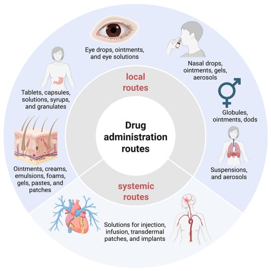
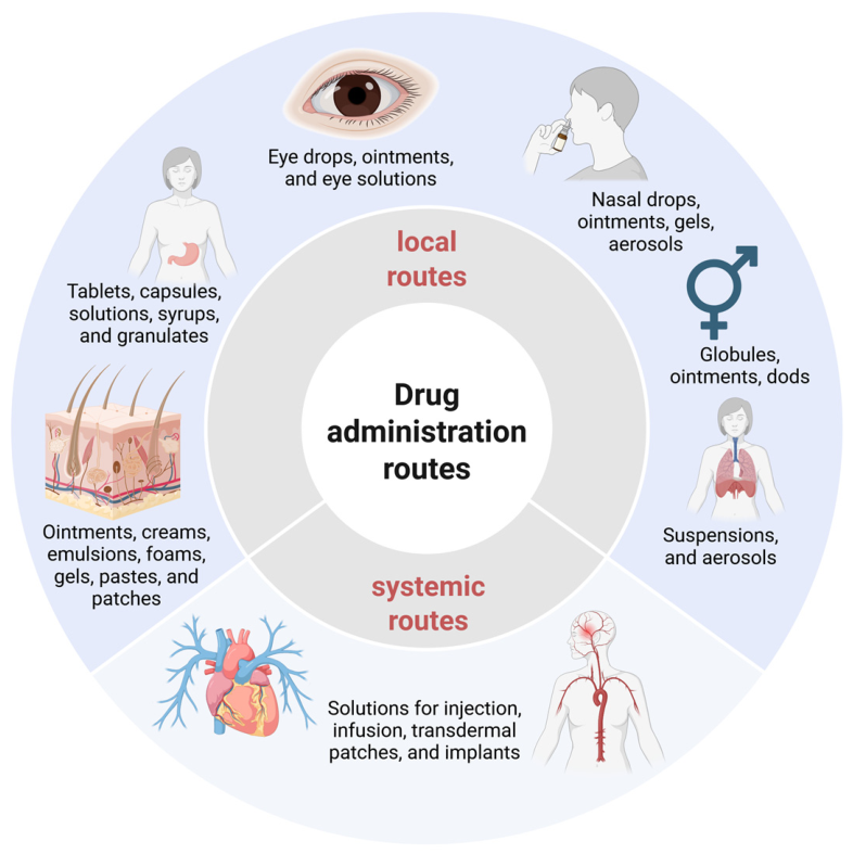
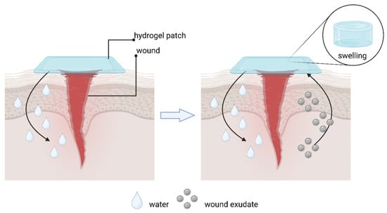
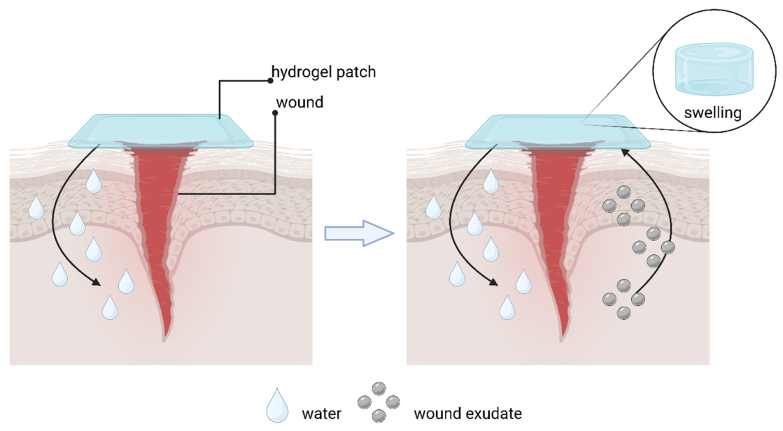
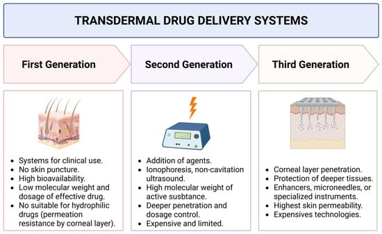
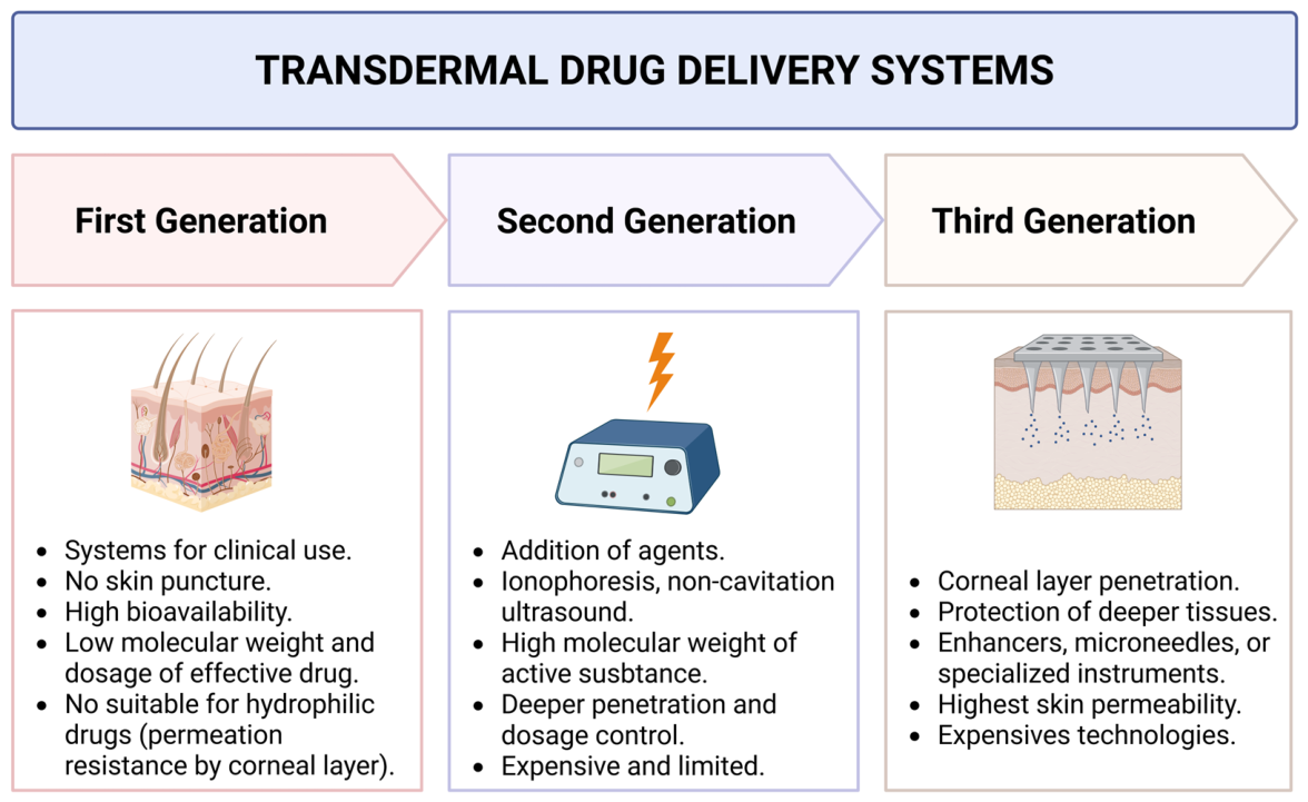
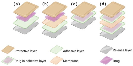
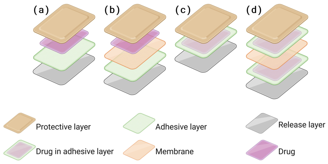
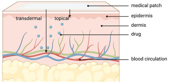
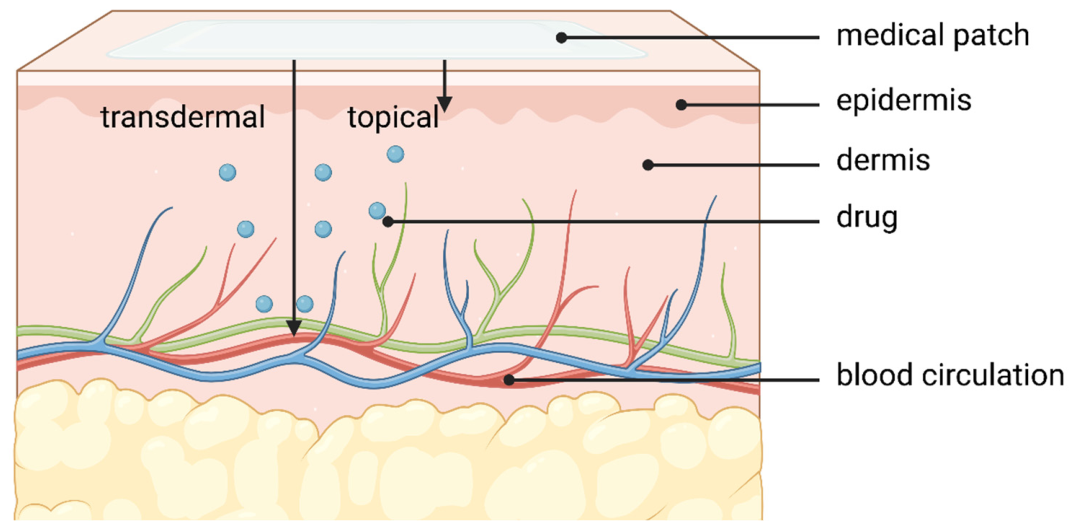
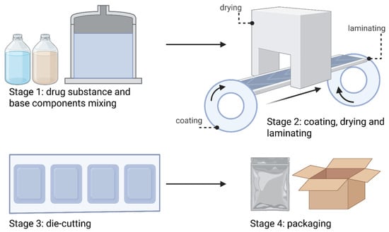
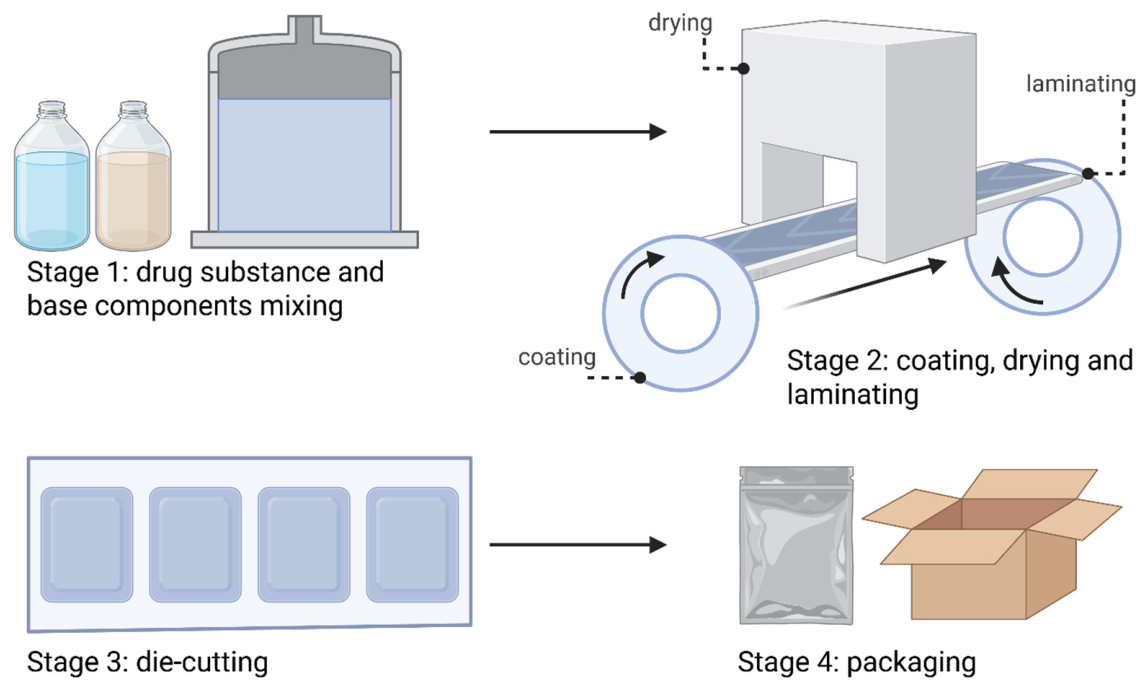
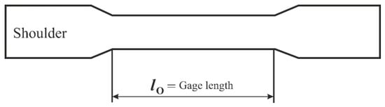
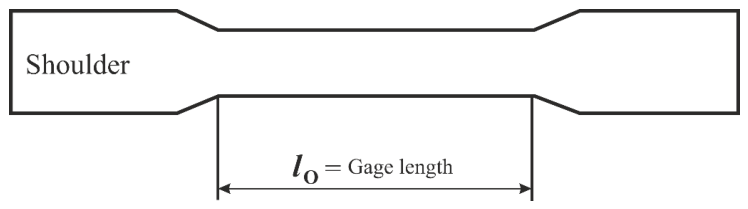
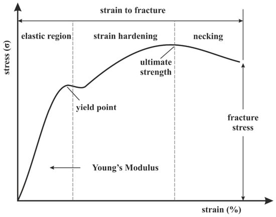
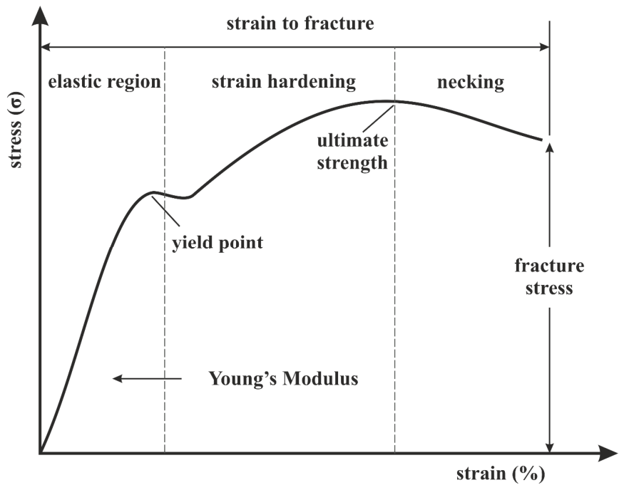
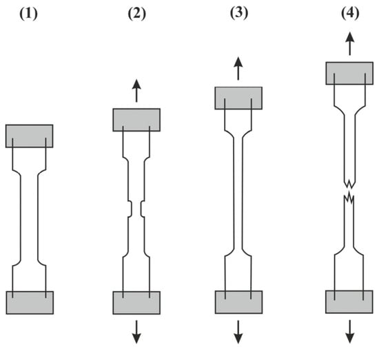
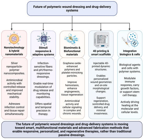
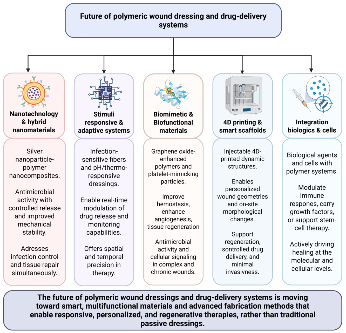

Disclaimer/Publisher’s Note: The statements, opinions and data contained in all publications are solely those of the individual author(s) and contributor(s) and not of MDPI and/or the editor(s). MDPI and/or the editor(s) disclaim responsibility for any injury to people or property resulting from any ideas, methods, instructions or products referred to in the content. |
© 2026 by the authors. Licensee MDPI, Basel, Switzerland. This article is an open access article distributed under the terms and conditions of the Creative Commons Attribution (CC BY) license.