


Simple Summary
Breast radiotherapy requires patients to maintain a reproducible treatment position throughout the course of treatment. However, changes in arm posture may alter the shape and position of the breast, potentially affecting how radiation is delivered. This study used a flexible breast-mimicking phantom to investigate this clinically relevant issue under controlled and reproducible conditions. The findings suggest that the sensitivity of radiation dose to changes in the postural position of breast radiotherapy may be related to the treatment technique.
Abstract
Accurate positioning is essential in breast radiotherapy; however, rigid phantoms do not reproduce posture-related anatomical deformation. We developed a breast-mimicking rubber and silicone phantom to evaluate dosimetric effects of arm-position-induced deformation in three-dimensional conformal radiotherapy (3D-CRT), intensity-modulated radiotherapy (IMRT), and volumetric-modulated arc therapy (VMAT). The phantom included a central internal marker and five surface optically stimulated luminescence dosimeter attachment points. Treatment plans were generated from computed tomography images to deliver 42.4 Gy in 16 fractions. Surface doses were measured with a linear accelerator in reference and modified arm-shift postures. 3D-CRT showed differences below 1.00% at nearly all points and a maximum deviation of −0.77% laterally. IMRT showed greater sensitivity, reaching 2.53% at the lower point. VMAT showed the largest changes: a 21.19% dose deficit (0.414 Gy) at the lower point and a −5.88% difference laterally. Arm position changes therefore produced localized surface-dose variation, particularly in the lower breast. These findings highlight the importance of careful arm positioning and reproducible setup when highly modulated techniques are used and suggest that deformable phantoms can complement conventional rigid quality-assurance tools. Under these experimental conditions, VMAT was more sensitive than IMRT and 3D-CRT at selected surface measurement points in this phantom model.
1. Introduction
In breast radiotherapy, various treatment modalities are widely used, including three-dimensional conformal radiotherapy (3D-CRT), intensity-modulated radiotherapy (IMRT), and volumetric-modulated arc therapy (VMAT) [1,2,3]. Although these techniques can achieve adequate target coverage while reducing radiation exposure to surrounding organs at risk, their dosimetric characteristics differ according to beam arrangement, modulation, field geometry, and dose gradients [4,5,6]. Therefore, accurate and reproducible patient positioning is increasingly important as radiotherapy techniques become more conformal and highly modulated. During simulation and treatment, patients with breast cancer are positioned supine on an inclined breast board, with one or both arms raised above the head. Because the breast consists of soft tissues, including fat, the reproducibility of its position is highly influenced by arm placement. Displacements in breast position can reach several millimeters, and such variations have been reported to occur more frequently and to a greater extent in patients with large breasts [7]. These positional changes can affect accurate dose delivery according to the radiation treatment plan; furthermore, this impact may vary among intensity-modulated radiation techniques, particularly across different skin positions. Previous studies have confirmed that inter-fractional motion and anatomical variations can significantly affect the actual delivered dose in breast IMRT [8]. A study quantifying breast deformation using daily cone-beam CT (CBCT) reported a maximum breast surface expansion of 2.4 ± 2.1 mm, with changes of >5 mm observed in 17% of treatment fractions [9]. Furthermore, sensitivity to setup deviations may differ among the VMAT, IMRT, and 3D-CRT plans. Conventional tangential 3D-CRT uses relatively broad and geometrically simple fields, whereas IMRT and VMAT achieve dose conformity through intensity modulation and multiple beam segments or continuously varying apertures. Thus, local changes in tissue thickness, surface curvature, or beam incidence may affect dose delivery differently among techniques [10,11]. However, clinical cases are influenced by multiple patient-specific factors, making it difficult to isolate the dosimetric effects of posture-induced deformation under controlled conditions.
In breast radiotherapy research, homogeneous water-equivalent phantoms, heterogeneous phantoms containing tissue-equivalent inserts, and anthropomorphic phantoms such as RANDO and CIRS are commonly utilized [12,13]. However, because most commercial phantoms have rigid or predefined external contours representing a single breast size and shape, they cannot adequately replicate variations in breast and skin geometry induced by changes in skeletal or arm positioning. To address this limitation, a breast-mimicking phantom was fabricated in-house to simulate breast skin movement and deformation in accordance with variations in arm position. Using this flexible phantom, we analyzed the specific locations that exhibited the most significant dosimetric variations during dose delivery across the 3D-CRT, IMRT, and VMAT techniques.
2. Materials and Methods
2.1. Breast-Mimicking Phantom Fabrication
In this study, a breast-mimicking phantom composed of rubber (HUmean: 143, mass density: 1.109 g/cm3, Smooth-On, Inc., Macungie, PA, USA) and silicone (HUmean: −621, mass density: 0.335 g/cm3) was fabricated to replicate the movement and deformation of the breast induced by variations in arm positioning. The phantom was designed to anatomically mimic the shapes of the human thorax and breast. Additionally, an internal marker was inserted at the center of the breast to assess whether the breast had shifted from its initial position. An optically stimulated luminescence dosimeter (OSLD; myOSLDchip, RadPro International GmbH, Remscheid, North Rhine-Westphalia, Germany) was used to measure the dose delivered to the skin surface of the phantom. The OSLD was pre-calibrated using 6 MV photon beams from the same linear accelerator (VitalBeam, Siemens Healthineers, Palo Alto, CA, USA) used in the experiment. Calibration was performed using a polymethyl methacrylate slab phantom consisting of 1 cm thick slabs. A 10 × 10 cm2 field was used with a source-to-surface distance of 100 cm. The OSLDs were positioned at a depth of 5 cm, with 10 cm of backscatter material. A Farmer-type ionization chamber was initially used to verify the beam output. The calibration results demonstrated that the measurement error was within ±1.00% relative to the reference dose, and this specific calibration factor was subsequently applied to all subsequent measurements.
2.2. CT Simulation and Treatment Planning
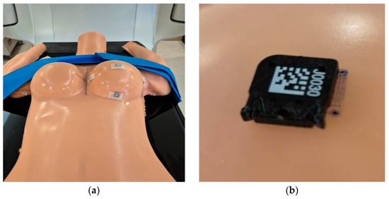
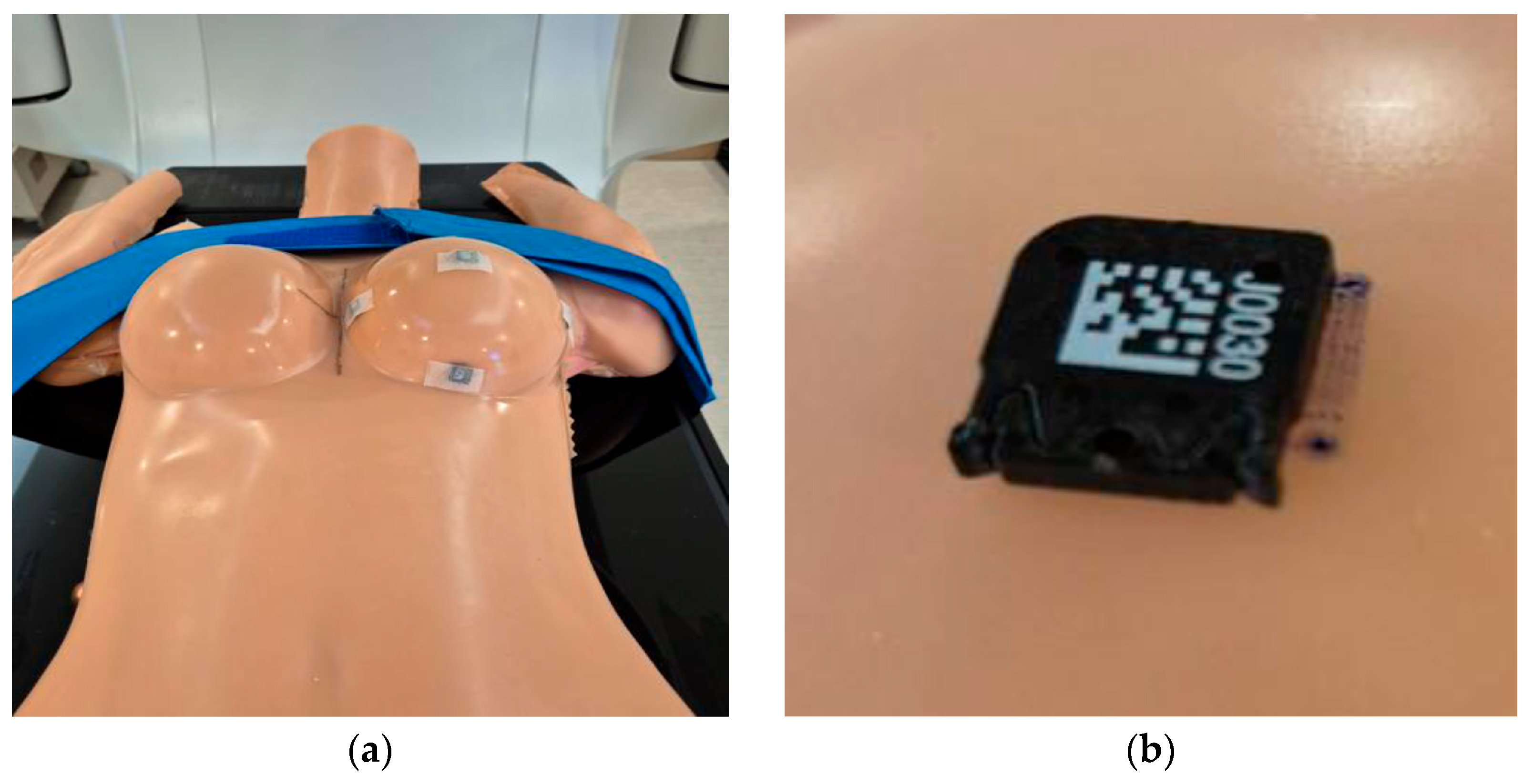
A CT simulator was used to acquire computed tomography (CT) images (Siemens go-sim; Siemens Healthineers, Munich, Germany) of the phantom. The phantom was positioned with its arms elevated and secured with straps to prevent displacement during scanning. To ensure measurement reproducibility, dummy OSLD chips were attached to five predefined points on the phantom during CT scanning, and the corresponding locations were marked to ensure consistent OSLD placement for all measurements (Figure 1).
Figure 1.
(a) Breast-mimicking phantom position, and (b) optically stimulated luminescence dosimeter location marking.
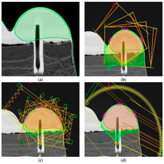
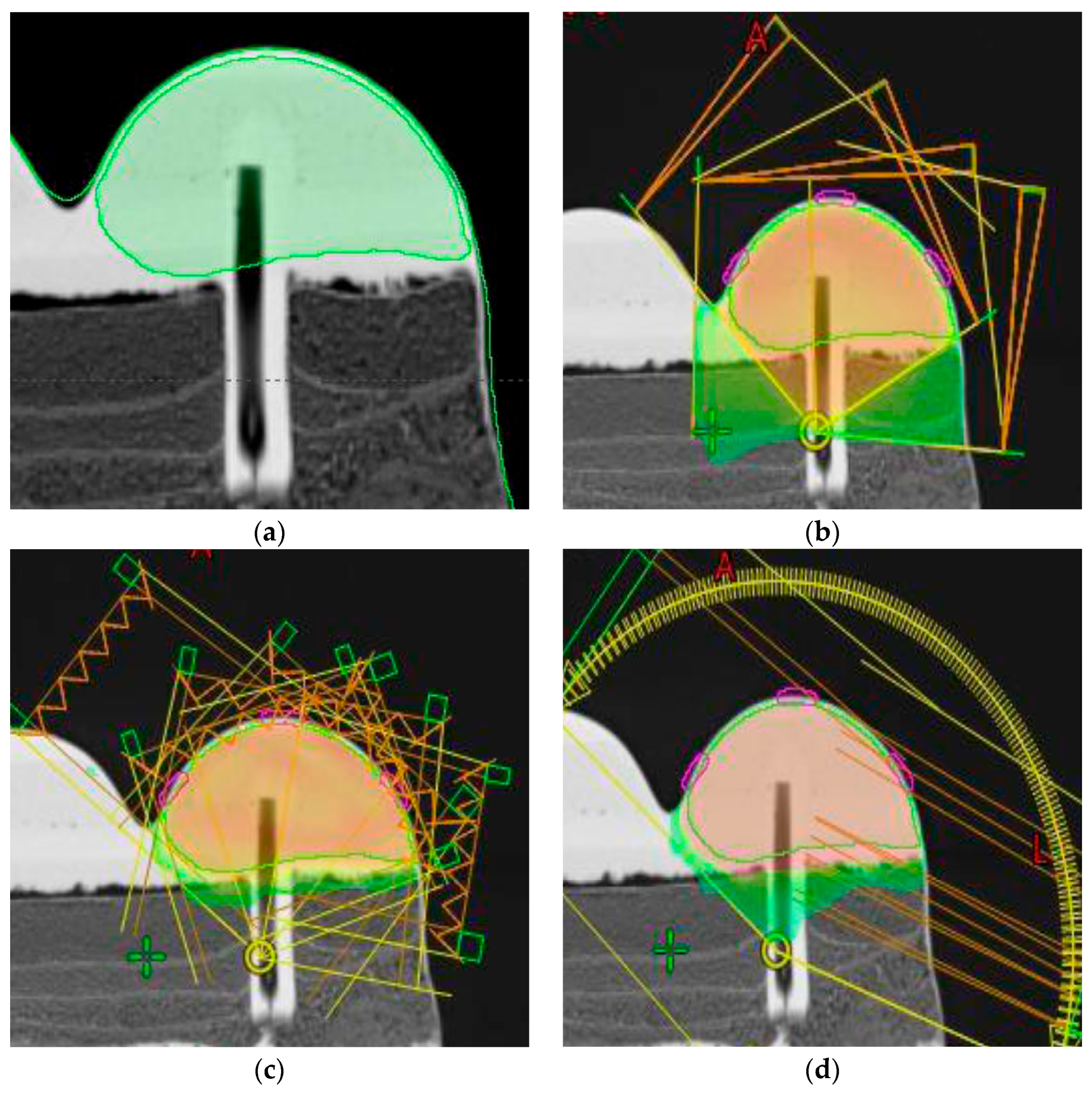
The center and reference positions of the phantom were established using a laser alignment system in the CT simulation room. Subsequently, the treatment isocenter and alignment reference points were marked on the phantom surface to ensure precise geometric reproducibility during treatment planning and subsequent irradiation. The CT images were acquired under standard clinical protocols for patients with breast cancer, with conditions of 120 kVp, 300 mAs, and 2.0 mm slice thickness. EclipseTM Treatment Planning System (version 16.0; Varian Medical Systems, Palo Alto, CA, USA) was used for treatment planning. The treatment plans for 3D-CRT, IMRT, and VMAT (AAA, PO, calculation resolution: 0.25 cm) were generated using phantom images acquired via CT simulation. In accordance with clinical breast radiotherapy protocols, the prescription dose was set to 42.4 Gy in 16 fractions of 2.65 Gy per fraction. As shown in Figure 2, a clinical target volume (CTV) was delineated within the breast region of the phantom. The treatment plans were optimized to ensure that 100% of the CTV received at least 95% of the prescribed dose, while the dose to the CTV did not exceed 105% of the prescribed dose. A 6 MV photon beam was used for all treatment plans. The 3D-CRT plan was designed by using four fields with gantry angles (GAs) of 60°, 95°, 316°, and 358°. For the IMRT plan, six static fields (GAs 10°, 40°, 70°, 100°, 310°, and 340°) were used, whereas the VMAT plan was optimized using two continuous arcs (GA 310°–110° and 110°–310°).
Figure 2.
(a) Clinical target volume, (b) three-dimensional conformal radiation therapy, (c) intensity-modulated radiation therapy, and (d) volumetric-modulated arc therapy plans.
2.3. Measurement of Surface Dose
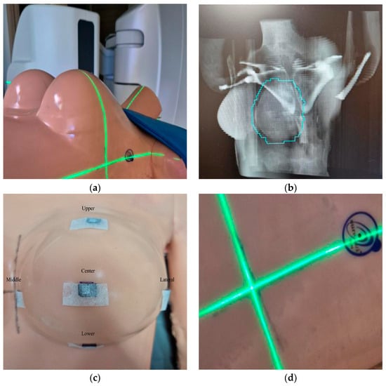
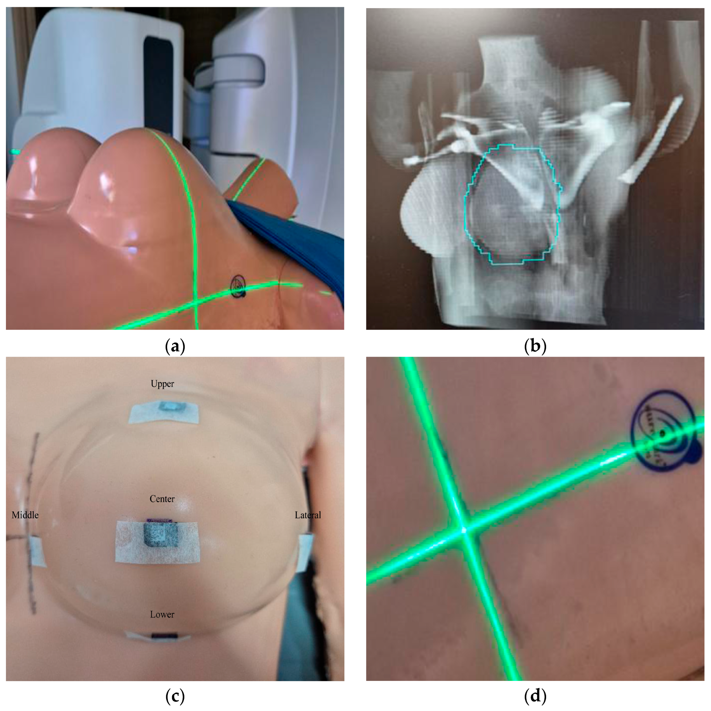
The phantom was aligned with the isocenter of the linear accelerator using room lasers and alignment reference markers indicated on the surface of the phantom. Planar images of the phantom were acquired using the onboard imaging system of the linear accelerator to verify that the phantom position during the CT simulation was accurately reproduced in the treatment room. The acquired images were compared with CT-based reference images to verify alignment of the phantom’s skeletal structures and external contours. After positional setup errors were corrected through image registration, radiation was delivered. The baseline phantom dose was acquired in the reference position with the arms elevated. In total, five OSLDs were attached to the phantom surface as shown in Figure 3. The 3D-CRT, IMRT, and VMAT treatment plans were delivered at this reference position to acquire the absorbed doses at the surface measurement points. To evaluate changes in the breast region and delivered dose induced by arm position adjustments, the arms were randomly repositioned from the reference setup, and laser displacement from the reference line was confirmed (Figure 3d). After adjusting the arms, the position of the phantom was verified using treatment room lasers and an image-guidance system. The same plans used for the baseline dose were delivered to the phantom. For each plan, the measurements were repeated three times to ensure reproducibility and data accuracy.
Figure 3.
(a) Phantom alignment, (b) cone-beam computed tomography image, (c) optically stimulated luminescence dosimeter locations, and (d) displacement between the reference line and laser.
2.4. Dose Evaluation
Dose measurement values obtained for each radiotherapy technique and arm position were calculated as the mean of three repeated measurements. Using the dose measured in the reference position with the arms elevated as the baseline, dose differences induced by changes in arm position were calculated as follows:
where A is the plan or reference position measurement value, and B is the shift measurement value.
Dose difference (Gy) = A − B
Subsequently, dose sensitivity to posture deviations was compared among the 3D-CRT, IMRT, and VMAT techniques.
3. Results
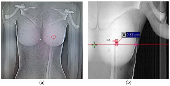
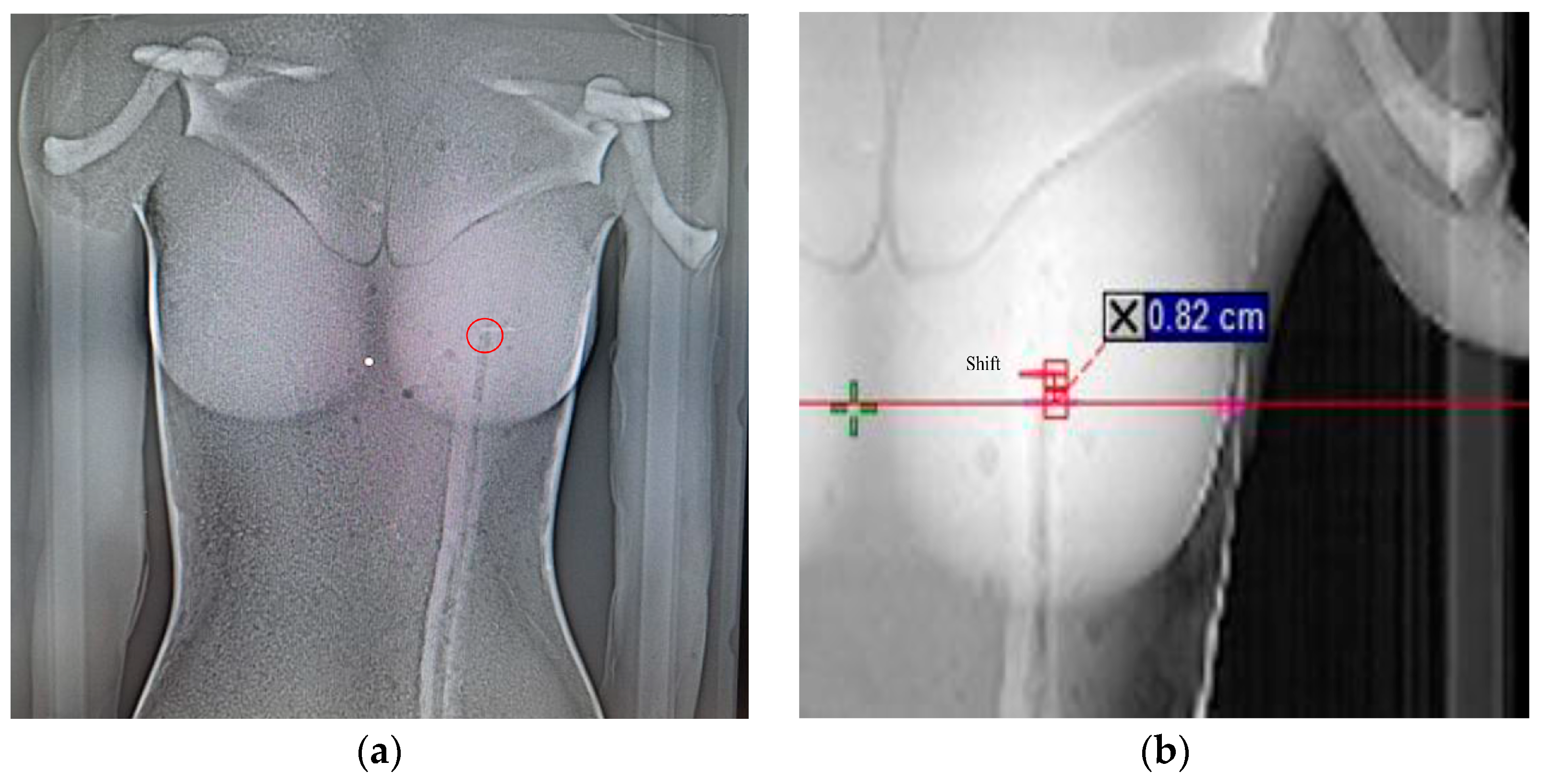
Altering the arm position of the breast-mimicking phantom induced a spatial displacement of 0.82 cm in the marker inserted at the central point of the breast relative to the reference position, as shown in Figure 4.
Figure 4.
(a) Displacement between the reference line and laser, and (b) positional displacement of the inserted marker according to variations in arm positioning.
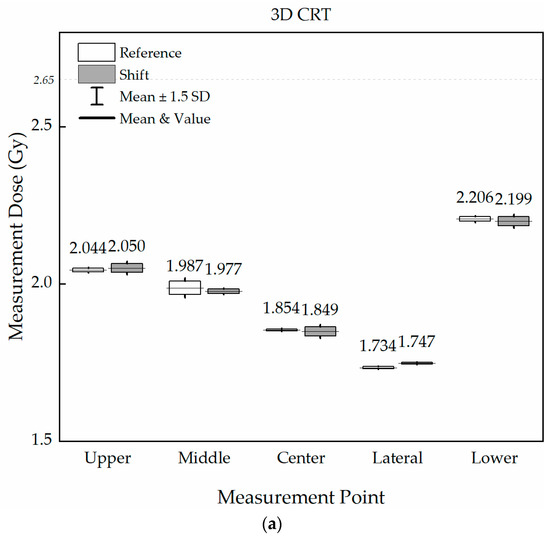
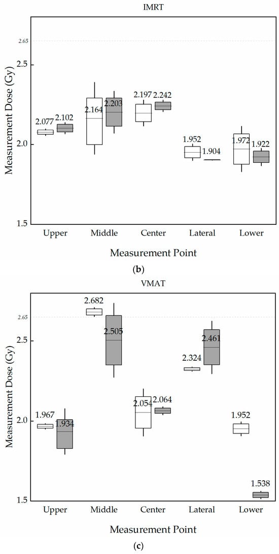
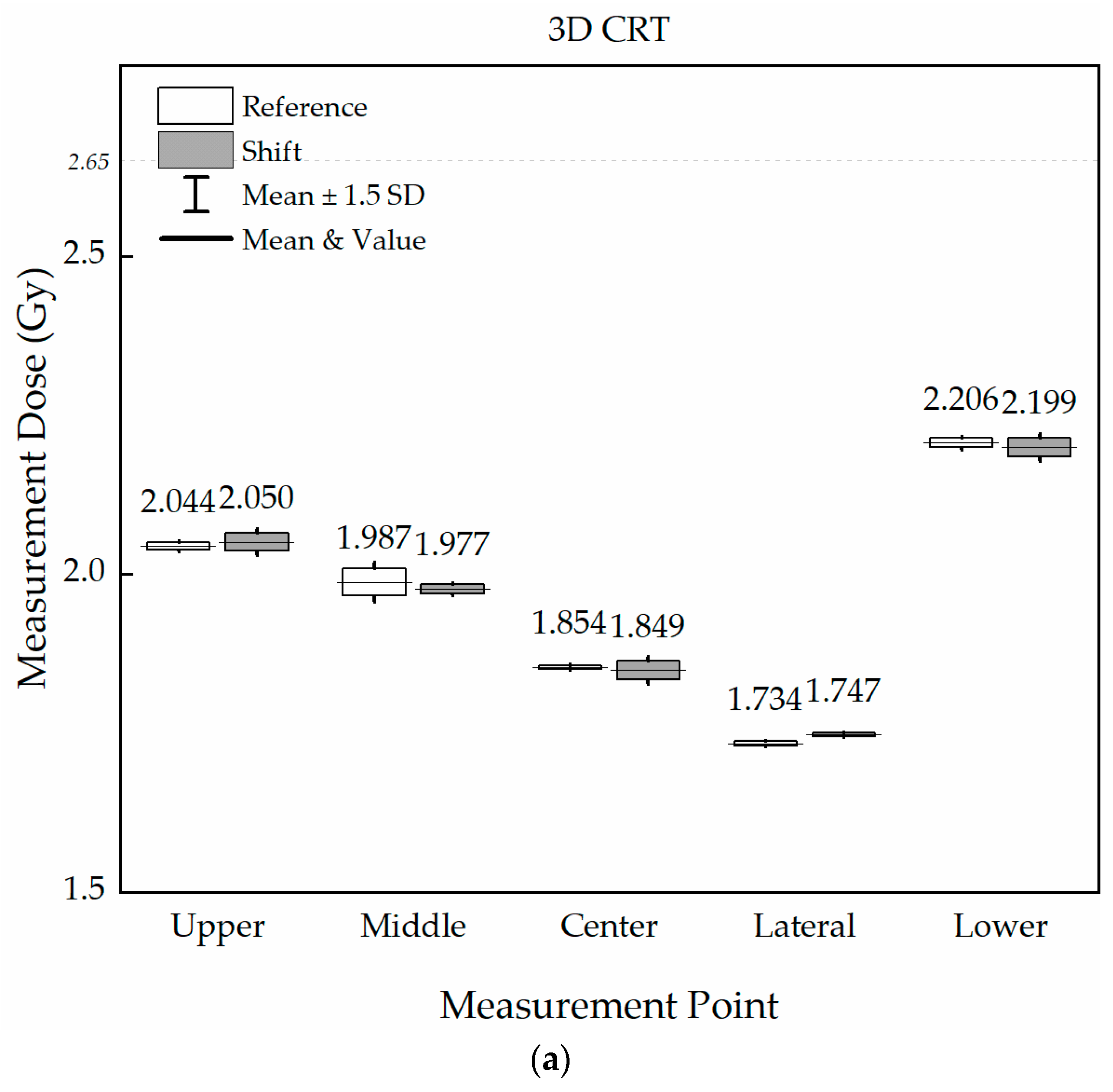
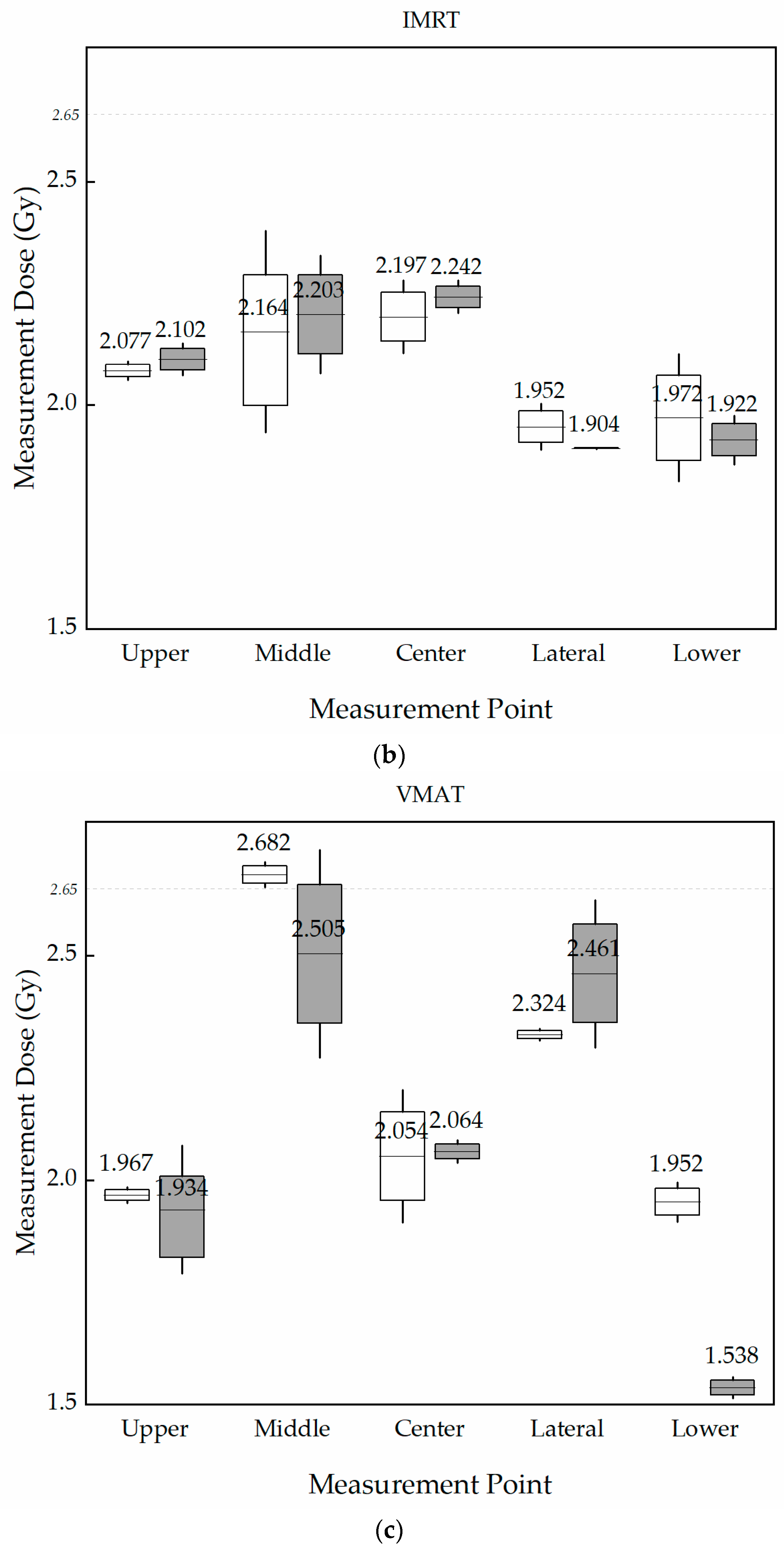
The planned doses, measured doses at the reference position, and measured doses (shift) following arm shift for 3D-CRT, IMRT, and VMAT are summarized in Table 1 and Figure 5.
Table 1.
Planned and measured doses according to radiotherapy techniques and measurement locations. (a) 3D-CRT; (b) IMRT; (c) VMAT.
Figure 5.
Dose difference (%) according to radiotherapy techniques and measurement locations, compared with the reference and shift positions: (a) three-dimensional conformal radiation therapy, (b) intensity-modulated radiation therapy, and (c) volumetric-modulated arc therapy.
The dosimetric sensitivity to these geometric alterations differed distinctly, depending on the choice of radiotherapy technique. For the 3D-CRT plans, the surface dose discrepancies between the reference and shifted postures remained exceptionally stable, with absolute variations restricted to less than 1.00% across all evaluated surface points (Table 1a). The maximum difference at the surface was observed at the lateral point, which exhibited a minor increase of −0.77% (1.734 Gy to 1.747 Gy, with an absolute difference of −0.013 Gy). The middle point showed a minimal difference of 0.53% (1.987 Gy to 1.977 Gy), whereas the lower point demonstrated a negligible difference of 0.32% (2.206 Gy to 2.199 Gy). The center point showed a variation of only 0.25%.
In contrast, IMRT plans demonstrated an elevated sensitivity to posture modifications compared to 3D-CRT, with surface dose differences ranging from −2.02% to 2.53% (Table 1b). The most pronounced dosimetric alteration on the surface occurred at the lower point, where the measured dose decreased by 2.53% (1.972 Gy to 1.922 Gy, representing a discrepancy of 0.050 Gy). The center and lateral points exhibited post-shift variations of −2.02% and 2.47%, respectively. At the upper point, discrepancy was minimized at −1.21%.
The VMAT plans were most affected by arm position adjustments, displaying substantial and nonuniform dose fluctuations across measurement locations (Table 1c). Notably, the surface dose at the lower point decreased dramatically from 1.952 Gy at the origin to 1.538 Gy after the shift, resulting in a critical dose deficit of 21.19% (0.414 Gy). This was the largest absolute percentage dose discrepancy observed across all tested modalities and measurement points throughout the study. Furthermore, substantial differences were observed at the middle and lateral points, with dose differences of 6.60% (2.682 Gy to 2.505 Gy) and −5.88% (2.324 Gy to 2.461 Gy), respectively. The center point demonstrated a stable difference of −0.50% (2.054 Gy to 2.064 Gy).
Across all three techniques, the center point showed relatively small dose variations, whereas greater changes were observed at peripheral measurement sites. Notably, VMAT demonstrated a nonuniform spatial response, with dose increase at the lower and lateral points but a decrease at the middle point, indicating that position-induced deformation affected surface dose differently according to measurement location.
4. Discussion
This study compared the impact of breast deformation induced by arm posture modifications on the actual measured doses across 3D-CRT, IMRT, and VMAT using an in-house breast-mimicking phantom. The myOSLDchip demonstrated good dose linearity, repeatability, and stability within the clinical radiotherapy dose range, supporting its potential use for in vivo dosimetry [14]. The study reported minimal angular dependence of the myOSLDchip, with a response difference of approximately 0.5% between 0° and 180°, while a variation of 2.2% ± 2.26% under a different phantom geometry remained within measurement uncertainty. Similarly, another study found no statistically significant systematic angular dependence (p = 0.997) across irradiation angles of 0°, 30°, 60°, and 90°, supporting the suitability of the myOSLDchip for clinical radiotherapy dosimetry. In this study, markings on the phantom surface were used to ensure consistent and reproducible placement of the OSLDs for all measurements; therefore, measurement variability attributable to differences in OSLD positioning was minimized [15].
The site-specific dose variations on the phantom surface differed distinctly depending on the radiotherapy technique, with maximum absolute percentage dose discrepancies recorded as −0.77% for 3D-CRT, 2.53% for IMRT, and 21.19% for VMAT. The most pronounced variation was observed at the lower VMAT site, with absolute discrepancies of 6.60% and 5.88% at the middle and lateral sites, respectively. These findings suggest that variations in arm positioning do not simply cause rigid translation of the entire breast but locally alter surface curvature, tissue thickness, and beam incidence angle, resulting in more pronounced dosimetric effects with highly modulated radiotherapy techniques.
The observation that the absolute dose discrepancies at almost all surface measurement points remained within 1.00% with 3D-CRT may be attributed to the relatively wide and simple irradiation fields. Previous patient-based studies have reported that when setup variations and soft-tissue deformations occur in the breast or chest wall, the target dose coverage of VMAT can degrade more severely than that of tangential irradiation techniques. This was consistent with our findings, in which 3D-CRT exhibited the smallest site-specific variations, whereas VMAT showed the largest.
In IMRT, the maximum absolute discrepancy was 2.53%, which was higher than that of 3D-CRT but lower than that of VMAT. Although IMRT can improve target conformity and homogeneity by modulating the intensity of multiple static fields, the localized dose gradients and field edges formed by each segment are susceptible to anatomical deformations. Liu et al. [1] reported that while IMRT and VMAT improve target conformity compared with 3D-CRT and VMAT reduces monitor units and treatment time, these modulation techniques involve different trade-offs in low-dose volumes and organs-at-risk (OAR) dose distributions. A comparative study by Chen et al. [16] demonstrated that the advantages of targeted and OAR sparing were inconsistent among techniques, with VMAT showing an increase in certain low-dose indices in the lungs and heart. Therefore, a superior planned dose distribution in modulated techniques does not inherently guarantee high robustness against positional changes, underscoring the necessity of simultaneously evaluating both plan quality and delivery robustness.
The significant dose variations observed at the lower (21.19%), middle (6.60%), and lateral (−5.88%) sites in VMAT can be attributed to its inherent characteristics, in which the total dose is synthesized through continuous intensity modulation and a large number of small apertures during gantry rotation. Specifically, morphological alterations of the breast and variations in the equivalent path length thickness caused by arm posture modifications accumulate across multiple control points, ultimately manifesting as localized dose increases or decreases. This interpretation is highly consistent with the findings of Rossi et al. [9], who demonstrated through CBCT-based dose recalculations that setup variations and soft-tissue alterations exert a more profound impact on target coverage in VMAT-based breast treatment compared to tangential techniques. Other studies have shown that breast deformation, swelling, and setup shifts can affect target coverage and that the magnitude of these dosimetric changes varies according to the treatment technique, including VMAT and tangential approaches [17,18].
The largest dose discrepancy observed at the lower point, corresponding to the inframammary region, may be relevant to clinical breast radiotherapy, particularly because this region is susceptible to anatomical variation and radiation-induced skin reactions. The inframammary fold (IMF) length was positively correlated with the severity of radiation-induced skin reactions, suggesting that anatomical characteristics of the IMF may contribute to increased susceptibility to treatment-related toxicity [19]. The IMF and axilla are also well-known areas where acute radiation dermatitis and moist desquamation frequently occur, owing to friction and the self-bolus effect caused by skin-to-skin contact [20,21]. The inferior and lateral aspects of the breast are highly susceptible to substantial morphological alterations depending on the patient’s breast size, gravitational sagging, degree of arm elevation, and skin infolding [7,22].
Previous studies have shown that setup variations, arm positioning, and breast geometry can affect the dosimetric accuracy of breast radiotherapy, with the impact varying by treatment technique [23,24,25,26]. CBCT- and deformable-registration-based analyses have further demonstrated that anatomical changes can be incorporated into dose recalculation to assess differences between planned and delivered doses [27,28]. These findings highlight the importance of both reproducible positioning and preservation of local breast geometry, particularly for highly modulated techniques.
The strength of this study lies in the direct comparison of 3D-CRT, IMRT, and VMAT under identical experimental conditions using an in-house breast-mimicking phantom capable of simulating breast movement. However, this study had several limitations. First, because it evaluated a single phantom and a limited range of arm position changes, it did not fully capture diverse breast sizes, thoracic shapes, and patient-specific soft-tissue characteristics. Future studies are needed to evaluate cumulative doses in patients with varying breast sizes and shapes or across a diverse range of phantoms by incrementally altering arm height, abduction angle, shoulder rotation, and torso tilt using repeated CT or CBCT-based dose recalculations and deformable image registration. Additional data may help establish technique-specific setup tolerances and repositioning criteria and determine the need for adaptive radiotherapy in patients with substantial anatomical variations.
5. Conclusions
In this study, a breast-mimicking phantom capable of simulating skin deformation was used to observe dosimetric variations induced by changes in arm positioning during breast radiotherapy. Substantial dose changes were observed in the inferior and lateral regions of the breast under VMAT, confirming that reproducibility of arm and breast morphology is a critical quality control factor in highly modulated rotational therapies. However, further investigation is required to evaluate technique-specific dose variations under diverse clinical patient posture deformations using a more advanced skin deformation phantom that can comprehensively replicate a wider range of arm positions.
Author Contributions
S.H.P.: methodology, investigation, data curation, writing—original draft. S.H.P. and J.C.: resources, software, investigation. J.C.: conceptualization, supervision, writing—review and editing. All authors have read and agreed to the published version of the manuscript.
Funding
This research received no external funding.
Institutional Review Board Statement
Not applicable.
Informed Consent Statement
Not applicable.
Data Availability Statement
Data are contained within the article.
Conflicts of Interest
The authors declare no conflicts of interest.
Abbreviations
The following abbreviations are used in this manuscript:
| 3D-CRT | Three-dimensional conformal radiotherapy |
| IMRT | Intensity-modulated radiotherapy |
| VMAT | Volumetric-modulated arc therapy |
| OSLD | Optically stimulated luminescence dosimeter |
| CT | Computed tomography |
| CBCT | Cone-beam computed tomography |
References
- Liu, H.; Chen, X.; He, Z.; Li, J.Y. Evaluation of 3D-CRT, IMRT and VMAT radiotherapy plans for left breast cancer based on clinical dosimetric study. Comput. Med. Imaging Graph. 2016, 54, 1–5. [Google Scholar] [CrossRef] [Scilit] [PubMed]
- Chang, J.S.; Chang, J.H.; Kim, N.; Kim, Y.B.; Shin, K.H.; Kim, K. Intensity Modulated Radiotherapy and Volumetric Modulated Arc Therapy in the Treatment of Breast Cancer: An Updated Review. J. Breast Cancer. 2022, 25, 349–365. [Google Scholar] [CrossRef] [Scilit] [PubMed]
- Ahmad, A.; Das, S.; Kharade, V. Dosimetric Study Comparing 3D Conformal Radiotherapy (3D-CRT), Intensity Modulated Radiotherapy (IMRT) and Volumetric Modulated Arc Therapy (VMAT) in Hypofractionated One-Week Radiotherapy Regimen in Breast Cancer. Cureus 2022, 14, e31860. [Google Scholar] [CrossRef] [Scilit] [PubMed]
- Padilla, L.; Kang, H.; Washington, M.; Hasan, Y.; Chmura, S.J.; Al-Hallaq, H. Assessment of interfractional variation of the breast surface following conventional patient positioning for whole-breast radiotherapy. J. Appl. Clin. Med. Phys. 2014, 15, 177–189. [Google Scholar] [CrossRef] [Scilit] [PubMed]
- Jin, G.H.; Chen, L.X.; Deng, X.W.; Liu, X.W.; Huang, Y.; Huang, X.B. A comparative dosimetric study for treating left-sided breast cancer for small breast size using five different radiotherapy techniques: Conventional tangential field, field-in-field, tangential-IMRT, multi-beam IMRT and VMAT. Radiat. Oncol. 2013, 8, 89. [Google Scholar] [CrossRef] [Scilit] [PubMed]
- Nithya, L.; Goel, V.; Sharma, D.; Vittal, K.; Marjara, N. Dosimetric Comparison of Different Planning Techniques in Left-sided Whole-Breast Irradiation: A Planning Study. J. Med. Phys. 2020, 45, 148–155. [Google Scholar] [CrossRef] [Scilit] [PubMed]
- Seppälä, J.; Vuolukka, K.; Virén, T.; Heikkilä, J.; Honkanen, J.T.J.; Pandey, A.; Al-Gburi, A.; Shah, M.; Sefa, S.; Koivumäki, T. Breast deformation during the course of radiotherapy: The need for an additional outer margin. Med. Phys. 2019, 65, 1–5. [Google Scholar] [CrossRef] [Scilit] [PubMed]
- Jain, P.; Marchant, T.; Green, M.; Watkins, G.; Davies, J.; McCarthy, C.; Loncaster, J.; Stewart, A.; Magee, B.; Moore, C.; et al. Inter-fraction motion and dosimetric consequences during breast intensity-modulated radiotherapy (IMRT). Radiat. Oncol. 2009, 90, 93–98. [Google Scholar] [CrossRef] [Scilit] [PubMed]
- Rossi, M.; Boman, E.; Skyttä, T.; Haltamo, M.; Laaksomaa, M.; Kapanen, M. Dosimetric effects of anatomical deformations and positioning errors in VMAT breast radiotherapy. J. Appl. Clin. Med. Phys. 2018, 19, 506–516. [Google Scholar] [CrossRef] [Scilit] [PubMed]
- Bogue, J.P.; Georgiou, A.; Weiss, E. Dosimetric comparison of VMAT with integrated skin flash to 3D field-in-field tangents for left breast irradiation. J. Appl. Clin. Med. Phys. 2019, 20, 31–41. [Google Scholar] [CrossRef] [Scilit] [PubMed]
- Mankinen, M.; Virén, T.; Seppälä, J. Interfractional variation in whole-breast VMAT irradiation: A dosimetric study with complementary SGRT and CBCT patient setup. Radiat. Oncol. 2024, 19, 21. [Google Scholar] [CrossRef] [Scilit] [PubMed]
- Kakade, N.R.; Kumar, R.; Srivastava, A.; Pilakkal, S.; Sharma, S.D.; Sapra, B.K. Application of Unlaminated Gafchromic EBT3 Film for Surface Dose Measurement in Breast Radiotherapy. J. Med. Phys. 2025, 50, 510–515. [Google Scholar] [CrossRef] [Scilit] [PubMed]
- Zhang, J.; Li, X.; Lu, M.; Zhang, Q.; Zhang, X.; Yang, R.; Chan, M.F.; Wen, J. A method for in vivo treatment verification of IMRT and VMAT based on electronic portal imaging device. Radiat. Oncol. 2021, 16, 232. [Google Scholar] [CrossRef] [Scilit] [PubMed]
- Davis, H.; Siebers, J.V.; Wijesooriya, K.; Mistro, M. Clinical validation of myOSLDchip: A beryllium oxide optically stimulated luminescent dosimeter (OSLD) system in radiotherapy dosimetry. J. Appl. Clin. Med. Phys. 2025, 26, e70094. [Google Scholar] [CrossRef] [Scilit] [PubMed]
- Goddard, L.C.; Basavatia, A.; Brodin, P.; Tome, W.A. An Investigation of the Dosimetric Uncertainties in the myOSLchip System: The Impact of Contributing Factors on Measurement Accuracy. Cureus 2025, 17, e88239. [Google Scholar] [CrossRef] [Scilit] [PubMed]
- Chen, S.N.; Ramachandran, P.; Deb, P. Dosimetric comparative study of 3DCRT, IMRT, VMAT, Ecomp, and Hybrid techniques for breast radiation therapy. Radiat. Oncol. 2020, 38, 270–281. [Google Scholar] [CrossRef] [Scilit] [PubMed]
- Schubert, L.K. The robustness of VMAT radiotherapy for breast cancer with tissue deformations. Med. Dosim. 2021, 46, e1–e8. [Google Scholar] [CrossRef] [Scilit] [PubMed]
- Gleeson, I. Comparing the robustness of different skin flash approaches using wide tangents, manual flash VMAT, and simulated organ motion robust optimization VMAT in breast and nodal radiotherapy. Med. Dosim. 2022, 47, 264–272. [Google Scholar] [CrossRef] [Scilit] [PubMed]
- Southworth, A.; Clough, H.; Roberts, N. An audit of radiation-induced skin reactions in the inframammary fold; does breast size impact on the severity of the reaction? Radiography 2020, 26, 192–197. [Google Scholar] [CrossRef] [Scilit] [PubMed]
- Ivica, R.; Aljasa, J.; Irena, O. Breast size impact on adjuvant radiotherapy adverse effects and dose parameters in treatment planning. Rad. Oncol. 2018, 52, 233–244. [Google Scholar] [CrossRef] [Scilit] [PubMed]
- Feng, H.; Wang, H.; Xu, L.; Ren, Y.; Ni, Q.; Yang, Z.; Ma, S.; Deng, Q.; Chen, X.; Xia, B.; et al. Prediction of radiation-induced acute skin toxicity in breast cancer patients using data encapsulation screening and dose-gradient-based multi-region radiomics technique: A multicenter study. Front. Oncol. 2022, 12, 1017435. [Google Scholar] [CrossRef] [Scilit] [PubMed]
- Park, S.H.; Choi, J. Analysis of Geometric and Dosimetric Effects of Bra Application to Support Large or Pendulous Breasts During Radiotherapy Planning: A Retrospective Single-Center Study. Technol. Cancer Res. Treat. 2021, 20, 15330338211027909. [Google Scholar] [CrossRef] [Scilit] [PubMed]
- Dekker, J.; Essers, M.; Verheij, M.; Kusters, M. Dose coverage and breath-hold analysis of breast cancer patients treated with surface-guided radiotherapy. Radiat. Oncol. 2023, 18, 72. [Google Scholar] [CrossRef] [Scilit] [PubMed]
- Kim, D.W.; Hong, C.S.; Son, J. Dosimetric analysis of six whole-breast irradiation techniques in supine and prone positions. Sci. Rep. 2024, 14, 14347. [Google Scholar] [CrossRef] [Scilit] [PubMed]
- Heikkilä, A.; Boman, E.; Rossi, M.; Vanharen, A.; Mankinen, M.; Postema, M.; Koivumäki, T. Dosimetric effect of rotational setup errors in volumetric modulated arc therapy and field-in-field treatment of left-sided breast cancer. Phys. Med. 2024, 117, 103203. [Google Scholar] [CrossRef] [Scilit] [PubMed]
- Yen, T.Y.; Chung, K.C.; Fu, H.M.; Feng, C.J.; Lien, K.Y.; Hsu, S.M. Estimation of the Surface Dose in Breast Irradiation by the Beam Incident Angle and the 1 cm Depth Dose. J. Clin. Med. 2022, 11, 2154. [Google Scholar] [CrossRef] [Scilit] [PubMed]
- Yoo, S.; Blitzblau, R.; McDuff, S.; Yin, F.F.; Cui, Y. Dosimetric variation in preoperative partial breast radiosurgery assessed by deformable image registrations. J. Radiosurg SBRT 2022, 8, 227–235. [Google Scholar] [PubMed]
- Sanklaa, K.; Pitikiattikul, C.; Rittiwong, M.; Thiamthan, N.; Turathong, S.; Sanghangthum, T. Surface dose measurement by optically stimulated luminescent dosimeter: A phantom study. Radiography 2024, 30, 1405–1410. [Google Scholar] [CrossRef] [Scilit] [PubMed]

Disclaimer/Publisher’s Note: The statements, opinions and data contained in all publications are solely those of the individual author(s) and contributor(s) and not of MDPI and/or the editor(s). MDPI and/or the editor(s) disclaim responsibility for any injury to people or property resulting from any ideas, methods, instructions or products referred to in the content. |
© 2026 by the authors. Licensee MDPI, Basel, Switzerland. This article is an open access article distributed under the terms and conditions of the Creative Commons Attribution (CC BY) license.