


Abstract
Background: Antisperm antibodies (ASA) are recognized as a potential immunological factor in male infertility; however, their prevalence and clinical correlates remain incompletely characterized in Middle Eastern populations. This study aimed to estimate the prevalence of clinically significant ASA positivity and investigate its associations with demographic, clinical, and semen characteristics among infertile men in Erbil, Kurdistan Region of Iraq. Methods: A multicenter retrospective cross-sectional study was conducted between July 2025 and March 2026 at urology and fertility centers in Erbil. A total of 465 infertile men were included after predefined data cleaning and MAR-test reclassification procedures. Demographic, clinical, and semen parameters were collected, and ASA status was assessed using the direct mixed antiglobulin reaction (MAR) test. Clinically significant positivity was defined as antibodies bound to ≥50% of motile spermatozoa. Associations between ASA status and participant characteristics were examined using bivariate and multivariable analyses. Additional exploratory analyses included k-means clustering of semen profiles and Random Forest machine-learning modelling to evaluate predictors of ASA positivity. Results: ASA positivity was identified in 53 of 465 participants, yielding a prevalence of 11.4% (95% CI: 8.7–14.7%). No significant associations were observed between ASA positivity and residence, smoking status, varicocele, infertility type, nutritional supplement use, sperm concentration, progressive motility, non-progressive motility, or sperm morphology (all p > 0.05). Men with secondary infertility demonstrated a modestly higher median ASA percentage than those with primary infertility; however, binary ASA positivity did not differ significantly between groups. Multivariable logistic regression showed limited explanatory capacity (AUC = 0.607), while a Random Forest classifier achieved only marginal discrimination on the test dataset (AUC = 0.567). Clustering analysis identified two distinct semen-quality phenotypes, but ASA prevalence did not differ significantly between clusters. Conclusions: Clinically significant ASA positivity was present in approximately one in nine infertile men attending participating centers in Erbil. Routine demographic, clinical, and semen characteristics were poor predictors of ASA status, and neither conventional statistical modelling nor machine-learning approaches reliably identified ASA-positive individuals. These findings suggest that ASA-associated infertility may not be adequately captured by standard infertility assessments alone and support the selective use of direct ASA testing within comprehensive male infertility evaluation. Larger prospective studies incorporating fertile controls, detailed reproductive histories, complementary antibody assays, and immunological biomarkers are warranted.
1. Introduction
Infertility is a major reproductive health problem affecting approximately 15% of couples of reproductive age worldwide. Male factors contribute solely or jointly to nearly half of infertility cases, making the systematic evaluation of male reproductive function an essential component of infertility care [1]. Conventional assessment is centered on semen analysis, including sperm concentration, motility, and morphology. However, these parameters do not fully explain reproductive failure in all patients. Some infertile men have normal or only mildly abnormal semen profiles but may have functional sperm defects caused by immunological mechanisms, particularly the presence of antisperm antibodies (ASA) [2].
ASAs are immunoglobulins, most commonly IgG and IgA, and less frequently IgM, that bind to antigens on the surface of spermatozoa. Their formation is thought to result from impaired testicular immune privilege or disruption of the blood–testis barrier, which normally prevents immune recognition of sperm-specific antigens [2,3]. Potential predisposing conditions include genital tract infection, testicular trauma, torsion, obstruction, genital or scrotal surgery, and varicocele [3]. The clinical effects of ASA depend on the antibody isotype, the location of antibody binding, the proportion of spermatozoa coated, and the functional properties of the affected sperm cells [4].
Sperm-bound antibodies may impair fertility through several mechanisms. These include reduced progressive motility, impaired migration through cervical mucus, interference with capacitation, altered sperm–zona pellucida interaction, inhibition of sperm–oocyte fusion, and, in some circumstances, complement-mediated sperm injury [2]. Accordingly, ASA may contribute to infertility even when sperm concentration is preserved. Their clinical relevance is particularly dependent on the proportion of motile spermatozoa carrying antibodies rather than on the mere detection of low-level immunological activity [4].
The reported prevalence of ASA among infertile men varies considerably. Estimates are influenced by the population studied, the proportion of men with unexplained or selected forms of infertility, the specimen analyzed, the antibody isotype measured, the assay used, and the threshold adopted for positivity [2,3,4,5]. Published estimates among infertile men commonly range from approximately 7% to 26%, whereas ASA are detected in only a small proportion of fertile men [3]. Direct sperm-based assays, such as the mixed antiglobulin reaction (MAR) test, provide clinically relevant information because they identify antibodies attached to spermatozoa. Nevertheless, differences between direct MAR, indirect MAR, immunobead testing, and serum-based enzyme-linked immunosorbent assays can produce substantially different prevalence estimates [2,4,5].
Regional studies demonstrate similar methodological and geographical variation. In Palestine, Yasin et al. reported ASA positivity in 9.5% of men from couples with unexplained infertility [6]. Iraqi studies have reported higher values, including approximately 22.8% among infertile men assessed by Salih et al., 21.2% in a Thi-Qar study using serological testing, and 48.4% among a specifically selected group of asthenospermic infertile men from Kirkuk [7,8,9]. Al-Assaf et al. also reported the highest ASA frequency among Iraqi infertile men with asthenozoospermia, suggesting that impaired motility may be an important clinical context in which sperm-bound antibodies are detected [10]. However, these findings cannot be directly compared without considering differences in patient selection, assay methodology, antibody classification, and clinical setting. Evidence from other regions further supports this variability; for example, a Nigerian study using serum enzyme-linked immunosorbent assay (ELISA) reported a substantially higher seroprevalence of IgG and IgA ASA than that typically reported in direct sperm-based studies [11]. A global review and survey spanning practices across Europe, Asia, Africa, the Middle East, Oceania, and the Americas likewise emphasized wide between-center variation in testing indications, assay methods, and positivity thresholds [12].
The association between ASA and conventional clinical or semen characteristics also remains uncertain. Some studies have linked ASA to reduced motility and varicocele-related infertility [4,5,8,10,13,14,15,16], whereas other investigations have reported weak or inconsistent relationships. Varicocele, infection, trauma, surgery, and obstruction may contribute to antibody formation by compromising the blood–testis barrier, but their relative importance may differ between populations. Similarly, demographic and lifestyle factors such as residence and smoking may act as markers of environmental exposure, healthcare access, occupational risk, or previous genitourinary disease rather than as direct causes of ASA production. These uncertainties support the need for locally derived data that examine ASA positivity alongside clinical history and semen characteristics.
No dedicated study had previously characterized ASA prevalence and its associated factors among infertile men attending fertility services in Erbil, Kurdistan Region of Iraq. Erbil contains a mixed urban and rural population served by public and private urology and fertility centers, and differences in healthcare access, referral patterns, environmental exposures, and the prevalence of genital tract conditions may influence the clinical profile of infertile men presenting for evaluation. Local data are therefore necessary to determine whether findings reported in other Iraqi and Middle Eastern populations are applicable to this setting.
The present multicenter cross-sectional study was designed to estimate the prevalence of clinically significant ASA positivity among infertile men in Erbil using the direct MAR test, with positivity defined as antibodies bound to at least 50% of motile spermatozoa. The study also examined whether ASA status was associated with demographic characteristics, smoking, nutritional supplement use, varicocele, primary or secondary infertility, sperm concentration, progressive and non-progressive motility, and sperm morphology. In addition, ASA percentage was analyzed as a continuous outcome, and exploratory clustering and machine learning approaches were used to assess whether combinations of semen parameters could identify clinically meaningful patient phenotypes or predict ASA positivity. By analyzing a corrected final cohort of 465 men, this study aims to provide a more reliable estimate of ASA positivity and clarify the extent to which routinely measured clinical and semen variables explain immunological findings in infertile men from Erbil.
2. Materials and Methods
2.1. Study Design and Setting
A multicenter retrospective cross-sectional study was conducted using routinely collected, anonymized clinical and laboratory records from urology and fertility clinics in Erbil Governorate, Kurdistan Region of Iraq, covering July 2025 to March 2026. Participating centers represented public and private tertiary-level facilities serving urban and surrounding rural catchment populations. The study involved no prospective intervention, additional specimen collection, or alteration of clinical care.
2.2. Study Population
The study enrolled a consecutive sample of adult men presenting with infertility, defined according to the World Health Organization (WHO) standard as the failure to achieve a clinical pregnancy after 12 or more months of regular, unprotected sexual intercourse with a female partner of known or assumed normal fertility [17].
Inclusion Criteria: Male patients aged 18 years or older—Diagnosis of primary or secondary infertility of at least 12 months’ duration—Attending participating urology or fertility centers in Erbil between July 2025 and March 2026—Willing to provide a semen sample by masturbation following 2–5 days of sexual abstinence—Able to give informed written consent.
Exclusion Criteria: History of azoospermia confirmed on two separate semen analyses (no spermatozoa available for MAR testing)—Active or recent (within 3 months) genital tract infection confirmed by positive microbiological culture or symptomatic urethritis/epididymo-orchitis—Known chromosomal abnormality or confirmed genetic cause of infertility (e.g., Klinefelter syndrome, Y chromosome microdeletion)—Prior or current use of systemic corticosteroids, immunosuppressants, or anabolic steroids within 3 months of enrolment—Incomplete data in the core study variables—Refusal to consent.
A total of 465 men met all inclusion criteria and formed the final analytical sample.
2.3. Data Collection
Data were extracted retrospectively from a structured clinical intake form, clinical examination records, and laboratory reports. The recorded variables included:
- Demographic and lifestyle: age (years), residence area (urban/rural), smoking status (non-smoker/smoker), and self-reported nutritional supplement use (no/yes). The source records did not consistently document product name, formulation, dose, or duration; therefore, supplement exposure was analyzed only as a binary variable;
- Clinical findings: varicocele status (absent/present, confirmed by clinical examination and scrotal Doppler ultrasound), infertility type (primary/secondary);
- Semen analysis parameters: sperm count (×106/mL), progressive motility (%), non-progressive motility (%), and morphology (% normal forms), assessed according to WHO 2021 reference criteria [17];
ASA Measurement
ASA status (positive/negative) and ASA percentage (%) were determined using the direct SpermMar Test IgG (FertiPro NV, Beernem, Belgium). Clinically significant positivity was defined a priori as antibodies bound to ≥50% of motile spermatozoa, consistent with the study protocol and the WHO sixth edition threshold retained for comparability [4,18].
For the direct MAR procedure, reagents and fresh, untreated semen were brought to room temperature and the latex suspension was mixed thoroughly. On a clean microscope slide, 10 microliters of semen, 10 microliters of IgG-coated latex particles, and 10 microliters of anti-human IgG antiserum were combined, mixed, and covered with a coverslip. The preparation was examined after 2–3 min by light microscopy at 400×-600× magnification. One hundred motile spermatozoa were assessed, and the percentage with attached latex particles was recorded; immotile spermatozoa were not counted. If no attachment was seen initially, the slide was re-examined after 10 min in a humid chamber. Testing required an adequate number of motile spermatozoa for valid scoring [18]. Because the direct MAR test scores antibody binding only on motile spermatozoa, samples with markedly reduced motility may yield fewer scorable cells and potentially reduced sensitivity. Only records with a reportable direct MAR result were included. This methodological constraint was considered when interpreting associations between ASA status and motility.
2.4. Sample Size
The achieved sample of 465 cases provides adequate precision for prevalence estimation. Assuming an expected ASA prevalence of 20% based on regional literature [3,6] and using a margin of error of ± 4% at a 95% confidence level, the minimum required sample size was calculated at approximately 384. The enrolled sample of 465 exceeds this minimum, providing a 95% confidence interval of ±3.7 percentage points around the observed prevalence.
2.5. Statistical Analysis
All primary analyses were performed using R version 4.3.3 (R Foundation for Statistical Computing, Vienna, Austria), with packages readr, dplyr, cluster, randomForest, caret, pROC, and ggplot2, and cross-verified against IBM Statistical Package for the Social Sciences (SPSS) Statistics Version 28.0 (IBM Corp., Armonk, NY, USA). Categorical variables are reported as frequencies and percentages; continuous variables are reported as means (±standard deviation, SD) or medians with interquartile ranges (IQR) as appropriate, based on normality assessment by the Kolmogorov–Smirnov test.
The prevalence of ASA positivity was estimated with a 95% confidence interval (CI) calculated using the Wilson score method. Subgroup prevalences were estimated for age group, residence area, smoking status, supplement use, varicocele status, infertility type, and sperm count category. Given 53 ASA-positive events, the fully adjusted 10-predictor model was treated as exploratory because the events-per-parameter ratio was approximately 5.3. Effect estimates, 95% confidence intervals, model discrimination, and calibration were emphasized rather than p-values alone. The Random Forest analysis was likewise considered exploratory and was evaluated only on the held-out test set.
Bivariate associations between ASA status (positive/negative) and each categorical covariate were assessed using the Pearson chi-square test (or Fisher’s exact test when cell counts were <5). For continuous semen parameters, group differences between ASA-positive and ASA-negative men were compared using the independent samples t-test or Mann–Whitney U test, depending on distributional assumptions.
Multivariable binary logistic regression was used to identify independent predictors of ASA positivity. Variables with p < 0.20 on bivariate analysis were entered as candidate predictors. As no variable met the p < 0.20 inclusion threshold after corrected bivariate analysis, all ten covariates were retained in the multivariable model a priori, consistent with the pre-specified analysis plan, and to ensure complete reporting. Three supplementary analyses were additionally conducted: (i) Spearman correlation and linear regression with ASA percentage as a continuous outcome, to detect dose–response patterns not captured by binary classification; (ii) unsupervised k-means clustering (k selected by silhouette criterion, validated by hierarchical clustering) applied to standardised semen parameters, to identify patient phenotype subgroups and examine whether ASA prevalence differed across clusters; and (iii) a class-weighted Random Forest classifier (ntree = 1000; negative: positive class weight ratio 1:7 to account for outcome imbalance) trained on a 70% stratified random split and evaluated on the held-out 30% test set, to rank variable importance using the mean decrease in Gini impurity. Results are expressed as adjusted odds ratios (aOR) with 95% CI. Statistical significance was set at p < 0.05 (two-tailed). Missing data were handled by complete-case analysis. During data cleaning, the following standardized data processing steps were applied: (i) two cases with missing or ambiguous infertility classification were reviewed and reconciled against clinical admission records; (ii) cases with progressive motility entries exceeding 100% were identified as data entry typos and verified against original laboratory logs; (iii) 52 cases in which the unadjusted sum of progressive and non-progressive motility exceeded 100% were mathematically corrected using proportional normalization to preserve the relative motility ratio; and (iv) 49 records had been provisionally labelled ASA-positive in the source spreadsheet despite recorded MAR binding values below the study’s prespecified ≥50% threshold. Audit against the numeric MAR results showed that this was a categorical coding error, not a repeat laboratory result or assay failure. The binary classification was therefore recalculated directly from the recorded MAR percentage using one uniform rule (negative, <50%; positive, ≥50%). No MAR percentage was changed. This yielded a final analytical sample of 465 participants across all clinical and laboratory parameters.
3. Results
3.1. Overall Prevalence of Antisperm Antibodies
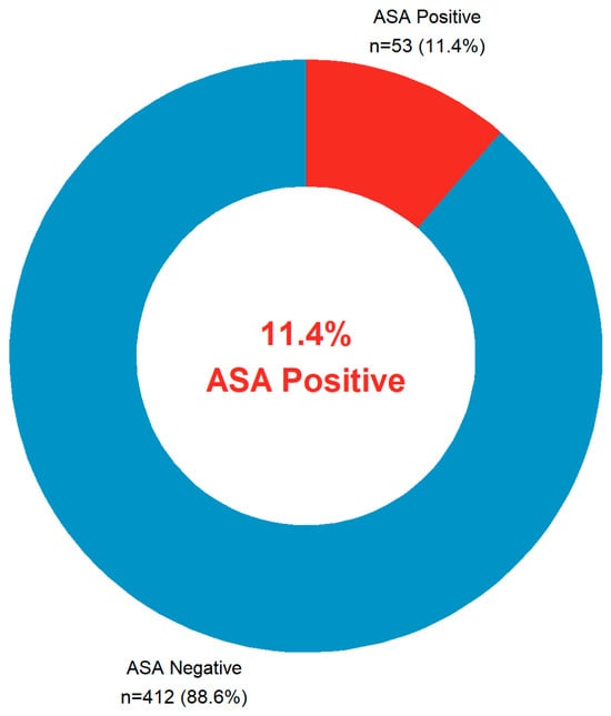
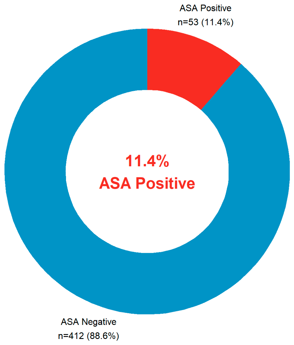
After data cleaning and reclassification (see Section 2.5), the final analytic cohort comprised 465 men, of whom 53 (11.4%; 95% CI 8.7–14.7%) were ASA positive by the direct MAR test (Table 1 and Figure 1). This prevalence falls within the mid-range reported among infertile male populations in the wider literature for MAR-based studies.
Table 1.
Overall ASA prevalence (n = 465).
Figure 1.
Antisperm antibody (ASA) status distribution in the study cohort (n = 465). ASA positivity was defined as ≥50% of motile spermatozoa bearing antibodies on the Direct Mixed Antiglobulin Reaction (MAR) test. Following data cleaning and reclassification, 53 men (11.4%; 95% CI 8.7–14.7%) were ASA positive and 412 (88.6%) were ASA negative.
3.2. ASA Positivity by Subgroup
ASA prevalence was numerically similar across all demographic and clinical subgroups (Table 2). Chi-square testing revealed no statistically significant association between ASA status and any categorical variable: residence (chi2 = 0.608, df = 1, p = 0.436), smoking status (chi2 = 1.502, df = 1, p = 0.220), varicocele (chi2 = 0.901, df = 1, p = 0.342), infertility type (chi2 = 0.601, df = 1, p = 0.439), or supplement use (chi2 = 0.014, df = 1, p = 0.905). Fisher’s exact odds ratios for all five comparisons included the null value (OR = 1.0) within their 95% confidence intervals, indicating no significant bivariate predictor of ASA positivity in this cohort.
Table 2.
ASA prevalence stratified by clinical and demographic subgroups.
3.3. ASA Percentage Distribution
Among the full cohort, 72.3% () had ASA percentages in the 1–10% range. High-titre positivity (≥50%) was found in 11.4% () of all participants, representing the clinically positive group by direct MAR test. A total of 374 men (80.4%) had a low but detectable MAR binding (1–49%), reflecting background immunological activity below the clinical positivity threshold (Table 3). On continuous analysis of ASA percentage across all 465 patients, no semen parameter showed a significant Spearman correlation with ASA percentage (rho range: −0.04 to +0.03; all ). However, men with secondary infertility had a significantly higher ASA percentage than those with primary infertility (median 7.5% [IQR 4–9] vs. 6.0% [IQR 3–9]; Mann–Whitney U, ), a finding not detectable by binary classification alone, as the two groups showed similar binary ASA-positive rates (13.3% vs. 10.7%; ).
Table 3.
Distribution of ASA percentage values across the full cohort (n = 465).
3.4. Age Distribution
The majority of infertile men were in their prime reproductive years, with 26.5% (n = 123) aged 26–30 years, 24.5% (n = 114) aged 31–35 years, and 19.8% (n = 92) aged 36–40 years. Only 0.4% (n = 2) were older than 50 years (Table 4). This age distribution is consistent with published Iraqi and regional fertility clinic data, reflecting the typical reproductive age presentation to infertility services.
Table 4.
Age group distribution of the study cohort (n = 465).
3.5. Sperm Count Distribution
The most common sperm count category was >100 million/mL (normozoospermia/polyzoospermia range), present in 185 men (39.8%). Severe oligospermia (<5 million/mL) was present in 20 men (4.3%), and oligospermia in the 5–15 million/mL range in 39 men (8.4%), consistent with the WHO 2021 lower reference limit of ≥16 million/mL (Table 5).
Table 5.
Sperm count categories (n = 465).
3.6. Progressive Motility Distribution
Severely impaired progressive motility (<10%) was the most common category, affecting 136 men (29.2%), placing this cohort well below the WHO 2021 reference threshold of ≥30% progressive motility. Only 8.2% (n = 38) demonstrated high progressive motility (>60%) (Table 6).
Table 6.
Progressive motility distribution (n = 465).
3.7. Sperm Morphology Distribution
The dominant morphology category was 2–4% normal forms, representing 192 men (41.3%), which falls below the WHO 2021 reference lower limit of ≥4% normal forms [17]. Combined with the 1–2% category (n = 38, 8.2%), a total of 49.5% of men (n = 230) had morphology below the reference limit, confirming a high burden of teratozoospermia in this cohort (Table 7).
Table 7.
Sperm morphology distribution (% normal forms, n = 465).
3.8. Multivariable Analysis of ASA Predictors
Mann–Whitney U tests comparing continuous semen parameters between ASA-positive and ASA-negative men revealed no statistically significant differences: age (median 33.0 vs. 33.0 years; p = 0.541), sperm count (100.0 vs. 72.0 million/mL; p = 0.129), progressive motility (25.0% vs. 16.5%; p = 0.126), non-progressive motility (30.0% vs. 25.0%; p = 0.187), and normal morphology (4.0% vs. 3.5%; p = 0.340), as shown in Table 8.
Table 8.
Multivariable logistic regression—predictors of ASA positivity (n = 465).
Table 8 presents the results of multivariable binary logistic regression, including all ten pre-specified covariates. After mutual adjustment, no variable independently predicted ASA positivity at the p < 0.05 threshold. The overall model had poor discriminative ability (McFadden R2 = 0.022; likelihood ratio test p = 0.708; AUC = 0.607; 95% CI 0.516–0.697), indicating that the measured demographic and semen variables collectively explain minimal variance in ASA status. Variance inflation factors (VIF) were all below 1.4, confirming the absence of multicollinearity.
3.9. Patient Phenotype Clustering by Semen Profile
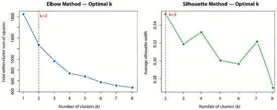
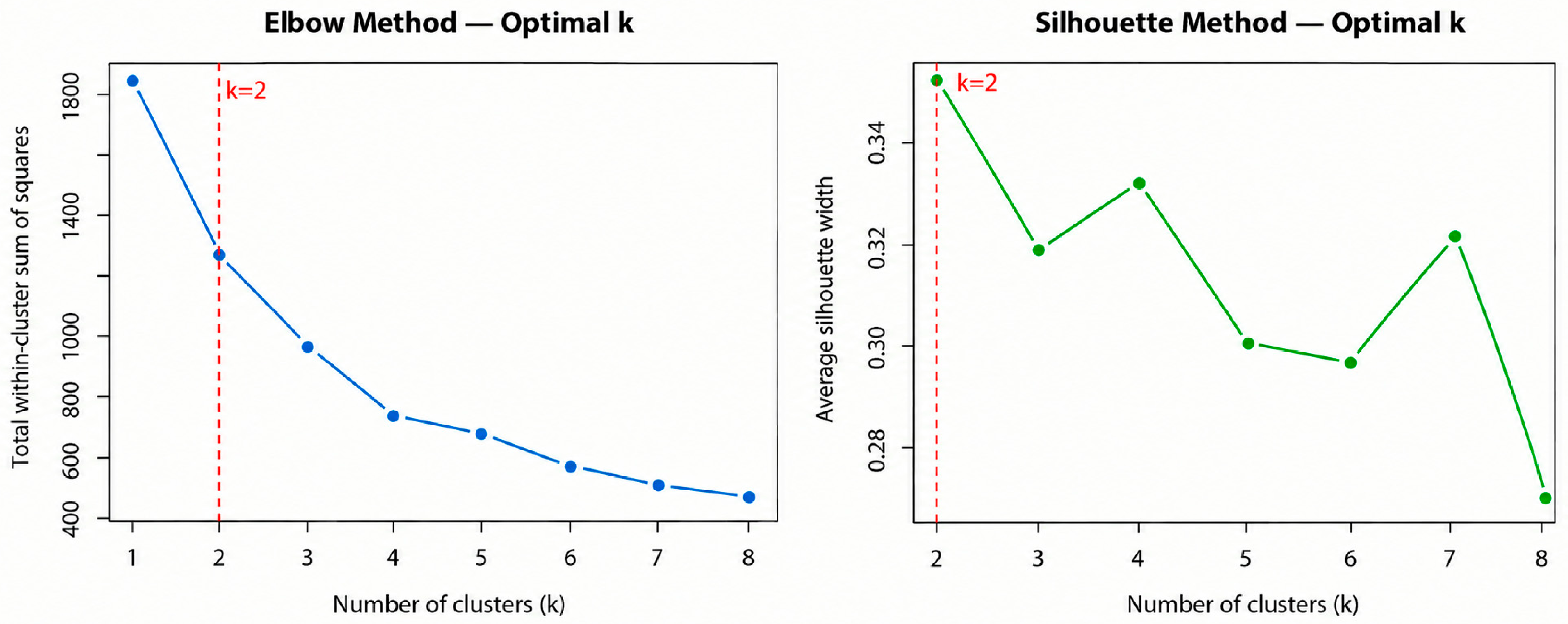
To explore whether natural patient subgroups existed based on semen quality, unsupervised k-means clustering was applied to the four standardized semen parameters: sperm count, progressive motility, non-progressive motility, and morphology. Both the elbow method and silhouette criterion independently identified k = 2 as the optimal number of clusters (average silhouette width = 0.353; Figure 2). The two-cluster solution was validated by hierarchical clustering with Ward linkage, which agreed with k-means assignment in 87.7% of patients.
Figure 2.
Determination of the optimal cluster number (k) for k-means clustering of semen profiles (n = 465). Left panel: Elbow method—total within-cluster sum of squares by k; the elbow occurs at k = 2. Right panel: Silhouette method—average silhouette width by k; k = 2 produced the highest value (0.353), independently confirming two as the optimal cluster number. The red dashed line marks k = 2.
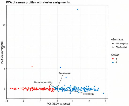
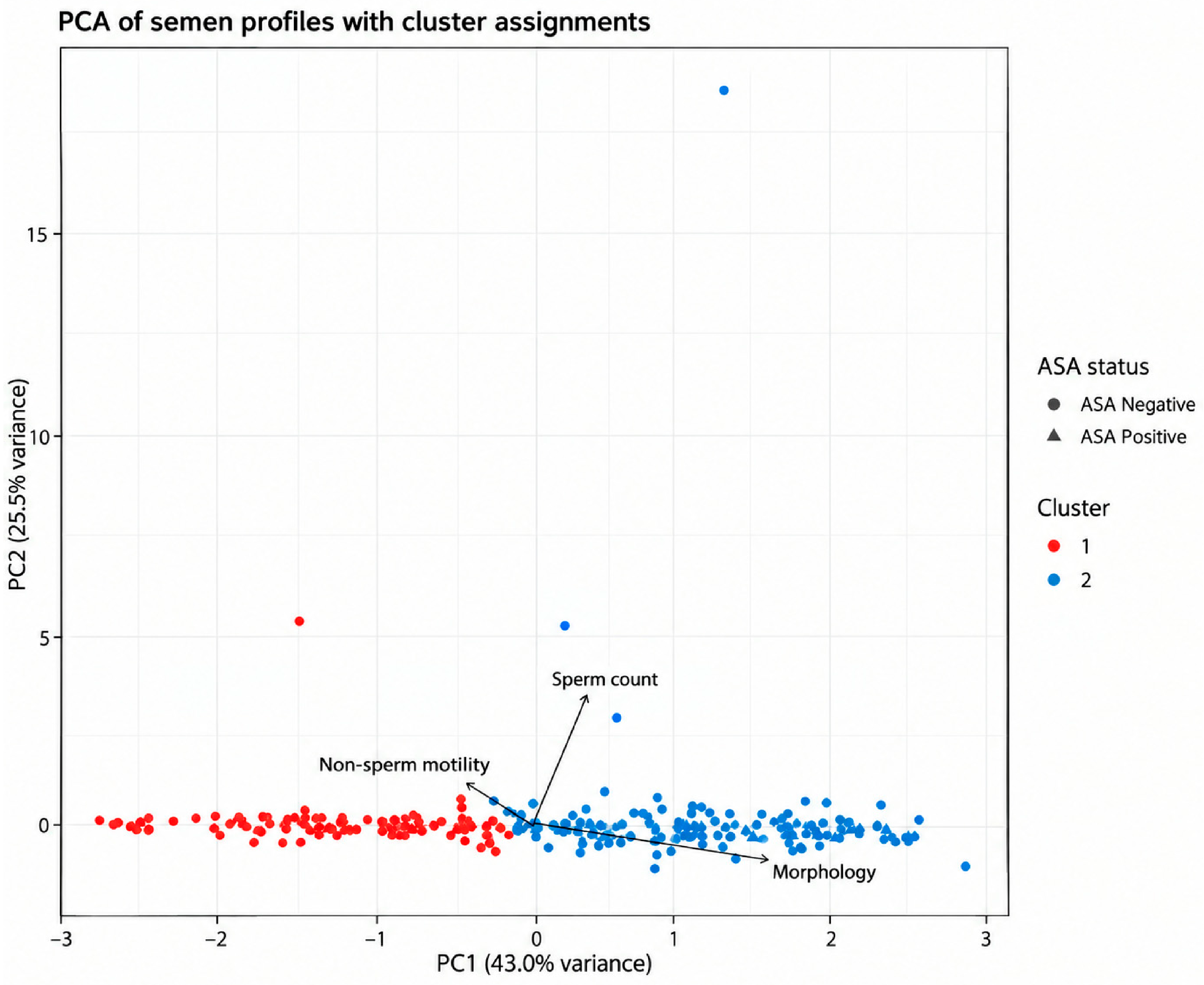
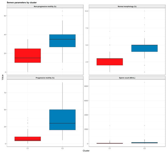
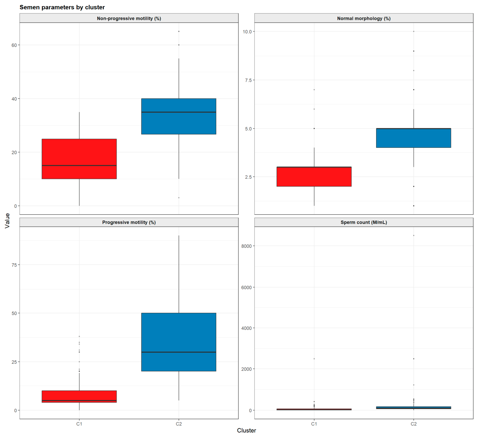
Cluster 1 (n = 201, 43.2%) was characterized by severe semen dysfunction across all parameters: mean sperm count 62.6 million/mL, progressive motility 8.2%, non-progressive motility 17.3%, and normal morphology 2.60%. Cluster 2 (n = 264, 56.8%) demonstrated relatively preserved semen quality: mean sperm count 183.6 million/mL, progressive motility 35.5%, non-progressive motility 33.7%, and normal morphology 4.54%. All four parameters differed significantly between clusters (Kruskal–Wallis p < 0.001 for all four). Principal component analysis (PCA) confirmed the cluster separation, with Principal Component 1 (PC1) (43.0% of variance, driven by motility and morphology) cleanly discriminating between the two groups (Figure 3). The within-cluster semen parameter distributions are shown in Figure 4.
Figure 3.
Principal component analysis (PCA) biplot of semen profiles with k-means cluster assignments (n = 465). Each point represents one patient. High-contrast color intensity indicates cluster membership (dark red = Cluster 1, severe semen dysfunction; dark blue = Cluster 2, relatively preserved semen quality), while distinct point shape indicates ASA status (circle = negative; triangle = positive). Semi-transparent plotting was used so overlapping observations remain visible. Arrows show variable loading directions. PC1 (43.0% of total variance) is driven by progressive motility, non-progressive motility, and morphology; PC2 (25.5%) primarily by sperm count. The two clusters are mainly separated along PC1.
Figure 4.
Semen parameters by cluster (n = 465). Boxplots show median, interquartile range, and outliers for non-progressive motility, normal morphology, progressive motility, and sperm concentration in Cluster 1 (red) and Cluster 2 (blue). All four parameters differed significantly between clusters (Kruskal–Wallis p < 0.001 for all). Note: the sperm count panel y-axis extends to 8500 million/mL due to extreme outliers in a few patients; the majority of values in both clusters fall below 500 million/mL.
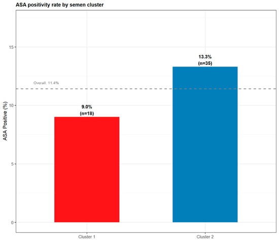
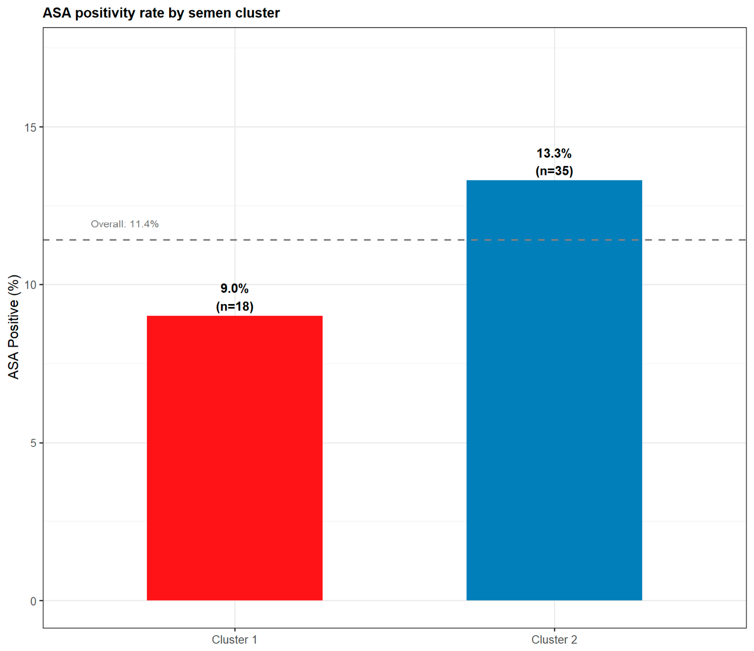
ASA positivity was numerically higher in Cluster 2 (13.3%, n = 35) than in Cluster 1 (9.0%, n = 18), though this difference did not reach statistical significance (Fisher’s exact OR = 1.55; 95% CI 0.82–3.01; p = 0.185; Figure 5). This pattern, higher ASA prevalence in the better-semen cluster, is consistent with the hypothesis that ASA-mediated motility impairment is most detectable in men with sufficient baseline motility for MAR testing, and that men with the most severe semen dysfunction may have ASA that is underdiagnosed by the direct MAR method.
Figure 5.
ASA positivity rate by semen quality cluster. Cluster 1 (red; n = 201, severe semen dysfunction) had an ASA positivity rate of 9.0% (n = 18), while Cluster 2 (blue; n = 264, relatively preserved semen quality) had a rate of 13.3% (n = 35). The dashed line indicates the overall cohort prevalence (11.4%). The difference was not statistically significant (Fisher’s exact OR = 1.55; 95% CI 0.82–3.01; p = 0.185).
3.10. Machine Learning Assessment of Predictor Importance
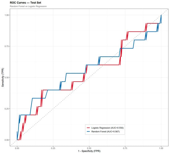
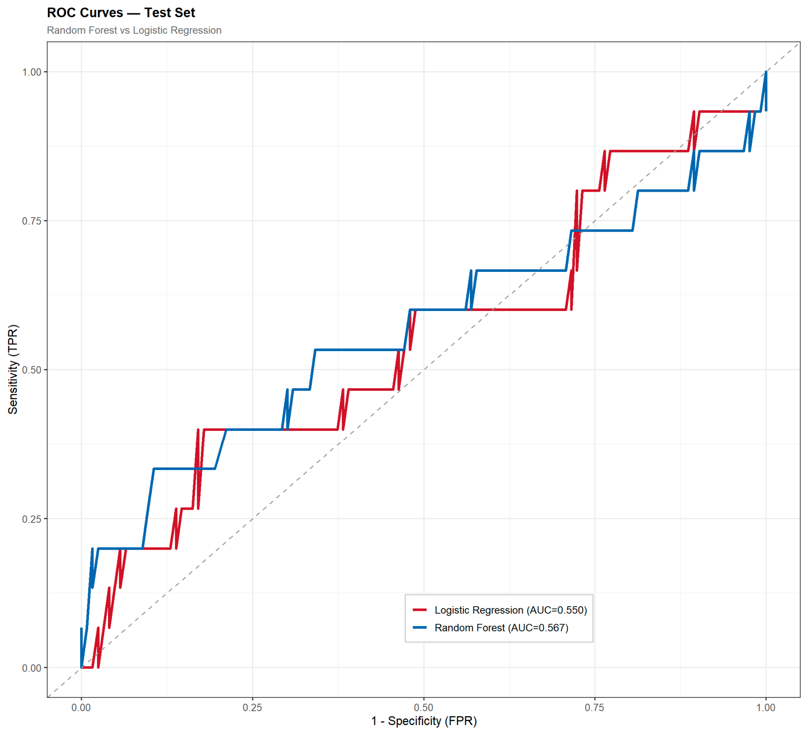
A class-weighted Random Forest classifier (ntree = 1000; negative: positive class weight ratio 1:7) was trained on 70% of the cohort (n = 327; ASA+ 11.6%) and evaluated on the held-out 30% (n = 138; ASA+ 10.9%). On the test set, the model achieved an AUC of 0.567 (95% CI 0.376–0.757), marginally exceeding logistic regression (AUC = 0.550; Figure 6). Both models performed only marginally above chance (AUC = 0.500), confirming that the measured clinical and semen variables carry minimal collective predictive signal for ASA positivity in this cohort.
Figure 6.
Receiver operating characteristic (ROC) curves for ASA positivity prediction on the held-out test set (n = 138; 30% of the cohort). Red line: logistic regression (AUC = 0.550); blue line: Random Forest classifier (AUC = 0.567). The diagonal dashed line represents chance performance (AUC = 0.500). Both models perform only marginally above chance, confirming that the measured clinical and semen variables carry minimal collective predictive signal for ASA status.
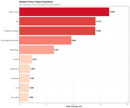
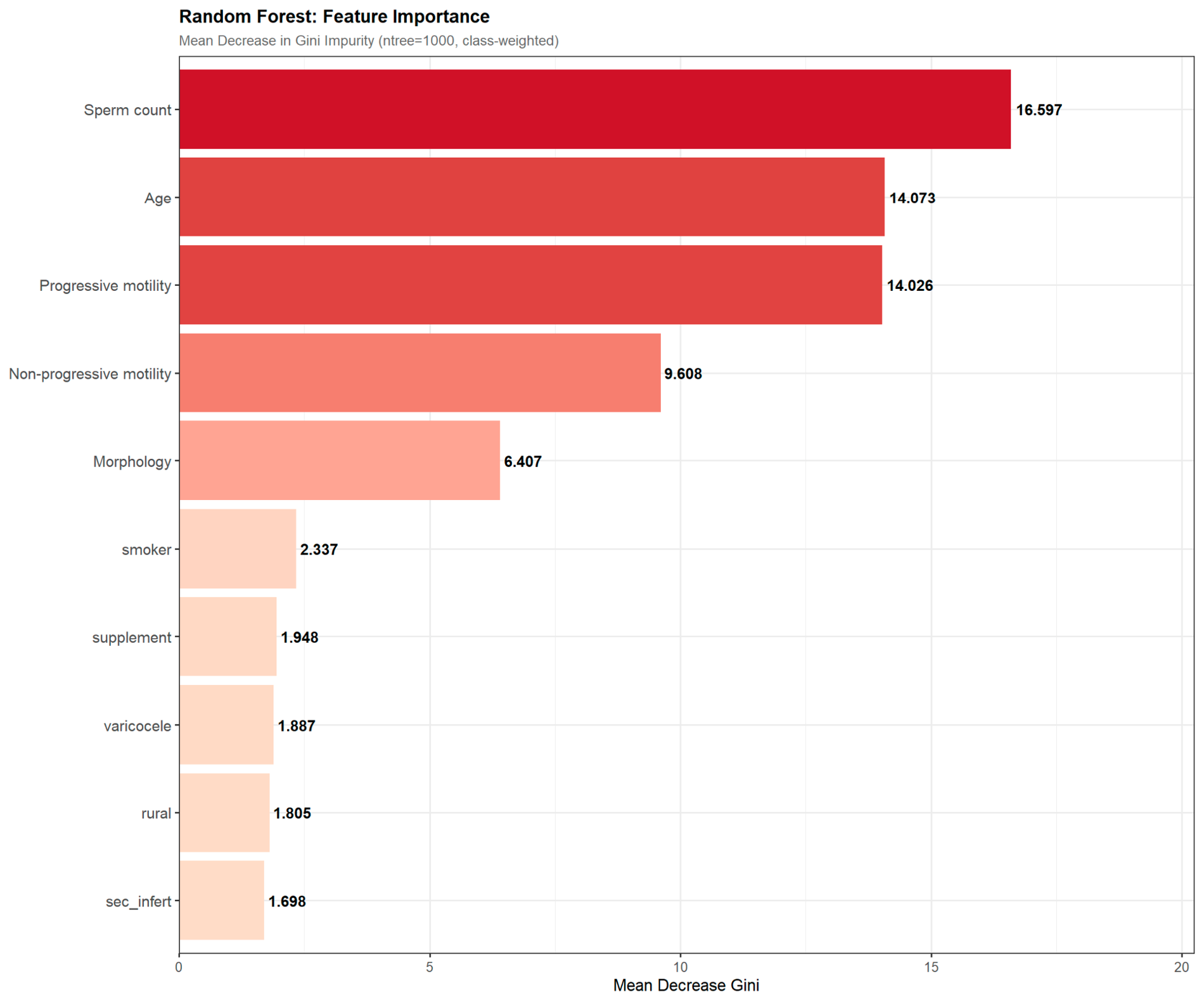
Feature importance analysis (Figure 7) ranked variables by mean decrease in Gini impurity. Continuous semen parameters consistently outranked binary clinical variables. The top three predictors were sperm count (Gini = 16.60), age (Gini = 14.07), and progressive motility (Gini = 14.03), each contributing approximately equally to model decisions and together accounting for over 70% of total Gini importance. Non-progressive motility (Gini = 9.61) and morphology (Gini = 6.41) ranked fourth and fifth. All five binary clinical variables—smoking, supplement use, varicocele, rural residence, and secondary infertility—each contributed less than 2.5 Gini units, collectively representing less than 12% of total model information.
Figure 7.
Random Forest feature importance for prediction of ASA positivity (n = 465; ntree = 1000; class weights: negative = 1, positive = 7). Variables were ranked by a mean decrease in Gini impurity. Continuous semen parameters (sperm count, age, progressive motility, non-progressive motility, morphology) together account for over 88% of total model information, while all five binary clinical variables each contribute less than 2.5 Gini units. The color gradient reflects importance magnitude (dark red = highest; light pink = lowest).
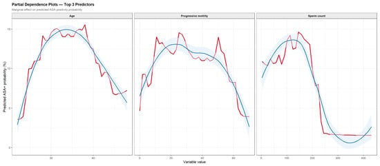
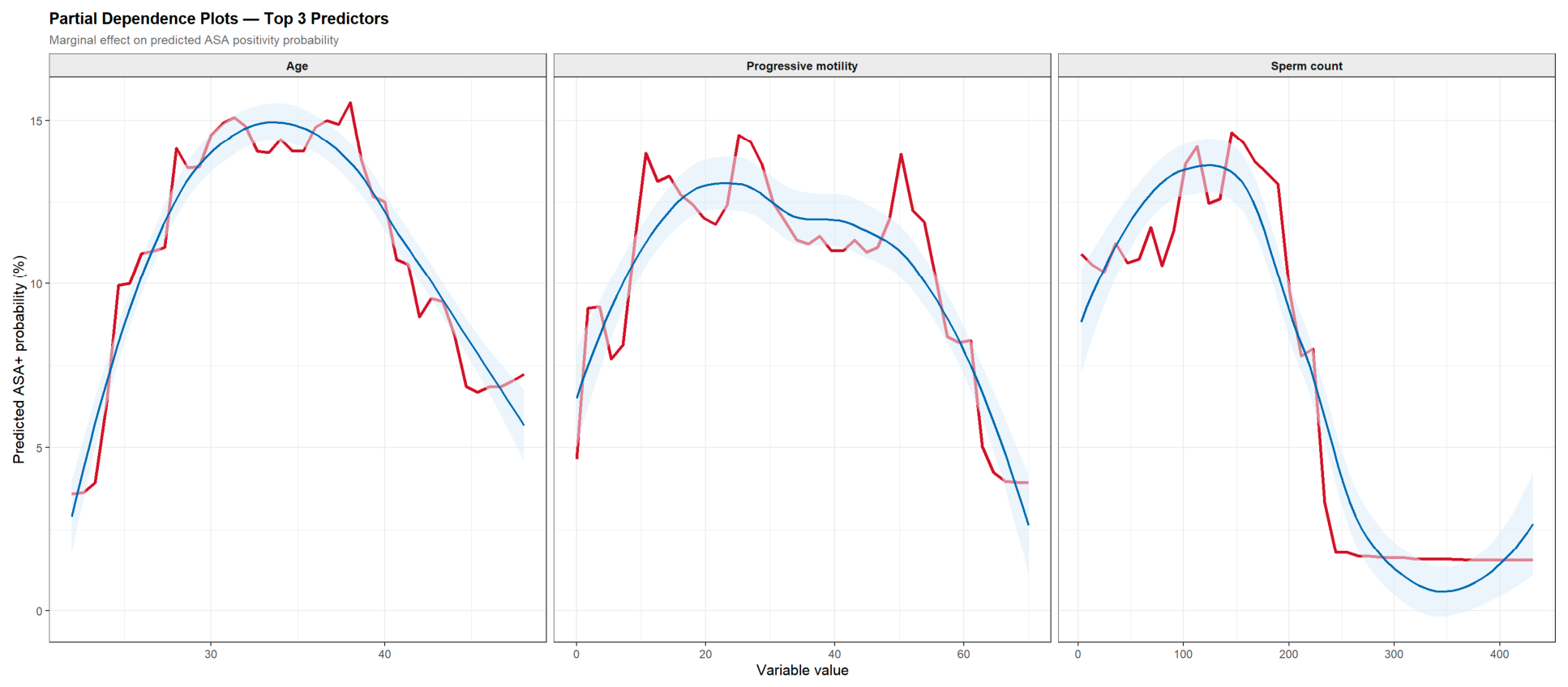
Partial dependence plots for the three top-ranked predictors revealed non-linear marginal effects of modest absolute magnitude (Figure 8). Predicted ASA probability peaked around ages 33–38 years, at intermediate progressive motility values (20–40%), and at intermediate sperm concentrations (100–200 million/mL), with declining predicted probability at more extreme values of each variable. These patterns are exploratory and hypothesis-generating, given the overall low AUC of the model.
Figure 8.
Partial dependence plots for the three highest-ranked Random Forest predictors of ASA positivity: Age (years), Progressive motility (%), and Sperm count (million/mL). The red line shows the marginal predicted ASA-positive probability across the observed range of each variable, holding all other variables at their observed distributions. The blue curve with shading shows the Locally Estimated Scatterplot Smoothing (LOESS) trend with 95% confidence interval. Age shows a non-linear relationship peaking around 33–38 years. Progressive motility peaks at intermediate values (20–40%). Sperm count peaks at 100–200 million/mL. All effects are modest in absolute magnitude, consistent with the low overall model AUC of 0.567.
4. Discussion
This multicenter retrospective cross-sectional study evaluated ASA positivity among infertile men attending urology and fertility centers in Erbil, Kurdistan Region of Iraq. After prespecified data cleaning and correction of MAR test classification, 53 of 465 participants were ASA-positive, corresponding to a prevalence of 11.4% (95% CI: 8.7–14.7%). This revised estimate is lower than the preliminary uncorrected value because 49 participants whose recorded MAR percentages were below the predefined 50% threshold were reclassified as ASA-negative. The corrected estimate should therefore be regarded as the primary result of the study.
The observed prevalence is consistent with the broad range reported among infertile men, although direct comparisons between studies should be made cautiously. ASA prevalence varies according to the characteristics of the study population, the underlying causes of infertility, the biological specimen analyzed, the antibody isotype assessed, and the diagnostic threshold applied [2,3,4,5]. Earlier literature has generally reported ASA positivity in approximately 7–26% of infertile men, whereas the prevalence in fertile populations is substantially lower [3]. The prevalence observed in the present study therefore supports the view that ASA may be clinically relevant in a meaningful minority of infertile men in Erbil, but it does not establish that ASA is the primary cause of infertility in all antibody-positive participants.
The current estimate is lower than several Iraqi reports but remains comparable to some regional findings. Salih et al. reported a prevalence of approximately 22.8% among infertile Iraqi men, while Hasony et al. reported a similar prevalence in Thi-Qar [7,9]. In contrast, Muhammad et al. observed a considerably higher prevalence of 48.4% among asthenospermic infertile men in Kirkuk [8]. The higher figure in that study may partly reflect its selective recruitment of men with impaired motility and its use of additional serum and seminal antibody assessments. Similarly, Al-Assaf et al. reported the highest ASA frequency among asthenozoospermic men, reaching 41.7% in that subgroup [10]. Conversely, Yasin et al. reported a lower prevalence of 9.5% among men from Palestine with unexplained infertility [6]. These differences illustrate that ASA prevalence cannot be interpreted independently of study selection criteria and laboratory methodology.
However, these findings cannot be directly compared without considering differences in patient selection, assay methodology, antibody classification, and clinical setting. Evidence from other regions further supports this variability. In Nigeria, Audu et al. reported substantially higher seroprevalence rates of IgG and IgA ASA using serum ELISA-based testing than are typically observed with direct sperm-based assays [11]. Likewise, an international review of ASA testing reported prevalence estimates ranging from approximately 2% to 15.6% among infertile men when standardized MAR or Immunobead methods were used, highlighting the substantial impact of assay methodology and positivity thresholds on reported prevalence rates. The prevalence observed in the present study (11.4%) therefore lies within the range commonly reported in contemporary MAR-based investigations and is considerably lower than estimates obtained in selected asthenospermic cohorts or studies utilizing serum antibody assays. Furthermore, recent reviews have emphasized that reported prevalence varies widely across Europe, Asia, Africa, and the Americas because of differences in study populations, infertility etiologies, laboratory protocols, and antibody detection techniques. A global survey involving specialists from multiple continents similarly demonstrated marked variation in the indications for ASA testing, preferred laboratory methods, and thresholds used to define clinically relevant positivity. Consequently, locally derived prevalence estimates remain important for interpreting the clinical significance of ASA within specific infertility populations [12,19].
The present study used the direct MAR test and defined clinical positivity as antibody binding to at least 50% of motile spermatozoa. This threshold is widely used in clinical andrology, but it identifies a specific category of clinically significant sperm-bound antibody activity rather than all immunological reactivity [2,4]. In the current cohort, low-level MAR binding was common, whereas only 11.4% of participants met the predefined clinical threshold. This distinction is important because detectable antibody binding below the positivity cut-off may not have the same biological or clinical significance as extensive coating of motile spermatozoa. Studies using serum ELISA, indirect MAR, immunobead testing, or lower positivity thresholds may consequently produce substantially different prevalence estimates [2,4,5,11,20].
Recent evidence suggests that the clinical relevance of ASA extends beyond simple detection and is strongly influenced by the degree of sperm coating, antibody class, and underlying immune mechanisms. A comprehensive review by Mukherjee and Gopalakrishnan highlighted that ASA-mediated infertility represents a heterogeneous disorder in which antibody binding may interfere with sperm transport, capacitation, acrosome reaction, and gamete interaction, while conventional semen analysis may underestimate the functional consequences of sperm autoimmunity. Furthermore, contemporary evidence indicates that disruption of the blood–testis barrier secondary to inflammation, infection, obstruction, or tissue injury remains the principal pathway leading to ASA formation, although the magnitude of fertility impairment varies substantially among affected individuals. These observations support the findings of the present study, where clinically significant ASA positivity was present in a subset of infertile men despite the absence of strong associations with routine semen parameters or common clinical characteristics [19].
No demographic or clinical variable examined in the present study was independently associated with ASA positivity. Residence, smoking, varicocele, infertility type, and supplement use were not significantly associated with the binary outcome in either bivariate or multivariable analyses. The adjusted model also showed limited explanatory capacity, with an AUC of 0.607 and a low McFadden pseudo-R2. These findings indicate that the variables available in this study were insufficient to identify men with ASA positivity reliably. A statistically nonsignificant result should not be interpreted as evidence that these factors have no biological relationship with ASA formation; rather, any associations may have been small, imprecisely estimated, or inadequately captured by the available variables.
The lack of a significant association between varicocele and ASA positivity contrasts with previous evidence. A recent systematic review and meta-analysis reported higher ASA positivity among men with varicocele assessed by the MAR method [15]. Earlier studies also described increased antisperm antibody activity in varicocele-related infertility [14,16], and Bozhedomov et al. proposed that immune-mediated sperm dysfunction may represent an important component of varicocele-associated subfertility [13]. The absence of a similar association in the present study may reflect the use of infertile men without a fertile control group, variation in varicocele severity, incomplete measurement of relevant immune and clinical variables, or the relatively limited number of participants with varicocele. The present result should therefore be interpreted as an absence of demonstrated association rather than evidence against a possible relationship.
Smoking was also not associated with ASA positivity. Although smoking is widely linked to impaired semen quality through oxidative and toxic effects, the formation of sperm-directed antibodies may depend more directly on disruption of testicular immune privilege, infection, obstruction, trauma, or surgery [2,3]. The binary smoking measure used in this study may not have captured dose, duration, waterpipe use, passive exposure, or concurrent environmental exposures. Consequently, the absence of an association indicates only that smoking status, as recorded here, did not discriminate between ASA-positive and ASA-negative participants.
Secondary infertility was associated with a slightly higher median ASA percentage in the continuous analysis, despite the absence of a significant difference in binary ASA positivity. This finding highlights the potential effect of outcome definition. Dichotomising MAR values at 50% may conceal modest shifts in antibody burden that remain detectable when the percentage of antibody-coated sperm is analyzed continuously. The finding is biologically plausible because men with secondary infertility may have experienced previous genital tract infection, surgery, trauma, obstruction, or other reproductive events capable of disrupting the blood–testis barrier [2,3]. Nevertheless, the difference was modest, and the cross-sectional design prevents determination of whether the higher antibody percentage contributed to secondary infertility or resulted from an underlying condition associated with it.
The semen profile of the cohort demonstrated substantial qualitative impairment. Severe reduction in progressive motility was common, and nearly half of participants had sperm morphology below the WHO 2021 reference threshold. At the same time, a large proportion of men had relatively high sperm concentrations. This pattern suggests that infertility in this clinical population was not explained solely by reduced sperm production. Motility and morphology abnormalities may be particularly important contributors to reproductive dysfunction, although semen analysis alone cannot determine the probability of natural conception for an individual couple [17].
Despite the high frequency of semen abnormalities, none of the continuous semen parameters differed significantly between ASA-positive and ASA-negative men. This finding differs from reports linking ASA with impaired motility and other semen-quality measures [4,5,8,10]. Possible explanations include the influence of the direct MAR test on motile spermatozoa, the severe semen abnormalities present throughout the cohort, the biological and analytical variability of semen analysis, and the possibility that ASA affects sperm function through mechanisms not fully reflected in a single routine semen examination.
The clustering analysis identified two broad semen phenotypes: one characterized by more severe impairment across sperm count, progressive motility, non-progressive motility, and morphology, and another with relatively preserved semen quality. ASA positivity was numerically higher in the relatively preserved semen cluster, but the difference was not statistically significant. This pattern should not be interpreted as evidence that better semen quality causes ASA positivity. One possible explanation is methodological: men with sufficient motile sperm may be more suitable for detecting sperm-bound antibodies with the direct MAR method, whereas severe motility impairment may limit the sensitivity or interpretability of the assay. This hypothesis requires direct evaluation using complementary methods, such as indirect MAR, immunobead testing, or isotype-specific assays. Moreover, the relationship between sperm autoimmunization and semen quality is complex because the immunological consequences of ASA may affect sperm function rather than conventional semen characteristics alone. Experimental and clinical evidence suggests that disruption of testicular immune privilege can lead to antibody-mediated impairment of fertilization processes, even in men whose routine semen parameters remain relatively preserved. These mechanisms may partially explain why ASA positivity was observed across both semen quality clusters in the present study [21].
Previous evidence indicates that ASA may impair reproductive potential through functional effects on spermatozoa, including reduced capacitation, impaired acrosome reaction, inhibition of sperm–zona pellucida binding, and interference with sperm–oocyte fusion. As a result, clinically relevant immunological infertility may occur even in men who demonstrate relatively preserved semen quality on conventional analysis, supporting the lack of a clear distinction between the semen profile clusters observed in this study [22].
The machine learning pipeline yielded complementary findings regarding feature discrimination. Although continuous semen parameters (sperm concentration, age, and progressive motility) consistently outranked categorical clinical factors in a mean decrease in Gini impurity, the overall Random Forest model demonstrated low discriminative capacity on the test set (; ). Consequently, variable importance rankings and partial dependence curves should be interpreted strictly as exploratory, hypothesis-generating patterns rather than established diagnostic predictors. These findings further confirm that routine semen parameters cannot serve as predictive surrogates for direct laboratory testing of ASA.
The poor predictive performance of the machine-learning models may reflect the complex biological pathways underlying immune infertility. Emerging literature has emphasized that antisperm antibody production is influenced by multiple interacting factors, including immune regulation, inflammatory responses, oxidative stress, and reproductive tract pathology. As a result, predictive models based solely on demographic and routine semen variables may fail to capture the mechanisms responsible for sperm autoimmunization. Future studies could therefore benefit from integrating immunological biomarkers, cytokine profiles, oxidative-stress markers, and molecular data alongside conventional semen characteristics to improve prediction of clinically relevant ASA positivity [19,23,24].
This study has several strengths. It included a relatively large clinical sample for a regional ASA study, recruited participants from multiple urology and fertility centers, used a prespecified MAR positivity threshold, and applied consistent correction of identified data-quality problems across the analysis. The study also examined ASA positivity both as a binary clinical outcome and as a continuous percentage, while incorporating conventional regression, unsupervised clustering, and exploratory machine-learning analyses. These complementary approaches provided a broader assessment of whether demographic, clinical, and semen variables could explain ASA status.
Several limitations require careful consideration. First, the cross-sectional design prevents conclusions about causality or temporal sequence. Second, recruitment from referral centers means that the prevalence may not represent all infertile men in Erbil or the broader Kurdistan Region. Third, no fertile control group was included; therefore, the study cannot determine whether ASA positivity was more frequent than in fertile men from the same population. Fourth, only the direct MAR method was used. The absence of indirect or isotype-specific testing may have led to under-identification of antibodies not detected by the selected assay. Fifth, important potential determinants, including previous genital tract infection, testicular trauma, hernia repair, infertility surgery, obstruction, and occupational or environmental exposures, were not systematically recorded. Sixth, the number of ASA-positive participants was relatively small for multivariable and machine-learning analyses, limiting statistical power and increasing the risk of unstable estimates. Finally, the data corrections demonstrate the importance of prospective laboratory quality control and independent verification of data entry. The absence of a fertile control group is a major limitation: the study estimates prevalence within a referred infertile cohort but cannot quantify excess ASA positivity relative to fertile men from the same population. In addition, direct MAR testing depends on motile spermatozoa. In men with marked asthenozoospermia, limited numbers of scorable motile cells may reduce sensitivity or precision and could bias comparisons toward apparently higher ASA prevalence among men with better-preserved motility. Complementary indirect MAR or immunobead testing should be considered in future studies when fewer than 100 motile spermatozoa can be evaluated.
The revised prevalence of 11.4% should therefore be interpreted as a clinically relevant but not unusually high frequency of ASA positivity among infertile men attending the participating centers. The findings do not support using residence, smoking, varicocele, infertility type, semen parameters, or machine-learning predictions as standalone substitutes for direct laboratory testing. ASA assessment may be most informative when interpreted alongside the clinical history, repeated semen analysis, evidence of unexplained infertility, and the couple’s reproductive context. Future studies should recruit fertile controls, use larger prospective cohorts, record specific risk factors for disruption of the blood–testis barrier, repeat semen and ASA measurements, and compare direct MAR with complementary antibody assays. Such studies would clarify whether ASA positivity represents a causal mechanism, a marker of previous genital tract injury, or a clinically meaningful but heterogeneous finding in infertile men.
5. Conclusions and Clinical Implications
In this multicenter study of infertile men attending urology and fertility centers in Erbil, clinically significant ASAs were detected in approximately one in nine participants. Although ASA positivity represented a relevant immunological finding within this population, it showed no significant association with routinely measured demographic, clinical, or semen characteristics. Furthermore, neither conventional statistical modelling nor machine-learning approaches were able to reliably identify ASA-positive individuals using the available variables.
These findings suggest that clinically relevant sperm autoimmunization may occur independently of many routinely assessed infertility markers and reinforce the importance of direct laboratory testing when ASA are clinically suspected. The results also highlight the complexity of immune-mediated male infertility and the limitations of relying solely on standard semen parameters to characterize reproductive dysfunction.
From a clinical perspective, ASA testing may provide additional value in selected patients, particularly in cases of unexplained infertility or discordance between semen quality and reproductive outcomes. Future prospective studies incorporating fertile controls, longitudinal follow-up, detailed reproductive histories, immunological biomarkers, and complementary antibody detection methods are needed to better define the biological and clinical significance of ASA in male infertility and to support evidence-based testing strategies in routine practice.
Author Contributions
Conceptualization, H.A.C., A.Q. and S.W.S.; methodology, H.A.C., A.Q., M.A.A. and S.W.S.; validation, S.W.S., C.A.O., A.A.B., M.O.A. and M.O.R.; formal analysis, H.A.C., A.Q. and S.W.S.; investigation, H.A.C., A.Q., M.A.A., C.A.O., A.A.B., M.O.A. and M.O.R.; resources, A.Q. and M.A.A.; data curation, H.A.C., A.Q. and M.A.A.; writing—original draft preparation, H.A.C. and S.W.S.; writing—review and editing, S.W.S., A.Q., C.A.O., A.A.B., M.O.A. and M.O.R.; visualization, H.A.C. and S.W.S.; supervision, S.W.S.; project administration, S.W.S.; funding acquisition, none. All authors have read and agreed to the published version of the manuscript.
Funding
This research was funded by Uppsala University, Sweden, under grant number 345GV478.
Institutional Review Board Statement
Ethical approval was obtained from the Scientific Research Ethics Committee of Erbil Polytechnic University (Approval No. 564/00228 HRE, 9 May 2025). All study procedures were conducted in accordance with relevant guidelines and regulations and with the principles of the Declaration of Helsinki.
Informed Consent Statement
The requirement for informed consent was waived by the Scientific Research Ethics Committee of Erbil Polytechnic University because the study used retrospectively collected and anonymized data.
Data Availability Statement
The data supporting the findings of this study are available from the corresponding author upon reasonable request, subject to ethical and institutional restrictions.
Acknowledgments
The authors gratefully acknowledge Uppsala University, Sweden, for supporting the APC. The authors also thank the participating institutions and healthcare professionals who contributed to data collection and verification.
Conflicts of Interest
The authors declare that they have no known competing financial interests or personal relationships that could have appeared to influence the work reported in this paper.
Abbreviations
The following abbreviations are used in this manuscript:
| AIC | Akaike Information Criterion |
| aOR | Adjusted Odds Ratio |
| ASA | Antisperm Antibodies |
| AUC | Area Under the Curve |
| CI | Confidence Interval |
| df | Degrees of Freedom |
| ELISA | Enzyme-Linked Immunosorbent Assay |
| FPR | False Positive Rate |
| IgA | Immunoglobulin A |
| IgG | Immunoglobulin G |
| IgM | Immunoglobulin M |
| IQR | Interquartile Range |
| LOESS | Locally Estimated Scatterplot Smoothing |
| MAR | Mixed Antiglobulin Reaction |
| OR | Odds Ratio |
| PC1/PC2 | Principal Component 1/Principal Component 2 |
| PCA | Principal Component Analysis |
| R2 | Pseudo Coefficient of Determination |
| ROC | Receiver Operating Characteristic |
| SD | Standard Deviation |
| SPSS | Statistical Package for the Social Sciences |
| TPR | True Positive Rate |
| VIF | Variance Inflation Factor |
| WHO | World Health Organization |
References
- Agarwal, A.; Mulgund, A.; Hamada, A.; Chyatte, M.R. A unique view on male infertility around the globe. Reprod. Biol. Endocrinol. 2015, 13, 37. [Google Scholar] [CrossRef] [Scilit] [PubMed]
- Leathersich, S.; Hart, R.J. Immune infertility in men. Fertil. Steril. 2022, 117, 1121–1131. [Google Scholar] [CrossRef] [Scilit] [PubMed]
- Heidenreich, A.; Bonfig, R.; Wilbert, D.M.; Strohmaier, W.L.; Engelmann, U.H. Risk factors for antisperm antibodies in infertile men. Am. J. Reprod. Immunol. 1994, 31, 69–76. [Google Scholar] [CrossRef] [Scilit] [PubMed]
- Kamphorst, K.; Faber, J.; van der Linden, P.J.Q. The Presence of Antisperm Antibodies in Semen of Subfertile Men—Positive ASA in Subfertile Men. Open J. Obstet. Gynecol. 2021, 11, 88–101. [Google Scholar] [CrossRef]
- Silva, A.F.N.; Schubert, M.; Kliesch, S.; Schlatt, S.; Ramalho-Santos, J.; Amaral, S.C.G. Prevalence and impact of antisperm antibodies on semen quality and male reproductive health aspects: A 10-years retrospective study. Andrology 2025, 13, 2150–2159. [Google Scholar] [CrossRef] [Scilit] [PubMed]
- Yasin, A.L.; Yasin, A.L.; Basha, W.S. The Epidemiology of Anti-Sperm Antibodies Among Couples with Unexplained Infertility in North West Bank, Palestine. J. Clin. Diagn. Res. 2016, 10, QC01–QC03. [Google Scholar] [CrossRef] [Scilit] [PubMed]
- Hasony, H.J.; Ghadhban, A.F.; Al-Kayata, E.S. Serological Evaluation of the Presence of Anti-Spermatozoa Antibody in Infertile Males in Thi-Qar Governorate. Thi-Qar Med. J. 2019, 18, 92–106. [Google Scholar] [CrossRef] [Scilit]
- Raad, H.N.; Muhammad, A.A.; Hussein, Q.A. Prevalence of Antisperm Antibodies in Asthenospermic Infertile Male. Kirkuk Univ. J. Sci. Stud. 2012, 7, 1–7. [Google Scholar] [CrossRef] [Scilit]
- Salih, I.; Rifat, U.N.; Abd, Z.H. The Role of Sperm-Bound Antibodies in Infertile Men in Iraq. Al-Anbar Med. J. 2009, 7, 53–59. [Google Scholar]
- Sulaiman Al-Assaf, A.I.; Ad’hiah, A.H.; Kadri, Z.H.; Abdul Al- Mageed, N.G.; Mustafa, L.K. Evaluation of Anti-sperm Antibodies and Some Cytokines Profile in Seminal Plasma of Iraqi Infertile Males. Al Mustansiriyah J. Pharm. Sci. 2013, 13, 155–163. [Google Scholar] [CrossRef] [Scilit]
- Audu, O.; Musa, B.O.P.; Adeiza, M.A.; Muhammed, A.; Ahmad, A.E.; Ega, B.; Opaluwa, S.A.; Shuaib, B.I. Sero-prevalence of IgG and IgA Antisperm Antibodies in Men with Infertility Attending Two Major Hospitals in Zaria, Nigeria. Health Sci. J. 2021, 15, 837. [Google Scholar]
- Gupta, S.; Sharma, R.; Agarwal, A.; Boitrelle, F.; Finelli, R.; Farkouh, A.; Saleh, R.; Abdel-Meguid, T.A.; Gül, M.; Zilaitiene, B.; et al. Antisperm Antibody Testing: A Comprehensive Review of Its Role in the Management of Immunological Male Infertility and Results of a Global Survey of Clinical Practices. World J. Men’s Health 2022, 40, 380–398. [Google Scholar] [CrossRef] [Scilit] [PubMed]
- Bozhedomov, V.A.; Lipatova, N.A.; Rokhlikov, I.M.; Alexeev, R.A.; Ushakova, I.V.; Sukhikh, G.T. Male fertility and varicocoele: Role of immune factors. Andrology 2014, 2, 51–58. [Google Scholar] [CrossRef] [Scilit] [PubMed]
- El-Gendy, A.; Younis, I.; El-Taweel, A.; Megahed, Y. Antisperm antibodies in varicocele related infertility. Med. J. Cairo Univ. 1994, 62, 189–193. [Google Scholar]
- Falcone, M.; Bocu, K.; Keskin, H.; Solorzano Vazquez, J.F.; Banthia, R.; Mahendran, T.; Deger, M.D.; Kv, V.; Mirko, P.; Harraz, A.M.; et al. Anti-sperm Antibody Positivity in Men with Varicocele: A Systematic Review and Meta-Analysis. World J. Men’s Health 2025, 43, 60–69. [Google Scholar] [CrossRef] [Scilit] [PubMed]
- Golomb, J.; Vardinon, N.; Homonnai, Z.T.; Braf, Z.; Yust, I. Demonstration of antispermatozoal antibodies in varicocele-related infertility with an enzyme-linked immunosorbent assay (ELISA). Fertil. Steril. 1986, 45, 397–402. [Google Scholar] [CrossRef] [Scilit] [PubMed]
- World Health Organization. WHO Laboratory Manual for the Examination and Processing of Human Semen, 6th ed.; World Health Organization: Geneva, Switzerland, 2021. [Google Scholar]
- FertiPro. SpermMar Test IgG: Test for the Detection of Anti-Sperm Antibodies. Available online: https://fertipro.com/2019/11/04/spermmar-igg/ (accessed on 9 April 2025).
- Mukherjee, A.G.; Gopalakrishnan, A.V. Anti-sperm Antibodies as an Increasing Threat to Male Fertility: Immunological Insights, Diagnostic and Therapeutic Strategies. Reprod. Sci. 2024, 31, 3303–3322. [Google Scholar] [CrossRef] [Scilit] [PubMed]
- Alongo, M.-A.M.; Sihalikyolo, J.; Agasa, S.B.; O’yandjo, A.M.; Mbutu, B.; Bosunga, G.K.; Okenge, J.-P.M. Anti-Sperm antibodies: Prevalence of Ig A and Ig G positive serology among infertile patients in Kisangani, Democratic Republic of congo. Open J. Obstet. Gynecol. 2019, 9, 707–714. [Google Scholar] [CrossRef]
- Fijak, M.; Meinhardt, A. The testis in immune privilege. Immunol. Rev. 2006, 213, 66–81. [Google Scholar] [CrossRef] [Scilit] [PubMed]
- Francavilla, F.; Santucci, R.; Barbonetti, A.; Francavilla, S. Naturally-occurring antisperm antibodies in men: Interference with fertility and clinical implications. An update. Front. Biosci. 2007, 12, 2890–2911. [Google Scholar] [CrossRef] [Scilit] [PubMed]
- Barratt, C.L.R.; Björndahl, L.; De Jonge, C.J.; Lamb, D.J.; Osorio Martini, F.; McLachlan, R.; Oates, R.D.; van der Poel, S.; St John, B.; Sigman, M.; et al. The diagnosis of male infertility: An analysis of the evidence to support the development of global WHO guidance-challenges and future research opportunities. Hum. Reprod. Update 2017, 23, 660–680. [Google Scholar] [CrossRef] [Scilit] [PubMed]
- Mass Lindenbaum, M.; Kuribayashi, S.; Lundy, S.D. Antisperm antibodies and autoimmunity in the era of assisted reproduction. Fertil. Steril. 2026, 125, 746–756. [Google Scholar] [CrossRef] [Scilit] [PubMed]

Disclaimer/Publisher’s Note: The statements, opinions and data contained in all publications are solely those of the individual author(s) and contributor(s) and not of MDPI and/or the editor(s). MDPI and/or the editor(s) disclaim responsibility for any injury to people or property resulting from any ideas, methods, instructions or products referred to in the content. |
© 2026 by the authors. Licensee MDPI, Basel, Switzerland. This article is an open access article distributed under the terms and conditions of the Creative Commons Attribution (CC BY) license.