


Abstract
PNH is an acquired ultrarare disease caused by a somatic mutation of PIGA gene located at band 22 of the short arm of the X chromosome, which is unable to carry out the synthesis of glycosil-phosphatidyl-inositol. The loss of complement regulators CD55 and CD59 is associated with the lack of the protective complement regulators in RBCs that become highly susceptible to complement-mediated lysis. PNH is characterized by intravascular hemolysis, high prevalence of thrombotic events, and a variable degree of bone marrow failure. It may arise from aplastic anemia, myelodysplasia and myeloproliferative neoplasms. Recently, a new clinical variant of PNH has been reported, the so-called “ahemolytic white blood cell PNH”, which may be associated with thrombotic episodes. In 2006, the introduction of eculizumab therapy has revolutionized the treatment for PNH patients. More recently, ravulizumab, crovalimab, iptacopan, danicopan, and pegcetacoplan have been introduced in the marketplace, with short/medium-term data supporting their safety and effectiveness. However, no trial has directly compared the clinical outcomes of the new drugs, and also the inclusion criteria in the various phase 3 trials were different, making a comparison between them difficult and unsatisfactory. It is conceivable that artificial intelligence and machine learning-based scoring models may be crucial for predicting the effectiveness of the anti-complement therapy.
1. Laboratory and Clinical Findings: Much More than Flow Cytometry
In 1866 William Gull described this disease for the first time; at that time, PNH was not a distinct entity from paroxysmal cold hemoglobinuria. In 1882 Paul Strübing showed that hemolysis took place during the night owing to the mild decrease in blood pH due to the accumulation of CO2 and lactic acid caused by breath slowing down during sleep. In 1911 the Dutch physician Abraham Albert Hijmans van den Bergh (1869–1943) demonstrated that hemolysis in PNH is related to an intrinsic red blood cell defect rather than to a plasma abnormality, as it occurs in paroxysmal cold hemoglobinuria. In the mid-1930s the USA hematologist Thomas Hale Ham (1905–1987) demonstrated that complement was the cause of hemolysis in PNH [1,2,3]. Nevertheless, only in the mid-1950s, a deep understanding of the complement innate or alternative pathway gave definitive confirmation to the etiological hypothesis suggested by Ham [4]. Ham has the credit of conceiving in 1939 a diagnostic tool based on acidified serum that was used worldwide despite its lack of both sensitivity and specificity until the early 90s, when it was ousted by flow cytometry [5]. PNH is also called Marchiafava–Micheli syndrome due to their amazing description of the disease.
PNH may be considered an extremely rare disease as it may be observed in less than one case in 200,000 subjects; it is more common in the Far East and in southeast Asia. No sex predilection has been noted; however, it arises more often during the third to fourth decade of life. In 10% of cases PNH arises in subjects younger than 20 years [1]. In comparison with PNH adults, pediatric PNH is characterized by smaller clones, more frequent bone marrow dysplastic involvement, and thrombotic events, and, finally, a better response to immunosuppressive therapy [3,6,7].
From a clinical point of view, PNH is characterized by intravascular hemolysis (chronic and acute), high prevalence of thrombotic events, involvement of smooth muscles (dysphagia and erectile dysfunction in affected men) and a variable degree of bone marrow failure and peripheral cytopenia.
Epidemiology of the disease remains difficult due to a number of confounding factors, such as the inclusion or exclusion of very small PNH clones (less than 1%) and or cases associated with bone marrow failure, such as hypoplastic anemia. In 1–2% of cases it may evolve in acute myeloblastic leukemia [8]. Spontaneous recovery may occur in less than 1% of the cases.
PNH is an acquired disorder which may arise from myelodysplastic syndromes (3–5%) or aplastic anemia (10–35%) [3,9,10]. In a recent multicenter study, Daddio et al. evaluate the prevalence of PNH clones (glycosyl-phosphatidyl-inositol lacking) in 119 Ph- negative myeloproliferative neoplasm (MPN) patients having anemia, LDH elevation, asthenia and history of thrombosis. Using a standardized diagnostic based on a single lyophilized template for granulocytes, monocytes, and erythrocytes, a prevalence of PNH-positive clones of 3.23% was found. PIG-A deletion was detected in PNH-positive cases along with mutations of several myeloid-related genes. These data derived from the analysis of MPN cases seem to suggest an association of CALR mutation and JAK2V617F mutation with PNH-positive clones, suggesting that the worsening of malignant process may be associated with the acquisition of multiple genetic mutations [11].
From a pathogenic point of view, it appears clear that erythrocyte lysis occurs due to pathologic intravascular activation of the complement alternative pathway, which represents a key of innate immunity that allows vertebrates to promptly provide early defenses against infective agents. The complement system is constituted by a large number of distinct plasma proteins that react with one another to opsonize pathogens and induce several inflammatory responses, which play a key role in infection control. Complement proteins are proteases that might be activated by proteolytic cleavage. The complement system is activated through a triggered enzyme cascade at sites of infection. In the early phases, the activation of a small number of complement proteins is enhanced by each successive enzymatic reaction, resulting in the rapid generation of a massive complement response. There are three distinct pathways through which a complement can be activated on pathogen surfaces. In normal conditions, the improper activation of lytic mechanisms is inhibited by the presence on the plasma membranes of erythrocyte and leukocyte (particularly neutrophils and monocytes) precursors of either CD55 factor, which promotes the MAC dissolution and regulates the synthesis and stability of both C3- and C5-convertases, or CD59 factor, which blocks the formation of plasma MAC inhibiting polymer C9 binding to the C5b-C8 complex [3].
CD55 and CD59 are kept adherent to the plasma membrane of red cells, neutrophil and monocyte precursors owing to a glycosyltransferase. Such enzyme catalyzes the binding of glucosamine to the phosphatidylinositol (PI) contained in the plasma membrane of red and white cell precursors owing to the etheric bridge (-O-), which arises between the central H-COH of glycerol (essential constituent of the phospholipidic bilayer of any plasma membrane) and one of the three –OH present in phosphate. Thus, the glycosylphosphatidylinositol (GPI) is formed.
The main pathogenic mechanism of PNH onset is due to a somatic mutation of PIG-A gene or phosphatidylinositol A glycan located at band 22 of the short arm of X chromosome (or Xp22.1), which is unable to carry out the synthesis of glycosyltransferase, which joins a glycosylic group to phosphatidylinositol. Thus, the capacity for defense AP (namely CD55 and CD59) to stick to the erythrocyte and leukocyte (particularly neutrophil and monocyte) precursor surface is lacking [12,13].
PIG-A gene is long at least 17 kb and has six exons; thus, it is a middle size gene. Splicing events may sometimes give rise to the synthesis of AP unable to protect the surface of erythrocyte and leukocyte precursors against aggressions by the circulating complement [3,14]. Splicing is known to consist in the breaking of phosphodiesterase bonds in mRNA and in successive casual linkage of remaining residues: it follows the production of different RNAs and thus of proteins different from the original patterns.
The severity of the clinical picture of a PNH depends on both the percent of erythrocytes and leukocytes affected by the genic mutation and on the degree of AP qualitative and/or quantitative deficiency. This “phenotypic mosaicism” has led authors of [1] to divide PNH red and white cells investigated through flow cytometry in three groups: (1) PNH I—cells which show on their surface normal AP concentration; this form may be considered subclinical and hemoglobinuria may be absent; (2) PNH II—cells which show a considerable AP deficiency (about 90%); in spite of this, they prove to be sufficiently resistant to complement-induced lysis; (3) PNH III—cells which lack absolutely AP and thus quite easily undergo spontaneous lysis; hemoglobinuria is constant or at least frequent.
The frequency of phenotypes I, II and III varies among patients. The association of PNH I and PNH III is the most common; the coexistence of all three types follows in decreasing order; the association of PNH I + PNH II is conversely the rarest type and may be considered as a subclinical form [15].
Careful longitudinal studies have ascertained that the abnormal PNH clone remains mostly unchanged (from 25 to 60% of cases); it may increase (from 15 to 50% of cases); more rarely (from 5 to 10% of cases) it may disappear.
As far as the detection of PNH cells in hematological malignancies is concerned, 20–60% of aplastic anemia cases may show PNH cells, while GPI-deficient cells may be reported in 5% of MDS and 3% of myeloproliferative neoplasms.
The main clinical manifestations of PNH are represented by hemolysis and consequent hemoglobinuria (frequent, persistent or variable from day to day or even during the day), anemia, asthenia, early physical weakness and exhaustibility during prolonged efforts, as well as occasional dysphagia, oropharyngeal pain, and impotence in men; abdominal pain may or may not be associated with thrombotic events occurring in unusual sites such suprahepatic (Budd-Chiari syndrome) or mesenteric veins. Brain vein thrombosis was also documented in several cases. Conversely thrombotic events in the arteries were seen in a few cases [3,16,17].
The thrombotic events are caused by (a) platelet activation induced by the release of free hemoglobin fragments and (b) ADP release into the blood by activated platelets. The nocturnal red cell lysis may be observed only in 25% of patients; the frequency of such event increases in infectious diseases owing to the presence of antibodies that activate complement through the classic pathway.
Laboratory investigations show the presence of dark (Coca-Cola) urine owing to the presence of hemoglobin, marked serum LDH increase (strictly related to the degree of intravascular hemolysis, IVH), indirect serum bilirubin increase, decreased serum haptoglobin as it is wasted by binding free hemoglobin released by lysed erythrocytes (in order to inhibit their oxidative activity), decrease in serum ferritin due to urinary iron loss, decrease in polymorphonuclear neutrophils and platelets owing to recurrent infections and bleeding, absence of spherocytic red cells at the microscope observation of a blood smear, Coombs test negativity, and high reticulocyte counts [3].
The finding of an insufficient reticulocyte number in relation to the anemia degree may lead to suspicion of the onset of a myelodysplastic pattern. Reticulocyte count plays a key role for the measurement of bone marrow response to the concomitant anemia.
At the onset of the disease, MCV may be increased due to the augmented reticulocyte production; during the course of the disease such index may decrease due to iron deficiency.
Renal failure is a common finding in PNH, although it often manifests as a subclinical entity. Acute kidney injury (AKI) or chronic kidney disease (CKD) are rare, accounting for 5–20% of the cases. A careful estimation of the incidence and prevalence of renal dysfunction is hardly evaluated; however, based on our personal experience as well as knowledge of the literature, the occurrence of acute renal failure in a PNH patient represents a criterion for the urgent beginning of anti-complement therapy, which is capable of reversing renal insufficiency [18,19,20,21,22].
Renal failure has been implicated as the leading cause of death in 8–18% of PNH population. Other authors have reported an eight-fold increase in mortality in PNH patients having concomitant renal dysfunction.
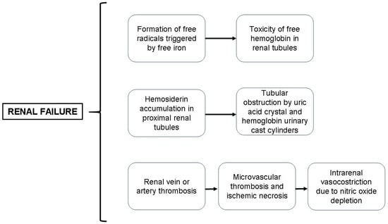
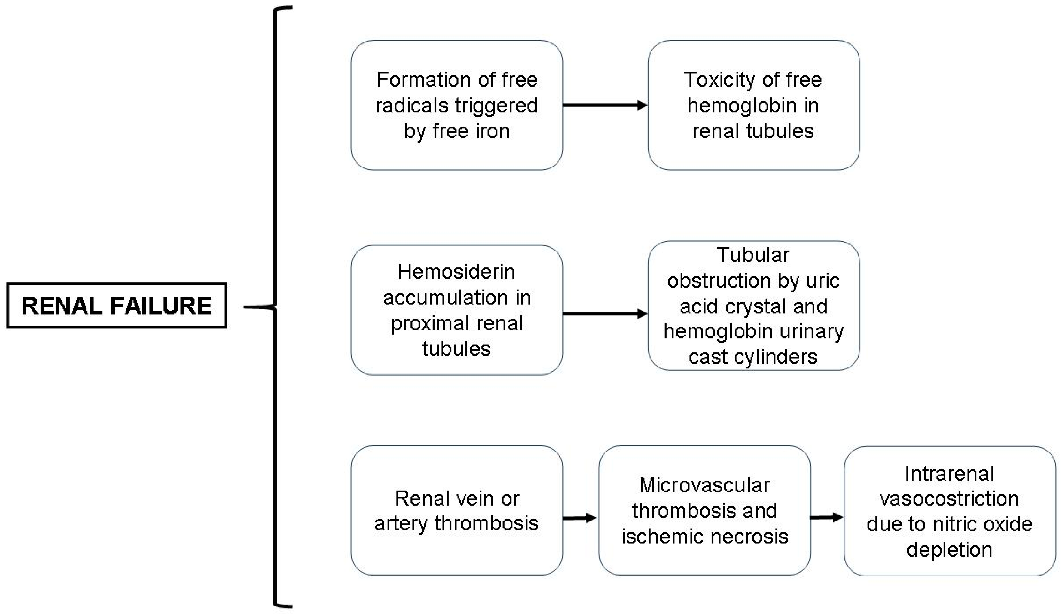
The main mechanisms underlying renal failure in PNH are illustrated in Figure 1.
Figure 1.
The main mechanisms underlying renal failure in PNH are illustrated in this figure.
Nowadays, the diagnosis of PNH is mainly based on the flow cytometry analysis of blood cells; the use of specific monoclonal antibodies (moAbs) capable of recognizing anchored proteins such as GPI, in combination with a great number of fluorochromes, make the test precise, sensitive, and specific for PNH. Leukocytes may also be investigated using a modified toxin (FLAER) capable of identifying the GPI molecules on both neutrophil granulocytes and monocytes. This method is rapid, very sensitive and capable of recognizing their presence in the blood of PNH microclones even in concentrations lower that 1% [23,24,25].
According to published guidelines for the diagnosis and monitoring of PNH using flow cytometry, the assessment of samples should be performed on leukocytes, as it is regarded as the best method for estimating the true size of a PNH clone. In this setting, both monocytes and neutrophils are suitable targets. Furthermore, it has been stated that the clone size measured in each population agrees relatively closely [26].
According to published guidelines, it has been suggested that the GPI-deficient molecules should be evaluated in granulocytes and at least one additional cell line—RBCs and/or monocytes—and that the proportion of GPI-deficient RBCs is significantly lower than that of WBCs due to hemolysis and/or recent transfusions [27].
In contrast, lymphocytes are not a suitable target because of their long life span and variable expression of many GPI-linked protein abnormalities in various B, T and NK subpopulations. Furthermore, it is well known that in most PNH patients the proportion of GPI-deficient RBCs is significantly lower than that of WBCs due to hemolysis and/or recent transfusions.
Moreover, as mentioned before, PNH patients may show three populations of PNH cells according to their normal (PNH I), partially deficient (PNH II), or completely deficient CD59 membrane expression (PNH III) [26,27].
In order to identify GPI-deficient clones in granulocytes, monocytes and erythrocytes, the following parameters should be evaluated: limit of detection (LOD) and lower limit of quantification (LLOQ) of the flow cytometric method. These parameters are calculated by assessing the total number of events acquired in the various flow gates based on the presence of a cluster of at least 30 PNH events.
As far as the measurement of PNH RBCs is concerned, appropriate staining methods are based on the use of MoAbs combinations containing anti CD235a-FITC/CD59-PE. The molecule CD235a (glycophorin A) is expressed on mature erythrocytes; thus, anti-CD235a antibodies allow an excellent identification of erythrocytes in comparison with other cells. Anti-CD59 antibodies allow to recognize and quantify normal and pathologic (PNH II and PNH III) erythrocytes among the mature CD235a+ RBC [11].
For diagnostic purposes, it is also mandatory to enumerate PNH cells in either neutrophil granulocytes or monocytes, since the examination of RBCs only may underestimate the severity of the clone size. In summary, I do believe that the diagnostic test could be performed with a single (lyophilized) template for granulocytes and monocytes consisting of a five-color measurement assay based on the FLAER-FITC/CD157-PE/CD64-PC7/CD15-APC/CD45-5 fluorescence channel and a single template for erythrocytes consisting of CD235a-FITC/CD59-PE/CD45-APC. Based on the total number of events acquired in the various gates of interest, a homogeneous cluster of at least 30 events could be detected in order to evaluate a PNH clone.
The main reasons leading physicians to order a PNH test are listed in Table 1.
Table 1.
The main reasons inducing physicians to order a PNH test are listed here.
The frequency and severity of thrombotic events in PNH patients may depend on the clone size. As a general consideration, it can be stated that thrombotic events represent the main cause of complications and death in PNH patients.
The main clinical categories of PNH, according to the size of PNH clone and laboratory and clinical characteristics, are illustrated in Table 2.
Table 2.
Clinical categories of PNH, according to the size of PNH clone and laboratory and clinical characteristics. Each category may or may not be associated with BMF.
2. Ahemolytic PNH
Recently, two publications [28,29] have shown a new clinical variant of PNH, the so-called white blood cell deficiency. The first description of the so-called ‘pure monocyte PNH”, was reported by Lanza, in which clones involved preferentially the monocytes with marginal RBC and neutrophil involvement. Since PNH is an acquired clonal disorder caused by a somatic mutation of the X-linked gene, PIG-A, it can be postulated that the deficiency of GPI-anchored proteins in the examined patients occurred at a later stage of progenitor cell maturation, namely at the monocyte differentiation phase. In this patient population (n.18 Italian cases), mild intravascular hemolysis was documented at diagnosis in four (22%) cases, supporting the evidence that PNH with a relatively small erythrocyte clone is less commonly associated with RBC destructions. Based on the high prevalence (22%) of bone marrow failures with low peripheral counts in the patient population, the presence of PNH III monocyte prevalence pattern may be speculated as another determinant for the hypoplastic PNH. Thrombotic events are detectable in this PNH variant. According to Tombul report, ahemolytic WBC PNH would constitute a novel PNH subtype in addition to the three categories proposed by PNH International PNH Interest Group currently including classic PNH, PNH in the setting of another defined bone marrow failure syndrome and subclinical PNH. It is characterized by frequent thrombotic events and a variable degree of bone marrow failure (see Table 2).
3. Role of Genetic Mutations in Disease Progression and in Assessing Treatment Response
The GPI-negative phenotype confers a survival advantage on PIG-A-mutated HSCs in patients’ bone marrow in which immune-mediated myelosuppression may occur, leading to a pathologic hematopoiesis [30,31]. According to Luzzatto’s point of view, in most PNH cases clonal expansion may result from the escape of glycosylphosphatidylinositol (GPI)-negative (PIGA-mutant) stem cells from a T-cell-mediated auto-immune attack on nonmutant stem cells [32]. Moreover, there is an accumulating body of evidence that immune privilege leading to clonal escape could only partially explain the overwhelming expansion of PNH clones in an hypoplastic BM. More recently, several authors have reported that the development of a number of intrinsic factors is involved in the survival advantages of PNH clones. In this context, chromosomal and gene abnormalities have been identified in some PNH cases [33,34,35]. In particular, clonal chromosomal abnormalities and somatic mutations have been documented in some publications involving PNH patients. The recent application of next-generation sequencing (NGS) and whole-genome sequencing (WGS) in hematological malignancies has allowed the identification of a higher level of genome complexity in either benign or malignant hematopoiesis. In the study by Shen et al., a stepwise acquisition of mutations and expansion of the most permissive subclones in PNH patients was found. The subsequent development of additional somatic mutations resulted in a complex hierarchical clonal architecture, which appears to be similar to that observed in myeloid neoplasms. More recently, further publications have shown that, in addition to PIG-A mutations, involvement of several myeloid-related genes, such as TET2, SUZ12, U2AF1, IDH, HMGA2, L3MBTL1, SGK2, CAL-R, BCR-ABL and JAK, were reported in PNH patients, suggesting stepwise clonal evolution derived from a singular stem cell clone [31,33,34,35,36].
In PNH, the demonstration of an involvement of genes that are strictly related to myeloid neoplasm pathogenesis has changed the genetic scenario of the disease.
At present, no systematic molecular, genetic, or functional complement investigations are performed to clarify the mechanisms of optimal or suboptimal responses to the various anti-complement therapies. We do believe that this area of investigation may be of great value for guiding precision use of anti-complement drugs and for tailoring treatment strategies to individual patients who fail to respond to a given agent. Interestingly, in a recent publication, it was found that the C3 MG-ring mutation rendered C3b resistant to inactivation and reduced pegcetacoplan binding, explaining persistent hemolysis following treatment with both anti C5 and anti-C3 therapies [37].
4. Therapy: Old and Novel Drugs—Where Are We Going!
Standard treatment options for hemolytic PNH include supportive treatments and, above all, complement inhibitors. Terminal complement C5 inhibition, with eculizumab or, more recently, with ravulizumab, has been found to be associated with a rapid and sustained improvement of clinical outcome measures as well as a significant decline in IVH. Eculizumab is administered intravenously at a dosage of 900 mg every 14 ± 2 days. The drug is usually well tolerated. The administration of a vaccine against Neisseria meningitidis should precede the start of drug administration, since C5 block can make patients more prone to infections, including meningococcal sepsis [38,39,40]. Intravenous drug administration increases hemoglobin levels and reduces the need of blood transfusions as well as the frequency of breakthrough hemolysis. However, an accumulating body of evidence has shown that responses to complement inhibition treatment are very heterogeneous from case to case [38,39,41,42,43]. Patients with bone marrow failure (BMF) tend to have smaller PNH clones resulting in milder IVH, and thus patients may experience limited benefit from treatment with the C5 inhibitor eculizumab. Based on real-life data, it can be stated that at least 1/3 of the treated patient population may experience mild reticulocyte increase and slight anemia; these findings are related to the occurrence of extravascular hemolysis (EVH), which is dependent on PNH erythrocyte opsonization mediated by activated C3; eculizumab, in fact, cannot inhibit C3 convertase. C5-treated patients may develop a variable degree of EVH, which is sometimes subclinical (Table 3).
Table 3.
Reasons for switching complement inhibition therapy (from terminal to proximal inhibition or from terminal to terminal, in case of BTH or patient characteristics/preferences).
Since C5 inhibition does not block the early steps of the complement cascade, PNH erythrocytes are opsonized by the C3 cleavage product C3b, leading to phagocytosis in the liver and spleen. Recent publications have reported that up to one-third of C5 inhibitor-treated patients will develop EVH, although surveys of patients receiving at least 3 months of C5 inhibitor therapy suggest that anemia and fatigue persist in an even greater proportion of patients. Common PNH symptoms (progressive Hb decrease, hemoglobinuria, weakness, drowsiness, lethargy) may appear again. Such patterns occur due to the fact that in some patients serum eculizumab concentrations fall below the levels (50 mg/L) capable of fully inhibiting C5. In some cases an increase in the drug dose from 900 to 1200 mg/14 days or shortening the infusion intervals, i.e., 900 mg/12 days, may improve anemia.
Regarding the development of novel compounds based on innovative drug delivery methods, an increased availability of subcutaneous (SC) drug administration options have been commercialized in PNH (pegcetacplan, crovalimab). SC injections reduce treatment duration and enhance patient comfort and satisfaction. Some companies are still focusing on improving practice efficiency and patient experience with the main aim of improving manual injection.
Pegcetacoplan is a C3 inhibitor that provides broader control of hemolysis (both IVH and EVH) compared to C5 inhibitors. The phase 3 PEGASUS study demonstrated the efficacy of pegcetacoplan in patients with hemolytic PNH who were anemic despite stable treatment with eculizumab [44,45]. Interestingly, patients having concomitant BMF may benefit from C3 inhibition, including those who show a suboptimal response to C5 inhibition. Across both PEGASUS and PRINCE studies, pegcetacoplan treatment was associated with improvements in hematological and clinical parameters regardless of bone marrow function status. This is a very interesting finding, since most of the complement inhibitors so far failed to improve hematological parameters in PNH patients with a concomitant aplastic anemia or MDS [46].
In two phase 3 trials, iptacopan monotherapy was administered orally over a 24-week period in patients with hemoglobin levels of less than 10 g/dL. In the first study, anti-C5-treated patients were randomly assigned to switch to iptacopan or to continue anti-C5 therapy. In the second, single-group trial, patients who had not received complement inhibitors and those who had lactate dehydrogenase (LDH) levels more than 1.5 times the upper limit of the normal range received iptacopan. The results showed that the use of iptacopan was associated with a significant improvement in hematologic and clinical outcomes in anti-C5-treated patients with persistent anemia and in PNH-naïve (untreated) patients [44,45].
These data have clearly demonstrated that blocking the complement system proximally at the alternative pathway with an oral factor B inhibitor is effective and safe and it resulted in a clinically meaningful increase in hemoglobin levels and a reduction in fatigue and in normal or near-normal hemoglobin levels in anti-C5-pre-treated PNH patients with persistent anemia [47,48,49,50,51].
Based on these findings, it can be postulated that the use of proximal complement inhibitors such as pegcetacoplan or iptacopan may significantly ameliorate anemia control in PNH patients.
Recently a new drug became available for PNH therapy. Factor D is the rate-limiting enzyme for the formation of C3 convertase, having the lowest plasma concentration of complement proteins and concentrations that remain stable during acute phase reactions, thus providing a strategic therapeutic target for alternative pathway regulation. Danicopan is a first-in-class oral factor D inhibitor (FD) indicated as an add-on therapy to ravulizumab or eculizumab for the treatment of signs and symptoms of EVH in adults with PNH. The long-term efficacy and safety of danicopan as add-on therapy to ravulizumab or eculizumab in PNH with significant EVH have been demonstrated [52,53,54] (Table 3).
The main reasons for switching complement inhibition therapy (from terminal to proximal inhibition or from terminal to terminal is illustrated in Table 3.
A comparative assessment of various anti-complement therapies available in the European marketplace is given in Table 4.
Table 4.
EMA-approved anti-complement therapies for PNH.
A systematic review of economic evaluations of the various drugs in use in PNH has been published at the beginning of 2026 [55]. Nine papers were included in the 351 screened records. Pegcetacoplan appeared as the best treatment since it was a dominant (less costly and more effective) option compared to C5 complement inhibitors and iptacopan. Eculizumab was not cost-effective compared with other complement inhibitors available in the marketplace or the standard of care. The current annual price of $12,300–$13,100 for adding danicopan to ravulizumab was not cost-effective compared to ravulizumab alone. The cost-effectiveness analyses from this review have clearly demonstrated that the newer options—pegcetacoplan and, to a lesser extent, iptacopan—are cost-effective and provide greater clinical utility than complement C5 inhibitors, especially eculizumab.
Over the last few years several novel drugs have been tested in phase 2/3 clinical trials, including the combined use of pozelimab, a fully human monoclonal antibody targeting C5, and cemdisiran, a small interfering RNA (siRNA) molecule that reduces hepatic C5 synthesis, as well as the MASP3 inhibitor drug, zaltenibart, which targets the alternative pathway. A further compound is represented by KP104, a novel bifunctional recombinant fusion protein comprising a humanized anti-C5 MoAb and the functional domain of complement regulator factor H, which can simultaneously inhibit both terminal and proximal complement activations. KP104 has demonstrated sustained long-term efficacy and safety in complement inhibitor-naïve PNH patients. However, at this stage, we cannot predict their real utility in the management of PNH patients.
An alternative therapeutic option is represented by the allogeneic hematopoietic stem cell transplantation (HSCT) mostly in cases with concomitant aplastic anemia, transfusion-dependent myelodysplasia, and severe thromboembolic events. However, the mortality rate associated with this procedure is high (10–40%) compared with anti-complement therapy, thus limiting its use in western countries.
As far as the role of supportive therapy is concerned, given the great functional importance of bivalent iron in transporting oxygen to tissues and its large urinary losses due to chronic and acute hemolysis, iron administration may prove to be quite useful. Such decision should be based on three important factors:
- (1)
- Bivalent iron administered orally is absorbed adequately only when the stomach has remained empty for at least 5–6 h; otherwise iron chelates with the food the patient has ingested and poorly absorbed or not absorbed at all;
- (2)
- Gastro-enteric tolerance for the oral iron is scanty and the treatment cannot be continued for a long time;
- (3)
- Intravenous iron (constantly in saline solution [9 g/L NaCl], otherwise the iron tends to bind to other components of the solution, especially to glucose, forming molecules too large to be utilizable); likewise intravenous iron nay induce the onset of both hemolytic crises and dangerous hypertensive events, mostly if the liquid does not descend sufficiently slowly (one drop/5 s); however, hemolysis may be avoided by administering eculizumab at the same time.
5. PNH Therapy in Pregnancy
PNH onset in pregnancy represents a serious event, which may prove to be even lethal. Although actual evidence shows that eculizumab is a risk-free drug [56,57], a recent study performed by Kelly et al. (2015) [56] on 75 PNH pregnant women treated with eculizumab has evidenced that (a) 8% of them have undergone miscarriages at the third month, (b) 4% bore dead fetuses, (c) 54% presented disease relapses that required an increase in eculizumab doses or shortening of administration intervals, (d) 9% had to undergo blood transfusion, and (e) 29% had premature births in the absence of plausible causes. In mother’s milk no trace of C5-inhibitor was found.
In pregnant women with PNH thrombocytopenia of various degrees of severity may occur, requiring platelet infusions whenever major hemorrhages take place. In pregnant women with PNH untreated with eculizumab, the frequency of thromboembolic events averages 10% of cases and may be associated with high mortality risk [16]. Treatment with warfarin is contraindicated in PNH pregnant women since this drug may give rise to teratogenic effects in the first trimester and favor the onset of bleeding in the final gestation months. If pregnancy is confirmed and anticoagulant therapy is necessary, the ideal drug is low-molecular-weight heparin, which has the advantage of avoiding the onset of thrombocytopenic patterns of autoimmune origin. The anticoagulant therapy must be shortly stopped during the delivery period and taken again as soon as possible, because in the puerperium, the thrombotic risk is increased.
In the case of pregnant women affected by subclinical PNH, no therapy is recommended. The only treatment so far approved for pregnant women with PNH who require anti-complement therapy is eculizumab. Preliminary data on ravulizamab have been recently collected. Few data on the use of pegcetacoplan, iptacopan or danicopan in pregnant women are available in the literature, making it contraindicated in this clinical setting.
6. Concluding Remarks: Some Consideration on Treatment Development and Diagnostic Tools
Nowadays, there is no doubt that the introduction of the eculizumab therapy in 2006 has revolutionized the treatment approach for PNH patients. Since then, C5 complement inhibition has been regarded as the “gold standard” therapy for this ultrarare disease. More recently, ravulizumab, crovalimab, and above all, iptacopan, danicopan, and pegcetacoplan have been introduced in the marketplace, with short-/medium-term data supporting their safety and effectiveness. However, no trial has directly compared the clinical outcomes of pegcetacoplan, danicopan add-on therapy, and iptacopan in PNH patient series. Moreover, either the inclusion criteria or study end points in the various phase 3 trials involving these novel proximal complement inhibitors were different, making a comparison difficult and unsatisfactory.
All three drugs showed benefit with few side effects and were able to control EVH. Pegcetacoplan has the longest duration of safety data available. Iptacopan is the only oral agent which can be used in monotherapy, making it an attractive treatment for young PNH patients. However, adherence to therapy represents a critical issue, along with the risk of development of concomitant gastroenteritis or the need of surgery which may negatively impact drug absorption. Danicopan is used orally in combination with a C5 inhibitor, so if there is an issue with adherence to danicopan, it is likely that the risk of IVH remains unchanged, due to the concomitant intravenous administration of C5 inhibitor.
Furthermore, based on recent discoveries, it can be postulated that the systematic application of molecular genetics and functional complement investigations may allow for a better understanding of the mechanisms underlying optimal or suboptimal responses to the various anti-complement therapies, thus allowing the down of precision medicine in the management of PNH.
In the next few years, a number of new developments will be available for the diagnosis and therapy of rare diseases, thus supporting clinicians in the optimal management of PNH [53,54,58]. Among these, artificial intelligence (AI) is rapidly becoming a primary source of information for patients and medical doctors, possibly influencing therapeutic interventions. Personally, we believe that AI may be used by a multispecialty team of physicians to predict a response to various anti-complement therapies based on patient-specific laboratory and clinical findings. A setup for machine learning inspired by biological neural networks may represent the starting point in which computational units referred to as neurons are arranged in a network that is composed of multiple layers of interconnected neurons, allowing it to learn complex patterns. In this setting, machine learning-based scoring models may be crucial for predicting the effectiveness of the anti-complement therapy. We believe that this novel technology may became more accurate and comprehensive in the near future, guiding clinicians in choosing the optimal treatment for the various patients, based on the presence of concomitant bone marrow failure. Finally, new drugs are under investigation; it is conceivable that the combined use of several anti-complement agents could improve clinical response in the coming years. However, although most next-generation PNH drugs have demonstrated sustained clinical benefits and a favorable safety profile, affirming the durable efficacy of these drugs in controlling both intravascular and extravascular hemolysis, the lack of long-term data on the dual inhibition of proximal and terminal complement pathway, especially in terms of infectious risk, makes this approach still experimental-phase-only.
Author Contributions
Conceptualization, F.L. and G.Z.; methodology, B.C., E.M. and M.C.; validation, F.L.; formal analysis, F.L.; investigation, B.C., E.M. and M.C.; resources, F.L. and E.M.; data curation, F.L. writing—original draft preparation, F.L.; writing—review and editing, F.L.; visualization, F.L. supervision, F.L.; project administration, F.L.; funding acquisition, F.L. All authors have read and agreed to the published version of the manuscript.
Funding
This paper was supported by Associazione Italiana Leucemie (AIL-Ravenna) and funds from the University of Bologna.
Data Availability Statement
No new data were created or analyzed in this study. Data sharing is not applicable to this article.
Acknowledgments
This paper was prepared under the auspices of the European Reference Center (ERN)—European Commission-EuroBloodnet. It was supported by AIL Ravenna (Associazione Italiana Leucemie).
Conflicts of Interest
Francesco Lanza was recipient of a grant research from Alexion and Pfizer.
References
- Parker, C.J. The pathophysiology of paroxysmal nocturnal hemoglobinuria. Exp. Hematol. 2007, 35, 523–533. [Google Scholar] [CrossRef] [Scilit] [PubMed]
- Levi, M. The inflammatory response. Williams Hematol. 2021, 18, 305–306. [Google Scholar]
- Parker, J.C. Paroxysmal nocturnal hemoglobinuria. Williams Hematol. 2021, 41, 626–627. [Google Scholar]
- Crosby, W.H. Paroxysmal nocturnal hemoglobinuria: A classic description by Paul Strübing in 1882, and a bibliography of the disease. Blood 1951, 6, 270–284. [Google Scholar] [CrossRef] [Scilit] [PubMed]
- Sharon, E.M.; Wandell, F.R. The use of monoclonal antibodies and flow cytometry in the diagnosis of paroxysmal nocturnal hemoglobinuria. Blood 1996, 87, 5332–5340. [Google Scholar] [CrossRef] [Scilit]
- Urbano-Ispizua, Á.; Muus, P.; Schrezenmeier, H.; Almeida, A.M.; Wilson, A.; Ware, R.E. Different clinical characteristics of paroxysmal nocturnal hemoglobinuria in pediatric and adult patients. Haematologica 2017, 102, e76–e79. [Google Scholar] [CrossRef] [Scilit] [PubMed]
- Bauters, T.; Bordon, V.; Robays, H.; Benoit, Y.; Dhooge, C. Successful use of eculizumab in a pediatric patient treated for paroxysmal nocturnal hemoglobinuria. J. Pediatr. Hematol. Oncol. 2012, 34, e346–e348. [Google Scholar] [CrossRef] [Scilit] [PubMed]
- Harris, J.W.; Koscick, R.; Lazarus, H.M.; Eshleman, J.R.; Medof, M.E. Leukemia arising out of paroxysmal nocturnal hemoglobinuria. Leuk. Lymphoma 1999, 32, 401–426. [Google Scholar] [CrossRef] [Scilit] [PubMed]
- Wang, S.A.; Pozdnyakova, O.; Jorgensen, J.L.; Medeiros, L.J.; Stachurski, D.; Anderson, M.; Raza, A.; Woda, B.A. Detection of paroxysmal nocturnal hemoglobinuria clones in patients with myelodysplastic syndromes and related bone marrow diseases, with emphasis on diagnostic pitfalls and caveats. Haematologica 2009, 94, 29–37. [Google Scholar] [CrossRef] [Scilit] [PubMed]
- Lanza, F.; Lazzari, M.C.; Brambilla, P.; Di Martino, G.; Spedini, P. An unusual association of paroxysmal nocturnal hemoglobinuria, myelodysplastic syndrome, and diffuse large B-cell non-Hodgkin lymphoma in a Caucasian man. Ann. Hematol. 2016, 95, 1555–1557. [Google Scholar] [CrossRef] [Scilit] [PubMed]
- D’Addio, A.; Rondoni, M.; Salvucci, M.; Marconi, G.; Bonifacio, M.; Tanasi, I.; Perbellini, O.; Carli, G.; Tosi, P.; Tomassetti, S.; et al. PNH clones prevalence study in ph-negative myeloproliferative neoplasms: A multicenter Italian study. Ann. Hematol. 2025, 104, 4487–4494. [Google Scholar] [CrossRef] [Scilit] [PubMed]
- Endo, M.; Ware, R.E.; Vreeke, T.M.; Singh, S.P.; Howard, T.A.; Tomita, A.; Holguin, M.H.; Parker, C.J. Molecular basis of the heterogeneity of expression of glycosyl phosphatidylinositol anchored proteins in paroxysmal nocturnal hemoglobinuria. Blood 1996, 87, 2546–2557. [Google Scholar] [CrossRef] [Scilit]
- Chen, R.; Walter, E.I.; Parker, G.; Lapurga, J.P.; Millan, J.L.; Ikehara, Y.; Udenfriend, S.; Medof, M.E. Mammalian glycophosphatidylinositol anchor transfer to proteins and posttransfer deacylation. Proc. Natl. Acad. Sci. USA 1998, 95, 9512–9517. [Google Scholar] [CrossRef] [Scilit] [PubMed]
- Fujita, M.; Kinoshita, T. GPI-anchor remodeling: Potential functions of GPI-anchors in intracellular trafficking and membrane dynamics. Biochim. Biophys. Acta 2012, 1821, 1050–1058. [Google Scholar] [CrossRef] [Scilit] [PubMed]
- Sugimori, C.; Mochizuki, K.; Qi, Z.; Sugimori, N.; Ishiyama, K.; Kondo, Y.; Yamazaki, H.; Takami, A.; Okumura, H.; Nakao, S. Origin and fate of blood cells deficient in glycosylphosphatidylinositol-anchored protein among patients with bone marrow failure. Br. J. Haematol. 2009, 147, 102–112. [Google Scholar] [CrossRef] [Scilit] [PubMed]
- Ray, J.G.; Burows, R.F.; Ginsberg, J.S.; Burrows, E.A. Paroxysmal nocturnal hemoglobinuria and the risk of venous thrombosis: Review and recommendations for management of the pregnant and nonpregnant patient. Haemostasis 2000, 30, 103–117. [Google Scholar] [CrossRef] [Scilit] [PubMed]
- Hill, A.; Kelly, R.J.; Hillmen, P. Thrombosis in paroxysmal nocturnal hemoglobinuria. Blood 2013, 121, 4985–4996. [Google Scholar] [CrossRef] [Scilit] [PubMed]
- Oliver, M.; Patriquin, C.J. Paroxysmal Nocturnal Hemoglobinuria: Current Management, Unmet Needs, and Recommendations. J. Blood Med. 2023, 14, 613–628. [Google Scholar] [CrossRef] [Scilit] [PubMed]
- Schrezenmeier, H.; Röth, A.; Araten, D.J.; Kanakura, Y.; Larratt, L.; Shammo, J.M.; Wilson, A.; Shayan, G.; Maciejewski, J.P. Baseline clinical characteristics and disease burden in patients with paroxysmal nocturnal hemoglobinuria (PNH): Updated analysis from the International PNH Registry. Ann. Hematol. 2020, 99, 1505–1514. [Google Scholar] [CrossRef] [Scilit] [PubMed]
- Villegas, A.; Núñez, R.; Gaya, A.; Cuevas-Ruiz, M.V.; Bosch, J.M.; Carral, A.; Arrizabalaga, B.; Gómez-Roncero, M.I.; Mora, A.; Bravo, P.; et al. Presence of acute and chronic renal failure in patients with paroxysmal nocturnal hemoglobinuria: Results of a retrospective analysis from the Spanish PNH Registry. Ann. Hematol. 2017, 96, 1727–1733. [Google Scholar] [CrossRef] [Scilit] [PubMed]
- Kokoris, S.I.; Gavriilaki, E.; Miari, A.; Travlou, A.; Kyriakou, E.; Anagnostopoulos, A.; Grouzi, E. Renal involvement in paroxysmal nocturnal hemoglobinuria: An update on clinical features, pathophysiology and treatment. Hematology 2018, 23, 558–566. [Google Scholar] [CrossRef] [Scilit] [PubMed]
- Al-Harbi, A.; Alfurayh, O.; Sobh, M.; Akhtar, M.; Tashkandy, M.A.; Shaaban, A. Paroxysmal nocturnal hemoglobinuria and renal failure. Saudi J. Kidney Dis. Transpl. 1998, 9, 147–151. [Google Scholar] [PubMed]
- Oldaker, T.; Whitby, L.; Saber, M.; Holden, J.; Wallace, P.K.; Litwin, V. ICCS/ ESCCA consensus guidelines to detect GPI-deficient cells in paroxysmal nocturnal hemoglobinuria (PNH) and related disorders part 4–assay validation and quality assurance. Cytom. B Clin. Cytom. 2018, 94, 67–81. [Google Scholar] [CrossRef] [Scilit] [PubMed]
- Cannizzo, E.; Raia, M.; De Propris, M.S.; Triolo, A.; Scarpati, B.; Marfia, A.; Stacchini, A.; Buccisano, F.; Lanza, F.; Regazzoli, A.; et al. Features, reason for testing, and changes with time of 583 paroxysmal nocturnal hemoglobinuria clones from 529 patients: A multicenter Italian study. Ann. Hematol. 2019, 98, 1083–1093. [Google Scholar] [CrossRef] [Scilit] [PubMed]
- Borowitz, M.J.; Craig, F.E.; Digiuseppe, J.A.; Illingworth, A.J.; Rosse, W.; Sutherland, D.R.; Wittwer, C.T.; Richards, S.J. Clinical Cytometry Society. Guidelines for the diagnosis and monitoring of paroxysmal nocturnal hemoglobinuria and related disorders by flow cytometry. Cytom. B Clin. Cytom. 2010, 78, 211–230. [Google Scholar] [CrossRef] [Scilit] [PubMed]
- Sutherland, D.R.; Illingworth, A.; Marinov, I.; Ortiz, F.; Andreasen, J.; Payne, D.; Wallace, P.K.; Keeney, M. ICCS/ESCCA consensus guidelines to detect GPI-deficient cells in paroxysmal nocturnal hemoglobinuria (PNH) and related disorders part 2-reagent selection and assay optimization for high-sensitivity testing. Cytom. B Clin. Cytom. 2018, 94, 23–48. [Google Scholar] [CrossRef] [Scilit] [PubMed]
- Brando, B.; Gatti, A.; Preijers, F. Flow cytometric diagnosis of paroxysmal nocturnal hemoglobinuria: Pearls and pitfalls—A critical review article. Electron. J. IFCC 2019, 30, 355–370. [Google Scholar] [PubMed] [PubMed Central]
- Tombul, Z.; Bahaj, W.; Ozturk, M.; Patel, B.; Toprak, A.; Ibrahim, I.; Chen, W.; Fuda, F.; Ogbue, O.D.; Gurnari, C.; et al. Ahemolytic PNH (white cell PNH): Clinical features and implications of a distinct phenotype of paroxysmal nocturnal haemoglobinuria. Br. J. Haematol. 2024, 5, 2121–2124. [Google Scholar] [CrossRef] [Scilit] [PubMed]
- Lanza, F.; Massari, E.; Marconi, G.; Poletti, G.; Krampera, M.; Lazzari, C.; Di Martino, G.; Raia, M.; Scalia, G. Description of 18 PNH patients with prevalent GPI-deficient monocytes. An Italian survey. Br. J. Haematol. 2025, 206, 1863–1867. [Google Scholar] [CrossRef] [Scilit] [PubMed]
- Lanza, F.; Seghatchian, J. An Overview of Current Position on Cell Therapy in Transfusion Science and Medicine: From Fictional Promises to Factual and Perspectives from Red Cell Substitution to Stem Cell Therapy. Transfus. Apher. Sci. 2020, 59, 102940. [Google Scholar] [CrossRef] [Scilit] [PubMed]
- Sugimori, C.; Padron, E.; Caceres, G.; Shain, K.; Sokol, L.; Zhang, L.; Tiu, R.; O’Keefe, C.L.; Afable, M.; Clemente, M.; et al. Paroxysmal nocturnal hemoglobinuria and concurrent JAK2(V617F) mutation. Blood Cancer J. 2012, 2, e63. [Google Scholar] [CrossRef] [Scilit] [PubMed]
- Luzzatto, L.; Nakao, S. Pathogenesis of paroxysmal nocturnal hemoglobinuria. Blood 2025, 145, 3075–3088. [Google Scholar] [CrossRef] [Scilit] [PubMed]
- Fraiman, Y.S.; Cuka, N.; Batista, D.; Vuica-Ross, M.; Moliterno, A.R. Development of paroxysmal nocturnal hemoglobinuria in CALR-positive myeloproliferative neoplasm. J. Blood Med. 2016, 7, 107–110. [Google Scholar] [CrossRef] [Scilit] [PubMed]
- Hosokawa, K.; Nakao, S. Somatic mutations and clonal expansions in paroxysmal nocturnal hemoglobinuria. Semin. Hematol. 2022, 59, 143–149. [Google Scholar] [CrossRef] [Scilit] [PubMed]
- Shen, W.; Clemente, M.J.; Hosono, N.; Yoshida, K.; Przychodzen, B.; Yoshizato, E.; Shiraishi, Y.; Miyano, S.; Ogawa, S.; Maciejewski, J.P.; et al. Deep sequencing reveals stepwise mutation acquisition in paroxysmal nocturnal hemoglobinuria. J. Clin. Invest. 2014, 124, 4529–4538. [Google Scholar] [CrossRef] [Scilit] [PubMed]
- Chotirat, S.; Thongnoppakhun, W.; Wanachiwanawin, W.; Auewarakul, C.U. Acquired somatic mutations of isocitrate dehydrogenases 1 and 2 (IDH1 and IDH2) in preleukemic disorders. Blood Cells Mol. Dis. 2015, 54, 286–291. [Google Scholar] [CrossRef] [Scilit] [PubMed]
- Rodríguez de Córdoba, S.; Reparaz Suevos, A.; González Sanz, S.; Fernández, F.J.; Vega, M.C.; Varela, E.C.; Lada Colunga, A.; Vallejo Llamas, J.C.; Gonzalez-Rodríguez, A.P. C3 mutations and poor pegcetacoplan response in paroxysmal nocturnal hemoglobinuria. Front. Immunol. 2025, 16, 1723596. [Google Scholar] [CrossRef] [Scilit] [PubMed]
- Parker, C.J. Eculizumab for paroxysmal nocturnal haemoglobinuria. Lancet 2009, 373, 759–767. [Google Scholar] [CrossRef] [Scilit] [PubMed]
- Hillmen, P.; Muus, P.; Röth, A.; Elebute, M.O.; Risitano, A.M.; Schrezenmeier, H.; Szer, J.; Browne, P.; Maciejewski, J.P.; Schubert, J.; et al. Long-term safety and efficacy of sustained eculizumab treatment in patients with paroxysmal nocturnal haemoglobinuria. Br. J. Haematol. 2013, 162, 62–73. [Google Scholar] [CrossRef] [Scilit] [PubMed]
- Luzzatto, L.; Gianfaldoni, G.; Notaro, R. Management of paroxysmal nocturnal haemoglobinuria: A personal view. Br. J. Haematol. 2011, 153, 709–720. [Google Scholar] [CrossRef] [Scilit] [PubMed]
- Kulasekararaj, A.; Lanza, F.; Arvanitakis, A.; Langemeijer, S.; Chonat, S.; Tombak, A.; Hanes, V.; Cao, J.; Mille, M.J.; Colbert, A.; et al. Comparative clinical efficacy and safety of biosimilar ABP 959 and eculizumab reference product in patients with paroxysmal nocturnal hemoglobinuria. Am. J. Hematol. 2024, 99, 2108–2117. [Google Scholar] [CrossRef] [Scilit] [PubMed]
- Kulasekararaj, A.G.; Hill, A.; Rottinghaus, S.T.; Langemeijer, S.; Wells, E.; Gonzalez-Fernandez, F.A.; Gaya, A.; Wook Lee, J.; Ojeda Gutierrez, E.; Piatek, E.I.; et al. Ravulizumab (ALXN1210) vs eculizumab in C5-inhibitor-experienced adult patients with PNH: The 302 study. Blood 2019, 133, 540–549. [Google Scholar] [CrossRef] [Scilit] [PubMed]
- Lee, J.W.; Kulasekararaj, A.G. Ravulizumab for the treatment of paroxysmal nocturnal hemoglbinuria. Exp. Opin. Biol. Ther. 2020, 20, 227–237. [Google Scholar] [CrossRef] [Scilit] [PubMed]
- Hillmen, P.; Szer, J.; Weitz, I.; Roth, A.; Hochsmann, B.; Panse, J.; Usuki, K.; Griffin, M.; Kiladjian, J.-J.; de Castro, C.; et al. Pegcetacoplan versus Eculizumab in Paroxysmal Nocturnal Hemoglobinuria. N. Engl. J. Med. 2021, 384, 1028–1037. [Google Scholar] [CrossRef] [Scilit] [PubMed]
- De Latour, R.P.; Szer, J.; Weitz, I.C.; Roth, A.; Hochsmann, B.; Panse, J.; Usuki, K.; Griffin, M.; Kiladjian, J.-J.; de Castro, C.M.; et al. Pegcetacoplan versus eculizumab in patients with paroxysmal nocturnal haemoglobinuria (PEGASUS):48-week follow-up of a randomised, open-label, phase 3, active-comparator, controlled trial. Lancet Haematol. 2022, 9, e648–e659. [Google Scholar] [CrossRef] [Scilit] [PubMed]
- Szer, J.; Panse, J.; Kulasekararaj, A.; Oliver, M.; Fattizzo, M.; Nishimura, J.; Horneff, R.; Szamosi, J.; de Latour, R.P. Moving toward Individual Treatment Goals with Pegcetacoplan in Patients with PNH and Impaired Bone Marrow Function. Int. J. Mol. Sci. 2024, 25, 8591. [Google Scholar] [CrossRef] [Scilit] [PubMed]
- Risitano, A.M.; Röth, A.; Soret, J.; Frieri, C.; Sicre de Fontbrune, F.; Marano, L.; Alashkar, D.; Benajiba, L.; Marotta, S.; Rozenberg, I.; et al. Addition of iptacopan, an oral factor B inhibitor, to eculizumab in patients with paroxysmal nocturnal haemoglobinuria and active haemolysis: An open-label, single-arm, phase 2, proof-of-concept trial. Lancet Haematol. 2021, 8, e344–e354. [Google Scholar] [CrossRef] [Scilit] [PubMed]
- De Latour, R.P.; Roeth, A.; Kulasekararaj, A.; Scheinberg, P.; Ueda, Y.; de Castro, C.M.; Di Bona, E.; Schrezenmeier, H.; Langemeijer, S.M.C.; Barcellini, W.; et al. Oral Monotherapy with Iptacopan, a Proximal Complement Inhibitor of Factor B, Has Superior Efficacy to Intravenous Terminal Complement Inhibition with Standard of Care Eculizumab or Ravulizumab and Favorable Safety in Patients with Paroxysmal Nocturnal Hemoglobinuria and Residual Anemia: Results from the Randomized, Active-Comparator-Controlled, Open-Label, Multicenter, Phase III Apply-PNH Study. Blood 2022, 140, LBA-2. [Google Scholar] [CrossRef] [Scilit]
- Notaro, R.; Luzzatto, L. Breakthrough hemolysis in PNH with proximal or terminal complement inhibition. N. Engl. J. Med. 2022, 387, 160–166. [Google Scholar] [CrossRef] [Scilit] [PubMed]
- Cella, D.; Johansson, P.; Ueda, Y.; Tomazos, I.; Gustovic, P.; Wang, A.; Patel, A.S.; Schrezenmeier, H. Clinically important change for the FACIT-Fatigue scale in paroxysmal nocturnal hemoglobinuria: A derivation from international PNH registry patient data. J. Patient Rep. Outcomes 2023, 7, 63. [Google Scholar] [CrossRef] [Scilit] [PubMed]
- Lee, J.W.; Jang, J.H.; Kim, J.S.; Yoon, S.S.; Lee, J.H.; Kim, Y.K.; Jo, D.Y.; Chung, J.; Sohn, S.K. Clinical signs and symptoms associated with increased risk for thrombosis in patients with paroxysmal nocturnal hemoglobinuria from a Korean Registry. Int. J. Hematol. 2013, 97, 749–757. [Google Scholar] [CrossRef] [Scilit] [PubMed]
- Kulasekararaj, A.; Griffin, M.; Piatek, C.; Shammo, J.; Nishimura, J.; Patriquin, C.; Schrezenmeier, H.; Barcellini, W.; Panse, J.; Gaya, A.; et al. Long-term efficacy and safety of danicopan as add-on therapy to ravulizumab or eculizumab in PNH with significant EVH. Blood 2025, 145, 811–822. [Google Scholar] [CrossRef] [Scilit] [PubMed]
- Gandhi, S.; Munro, I.; Shodimu, V.; Webb, N.; Pannagl, K.; Wiyani, A.; Balp, M.M. Efficacy and Safety of Treatments for Paroxysmal Nocturnal Hemoglobinuria: A Systematic Literature Review. J. Clin. Med. 2016, 15, 4217. [Google Scholar] [CrossRef] [Scilit] [PubMed]
- Ishaque, R.; Talpur, A.S.; Sidra, H.; Memon, H.; Memon, F.T.; Talat, F.; Rafiq, R.; Rayamajhi, R. Comparative Efficacy of Complement Inhibitors in Complement Inhibitor-Naïve PNH: A Systematic Review With Supportive Exploratory Network Meta-Analysis of Randomized Trials. EJHaem 2026, 7, e70250. [Google Scholar] [CrossRef] [Scilit] [PubMed]
- Moradi, S.; Khajeamiri, Y.; Zandi, S.; Keramati, M.; Mohammadnezhad, G. Disease-modifying treatments in paroxysmal nocturnal hemoglobinuria: A systematic review of economic evaluations. Cost Eff. Resour. Alloc. 2025, 24, 18. [Google Scholar] [CrossRef] [Scilit] [PubMed]
- Kelly, R.J.; Höchsmann, B.; Szer, J.; Kulasekararaj, A.; de Guibert, S.; Röth, A.; Weitz, I.C.; Armstrong, E.; Risitano, A.M.; Patriquin, C.J.; et al. Eculizumab in Pregnant Patients with Paroxysmal Nocturnal Hemoglobinuria. N. Engl. J. Med. 2015, 373, 1032–1039. [Google Scholar] [CrossRef] [Scilit] [PubMed]
- Miyasaka, N.; Miura, O. Pregnancy in Paroxysmal Nocturnal Hemoglobinuria; Springer: Berlin, Germany, 2017; Volume 4, pp. 89–102. [Google Scholar]
- Worker, A.; Mahon, H.; Sams, J.; Boardman-Pretty, F.; Marchini, E.; Dubis, R.; Warren, A.; Stockdale, J.; Kumar, J.; Varones, E.; et al. A machine learning algorithm for the detection of paroxysmal nocturnal haemoglobinuria (PNH) in UK primary care electronic health records. Orphanet J. Rare Dis. 2024, 19, 378. [Google Scholar] [CrossRef] [Scilit] [PubMed]

Disclaimer/Publisher’s Note: The statements, opinions and data contained in all publications are solely those of the individual author(s) and contributor(s) and not of MDPI and/or the editor(s). MDPI and/or the editor(s) disclaim responsibility for any injury to people or property resulting from any ideas, methods, instructions or products referred to in the content. |
© 2026 by the authors. Licensee MDPI, Basel, Switzerland. This article is an open access article distributed under the terms and conditions of the Creative Commons Attribution (CC BY) license.