


Abstract
Regulatory CD4+ T cells (Tregs) are essential for immune tolerance, tissue repair, and control of inflammation, but functional human Tregs cannot be identified reliably by a single protein. This targeted narrative review evaluates protein-level and proteomics-supported Treg signatures with explicit attention to species, sample source, analytical platform, validation strategy, and intended use. Primary human LC-MS/MS studies reveal pathway-level differences involving T-cell receptor signaling, metabolism, lysosomal activity, and lineage protection, whereas murine proteomic studies provide mechanistic candidates such as Themis1 but do not establish human biomarkers. CyTOF, functional single-cell protein profiling, spatially resolved protein imaging, and multi-omics further resolve phenotypic and tissue heterogeneity. Established CD25high/CD127low/FOXP3-based panels support enrichment and phenotyping; CTLA-4, ICOS, TIGIT, GITR, PD-1, chemokine receptors, suppressive enzymes, metabolic proteins, and emerging candidates report functional or tissue states but are not Treg-exclusive. Cancer currently provides the strongest tissue-level and prognostic evidence, whereas data in healthy aging, cardiovascular and metabolic disease, osteoarthritis, and neurodegeneration remain heterogeneous and mainly exploratory. Across contexts, validated diagnostic sensitivity, specificity, reference ranges, prospective clinical utility, and inter-laboratory reproducibility are largely absent. Treg immunoproteomics is therefore best regarded as a discovery and stratification framework rather than a standardized clinical diagnostic test.
1. Introduction
Human regulatory CD4+ T cells maintain immune tolerance, restrain excessive inflammation, and support tissue repair, but they comprise multiple developmental and functional states [1,2,3]. Thymus-derived, peripherally induced, activated, tissue-resident, and disease-associated Tregs differ in lineage stability, suppressive mechanisms, trafficking, and molecular phenotype [2,4,5]. This review focuses on CD4+ Tregs; other regulatory lymphocyte populations are outside its principal scope.
The central biomarker problem is that no single protein is exclusive to all functional human Tregs. FOXP3 is biologically central, but activated conventional human CD4+ T cells can express it transiently, and FOXP3+ human populations include suppressive and non-suppressive states [6,7]. High CD25 with low CD127 improves enrichment of viable Tregs but does not itself measure lineage stability or suppressive potency [8,9]. Checkpoint, co-stimulatory, chemokine, and metabolic proteins are likewise shared with non-Treg states [3,10]. Consequently, the meaning of a marker depends on the intended use: analytical identification or enrichment, assessment of lineage stability, measurement of suppressive potency, disease stratification, prognosis, or prediction of treatment response.
Aging increases the clinical importance of this distinction. Inflammaging, immunosenescence, altered tissue repair, chronic antigen exposure, and changes in tumor surveillance can reshape Treg abundance and function. Regulatory activity may protect tissues from inflammatory injury, yet it impairs antimicrobial or antitumor immunity when excessive or mislocalized [4,5,11]. Evidence obtained in an age-associated disease is not automatically evidence of an aging-specific biomarker; age distribution, multimorbidity, treatment, tissue source, and disease stage must be reported.
Protein-level methods contribute information that cannot be inferred fully from RNA abundance or epigenetic state. Primary human LC-MS/MS studies can identify unexpected proteins and coordinated pathways in purified Treg populations [12,13,14]. CyTOF and functional single-cell profiling resolve multi-marker states and connect phenotype with suppressive behavior [15,16]. Spatial protein imaging links selected markers to tissue architecture [17]. Murine proteomics and proteogenomics remain valuable mechanisms, but findings such as Themis1-related regulation require explicit separation from primary human evidence [18,19]. A broad review of TCR signaling provides pathway context for interpreting adaptor and kinase findings, but it is not used as evidence of Treg-specific biomarker performance [20].
This review therefore asks four questions: which human Treg markers and signatures are supported by protein-level evidence; which findings are derived from human versus preclinical systems; how tissue and aging-related disease contexts alter interpretation; and what validation is required before a protein signature can be used clinically? The organizing principle is not a list of isolated molecules but a platform-aware map linking study design, biological meaning, and translational readiness.
2. Literature Search and Evidence Classification
This article is a targeted narrative review. PubMed was searched and the evidence base was updated using combinations of (“regulatory T cell” OR Treg) with proteom*, “mass spectrometry”, LC-MS/MS, CyTOF, “mass cytometry”, “single-cell protein”, “spatial protein”, aging, immunosenescence, cardiovascular disease, atherosclerosis, diabetes, osteoarthritis, neurodegeneration, Alzheimer disease, cancer, and the individual marker names discussed below. Reference lists of eligible primary studies and major reviews were screened to identify additional reports.
English-language peer-reviewed studies were retained when they reported primary protein-level or proteogenomic data, high-dimensional Treg phenotyping, functional validation, tissue localization, or human aging/disease cohorts relevant to marker interpretation. Transcriptomic and epigenetic studies were included only when they clarified lineage stability, tissue identity, or interpretation of protein findings. Studies without separable Treg evidence and non-peer-reviewed reports were excluded. For claim-level citation, primary human studies were preferred when available; reviews were used for synthesis, animal studies were labelled explicitly, and references were assigned to the specific sentence they support rather than collected in broad paragraph-level blocks. Because the review is narrative, it was not preregistered and no formal meta-analysis or risk-of-bias instrument was applied.
Evidence was classified operationally. Level 1 denotes established human phenotyping or lineage evidence reproduced across methods or cohorts. Level 2 denotes supported human functional or tissue-context evidence with orthogonal or biological validation but incomplete Treg specificity. Level 3 denotes human discovery evidence from proteomics, CyTOF, spatial protein analysis, or a limited clinical cohort without adequate independent diagnostic validation. Level 4 denotes preclinical or exploratory evidence from murine, in vitro, or single-setting studies that lacks direct confirmation in primary human Tregs. These levels describe evidential maturity, not biological importance. In this review, protein-level evidence refers to targeted or direct measurement of proteins by cytometry, imaging, immunoassays, or functional protein assays. Proteomics-supported evidence refers to findings derived from, or strengthened by, broad protein-profiling approaches such as LC-MS/MS or high-dimensional protein panels; such findings are treated as discovery-level unless supported by independent human validation.
3. Protein-Level Platforms and Evidence Hierarchy
Different platforms answer different questions. Conventional flow cytometry is efficient for enrichment and validation of predefined marker combinations but is not an unbiased discovery method [8,10,21]. LC-MS/MS can detect broad protein signatures and unexpected candidates, although it generally requires purified cells, sufficient input material, and careful control of isolation and activation [12,13,14]. CyTOF provides high-dimensional single-cell phenotyping but remains limited to a predefined antibody panel [15,16]. Multiplex immunohistochemistry and related spatial protein methods preserve tissue architecture but should not be described as unbiased spatial proteomics when only selected proteins are measured [17].
Platform provenance is therefore part of the evidence. A receptor measured after CD25high/CD127low gating answers a different question from a protein discovered by LC-MS/MS in independently sorted populations (Table 1). Likewise, a CyTOF association identifies a high-dimensional cellular state but cannot reveal proteins absent from the panel. The term “proteomics-supported” is used here for findings derived from or reinforced by broad protein profiling; “protein-level” is used for targeted cytometry, imaging, and functional protein assays.
Table 1.
Protein-level platforms and translational readiness.
Primary human LC-MS/MS studies provide the most direct discovery evidence for human Treg proteomes. Mensink et al., Weerakoon et al., and Cuadrado et al. characterized purified human Treg populations and identified differences in signaling, metabolism, surface proteins, and cellular identity [12,13,14]. Schmidt et al. integrated time-resolved human proteomic and transcriptomic data during induced Treg differentiation and functionally screened candidate regulators of FOXP3 [22]. These studies are informative but small and heterogeneous in donor number, cell definition, activation, and analytical workflow.
Duguet et al. and Kanno et al. are mechanistically valuable but used murine material [18,19]. The Themis1 study combined proteomics with genetic and functional experiments in mice, while the proteogenomic differentiation study examined murine CD4+ T-cell subsets. These reports should support preclinical mechanism, not direct claims about validated human Treg biomarkers.
CyTOF is particularly informative for mapping state heterogeneity. Barcenilla et al. identified Treg and natural-killer-cell subsets associated with high risk of type 1 diabetes using mass cytometry [15]. Søndergaard et al. developed single-cell suppression profiling that combines a 52-marker protein panel with functional readouts in a human PBMC system [16]. These platforms move beyond cell counting, but neither constitutes a standardized diagnostic assay without prospective replication and predefined performance thresholds.
Spatially resolved protein analysis is most mature in oncology. Bergsland et al. used multiplex fluorescence immunohistochemistry and digital spatial analysis to relate CD25-expressing regulatory populations to prognosis in colorectal cancer [17]. Such work is clinically relevant because location and neighboring immune cells modify the meaning of Treg abundance, but the technology measures selected targets rather than the whole proteome.
What Proteomics Adds Beyond Cytometry, Transcriptomics, and Epigenetics
Proteomics does not replace established Treg identification strategies; it adds a complementary evidence layer. Flow cytometry remains practical for enriching CD4+CD25highCD127low cells [8,10,21]. FOXP3 Treg-specific demethylated region (TSDR) analysis helps distinguish stable lineage identity from activation-associated FOXP3 expression [6,9]. Transcriptomics describes regulatory programs and plasticity, whereas protein profiling measures receptors, enzymes, signaling intermediates, and metabolic machinery that execute function [12,14,22].
The preselection problem is important. When LC-MS/MS is performed after a canonical Treg gate, the result is a proteomic characterization of a predefined population, not proof that each detected protein is independently Treg-specific [12,13,14]. Conversely, CyTOF and functional single-cell profiling can test many proteins in single cells, but their conclusions are constrained by antibody availability and panel design [15,16]. A credible candidate should therefore be interpreted together with the sorting definition, comparator population, donor characteristics, and orthogonal validation; GPA33 illustrates the value of such multi-layer confirmation [23].
Concordance across molecular layers strengthens inference. Protein detection, functional suppression, stable epigenetic identity, and replication in an independent human cohort provide more compelling evidence than any single layer. Divergence is also informative: RNA-protein discordance, post-translational regulation, isoform differences, and tissue-specific expression may explain why a transcriptomic signature fails as a protein biomarker.
For translation, discovery and clinical validation must be separated. Discovery asks whether proteins or pathways differ between well-defined cell states. Analytical validation asks whether the assay is precise, reproducible, and robust to sample handling. Clinical validation asks whether a prespecified signature improves identification, prognosis, or treatment decisions in an independent population. Most current Treg studies address discovery or biological validation rather than diagnostic accuracy.
4. Protein-Level Marker Classes and Intended Use
Treg-associated proteins fall into functional classes that differ in specificity and intended use. FOXP3 informs lineage-associated biology but is not sufficient to distinguish all suppressive from activated non-suppressive human CD4+ cells [6,7,24]. Helios can aid subset interpretation, whereas Eos and BATF report regulatory or tissue-adapted transcriptional programs [25,26,27]. Surface markers such as CD25 and low CD127 provide practical access to circulating Tregs, yet overlap with activated conventional cells requires a multi-marker gate [8,10,21].
Suppressive and co-regulatory proteins include CTLA-4, TIGIT, PD-1/PD-L1, ICOS, GITR, CD39, CD73, and cytokine-associated pathways [3,10]. CTLA-4-mediated control of CD80/CD86 has direct mechanistic support [28,29], whereas CD73 can contribute to adenosine-dependent suppression in expanded human Tregs [30]. IL-10- and TGF-beta-associated programs further describe regulatory function but are not Treg-exclusive diagnostic markers [3,31,32]. Treg-derived IL-35-coated extracellular vesicles provide a specific experimental example of transferable suppressive signaling [33]. Granzyme-producing CD4+ T-cell literature is retained only as a functional differential, because these populations are not synonymous with Tregs [34].
Chemokine receptors add spatial meaning. CCR4 is linked to recruitment into selected tumors [35,36]. CCR7 is linked to lymphoid trafficking and gastric-cancer Treg migration [37]. CCR8 marks highly activated tumor-infiltrating human Tregs [38,39,40]. Other receptors, including CCR2, CCR5, CCR6, and CXCR3, can report inflammatory or non-lymphoid recruitment, but interpretation requires tissue source, disease stage, and co-expression with lineage markers [21,41].
Proteomics-supported candidates include proteins involved in TCR signaling, metabolism, ferritin and lysosomal pathways, and lineage protection [12,14,22]. GPA33 has human multi-layer support for stable Treg subsets [23]. RLTPR and TRIM22 remain discovery-level human candidates [14,22]. CD36 has direct mechanistic support in tumor Treg metabolism [42]. Themis1 is supported most strongly by murine proteomics and broader preclinical THEMIS biology [18,43]. These candidates should be reported separately with species, assay, and validation status.
A marker panel should state its objective before performance is discussed. Identification or enrichment, lineage stability, suppressive potency, tissue localization, disease stratification, prognosis, and treatment-response prediction are distinct use cases. A panel can be biologically plausible for research stratification yet remain unsuitable for clinical diagnosis because sensitivity, specificity, reference ranges, and prospective utility have not been established (Figure 1).
Figure 1.
Functional classes of human Treg protein-level markers and the rule of multi-marker interpretation.
The figure groups Treg-associated proteins into five functional classes and states the interpretive rule that governs their combined use. Identification and enrichment markers (CD25high, the high-affinity IL-2 receptor alpha chain; CD127low, the IL-7 receptor alpha chain; and FOXP3, forkhead box protein 3) are measured by targeted protein assays (flow cytometry with intracellular staining) and support enrichment and phenotyping only. Lineage and stability markers (FOXP3; Helios, an Ikaros-family transcription factor; and TSDR, the FOXP3 Treg-specific demethylated region) combine targeted protein assays with a complementary epigenetic assay (TSDR is a DNA-methylation readout, not a protein). Functional-state markers (CTLA-4, cytotoxic T-lymphocyte antigen 4; ICOS, inducible T-cell co-stimulator; TIGIT, T-cell immunoreceptor with Ig and ITIM domains; GITR, glucocorticoid-induced TNFR-related protein; and PD-1, programmed cell death protein 1) derive from targeted protein assays and functional validation studies and report activation or suppression state rather than lineage identity. Trafficking and tissue-context markers (CCR4, CCR7, CCR8 and related chemokine receptors) derive from targeted protein assays, tissue studies, and spatial protein imaging. Proteomics-supported pathways (T-cell receptor [TCR] signaling, metabolism, lysosomal proteins, and additional candidate proteins) originate from proteomic discovery, principally LC-MS/MS (liquid chromatography-tandem mass spectrometry), and are shown at the pathway level rather than as individual validated biomarkers.
Marker origin is indicated conceptually by class: identification, lineage, functional-state, and trafficking markers rest primarily on targeted protein assays with supporting functional validation, whereas the pathway class rests on proteomic discovery. These origins denote how a marker was generated, not equivalent diagnostic claims.
Evidence maturity follows the operational hierarchy used throughout the manuscript. Level 1, established human phenotyping or lineage evidence reproduced across methods or cohorts; Level 2, supported human functional or tissue-context evidence with orthogonal or biological validation but incomplete Treg specificity; Level 3, human discovery evidence (proteomics, CyTOF [cytometry by time-of-flight/mass cytometry], spatial protein analysis, or a limited clinical cohort) lacking adequate independent diagnostic validation; Level 4, preclinical or exploratory evidence (murine, in vitro, or single-setting) without direct confirmation in primary human Tregs. These levels describe evidential maturity, not biological importance.
4.1. Practical Marker Combinations and Human Cohort Evidence
For circulating human Tregs, CD3+CD4+CD25highCD127low with intracellular FOXP3 remains a common operational starting point [7,8]. It supports enrichment and phenotyping but does not directly establish suppressive function or clinical diagnosis because activated conventional cells can transiently express FOXP3 [6]. TSDR demethylation can add lineage-stability evidence when the study question justifies an epigenetic assay [9].
Human cohorts usually apply targeted cytometry rather than standardized proteomic diagnostics. FOXP3/Helios co-expression has been evaluated for human Treg identification and stability [25,44]. CyTOF-defined subsets have been associated with type 1 diabetes risk [15]. Disease-specific panels add checkpoint or tissue markers in selected cohorts [45,46]. Clinical interpretation should report the full gate, sample handling, cohort age, treatment, tissue, and whether the finding was validated independently.
No reviewed marker combination has yet demonstrated the complete package expected of a routine diagnostic assay: a locked protocol, analytical reproducibility, predefined thresholds, reference intervals, sensitivity and specificity, prospective clinical utility, and external validation (Table 2).
Table 2.
Study-level evidence map for representative protein-level and proteomics-supported Treg studies.
4.2. Tissue-Specific Signatures and Limits of Cross-Tissue Panels
Peripheral-blood and tissue-resident Tregs should not be treated as interchangeable. Human tumor Tregs display distinct BATF- and CCR8-associated programs [27,38,39]. Human blood Tregs can acquire non-lymphoid tissue-resident features after TNFR2 costimulation [47], whereas BATF-dependent precursor programs are supported predominantly by preclinical tissue-Treg studies [48]. CNS-associated and synovial regulatory populations introduce additional compartment-specific biology [49,50].
A panel developed in blood may be appropriate for immune monitoring but fail to represent atherosclerotic lesions, synovium, CNS-associated inflammation, or a tumor microenvironment. Atherosclerotic Tregs can undergo stability and plasticity changes [41,51,52]. Synovial Tregs vary by joint compartment [50]. CNS/Alzheimer studies describe disease- or stage-dependent regulatory states [49,53,54]. In tumors, spatial arrangement [17], BATF programs [27], metabolic adaptation [42], and treatment [46] can reshape the protein phenotype.
Tissue source should therefore be part of the biomarker definition. Reports should specify whether a signature was derived from peripheral blood, an ex vivo sorted population, an in vitro differentiated population, inflamed tissue, or tumor, and whether it was reproduced in a second compartment or cohort (Table 3).
Table 3.
Marker classes, intended use, and evidence status.
Spatial context can reverse simple interpretations. In colorectal cancer, multiplex spatial analysis linked CD25-expressing regulatory populations with outcome [17]. In lung cancer, CCR8+ Tregs were linked to suppressive function and cytotoxic T-cell context [40]. Across tumors, the ratio and proximity of regulatory, cytotoxic, myeloid, and stromal cells may be more informative than a Treg marker alone. Broad lung-cancer immune-microenvironment signatures are retained as outcome context, not as separable Treg protein evidence [55].
5. Detailed Evidence Map of Treg Markers
5.1. Core Phenotyping Markers: CD25 and CD127
CD25, the alpha chain of the high-affinity IL-2 receptor, is among the most established protein-level markers for identifying and enriching human Tregs at the phenotyping level. Its biological relevance reflects the dependence of Tregs on exogenous IL-2 for maintenance and suppressive fitness [1,10]. However, CD25 is induced on activated conventional T cells and is not Treg-exclusive [7,21]. It is an established phenotyping component, not an independent diagnostic marker. CD25 is also being developed as a therapeutic target in oncology, but therapeutic tractability does not establish Treg-specific diagnostic performance [56].
Low CD127, the alpha chain of the IL-7 receptor, is commonly combined with high CD25 to enrich viable human Tregs. The inverse association between CD127 and FOXP3, together with functional suppression data, supports its value as a surface surrogate [8]. CD127low does not itself measure suppressive potency, TSDR demethylation, FOXP3 stability, or disease-specific function [8,9].
Clinical and translational reports should describe CD25/CD127/FOXP3 combinations as enrichment or phenotyping panels unless functional or outcome data justify a stronger claim. Performance depends on gating, sample handling, activation state, tissue, and whether intracellular staining is feasible.
5.2. Transcriptional Regulators: FOXP3, Helios, Eos, and BATF
FOXP3 is central to Treg biology and controls a broad regulatory network; genetic defects and structural studies establish its mechanistic importance [24,57]. In humans, activated conventional CD4+ T cells can transiently express FOXP3, and human FOXP3+ populations include both suppressive and non-suppressive subsets [6,7]. FOXP3 is intracellular and comprises isoforms and post-translational states that are not captured by total abundance [58,59,60]. FOXP3 alone is therefore neither a complete functional assay nor a universal clinical marker.
Helios can support interpretation of Treg stability or developmental history, but it is a marker rather than an invariant driver of human Treg identity [25,44]. Human disease studies show that Helios and related Ikaros-family signals can vary with inflammatory context and TSDR status [26,61]. Helios should therefore not be used as a universal marker of thymic origin. A broader review of Helios co-expression across human diseases is retained as synthesis evidence and does not supersede direct human stability studies [62].
Eos and BATF illustrate the value of intracellular regulatory proteins. Eos belongs to the Ikaros-family program implicated in human Treg dysregulation [26]. BATF contributes to Treg homeostasis and tissue adaptation in preclinical systems [48,63] and controls the activation program of human tumor Tregs [27]. Their mechanistic relevance does not automatically make them practical surface biomarkers. A recent BATF review provides broader immune-differentiation and autoimmune context, whereas the Treg-specific claims in this review remain anchored to primary studies [64].
The strongest interpretation combines FOXP3 with phenotypic, epigenetic, functional, and tissue information. FOXP3 isoform composition can affect regulatory stability [59,65]. Post-translational modifications modulate suppressive activity [58]. Dynamic chromatin interaction can alter function without a simple relationship to total protein signal [24,60]. TSDR demethylation provides a complementary lineage-stability measure [9].
5.3. Suppressive and Co-Regulatory Receptor Module
ICOS defines functionally distinct human FOXP3+ Treg subsets in thymus and blood; ICOS+ cells show an IL-10-associated suppressive program [66]. Across immune diseases, ICOS is best interpreted as an activation/suppression-state marker rather than a lineage-specific diagnostic protein [67]. Its abundance may also reflect broader co-stimulation or treatment effects. Distinct ICOS and CD28 transcriptional programs in conventional naive human CD4+ T cells show that ICOS biology extends beyond Tregs [68]. Preclinical mucosal work demonstrates cooperation between ICOSL and IL-10 in host-microbiota mutualism [69]. Human and murine rheumatoid-arthritis data identify pathogenic ICOSL+ B-cell states, underscoring that ligand abundance is not a Treg-specific readout [70]. Pan-cancer ICOS associations are retained as bulk-tissue context rather than evidence of a Treg biomarker [71]. Breast-cancer transcriptomic analyses likewise cannot be equated directly with ICOS protein abundance on Tregs [72]. Oncology outcome analyses of ICOS and ICOSL provide clinical context but have limited cell-source attribution [73].
CTLA-4 is among the best-supported functional mediators of Treg suppression. It competes with CD28 for CD80/CD86 and can deplete these ligands from antigen-presenting cells, thereby modifying co-stimulation and PD-L1 availability [28,29,74]. In human cancer, CTLA-4 is a therapeutically relevant Treg pathway [75], while SLE data show that expression can vary with disease activity and induced-Treg phenotype [45]. Because CTLA-4 is also induced in activated non-Treg cells and traffics dynamically, it is a functional module rather than a stand-alone identifier. Treatment-associated CTLA-4 and PD-L1 changes in relapsing-remitting multiple sclerosis further illustrate medication and disease dependence [76]. A breast-cancer review is retained for tumor-level CTLA-4 context, not for Treg-specific diagnostic validation [77].
TIGIT identifies a functionally distinct Treg subset capable of preferentially suppressing Th1 and Th17 responses, although the supporting study included substantial preclinical work alongside human assays [78]. In chronic liver disease, TIGIT can have divergent roles in regulatory and effector compartments [79]. Tumor-targeting studies further emphasize context dependence [80]. TIGIT should therefore be interpreted with CD226, activation markers, tissue source, and treatment context. TIGIT-knockout tumor models provide additional preclinical therapeutic evidence but do not validate a human Treg marker [81].
GITR influences both Tregs and effector T cells. Autoimmune literature supports context-dependent GITR/GITRL signaling [82], gastrointestinal tumor studies show divergent multicellular responses to GITR/TIGIT intervention [80], and atherosclerosis work combines mouse mechanism with human plaque associations [51]. Increased GITR may therefore indicate activation or tissue adaptation as well as regulation; the cell type and clinical endpoint must be specified. A liver-disease and hepatocellular-carcinoma review adds organ-specific GITR/GITRL context but not diagnostic qualification [83].
The PD-1/PD-L1 axis contributes to Treg development and function in cancer-related contexts [84] and sits within a broader signaling network shaped by chronic antigen exposure [85]. PD-L1 can increase with cellular senescence and aging [86], while ovarian-cancer data show that the tumor microenvironment can promote effector Treg differentiation and resistance to PD-1 blockade [46]. Treg-directed strategies in PD-1/PD-L1 therapy remain context-dependent [87]. PD-1-related abundance can reflect activated or exhaustion-like states across multiple T-cell populations. The PD-1 interactome literature is retained to show pathway complexity and should not be read as Treg-specific marker evidence [88].
5.4. Chemokine Receptors and Tissue-Homing Signatures
Chemokine receptors link Treg phenotype to location. CCR4, CCR7, and CCR8 have direct human tumor or migration evidence [35,37,40]. CCR2, CCR5, CCR6, CXCR3, and CXCR4 are better treated as contextual trafficking markers drawn from broader surface-marker and plasticity literature [21,41]. These associations are conditional rather than absolute.
CCR4 participates in tumor recruitment through CCL17/CCL22-related pathways and can be driven by a FOXP3/HAT1 program in breast cancer [35]. Its therapeutic relevance has been reviewed across cancers [36]. CCR7 supports lymphoid trafficking and has been linked directly to Treg migration and prognosis in gastric cancer [37]. Neither receptor is Treg-exclusive. Bulk head-and-neck cancer analyses link CCR4 with prognosis and immune infiltration, but they do not isolate the Treg-derived signal [89].
Transcriptomic studies of human breast and colorectal tumors identified CCR8-enriched tumor-Treg programs [38,39]. In human lung cancer, CCR8 protein marked highly activated Tregs and was linked to suppression of cytotoxic T-cell function and clinical outcome [40]. This supports CCR8 as a tissue-context and therapeutic-target candidate, not as a universal circulating Treg marker.
A chemokine-receptor panel should always report sample compartment, inflammatory stage, co-expression with lineage markers, and clinical endpoint. A CCR4-rich tumor signature and a CCR7-rich circulating phenotype represent different biological questions.
5.5. Proteomics-Derived Candidates and Pathway-Level Markers
Human LC-MS/MS studies have revealed coordinated differences in TCR signaling, metabolic adaptation, lysosomal and catabolic proteins, ferritin chains, and pathways that protect cellular identity [12,14,22]. Weerakoon et al. provide an openly available high-resolution human Treg proteomic dataset that can support reanalysis and candidate testing [13]. These results are strongest as pathway-level evidence because the specificity and diagnostic performance of many individual proteins remain unknown.
Cuadrado et al. reported candidate groups including GK, UGP2, SHMT2, FTH1, FTL, ASAH1, GGH, GUSB, SGSH, and PLBD2 in human Treg proteomic analyses [14]. Mensink et al. and Schmidt et al. provide complementary differentiation- and pathway-level signatures [12,22]. Their utility is to define coordinated biological states for testing in independent cohorts, not to support single-protein clinical claims.
GPA33 has comparatively strong human support. Integrated proteomic, transcriptomic, flow-cytometric, functional, and TSDR analyses identified GPA33-positive populations with stable regulatory features [23]. Nevertheless, clinical specificity, reference intervals, and prospective utility remain to be established.
Themis1 requires a different evidential label. Duguet et al. identified Themis1-related differences by murine Treg proteomics and validated suppressive mechanisms in mouse models [18]. This is high-value preclinical evidence, reinforced by broader THEMIS biology [43], but direct replication in primary human Tregs is insufficient for classification as a core human phenotyping marker.
RLTPR and TRIM22 emerged within human proteomic/transcriptomic analyses as candidate regulators or discriminators [14,22]. CD36 has direct mechanistic support for metabolic adaptation and suppressive fitness of intratumoral Tregs, but the strongest causal evidence is preclinical with supporting human expression data [42]. These candidates are not equally mature and should be reported individually with species and validation status.
Destabilization and transition signals are also informative. Human proteomic work implicates STAT4- and NFATC2-associated changes in protection or loss of Treg identity [14,22]. ITK-dependent balancing of Th17 and Treg differentiation is supported predominantly by mechanistic preclinical data [90], as are several findings from murine Treg proteomics and differentiation studies [18,19]. At the current evidence level, the most appropriate translational models will probably combine practical surface phenotyping with validated pathway scores, tissue context, and a functional endpoint. A broad JAK/STAT review is retained for pathway nomenclature and disease context, not as direct Treg biomarker validation [91].
6. Aging and Age-Associated Diseases
Aging should be treated as an interpretive variable, not as a label applied to every disease common in older adults. Age-related immune remodeling can change Treg abundance, activation, suppressive function, metabolism, and tissue distribution. Inflammaging may increase the need for regulation, while immunosenescence can make the same suppressive program detrimental to infection or tumor surveillance [4,5,11]. Age-stratified analyses in head-and-neck cancer show that T-cell and Treg phenotypes can vary with age within a specific malignancy, reinforcing the need to avoid generalization to healthy aging [92].
Cardiovascular disease and atherosclerosis illustrate context dependence. Treg subsets can restrain vascular inflammation, while instability and conversion toward Th1-, Th17-, or Tfh-like programs may be more informative than total frequency [41,52]. CTLA-4, GITR, chemokine receptors, and transition markers should therefore be treated as components of a plaque-related immunoregulatory signature, not independent cardiovascular diagnostics. A recent cardiovascular checkpoint review adds broader clinical context while the Treg-specific interpretation remains anchored to focused atherosclerosis studies [93].
The GITR study in atherosclerosis is especially instructive because it combined mouse mechanism with associations in human plaque and cerebrovascular events [51]. Such mixed-species evidence is translationally valuable but should not be summarized as direct validation of a Treg-specific human blood test.
Diabetes and metabolic disease involve coupled regulatory and effector remodeling. A meta-analysis documents shifts in Treg/Th17 frequencies and IL-17/IL-10 in type 2 diabetes [94], while human prediabetes data link Treg function to effector inflammation [95]. Low-dose IL-2 and adoptive Treg studies address therapeutic modulation in type 1 diabetes [96]. Broader synthesis places these findings within Th17/Treg imbalance across metabolic disease [97]. CyTOF has identified immune subsets associated with type 1 diabetes risk [15], but no standardized proteomic diagnostic panel has emerged. A general diabetes/Treg review is retained for clinical synthesis but is not used as primary evidence for a protein signature [98].
CD36 has direct mechanistic support in intratumoral Treg metabolic adaptation [42], and broader Treg-metabolism literature places lipid handling within tumor immune regulation [99]. It has not been validated as a prediabetes or diabetes biomarker. Human metabolic cohorts should test it alongside cell identity, functional suppression, metabolic status, age, and treatment.
Osteoarthritis evidence is predominantly genetic-association and tissue immunophenotyping rather than proteomic. Genetic evidence has linked CD25-related immune traits with hip osteoarthritis [100], while tissue studies show that Treg infiltration and phenotype vary with pain, disability, and compartment [50]. Claims about an osteoarthritis protein signature remain exploratory.
In neurodegenerative disease, preclinical work supports roles for Tregs in CNS repair and myelin regeneration [49], whereas a human Alzheimer disease study examined ex vivo restoration of dysfunctional Tregs [53]. Human studies in mild cognitive impairment, Alzheimer dementia, and multiple sclerosis report stage- and disease-dependent differences in regulatory populations [54,101]. FOXP3, PD-1, CD73, and cytokine-related features are candidate components of neuroimmune signatures, not established diagnostic biomarkers.
Cancer provides the most developed translational setting because tissue can be sampled and linked with prognosis or treatment response. Human tumor Tregs exhibit BATF-dependent activation and checkpoint-rich programs [27,46,75]. CCR4 contributes to tumor recruitment [35,36]. CCR8-enriched states are supported by human breast, colorectal, and lung-cancer studies [38,39,40]. CD36 supports metabolic adaptation of intratumoral Tregs [42]. Spatial studies further show that localization and neighboring immune cells can alter the prognostic meaning of Treg abundance [17,40]. A dedicated review of human FOXP3 in the tumor microenvironment provides synthesis across tumor-cell and immune-cell sources, requiring careful cell attribution [102]. NK-Treg cross-talk literature in hepatocellular carcinoma illustrates the multicellular nature of tumor immune regulation [103].
Metabolic and hypoxia-related programs may support tumor Treg adaptation. Human expanded Tregs can acquire HIF1A-associated CD73 expression [30], and cancer-metabolism studies implicate lipid utilization and CD36-dependent fitness [42,99]. These are pathway components, not validated stand-alone diagnostics. A broader CD36 tumor-microenvironment review provides pathway context, while Treg-specific causal interpretation remains anchored to the mechanistic study [42,104] (Figure 2).
Figure 2.
Interpretive framework for Treg protein-level signatures in aging and age-associated diseases.
The figure shows that a Treg protein-level signature is multidimensional (lineage, suppressive state, trafficking, and metabolism) and must be interpreted together with tissue source, age, treatment, and disease stage, then evaluated separately in each disease context (healthy aging/inflammaging, cardiovascular disease/atherosclerosis, diabetes and metabolic disease, osteoarthritis and neurodegeneration, and cancer/tumor microenvironment). The top statement emphasizes that aging-specific evidence must be distinguished from evidence obtained only in diseases that become more common with age.
Marker and signature origin: components should be interpreted according to how they were generated, that is, proteomic discovery (LC-MS/MS or high-dimensional protein profiling), targeted protein assay (flow cytometry, immunoassay, or immunohistochemistry), spatial protein analysis (multiplex tissue imaging), or functional validation study (suppression or mechanistic assays). Most disease-context signatures currently combine targeted protein assays with functional or spatial evidence, whereas pathway- and metabolism-level features (for example, CD36-associated lipid metabolism) originate from proteomic discovery with variable, and largely preclinical, mechanistic validation.
Evidence categories follow the same Level 1–4 hierarchy defined in the text and in Figure 1: Level 1, established human phenotyping or lineage evidence; Level 2, supported human functional or tissue-context evidence with orthogonal validation but incomplete specificity; Level 3, human discovery evidence without adequate independent diagnostic validation; Level 4, preclinical or exploratory evidence. Across contexts, cancer currently carries the strongest tissue-level and prognostic evidence (Level 2–3), whereas healthy aging, cardiovascular and metabolic disease, osteoarthritis, and neurodegeneration remain heterogeneous and mainly exploratory (predominantly Level 3–4).
The figure is an interpretive framework and does not imply that any marker or signature has achieved routine diagnostic readiness. Cohort age, tissue source, treatment, disease stage, analytical platform, and prespecified intended use should be reported so that disease-associated findings are not overgeneralized to aging.
Across all conditions, studies must distinguish aging-specific evidence from disease-specific evidence. A rigorous cohort report should include age distribution, sex, comorbidities, medication, tissue source, cell-isolation strategy, platform, disease stage, treatment status, and a prespecified intended use. Without this information, conflicting findings cannot be resolved and disease-associated markers may be overgeneralized to aging (Table 4).
Table 4.
Disease-context interpretation of Treg signatures.
7. Translational Barriers and Clinical Readiness
Current evidence does not support routine clinical diagnosis based on a single Treg protein or an unvalidated multi-marker score. Human proteomic studies are primarily discovery or biological validation studies [12,13,14]. GPA33 and single-cell suppression profiling illustrate orthogonal validation without complete diagnostic qualification [16,23]. Spatial outcome studies add clinical association but not standardized test performance [17]. Clinical implementation requires a defined intended use, standardized pre-analytics, reproducible cell isolation, locked analytical protocols, reference materials, prespecified thresholds, sensitivity and specificity estimates, prospective validation, and evidence that the result improves a clinical decision.
Biological heterogeneity is as important as technical variation. Tregs are rare, responsive to handling and activation, and distributed differently across blood and tissues. A signature may fail because the cell population, comparator, tissue, or clinical endpoint is poorly defined even when the assay itself is precise.
Proteoform complexity adds another layer. FOXP3 isoforms can differ in functional consequences [59,65]. Post-translational modifications regulate suppressive activity [58]. Dynamic chromatin interactions can alter function without proportional changes in total protein abundance [24,60]. Similar considerations apply to receptor signaling and metabolic proteins.
Therapeutic target studies and diagnostic studies should not be conflated. CTLA-4, TIGIT, and GITR are actionable or investigational immune-regulatory pathways in selected cancer or autoimmune settings [75,80,82]. CCR4 and CCR8 are tissue-directed therapeutic candidates in cancer [36,40]. PD-1/PD-L1-directed strategies can alter Treg states without making PD-1 a diagnostic Treg marker [84,87]. CD36 is a mechanistic tumor-Treg target candidate [42]. Therapeutic relevance does not establish diagnostic specificity. Combined ICOS costimulation and CTLA-4 blockade can remodel tumor-associated macrophages, illustrating that therapeutic effects may be mediated across cell types [105].
Machine learning may integrate high-dimensional signatures, yet it cannot correct weak cohort design, leakage between training and validation sets, batch effects, or an ill-defined endpoint. Models should use interpretable features, independent external cohorts, calibration, decision-curve analysis where appropriate, and transparent reporting before clinical claims are made.
Operational Limitations of LC-MS/MS and CyTOF
LC-MS/MS results depend on cell purity, sample preparation, peptide recovery, instrument performance, missing-value handling, normalization, and quantification strategy. Small differences in gating or activation can materially alter a rare-cell proteome. Cross-laboratory translation will require reference samples, standardized operating procedures, and external quality control.
CyTOF depends on antibody specificity, metal conjugation, panel design, signal normalization, acquisition stability, and batch correction. Rare subsets require adequate event counts, and computational clustering must be tested against transparent manual or supervised definitions.
A clinically usable assay would require a locked sample-to-result workflow, acceptable turnaround time and cost, traceable calibration, predefined quality-control failure rules, and validation in the population for which the result will be used.
8. Future Directions
The near-term priority is validation rather than continued accumulation of candidate names. Human studies should compare a prespecified set of signatures across independent cohorts, age groups, tissues, and platforms, while reporting reproducibility and functional correlation.
A clinically informative study design would combine an analytically practical en-richment panel with an orthogonal lineage measure, a protein pathway score, and a direct or surrogate functional endpoint. This would test whether proteomics-derived information improves in conventional phenotyping rather than merely correlating with it.
Longitudinal and treatment-aware cohorts are needed to distinguish stable traits from transient activation. Serial sampling can clarify whether a signature predicts progression, responds to therapy, or simply reflects current inflammation.
Spatial and functional single-cell protein methods should be prioritized for tissue-resident Tregs. Spatial studies can link Treg markers with cellular neighborhoods and outcome [17], while human blood-to-tissue differentiation models clarify tissue adaptation [47]. Functional single-cell suppression profiling directly connects phenotype with suppressive behavior [16], and CCR8-focused tumor studies illustrate how protein state, tissue context, and effector-cell interaction can converge [40].
Human and animal evidence should be connected through explicit translational steps. Candidates emerging from murine proteomics, such as Themis1-related pathways, should first be confirmed in primary human Tregs, then tested across tissues and diseases, and only afterward evaluated for analytical and clinical performance [18,19].
Finally, dynamic Th17/Treg and stability transitions should be studied as integrated immunoproteomic processes. Metabolic-disease evidence supports coordinated changes in Treg/Th17 balance and effector inflammation [94,95,97]. ITK-dependent differentiation and murine proteogenomic studies identify candidate transition pathways [19,90], while atherosclerosis literature highlights the importance of Treg stability and plasticity [41]. STAT-, NFAT-, SMAD-, and ROR-related signals require longitudinal human protein data and functional assays rather than isolated expression measurements.
9. Conclusions
Human Treg identification is a multi-layer problem. CD25high/CD127low/FOXP3-based panels are useful for enrichment and phenotyping, but lineage stability, suppressive state, tissue localization, and clinical use require additional evidence. FOXP3 remains biologically central but is not a stand-alone clinical gold standard.
Protein-level and proteomics-supported technologies have shifted the field from isolated markers toward platform-aware signatures that integrate lineage, signaling, suppression, trafficking, metabolism, and tissue context. The evidence is strongest when primary human protein data converge with functional, epigenetic, spatial, and independent cohort validation. Murine proteomics should be labeled as preclinical mechanism rather than direct human biomarker evidence.
At present, Treg immunoproteomics is a strong discovery and research-stratification framework, not a standardized clinical diagnostic approach. No currently available protein marker or multi-marker Treg signature has yet achieved sufficient analytical and clinical validation for routine diagnostic implementation. Progress depends on study-level evidence reporting, precise intended-use definitions, harmonized workflows, independent prospective validation, and separation of aging-specific biology from disease-associated immune remodeling.
Author Contributions
Conceptualization, E.A.B. and A.V.C.; methodology, E.A.B. and A.V.C.; investigation, E.A.B. and A.V.C.; resources, A.V.C.; data curation, E.A.B. and A.V.C.; writing—original draft preparation, E.A.B., A.V.C. and M.S.A.; writing—review and editing, E.A.B., A.V.C. and M.S.A.; supervision, A.V.C. and M.S.A.; project administration, A.V.C. and M.S.A.; funding acquisition, A.V.C. All authors have read and agreed to the published version of the manuscript.
Funding
This work was financed by the Ministry of Science and Higher Education of the Russian Federation within the framework of Agreement No. 075-15-2024-643.
Data Availability Statement
No new data were created or analyzed in this study.
Conflicts of Interest
The authors declare no conflicts of interest.
Abbreviations
The following abbreviations are used in this manuscript:
| APC | Antigen-presenting cells |
| BATF | Basic leucine zipper transcription factor ATF-like |
| CTLA-4 | Cytotoxic T lymphocyte antigen 4 |
| FOXP3 | Forkhead box protein 3 |
| GITR | Glucocorticoid-induced tumor necrosis factor receptor (TNFR)-related protein |
| ICOS | Inducible T cell co-stimulator |
| IL | Interleukin |
| iTregs | TGF-β-induced Tregs |
| MHC | Major histocompatibility complex |
| NF-κB | Nuclear factor kappa B |
| NFAT | Nuclear factor of activated T cells |
| NK | Natural killer cells |
| OA | Osteoarthritis |
| PD-L1 | Programmed death ligand 1 |
| pTregs | Peripherally induced Tregs |
| RA | Rheumatoid arthritis |
| TCR | T cell receptor |
| Teff | Effector T cells |
| Tconv | Conventional T cells |
| TIGIT | T cell immunoreceptor with Ig and ITIM domains |
| sTME | Tumor microenvironment |
| Tregs | Regulatory T cells |
| tTregs | Thymus-derived Tregs |
References
- Dikiy, S.; Rudensky, A.Y. Principles of regulatory T cell function. Immunity 2023, 56, 240–255. [Google Scholar] [CrossRef] [Scilit] [PubMed]
- Raugh, A.; Allard, D.; Bettini, M. Nature vs. nurture: FOXP3, genetics, and tissue environment shape Treg function. Front. Immunol. 2022, 13, 911151. [Google Scholar] [CrossRef] [Scilit] [PubMed] [PubMed Central]
- Goldmann, O.; Nwofor, O.V.; Chen, Q.; Medina, E. Mechanisms underlying immunosuppression by regulatory cells. Front. Immunol. 2024, 15, 1328193. [Google Scholar] [CrossRef] [Scilit] [PubMed] [PubMed Central]
- Deng, B.; Zhang, W.; Zhu, Y.; Li, Y.; Li, D.; Li, B. FOXP3+ regulatory T cells and age-related diseases. FEBS J. 2022, 289, 319–335. [Google Scholar] [CrossRef] [Scilit] [PubMed]
- Rocamora-Reverte, L.; Melzer, F.L.; Würzner, R.; Weinberger, B. The Complex Role of Regulatory T Cells in Immunity and Aging. Front. Immunol. 2021, 11, 616949. [Google Scholar] [CrossRef] [Scilit] [PubMed] [PubMed Central]
- Wang, J.; Ioan-Facsinay, A.; van der Voort, E.I.H.; Huizinga, T.W.J.; Toes, R.E.M. Transient expression of FOXP3 in human activated nonregulatory CD4+ T cells. Eur. J. Immunol. 2007, 37, 129–138. [Google Scholar] [CrossRef] [Scilit] [PubMed]
- Miyara, M.; Yoshioka, Y.; Kitoh, A.; Shima, T.; Wing, K.; Niwa, A.; Parizot, C.; Taflin, C.; Heike, T.; Valeyre, D.; et al. Functional delineation and differentiation dynamics of human CD4+ T cells expressing the FoxP3 transcription factor. Immunity 2009, 30, 899–911. [Google Scholar] [CrossRef] [Scilit] [PubMed]
- Liu, W.; Putnam, A.L.; Xu-Yu, Z.; Szot, G.L.; Lee, M.R.; Zhu, S.; Gottlieb, P.A.; Kapranov, P.; Gingeras, T.R.; de St Groth, B.F.; et al. CD127 expression inversely correlates with FoxP3 and suppressive function of human CD4+ T reg cells. J. Exp. Med. 2006, 203, 1701–1711. [Google Scholar] [CrossRef] [Scilit] [PubMed] [PubMed Central]
- Baron, U.; Floess, S.; Wieczorek, G.; Baumann, K.; Grutzkau, A.; Dong, J.; Thiel, A.; Boeld, T.J.; Hoffmann, P.; Edinger, M.; et al. DNA demethylation in the human FOXP3 locus discriminates regulatory T cells from activated FOXP3+ conventional T cells. Eur. J. Immunol. 2007, 37, 2378–2389. [Google Scholar] [CrossRef] [Scilit] [PubMed]
- Haddadi, M.H.; Negahdari, B. Clinical and diagnostic potential of regulatory T cell markers: From bench to bedside. Transpl. Immunol. 2022, 70, 101518. [Google Scholar] [CrossRef] [Scilit] [PubMed]
- Thomas, A.L.; Godarova, A.; Wayman, J.A.; Miraldi, E.R.; Hildeman, D.A.; Chougnet, C.A. Accumulation of immune-suppressive CD4+ T cells in aging–tempering inflammaging at the expense of immunity. Semin. Immunol. 2023, 70, 101836. [Google Scholar] [CrossRef] [Scilit] [PubMed] [PubMed Central]
- Mensink, M.; Schrama, E.; Cuadrado, E.; Amsen, D.; de Kivit, S.; Borst, J. Proteomics reveals unique identities of human TGF-β-induced and thymus-derived CD4+ regulatory T cells. Sci. Rep. 2022, 12, 20268. [Google Scholar] [CrossRef] [Scilit] [PubMed] [PubMed Central]
- Weerakoon, H.; Miles, J.J.; Lepletier, A.; Hill, M.M. A high-resolution mass spectrometry based proteomic dataset of human regulatory T cells. Data Brief 2021, 40, 107687. [Google Scholar] [CrossRef] [Scilit] [PubMed] [PubMed Central]
- Cuadrado, E.; van den Biggelaar, M.; de Kivit, S.; Chen, Y.Y.; Slot, M.; Doubal, I.; Meijer, A.; van Lier, R.A.W.; Borst, J.; Amsen, D. Proteomic Analyses of Human Regulatory T Cells Reveal Adaptations in Signaling Pathways that Protect Cellular Identity. Immunity 2018, 48, 1046–1059.e6. [Google Scholar] [CrossRef] [Scilit] [PubMed]
- Barcenilla, H.; Åkerman, L.; Pihl, M.; Ludvigsson, J.; Casas, R. Mass Cytometry Identifies Distinct Subsets of Regulatory T Cells and Natural Killer Cells Associated With High Risk for Type 1 Diabetes. Front. Immunol. 2019, 10, 982. [Google Scholar] [CrossRef] [Scilit] [PubMed] [PubMed Central]
- Søndergaard, J.N.; Tulyeu, J.; Priest, D.; Sakaguchi, S.; Wing, J.B. Single cell suppression profiling of human regulatory T cells. Nat. Commun. 2025, 16, 1325. [Google Scholar] [CrossRef] [Scilit] [PubMed] [PubMed Central]
- Bergsland, C.H.; Jeanmougin, M.; Moosavi, S.H.; Svindland, A.; Bruun, J.; Nesbakken, A.; Sveen, A.; Lothe, R.A. Spatial analysis and CD25-expression identify regulatory T cells as predictors of a poor prognosis in colorectal cancer. Mod. Pathol. 2022, 35, 1236–1246. [Google Scholar] [CrossRef] [Scilit] [PubMed] [PubMed Central]
- Duguet, F.; Locard-Paulet, M.; Marcellin, M.; Chaoui, K.; Bernard, I.; Andreoletti, O.; Lesourne, R.; Burlet-Schiltz, O.; Gonzalez de Peredo, A.; Saoudi, A. Proteomic Analysis of Regulatory T Cells Reveals the Importance of Themis1 in the Control of Their Suppressive Function. Mol. Cell. Proteom. 2017, 16, 1416–1432. [Google Scholar] [CrossRef] [Scilit] [PubMed] [PubMed Central]
- Kanno, T.; Konno, R.; Miyako, K.; Nakajima, T.; Yokoyama, S.; Sasamoto, S.; Asou, H.K.; Ohzeki, J.; Kawashima, Y.; Hasegawa, Y.; et al. Characterization of proteogenomic signatures of differentiation of CD4+ T cell subsets. DNA Res. 2023, 30, dsac054. [Google Scholar] [CrossRef] [Scilit] [PubMed] [PubMed Central]
- Shah, K.; Al-Haidari, A.; Sun, J.; Kazi, J.U. T cell receptor (TCR) signaling in health and disease. Signal Transduct. Target. Ther. 2021, 6, 412. [Google Scholar] [CrossRef] [Scilit] [PubMed] [PubMed Central]
- Wegrzyn, A.S.; Kedzierska, A.E.; Obojski, A. Identification and classification of distinct surface markers of T regulatory cells. Front. Immunol. 2023, 13, 1055805. [Google Scholar] [CrossRef] [Scilit] [PubMed] [PubMed Central]
- Schmidt, A.; Marabita, F.; Kiani, N.A.; Gross, C.C.; Johansson, H.J.; Éliás, S.; Rautio, S.; Eriksson, M.; Fernandes, S.J.; Silberberg, G.; et al. Time-resolved transcriptome and proteome landscape of human regulatory T cell (Treg) differentiation reveals novel regulators of FOXP3. BMC Biol. 2018, 16, 47. [Google Scholar] [CrossRef] [Scilit] [PubMed] [PubMed Central]
- Opstelten, R.; de Kivit, S.; Slot, M.C.; van den Biggelaar, M.; Iwaszkiewicz-Grześ, D.; Gliwiński, M.; Scott, A.M.; Blom, B.; Trzonkowski, P.; Borst, J.; et al. GPA33: A Marker to Identify Stable Human Regulatory T Cells. J. Immunol. 2020, 204, 3139–3148. [Google Scholar] [CrossRef] [Scilit] [PubMed]
- Zhang, W.; Leng, F.; Wang, X.; Ramirez, R.N.; Park, J.; Benoist, C.; Hur, S. FOXP3 recognizes microsatellites and bridges DNA through multimerization. Nature 2023, 624, 433–441. [Google Scholar] [CrossRef] [Scilit] [PubMed] [PubMed Central]
- Morina, L.; Jones, M.E.; Oguz, C.; Kaplan, M.J.; Gangaplara, A.; Fitzhugh, C.D.; Kanakry, C.G.; Shevach, E.M.; Buszko, M. Co-expression of Foxp3 and Helios facilitates the identification of human T regulatory cells in health and disease. Front. Immunol. 2023, 14, 1114780. [Google Scholar] [CrossRef] [Scilit] [PubMed] [PubMed Central]
- Dittrich-Salamon, M.; Meyer, A.; Yan, S.; Steinbach-Knödgen, E.; Kotschenreuther, K.; Stahl, D.; Tho Pesch, C.; Schiller, J.; Byrtus, F.; Jochimsen, D.; et al. Regulatory T Cells from Patients with Rheumatoid Arthritis Are Characterized by Reduced Expression of Ikaros Zinc Finger Transcription Factors. Cells 2022, 11, 2171. [Google Scholar] [CrossRef] [Scilit] [PubMed] [PubMed Central]
- Itahashi, K.; Irie, T.; Yuda, J.; Kumagai, S.; Tanegashima, T.; Lin, Y.T.; Watanabe, S.; Goto, Y.; Suzuki, J.; Aokage, K.; et al. BATF epigenetically and transcriptionally controls the activation program of regulatory T cells in human tumors. Sci. Immunol. 2022, 7, eabk0957. [Google Scholar] [CrossRef] [Scilit] [PubMed]
- Wardell, C.M.; MacDonald, K.N.; Levings, M.K.; Cook, L. Cross talk between human regulatory T cells and antigen-presenting cells: Lessons for clinical applications. Eur. J. Immunol. 2021, 51, 27–38. [Google Scholar] [CrossRef] [Scilit] [PubMed]
- Tekguc, M.; Wing, J.B.; Osaki, M.; Long, J.; Sakaguchi, S. Treg-expressed CTLA-4 depletes CD80/CD86 by trogocytosis, releasing free PD-L1 on antigen-presenting cells. Proc. Natl. Acad. Sci. USA 2021, 118, e2023739118. [Google Scholar] [CrossRef] [Scilit] [PubMed] [PubMed Central]
- Jarvis, L.B.; Rainbow, D.B.; Coppard, V.; Howlett, S.K.; Georgieva, Z.; Davies, J.L.; Mullay, H.K.; Hester, J.; Ashmore, T.; Van Den Bosch, A.; et al. Therapeutically expanded human regulatory T-cells are super-suppressive due to HIF1A induced expression of CD73. Commun. Biol. 2021, 4, 1186. [Google Scholar] [CrossRef] [Scilit] [PubMed] [PubMed Central]
- Chen, W. TGF-β Regulation of T Cells. Annu. Rev. Immunol. 2023, 41, 483–512. [Google Scholar] [CrossRef] [Scilit] [PubMed]
- Ramos-Ramírez, P.; Malmhäll, C.; Tliba, O.; Rådinger, M.; Bossios, A. Adiponectin/AdipoR1 Axis Promotes IL-10 Release by Human Regulatory T Cells. Front Immunol. 2021, 12, 677550. [Google Scholar] [CrossRef] [Scilit] [PubMed] [PubMed Central]
- Sullivan, J.A.; Tomita, Y.; Jankowska-Gan, E.; Lema, D.A.; Arvedson, M.P.; Nair, A.; Bracamonte-Baran, W.; Zhou, Y.; Meyer, K.K.; Zhong, W.; et al. Treg-Cell-Derived IL-35-Coated Extracellular Vesicles Promote Infectious Tolerance. Cell Rep. 2020, 30, 1039–1051.e5. [Google Scholar] [CrossRef] [Scilit] [PubMed] [PubMed Central]
- Park, S.; Anderson, N.L.; Canaria, D.A.; Olson, M.R. Granzyme-Producing CD4 T Cells in Cancer and Autoimmune Disease. Immunohorizons 2021, 5, 909–917. [Google Scholar] [CrossRef] [Scilit] [PubMed]
- Sarkar, T.; Dhar, S.; Chakraborty, D.; Pati, S.; Bose, S.; Panda, A.K.; Basak, U.; Chakraborty, S.; Mukherjee, S.; Guin, A.; et al. FOXP3/HAT1 Axis Controls Treg Infiltration in the Tumor Microenvironment by Inducing CCR4 Expression in Breast Cancer. Front. Immunol. 2022, 13, 740588. [Google Scholar] [CrossRef] [Scilit] [PubMed] [PubMed Central]
- Yoshie, O. CCR4 as a Therapeutic Target for Cancer Immunotherapy. Cancers 2021, 13, 5542. [Google Scholar] [CrossRef] [Scilit] [PubMed] [PubMed Central]
- Xu, D.; Liu, X.; Ke, S.; Guo, Y.; Zhu, C.; Cao, H. CCL19/CCR7 drives regulatory T cell migration and indicates poor prognosis in gastric cancer. BMC Cancer 2023, 23, 464. [Google Scholar] [CrossRef] [Scilit] [PubMed] [PubMed Central]
- Plitas, G.; Konopacki, C.; Wu, K.; Bos, P.D.; Morrow, M.; Putintseva, E.V.; Chudakov, D.M.; Rudensky, A.Y. Regulatory T cells exhibit distinct features in human breast cancer. Immunity 2016, 45, 1122–1134. [Google Scholar] [CrossRef] [Scilit] [PubMed]
- De Simone, M.; Arrigoni, A.; Rossetti, G.; Gruarin, P.; Ranzani, V.; Politano, C.; Chudakov, D.M.; Rudensky, A.Y. Transcriptional landscape of human tissue lymphocytes unveils uniqueness of tumor-infiltrating T regulatory cells. Immunity 2016, 45, 1135–1147. [Google Scholar] [CrossRef] [Scilit] [PubMed]
- Haruna, M.; Ueyama, A.; Yamamoto, Y.; Hirata, M.; Goto, K.; Yoshida, H.; Higuchi, N.; Yoshida, T.; Kidani, Y.; Nakamura, Y.; et al. The impact of CCR8+ regulatory T cells on cytotoxic T cell function in human lung cancer. Sci. Rep. 2022, 12, 5377. [Google Scholar] [CrossRef] [Scilit] [PubMed]
- Ali, A.J.; Makings, J.; Ley, K. Regulatory T Cell Stability and Plasticity in Atherosclerosis. Cells 2020, 9, 2665. [Google Scholar] [CrossRef] [Scilit] [PubMed] [PubMed Central]
- Wang, H.; Franco, F.; Tsui, Y.C.; Xie, X.; Trefny, M.P.; Zappasodi, R.; Mohmood, S.R.; Fernández-García, J.; Tsai, C.-H.; Schulze, I.; et al. CD36-mediated metabolic adaptation supports regulatory T cell survival and function in tumors. Nat. Immunol. 2020, 21, 298–308. [Google Scholar] [CrossRef] [Scilit] [PubMed] [PubMed Central]
- Mélique, S.; Yang, C.; Lesourne, R. Negative times negative equals positive, THEMIS sets the rule on thymic selection and peripheral T cell responses. Biomed. J. 2022, 45, 334–346. [Google Scholar] [CrossRef] [Scilit] [PubMed] [PubMed Central]
- Lam, A.J.; Uday, P.; Gillies, J.K.; Levings, M.K. Helios is a marker, not a driver, of human Treg stability. Eur. J. Immunol. 2022, 52, 75–84. [Google Scholar] [CrossRef] [Scilit] [PubMed]
- Zhao, L.; Zhou, X.; Zhou, X.; Wang, H.; Gu, L.; Ke, Y.; Zhang, M.; Ji, X.; Yang, X. Low expressions of PD-L1 and CTLA-4 by induced CD4+CD25+ Foxp3+ Tregs in patients with SLE and their correlation with the disease activity. Cytokine 2020, 133, 155119. [Google Scholar] [CrossRef] [Scilit] [PubMed]
- Wang, C.; Yang, M.; Zhong, Y.; Cao, K.; Wang, X.; Zhang, C.; Wang, Y.; He, M.; Lu, J.; Zhang, G.; et al. Immunosuppressive JAG2+ tumor-associated neutrophils hamper PD-1 blockade response in ovarian cancer by mediating the differentiation of effector regulatory T cells. Cancer Commun. 2025, epub ahead of print. [Google Scholar] [CrossRef] [Scilit] [PubMed]
- Mensink, M.; Verleng, L.J.; Schrama, E.; Janssen, G.M.; Tjokrodirijo, R.T.; van Veelen, P.A.; Jiang, Q.; Pascutti, M.F.; van der Hoorn, M.L.; Eikmans, M.; et al. Tregs from human blood differentiate into nonlymphoid tissue-resident effector cells upon TNFR2 costimulation. JCI Insight 2024, 9, e172942. [Google Scholar] [CrossRef] [Scilit] [PubMed] [PubMed Central]
- Delacher, M.; Imbusch, C.D.; Hotz-Wagenblatt, A.; Mallm, J.P.; Bauer, K.; Simon, M.; Riegel, D.; Rendeiro, A.F.; Bittner, S.; Sanderink, L.; et al. Precursors for Nonlymphoid-Tissue Treg Cells Reside in Secondary Lymphoid Organs and Are Programmed by the Transcription Factor BATF. Immunity 2020, 52, 295–312.e11. [Google Scholar] [CrossRef] [Scilit] [PubMed] [PubMed Central]
- Dombrowski, Y.; O’Hagan, T.; Dittmer, M.; Penalva, R.; Mayoral, S.R.; Bankhead, P.; Fleville, S.; Eleftheriadis, G.; Zhao, C.; Naughton, M.; et al. Regulatory T cells promote myelin regeneration in the central nervous system. Nat. Neurosci. 2017, 20, 674–680. [Google Scholar] [CrossRef] [Scilit] [PubMed] [PubMed Central]
- Nees, T.A.; Zhang, J.A.; Platzer, H.; Walker, T.; Reiner, T.; Tripel, E.; Moradi, B.; Rosshirt, N. Infiltration Profile of Regulatory T Cells in Osteoarthritis-Related Pain and Disability. Biomedicines 2022, 10, 2111. [Google Scholar] [CrossRef] [Scilit] [PubMed] [PubMed Central]
- Shami, A.; Atzler, D.; Bosmans, L.A.; Winkels, H.; Meiler, S.; Lacy, M.; van Tiel, C.; Ta Megens, R.; Nitz, K.; Baardman, J.; et al. Glucocorticoid-induced tumour necrosis factor receptor family-related protein (GITR) drives atherosclerosis in mice and is associated with an unstable plaque phenotype and cerebrovascular events in humans. Eur. Heart J. 2020, 41, 2938–2948. [Google Scholar] [CrossRef] [Scilit] [PubMed] [PubMed Central]
- Xia, Y.; Gao, D.; Wang, X.; Liu, B.; Shan, X.; Sun, Y.; Ma, D. Role of Treg cell subsets in cardiovascular disease pathogenesis and potential therapeutic targets. Front. Immunol. 2024, 15, 1331609. [Google Scholar] [CrossRef] [Scilit] [PubMed] [PubMed Central]
- Faridar, A.; Thome, A.D.; Zhao, W.; Thonhoff, J.R.; Beers, D.R.; Pascual, B.; Masdeu, J.C.; Appel, S.H. Restoring regulatory T-cell dysfunction in Alzheimer’s disease through ex vivo expansion. Brain Commun. 2020, 2, fcaa112. [Google Scholar] [CrossRef] [Scilit] [PubMed] [PubMed Central]
- Fu, J.; Duan, J.; Mo, J.; Xiao, H.; Huang, Y.; Chen, W.; Xiang, S.; Yang, F.; Chen, Y.; Xu, S. Mild Cognitive Impairment Patients Have Higher Regulatory T-Cell Proportions Compared With Alzheimer’s Disease-Related Dementia Patients. Front. Aging Neurosci. 2021, 12, 624304. [Google Scholar] [CrossRef] [Scilit] [PubMed] [PubMed Central]
- Wang, Q.; Zhou, D.; Wu, F.; Liang, Q.; He, Q.; Peng, M.; Yao, T.; Hu, Y.; Qian, B.; Tang, J.; et al. Immune Microenvironment Signatures as Biomarkers to Predict Early Recurrence of Stage Ia-b Lung Cancer. Front. Oncol. 2021, 11, 680287. [Google Scholar] [CrossRef] [Scilit] [PubMed] [PubMed Central]
- Peng, Y.; Tao, Y.; Zhang, Y.; Wang, J.; Yang, J.; Wang, Y. CD25: A potential tumor therapeutic target. Int. J. Cancer. 2023, 152, 1290–1303. [Google Scholar] [CrossRef] [Scilit] [PubMed]
- Mertowska, P.; Mertowski, S.; Podgajna, M.; Grywalska, E. The Importance of the Transcription Factor FOXP3 in the Development of Primary Immunodeficiencies. J. Clin. Med. 2022, 11, 947. [Google Scholar] [CrossRef] [Scilit] [PubMed] [PubMed Central]
- Deng, G.; Song, X.; Fujimoto, S.; Piccirillo, C.A.; Nagai, Y.; Greene, M.I. FOXP3 Post-translational Modifications and Treg Suppressive Activity. Front. Immunol. 2019, 10, 2486. [Google Scholar] [CrossRef] [Scilit] [PubMed] [PubMed Central]
- Du, J.; Wang, Q.; Yang, S.; Chen, S.; Fu, Y.; Spath, S.; Domeier, P.; Hagin, D.; Anover-Sombke, S.; Haouili, M.; et al. FOXP3 exon 2 controls Treg stability and autoimmunity. Sci. Immunol. 2022, 7, eabo5407. [Google Scholar] [CrossRef] [Scilit] [PubMed] [PubMed Central]
- He, M.; Zong, X.; Xu, B.; Qi, W.; Huang, W.; Djekidel, M.N.; Zhang, Y.; Pagala, V.R.; Li, J.; Hao, X.; et al. Dynamic FOXP3-chromatin interaction controls tunable Treg cell function. J. Exp. Med. 2024, 221, e20232068. [Google Scholar] [CrossRef] [Scilit] [PubMed] [PubMed Central]
- Zafari, P.; Yari, K.; Mostafaei, S.; Iranshahi, N.; Assar, S.; Fekri, A.; Taghadosi, M. Analysis of Helios gene expression and Foxp3 TSDR methylation in the newly diagnosed Rheumatoid Arthritis patients. Immunol. Investig. 2018, 47, 632–642. [Google Scholar] [CrossRef] [Scilit] [PubMed]
- Yu, W.Q.; Ji, N.F.; Gu, C.J.; Wang, Y.L.; Huang, M.; Zhang, M.S. Coexpression of Helios in Foxp3+ Regulatory T Cells and Its Role in Human Disease. Dis. Markers 2021, 2021, 5574472. [Google Scholar] [CrossRef] [Scilit] [PubMed] [PubMed Central]
- Khatun, A.; Wu, X.; Qi, F.; Gai, K.; Kharel, A.; Kudek, M.R.; Fraser, L.; Ceicko, A.; Kasmani, M.Y.; Majnik, A.; et al. BATF is Required for Treg Homeostasis and Stability to Prevent Autoimmune Pathology. Adv. Sci. 2023, 10, e2206692. [Google Scholar] [CrossRef] [Scilit] [PubMed] [PubMed Central]
- Wang, X.; Hong, Y.; Zou, J.; Zhu, B.; Jiang, C.; Lu, L.; Tian, J.; Yang, J.; Rui, K. The role of BATF in immune cell differentiation and autoimmune diseases. Biomark. Res. 2025, 13, 22. [Google Scholar] [CrossRef] [Scilit] [PubMed] [PubMed Central]
- He, M.; Feng, Y. Elusive modes of FOXP3 activity in versatile regulatory T cells. Front. Immunol. 2025, 15, 1533823. [Google Scholar] [CrossRef] [Scilit] [PubMed] [PubMed Central]
- Ito, T.; Hanabuchi, S.; Wang, Y.H.; Park, W.R.; Arima, K.; Bover, L.; Qin, F.X.F.; Gilliet, M.; Liu, Y.J. Two functional subsets of FOXP3+ regulatory T cells in human thymus and periphery. Immunity 2008, 28, 870–880. [Google Scholar] [CrossRef] [Scilit] [PubMed] [PubMed Central]
- Li, D.Y.; Xiong, X.Z. ICOS+ Tregs: A Functional Subset of Tregs in Immune Diseases. Front. Immunol. 2020, 11, 2104, Erratum in Front. Immunol. 2021, 12, 701515. https://doi.org/10.3389/fimmu.2021.701515. [Google Scholar] [CrossRef] [PubMed] [PubMed Central]
- Gigliotti, C.L.; Boggio, E.; Favero, F.; Incarnato, D.; Santoro, C.; Oliviero, S.; Rojo, J.M.; Zucchelli, S.; Persichetti, F.; Baldanzi, G.; et al. Specific transcriptional programs differentiate ICOS from CD28 costimulatory signaling in human Naïve CD4+ T cells. Front. Immunol. 2022, 13, 915963. [Google Scholar] [CrossRef] [Scilit] [PubMed] [PubMed Central]
- Landuyt, A.E.; Klocke, B.J.; Duck, L.W.; Kemp, K.M.; Muir, R.Q.; Jennings, M.S.; Blum, S.I.; Tse, H.M.; Lee, G.; Morrow, C.D.; et al. ICOS ligand and IL-10 synergize to promote host-microbiota mutualism. Proc. Natl. Acad. Sci. USA 2021, 118, e2018278118. [Google Scholar] [CrossRef] [Scilit] [PubMed] [PubMed Central]
- Ding, S.; Sun, Z.; Jiang, J.; Chang, X.; Shen, Y.; Gu, Y.; Liu, C. Inducible costimulator ligand (ICOSL) on CD19+ B cells is involved in immunopathological damage of rheumatoid arthritis (RA). Front. Immunol. 2022, 13, 1015831. [Google Scholar] [CrossRef] [Scilit] [PubMed] [PubMed Central]
- Zhao, X.; Wang, Y.; Jiang, X.; Mo, B.; Wang, C.; Tang, M.; Rong, Y.; Zhang, G.; Hu, M.; Cai, H. Comprehensive analysis of the role of ICOS (CD278) in pan-cancer prognosis and immunotherapy. BMC Cancer 2023, 23, 194. [Google Scholar] [CrossRef] [Scilit] [PubMed] [PubMed Central]
- Wang, P.; Zhang, Q.; Zhang, H.; Shao, J.; Zhang, H.; Wang, Z. Molecular and clinical characterization of ICOS expression in breast cancer through large-scale transcriptome data. PLoS ONE 2023, 18, e0293469. [Google Scholar] [CrossRef] [Scilit] [PubMed] [PubMed Central]
- Nikanjam, M.; Kato, S.; Nishizaki, D.; Barkauskas, D.A.; Pabla, S.; Nesline, M.K.; Conroy, J.M.; Naing, A.; Kurzrock, R. ICOS and ICOS ligand: Expression patterns and outcomes in oncology patients. Ther. Adv. Med. Oncol. 2025, 17, 17588359251330514. [Google Scholar] [CrossRef] [Scilit] [PubMed] [PubMed Central]
- Kim, G.R.; Choi, J.M. Current Understanding of Cytotoxic T Lymphocyte Antigen-4 (CTLA-4) Signaling in T-Cell Biology and Disease Therapy. Mol. Cells 2022, 45, 513–521. [Google Scholar] [CrossRef] [Scilit] [PubMed] [PubMed Central]
- Sobhani, N.; Tardiel-Cyril, D.R.; Davtyan, A.; Generali, D.; Roudi, R.; Li, Y. CTLA-4 in Regulatory T Cells for Cancer Immunotherapy. Cancers 2021, 13, 1440. [Google Scholar] [CrossRef] [Scilit] [PubMed] [PubMed Central]
- Derakhshani, A.; Asadzadeh, Z.; Safarpour, H.; Leone, P.; Shadbad, M.A.; Heydari, A.; Baradaran, B.; Racanelli, V. Regulation of CTLA-4 and PD-L1 Expression in Relapsing-Remitting Multiple Sclerosis Patients after Treatment with Fingolimod, IFNβ-1α, Glatiramer Acetate, and Dimethyl Fumarate Drugs. J. Pers. Med. 2021, 11, 721. [Google Scholar] [CrossRef] [Scilit] [PubMed] [PubMed Central]
- Kern, R.; Panis, C. CTLA-4 Expression and Its Clinical Significance in Breast Cancer. Arch. Immunol. Ther. Exp. 2021, 69, 16. [Google Scholar] [CrossRef] [Scilit] [PubMed]
- Joller, N.; Lozano, E.; Burkett, P.R.; Patel, B.; Xiao, S.; Zhu, C.; Xia, J.; Tan, T.G.; Sefik, E.; Yajnik, V.; et al. Treg cells expressing the coinhibitory molecule TIGIT selectively inhibit proinflammatory Th1 and Th17 cell responses. Immunity 2014, 40, 569–581. [Google Scholar] [CrossRef] [Scilit] [PubMed] [PubMed Central]
- Bozward, A.G.; Davies, S.P.; Fiancette, R.; Wootton, G.E.; Faustini, S.; Kwok, H.F.; Richardson, N.; Morris, S.M.; Kayani, K.; Middleton, G.; et al. The dual role of TIGIT in regulatory and effector T cells in chronic liver disease. JHEP Rep. 2025, 7, 101405. [Google Scholar] [CrossRef] [Scilit] [PubMed]
- Sathe, A.; Ayala, C.; Bai, X.; Grimes, S.M.; Lee, B.; Kin, C.; Shelton, A.; Poultsides, G.; Ji, H.P. GITR and TIGIT immunotherapy provokes divergent multicellular responses in the tumor microenvironment of gastrointestinal cancers. Genome Med. 2023, 15, 100. [Google Scholar] [CrossRef] [Scilit] [PubMed] [PubMed Central]
- Rishiq, A.; Bsoul, R.; Pick, O.; Mandelboim, O. Studying TIGIT activity against tumors through the generation of knockout mice. Oncoimmunology 2023, 12, 2217735. [Google Scholar] [CrossRef] [Scilit] [PubMed] [PubMed Central]
- Tian, J.; Zhang, B.; Rui, K.; Wang, S. The Role of GITR/GITRL Interaction in Autoimmune Diseases. Front. Immunol. 2020, 11, 588682. [Google Scholar] [CrossRef] [Scilit] [PubMed] [PubMed Central]
- Papadakos, S.P.; Chatzikalil, E.; Vakadaris, G.; Reppas, L.; Arvanitakis, K.; Koufakis, T.; Siakavellas, S.I.; Manolakopoulos, S.; Germanidis, G.; Theocharis, S. Exploring the Role of GITR/GITRL Signaling: From Liver Disease to Hepatocellular Carcinoma. Cancers 2024, 16, 2609. [Google Scholar] [CrossRef] [Scilit] [PubMed] [PubMed Central]
- Cai, J.; Wang, D.; Zhang, G.; Guo, X. The Role of PD-1/PD-L1 Axis in Treg Development and Function: Implications for Cancer Immunotherapy. OncoTargets Ther. 2019, 12, 8437–8445. [Google Scholar] [CrossRef] [Scilit] [PubMed] [PubMed Central]
- Patsoukis, N.; Wang, Q.; Strauss, L.; Boussiotis, V.A. Revisiting the PD-1 pathway. Sci. Adv. 2020, 6, eabd2712. [Google Scholar] [CrossRef] [Scilit] [PubMed] [PubMed Central]
- Onorati, A.; Havas, A.P.; Lin, B.; Rajagopal, J.; Sen, P.; Adams, P.D.; Dou, Z. Upregulation of PD-L1 in Senescence and Aging. Mol. Cell Biol. 2022, 42, e0017122. [Google Scholar] [CrossRef] [Scilit] [PubMed] [PubMed Central]
- Zhulai, G.; Oleinik, E. Targeting regulatory T cells in anti-PD-1/PD-L1 cancer immunotherapy. Scand. J. Immunol. 2022, 95, e13129. [Google Scholar] [CrossRef] [Scilit] [PubMed]
- Wang, Q.; Bardhan, K.; Boussiotis, V.A.; Patsoukis, N. The PD-1 Interactome. Adv. Biol. 2021, 5, e2100758. [Google Scholar] [CrossRef] [Scilit] [PubMed] [PubMed Central]
- Zhang, Y.; Chen, K.; Li, L.; Mao, W.; Shen, D.; Yao, N.; Zhang, L. CCR4 is a prognostic biomarker and correlated with immune infiltrates in head and neck squamous cell carcinoma. Ann. Transl. Med. 2021, 9, 1443. [Google Scholar] [CrossRef] [Scilit] [PubMed] [PubMed Central]
- Anannya, O.; Huang, W.; August, A. The kinase ITK controls a Ca2+-mediated switch that balances TH17 and Treg cell differentiation. Sci. Signal. 2024, 17, eadh2381. [Google Scholar] [CrossRef] [Scilit] [PubMed] [PubMed Central]
- Hu, Q.; Bian, Q.; Rong, D.; Wang, L.; Song, J.; Huang, H.S.; Zeng, J.; Mei, J.; Wang, P.Y. JAK/STAT pathway: Extracellular signals, diseases, immunity, and therapeutic regimens. Front. Bioeng. Biotechnol. 2023, 11, 1110765. [Google Scholar] [CrossRef] [Scilit] [PubMed] [PubMed Central]
- Jeske, S.S.; Schuler, P.J.; Doescher, J.; Theodoraki, M.N.; Laban, S.; Brunner, C.; Hoffmann, T.K.; Wigand, M.C. Age-related changes in T lymphocytes of patients with head and neck squamous cell carcinoma. Immun. Ageing 2020, 17, 3. [Google Scholar] [CrossRef] [Scilit] [PubMed] [PubMed Central]
- Huang, S.; Kang, Y.; Liu, T.; Xiong, Y.; Yang, Z.; Zhang, Q. The role of immune checkpoints PD-1 and CTLA-4 in cardiovascular complications leading to heart failure. Front. Immunol. 2025, 16, 1561968. [Google Scholar] [CrossRef] [Scilit] [PubMed] [PubMed Central]
- Zi, C.; He, L.; Yao, H.; Ren, Y.; He, T.; Gao, Y. Changes of Th17 cells, regulatory T cells, Treg/Th17, IL-17 and IL-10 in patients with type 2 diabetes mellitus: A systematic review and meta-analysis. Endocrine 2022, 76, 263–272. [Google Scholar] [CrossRef] [Scilit] [PubMed]
- Liu, R.; Pugh, G.H.; Tevonian, E.; Thompson, K.; Lauffenburger, D.A.; Kern, P.A.; Nikolajczyk, B.S. Regulatory T Cells Control Effector T Cell Inflammation in Human Prediabetes. Diabetes 2022, 71, 264–274. [Google Scholar] [CrossRef] [Scilit] [PubMed] [PubMed Central]
- Dong, S.; Hiam-Galvez, K.J.; Mowery, C.T.; Herold, K.C.; Gitelman, S.E.; Esensten, J.H.; Liu, W.; Lares, A.P.; Leinbach, A.S.; Lee, M.; et al. The effect of low-dose IL-2 and Treg adoptive cell therapy in patients with type 1 diabetes. JCI Insight 2021, 6, e147474. [Google Scholar] [CrossRef] [Scilit] [PubMed] [PubMed Central]
- Zhang, S.; Gang, X.; Yang, S.; Cui, M.; Sun, L.; Li, Z.; Wang, G. The Alterations in and the Role of the Th17/Treg Balance in Metabolic Diseases. Front. Immunol. 2021, 12, 678355. [Google Scholar] [CrossRef] [Scilit] [PubMed] [PubMed Central]
- Qin, W.; Sun, L.; Dong, M.; An, G.; Zhang, K.; Zhang, C.; Meng, X. Regulatory T Cells and Diabetes Mellitus. Hum. Gene Ther. 2021, 32, 875–881. [Google Scholar] [CrossRef] [Scilit] [PubMed]
- Kim, J.; Li, J.; Wei, J.; Lim, S.A. Regulatory T Cell Metabolism: A Promising Therapeutic Target for Cancer Treatment? Immune Netw. 2025, 25, e13. [Google Scholar] [CrossRef] [Scilit] [PubMed] [PubMed Central]
- Luo, H.; Zhu, Y.; Guo, B.; Ruan, Z.; Liu, Z.; Fan, Z.; Zhao, S. Causal relationships between CD25 on immune cells and hip osteoarthritis. Front. Immunol. 2023, 14, 1247710. [Google Scholar] [CrossRef] [Scilit] [PubMed] [PubMed Central]
- Ciccocioppo, F.; Lanuti, P.; Pierdomenico, L.; Simeone, P.; Bologna, G.; Ercolino, E.; Buttari, F.; Fantozzi, R.; Thomas, A.; Onofrj, M.; et al. The Characterization of Regulatory T-Cell Profiles in Alzheimer’s Disease and Multiple Sclerosis. Sci. Rep. 2019, 9, 8788. [Google Scholar] [CrossRef] [Scilit] [PubMed] [PubMed Central]
- Wang, J.; Gong, R.; Zhao, C.; Lei, K.; Sun, X.; Ren, H. Human FOXP3 and tumour microenvironment. Immunology 2023, 168, 248–255. [Google Scholar] [CrossRef] [Scilit] [PubMed]
- Bozward, A.G.; Warricker, F.; Oo, Y.H.; Khakoo, S.I. Natural Killer Cells and Regulatory T Cells Cross Talk in Hepatocellular Carcinoma: Exploring Therapeutic Options for the Next Decade. Front. Immunol. 2021, 12, 643310. [Google Scholar] [CrossRef] [Scilit] [PubMed] [PubMed Central]
- Liao, X.; Yan, S.; Li, J.; Jiang, C.; Huang, S.; Liu, S.; Zou, X.; Zhang, G.; Zou, J.; Liu, Q. CD36 and Its Role in Regulating the Tumor Microenvironment. Curr. Oncol. 2022, 29, 8133–8145. [Google Scholar] [CrossRef] [Scilit] [PubMed] [PubMed Central]
- Sharma, N.; Fan, X.; Atolagbe, O.T.; Ge, Z.; Dao, K.N.; Sharma, P.; Allison, J.P. ICOS costimulation in combination with CTLA-4 blockade remodels tumor-associated macrophages toward an antitumor phenotype. J. Exp. Med. 2024, 221, e20231263. [Google Scholar] [CrossRef] [Scilit] [PubMed] [PubMed Central]
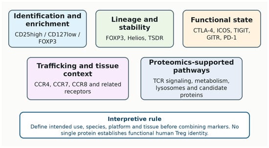
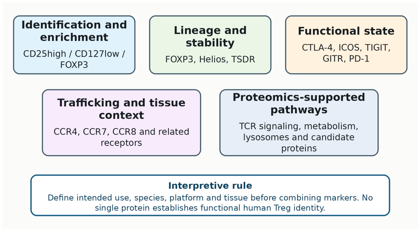
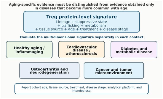
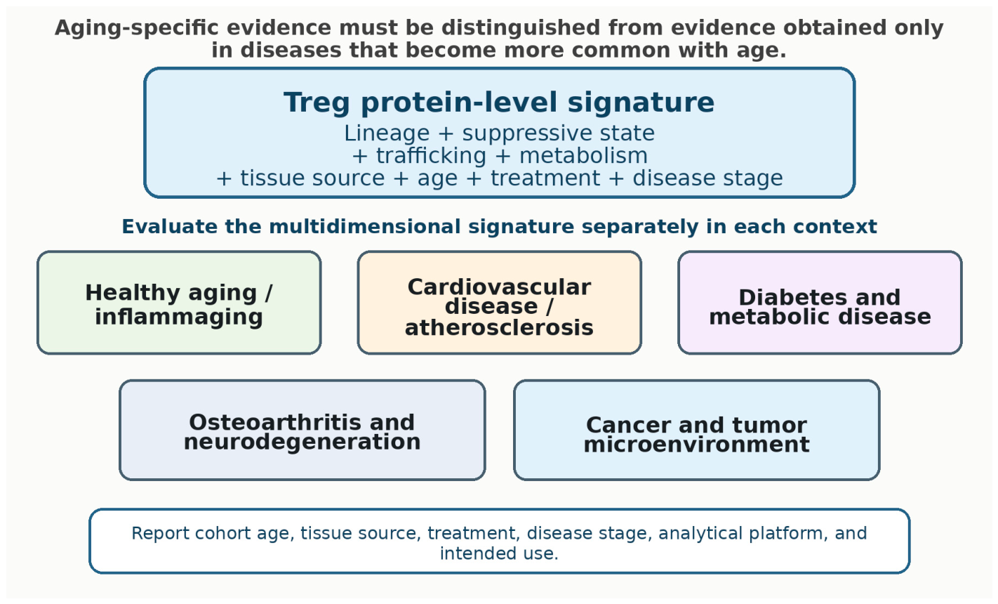

Disclaimer/Publisher’s Note: The statements, opinions and data contained in all publications are solely those of the individual author(s) and contributor(s) and not of MDPI and/or the editor(s). MDPI and/or the editor(s) disclaim responsibility for any injury to people or property resulting from any ideas, methods, instructions or products referred to in the content. |
© 2026 by the authors. Licensee MDPI, Basel, Switzerland. This article is an open access article distributed under the terms and conditions of the Creative Commons Attribution (CC BY) license.