


Abstract
Oral squamous cell carcinoma (OSCC) shows substantial immune and clinical heterogeneity that is not fully captured by conventional clinicopathologic risk factors. This systematic review synthesized primary studies evaluating immune biomarker signatures in OSCC identified through spatial transcriptomics, single-cell transcriptomics, and artificial intelligence (AI)-enabled pathology. PubMed/MEDLINE, Scopus, and Embase were searched without language or date restrictions. Eligible studies included original human OSCC investigations reporting immune-relevant biomarker outputs derived from spatial/single-cell transcriptomics or AI-enabled pathology. Nine studies met the inclusion criteria. Six used spatial and/or single-cell transcriptomic approaches, and three used AI-enabled pathology applied to histopathological whole-slide images. Functional meta-synthesis identified four interconnected domains: AI-derived tissue immune infiltration for prognostic stratification; T-cell states and tertiary lymphoid structure-associated antitumor immunity; spatial and metabolic immunosuppressive niches; and stromal–myeloid programs linked to T-cell exhaustion and resistance. Quantitative synthesis was considered but not performed because no group of studies was sufficiently comparable in biomarker construct, comparator, outcome, and effect measure. Clinical confidence remains limited by heterogeneity and prospective validation gaps. These findings suggest that emerging OSCC immune biomarkers may integrate tissue architecture, cellular states, and stromal–immune interactions; however, the current evidence remains exploratory and requires standardized, prospective validation before clinical translation can be considered.
1. Introduction
Oral squamous cell carcinoma (OSCC) remains one of the most clinically consequential malignancies of the oral cavity. It represents the dominant histologic type of oral cancer and contributes substantially to the global burden of lip and oral cavity cancer, with morbidity related not only to mortality but also to functional impairment after treatment, including effects on speech, swallowing, mastication, and quality of life [1,2]. Despite advances in surgery, radiotherapy, systemic therapy, and reconstructive approaches, risk stratification in OSCC still relies heavily on clinicopathologic variables such as tumor size, nodal status, depth of invasion, margin status, extracapsular extension, lymphovascular invasion, and perineural invasion. These features remain essential, but they do not fully explain why patients with apparently similar stages and histology experience divergent patterns of recurrence, metastatic progression, treatment response, and survival. This limitation is particularly relevant in the era of precision oncology, where biological heterogeneity and host immune contexture increasingly shape clinical outcomes.
The therapeutic landscape of head and neck squamous cell carcinoma has changed with the introduction of immune checkpoint blockade. Nivolumab improved survival in recurrent or metastatic disease after platinum therapy, and pembrolizumab, alone or combined with chemotherapy, became a first-line option for selected recurrent or metastatic cases based in part on programmed death ligand 1 (PD-L1) expression [3,4]. These developments established immune biomarkers as clinically relevant tools in head and neck oncology. However, the benefit of immune checkpoint inhibitors is uneven, and many patients do not achieve durable response. This variability reflects a more complex biology than can be captured by a single immune marker. The cancer-immunity cycle, immune exclusion, regulatory cell populations, antigen presentation, checkpoint co-expression, stromal barriers, metabolic reprogramming, and spatial arrangement of immune and malignant cells all influence whether a tumor behaves as immune-inflamed, immune-excluded, or immune-cold [5,6]. Therefore, immune biomarker discovery in OSCC must move beyond static, single-analyte assessment toward integrated signatures that account for cellular state, spatial organization, and tissue architecture.
Previous evidence has already shown that immune infiltrates are clinically meaningful in OSCC and related head and neck cancers. Systematic reviews and meta-analyses have assessed PD-L1 expression, CD4+ and CD8+ tumor-infiltrating lymphocytes (TILs), and broader immune-cell infiltrates, generally supporting the prognostic relevance of immune contexture while also highlighting substantial heterogeneity in markers, scoring systems, anatomical subsites, cutoffs, and outcome definitions [7,8,9,10]. More recent work has focused specifically on TILs in OSCC, showing that lymphocytic infiltration can be associated with survival outcomes, but also emphasizing inconsistent independence after adjustment and limitations in reproducibility when evaluation is based on conventional visual assessment [10]. These prior reviews are important, but they mainly summarize immunohistochemical or histopathologic markers individually. They do not fully capture how immune biomarkers emerge from high-resolution cellular technologies, spatially resolved transcriptomic platforms, and artificial intelligence (AI)-enabled pathology.
Single-cell RNA sequencing and spatial transcriptomics have created a new opportunity to characterize the OSCC tumor immune microenvironment at a level of resolution that was not possible with bulk assays. Foundational single-cell studies in head and neck cancer demonstrated that malignant, stromal, and immune compartments are highly heterogeneous and that interactions between tumor cells, fibroblasts, macrophages, dendritic cells, and T-cell subsets influence tumor behavior [11]. Spatial transcriptomics added a complementary dimension by preserving tissue location while measuring gene expression, enabling researchers to examine whether immune suppression, T-cell exhaustion, tertiary lymphoid structures, myeloid-derived suppressor cells, cancer-associated fibroblast programs, and chemokine-mediated immune recruitment occur in distinct tumor niches rather than uniformly across the lesion [12,13]. Such information is directly relevant to immuno-oncology because response and resistance to immune checkpoint blockade depend not only on the presence of immune cells but also on their phenotype, proximity to tumor cells, communication with stromal populations, and location within or outside invasive fronts.
In parallel, AI-enabled pathology has expanded the ability to quantify tissue immune biomarkers from hematoxylin and eosin-stained whole-slide images and digital immunopathology. Conventional TIL assessment is clinically attractive because it is tissue-based and potentially scalable, but manual estimation is vulnerable to interobserver variability, differences in field selection, and inconsistent compartmental definitions. Systematic reviews of AI and deep learning in oral cancer have shown rapid growth in diagnostic and prognostic applications, including histopathological image analysis, but most existing syntheses are broad and focus on detection, classification, or general prognosis rather than specifically on immune biomarker signatures [14,15,16]. AI-enabled pathology can potentially bridge morphology and immunobiology by converting spatial tissue patterns into reproducible quantitative variables, particularly when models distinguish tumor nests, stroma, lymphocytic infiltration, and immune-cell-rich regions. Nevertheless, whether these AI-derived immune signals align with transcriptomic immune states, spatial immunosuppressive niches, or clinically meaningful outcomes in OSCC has not been systematically synthesized.
The current evidence base therefore contains an important knowledge gap. Earlier reviews have examined PD-L1, TILs, immune cells, machine learning, or deep learning in oral cancer as separate topics, but no systematic review has specifically integrated immune biomarker signatures in OSCC identified through spatial transcriptomics, single-cell transcriptomics, and AI-enabled pathology. The added value of this review is therefore not simply that it summarizes newer technologies but that it connects mechanistic immune states identified at single-cell and spatial resolution with scalable tissue-level immune features derived from AI-enabled pathology. This integrated perspective helps position emerging OSCC biomarkers along a translational continuum, from biological discovery to computationally reproducible tissue assessment and potential clinical stratification. This distinction is not merely methodological. A review focused on this intersection can clarify whether the field is converging on recurring functional immune programs, such as exhausted T-cell states, regulatory T-cell enrichment, macrophage- or myeloid-driven immunosuppression, cancer-associated fibroblast-mediated immune exclusion, tertiary lymphoid structure-associated antitumor immunity, and computationally derived TIL patterns. It can also clarify whether these signatures are linked to prognosis, treatment response, or resistance, and whether the available evidence provides sufficiently comparable quantitative estimates for formal synthesis.
The focus on spatial transcriptomics, single-cell transcriptomics, and AI-enabled pathology was intentional because these approaches provide complementary levels of translational information: cellular-state resolution, spatial tissue organization, and scalable histopathological assessment. Other multiplexed immune-profiling technologies are valuable for tumor immune characterization, but they were outside the predefined scope of this review unless integrated with one of these eligible platform domains.
This systematic review was designed to fill that gap by synthesizing primary studies that evaluated immune biomarker signatures in OSCC using spatial transcriptomics, single-cell transcriptomics, and AI-enabled pathology. The primary objective was to characterize and functionally synthesize the immune biomarker signatures identified by these technologies, with attention to their biological source, spatial or cellular context, and clinical interpretation. Secondary objectives were to map the comparator structures used across studies, examine reported associations with prognosis, treatment response, and resistance, and assess whether the available quantitative estimates were sufficiently comparable for formal synthesis. By combining functional meta-synthesis with an explicit appraisal of quantitative synthesis feasibility, this review aimed to provide a structured account of how emerging multi-resolution transcriptomic and AI-enabled pathology approaches are reshaping immune biomarker discovery in OSCC.
2. Materials and Methods
This systematic review with functional meta-synthesis was conducted and reported in accordance with the Preferred Reporting Items for Systematic Reviews and Meta-Analyses (PRISMA) 2020 statement [17] and its explanation and elaboration document [18]. The protocol was registered in PROSPERO (CRD420261377852). The search strategy was prepared with attention to transparency and reproducibility, following the principles of the PRISMA literature search extension (PRISMA-S) for reporting literature searches in systematic reviews [19].
2.1. Eligibility Criteria
Eligibility criteria were defined using a PICOS-based framework adapted to the translational and biomarker-oriented nature of the review.
Participants (P): Eligible studies included patients with histologically confirmed oral squamous cell carcinoma (OSCC), including oral cavity squamous cell carcinoma and oral tongue squamous cell carcinoma. Studies involving gingival, buccal mucosal, floor-of-mouth, alveolar ridge, hard palate, retromolar trigone, or oral tongue OSCC were considered eligible when results were reported specifically for oral cavity disease. Studies of head and neck squamous cell carcinoma were included only when OSCC or oral cavity data were separable.
Intervention/exposure (I): The exposure of interest was the assessment of immune biomarker signatures in OSCC using spatial transcriptomics, single-cell transcriptomics, or AI-enabled pathology, including digital or computational analysis of histopathological images when these approaches generated immune-relevant tissue features. Eligible studies were required to report immune-relevant biomarker outputs derived from these platforms, including cellular, spatial, molecular, or computational features of the tumor immune microenvironment.
Comparisons (C): Because biomarker studies in this field commonly vary in design and analytical platform, the comparator was defined broadly as any contrast reflecting variation in immune biomarker status, distribution, or activity within or across study populations. Eligible comparisons included, but were not limited to, differences between levels or categories of biomarker expression, distinct immune phenotypes, spatial or tissue-based contrasts, or alternative assessment approaches (e.g., conventional versus computational methods). The comparator was accepted as defined by each primary study, provided that it enabled an interpretable evaluation of immune biomarker behavior in relation to the reported outcomes.
Outcomes (O): The primary outcome was the identification and functional characterization of immune biomarker signatures derived from spatial transcriptomics, single-cell transcriptomics, or AI-enabled pathology in OSCC. Outcomes of interest included the biological source and context of each biomarker, its spatial or cellular organization, and its reported clinical interpretation.
Secondary outcomes included reported associations between immune biomarker signatures and prognosis, treatment response, resistance, or biomarker-based risk stratification. The review also extracted the comparator structure used in each study and any quantitative estimates relevant to the assessment of quantitative synthesis feasibility, including survival effect estimates or performance measures of AI-enabled prognostic models when available.
Study design (S): Original human primary studies were eligible, including retrospective or prospective observational cohorts, translational tissue-based studies, single-cell or spatial transcriptomic studies with original human data generation, and AI-enabled pathology studies using original human histopathology data. Studies with clinical, internal, external, or functional validation cohorts were eligible.
2.2. Exclusion Criteria
Studies were excluded if they focused exclusively on non-immune biomarkers; included non-squamous oral malignancies without OSCC-specific results; evaluated benign, dysplastic, or premalignant lesions without OSCC-specific analysis; used animal-only, cell-line-only, or in vitro-only models without human tissue or clinical data; reported radiomics-only models without pathology or transcriptomic immune biomarker assessment; presented bulk transcriptomics-only signatures without spatial or single-cell resolution and without AI-enabled pathology assessment; or provided purely methodological image-classification outputs without an interpretable immune biomarker. Multiplexed immune-profiling platforms, including CyTOF, CODEX, imaging mass cytometry, or multiplexed ion beam imaging, were not considered stand-alone eligibility criteria unless the study also generated eligible spatial transcriptomic, single-cell transcriptomic, or AI-enabled pathology outputs in OSCC. Reviews, systematic reviews, meta-analyses, protocols, editorials, comments, letters without original data, conference abstracts without sufficient data, and purely computational re-analyses of public datasets without new original human data generation or without a clearly reportable OSCC-specific biomarker outcome were also excluded.
2.3. Information Sources and Search Strategy
A comprehensive literature search was performed in PubMed/MEDLINE, Scopus, and Embase. Searches were conducted without language or date restrictions and included records available up to March 2026. Only published studies were sought. Additional studies were identified through backward citation searching of included articles, forward citation tracking, and manual checking of relevant reference lists. Authors were contacted when essential information required for eligibility assessment, risk-of-bias assessment, or quantitative synthesis was not available in the published report.
The search strategy combined controlled vocabulary and free-text terms related to OSCC, immune biomarkers, tumor immune microenvironment, spatial transcriptomics, single-cell transcriptomics, artificial intelligence, AI-enabled pathology, digital pathology, computational pathology, and tumor-infiltrating lymphocytes. The complete reproducible search strategy for PubMed/MEDLINE, Scopus, and Embase, including controlled vocabulary terms, free-text terms, Boolean operators, and database-specific adaptations, is provided in Supplementary Table S1.
2.4. Selection Process
Records retrieved from all databases were exported to a reference-management platform, and duplicates were removed before screening. Two reviewers independently screened titles and abstracts. Reports considered potentially eligible by either reviewer were assessed in full text. Disagreements at title/abstract or full-text stage were resolved through discussion; if consensus could not be reached, a third reviewer adjudicated.
2.5. Data Collection Process
Data extraction was performed independently by two reviewers using a prepiloted extraction form specifically designed for immune biomarker, transcriptomic, and AI-enabled pathology studies. The form was tested on a subset of included studies and refined before full extraction. Discrepancies were resolved by discussion and, when necessary, by consultation with a third reviewer.
Extracted information included bibliographic details, country, study setting, study design, sample size, OSCC subsite, clinical stage, treatment context, tissue source, platform used, biomarker class, comparator definition, outcome definition, validation approach, statistical method, and effect estimates or model-performance metrics. Where available, adjusted estimates were prioritized over unadjusted estimates for the assessment of quantitative comparability. For time-to-event outcomes, hazard ratios and 95% confidence intervals were extracted directly when reported. If not directly available, established methods for deriving time-to-event estimates from published information were considered [20].
2.6. Data Items
The following variables were extracted from each study: first author, year of publication, journal, country, participant number, OSCC subsite, inclusion criteria, specimen type, tumor stage, treatment status, sequencing or image-analysis platform, immune biomarker definition, cell populations or spatial niches evaluated, comparator category, clinical endpoint, statistical model, confounders included in adjusted analyses, internal or external validation, functional validation, reporting of missing data, availability of code or data, and effect estimates relevant to the assessment of quantitative comparability. For AI-enabled pathology studies, additional data included image source, annotation process, ground-truth definition, segmentation or model architecture, training and validation design, performance metrics, and whether immune-cell-derived features were explicitly incorporated.
2.7. Study Risk of Bias Assessment
Risk of bias was assessed independently by two reviewers using tools appropriate to study design and objective. Prognostic factor studies were evaluated using the Quality In Prognosis Studies (QUIPS) tool, which considers study participation, attrition, prognostic factor measurement, outcome measurement, confounding, and statistical analysis and reporting [21]. Prediction model studies, including AI-enabled prognostic models where applicable, were assessed using PROBAST, which evaluates risk of bias and applicability across participants, predictors, outcome, and analysis [22].
For mechanistic translational studies using single-cell or spatial transcriptomics that were not fully captured by QUIPS or PROBAST, a structured narrative appraisal was applied. This appraisal considered participant and specimen selection, tissue and spatial-region definition, biomarker measurement reproducibility, analytical transparency, handling of batch effects, outcome definition, validation strategy, and selective reporting. Risk-of-bias judgments were tabulated and incorporated into the interpretation of the functional meta-synthesis and the assessment of quantitative synthesis feasibility.
To improve transparency, overall risk-of-bias judgments were anchored a priori as low, moderate, moderate-to-high, or high concern according to the number, severity, and clinical relevance of limitations identified within each applicable appraisal domain. Low concern indicated no major limitation likely to affect interpretation. Moderate concern indicated limitations that could affect interpretation but did not invalidate the main finding. Moderate-to-high concern indicated several limitations or at least one important limitation that substantially reduced clinical inference. High concern indicated major limitations likely to compromise prognostic or clinical interpretation. Pre-consensus inter-rater agreement for overall risk-of-bias judgments was assessed using weighted Cohen’s kappa.
2.8. Certainty of Evidence Assessment
The certainty of evidence was assessed narratively across outcome domains because the included studies were heterogeneous in platform, biomarker construct, comparator definition, clinical endpoint, and effect measure. The narrative certainty assessment considered risk of bias, inconsistency across studies, indirectness of clinical inference, imprecision related to small patient-level cohorts or limited validation, and the maturity of prospective clinical evidence. A formal GRADE rating [23] or simplified GRADE table was not applied because no outcome domain contained a sufficiently comparable set of studies to support meaningful domain-level certainty grading.
2.9. Data Synthesis and Statistical Analysis
A structured functional meta-synthesis was the primary synthesis approach because substantial clinical, technological, and analytical heterogeneity was expected across biomarker classes, comparator definitions, and reported outcomes.
In this review, functional meta-synthesis was defined as a structured qualitative and interpretive synthesis approach used to organize heterogeneous biomarker findings according to biological function, platform-derived output, comparator structure, clinical interpretation, and potential translational relevance, rather than as a formal computational or quantitative meta-analysis. This approach was used to identify recurring and divergent immune-biomarker patterns across studies when statistical pooling was not appropriate.
The synthesis was performed in reproducible steps.
First, included studies were grouped by platform domain: spatial/single-cell transcriptomics and AI-enabled pathology.
Second, each immune biomarker was mapped according to its biological unit of analysis, such as T-cell state, tertiary lymphoid structure-associated immunity, myeloid-derived suppressor cell activity, macrophage-related program, cancer-associated fibroblast-immune interaction, spatial immune niche, or tissue-level TIL quantification.
Third, the comparator used in each study was coded as high versus low biomarker, positive versus negative biomarker, immune-hot versus immune-cold, tumor versus adjacent normal tissue, spatial-region contrast, stage-based contrast, treatment-sensitive versus resistant contrast, or AI versus manual assessment.
Fourth, biomarker findings were categorized by predominant clinical or biological function: immune phenotype definition, immune suppression, prognosis, treatment response, treatment resistance, or model-based risk stratification.
Fifth, the direction of association was extracted and harmonized as favorable, unfavorable, mixed, or unclear according to its relationship with survival, recurrence, treatment response, or immune suppression.
Sixth, a study-by-biomarker matrix was built to identify convergent and divergent findings across platforms.
Seventh, candidate functional domains were refined through reviewer discussion, and disagreements in coding were resolved by consensus.
The four final domains were derived from recurring patterns identified across the study-by-biomarker matrix, particularly when multiple studies or platform outputs pointed to conceptually related immune functions, including tissue immune infiltration, T-cell/TLS-associated immunity, spatial immune suppression, and stromal–myeloid resistance biology.
Eighth, the final synthesis integrated platform, biomarker class, comparator, outcome, and validation status to describe immune biomarker signatures with potential translational relevance.
Quantitative synthesis was considered when studies were sufficiently comparable in biomarker construct, comparator definition, outcome, and effect measure. For time-to-event outcomes, hazard ratios were extracted when reported with sufficient information. Adjusted estimates were prioritized over unadjusted estimates when both were available. When model-performance metrics, such as the concordance index (c-index) or area under the receiver operating characteristic curve (AUC), were reported, these were summarized descriptively and considered for quantitative synthesis only if outcome definition, prediction target, and validation setting were sufficiently comparable. If quantitative synthesis was not appropriate, findings were synthesized narratively within the functional meta-synthesis framework.
When quantitative pooling was feasible, statistical heterogeneity was planned to be assessed using visual inspection of forest plots, tau-squared, and I-squared statistics [24]. Small-study effects or publication bias were planned to be assessed only when at least ten sufficiently comparable studies were available for the same outcome and effect measure.
3. Results
3.1. Study Selection Process
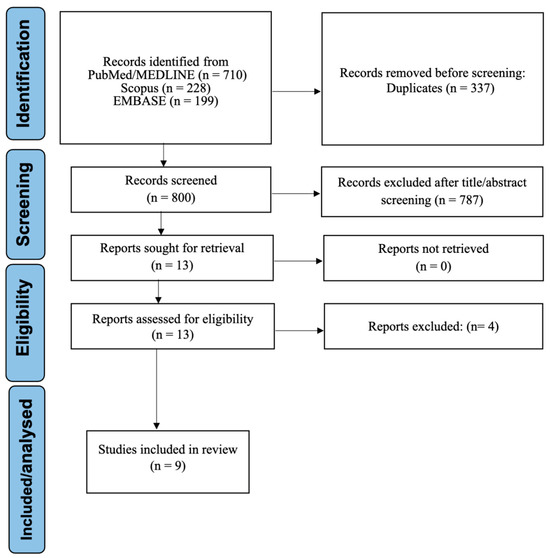
The study selection process is summarized in the PRISMA 2020 flow diagram (Figure 1), which presents record identification, duplicate removal, title/abstract screening, full-text eligibility assessment, and final study inclusion.
Figure 1.
PRISMA 2020 flow diagram of study identification, screening, eligibility assessment, and inclusion.
The database search identified 1137 records. After removal of 337 duplicate records, 800 unique records were screened by title and abstract. Of these, 787 records were excluded. This high exclusion rate reflected the broad and sensitive search strategy, which retrieved studies related to OSCC, immune biomarkers, transcriptomics, digital pathology, artificial intelligence, and tumor-infiltrating lymphocytes, whereas the final eligibility criteria required the intersection of these topics. Most excluded records did not focus specifically on OSCC, did not report immune-relevant biomarker assessment, used bulk transcriptomic or conventional biomarker approaches without spatial/single-cell resolution or AI-enabled pathology, involved non-human or non-primary study designs, or represented publication types incompatible with the review question.
Thirteen articles were retrieved for full-text assessment. After full-text evaluation, four studies were excluded because they did not meet the final eligibility requirements regarding original human data generation, study design, or suitability for the immune biomarker framework. The excluded full-text articles and their reasons for exclusion are presented in Supplementary Table S2. Finally, nine studies met all inclusion criteria and were included in the systematic review and functional meta-synthesis [25,26,27,28,29,30,31,32,33].
The database search identified 1137 records. After removal of 337 duplicate records, 800 unique records were screened by title and abstract. Of these, 787 records were excluded because they did not meet the predefined eligibility criteria. The most frequent reasons for exclusion at this stage were lack of focus on OSCC, absence of immune-relevant biomarker assessment, use of bulk transcriptomic or conventional biomarker approaches without spatial/single-cell resolution or AI-enabled pathology, non-human or non-primary study design, and publication type incompatible with the review question.
3.2. Descriptive Characteristics of Included Studies
The nine included studies were published between 2021 and 2026 and represented two major technological domains: spatial/single-cell transcriptomics and AI-enabled pathology. Six studies used single-cell RNA sequencing, spatial transcriptomics, or combined spatial and single-cell approaches [25,26,29,31,32,33], whereas three studies used AI-enabled pathology applied to histopathological whole-slide images [27,28,30].
The evidence base was heterogeneous in scale and unit of analysis. The transcriptomic studies ranged from small, deeply profiled OSCC cohorts to larger multi-sample or validation-based analyses, including studies that analyzed thousands of individual cells, spatial tissue spots, paired tumor/adjacent normal tissues, multiregional tumor samples, or independent tissue-validation cohorts [25,26,29,31,32,33]. The AI-enabled pathology studies generally involved larger image-based cohorts, including whole-slide images from institutional or multicenter OSCC series and computational workflows for immune-cell or TIL quantification [27,28,30]. This diversity reflects the different analytical purposes of the included studies: high-resolution immune-state discovery in transcriptomic studies and prognostic tissue-based immune quantification in AI pathology studies.
The transcriptomic studies primarily addressed immune-cell states, spatial immune niches, stromal–immune interactions, T-cell dysfunction, tertiary lymphoid structure-related immunity, and immunosuppressive myeloid or fibroblast-associated programs. The AI-enabled pathology studies focused on computational quantification of immune infiltration, particularly TIL-related or infiltrating immune-cell features, and their relationship with prognostic stratification. The main descriptive characteristics of the included studies are summarized in Table 1.
Table 1.
Descriptive Characteristics of Included Studies.
3.3. Platforms and Biomarker Classes
The included evidence was organized into two platform domains. The first domain comprised spatial and single-cell transcriptomic studies, which provided high-resolution characterization of immune-cell states and spatial immune organization in OSCC [25,26,29,31,32,33]. The second domain comprised AI-enabled pathology studies, which extracted immune-relevant features from histopathological whole-slide images, mainly through quantification or localization of TILs and infiltrating immune-cell patterns [27,28,30].
Across transcriptomic studies, recurrent immune-biomarker themes included T-cell dysfunction or exhaustion, regulatory immune-cell enrichment, myeloid-mediated immune suppression, fibroblast–immune crosstalk, spatial metabolic niches, and tertiary lymphoid structure (TLS)-associated antitumor immunity. Across AI-enabled pathology studies, the dominant biomarker class was tissue-level immune infiltration, particularly stromal or compartment-specific TIL quantification. The biomarker classes and their interpretive roles are summarized in Table 2.
Table 2.
Immune biomarker domains and platform-derived outputs.
3.4. Comparator Structures Across Studies
Comparator structures varied across studies but were interpretable within the biomarker framework. AI-enabled pathology studies mainly used contrasts based on immune-cell density, computational versus manual assessment, or model-based prognostic stratification [27,28,30]. Transcriptomic studies relied mainly on cellular-state, spatial-region, tissue-based, TLS-related, or treatment-response contrasts [25,26,29,31,32,33]. Comparator structures and their relevance to outcomes are summarized in Table 3.
Table 3.
Comparator structures and outcome relevance.
3.5. Outcomes Related to Prognosis, Response, Resistance, and Risk Stratification
The included studies differed in the type and maturity of clinical outcome reporting. AI-enabled pathology studies provided the most directly interpretable prognostic outputs, including AI-assessed stromal TILs, AI pathomics-based prognostic modeling, and automated TIL-enriched phenotypes [27,28,30]. Among transcriptomic studies, the clearest clinical associations involved TCF1/TCF7-positive T cells and TLS-related immunity, FAP–WNT2–C1QC-associated resistance, and T-cell cluster-derived prognostic signatures [26,32,33]. Other transcriptomic studies mainly contributed mechanistic or spatial evidence regarding immune suppression, tissue-region heterogeneity, and immune niche formation [25,29,31]. The clinical or analytical outcomes, reported quantitative metrics, and potential relevance for quantitative synthesis are summarized in Table 4.
Table 4.
Outcome and quantitative synthesis relevance.
3.6. Functional Meta-Synthesis
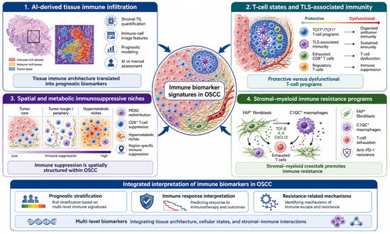
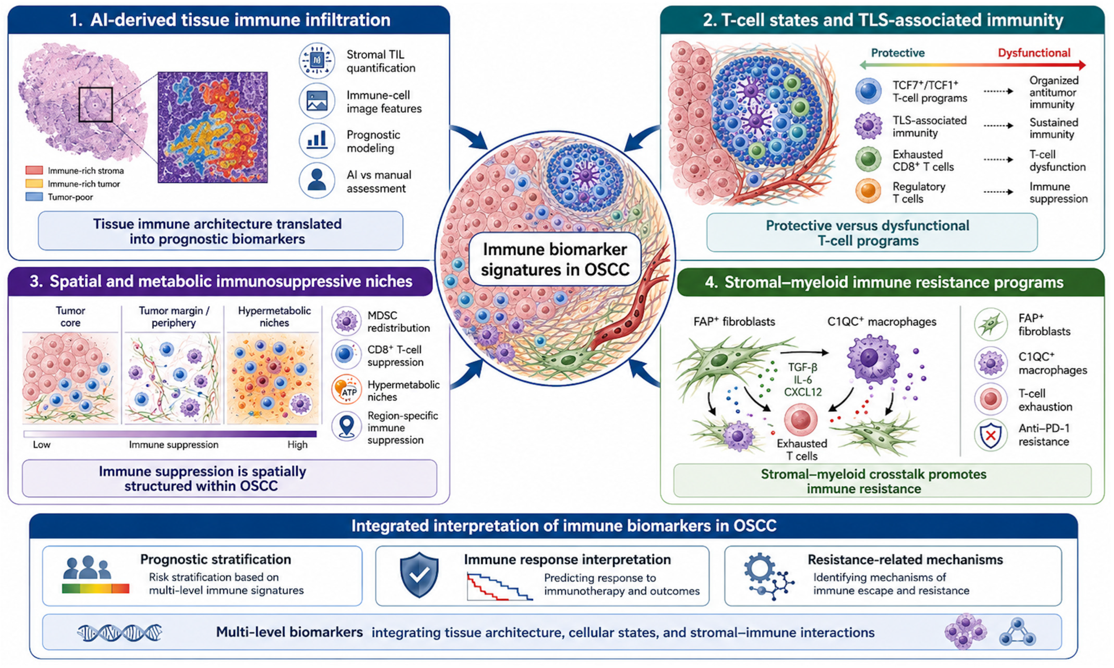
The functional meta-synthesis grouped the nine included studies according to the biological and analytical function of the reported immune biomarkers rather than by platform alone. Four recurrent and partially interconnected domains emerged: (1) AI-derived tissue immune infiltration for prognostic stratification; (2) T-cell states and tertiary lymphoid structure-related antitumor immunity; (3) spatial and metabolic immunosuppressive niches; and (4) stromal–myeloid immune programs linked to T-cell exhaustion and therapeutic resistance (Figure 2).
Figure 2.
Functional meta-synthesis of immune biomarker signatures in oral squamous cell carcinoma identified through spatial and single-cell transcriptomics and artificial intelligence-enabled pathology. Schematic summary of the four interconnected functional domains identified in the meta-synthesis of included studies: (1) AI-derived tissue immune infiltration for prognostic stratification, supported by AI-enabled pathology studies evaluating tumor-infiltrating lymphocytes and infiltrating immune-cell features [27,28,30]; (2) T-cell states and tertiary lymphoid structure-associated immunity, including TCF1/TCF7-positive T-cell programs, exhausted CD8-positive T cells, regulatory T cells, and regional immune heterogeneity [29,32,33]; (3) spatial and metabolic immunosuppressive niches, including stage-dependent or region-dependent immune suppression, hypermetabolic tumor niches, myeloid-derived suppressor cell redistribution, and stromal-mediated Treg recruitment [25,29,31]; and (4) stromal–myeloid immune resistance programs, particularly FAP-positive fibroblast and C1QC-positive macrophage interactions associated with T-cell exhaustion and anti-PD-1 resistance [26]. Together, these domains illustrate how emerging immune biomarkers in OSCC integrate tissue architecture, cellular states, spatial immune organization, and stromal–immune interactions. The lower panel summarizes the main translational implications of these signatures, including prognostic stratification, interpretation of immune response, and identification of resistance-related mechanisms.
The first domain comprised the AI-enabled pathology studies, which consistently identified tissue-level immune infiltration as a clinically relevant biomarker class [27,28,30]. Cai et al. [27] showed that AI-derived pathomics signatures integrating tumor-cell and infiltrating immune-cell information improved prognostic modeling in OSCC. Lee et al. [28] demonstrated that AI-based stromal TIL quantification provided better prognostic discrimination than manual assessment in oral tongue squamous cell carcinoma. Similarly, Crispino et al. [30] showed that automated T-lymphocyte detection enabled the identification of TIL-enriched phenotypes associated with more favorable survival. Collectively, these studies defined a functional domain in which computational pathology translated tissue immune architecture into prognostically informative biomarkers.
The second domain captured T-cell differentiation states and organized antitumor immune responses, particularly those related to tertiary lymphoid structures and T-cell functional heterogeneity [29,32,33]. Peng et al. [32] identified a TCF1/TCF7-positive T-cell subset associated with tertiary lymphoid structures and favorable prognosis, suggesting that organized local immune responses may mark more effective tumor control. In contrast, Chen et al. [33] described intratumoral enrichment of exhausted CD8-positive T cells and regulatory CD4-positive T cells, together with TOX-related dysfunction, thereby highlighting suppressive T-cell programs within the OSCC microenvironment. Kim et al. [29] further expanded this domain by showing that multiregional OSCC samples contained heterogeneous immune states, including cytotoxic exhausted T cells and immunosuppressive programs associated with partial epithelial–mesenchymal transition. Taken together, these studies indicated that T-cell-related biomarkers in OSCC span a continuum from organized and potentially protective immune states to dysfunctional and suppressive states.
The third domain corresponded to spatially organized immunosuppressive niches, in which immune suppression was shaped by tissue location, tumor progression, or metabolic context [25,29,31]. Li et al. [25] showed that OSCC progression was accompanied by spatial redistribution of myeloid-derived suppressor cells toward regions enriched in CD8-positive T cells, supporting a model of stage-dependent immune suppression mediated by annexin A1–formyl peptide receptor family (ANXA1–FPR) signaling. Liu et al. [31] demonstrated that hypermetabolic tumor regions were linked to inflammatory cancer-associated fibroblast transformation, C-X-C motif chemokine ligand 12 (CXCL12) expression, regulatory T cells (Treg) recruitment, and transforming growth factor beta (TGF-β)-associated immune suppression. Kim et al. [29] also contributed to this domain by showing that the tumor periphery displayed distinct immunosuppressive states compared with other tumor regions. Overall, these studies showed that immune biomarker signatures in OSCC are not only cell-type specific but also spatially structured, with suppressive programs concentrating in biologically distinct tumor niches.
The fourth domain represented stromal–myeloid programs associated with immune resistance, with the clearest example provided by Chen et al. [26]. This study identified a fibroblast activation protein–Wnt family member 2–complement C1q C chain (FAP–WNT2–C1QC) axis in which FAP-positive fibroblasts promoted infiltration of C1QC-positive macrophages, enhanced T-cell exhaustion, and contributed to poor prognosis and anti- programmed cell death protein 1 (PD-1) resistance. This domain was conceptually supported by Li et al. [25] and Liu et al. [31], which also highlighted suppressive interactions involving myeloid cells, stromal programs, and spatially restricted immune dysfunction. In functional terms, this domain extended the synthesis from biomarker identification toward a resistance-oriented framework, linking stromal and myeloid interactions to immunotherapy-relevant biology.
Across these four domains, the synthesis revealed a consistent pattern in which OSCC immune biomarkers ranged from favorable immune organization, characterized by high TIL infiltration and TLS-associated TCF7-positive T-cell programs, to adverse suppressive ecosystems, characterized by exhausted T cells, Tregs, myeloid-derived suppressor cells, inflammatory fibroblasts, and macrophage-mediated immune dysfunction. Thus, despite methodological heterogeneity, the included studies converged on a common biological interpretation: immune biomarker signatures in OSCC are best understood as multi-level features integrating tissue architecture, cellular state, and stromal–immune interactions, with direct implications for prognosis, treatment response, and resistance.
3.7. Quantitative Synthesis Feasibility
Quantitative synthesis was considered according to the criteria defined in the methodology; however, no pooled meta-analysis was performed because the included studies were not sufficiently comparable in biomarker construct, comparator definition, outcome, and effect measure. The closest candidate domain was the AI-enabled pathology group evaluating tissue immune infiltration and TIL-related prognostic markers [27,28,30]. However, the quantitative outputs differed substantially across studies.
Among AI-enabled pathology studies, quantitative outputs were clinically related but statistically incompatible: Lee et al. [28] reported a multivariable hazard ratio for AI-assessed stromal TILs, Crispino et al. [30] reported Kaplan–Meier/log-rank comparisons for TIL-hot and stroma-hot phenotypes, and Cai et al. [27] reported c-index and AUC metrics for AI pathomics models. Therefore, the assumptions required for quantitative pooling were not met, and the available numerical results were summarized descriptively within the functional meta-synthesis framework.
3.8. Risk of Bias Assessment
Risk-of-bias assessment showed that the included evidence had a heterogeneous methodological profile, reflecting the different purposes of AI-enabled pathology studies and spatial/single-cell transcriptomic investigations. In the AI-enabled pathology domain, the main concerns were related to retrospective design, annotation and model-development procedures, sample size or validation-set limitations, and incomplete comparability of prognostic estimates across studies. Although the AI-based studies generally provided the most clinically oriented outcomes, their prognostic applicability was affected by differences in model architecture, outcome definition, validation strategy, and reporting of performance metrics [27,28,30].
For spatial and single-cell transcriptomic studies, the main limitations were not primarily related to conventional outcome measurement, but to the translational nature of the evidence. Common concerns included small patient-level sample sizes despite large cell-level or spot-level datasets, selective tissue sampling, complex computational integration, heterogeneous validation strategies, and limited prospective clinical validation [25,26,29,31,32,33]. These limitations did not undermine the value of the studies for functional meta-synthesis, but they reduced the strength of direct clinical inference and limited the feasibility of quantitative synthesis. Overall risk-of-bias judgments and study-specific considerations are summarized in Table 5. The anchoring criteria used to assign overall concern levels are provided in the note below Table 5. The pre-consensus inter-rater agreement matrix is presented in Supplementary Table S3, with an overall weighted Cohen’s kappa of 0.91, indicating high agreement between reviewers before consensus.
Table 5.
Study risk-of-bias assessment.
Availability of public raw data, processed data, analysis code, and validation datasets varied across studies and is summarized in Supplementary Table S4. This information was considered relevant to reproducibility, external validation, and interpretation of translational readiness.
3.9. Certainty of Evidence Assessment
The certainty of evidence was assessed narratively across the main outcome domains because the included studies were heterogeneous in platform, biomarker construct, comparator definition, and reported effect measures. A formal GRADE assessment for a pooled quantitative outcome was not applied because no outcome domain included a sufficient number of studies with comparable effect estimates suitable for meta-analysis. Therefore, certainty judgments were interpreted as GRADE-informed narrative ratings, considering risk of bias, inconsistency, indirectness, imprecision, and publication bias.
Overall, the certainty of evidence was low to very low for most clinical inferences. The strongest and most clinically interpretable evidence was found in the AI-enabled pathology domain, where studies consistently supported the prognostic relevance of tissue immune infiltration or immune-cell-derived image features [27,28,30]. However, certainty was limited by retrospective designs, heterogeneous model architectures, differences in effect measures, and incomplete comparability between hazard ratios, Kaplan–Meier/log-rank analyses, and model-performance metrics. Evidence for T-cell states, tertiary lymphoid structure-associated immunity, and spatial immunosuppressive niches was biologically coherent and supported by high-resolution single-cell or spatial transcriptomic data [25,26,29,31,32,33], but certainty for clinical application was reduced by small patient-level sample sizes, indirect validation strategies, and limited prospective outcome assessment. The certainty judgments by outcome domain are summarized in Table 6.
Table 6.
Narrative certainty of evidence assessment by outcome domain.
4. Discussion
This systematic review synthesized emerging evidence on immune biomarker signatures in OSCC derived from spatial transcriptomics, single-cell transcriptomics, and AI-enabled pathology. The main finding was that immune biomarkers in OSCC are increasingly being defined as multi-level signatures rather than isolated markers. Across the nine included studies, the evidence converged on four functional domains: AI-derived tissue immune infiltration for prognostic stratification; T-cell states and tertiary lymphoid structure-associated antitumor immunity; spatial and metabolic immunosuppressive niches; and stromal–myeloid programs linked to T-cell exhaustion and therapeutic resistance [25,26,27,28,29,30,31,32,33]. These findings are consistent with the broader concept that the tumor immune microenvironment is not a passive background feature but a dynamic ecosystem shaped by malignant cells, immune infiltrates, stromal populations, metabolic pressure, and tissue architecture [34]. Importantly, this review differs from prior syntheses by integrating immune-biomarker evidence generated from high-resolution cellular, spatial, and computational pathology platforms rather than focusing exclusively on conventional immunohistochemical markers or general AI-based oral cancer diagnosis.
The platform-level synthesis showed that spatial and single-cell transcriptomic studies primarily contributed mechanistic resolution, whereas AI-enabled pathology studies provided the most clinically oriented prognostic outputs. Single-cell and spatial technologies allowed the included studies to resolve immune-cell states, spatial niches, and cell–cell interaction axes that would not be detectable using bulk transcriptomic or conventional immunohistochemical approaches [25,26,29,31,32,33]. This finding is biologically plausible, because single-cell and spatially resolved methods can identify heterogeneity within T-cell, myeloid, fibroblast, and tumor-cell compartments while preserving, at least in spatial platforms, the anatomical relationships that influence immune function. In contrast, AI-enabled pathology studies translated tissue immune architecture into quantifiable image-derived variables, particularly TIL-related or infiltrating immune-cell features [27,28,30]. This distinction is important: transcriptomic studies explained biological mechanisms, whereas AI-enabled pathology studies moved closer to scalable prognostic assessment. The complementarity of these approaches supports the rationale for integrated immuno-oncology biomarkers that combine tissue architecture, spatial context, and cellular phenotype. Similar principles underlie immune-contexture frameworks such as the Immunoscore, in which the location and density of immune cells provide prognostic information beyond conventional staging [35].
It is important to clarify that the review did not treat biomarker-discovery studies and prognostic-model studies as equivalent forms of evidence. The spatial and single-cell transcriptomic studies were interpreted mainly as discovery-oriented or mechanistic investigations because they identified immune-cell states, spatial niches, and stromal–immune interactions that may explain OSCC immune heterogeneity. By contrast, AI-enabled pathology studies were interpreted as more clinically oriented prognostic investigations because they quantified tissue-level immune features, such as tumor-infiltrating lymphocytes or infiltrating immune-cell patterns, in relation to survival or model-performance outcomes. Therefore, differences between models were not pooled or directly compared as if they measured the same construct. Instead, they were synthesized according to their platform, biomarker output, comparator structure, outcome type, and translational maturity. This distinction explains why the review integrates biomarker discovery and prognostic modeling within a common immune-biomarker framework while maintaining separate interpretation of their evidentiary roles.
The comparator structures summarized in Table 3 further explain why conventional quantitative synthesis was not appropriate. Across studies, the analytical contrasts ranged from AI-derived immune-cell density and model-based prognostic stratification to tumor-region, metabolic-niche, TLS-related, tumor-versus-adjacent tissue, and treatment-response contrasts. This heterogeneity reflects the exploratory and translational nature of OSCC immune-biomarker discovery, but it also prevents direct pooling because the analytical units differ substantially across platforms.
Regarding prognosis, response, resistance, and risk stratification, the most clinically interpretable findings came from AI-enabled pathology and selected T-cell/TLS-related transcriptomic studies. Lee et al. [28] provided directly extractable prognostic evidence for AI-assessed stromal TILs, Cai et al. [27] reported prognostic model performance using tumor-cell and infiltrating immune-cell image features, and Crispino et al. [30] supported the survival relevance of TIL-enriched phenotypes. These findings are consistent with previous evidence showing that immune infiltrates and TILs can be prognostically meaningful in oral cancer and HNSCC, while also reinforcing that their interpretation depends on cellular phenotype, anatomical subsite, tissue compartment, scoring method, and statistical adjustment [8,9,10].
The functional meta-synthesis provided a biological interpretation that could not be obtained through effect-size pooling alone. Across platforms, the included studies supported a model in which OSCC immune biomarkers operate as interacting programs rather than isolated predictors. Favorable immune organization was represented by TIL-enriched tissue architecture and TLS-associated TCF1/TCF7-positive T-cell programs, whereas adverse immune ecosystems involved exhausted T cells, Tregs, MDSC redistribution, hypermetabolic immunosuppressive niches, and stromal–myeloid interactions associated with anti-PD-1 resistance [25,26,27,28,29,30,31,32,33]. This synthesis therefore links tissue architecture, spatial context, cellular state, and resistance biology within a unified immune-biomarker framework.
The absence of quantitative synthesis should be interpreted as a methodological finding rather than a limitation alone. As shown in Section 3.7, the included studies were biologically related but differed in biomarker construct, comparator, outcome, and effect measure. This reinforces the need for more consistent prognostic-marker reporting in future OSCC studies using spatial, single-cell, or AI-enabled pathology platforms, particularly regarding cohort definition, assay methods, endpoint specification, effect estimates, and model reporting.
Risk-of-bias findings also shaped interpretation. AI-enabled pathology studies were closer to clinical application but remained affected by retrospective designs, model-development choices, annotation procedures, validation constraints, and heterogeneous performance reporting [27,28,30]. These concerns are consistent with previous reviews of AI and deep learning in oral cancer, which reported promising diagnostic or prognostic performance but emphasized variability in datasets, architectures, validation approaches, interpretability, and reporting quality [14,15,16]. Therefore, AI-derived immune biomarkers in OSCC should be considered promising but not yet sufficiently validated for routine clinical use.
Spatial and single-cell transcriptomic studies had a different risk profile. Their main value was biological discovery, but clinical certainty was limited by small patient-level cohorts, selective tissue sampling, complex computational pipelines, heterogeneous validation strategies, and limited prospective evaluation [25,26,29,31,32,33]. Accordingly, the narrative certainty assessment indicated low confidence in most clinical inferences. This does not negate the biological relevance of the identified signatures but indicates that the current evidence remains closer to biomarker discovery than to clinical implementation [34,35,36].
This review has limitations. The evidence base was small and methodologically heterogeneous, with only nine eligible studies differing in anatomical subsite, sample type, platform, comparator structure, clinical endpoint, and effect-measure reporting. Several transcriptomic studies were based on small patient-level cohorts despite large numbers of cells or spatial spots, and some prognostic analyses relied on external datasets or non-OSCC-specific validation settings. Therefore, the current evidence remains primarily hypothesis-generating and biologically informative, rather than sufficient for immediate clinical implementation of these biomarkers in OSCC risk stratification or immunotherapy decision-making.
These limitations are balanced by several strengths. The review addressed a focused and timely question aligned with precision immuno-oncology, separated OSCC-specific evidence from broader HNSCC or oral cancer literature whenever possible, and integrated spatial/single-cell transcriptomics with AI-enabled pathology. The use of functional meta-synthesis allowed biologically related but methodologically incompatible studies to be interpreted without forcing inappropriate quantitative pooling. The review also situates OSCC immune-biomarker discovery within broader debates about AI reporting, reproducibility, and clinical translation, including the need for transparent data description, model validation, human–AI interaction reporting, and performance assessment [37,38,39,40].
The clinical and translational implications are promising but should be interpreted cautiously. AI-enabled TIL assessment and immune-cell pathomics may eventually support reproducible tissue-based risk stratification, while single-cell and spatial transcriptomic biomarkers may help identify immune-excluded, immune-suppressed, TLS-enriched, or stromal–myeloid resistant tumor ecosystems [25,26,27,28,29,30,31,32,33]. From a companion diagnostic perspective, integrated models combining spatially resolved immune context, cellular functional states, and AI-derived tissue features could help distinguish immune-inflamed, immune-excluded, and immunosuppressive OSCC profiles, thereby supporting more refined immunotherapy stratification. However, none of these biomarkers is ready for routine clinical use without standardized assays, reproducible cutoffs, external validation, prospective evaluation, and clinically interpretable decision thresholds [41,42,43].
Future studies should prioritize methodological standardization and clinical validation. Spatial and single-cell studies should report patient-level sample size, tumor subsite, tissue sampling location, preprocessing pipelines, batch correction, spatial-region definitions, cell-type annotation criteria, and validation strategy. AI-enabled pathology studies should report annotation procedures, patient-level data partitioning, model architecture, calibration, external validation, error analysis, and explainability. Prognostic studies should provide adjusted hazard ratios with confidence intervals, clearly defined endpoints, covariates, and sufficient survival data for evidence synthesis. Multicenter and prospective studies, including trial-embedded biomarker analyses, are needed to determine whether these signatures can predict response or resistance to immune checkpoint blockade and whether transcriptomic immune states correspond to scalable AI-derived tissue features [44,45,46]. Future AI-enabled prognostic or predictive model studies in OSCC should also follow TRIPOD+AI reporting guidance to improve transparency in model development, validation, calibration, and clinical interpretability [47].
These considerations indicate that the field is moving toward biologically integrated immune biomarker models, but that clinical translation will depend on standardized reporting, reproducible validation, and direct linkage with OSCC-specific outcomes.
5. Conclusions
This systematic review suggests that immune biomarker discovery in OSCC is beginning to move beyond isolated markers toward integrated, multi-resolution signatures derived from spatial transcriptomics, single-cell transcriptomics, and AI-enabled pathology. Across the included studies, these emerging signatures were mainly related to tissue immune infiltration, T-cell functional states, TLS-associated antitumor immunity, spatially organized immunosuppressive niches, and stromal–myeloid resistance programs. However, the current evidence remains insufficiently standardized for quantitative pooling or routine clinical implementation. Future studies should prioritize OSCC-specific cohorts, harmonized comparator definitions, transparent AI and transcriptomic workflows, external validation, and clinically interpretable effect estimates. At present, these biomarkers should be viewed as promising but exploratory signals that may inform future research on prognosis, treatment-response assessment, and immuno-oncology stratification in OSCC after adequate prospective validation.
Supplementary Materials
The following supporting information can be downloaded at https://www.mdpi.com/article/10.3390/immuno6020038/s1: Table S1. Structured search strategies. Table S2. Full-text articles excluded with reasons [48,49,50,51]. Table S3. Pre-consensus inter-rater agreement for overall risk-of-bias judgments. Table S4. Availability of public data, code, and validation datasets in included studies.
Author Contributions
C.M.A. performed the conceptualization, data curation, data analysis, manuscript writing, and revision of the manuscript; E.P.-V. and A.I.D.-L. performed the data curation, data analysis, and revision of the manuscript; A.M.V.-B. performed the data curation, data analysis, and revision of the manuscript. All authors have read and agreed to the published version of the manuscript.
Funding
This research received no external funding.
Institutional Review Board Statement
Not applicable.
Data Availability Statement
The original contributions presented in this study are included in the article/Supplementary Material. Further inquiries can be directed to the corresponding author.
Conflicts of Interest
The authors declare no conflicts of interest.
References
- Sung, H.; Ferlay, J.; Siegel, R.L.; Laversanne, M.; Soerjomataram, I.; Jemal, A.; Bray, F. Global cancer statistics 2020: GLOBOCAN estimates of incidence and mortality worldwide for 36 cancers in 185 countries. CA Cancer J. Clin. 2021, 71, 209–249. [Google Scholar] [CrossRef] [Scilit]
- Johnson, D.E.; Burtness, B.; Leemans, C.R.; Lui, V.W.Y.; Bauman, J.E.; Grandis, J.R. Head and neck squamous cell carcinoma. Nat. Rev. Dis. Primers 2020, 6, 92. [Google Scholar] [CrossRef] [Scilit]
- Ferris, R.L.; Blumenschein, G., Jr.; Fayette, J.; Guigay, J.; Colevas, A.D.; Licitra, L.; Harrington, K.; Kasper, S.; Vokes, E.E.; Even, C.; et al. Nivolumab for recurrent squamous-cell carcinoma of the head and neck. N. Engl. J. Med. 2016, 375, 1856–1867. [Google Scholar] [CrossRef] [Scilit]
- Burtness, B.; Harrington, K.J.; Greil, R.; Soulieres, D.; Tahara, M.; de Castro, G., Jr.; Psyrri, A.; Basté, N.; Neupane, P.; Bratland, A.; et al. Pembrolizumab alone or with chemotherapy versus cetuximab with chemotherapy for recurrent or metastatic squamous cell carcinoma of the head and neck (KEYNOTE-048): A randomised, open-label, phase 3 study. Lancet 2019, 394, 1915–1928. [Google Scholar] [CrossRef] [Scilit]
- Chen, D.S.; Mellman, I. Oncology meets immunology: The cancer-immunity cycle. Immunity 2013, 39, 1–10. [Google Scholar] [CrossRef] [Scilit]
- Galon, J.; Bruni, D. Tumor immunology and tumor evolution: Intertwined histories. Immunity 2020, 52, 55–81. [Google Scholar] [CrossRef] [Scilit] [PubMed]
- Yang, W.F.; Wong, M.C.M.; Thomson, P.J.; Li, K.Y.; Su, Y.X. The prognostic role of PD-L1 expression for survival in head and neck squamous cell carcinoma: A systematic review and meta-analysis. Oral Oncol. 2018, 86, 81–90. [Google Scholar] [CrossRef] [Scilit] [PubMed]
- Hadler-Olsen, E.; Wirsing, A.M. Tissue-infiltrating immune cells as prognostic markers in oral squamous cell carcinoma: A systematic review and meta-analysis. Br. J. Cancer 2019, 120, 714–727. [Google Scholar] [CrossRef] [Scilit]
- Borsetto, D.; Tomasoni, M.; Payne, K.; Polesel, J.; Deganello, A.; Bossi, P.; Tysome, J.R.; Masterson, L.; Tirelli, G.; Tofanelli, M.; et al. Prognostic significance of CD4+ and CD8+ tumor-infiltrating lymphocytes in head and neck squamous cell carcinoma: A systematic review and meta-analysis. Cancers 2021, 13, 781. [Google Scholar] [CrossRef] [Scilit] [PubMed]
- Wongpattaraworakul, W.; Choi, A.; Buchakjian, M.R.; Lanzel, E.A.; Simons, A.L. Prognostic role of tumor-infiltrating lymphocytes in oral squamous cell carcinoma. BMC Cancer 2024, 24, 766. [Google Scholar] [CrossRef] [Scilit]
- Puram, S.V.; Tirosh, I.; Parikh, A.S.; Patel, A.P.; Yizhak, K.; Gillespie, S.; Rodman, C.; Luo, C.L.; Mroz, E.A.; Emerick, K.S.; et al. Single-cell transcriptomic analysis of primary and metastatic tumor ecosystems in head and neck cancer. Cell 2017, 171, 1611–1624.e24. [Google Scholar] [CrossRef] [Scilit]
- Stahl, P.L.; Salmen, F.; Vickovic, S.; Lundmark, A.; Navarro, J.F.; Magnusson, J.; Giacomello, S.; Asp, M.; Westholm, J.O.; Huss, M.; et al. Visualization and analysis of gene expression in tissue sections by spatial transcriptomics. Science 2016, 353, 78–82. [Google Scholar] [CrossRef] [Scilit]
- Liu, W.; Puri, A.; Fu, D.; Chen, L.; Wang, C.; Kellis, M.; Yang, J. Dissecting the tumor microenvironment in response to immune checkpoint inhibitors via single-cell and spatial transcriptomics. Clin. Exp. Metastasis 2024, 41, 313–332. [Google Scholar] [CrossRef] [Scilit]
- Alabi, R.O.; Youssef, O.; Pirinen, M.; Elmusrati, M.; Makitie, A.A.; Leivo, I.; Almangush, A. Machine learning in oral squamous cell carcinoma: Current status, clinical concerns and prospects for future—A systematic review. Artif. Intell. Med. 2021, 115, 102060. [Google Scholar] [CrossRef] [Scilit]
- Warin, K.; Suebnukarn, S. Deep learning in oral cancer—A systematic review. BMC Oral Health 2024, 24, 212. [Google Scholar] [CrossRef] [Scilit]
- Khanagar, S.B.; Alkadi, L.; Alghilan, M.A.; Kalagi, S.; Awawdeh, M.; Bijai, L.K.; Vishwanathaiah, S.; Aldhebaib, A.; Singh, O.G. Application and performance of artificial intelligence (AI) in oral cancer diagnosis and prediction using histopathological images: A systematic review. Biomedicines 2023, 11, 1612. [Google Scholar] [CrossRef] [Scilit] [PubMed]
- Page, M.J.; McKenzie, J.E.; Bossuyt, P.M.; Boutron, I.; Hoffmann, T.C.; Mulrow, C.D.; Shamseer, L.; Tetzlaff, J.M.; Akl, E.A.; Brennan, S.E.; et al. The PRISMA 2020 statement: An updated guideline for reporting systematic reviews. BMJ 2021, 372, n71. [Google Scholar] [CrossRef] [Scilit] [PubMed]
- Page, M.J.; Moher, D.; Bossuyt, P.M.; Boutron, I.; Hoffmann, T.C.; Mulrow, C.D.; Shamseer, L.; Tetzlaff, J.M.; Akl, E.A.; Brennan, S.E.; et al. PRISMA 2020 explanation and elaboration: Updated guidance and exemplars for reporting systematic reviews. BMJ 2021, 372, n160. [Google Scholar] [CrossRef]
- Rethlefsen, M.L.; Kirtley, S.; Waffenschmidt, S.; Ayala, A.P.; Moher, D.; Page, M.J.; Koffel, J.B.; PRISMA-S Group. PRISMA-S: An extension to the PRISMA statement for reporting literature searches in systematic reviews. Syst. Rev. 2021, 10, 39. [Google Scholar] [CrossRef] [Scilit]
- Tierney, J.F.; Stewart, L.A.; Ghersi, D.; Burdett, S.; Sydes, M.R. Practical methods for incorporating summary time-to-event data into meta-analysis. Trials 2007, 8, 16. [Google Scholar] [CrossRef] [Scilit] [PubMed]
- Hayden, J.A.; van der Windt, D.A.; Cartwright, J.L.; Cote, P.; Bombardier, C. Assessing bias in studies of prognostic factors. Ann. Intern. Med. 2013, 158, 280–286. [Google Scholar] [CrossRef] [Scilit] [PubMed]
- Wolff, R.F.; Moons, K.G.M.; Riley, R.D.; Whiting, P.F.; Westwood, M.; Collins, G.S.; Reitsma, J.B.; Kleijnen, J.; Mallett, S.; for the PROBAST Group. PROBAST: A tool to assess the risk of bias and applicability of prediction model studies. Ann. Intern. Med. 2019, 170, 51–58. [Google Scholar] [CrossRef] [Scilit]
- Guyatt, G.H.; Oxman, A.D.; Vist, G.E.; Kunz, R.; Falck-Ytter, Y.; Alonso-Coello, P.; Schünemann, H.J. GRADE: An emerging consensus on rating quality of evidence and strength of recommendations. BMJ 2008, 336, 924–926. [Google Scholar] [CrossRef] [Scilit] [PubMed]
- Higgins, J.P.T.; Thompson, S.G.; Deeks, J.J.; Altman, D.G. Measuring inconsistency in meta-analyses. BMJ 2003, 327, 557–560. [Google Scholar] [CrossRef] [Scilit] [PubMed]
- Li, F.; Han, Y.; Ou, F.; Deng, L.; Li, H.; Yu, X.; Yi, Y.; Ma, R.; Wu, Z.; You, Z.; et al. Spatial heterogeneity of MDSCs mediated by ANXA1-FPRs signaling drives immune suppression in OSCC progression. Nat. Commun. 2026, 17, 2535. [Google Scholar] [CrossRef] [Scilit] [PubMed]
- Chen, Z.; Feng, X.; Pathak, J.L.; Fu, Z.; Chen, X.; Zeng, L.; Zheng, Y.; Qiu, Q.; Qiao, L.; Wu, L. FAP+ fibroblasts promote C1QC+ macrophage infiltration via WNT2 signaling to exacerbate T cell exhaustion in oral squamous cell carcinoma. Cancer Lett. 2026, 646, 218410. [Google Scholar] [CrossRef] [Scilit]
- Cai, X.J.; Peng, C.R.; Ding, C.Y.; Cui, Y.Y.; Gao, L.; Xu, Z.X.; Li, L.; Zhang, J.-Y.; Li, T.-J. Tumor cell- and infiltrating immune cell-based supervised learning artificial intelligence multimodal platform for tumor prognosis. npj Precis. Oncol. 2025, 9, 348. [Google Scholar] [CrossRef] [Scilit]
- Lee, J.; Fan, M.; Jo, D.; Lee, J.; Song, J.S.; Lee, H.J.; Choi, S.-H.; Nam, S.Y.; Lee, Y.S.; Kim, D.-J.; et al. Assessment of established prognostic factors and artificial intelligence-based evaluation of tumor-infiltrating lymphocytes in oral tongue squamous cell carcinoma. Oral Oncol. 2025, 167, 107448. [Google Scholar] [CrossRef] [Scilit]
- Kim, S.; Kee, H.J.; Kim, D.; Jang, J.; Jeong, H.O.; Sim, N.S.; Selig, M.; Ihlow, J.; Penter, L.; Hwang, T.; et al. Multiregional single-cell transcriptomics reveals an association between partial EMT and immunosuppressive states in oral squamous cell carcinoma. iScience 2025, 28, 112988. [Google Scholar] [CrossRef] [Scilit]
- Crispino, A.; Varricchio, S.; Ilardi, G.; Russo, D.; Di Crescenzo, R.M.; Staibano, S.; Merolla, F. A Digital Workflow for Automated Assessment of Tumor-Infiltrating Lymphocytes in Oral Squamous Cell Carcinoma Using QuPath and a StarDist-Based Model. Pathologica 2024, 116, 390–403. [Google Scholar] [CrossRef] [Scilit]
- Liu, Z.; Zhang, Z.; Zhang, Y.; Zhou, W.; Zhang, X.; Peng, C.; Ji, T.; Zou, X.; Zhang, Z.; Ren, Z. Spatial transcriptomics reveals that metabolic characteristics define the tumor immunosuppression microenvironment via iCAF transformation in oral squamous cell carcinoma. Int. J. Oral Sci. 2024, 16, 9. [Google Scholar] [CrossRef] [Scilit]
- Peng, Y.; Xiao, L.; Rong, H.; Ou, Z.; Cai, T.; Liu, N.; Li, B.; Zhang, L.; Wu, F.; Lan, T.; et al. Single-cell profiling of tumor-infiltrating TCF1/TCF7+ T cells reveals a T lymphocyte subset associated with tertiary lymphoid structures/organs and a superior prognosis in oral cancer. Oral Oncol. 2021, 119, 105348. [Google Scholar] [CrossRef] [Scilit] [PubMed]
- Chen, J.; Yang, J.; Li, H.; Yang, Z.; Zhang, X.; Li, X.; Wang, J.; Zhang, Y.; Chen, S.; Song, M. Single-cell transcriptomics reveal the intratumoral landscape of infiltrated T-cell subpopulations in oral squamous cell carcinoma. Mol. Oncol. 2021, 15, 866–886. [Google Scholar] [CrossRef] [Scilit] [PubMed]
- Binnewies, M.; Roberts, E.W.; Kersten, K.; Chan, V.; Fearon, D.F.; Merad, M.; Coussens, L.M.; Gabrilovich, D.I.; Ostrand-Rosenberg, S.; Hedrick, C.C.; et al. Understanding the tumor immune microenvironment (TIME) for effective therapy. Nat. Med. 2018, 24, 541–550. [Google Scholar] [CrossRef] [Scilit]
- Pagès, F.; Mlecnik, B.; Marliot, F.; Bindea, G.; Ou, F.S.; Bifulco, C.; Lugli, A.; Zlobec, I.; Rau, T.T.; Berger, M.D.; et al. International validation of the consensus Immunoscore for the classification of colon cancer: A prognostic and accuracy study. Lancet 2018, 391, 2128–2139. [Google Scholar] [CrossRef] [Scilit] [PubMed]
- McShane, L.M.; Altman, D.G.; Sauerbrei, W.; Taube, S.E.; Gion, M.; Clark, G.M.; Statistics Subcommittee of NCI-EORTC Working Group. Reporting recommendations for tumor marker prognostic studies (REMARK). J. Natl. Cancer Inst. 2005, 97, 1180–1184. [Google Scholar] [CrossRef] [Scilit]
- Sautès-Fridman, C.; Petitprez, F.; Calderaro, J.; Fridman, W.H. Tertiary lymphoid structures in the era of cancer immunotherapy. Nat. Rev. Cancer 2019, 19, 307–325. [Google Scholar] [CrossRef] [Scilit]
- Zhang, Y.; Wang, D.; Peng, M.; Tang, L.; Ouyang, J.; Xiong, F.; Guo, C.; Tang, Y.; Zhou, Y.; Liao, Q.; et al. Single-cell RNA sequencing in cancer research. J. Exp. Clin. Cancer Res. 2021, 40, 81. [Google Scholar] [CrossRef] [Scilit]
- Mongan, J.; Moy, L.; Kahn, C.E., Jr. Checklist for Artificial Intelligence in Medical Imaging (CLAIM): A guide for authors and reviewers. Radiol. Artif. Intell. 2020, 2, e200029. [Google Scholar] [CrossRef] [Scilit]
- Liu, X.; Cruz Rivera, S.; Moher, D.; Calvert, M.J.; Denniston, A.K.; SPIRIT-AI and CONSORT-AI Working Group. Reporting guidelines for clinical trial reports for interventions involving artificial intelligence: The CONSORT-AI extension. Nat. Med. 2020, 26, 1364–1374. [Google Scholar] [CrossRef] [Scilit]
- Barrett, R.L.; Puré, E. Cancer-associated fibroblasts and their influence on tumor immunity and immunotherapy. eLife 2020, 9, e57243. [Google Scholar] [CrossRef] [Scilit]
- Schumacher, T.N.; Thommen, D.S. Tertiary lymphoid structures in cancer. Science 2022, 375, eabf9419. [Google Scholar] [CrossRef] [Scilit]
- Gabrilovich, D.I. Myeloid-derived suppressor cells. Cancer Immunol. Res. 2017, 5, 3–8. [Google Scholar] [CrossRef] [Scilit] [PubMed]
- Lekadir, K.; Frangi, A.F.; Porras, A.R.; Glocker, B.; Cintas, C.; Langlotz, C.P.; Weicken, E.; Asselbergs, F.W.; Prior, F.; Collins, G.S.; et al. FUTURE-AI: International consensus guideline for trustworthy and deployable artificial intelligence in healthcare. BMJ 2025, 388, e081554. [Google Scholar] [CrossRef] [Scilit]
- Ardila, C.M.; Pineda-Vélez, E.; Vivares-Builes, A.M.; Díaz-Laclaustra, A.I. Artificial-Intelligence-Based Radiologic, Histopathologic, and Molecular Models for the Diagnosis and Classification of Malignant Salivary Gland Tumors: A Systematic Review and Functional Meta-Synthesis. Med. Sci. 2026, 14, 183. [Google Scholar] [CrossRef] [Scilit]
- Ardila, C.M.; Pineda-Vélez, E.; Vivares-Builes, A.M. Computational Stemness and Cancer Stem Cell Markers in Oral Squamous Cell Carcinoma: A Systematic Review, Dual Meta-Analysis, and Functional Meta-Synthesis. Med. Sci. 2025, 14, 21. [Google Scholar] [CrossRef] [Scilit] [PubMed]
- Collins, G.S.; Moons, K.G.M.; Dhiman, P.; Riley, R.D.; Beam, A.L.; Van Calster, B.; Ghassemi, M.; Liu, X.; Reitsma, J.B.; van Smeden, M.; et al. TRIPOD+AI statement: Updated guidance for reporting clinical prediction models that use regression or machine learning methods. BMJ 2024, 385, e078378. [Google Scholar] [CrossRef] [Scilit] [PubMed]
- Song, H.; Lou, C.; Ma, J.; Gong, Q.; Tian, Z.; You, Y.; Ren, G.; Guo, W.; Wang, Y.; He, K.; et al. Single-Cell Transcriptome Analysis Reveals Changes of Tumor Immune Microenvironment in Oral Squamous Cell Carcinoma After Chemotherapy. Front. Cell Dev. Biol. 2022, 10, 914120. [Google Scholar] [CrossRef] [Scilit]
- Iwasa, Y.-i.; Nakajima, T.; Hori, K.; Yokota, Y.; Kitoh, R.; Uehara, T.; Takumi, Y. A Spatial Transcriptome Reveals Changes in Tumor and Tumor Microenvironment in Oral Cancer with Acquired Resistance to Immunotherapy. Biomolecules 2023, 13, 1685. [Google Scholar] [CrossRef] [Scilit]
- Le Meitour, Y.; Foy, J.P.; Guinand, M.; Michon, L.; Karabajakian, A.; Fayette, J.; Saintigny, P.; Mahtouk, K. Uncovering immune checkpoint heterogeneity in oral squamous cell carcinoma using single cell RNA-sequencing data highlights three subgroups of patients with distinct immune phenotypes. Oral Oncol. 2024, 149, 106680. [Google Scholar] [CrossRef] [Scilit] [PubMed]
- Shi, Y.; Shao, Y.; Xia, B.; Yang, W.; Wang, Y.; Zhang, Y. Multi-omics exploration of CAV1+ tumor epithelial subcelltype in oral squamous cell carcinoma and its impact on the immune microenvironment. Discov. Oncol. 2025, 16, 1006. [Google Scholar] [CrossRef] [Scilit]

Disclaimer/Publisher’s Note: The statements, opinions and data contained in all publications are solely those of the individual author(s) and contributor(s) and not of MDPI and/or the editor(s). MDPI and/or the editor(s) disclaim responsibility for any injury to people or property resulting from any ideas, methods, instructions or products referred to in the content. |
© 2026 by the authors. Licensee MDPI, Basel, Switzerland. This article is an open access article distributed under the terms and conditions of the Creative Commons Attribution (CC BY) license.