


Abstract
Cutaneous leishmaniasis remains a therapeutic challenge due to toxicity, parasitic resistance, and the limitations of conventional drugs. In this context, combination therapies using natural immunomodulatory compounds have emerged as a promising alternative. This study evaluated the antileishmanial, cytotoxic, and immunomodulatory effects of gallic acid (GA) combined with paromomycin sulfate (PAR) using in vitro models. IC50 values were determined in promastigote and amastigote forms of Leishmania (Viannia) guyanensis, and CC50 values were assessed in RAW 264.7 macrophages. Peripheral blood mononuclear cells (PBMCs) from healthy volunteers (n = 13) were stimulated with GA, PAR, GA + PAR, or controls to evaluate lymphoproliferation and cytokine production. GA showed higher activity against intracellular amastigotes (IC50 = 11.4 µM), while GA + PAR also demonstrated activity (IC50 = 20.5 µM). PAR alone showed limited efficacy. The combination exhibited limited antiparasitic activity, with effects mainly at higher concentrations and without consistent superiority over GA monotherapy. Immunologically, GA + PAR displayed a balanced cytokine profile without inducing excessive proliferation. These findings suggest a complementary immunomodulatory effect, although further studies are needed to confirm its therapeutic relevance.
1. Introduction
Cutaneous leishmaniasis (CL) is an infectious, non-contagious disease caused by flagellated protozoa of the genus Leishmania, transmitted by the bite of infected female sand flies. It is endemic in approximately 90 countries and is classified by the World Health Organization (WHO) as one of the 21 Neglected Tropical Diseases (NTDs), predominantly affecting socioeconomically vulnerable populations with limited access to healthcare services [1].
The immune response to Leishmania infection is complex and involves a coordinated interplay between innate and adaptive immunity. Initially, macrophages and neutrophils act to control parasite replication through the production of reactive oxygen species and nitric oxide, which subsequently influences T lymphocyte differentiation and disease outcome [2,3,4].
Protective immunity is mainly associated with a Th1-type response, characterized by cytokines such as interferon-gamma (IFN-γ), tumor necrosis factor (TNF-α), and interleukin-12 (IL-12), which promote parasite clearance. In contrast, Th2 responses, marked by IL-4 and IL-5 production, may contribute to parasite persistence, while Th17 responses (IL-17) are linked to inflammation. Regulatory T cells (Treg) and IL-10 play a critical role in limiting excessive immune responses and maintaining homeostasis [5,6,7,8]. Imbalance among these pathways often necessitates pharmacological intervention.
Current treatment for CL includes drugs such as meglumine antimoniate, amphotericin B, pentamidine isethionate, and miltefosine. Despite their effectiveness, these therapies are limited by toxicity, complex administration routes, and the emergence of resistant strains, which negatively impact treatment adherence and outcomes [9,10,11].
In this context, combination therapy has been proposed as a strategy to improve efficacy while reducing toxicity and delaying resistance. Paromomycin sulfate, an aminoglycoside antibiotic that inhibits protein synthesis by binding to the 30S ribosomal subunit, has been recommended by the Pan American Health Organization for combination regimens in CL treatment. In parallel, naturally derived bioactive compounds have gained attention as complementary therapeutic agents.
Among these, gallic acid (GA), a phenolic compound widely distributed in plants, exhibits antioxidant, antimicrobial, anti-inflammatory, and immunomodulatory properties. Previous studies have demonstrated that GA can enhance nitric oxide (NO) production and macrophage microbicidal activity, suggesting a potential role in parasite control [12,13,14].
Therefore, investigating the combined use of GA and PAR may provide insights into complementary mechanisms of action, particularly in terms of immune modulation rather than direct synergistic antiparasitic effects.
Thus, this study aimed to evaluate the in vitro effects of gallic acid combined with paromomycin sulfate against Leishmania (Viannia) guyanensis, focusing on antiparasitic activity, cytotoxicity in RAW 264.7 macrophages, and immunomodulatory responses in peripheral blood mononuclear cells (PBMCs) from healthy volunteers. This approach aims to investigate the potential of this combination as an adjunct strategy to address limitations related to toxicity and resistance in CL treatment.
2. Materials and Methods
2.1. Obtaining Chemical Samples
Gallic acid, paromomycin sulfate, and miltefosine were purchased from Sigma Aldrich® (St. Louis, MO, USA). Glucantime® (meglumine antimoniate) [Sanofi-Aventis Farmacêutica Ltda, São Paulo, Brazil] was provided by the Dr. Heitor Vieira Dourado Tropical Medicine Foundation (FMT-HVD).
2.2. Parasite Maintenance
The species Leishmania (Viannia) guyanensis (MHO/BR/1975/M4147) was used, maintained at the Leishmaniasis and Chagas Disease Laboratory of the National Institute for Amazonian Research (INPA). The promastigote forms were cultured in DMEM (Dulbecco’s Modified Eagle Medium) [Sigma Aldrich®, Saint Louis, MO, USA], supplemented with 10% inactivated fetal bovine serum (SFBi) [Gibco, Thermo Fisher Scientific, São Paulo, SP, Brazil], at 25 °C.
2.3. Determination of Antileishmanial Activity and 50% Inhibitory Concentration (IC50) of Chemical Samples
Promastigotes (106 cells/mL) were incubated with gallic acid (GA) or paromomycin sulfate (PAR) at concentrations of 0.1, 1, 10, 100, and 150 µM. For the combinatorial assays (GA + PAR), concentrations of 0.05 + 0.05, 0.5 + 0.5, 5 + 5, 50 + 50, and 75 + 75 µM were used. The 1:1 ratio was selected as an initial exploratory approach to evaluate the combined effect of both compounds under equivalent concentrations, allowing direct comparison with monotherapies. Further studies are required to explore optimized ratios. Glucantime® (17; 34; 68; 137 and 273 μM) and miltefosine (0.1; 1; 10; 50 and 100 μM) were used as reference drugs, and DMSO (Dimethyl sulfoxide/Merck) at a concentration of 1% was used as a negative control. The cultures were kept at 25 °C in a BOD incubator (Eletrolab Indústria e Comércio de Equipamentos para Laboratório Ltda., São Paulo, SP, Brazil) for 24 and 48 h. Parasite viability was assessed by the MTT (3-[4,5-dimethylthiazol-2-yl]-2,5-diphenyl tetrazolium bromide) colorimetric assay. Absorbance was measured at 595 nm in a spectrophotometer (BioTek® Instruments, Inc., Agilent Technologies, Winooski, VT, USA), and the values of concentration that inhibit parasite growth by 50% (IC50) were determined by nonlinear regression. The assays were performed in triplicate in three independent experiments. The classification of compound activity (e.g., high, moderate, or low) was interpreted based on IC50 values within the tested concentration range, considering comparative performance across experimental conditions rather than fixed categorical thresholds [15].
2.4. In Vitro Cytotoxicity Assays
The cytotoxicity of the substances was evaluated by determining the 50% cytotoxic concentration (CC50) in uninfected macrophages of the RAW 264.7 cell line. The cells (105 cells/mL) were cultured in 96-well plates containing DMEM medium supplemented with 10% FBS for 24 h in an incubator at 37 °C and 5% CO2. After this period, the cells were treated with gallic acid and paromomycin sulfate (0.1, 1, 10, 100, and 150 µM), the combination (0.05 + 0.05, 0.5 + 0.5, 5 + 5, 50 + 50, and 75 + 75 µM), miltefosine (0.1, 1, 10, 50, and 100 μM), and Glucantime® (17, 34, 68, 137, and 273 µM). 1% DMSO was used as a negative control. The plates were incubated for 24 and 48 h, and cell viability was determined by the MTT assay. Absorbance was measured at 570 nm in a spectrophotometer, and CC50 values were calculated by nonlinear regression. The assays were performed in triplicate in three independent experiments [16].
2.5. In Vitro Antileishmanial Activity in Amastigotes Internalized in Macrophages
Murine macrophages of the RAW 264.7 strain (105/mL) were cultured in a 24-well plate, where sterile coverslips were previously placed in the wells. The cells were infected with L. guyanensis promastigotes (106/mL) and treated with different concentrations of gallic acid and paromomycin sulfate (10, 50, 100, and 150 μM) and their combination (5 + 5, 25 + 25, 50 + 50; 75 + 75 μM). Miltefosine (3; 6; 12.5 and 24 μM) and Glucantime® (2.73; 27.3 and 273 μM) were used as positive controls, and 1% DMSO was used as a negative control. After 48 h of incubation at 37 °C, the wells were washed, the glass coverslips were removed, and the infected cells were fixed and stained using the panoptic method. The number of infected macrophages (100 macrophages per slide/replicate) was evaluated. These parameters were evaluated by observation under an optical microscope at 1000X magnification [17]. The assays were performed in triplicate in three independent experiments.
2.6. Cell Selectivity Index (SI)
To determine the selectivity of concentrations with antileishmanial activity against RAW 264.7 macrophages, the selectivity index (SI) was obtained by dividing the 50% cytotoxic concentration (CC50) by the 50% inhibitory concentration (IC50) for amastigotes for 48 h, using the following equation:
SI = (CC50 in RAW 264.7 macrophages)/(IC50 amastigote)
The SI was used as an indicator of compound selectivity, where higher values reflect greater toxicity to the parasite relative to host cells, suggesting a more favorable therapeutic profile. Conversely, lower SI values indicate reduced selectivity and potential host cell toxicity.
2.7. Ethical Aspects
The study was approved by the Human Research Ethics Committee (CEP) of the Federal University of Amazonas (CAAE: 81512524.3.0000.5020). A total of 13 healthy individuals participated in this project, who, after agreeing to participate, signed the Free and Informed Consent Form (FICF).
2.8. Eligibility Criteria
2.8.1. Inclusion Criteria
Individuals regardless of gender and/or sexual orientation, aged 18 to 59 years; individuals who agree to participate in the project and sign the informed consent form; with no history of clinical manifestations of lesions caused by Leishmania; who had not visited rural or peri-urban areas within the previous fifteen days. This criterion was adopted to minimize the inclusion of participants with potential recent exposure to Leishmania vectors or other environmental factors that could influence immune responses.
2.8.2. Exclusion Criteria
Pregnant women; known diagnosis of HIV infection or other immunodeficiencies or use of immunosuppressants; comorbidities such as diabetes; known diagnosis of autoimmune diseases.
2.9. Blood Sample Collection and PBMC Extraction
The isolation of peripheral blood mononuclear cells (PBMCs) is described in more detail in Silva et al. (2021) [18]. In summary, 8 mL of peripheral venous blood from healthy individuals was collected in heparinized polypropylene tubes. The blood was diluted in complete RPMI-1640 medium, and Ficoll-Histopaque (Histopaque-1077, Sigma-Aldrich®, Saint Louis, MO, USA) was added. After centrifugation at 2200 rpm for 30 min, the PBMCs were washed twice with complete RPMI-1640 medium and centrifuged at 1800 rpm for 10 min at 4 °C. The PBMCs were then resuspended in 1 mL of supplemented RPMI medium, and the PBMC concentration was adjusted to 2 × 10⁵ cells mL−1.
2.10. Cell Proliferation Assay
In 96-well flat-bottom culture plates, the PBMCs obtained were organized in triplicate, with 100 μL/well (2 × 10⁵/mL). The cells were sensitized with gallic acid and paromomycin sulfate samples in monotherapy (50 μM) and their combination GA + PAR (25 + 25 μM). Phytohemagglutinin (PHA) (10 μg/mL) and the conventional drug Glucantime® (273 μM) were used as positive controls for lymphoproliferative response, while cells that received no stimulation were used as negative controls. Next, the PBMCs were incubated in a humidified atmosphere (95%) with 5% CO2 for 72 h at 37 °C, and after 18 h of incubation, BrdU (5-bromo-2′-deoxyuridine) [10 mM] was added [18].
The colorimetric ELISA assay was performed with the BrdU Cell Proliferation ELISA Kit (Roche, Applied Sciences, Pleasanton, CA, USA), following the manufacturer’s guidelines. Finally, BrdU incorporation was evaluated based on the mean increase in optical density by spectrophotometry (absorbance at 450 nm). The stimulation of lymphoproliferation by each substance was determined based on the relative value between the arithmetic mean of the triplicates of the positive control and the negative control, in relation to the wells with stimulated cells.
2.11. Obtaining Culture Supernatant for Cytokine Identification
After the incubation period of the cell proliferation plates, centrifugation was performed at 2200 rpm for 15 min to collect the culture supernatants. The supernatants from each well were transferred individually to cryopreservation tubes and stored at −80 °C until cytokine assay by flow cytometry.
2.12. Calculation of Relative Lymphoproliferative Response and Normalization Strategy
Lymphocyte proliferation was evaluated using the 5-bromo-2′-deoxyuridine (BrdU) colorimetric incorporation assay, with the response determined based on the optical density (O.D.) measured in a microplate reader. Initially, all readings were corrected for the blank value (wells containing only culture medium and reagents) to eliminate background interference.
Cultures were established with 2 × 10⁵ cells per well and subjected to the following conditions: basal control (unstimulated cells), experimental stimuli, and positive proliferation control using phytohemagglutinin (PHA). PHA was used as a nonspecific mitogenic stimulus and considered a reference for maximum proliferative capacity in the assay.
As the different conditions were evaluated from cells from the same individual, all calculations were performed intraindividually, preserving the paired design of the study.
The lymphoproliferative response relative to the basal control was calculated according to the formula:
Relative proliferation (%) = (“O.D. of the sample”)/“O.D. of the baseline control” × 100
In this model, the baseline control of each participant was considered an internal reference (100%). A 50% cutoff was further applied to identify higher-magnitude proliferative responses, based on previously established thresholds in lymphocyte proliferation assays, thereby enabling standardized intraindividual comparisons of biologically relevant responses [19]. To assess the response magnitude relative to the positive control (PHA), an additional intraindividual normalization was performed:
Relative response to PHA (%) = (Value of the stimulus)/(Value obtained with PHA) × 100
To standardize the analysis, values were capped at 100%, ensuring that no experimental condition exceeded the response observed for PHA in the same individual. Values above this threshold were adjusted accordingly. This normalization was applied to enable intraindividual comparisons and to prevent overestimation of proliferative responses relative to the positive control.
2.13. Cytokine Assay
Cytokines were quantified by flow cytometry using the BD FACSCanto™ II cytometer (BD® Biosciences, San Diego, CA, USA), with analysis by FCAP Array v3 software, using the Cytometric Bead Array (CBA) technique. The BD™ Human Th1/Th2/Th17 Cytokine kit (Cat. No. 560484, lot 29132; BD® Biosciences, San Diego, CA, USA) was used, following the manufacturer’s instructions. The following cytokines were measured: IL-2, IL-4, IL-6, IL-10, TNF, IFN-γ, and IL-17A.
2.14. Statistical Analyses
Parasite cell multiplication was determined by fitting sigmoidal curves using GraphPad Prism 9.0 software (San Diego, CA, USA), with calculation of the respective 95% confidence intervals. Statistical analyses of lymphoproliferation and cytokine levels were also performed in GraphPad Prism 9.0. After evaluating data distribution, the variables were considered nonparametric. Given the paired experimental design, all intraindividual comparisons were performed using the Wilcoxon signed-rank test. When comparisons involved more than two related conditions, the Friedman test followed by appropriate post hoc analysis was applied. No statistical tests for independent groups were used, as all experimental conditions were evaluated within the same individuals. Correlation analysis between immunological variables was performed using Spearman’s correlation coefficient to identify monotonic associations between cytokine levels and the evaluated parameters. Differences were considered statistically significant when p < 0.05.
3. Results and Discussion
The therapeutic management of cutaneous leishmaniasis is based on the clinical presentation of the disease, the infecting species, and the host’s immune response. Despite the existence of established protocols, currently available drugs have significant limitations, such as high toxicity, long treatment times, and clinical failures, which reinforce the need for alternative and safer strategies. In this context, combination therapy has been explored as a promising approach, allowing for the reduction of individual doses, minimizing toxic effects, and potentially increasing therapeutic efficacy [20,21,22].
Phenolic compounds, such as gallic acid (GA), have been extensively investigated for their antileishmanial activity, mainly associated with the induction of oxidative stress and mitochondrial dysfunction, in a concentration and exposure time-dependent manner. Preclinical studies have been conducted with this substance, demonstrating that GA acts as an inhibitor of Leishmania arginase, a key enzyme in parasite metabolism, and shows relevant therapeutic potential against different species of the genus Leishmania, although it remains in the preclinical stage of investigation [23].
Studies conducted by this research group reinforce the therapeutic potential of phenolic compounds with antileishmanial activity, based on the investigation of methyl gallate, an ester of gallic acid, isolated from Libidibia ferrea, a plant species native to Brazil. Evidence previously obtained by the group demonstrates that this compound has relevant antileishmanial activity and does not exhibit cytotoxicity in primary cultured murine macrophages (BALB/c), reinforcing the potential of phenolic compounds as promising candidates for the development of new therapeutic approaches in the control of CL [24,25,26].
In contrast, paromomycin sulfate (PAR), an aminoglycoside with established clinical use, is a drug already employed in therapeutic protocols for the treatment of CL, generally used in combination regimens. Several studies demonstrate the antileishmanial efficacy of this substance, as well as its safety profile, both alone and in combination with other drugs, reinforcing its role as a therapeutic alternative to antimonials [27,28,29]. Additionally, the study by Carter et al. (2022) [30] indicates that PAR may interfere with essential metabolic pathways of the parasite, such as polyamine metabolism, considered a promising therapeutic target in Leishmania. Thus, this study evaluated the antileishmanial activity of the combination of gallic acid (GA) and paromomycin sulfate (PAR), as well as the effect of each compound alone, on promastigotes and amastigotes of Leishmania guyanensis.
In antileishmanial assays with promastigotes, GA monotherapy at the highest concentrations (150 and 100 µM) showed a greater inhibitory effect when compared to PAR and the GA + PAR combination, which did not demonstrate synergistic interaction under the tested conditions. Analysis of the 50% inhibitory concentration (IC50) values revealed a numerical reduction for GA over 48 h (from 142.5 to 126.5 µM); however, this difference was not statistically significant. In contrast, PAR alone and the combination of GA + PAR did not reach IC50 at the concentrations tested, indicating that concentrations above 150 µM would be necessary to produce a significant effect on promastigotes (Table 1).
Table 1.
Evaluation of the antileishmanial biological activity of the samples against the promastigote and amastigote forms of Leishmania guyanensis, cytotoxicity in RAW 264.7 macrophage cells, and selectivity index in relation to amastigotes. Data are expressed as mean ± standard deviation (SD) from three independent experiments performed in triplicate (n = 9).
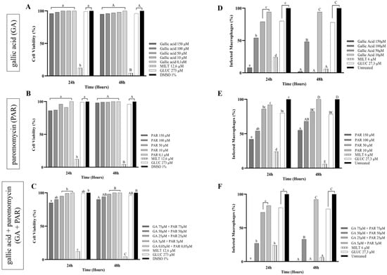
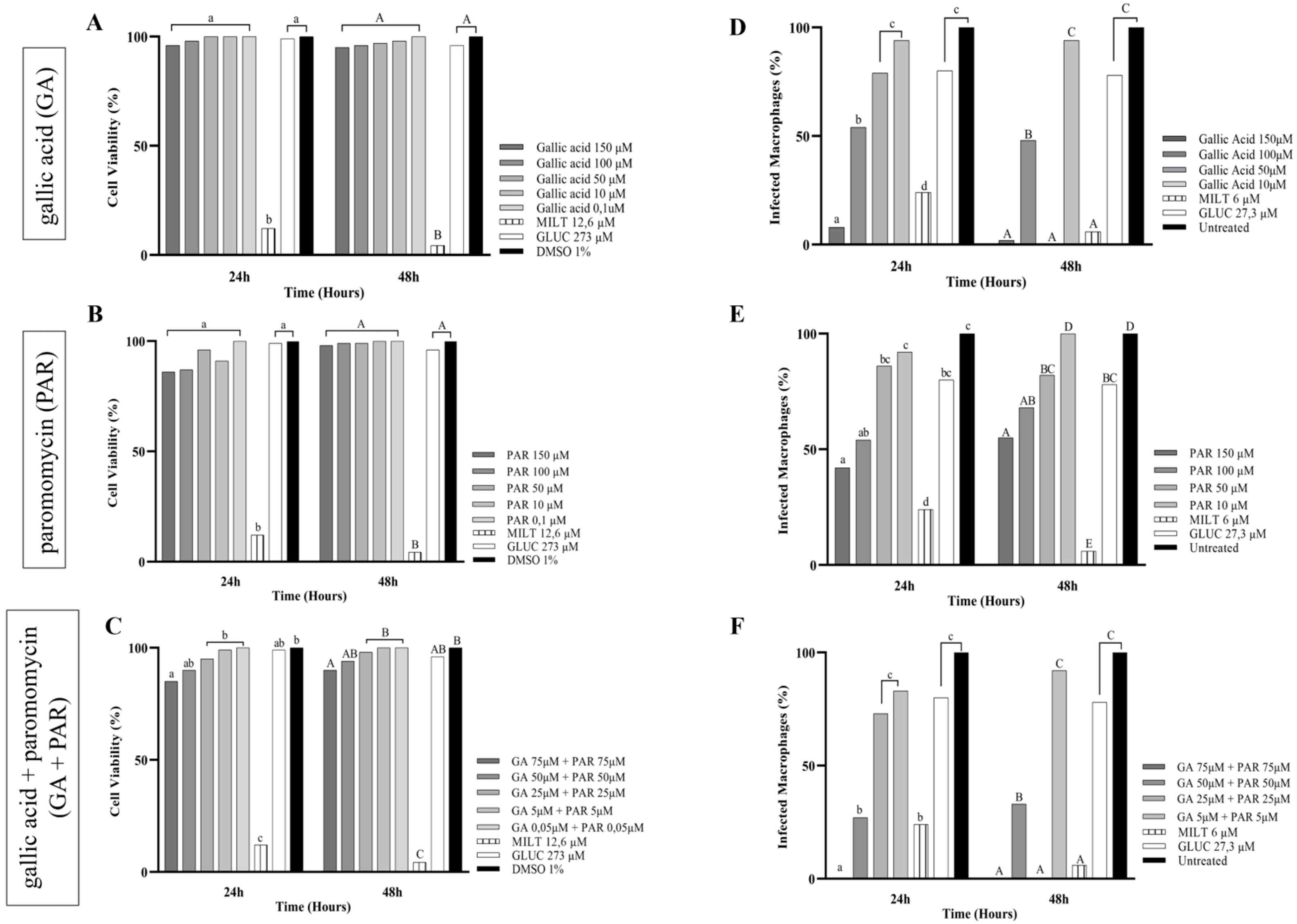
In cytotoxicity assays on RAW 264.7 macrophages, GA and PAR did not show significant cytotoxic effect at any of the concentrations tested (Figure 1). However, the GA + PAR combination showed a small decrease in cell population at its highest concentrations (150 and 100 µM) (Figure 1C). The underlying mechanism responsible for this effect was not investigated in the present study and therefore remains unclear. In contrast, PAR showed a favorable safety profile, with CC50 values not determined within the tested concentration range, indicating that cytotoxicity was not observed at the highest concentration evaluated (150 µM) (Table 1). These findings are consistent with previous studies demonstrating good tolerability of the association between paromomycin sulfate and pyrvinium pamoate, both in axenic and intramacrophage amastigote models of L. infantum and in murine organoids [31].
Figure 1.
(A–C)—Cell viability (%) and (D–F)—Percentage of infected macrophages after treatment with gallic acid (GA), paromomycin (PAR), and GA + PAR for 24 h and 48 h. Miltefosine (MILT) and Glucantime® (GLUC) were used as positive controls, 1% DMSO and untreated cells as negative controls. Statistical differences between groups are represented by letters. For the 24-h time frame, lowercase letters (a, b, c) were used, and for 48 h, uppercase letters (A, B, C) were used. Groups identified with the same letter do not show a statistically significant difference between them, while groups with different letters differ significantly (p < 0.05), according to nonparametric analysis using the Kruskal–Wallis test followed by Dunn’s post hoc test.
Miltefosine, used as one of the positive controls, showed high antileishmanial efficacy, with IC50 between 6.3 and 7.4 µM, but was associated with relevant cytotoxicity (CC50 between 24.8 and 29.1 µM), in agreement with studies describing its cytotoxic and genotoxic effects in human leukocyte cells, associated with the induction of DNA damage and cell apoptosis, reinforcing its cytotoxicity profile [32]. However, the high standard deviation observed for miltefosine, particularly in amastigote assays, indicates considerable data variability, which may limit its reliability as a comparative control in this experimental model.
In intracellular amastigotes, GA treatment resulted in a concentration-dependent reduction in the infection rate of macrophages, indicating relevant antiparasitic activity under the tested conditions. In contrast, PAR exhibited limited efficacy, with no consistent reduction in infection rates observed over time. Although numerical differences between parasite forms were observed, these were not statistically significant (Table 1).
The GA + PAR combination demonstrated activity primarily at higher concentrations, showing an intermediate profile compared to the individual compounds, without consistent superiority over GA monotherapy. The antileishmanial activity of gallic acid (GA) has been consistently associated with the inhibition of parasite arginase, a key enzyme involved in polyamine biosynthesis and parasite survival. Inhibition of this enzyme compromises the production of essential polyamines and may redirect L-arginine metabolism toward nitric oxide (NO) synthesis, enhancing the host’s microbicidal response. Previous studies have demonstrated that GA can modulate macrophage function by increasing NO production and lysosomal activity in infected cells, supporting its role as an immunomodulatory compound with potential relevance to parasite control [31,32,33]. Importantly, structural and functional differences between parasite and host arginase enzymes may allow selective targeting, reinforcing the therapeutic rationale for GA as a candidate with reduced host toxicity.
In turn, the activity profile of PAR against amastigotes reinforces the limitations of its use in monotherapy and highlights the importance of combination therapeutic approaches to increase antiparasitic efficacy. In agreement with these findings, Coser et al. (2021) [34] demonstrated that PAR is consistently more active against intracellular amastigotes of L. braziliensis, L. amazonensis, and L. guyanensis (IC50 of 0.62 to 108.6 µM) than against promastigotes (IC50 of 4.95 to 205 µM), despite the high variability in susceptibility among species, corroborating its better performance against the intracellular form of the parasite.
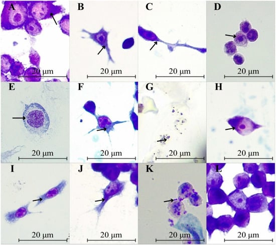
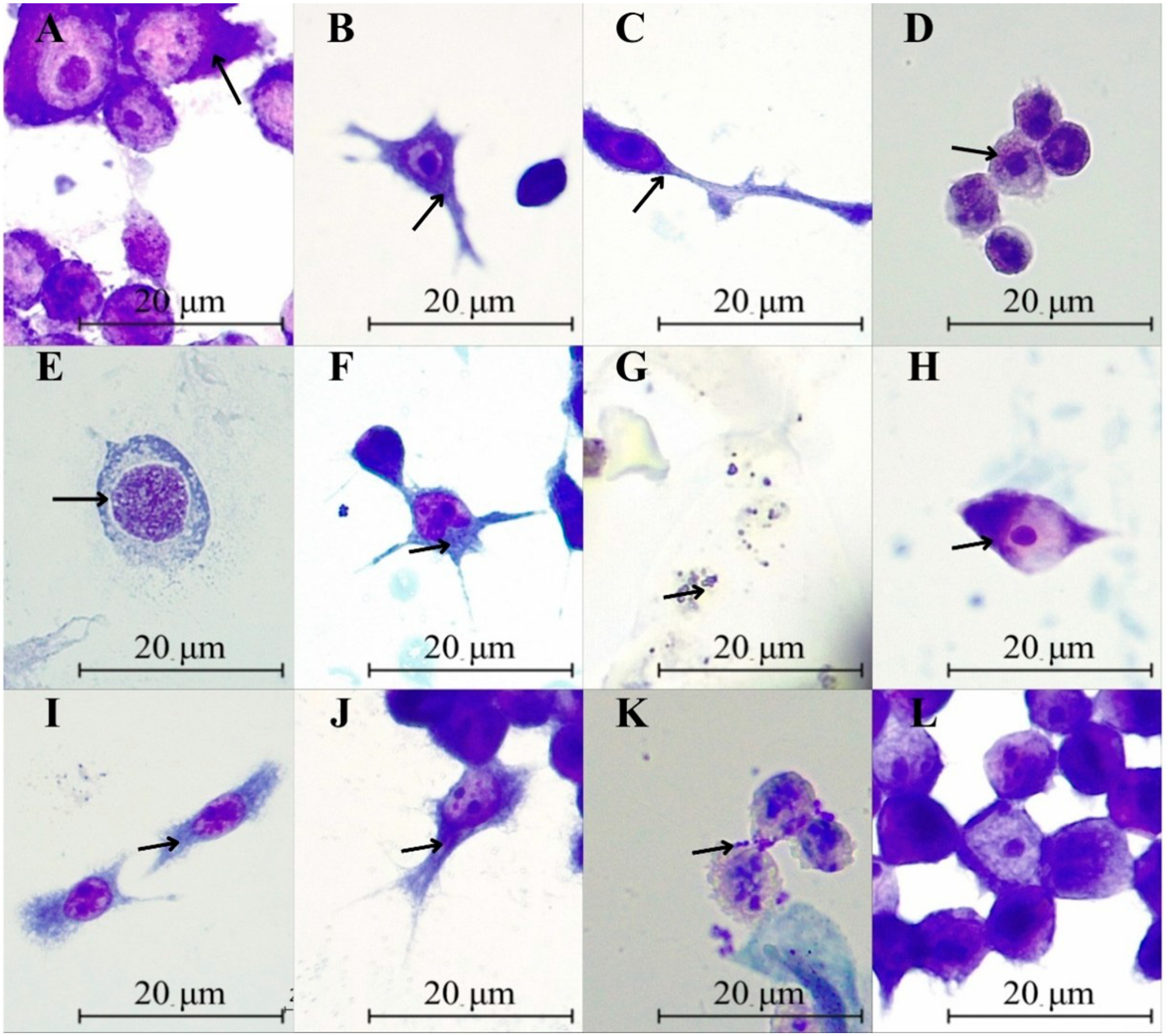
Morphological analysis indicated a reduction in the infection rate after treatment with GA at a concentration of 150 µM, evidenced by the observation of rare structures compatible with amastigotes, indicated by the arrow in Figure 2A. In contrast, the 10 µM concentration showed a higher number of infected macrophages, indicating lower antiparasitic efficacy, associated with slight morphological changes in macrophages (Figure 2B). Treatment with PAR, illustrated in Figure 2C and Figure 2D (150 and 10 µM, respectively), revealed a higher infection rate with intracellular amastigotes indicated by the arrows, especially at the lower concentration, associated with the presence of cytoplasmic extensions.
Figure 2.
Morphological analysis of RAW 264.7 macrophages infected with Leishmania guyanensis amastigotes after treatment with different compounds. Infected macrophages were treated with gallic acid (GA) at 150 μM (A) and 10 μM (B); paromomycin sulfate (PAR) at 150 μM (C) and 10 μM (D); the combination GA + PAR at 150 μM (E) and 10 μM (F); miltefosine at 24 μM (G) and 3 μM (H); and Glucantime® at 100 μM (I) and 1 μM (J). Panel (K) represents the negative control (1% DMSO), panel (L) the untreated infected control, and panel M the uninfected control. Overall, the morphological analysis indicates a concentration-dependent reduction in infected macrophages, with more pronounced antiparasitic effects observed at higher concentrations. However, this analysis is qualitative and should be interpreted as supportive evidence, not as a quantitative measure of antiparasitic activity.
In the GA + PAR combination, a more pronounced reduction in the rate of infected macrophages was observed at a concentration of 150 µM, accompanied by changes in the cells, such as an increase in nuclear volume indicated by the arrow in Figure 2E. On the other hand, at a concentration of 10 µM (Figure 2F), a higher number of infected macrophages was observed, reinforcing the dose-dependent nature of the combined therapy. In contrast, miltefosine exhibited high antiparasitic activity associated with marked cytotoxicity, evidenced in Figure 2G by the presence of cellular debris indicated by the arrow. Finally, Glucantime® showed a partial reduction in infected macrophages, with residual amastigotes still indicated by the arrows in Figure 2I,J.
Regarding the hematological parameters of healthy volunteers, it was observed that, in general, the total white blood cell (WBC) count remained within the established reference range (4000–10,000 cells/mm3) for most individuals, with only a slightly reduced count identified in volunteer 6. The percentages of segmented neutrophils remained within the reference values of all participants. Monocyte proportions were predominantly normal, with low interindividual variability, while lymphocyte percentages showed moderate variability, with some individuals exhibiting values above or below the reference range, which may reflect normal biological variation among healthy individuals rather than pathological alterations [35,36] (Supplementary Table S1).
Overall, these findings suggest that minor deviations from reference ranges did not indicate underlying clinical conditions and are consistent with expected physiological variability. These results indicate the preservation of immune homeostasis among study participants, with no evidence of acute infection, inflammatory processes, or hematological disorders. Thus, the data supports the suitability of volunteers for subsequent immunological assays, minimizing the possibility of baseline hematological bias in the experimental results.
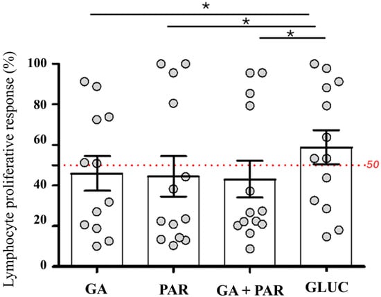
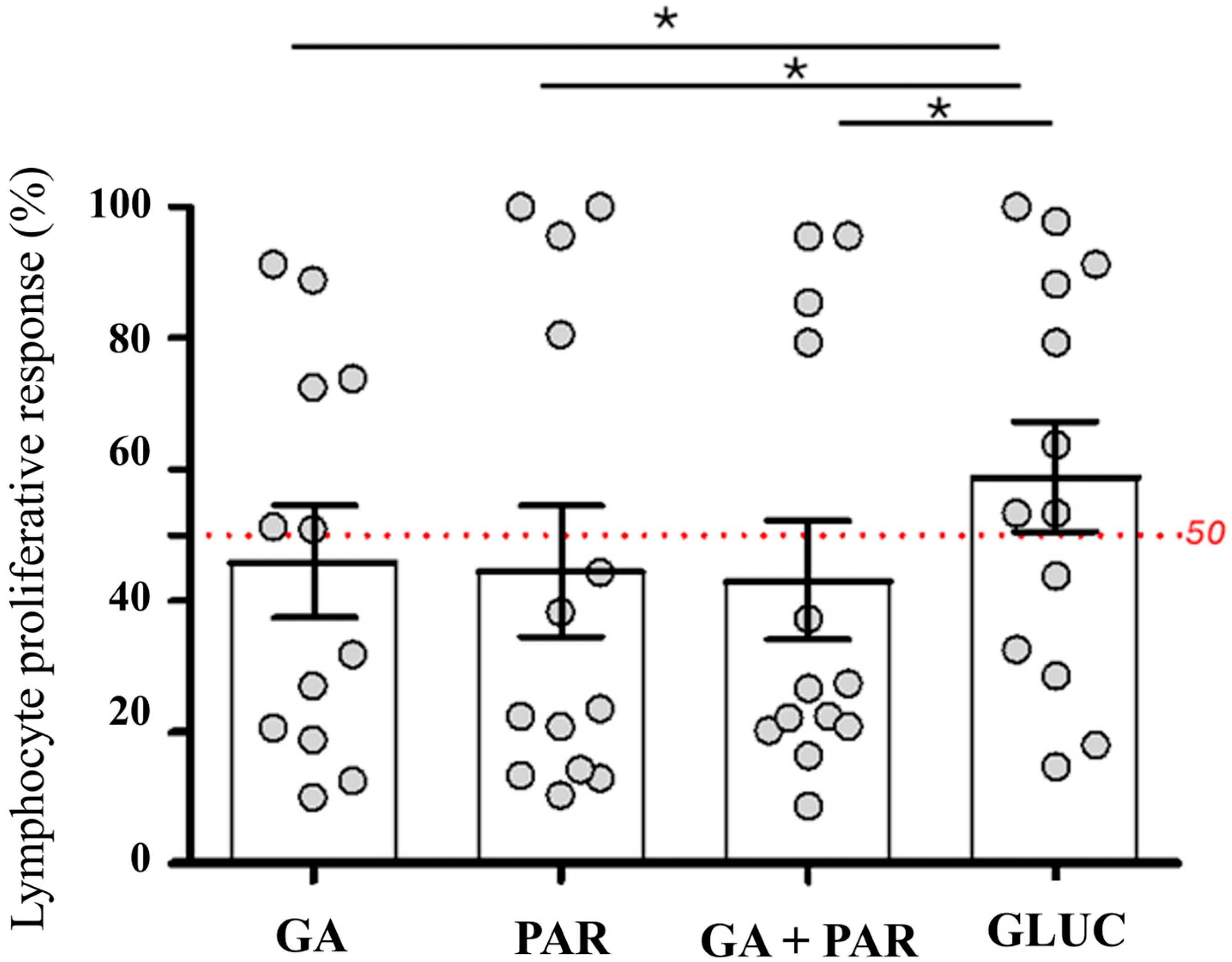
The relative lymphoproliferative response was evaluated under GA, PAR, GA + PAR, and control drug (Glucantime®—GLUC) conditions, with a cutoff point of 50% adopted to characterize proliferative responses of greater magnitude. All analyses were conducted intraindividually, respecting the paired study design (Figure 3).
Figure 3.
Relative lymphoproliferative response (%) of PBMCs stimulated with gallic acid (GA), paromomycin (PAR), GA + PAR combination, and the control drug Glucantime® (GLUC). Each point represents one volunteer (n = 13), and the dashed line indicates the 50% cutoff point used to define responses of greater proliferative magnitude. Intraindividual comparisons were performed using the paired Wilcoxon test and showed significant differences between GLUC and the experimental conditions (GA: p = 0.0186; PAR: p = 0.0479; GA + PAR: p = 0.0161), indicating lower proliferative intensity in the tested conditions compared with the control drug. *: p < 0.05 indicates a statistically significant difference compared with GLUC.
The intraindividual comparison of responses was performed using the Wilcoxon signed-rank test for paired samples, which is appropriate for dependent data with nonparametric distribution. The analyses showed that the responses obtained under the GA, PAR, and GA + PAR conditions were significantly different from those observed for the control drug (GLUC), with p values equal to 0.0186, 0.0479, and 0.0161, respectively.
Overall, the results indicate that, when compared to the control drug, the experimental conditions showed lower proliferative magnitude, both in terms of the distribution of values and the proportion of individuals who exceeded the established cutoff point. These findings reinforce the role of GLUC as the maximum reference for proliferative response in the present assay.
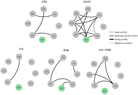
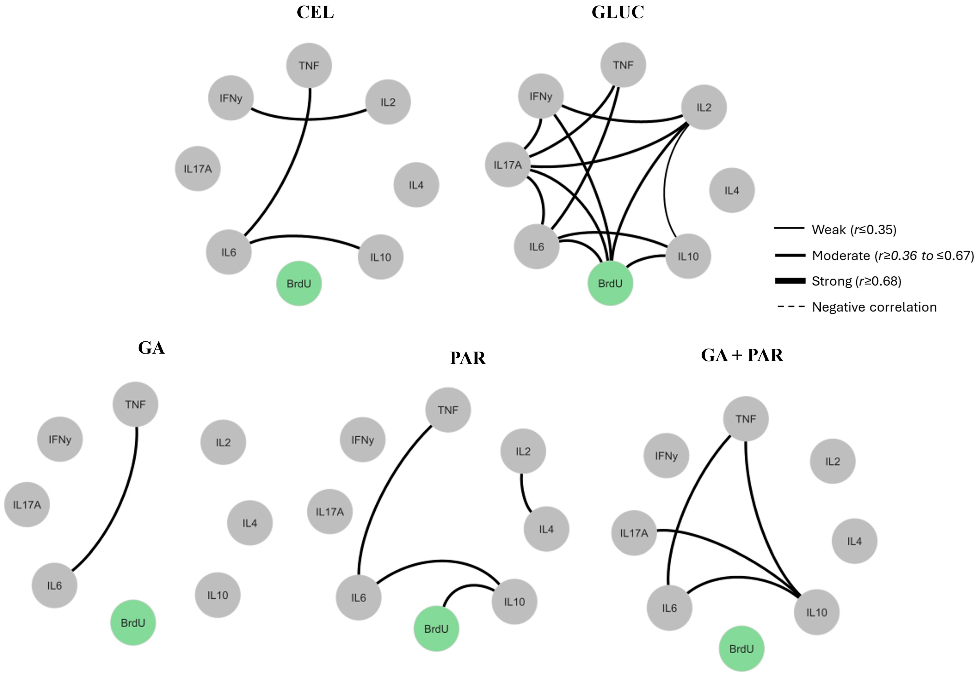
Spearman’s correlation analysis revealed distinct immunomodulatory profiles between treatments. In the basal condition (CEL), a limited set of significant correlations was observed. Among them, positive correlations between IL-6/IL-10 (r2 = 0.89, p = 0.001) and IL-6/TNF (r2 = 0.77, p = 0.011) stood out, suggesting co-regulation of these cytokines at rest. Positive correlations between IL-2/IFN-γ (r2 = 0.21, p = 0.014) were also identified, in addition to a direct association between IL-2 and proliferative parameters, indicating the influence of this mediator on basal proliferation activity (Figure 4).
Figure 4.
Spearman correlation matrices between cytokines, cell proliferation, and BrdU optical density under different experimental conditions. The matrices represent the correlations between the levels of cytokines IFN-γ, TNF, IL-2, IL-4, IL-6, IL-10, and IL-17A and the optical density of BrdU, an indicator of cell proliferation, under control conditions (CON), stimulation with Glucantime® (GLUC), gallic acid (GA), paromomycin sulfate (PAR), and gallic acid + paromomycin (GA + PAR) association. The lines indicate significant correlations, with thickness proportional to the intensity of the correlation (weak, moderate, or strong). The absence of a connection indicates non-significant correlations (p > 0.05).
GA presented a more restricted profile, characterized by occasional modulation of inflammatory axes, evidenced in Figure 4 by the positive association between TNF and IL-6 (r2 = 0.93, p = 0.001), without direct correlation with cell proliferation. The absence of multiple associations with the BrdU assay may suggest that its regulatory effect is not directly linked to proliferative responses. These findings are consistent with the literature, which describes GA as a selective immunomodulator capable of interfering with central inflammatory pathways and reducing mediators such as TNF-α and IL-6 without inducing cell proliferation [12,37].
Treatment with PAR showed an intermediate pattern, identifying positive correlations between IL-2 and IL-4 (r2 = 0.79, p = 0.01), as well as associations that were also present in the baseline condition, such as IL-10/IL-6 (r2 = 0.68, p = 0.04) and TNF/IL-6 (r2 = 0.91, p = 0.001). A positive association between IL-10 and proliferation-related parameters (r2 = 0.69, p = 0.03) was observed; however, this relationship should be interpreted cautiously and does not imply a direct causal effect. This finding is consistent with the reported role of IL-10 in the regulation of immune responses, particularly in limiting excessive inflammation during parasitic infections, although its relationship with lymphocyte proliferation may vary depending on the experimental context [38].
The combined GA + PAR treatment exhibited an integrated profile, combining characteristics observed in the isolated treatments. Positive correlations were found between IL-6/IL-10 (r2 = 0.79, p = 0.01), TNF/IL-6 (r2 = 0.96, p < 0.001), TNF/IL-10 (r2 = 0.68, p = 0.03), and IL-17/IL-10 (r2 = 0.68, p = 0.03), as shown in Figure 4, indicating coordinated cytokine patterns rather than evidence of synergistic interaction. However, unlike the other treatments, no significant correlations involving variables related to cell proliferation were observed, indicating that the combination can modulate inflammatory pathways without directly impacting proliferative mechanisms. This profile is consistent with studies describing combination regimens capable of inducing synergistic immunomodulatory effects, promoting inflammation control without compromising the effector response necessary for parasite control [39].
Finally, treatment with GLUC induced a complex network of interactions (r2 > 0.68, p < 0.05) between pro-inflammatory and regulatory cytokines (IL-2, IL-6, IL-17, IL-10, and IFN-γ), consistent with its broad immunological effect (Figure 4). Although associations with proliferation-related variables were observed, these findings should be interpreted as correlations and not as evidence of direct modulation of cell proliferation. These results are in line with studies describing pentavalent antimonials as inducers of an intense and poorly selective immune response [40].
In summary, these results indicate that the GA + PAR combination shows limited antiparasitic activity observed at higher concentrations, without consistent superiority over monotherapy. These findings reinforce the relevance of future studies exploring trials with PBMCs from patients diagnosed with CL, which will allow the evaluation of immune response modulation in a clinical context considering the inflammatory state, parasite load, and immunological heterogeneity characteristic of the disease, contributing to the consolidation of this combination as a potential adjuvant or alternative therapeutic strategy.
4. Conclusions
The results show that gallic acid and GA + PAR combination exhibit antileishmanial activity (IC50 of 11.4 and 20.5 μM, respectively), particularly against intracellular amastigotes of Leishmania guyanensis at the highest concentrations tested after 48 h of exposure. However, the combination demonstrated limited antiparasitic activity, with effects observed mainly at higher concentrations and without consistent superiority over GA monotherapy. Although the GA + PAR association showed a slight reduction in RAW 264.7 cell viability, it did not induce exacerbated proliferative or inflammatory responses in PBMCs from healthy volunteers. Instead, it was associated with coordinated correlations between pro-inflammatory and regulatory cytokines (IL-6/IL-10, TNF/IL-6, TNF/IL-10, IL-17/IL-10), without inducing significant proliferative responses under the experimental conditions evaluated. These findings suggest a potential complementary immunomodulatory role of the GA + PAR combination rather than a clearly enhanced antiparasitic efficacy. However, these results should be interpreted cautiously due to the limited sample size and require validation in larger and clinically relevant populations, particularly involving PBMCs from patients with cutaneous leishmaniasis, to better understand the therapeutic relevance of this combination.
Supplementary Materials
The following supporting information can be downloaded at: https://www.mdpi.com/article/10.3390/immuno6020032/s1, Supplementary Table S1. Hematological parameters of healthy volunteers, including total white blood cell (WBC) count and percentage distribution of segmented cells, monocytes, and lymphocytes in peripheral blood, according to established reference values.
Author Contributions
Conceptualization, R.S.B.H., B.B.J. and A.M.R.F.-P.; methodology, R.S.B.H., J.C.d.A., L.G.d.S., S.T.V., M.A.d.S., R.B.S., C.L.S.C., E.Y.C.d.O., A.G.d.C. and A.M.A.M.; formal analysis, R.S.B.H., Y.O.C. and B.B.J.; writing—review and editing R.S.B.H., B.B.J., A.M.R.F.-P. and J.F.M.B.; supervision, B.B.J., A.M.R.F.-P. and J.F.M.B.; project administration R.S.B.H.; funding acquisition, A.M.R.F.-P. All authors have read and agreed to the published version of the manuscript.
Funding
This research was financially supported by the National Council for Scientific and Technological Development (CNPq) and the Foundation of Support to Research of the State of Amazonas (FAPEAM). The APC was funded by the PDPG Program of the Coordination for the Improvement of Higher Education Personnel (CAPES).
Institutional Review Board Statement
The study was conducted in accordance with the Declaration of Helsinki and approved by the Human Research Ethics Committee of the Federal University of Amazonas (decision number 81512524.3.0000.5020).
Informed Consent Statement
Informed consent was obtained from all subjects involved in the study.
Data Availability Statement
The raw data supporting the conclusions of this article will be made available by the authors on request.
Acknowledgments
The authors gratefully acknowledge all individuals and institutions involved in the development of this study. We extend our sincere thanks to the collaborators, technical staff, research groups, and supporting institutions whose contributions, assistance, and encouragement were essential for the successful completion of this work.
Conflicts of Interest
The authors declare no conflicts of interest.
Abbreviations
| BrdU | 5-bromo-2′-deoxyuridine |
| CC50 | Cytotoxic concentration 50% |
| CL | Cutaneous leishmaniasis |
| OD | Optical density |
| DMEM | Dulbecco’s Modified Eagle Medium |
| GA | Gallic acid |
| GA + PAR | Gallic acid + paromomycin sulfate |
| IC50 | 50% inhibitory concentration |
| IFN-γ | Interferon-gamma |
| IL-2 | Interleukin 2 |
| IL-4 | Interleukin 4 |
| IL-6 | Interleukin 6 |
| IL-10 | Interleukin 10 |
| IL-17 | Interleukin 17 |
| INPA | National Institute for Amazonian Research |
| L. guyanensis | Leishmania guyanensis |
| MTT | 3-[4,5-dimethylthiazol-2-yl]-2,5-diphenyl tetrazolium bromide |
| NO | Nitric Oxide |
| NTD | Neglected Tropical Diseases |
| PAR | Paromomycin Sulfate |
| PBMCs | Peripheral Blood Mononuclear Cells |
| PBS | Phosphate-Buffered Saline |
| PHA | Phytohemagglutinin |
| RPMI 1640 | Rosewell Park Memorial Institute medium 1640 |
| SD | Standard deviation |
| SDS | Sodium dodecyl sulfate |
| SFBi | Inactivated fetal bovine serum |
| SI | Selectivity index |
| TCLE | Free and informed consent form |
| Th1 | Functional subgroup of type 1 helper T lymphocytes |
| Th17 | Functional subgroup of type 17 T helper lymphocytes |
| Th2 | Functional subgroup of type 2 T helper lymphocytes |
| TNF | Tumor necrosis factor |
| TNF-α | tumor necrosis factor alpha |
| Treg | Functional subgroup of regulatory T helper lymphocytes |
| WHO | World Health Organization |
References
- Magalhães, A.R.; Codeço, C.T.; Svenning, J.C.; Escobar, L.E.; Van de Vuurst, P.; Gonçalves-Souza, T. Neglected tropical diseases risk correlates with poverty and early ecosystem destruction. Infect. Dis. Poverty 2023, 12, 32. [Google Scholar] [CrossRef] [Scilit] [PubMed]
- Burza, S.; Croft, S.L.; Boelaert, M. Leishmaniasis. Lancet 2018, 392, 951–970. [Google Scholar] [CrossRef] [Scilit] [PubMed]
- Horta, M.F.; Andrade, L.O.; Martins-Duarte, E.S.; Castro-Gomes, T. Cell invasion by intracellular parasites—The many roads to infection. J. Cell Sci. 2020, 133, jcs232488. [Google Scholar] [CrossRef] [Scilit] [PubMed]
- Rafique, A.; Sani, S.S.; Sultana, S.; Sultana, T.; Ashraf, A.; Mahmood, M.S. Cutaneous leishmaniasis. In Leishmania Parasites—Epidemiology, Immunopathology and Hosts; IntechOpen: London, UK, 2023. [Google Scholar] [CrossRef] [Scilit]
- Scott, P.; Novais, F.O. Cutaneous leishmaniasis: Immune responses in protection and pathogenesis. Nat. Rev. Immunol. 2016, 16, 581–592. [Google Scholar] [CrossRef] [Scilit]
- Yasmin, H.; Adhikary, A.; Al-Ahdal, M.N.; Roy, S.; Kishore, U. Host–pathogen interaction in leishmaniasis: Immune response and vaccination strategies. Immuno 2022, 2, 218–254. [Google Scholar] [CrossRef] [Scilit]
- Singh, V.K.; Tiwari, R.; Rajneesh; Kumar, A.; Chauhan, S.B.; Sudarshan, M.; Kumar, R. Advancing treatment for leishmaniasis: From overcoming challenges to embracing therapeutic innovations. ACS Infect. Dis. 2024, 11, 47–68. [Google Scholar] [CrossRef] [Scilit]
- Morales-Primo, A.U.; Becker, I.; Pedraza-Zamora, C.P.; Zamora-Chimal, J. Th17 cell and inflammatory infiltrate interactions in cutaneous leishmaniasis. Immune Netw. 2024, 24, e14. [Google Scholar] [CrossRef] [Scilit]
- Akbari, M.; Oryan, A.; Hatam, G. Application of nanotechnology in treatment of leishmaniasis. Acta Trop. 2017, 172, 86–90. [Google Scholar] [CrossRef] [Scilit]
- Tiwari, N.; Gedda, M.R.; Tiwari, V.K.; Singh, S.P.; Singh, R.K. Limitations of current therapeutic options. Mini Rev. Med. Chem. 2018, 18, 26–41. [Google Scholar] [CrossRef] [Scilit]
- Roatt, B.M.; Oliveira Cardoso, J.M.; Brito, R.C.F.; Coura-Vital, W.; Oliveira Aguiar-Soares, R.D.; Reis, A.B. Recent advances in leishmaniasis treatment. Appl. Microbiol. Biotechnol. 2020, 104, 8965–8977. [Google Scholar] [CrossRef] [Scilit]
- Bai, J.; Zhang, Y.; Tang, C.; Hou, Y.; Ai, X.; Chen, X.; Meng, X. Gallic acid: Pharmacological activities. Biomed. Pharmacother. 2021, 133, 110985. [Google Scholar] [CrossRef] [Scilit]
- Keshav, P.; Goyal, D.K.; Kaur, S. Antileishmanial potential of gallic acid. Parasite Immunol. 2021, 43, e12875. [Google Scholar] [CrossRef] [Scilit] [PubMed]
- Pratyusha, S. Phenolic compounds in plant defense. In Plant Stress Physiology; IntechOpen: London, UK, 2022. [Google Scholar] [CrossRef] [Scilit]
- Stroppa, P.H.; Antinarelli, L.M.; Carmo, A.M.; Gameiro, J.; Coimbra, E.S.; Silva, A.D. Triazole salts against Leishmania. Bioorg. Med. Chem. 2017, 25, 3034–3045. [Google Scholar] [CrossRef] [Scilit]
- Antinarelli, L.M.; Pinto, N.C.; Scio, E.; Coimbra, E.S. Brazilian plants with antileishmanial activity. An. Acad. Bras. Cienc. 2015, 87, 733–742. [Google Scholar] [CrossRef] [Scilit] [PubMed]
- Comandolli-Wyrepkowski, C.D.; Jensen, B.B.; Grafova, I.; dos Santos, P.A.; Barros, A.M.C.; Soares, F.V.; Barcellos, J.F.M.; da Silva, A.F.; Grafov, A.; Franco, A.M.R. Libidibia ferrea extracts. Acta Amazon. 2017, 47, 331–340. [Google Scholar] [CrossRef] [Scilit]
- Silva, E.O.; Borges, P.F.C.; Santana, R.B.; Moura, H.S.D.; Barcellos, J.F.M.; Jensen, B.B.; Pinheiro, F.G.; Naiff, M.F.; Espir, T.T.; Franco, A.M.R. Lymphoproliferation in leishmaniasis. Acta Bras. 2021, 5, 97–102. [Google Scholar] [CrossRef] [Scilit]
- Ganesan, N.; Ronsmans, S.; Hoet, P. Methods to assess lymphocyte proliferation. Cells 2023, 12, 386. [Google Scholar] [CrossRef] [Scilit]
- Sasidharan, S.; Saudagar, P. Leishmaniasis overview. Parasitol. Res. 2021, 120, 1541–1554. [Google Scholar] [CrossRef] [Scilit]
- Mann, S.; Frasca, K.; Scherrer, S.; Henao-Martínez, A.F.; Newman, S.; Ramanan, P.; Suarez, J.A. Review of leishmaniasis. Curr. Trop. Med. Rep. 2021, 8, 121–132. [Google Scholar] [CrossRef] [Scilit]
- Mathison, B.A.; Bradley, B.T. Clinical aspects of leishmaniasis. Lab. Med. 2023, 54, 363–371. [Google Scholar] [CrossRef] [Scilit] [PubMed]
- Carter, N.S.; Stamper, B.D.; Elbarbry, F.; Nguyen, V.; Lopez, S.; Kawasaki, Y.; Poormohamadian, R.; Roberts, S.C. Arginase as therapeutic target. Microorganisms 2021, 9, 267. [Google Scholar] [CrossRef] [Scilit]
- Bacha, T.D.J.; Jensen, B.B.; Rodrigues, D.D.C.; da Silva, M.E.T.; Araújo, A.O.; Geraldi, K.S.C.R.d.S.; Valadão, R.C.; Pinheiro, F.G.; Franco, A.M.R. Microemulsion for leishmaniasis. Acta Amazon. 2023, 53, 246–253. [Google Scholar] [CrossRef] [Scilit]
- Rebecca, S.B.H.; Bruno, B.J.; Richaelly, P.d.S.; Paula, F.d.C.B.; Francimeire, G.P.; Mayara, A.d.S.; Ana, C.d.S.C.; Juliana, M.d.M.; Antonia, M.R.F. An in vitro evaluation of the antileishmanial and cytotoxic activity of methyl gallate associated with conventional treatment for cutaneous leishmaniasis. Afr. J. Pharm. Pharmacol. 2024, 18, 48–54. [Google Scholar] [CrossRef] [Scilit]
- Jensen, B.B.; Rodrigues dos Santos, K.S.C.; Comandolli-Wyrepkowski, C.D.; da Silva, F.M.A.; de Souza, C.C.; Lima, M.d.P.; Barcellos, J.F.M.; de Souza, T.P.; Rolim, F.N.A.; Pinheiro, F.G.; et al. Phenolic microemulsion. Eur. J. Microbiol. Immunol. 2025, 15, 222–231. [Google Scholar] [CrossRef] [Scilit]
- Kim, D.H.; Chung, H.J.; Bleys, J.; Ghohestani, R.F. Paromomycin meta-analysis. PLoS Negl. Trop. Dis. 2009, 3, e381. [Google Scholar] [CrossRef] [Scilit]
- Ben Salah, A.; Ben Messaoud, N.; Guedri, E.; Zaatour, A.; Ben Alaya, N.; Bettaieb, J.; Gharbi, A.; Hamida, N.B.; Boukthir, A.; Chlif, S.; et al. Topical paromomycin. N. Engl. J. Med. 2013, 368, 524–532. [Google Scholar] [CrossRef] [Scilit] [PubMed]
- Sosa, N.; Pascale, J.M.; Jiménez, A.I.; Norwood, J.A.; Kreishman-Detrick, M.; Weina, P.J.; Lawrence, K.; McCarthy, W.F.; Adams, R.C.; Scott, C.; et al. Paromomycin in New World CL. PLoS Negl. Trop. Dis. 2019, 13, e0007253. [Google Scholar] [CrossRef] [Scilit] [PubMed]
- Carter, N.S.; Kawasaki, Y.; Nahata, S.S.; Elikaee, S.; Rajab, S.; Salam, L.; Alabdulal, M.Y.; Broessel, K.K.; Foroghi, F.; Abbas, A.; et al. Polyamine metabolism. Med. Sci. 2022, 10, 24. [Google Scholar] [CrossRef] [Scilit]
- Melcón-Fernández, E.; Galli, G.; Balaña-Fouce, R.; García-Fernández, N.; Martínez-Valladares, M.; Reguera, R.M.; García-Estrada, C.; Pérez-Pertejo, Y. Drug synergy. Trop. Med. Infect. Dis. 2024, 9, 30. [Google Scholar] [CrossRef] [Scilit]
- Pessenda, G.; Silva, J.S. Arginase in persistence. Parasite Immunol. 2020, 42, e12722. [Google Scholar] [CrossRef] [Scilit]
- Badirzadeh, A.; Taheri, T.; Taslimi, Y.; Abdossamadi, Z.; Heidari-Kharaji, M.; Gholami, E.; Sedaghat, B.; Niyyati, M.; Rafati, S. Arginase activity. PLoS Negl. Trop. Dis. 2017, 11, e0005774. [Google Scholar] [CrossRef] [Scilit]
- Coser, E.M.; Ferreira, B.A.; Yamashiro-Kanashiro, E.H.; Lindoso, J.A.L.; Coelho, A.C. Paromomycin susceptibility. Acta Trop. 2021, 215, 105806. [Google Scholar] [CrossRef] [Scilit]
- Wakeman, L.; Al-Ismail, S.; Benton, A.; Gibbs, A.; Hartnell, S.; Morris, K.; Munro, R. Robust, routine haematology reference ranges for healthy adults. Int. J. Lab. Hematol. 2007, 29, 279–283. [Google Scholar] [CrossRef] [Scilit] [PubMed]
- Abbas, A.B.; Aldomaini, A.; Al-Qadri, A.A.; Algorbani, Z.; Aljamali, S.; Alsiri, S.; Alghorbani, K.; Osba’a, S.A. Blood reference values. J. Blood Med. 2024, 15, 513–522. [Google Scholar] [CrossRef] [Scilit]
- Li, K.; Gong, Q.; Lu, B.; Huang, K.; Tong, Y.; Mutsvene, T.E.; Lin, M.; Xu, Z.; Lu, F.; Li, X.; et al. Gallic acid effects. Eye Vis. 2023, 10, 17. [Google Scholar] [CrossRef] [Scilit]
- Stijlemans, B.; Schoovaerts, M.; De Baetselier, P.; Magez, S.; De Trez, C. MIF and IL-10 balance. Front. Immunol. 2022, 13, 865395. [Google Scholar] [CrossRef] [Scilit]
- Ramos, T.D.; Silva, J.D.; Fonseca-Martins, A.M.; Pratti, J.E.d.S.; Firmino-Cruz, L.; Maciel-Oliveira, D.; Dos-Santos, J.S.; Tenorio, J.I.N.; de Araujo, A.F.; Freire-De-Lima, C.G.; et al. Combined therapy in leishmaniasis. Stem Cell Res. Ther. 2020, 11, 374. [Google Scholar] [CrossRef] [Scilit] [PubMed]
- Gonzalez-Fajardo, L.; Fernández, O.L.; McMahon-Pratt, D.; Saravia, N.G. Ex vivo response. PLoS Negl. Trop. Dis. 2015, 9, e0003820. [Google Scholar] [CrossRef] [Scilit]

Disclaimer/Publisher’s Note: The statements, opinions and data contained in all publications are solely those of the individual author(s) and contributor(s) and not of MDPI and/or the editor(s). MDPI and/or the editor(s) disclaim responsibility for any injury to people or property resulting from any ideas, methods, instructions or products referred to in the content. |
© 2026 by the authors. Licensee MDPI, Basel, Switzerland. This article is an open access article distributed under the terms and conditions of the Creative Commons Attribution (CC BY) license.