


Abstract
Background: Proton pump inhibitors (PPIs) have been associated with lung dysbiosis and increased respiratory risk. Micro-aspiration is a proposed mechanism, but reliable biomarkers remain elusive. This study evaluates the potential of fluorescein as a biomarker of micro-aspiration and PPI-associated pulmonary risk. Methods: We conducted a retrospective analysis of 137 bronchial washing fluid samples from patients with pulmonary conditions to assess microbial colonization in relation to PPI use. Bacterial burden was determined by culture and PCR and categorized as 0, 1 or ≥2 pathogens. Micro-aspiration was evaluated by quantifying fluorescein-laden macrophages in bronchoalveolar lavage following oral fluorescein administration. Associations between PPI use, fluorescein levels and pathogen burden were analyzed using adjusted ordinal regression models. Results: PPI use was associated with higher odds of increased pathogen burden, though not statistically significant (OR = 1.40, 95% CI: 0.71–2.75, p = 0.33). Fluorescein-laden macrophages were higher in PPI users (41.5 versus 31.2 ng/mL), but showed no meaningful correlation with pathogen load (p = 0.09). Corticosteroid therapy was significantly associated with Gram stain results (OR = 2.37, 95% CI: 1.12–5.15, p = 0.03). Conclusions: These findings suggest a potential link between PPI use and airway colonization. Fluorescein shows promise as a biomarker for micro-aspiration, but its clinical utility requires further validation.
1. Background
Proton pump inhibitors (PPIs) are widely used to suppress gastric acid production [1]. However, this effect has been associated with an increased risk of community-acquired pneumonia, likely due to disruption of the gastric acid barrier [2,3,4,5,6]. By reducing gastric acidity, PPIs may allow the migration of gastrointestinal bacteria from the small intestine and impair the elimination of ingested pathogens, thereby allowing bacterial survival in the gastrointestinal tract. This, in turn, may enable their translocation to the respiratory tract via micro-aspiration, ultimately contributing to pulmonary colonization. Repeated micro-aspiration is thought to increase microbial colonization and induce alterations in the pulmonary microbiome. Such dysbiosis can promote inflammation and contribute to structural lung damage, increasing susceptibility to respiratory infections [7,8].
The lung microbiome has emerged as a dynamic and critical regulator of respiratory health and disease, with bacterial burden exerting effects that extend well beyond acute infections such as pneumonia or lung abscess. Rather than acting in isolation, the respiratory microbiota interacts intricately with host immune mechanisms, necessitating interpretation within a broader pathophysiological context. Commensal microorganisms actively shape immune homeostasis by influencing pattern recognition receptor signaling, cytokine release, and alveolar macrophage activity [9,10]. In chronic pulmonary conditions, including chronic obstructive pulmonary disease and bronchiectasis, increased bacterial load is consistently associated with greater disease severity, more rapid decline in lung function, and a higher frequency of exacerbations [11,12,13]. Importantly, both the total microbial burden and the composition of the microbial community contribute to clinical outcomes. Chronic colonization is often characterized by diminished microbial diversity alongside the dominance of particular taxa, factors that may further drive disease progression [14,15,16]. Although it remains unclear whether increased bacterial burden and microbial dysbiosis are primary drivers or secondary consequences of airway damage, accumulating longitudinal evidence suggests a bidirectional relationship. These findings highlight the coexistence of causal and consequential mechanisms in disease evolution and emphasize the need for further mechanistic investigations [9,17].
Numerous studies have investigated the use of fluorescent dyes for the detection of aspiration, demonstrating advantages over conventional methods such as colored dyes [18,19]. Fluorescein is a synthetic fluorescent dye that has established itself as a versatile marker across multiple medical fields, including neurosurgery, gastroenterology and ophthalmology, owing to its strong visibility, low toxicity and low cost [20,21,22,23]. It has also been described as a useful agent for the diagnostic assessment of swallowing disorders [24]. In this context, high-resolution fluorescence imaging has been used to visualize structural alterations to the alveolar scaffold in lung injury [25]. This highlights the feasibility and growing use of fluorescence-based techniques in pulmonary research. Additionally, fluorescence-based methods have been integrated with bronchoalveolar lavage (BAL) analysis to characterize cellular components, including macrophage populations. These approaches have also been used to quantify drug deposition in pulmonary disease [26,27].
Despite these advances, reliable and minimally invasive biomarkers for micro-aspiration remain limited. Most current approaches rely on indirect clinical outcomes or sputum-based analyses, which are susceptible to contamination from upper airway flora and may not accurately reflect lower airway processes [4,28,29,30]. In contrast, bronchoscopic sampling of bronchial washing fluid (BWF) enables direct assessment of the pulmonary compartment and provides a more accurate representation of airway microbial colonization.
In this study, we investigated the association between PPI therapy and bacterial burden in the lower airways using BWF obtained via bronchoscopy. Furthermore, we evaluated fluorescein-laden macrophages as a surrogate marker of micro-aspiration and their relationship with airway colonization. We found higher levels of fluorescein-laden macrophages in BAL among PPI users compared with non-users, although no meaningful association was observed between fluorescein levels and pathogen burden. To our knowledge, this is the first study to apply intracellular fluorescein uptake in BAL macrophages following oral fluorescein administration as a marker of micro-aspiration. Given the widespread use of acid-suppressive therapy in clinical practice, improved understanding of its potential pulmonary consequences is of clinical relevance, particularly in patients at increased risk of respiratory infections.
2. Methods
2.1. Study Design and Population
We conducted a retrospective, single-center observational study at the Department of Pulmonology at the Medical University of Vienna. The ethics committee of the Medical University of Vienna approved the protocol of this retrospective study (1876/2024, 20 March 2025). Written informed consent was not required owing to the retrospective study design.
2.2. Patient Characteristics and PPI Use
Adult patients with various pulmonary diseases who underwent clinically indicated bronchoscopy with bronchial washing fluid and bronchoalveolar lavage between 2020 and 2024 were included in the study. Patient characteristics included sex, age, smoking status, and relevant comorbidities, such as gastro-esophageal reflux (diagnosed by gastroscopy or pH-metry), hiatal hernia, previous gastrointestinal surgery, gastritis and systemic sclerosis. Use of PPIs, limited to pantoprazole and esomeprazole in this study, was determined based on medical records. Documented doses included 20, 40 or 80 mg per day. For the comparative analysis, the esomeprazole doses were converted to pantoprazole equivalents by applying a correction factor of 2:1, reflecting the higher acid-suppressive potency of esomeprazole, which is supported by clinical practice and literature [31,32,33]. The data were extracted from the patient files and bronchoscopy reports.
2.3. Pathogen Load and Microbiological Assessment
Bronchial washing fluid microbiology was evaluated using both culture-based methods and polymerase chain reaction (PCR) techniques. Briefly, bronchial washing samples were transported to the microbiology laboratory and processed within two hours of collection. If immediate processing was not feasible, samples were stored at 2–8 °C for up to 24 h. Quantitative culture was performed using a calibrated 0.01 mL (10 µL) disposable inoculating loop in combination with a standardized streaking technique on rotating agar plates, enabling precise estimation of bacterial load. To ensure comprehensive pathogen detection, samples were cultured on multiple media. Aerobic cultures were incubated at 35–37 °C in a CO2-enriched atmosphere for up to seven days on BD Columbia agar III with 5% sheep blood (Becton Dickinson GmbH, Heidelberg, Germany) and BD chocolate II agar with bacitracin (selective for Haemophilus species; Becton Dickinson GmbH, Heidelberg, Germany). MacConkey agar No. 3 (Thermo Fisher Scientific, Oxoid Ltd., Basingstoke, UK) was additionally used for aerobic cultivation over the same period. Anaerobic cultures were performed on Brucella blood agar supplemented with haemin and vitamin K1 (Becton Dickinson GmbH, Heidelberg, Germany), as well as Schaedler kanamycin–vancomycin agar containing 5% sheep blood (Becton Dickinson GmbH, Heidelberg, Germany), under anaerobic conditions at 35–37 °C (GENbag Anaer, bioMérieux SA, Marcy-l’Étoile, France). All culture plates were examined daily for up to seven days for evidence of microbial growth. Isolates were identified using matrix-assisted laser desorption ionization time-of-flight mass spectrometry (MALDI-TOF MS; MALDI Biotyper® Sirius IVD System, Bruker Daltonics GmbH & Co. KG, Bremen, Germany), with the corresponding MBT Compass HT IVD software version 5.2.320 and the MBT IVD Library version 2023 (Bruker Daltonics GmbH & Co. KG, Bremen, Germany). For molecular pathogen detection, the same samples were analyzed using an automated multiplex PCR platform (BioFire® FilmArray® TORCH System, FilmArray® Pneumonia Panel plus, BioFire Diagnostics, LLC, Salt Lake City, UT, USA), which targets 18 bacterial and 9 viral pathogens commonly associated with pneumonia. Testing was performed according to the manufacturer’s instructions using 200 µL of sample per analysis. The system integrates automated cell lysis, nucleic acid extraction and purification, followed by nested multiplex PCR amplification, with detection based on endpoint melting curve analysis.
Pathogen burden was categorized as 0, 1, or ≥2 detected pathogens. Viral pathogens were excluded from this classification due to their transient nature. Gram stain findings were categorized as negative flora, exclusively Gram-positive flora, or Gram-negative/mixed flora. If the same pathogen was identified by both PCR and culture in a single patient, it was counted as a single detection. This categorical approach was chosen to facilitate robust statistical analysis despite heterogeneous microbiological findings, including cases with single-pathogen detection. It also reflects clinically relevant distinctions between absent, mono-microbial, and polymicrobial colonization, the latter being typical of aspiration-related processes [34,35,36,37,38]. Given that gastrointestinal-associated bacteria are predominantly Gram-negative and aspiration-related colonization is often polymicrobial, Gram stain classification provides an additional layer of analytical relevance [37,38,39,40,41,42]. Quantitatively assessed oropharyngeal flora were excluded from the analysis, as these findings may reflect upper airway contamination rather than true lower respiratory tract colonization.
2.4. Fluorescein Administration and Bronchoalveolar Lavage Analysis
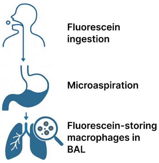
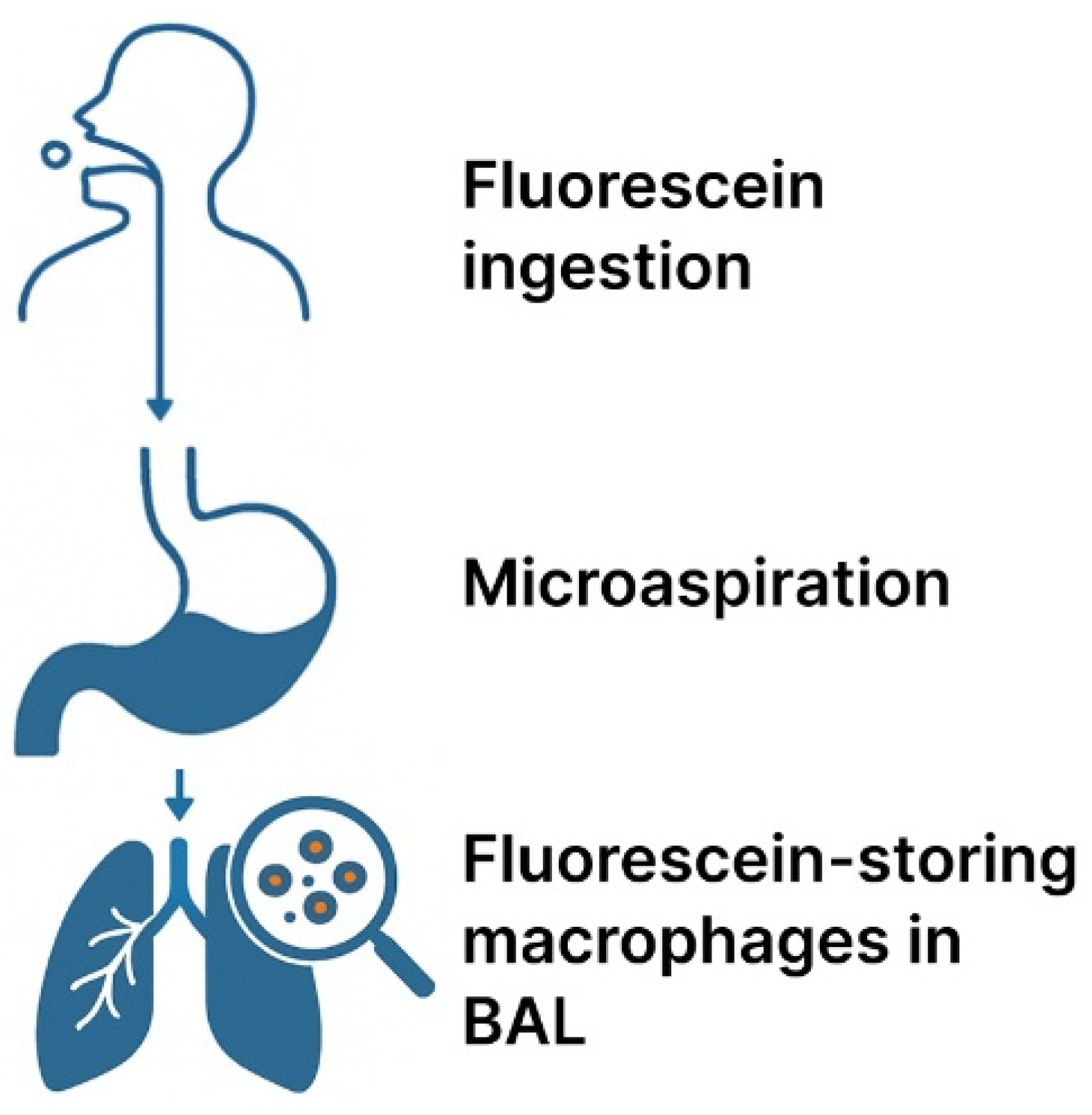
Fluorescein has been used as a marker of aspiration in swallowing disorders and to visualize structural alterations in alveolar tissue. It has also been incorporated into BAL analysis to characterize cellular components, including macrophage populations [18,19,24,25,26]. Bronchial washing and bronchoalveolar lavage were performed in each patient using 40 mL and 200 mL of 0.9% saline, respectively. Fluorescein measurements were obtained exclusively from BAL samples. To reduce the possibility of false-positive results due to sample contamination or incorrect swallowing and to ensure proper stomach emptying, patients ingested 100 mg of fluorescein with yogurt 9–12 h prior to bronchoscopy. Patients otherwise followed the standard BAL protocol, including fasting prior to the procedure and inpatient admission for observation. Bronchoalveolar lavage was performed in the middle lobe and analyzed immediately for macrophages storing fluorescein (Figure 1). In brief, bronchoalveolar lavage samples were filtered and centrifuged, and the cells were counted as previously described [43]. One million cells were analyzed by flow cytometry using a CytoFLEX V5-B5-R3 instrument (Beckman Coulter, Brea, CA, USA) to determine the proportion of fluorescein-positive macrophages. This method does not directly visualize aspiration events but instead assesses the accumulation of aspirated material in the lower respiratory tract over time. Currently, no validated fluorescein concentration thresholds for the diagnosis of micro-aspiration exist in the literature. In our cohort, a threshold of >10 ng/mL consistently corresponded to a clear separation between background fluorescence levels and values suggestive of clinically relevant micro-aspiration. This cutoff was selected based on clinical experience to distinguish true aspiration from low-level systemic absorption following oral administration, which is expected to produce only minimal pulmonary fluorescence. Accordingly, values > 10 ng/mL were considered indicative of significant micro-aspiration. Due to the known pH sensitivity of fluorescein [44], potential fluorescence quenching in acidic environments was considered when interpreting the results, although this effect was not formally modeled in the present analysis. From a causal perspective, micro-aspiration may act as an intermediate variable in the association between PPI use and airway colonization; accordingly, adjustment for fluorescein could attenuate the observed relationship. In this study, fluorescein was not treated as a confounder but rather as an exploratory marker intended to contextualize the potential role of micro-aspiration in the observed associations.
Figure 1.
Schematic workflow illustrating the use of fluorescein as a marker of micro-aspiration. Fluorescein (100 mg) was ingested with yogurt 9–12 h prior to bronchoscopy. BAL was performed with 200 mL saline in the middle lobe and analyzed immediately for fluorescein-storing macrophages.
2.5. Statistical Analysis
Descriptive statistics were used to summarize patient characteristics and explore relationships between relevant variables. Correlation between pathogen load and fluorescence levels was assessed using the Spearman correlation coefficient. To further investigate the association between PPI intake and pathogen counts, ordinal regression analysis was performed. This model estimates the odds of belonging to a higher pathogen load category while accounting for the ordinal structure of the outcome. Analyses were adjusted for sex, age, fluorescence levels, corticosteroid use and number of indication diagnoses. The method models the relationship between an ordinal dependent variable and one or more independent variables. It assumes that the odds of being in a higher category are proportional across all levels of the predictors (i.e., the proportional odds assumption). Fluorescein was included as an exploratory variable reflecting potential micro-aspiration. Model assumptions were tested, and appropriate diagnostics and sensitivity analyses were performed to ensure the robustness of the results. The results of the regression analysis are presented as odds ratios (ORs) with their corresponding 95% confidence intervals (CIs) and p-values. Gram stain, categorized as an ordinal variable (0 = no evidence of pathogens; 1 = Gram-positive; and 2 = Gram-negative or mixed Gram-positive and Gram-negative), was analyzed using the same approach as for pathogen load. Due to the small number of missing values, a complete case analysis was performed for the Spearman correlation and regression analyses. Specifically, data on pathogen load were unavailable for four patients (three female and one male patient) who had no recorded microbiological or PCR results in their medical files (Table S1 in the Supplementary Materials file). All analyses were conducted using R (version 4.4.2), and a significance level of 0.05 was used. A more detailed description of the statistical analysis is provided in the Supplementary Materials file.
3. Results
3.1. Patient Characteristics and Bronchoscopic Indication
A total of 140 patients (63.5% female, mean age 56.8 years) were included in this retrospective analysis. Only four patients (2.9%) were active smokers. Indications for bronchoscopy included suspected micro-aspiration (47.4%), severe asthma (28.5%), chronic cough (27.7%), suspected interstitial lung disease (15.3%) and other pulmonary conditions (8%, including bronchiectasis, post-COVID-19 syndrome, nontuberculous mycobacterial (NTM) infection and suspicious CT findings). Relevant comorbidities included gastroesophageal reflux, diagnosed by gastroscopy or pH-metry (29.9%), hiatal hernia (4.4%), previous gastrointestinal surgery (1.5%), gastritis (12.4%), and systemic sclerosis (3.6%) (Table 1, Table 2 and Table 3).
Table 1.
Patient characteristics of the study population undergoing bronchoscopy for suspected pulmonary conditions, including micro-aspiration. * Age is presented as mean for the overall study population.
Table 2.
Clinical indications for bronchoscopy.
Table 3.
Relevant patient comorbidities.
Out of 140 screened patients, three were excluded due to missing microbiological (culture) or PCR data. A total of 137 patients were included in the final analysis. In seven patients, pathogen counts were adjusted following exclusion of transient viral detections (Influenza-A, Rhinovirus, Enterovirus, Adenovirus, and Coronavirus). The most frequently detected pathogens were Haemophilus influenzae (n = 27), Staphylococcus aureus (n = 24), Candida albicans (n = 10), Pseudomonas aeruginosa (n = 9), Escherichia coli (n = 6), Moraxella catarrhalis (n = 6), Streptococcus agalactiae (n = 4), and Streptococcus pneumoniae (n = 4). Serratia marcescens, Candida glabrata, Enterobacter cloacae, Klebsiella aerogenes, Klebsiella pneumoniae, Klebsiella oxytoca, Proteus mirabilis, Neisseria meningitidis, Achromobacter xylosoxidans, Streptococcus pyogenes (group A), Citrobacter koseri, Aspergillus flavus, and Candida krusei were only detected in a few cases by culture or PCR. Of the 137 included patients, 50 (36.5%) were receiving PPI therapy, while the remaining patients were either PPI naive or had no recent exposure to acid-suppressive treatment (Table S1).
3.2. Pathogen Load and PPI Association
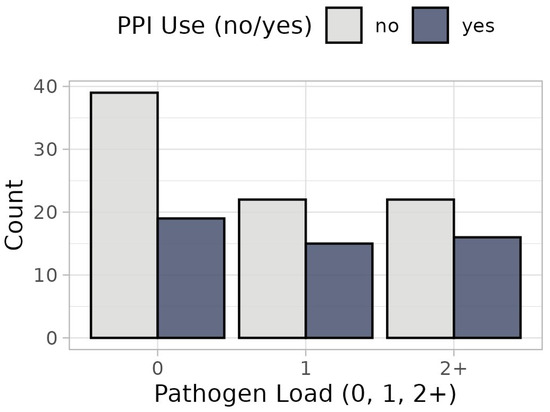
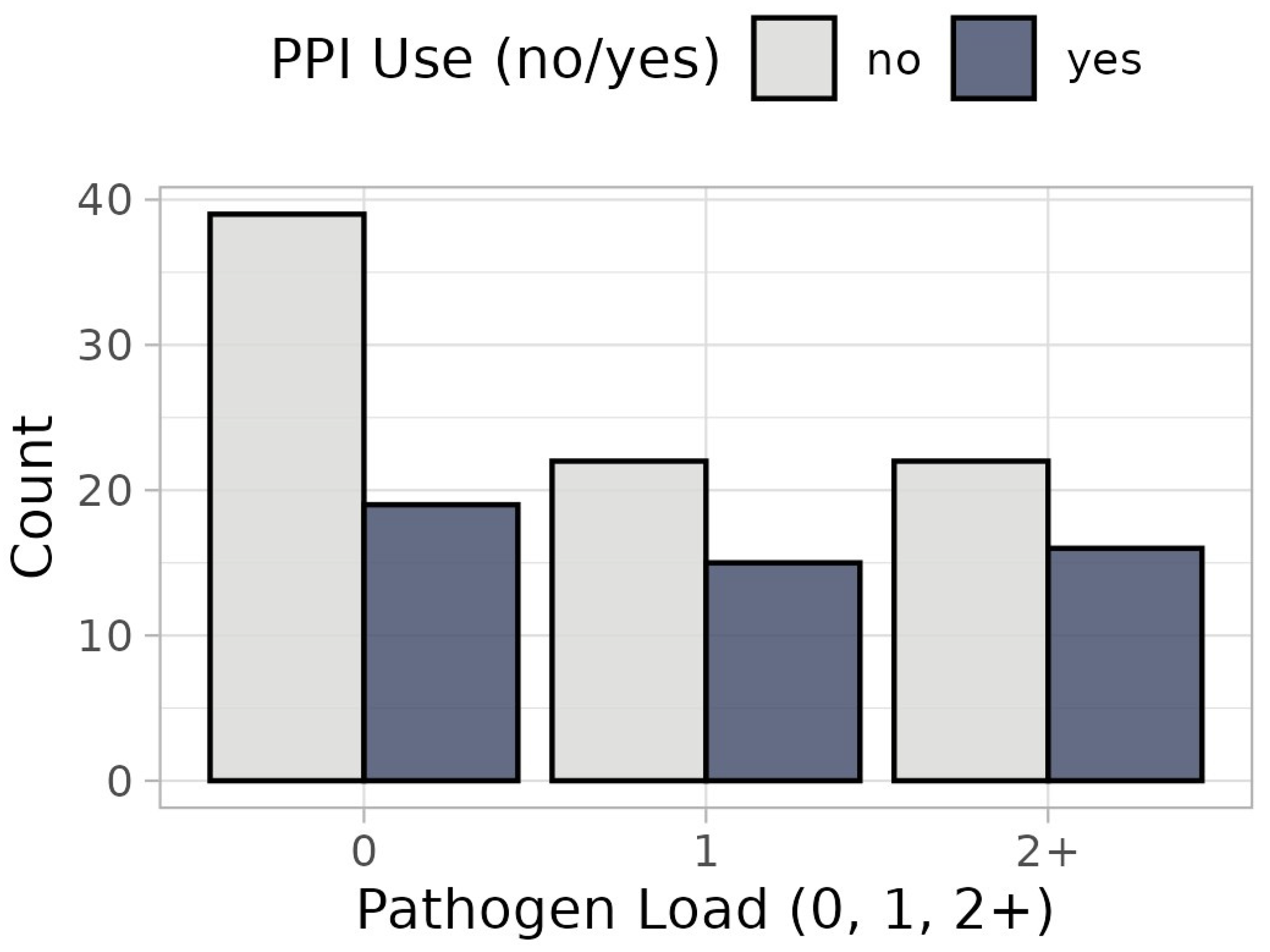
Overall, pathogens were detected in 57.7% of all patients who underwent bronchoscopy. Pathogens were more frequently detected in PPI users than in non-users (62% versus 50.6%, respectively) (Figure 2). Multivariable ordinal regression showed increased odds of bacterial load for PPI use but the effect was not statistically significant (OR = 1.40, 95% CI: 0.71–2.75, p = 0.30). Female patients showed lower odds of pathogen load than male patients (OR = 0.40, 95% CI: 0.20–0.79, p = 0.009). No significant association of corticosteroid use with bacterial load could be observed (OR = 1.56, 95% CI: 0.78–3.13, p = 0.20) (Table S2, Figure S1).
Figure 2.
Pathogen load according to PPI use (no vs. yes): Bar charts showing the distribution of pathogen load stratified by PPI treatment. Grey bars represent the frequency of participants without PPI treatment; blue bars represent those with PPI treatment.
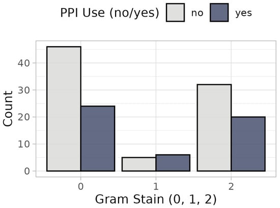
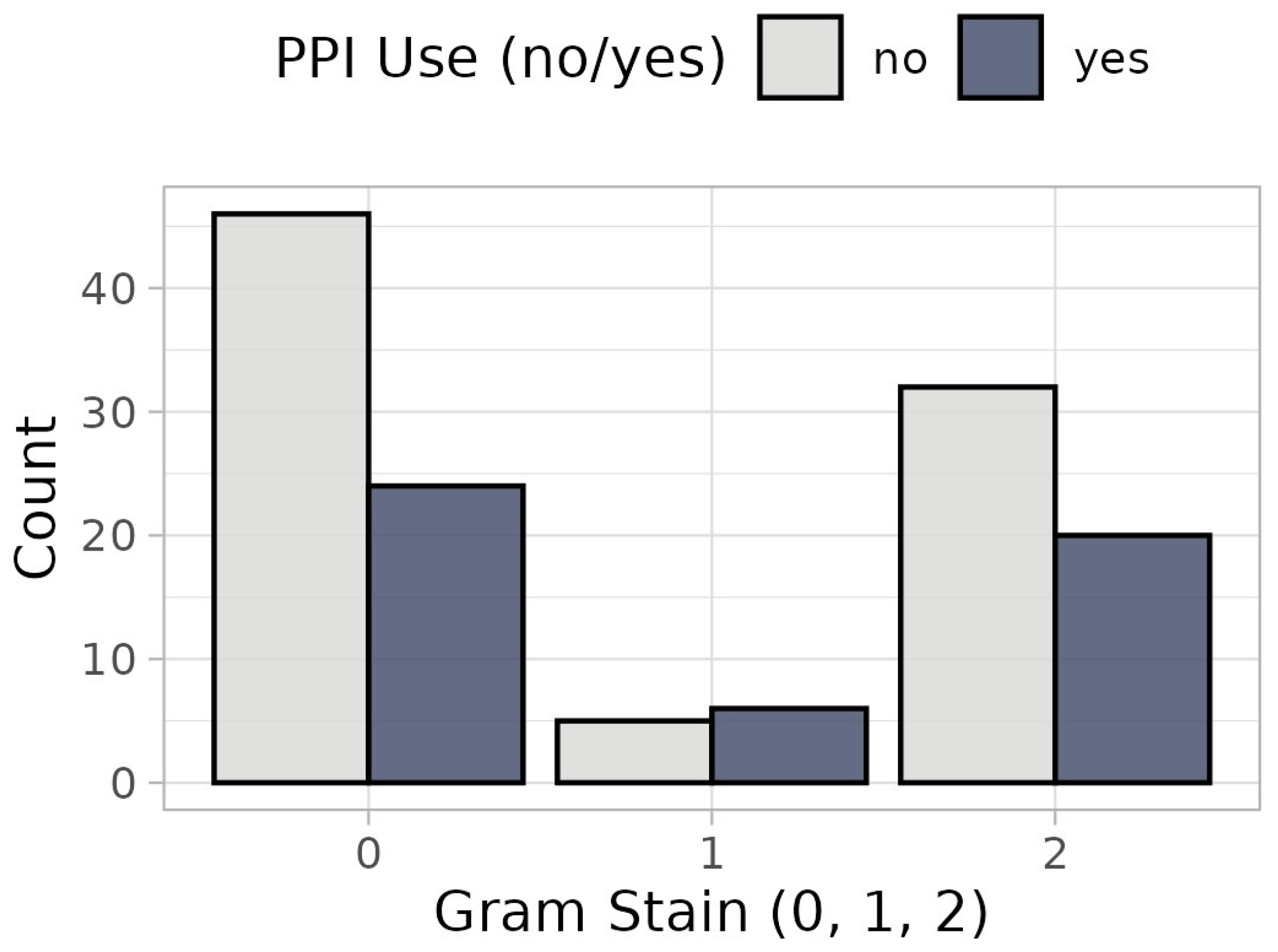
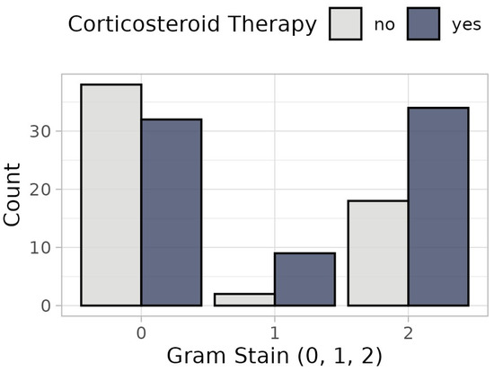
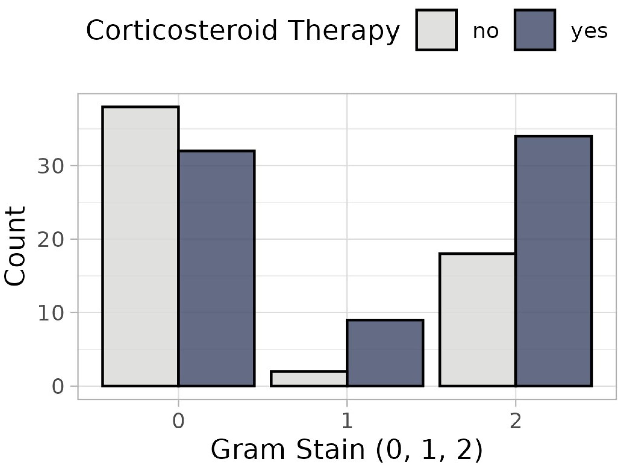
Gram stain findings were categorized to reflect increasing likelihood of gastrointestinal origin: 0 = no pathogens detected, 1 = Gram-positive organisms and 2 = Gram-negative or mixed Gram-positive and Gram-negative organisms. PPI therapy was not significantly associated with Gram stain outcomes (OR = 1.18, 95% CI: 0.57–2.44, p = 0.7; Figure 3). Corticosteroid therapy was significantly associated with Gram stain results (OR = 2.37, 95% CI: 1.12–5.15, p = 0.025; Figure 4). Female sex was associated with significantly lower odds of harboring pathogens of intestinal origin compared with males (OR = 0.37, 95% CI: 0.17–0.77, p = 0.01) (Table S3, Figure S2).
Figure 3.
Gram stain results according to PPI use (no vs. yes): Bar charts showing the distribution of Gram stain categories stratified by PPI treatment. Grey bars represent the frequency of participants without PPI treatment; blue bars represent those with PPI treatment.
Figure 4.
Gram stain results according to corticosteroids use (no vs. yes): Bar charts showing the distribution of Gram stain categories stratified by corticosteroid treatment. Grey bars represent the frequency of participants without corticosteroid treatment; blue bars represent those with corticosteroid treatment.
Corticosteroid use was more frequent among patients with Gram-negative or mixed Gram-positive and Gram-negative organisms, as well as those with Gram-positive findings, compared with patients without detectable pathogens. Patients with no evidence of pathogens (category 0) had a lower proportion of corticosteroid use (Figure 4).
3.3. Fluorescein Analysis and Its Correlation with Pathogen Load
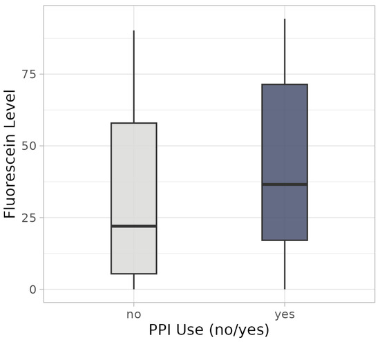
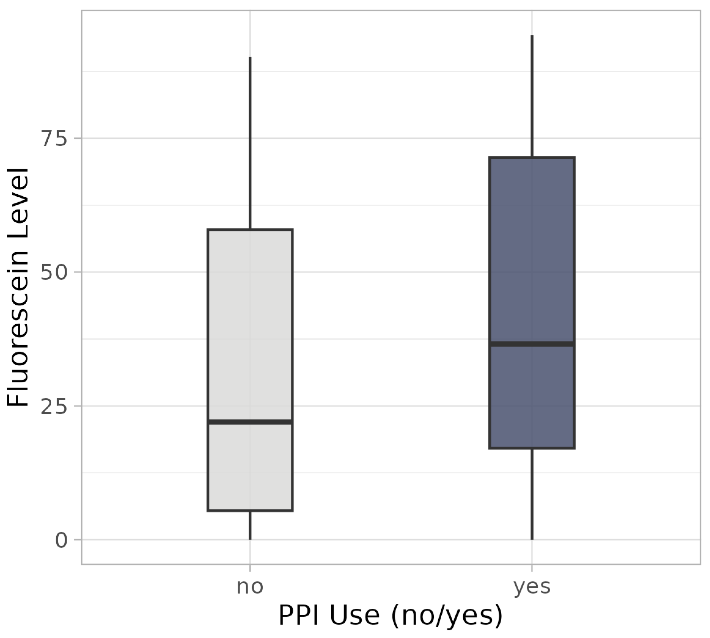
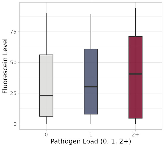
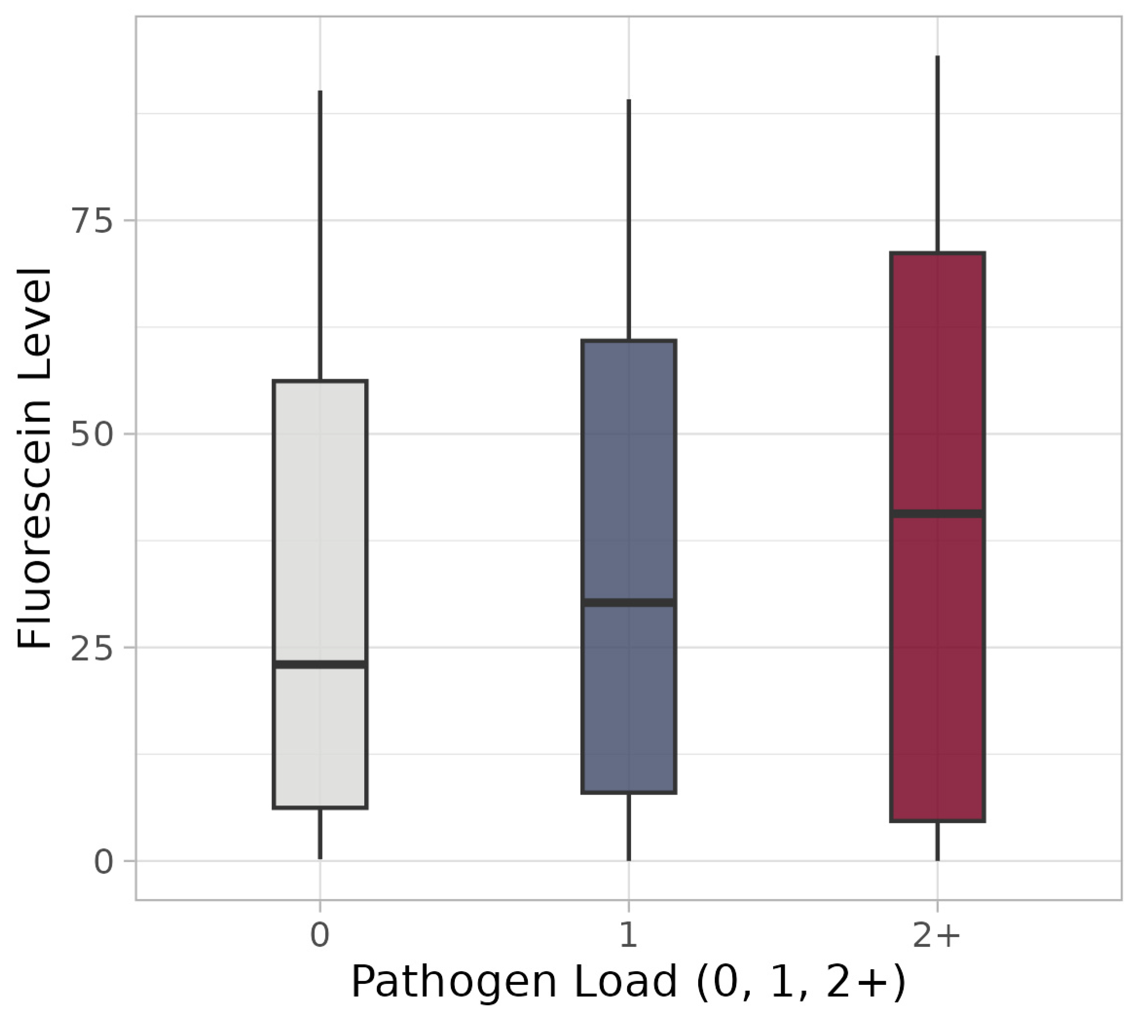
The prevalence of clinically significant micro-aspiration (BAL fluorescein > 10 ng/mL) and the mean levels of fluorescein-storing macrophages were higher in PPI users than in non-PPI users (78.0% vs. 63.2% and 41.5 ng/mL vs. 31.2 ng/mL, respectively) (Figure 5). However, the correlation with pathogen load was negligible (ρ = 0.09, p = 0.32). The box plots indicate a slight increase in fluorescence levels with the number of pathogens detected; however, dispersion is higher when two or more pathogens are detected (Figure 6). Nevertheless, the proportion of observations exceeding the fluorescence cutoff value of 10 remains consistent across all pathogen load categories.
Figure 5.
Fluorescence according to PPI use (no vs. yes). Box plots showing fluorescein levels stratified by proton pump inhibitor (PPI) use. The grey box plot represents participants without PPI treatment; the blue box plot represents those with PPI treatment.
Figure 6.
Fluorescence according to pathogen load: Box plots showing fluorescein levels stratified by pathogen load. Groups are defined by the number of pathogens detected: grey indicates 0 pathogens; blue, 1 pathogen; and red, 2 or more pathogens.
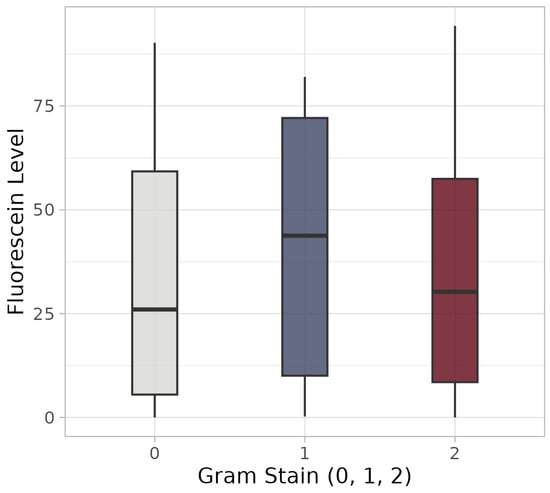
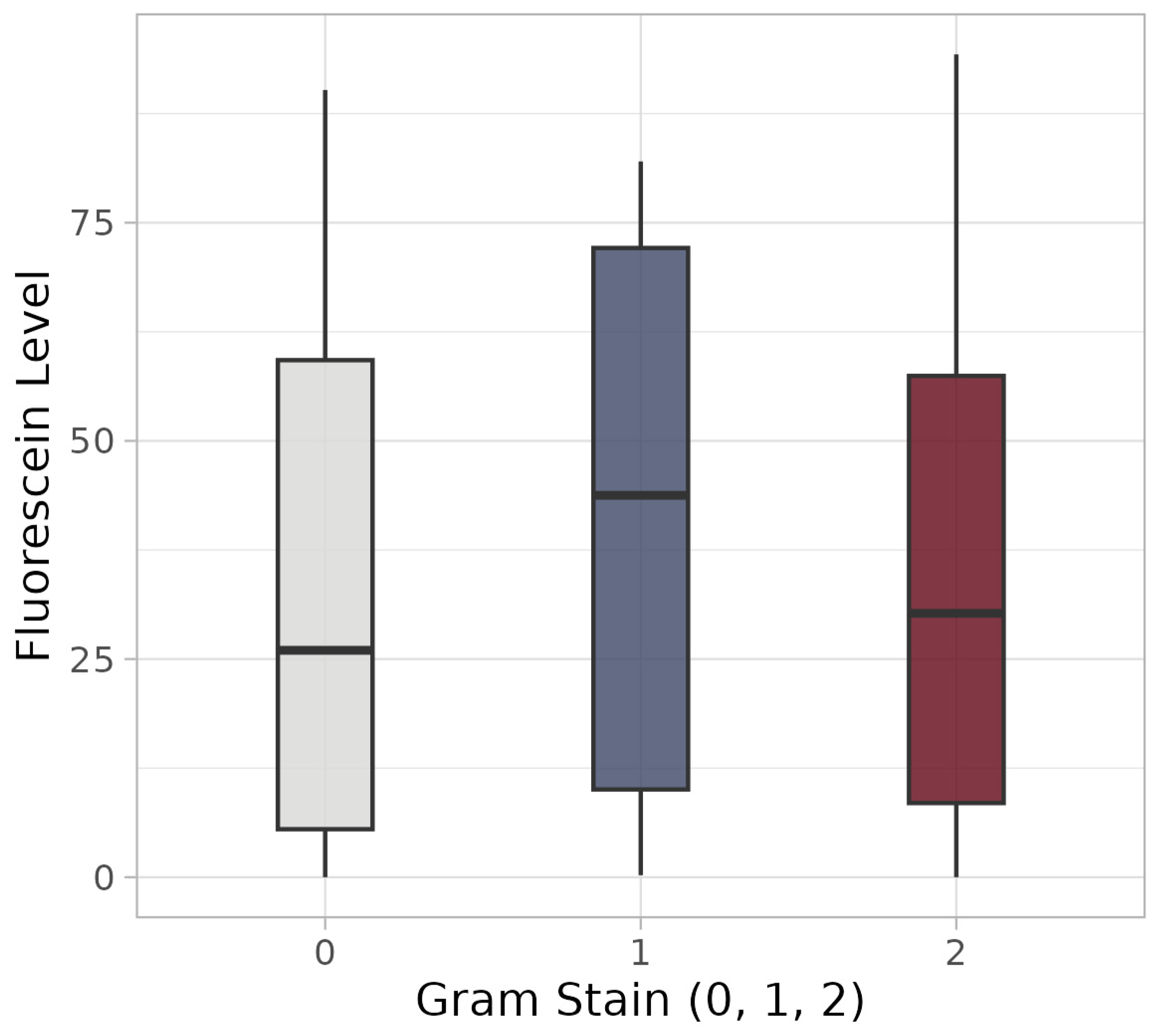
Fluorescence levels were highest among patients with Gram-positive stains (as well as those with unobserved Gram stains) (Figure 7). The proportion of patients with fluorescence levels above the cutoff of 10 did not differ notably across the different Gram stain categories.
Figure 7.
Fluorescence according to Gram stain results: Box plots showing fluorescein levels stratified by Gram stain category. Grey indicates samples with no evidence of pathogens (Gram stain = 0); blue represents samples with Gram-positive bacteria (Gram stain = 1); and red corresponds to samples with Gram-negative or mixed bacteria (Gram stain = 2).
4. Discussion
In recent years, increasing attention has been given to the complex interaction between the pulmonary microbiome and host immunity. Alterations in microbial composition have been associated with the development and progression of lung disease, reflecting the two-way interaction between host factors and bacterial communities [14,17,45]. PPIs are widely prescribed, and their use has been linked to an increased risk of pneumonia, though the underlying mechanisms are not fully understood [3,4,28,29]. One proposed explanation is that gastric acid suppression impairs the elimination of gastrointestinal pathogens, facilitating their micro-aspiration into the lower airways. While aspiration-related changes may be evident on chest imaging in cases of overt aspiration, this is generally not the case for micro-aspiration, which is typically subclinical and not reliably detected on chest X-ray [46]. Moreover, the patients in this study were not assessed in the context of acute aspiration events, but rather for chronic conditions or follow-up of underlying disease. For these reasons, chest radiography was not considered an appropriate or sufficiently sensitive method to assess micro-aspiration in this cohort. Micro-aspiration is increasingly recognized as a contributor to disease progression and symptom burden in various pulmonary conditions [47,48,49]. However, it is difficult to detect due to the lack of a widely accepted, validated diagnostic marker. Biomarkers such as pepsin and α-amylase in tracheal aspirates or bronchoalveolar lavage fluid have been investigated as potential diagnostic markers, but none have been clinically validated or adopted for routine use [50,51,52].
Fluorescein has been proposed as an alternative marker of aspiration and has been shown to be feasible in other clinical contexts, including bronchoalveolar lavage-based cellular analyses [18,19,24,25,26,27]. However, its use in the context of micro-aspiration in lung disease and PPI-associated respiratory risk is novel and therefore subject to important methodological limitations. Fluorescein fluorescence is pH-dependent and may be attenuated in acidic environments, which could lead to an underestimation of micro-aspiration in individuals not receiving acid suppression therapy [44]. Additionally, measured concentrations may be influenced by systemic absorption, mucociliary clearance, and variability in BAL sampling techniques, as well as the potential for false-positive results due to macro-aspiration. To address these limitations, we implemented a standardized 9–12 h interval between oral administration and bronchoscopy. This timing was chosen to minimize the risk of macro-aspiration while still allowing sufficient time for potential micro-aspiration events to occur. Furthermore, a predefined threshold based on clinical experience was applied to minimize the misclassification of low-level fluorescence as a systemic background signal.
This retrospective observational study investigated the association between proton pump inhibitor (PPI) use, pulmonary bacterial colonization, and micro-aspiration using orally administered fluorescein as an exploratory marker in bronchoalveolar lavage fluid. To our knowledge, this is the first study to use direct lower airway sampling rather than sputum-based analysis in this context, thereby enabling a more accurate assessment of lower respiratory tract processes. Although no statistically significant associations were observed between PPI use and bacterial load in bronchial washing fluid or between fluorescein levels and pathogen burden, trends were identified. Higher pathogen loads were observed among patients undergoing PPI therapy, and increased fluorescein concentrations tended to increase with a greater number of detected pathogens. While these findings suggest a possible link between micro-aspiration and airway colonization, they do not allow for definitive conclusions.
Several limitations must be acknowledged. The retrospective design limits control over data collection, introduces the risk of selection and information bias, and restricts the ability to adjust for confounding factors in a standardized manner. The relatively small sample size reduces statistical power and increases susceptibility to random variation. Additionally, the study population was heterogeneous with respect to underlying pulmonary diseases, which may have influenced the occurrence and detection of micro-aspiration. Underlying lung pathology, corticosteroid therapy, and acid suppression itself may independently alter airway microbial ecology and promote colonization [2,48,53]. Notably, corticosteroid use in our cohort was associated with a higher prevalence of Gram-negative or mixed flora, suggesting a potential modulatory effect on airway microbiota.
The interpretation of the findings is complicated by the inherent complexity of the pulmonary microbiome and the necessity of grouping pathogens into clinically meaningful categories. Although this approach is reductive, it was necessary to ensure interpretability in a heterogeneous dataset. Aspiration-related colonization is typically polymicrobial and often dominated by Gram-negative organisms; thus, this classification strategy is consistent with prior literature and adds clinical relevance [37,38,42]. At the same time, PPI-associated alterations in the qualitative composition of the lung microbiome may be clinically relevant beyond simple quantitative pathogen burden. The pulmonary microbiome is a highly dynamic ecosystem in which subtle, chronic shifts in microbial balance may exert disease-modifying effects over time yet remain difficult to quantify and are frequently underrecognized in clinical practice [2,53]. In this context, interventions that influence this system, including long-term acid suppression, should be carefully evaluated for potential unintended consequences. In patients with unexplained chronic respiratory symptoms, particularly those with gastrointestinal comorbidities, targeted evaluation for micro-aspiration may be warranted [48]. Despite these limitations, the study should be considered hypothesis-generating. It introduces a novel, integrative approach that combines a bronchial washing-based microbiological assessment with an exploratory bronchoalveolar lavage-based marker of aspiration. This framework may contribute to a more comprehensive understanding of the potential interplay between gastroesophageal processes and pulmonary microbial colonization. While fluorescein is not yet a validated micro-aspiration biomarker, it is a simple, cost-effective, and safe tool that could be used in future studies.
In conclusion, our findings do not definitively establish a role for fluorescein as a marker of micro-aspiration or a mechanistic link between PPI therapy and increased bacterial colonization. However, the observed trends suggest a potential association that warrants further investigation. Future prospective studies with larger, more homogeneous cohorts and standardized protocols are essential to validate fluorescein as a biomarker and clarify the role of micro-aspiration in PPI-associated respiratory risk.
5. Conclusions
This retrospective observational study investigated the association between PPI treatment and pulmonary bacterial colonization using fluorescein-labeled macrophages as an exploratory biomarker of micro-aspiration in bronchoalveolar lavage samples. To our knowledge, this is the first study to apply direct lower airway sampling in this context rather than sputum-based analysis. No statistically significant association was observed between PPI use and bacterial load, nor between fluorescein levels and pathogen burden. However, trends toward higher bacterial loads in patients receiving PPI therapy and increased fluorescein concentrations in samples with greater pathogen counts suggest a potential relationship between micro-aspiration and airway colonization, although definitive conclusions cannot be drawn.
Although these findings do not warrant immediate changes in clinical practice, they highlight a possible interaction between acid suppression therapy and airway microbial imbalance. Fluorescein-enhanced BAL may represent a supportive exploratory tool for future studies aimed at stratifying aspiration risk. Larger prospective cohorts with standardized methodologies will be required to further evaluate fluorescein as a potential biomarker in pulmonary disease and to better define the gastroenterology–pulmonology interface.
Ultimately, improved understanding of the interplay between micro-aspiration, microbial colonization, and airway inflammation may contribute to more targeted preventive strategies for pneumonia, individualized treatment approaches, and more judicious use of acid-suppressive therapy in at-risk populations.
Supplementary Materials
The following supporting information can be downloaded at https://www.mdpi.com/article/10.3390/jor6020007/s1: Table S1: Summary statistics for demographic and clinical variables, stratified by pathogen load outcome and for the overall sample; Table S2: Ordinal regression results evaluating the association between pathogen load and PPI, adjusted for demographic and clinical covariates; Table S3: Ordinal regression results evaluating the association between Gram stain and PPI, adjusted for demographic and clinical covariates; Table S4: Summary statistics for demographic and clinical variables, stratified by PPI use and for the overall sample; Figure S1: Visualization of ordinal regression results evaluating the association between pathogen load and PPI, adjusted for demographic and clinical covariates; Figure S2: Visualization of ordinal regression results evaluating the association between Gram stain and PPI, adjusted for demographic and clinical covariates.
Author Contributions
L.N.: co-planned the study design, provided overall supervision and data interpretation and prepared the main manuscript; C.W.: performed the statistical analysis of the dataset and created the figures; P.S.: performed the microbiological testing and assisted with data interpretation; D.G.: co-planned the study and performed the bronchoscopies; M.I.: provided the necessary resources and infrastructure; A.E.-G.: performed fluorescein analysis of all bronchoalveolar lavage samples and prepared the revised manuscript. All authors have read and agreed to the published version of the manuscript.
Funding
This research was funded by the Medical University of Vienna and the Austrian Science Fund (FWF) (grant number 10.55776/PAT2343825), which was awarded to A.E.-G.
Institutional Review Board Statement
The study was conducted in accordance with the Declaration of Helsinki. This study was conducted according to a protocol that was approved by the Ethics Committee of the Medical University of Vienna (EK-Nr. 1876/2024, 20 March 2025).
Informed Consent Statement
Due to the retrospective nature of the study, consent for publication was waived.
Data Availability Statement
The original contributions presented in this study are included in the article and Supplementary Materials. Further inquiries can be directed to the corresponding authors.
Acknowledgments
We would like to express our sincere gratitude to all the bronchoscopists and assisting personnel for their dedicated work in performing the procedures. We also thank the Division of Clinical Microbiology team and the Department of Pulmonology research laboratory team at the Medical University of Vienna for their careful analyses and technical support, which made this study possible.
Conflicts of Interest
The authors declare that they have no competing interests.
References
- Scarpignato, C.; Gatta, L.; Zullo, A.; Blandizzi, C. Effective and safe proton pump inhibitor therapy in acid-related diseases—A position paper addressing benefits and potential harms of acid suppression. BMC Med. 2016, 14, 179. [Google Scholar] [CrossRef] [Scilit] [PubMed]
- Whiteside, S.A.; McGinniss, J.E.; Collman, R.G. The lung microbiome: Progress and promise. J. Clin. Investig. 2021, 131, e150473. [Google Scholar] [CrossRef] [Scilit] [PubMed]
- Maret-Ouda, J.; Panula, J.; Santoni, G.; Xie, S.; Lagergren, J. Proton pump inhibitor use and risk of pneumonia: A self-controlled case series study. J. Gastroenterol. 2023, 58, 734–740. [Google Scholar] [CrossRef] [Scilit] [PubMed]
- Herzig, S.J.; Howell, M.D.; Ngo, L.H.; Marcantonio, E.R. Acid-suppressive medication use and the risk for hospital-acquired pneumonia. JAMA 2009, 301, 2120–2128. [Google Scholar] [CrossRef] [Scilit] [PubMed]
- Lambert, A.A.; Lam, J.O.; Paik, J.J.; Ugarte-Gil, C.; Drummond, M.B.; Crowell, T.A. Risk of Community-Acquired Pneumonia with Outpatient Proton-Pump Inhibitor Therapy: A Systematic Review and Meta-Analysis. PLoS ONE 2015, 10, e0128004. [Google Scholar] [CrossRef] [Scilit] [PubMed]
- Nehra, A.K.; Alexander, J.A.; Loftus, C.G.; Nehra, V. Proton Pump Inhibitors: Review of Emerging Concerns. Mayo Clin. Proc. 2018, 93, 240–246. [Google Scholar] [CrossRef] [Scilit] [PubMed]
- Li, R.; Li, J.; Zhou, X. Lung microbiome: New insights into the pathogenesis of respiratory diseases. Signal Transduct. Target. Ther. 2024, 9, 19. [Google Scholar] [CrossRef] [Scilit] [PubMed]
- Yang, D.; Xing, Y.; Song, X.; Qian, Y. The impact of lung microbiota dysbiosis on inflammation. Immunology 2019, 159, 156. [Google Scholar] [CrossRef] [Scilit] [PubMed]
- Budden, K.F.; Shukla, S.D.; Rehman, S.F.; Bowerman, K.L.; Keely, S.; Hugenholtz, P.; Armstrong-James, D.; Adcock, I.M.; Chotirmall, S.H.; Chung, K.F.; et al. Functional effects of the microbiota in chronic respiratory disease. Lancet Respir. Med. 2019, 7, 907–920. [Google Scholar] [CrossRef] [Scilit] [PubMed]
- Brown, R.L.; Sequeira, R.P.; Clarke, T.B. The microbiota protects against respiratory infection via GM-CSF signaling. Nat. Commun. 2017, 8, 1512. [Google Scholar] [CrossRef] [Scilit] [PubMed]
- Garcha, D.S.; Thurston, S.J.; Patel, A.R.C.; Mackay, A.J.; Goldring, J.J.P.; Donaldson, G.C.; McHugh, T.D.; Wedzicha, J.A. Changes in prevalence and load of airway bacteria using quantitative PCR in stable and exacerbated COPD. Thorax 2012, 67, 1075–1080. [Google Scholar] [CrossRef] [Scilit] [PubMed]
- Sethi, S.; Murphy, T.F. Infection in the Pathogenesis and Course of Chronic Obstructive Pulmonary Disease. N. Engl. J. Med. 2008, 359, 2355–2365. [Google Scholar] [CrossRef] [Scilit] [PubMed]
- Wilkinson, T.M.A.; Patel, I.S.; Wilks, M.; Donaldson, G.C.; Wedzicha, J.A. Airway bacterial load and FEV1 decline in patients with chronic obstructive pulmonary disease. Am. J. Respir. Crit. Care Med. 2003, 167, 1090–1095. [Google Scholar] [CrossRef] [Scilit] [PubMed]
- Caverly, L.J.; Huang, Y.J.; Sze, M.A. Past, Present, and Future Research on the Lung Microbiome in Inflammatory Airway Disease. Chest 2019, 156, 376–382. [Google Scholar] [CrossRef] [Scilit] [PubMed]
- Flume, P.A.; Chalmers, J.D.; Olivier, K.N. Advances in bronchiectasis: Endotyping, genetics, microbiome, and disease heterogeneity. Lancet 2018, 392, 880–890. [Google Scholar] [CrossRef] [Scilit] [PubMed]
- Lin, Z.; Jiang, Y.; Liu, H.; Yang, J.; Yang, B.; Zhang, K.; Tang, P.; Xiang, B.; Sun, B. Airway microbiota and immunity associated with chronic obstructive pulmonary disease severity. J. Transl. Med. 2025, 23, 962. [Google Scholar] [CrossRef] [Scilit] [PubMed]
- Faner, R.; Sibila, O.; Agustí, A.; Bernasconi, E.; Chalmers, J.D.; Huffnagle, G.B.; Manichanh, C.; Molyneaux, P.L.; Paredes, R.; Perez, B.; et al. The microbiome in respiratory medicine: Current challenges and future perspectives. Eur. Respir. J. 2017, 49, 1602086. [Google Scholar] [CrossRef] [Scilit] [PubMed]
- Nishimura, G.; Wakamatsu, K.; Koike, T.; Yamada, Y.; Niwa, H.; Michiwaki, Y. Fluorescence Method to Assess Aspiration Risk. In Proceedings of the European Conferences on Biomedical Optics 2025, München Germany, 22–26 June 2025. [Google Scholar] [CrossRef] [Scilit]
- Assessment of Aspiration in Patients with Tracheostomies: Comparison of the Bedside Colored Dye Assessment with Videofluoroscopic Examination. Available online: https://pubmed.ncbi.nlm.nih.gov/11262550/ (accessed on 11 April 2026).
- Das, D.; Deka, P.; Bhattacharjee, H.; Deshmukh, S.; Gupta, P.; Deka, A.; Sultana, S. Fluorescein dye as a novel cost-effective approach for staining raw specimens in ophthalmic pathology. Indian J. Ophthalmol. 2020, 68, 2175. [Google Scholar] [CrossRef] [Scilit] [PubMed]
- Prieto, S.P.; Lai, K.K.; Laryea, J.A.; Mizell, J.S.; Mustain, W.C.; Muldoon, T.J. Fluorescein as a topical fluorescent contrast agent for quantitative microendoscopic inspection of colorectal epithelium. Biomed. Opt. Express 2017, 8, 2324. [Google Scholar] [CrossRef] [Scilit] [PubMed]
- Falco, J.; Cavallo, C.; Vetrano, I.G.; de Laurentis, C.; Siozos, L.; Schiariti, M.; Broggi, M.; Ferroli, P.; Acerbi, F. Fluorescein Application in Cranial and Spinal Tumors Enhancing at Preoperative MRI and Operated with a Dedicated Filter on the Surgical Microscope: Preliminary Results in 279 Patients Enrolled in the FLUOCERTUM Prospective Study. Front. Surg. 2019, 6, 49. [Google Scholar] [CrossRef] [Scilit] [PubMed]
- Behbahaninia, M.; Martirosyan, N.L.; Georges, J.; Udovich, J.A.; Kalani, M.Y.S.; Feuerstein, B.G.; Nakaji, P.; Spetzler, R.F.; Preul, M.C. Intraoperative fluorescent imaging of intracranial tumors: A review. Clin. Neurol. Neurosurg. 2013, 115, 517–528. [Google Scholar] [CrossRef] [Scilit] [PubMed]
- Zeinerová, L.; Černý, M.; Homoláč, M.; Školoudík, L.; Šatanková, J.; Chrobok, V. Use of fluorescein to improve the sensitivity of flexible endoscopic evaluation of swallowing. Otorinolaryngol. Foniatr. 2023, 72, 127–135. [Google Scholar] [CrossRef] [Scilit]
- Lindstedt, S.; Wang, Q.; Niroomand, A.; Stenlo, M.; Hyllen, S.; Pierre, L.; Olm, F.; Bechet, N.B. High resolution fluorescence imaging of the alveolar scaffold as a novel tool to assess lung injury. Sci. Rep. 2024, 14, 6662. [Google Scholar] [CrossRef] [Scilit] [PubMed]
- Zlotnikov, I.D.; Kudryashova, E.V. Polymeric Infrared and Fluorescent Probes to Assess Macrophage Diversity in Bronchoalveolar Lavage Fluid of Asthma and Other Pulmonary Disease Patients. Polymers 2024, 16, 3427. [Google Scholar] [CrossRef] [Scilit] [PubMed]
- Gelfand, C.A.; Wang, Y.; Chandan, G.; Liu, J.; Madrigal, S.; Sakurai, R.; Yu, C.; Guerra, C.; Segal, R.; Rehan, V.K. A Rapid Fluorescence Method for In Vivo Quantitation of Lung Deposition of a Nebulized Drug: Multiple Uses for Advancing Aerosolized Drug Development and Specific Insight Regarding Aerosolized Vitamin A for Preventing Bronchopulmonary Dysplasia. Methods Protoc. 2025, 8, 140. [Google Scholar] [CrossRef] [Scilit] [PubMed]
- Meijvis, S.C.A.; Cornips, M.C.A.; Voorn, G.P.; Souverein, P.C.; Endeman, H.; Biesma, D.H.; Leufkens, H.G.M.; van de Garde, E.M.W. Microbial evaluation of proton-pump inhibitors and the risk of pneumonia. Eur. Respir. J. 2011, 38, 1165–1172. [Google Scholar] [CrossRef] [Scilit] [PubMed]
- Eurich, D.T.; Sadowski, C.A.; Simpson, S.H.; Marrie, T.J.; Majumdar, S.R. Recurrent community-acquired pneumonia in patients starting acid-suppressing drugs. Am. J. Med. 2010, 123, 47–53. [Google Scholar] [CrossRef] [Scilit] [PubMed]
- De Jager, C.P.C.; Wever, P.C.; Gemen, E.F.A.; Van Oijen, M.G.H.; Van Gageldonk-Lafeber, A.B.; Siersema, P.D.; Kusters, G.C.M.; Laheij, R.J.F. Proton pump inhibitor therapy predisposes to community-acquired Streptococcus pneumoniae pneumonia. Aliment. Pharmacol. Ther. 2012, 36, 941–949. [Google Scholar] [CrossRef] [Scilit] [PubMed]
- Hunfeld, N.G.; Touw, D.J.; Mathot, R.A.; Mulder, P.G.H.; Van Schaik, R.H.; Kuipers, E.J.; Kooiman, J.C.; Geus, W.P. A comparison of the acid-inhibitory effects of esomeprazole and pantoprazole in relation to pharmacokinetics and CYP2C19 polymorphism. Aliment. Pharmacol. Ther. 2010, 31, 150–159. [Google Scholar] [CrossRef] [Scilit] [PubMed]
- Pawlotsky, J.M.; Negro, F.; Aghemo, A.; Berenguer, M.; Dalgard, O.; Dusheiko, G.; Marra, F.; Puoti, M.; Wedemeyer, H. EASL recommendations on treatment of hepatitis C: Final update of the series. J. Hepatol. 2020, 73, 1170–1218. [Google Scholar] [CrossRef] [Scilit] [PubMed]
- Thomson, A.B.R. Standard doses of the oral proton pump inhibitors are clinically equivalent: A comparison. Curr. GERD Rep. 2007, 1, 223–232. [Google Scholar] [CrossRef] [Scilit]
- Bartlett, J.G. Anaerobic bacterial infections of the lung. Chest 1987, 91, 901–909. [Google Scholar] [CrossRef] [Scilit] [PubMed]
- Brook, I. Anaerobic pulmonary infections in children. Pediatr. Emerg. Care 2004, 20, 636–640. [Google Scholar] [CrossRef] [Scilit] [PubMed]
- Bartlett, J.G.; Gorbach, S.L. The triple threat of aspiration pneumonia. Chest 1975, 68, 560–566. [Google Scholar] [CrossRef] [Scilit] [PubMed]
- Fadell, F.; Saliba, R.; El-Solh, A.A. Bacteriology of Aspiration Pneumonia: The Lung Microbiome and the Changing Microbial Etiology. Semin. Respir. Crit. Care Med. 2024, 45, 626–633. [Google Scholar] [CrossRef] [Scilit] [PubMed]
- Dickson, R.P. The Lung Microbiome and ARDS. It Is Time to Broaden the Model. Am. J. Respir. Crit. Care Med. 2018, 197, 549–551. [Google Scholar] [CrossRef] [Scilit] [PubMed]
- Morris, A.; Beck, J.M.; Schloss, P.D.; Campbell, T.B.; Crothers, K.; Curtis, J.L.; Flores, S.C.; Fontenot, A.P.; Ghedin, E.; Huang, L.; et al. Comparison of the respiratory microbiome in healthy nonsmokers and smokers. Am. J. Respir. Crit. Care Med. 2013, 187, 1067–1075. [Google Scholar] [CrossRef] [Scilit] [PubMed]
- Hayashi, M.; Iwasaki, T.; Yamazaki, Y.; Takayasu, H.; Tateno, H.; Tazawa, S.; Kato, E.; Wakabayashi, A.; Yamaguchi, F.; Tsuchiya, Y.; et al. Clinical features and outcomes of aspiration pneumonia compared with non-aspiration pneumonia: A retrospective cohort study. J. Infect. Chemother. 2014, 20, 436–442. [Google Scholar] [CrossRef] [Scilit] [PubMed]
- Tay, W.H.; Chong, K.K.L.; Kline, K.A. Polymicrobial–Host Interactions during Infection. J. Mol. Biol. 2016, 428, 3355–3371. [Google Scholar] [CrossRef] [Scilit] [PubMed]
- Miller, J.M.; Binnicker, M.J.; Campbell, S.; Carroll, K.C.; Chapin, K.C.; Gonzalez, M.D.; Harrington, A.; Jerris, R.C.; Kehl, S.C.; Leal, S.M.; et al. Guide to Utilization of the Microbiology Laboratory for Diagnosis of Infectious Diseases: 2024 Update by the Infectious Diseases Society of America (IDSA) and the American Society for Microbiology (ASM). Clin. Infect. Dis. 2024, 36, 941–949. [Google Scholar] [CrossRef] [Scilit] [PubMed]
- El-Gazzar, A.; Aschenbrenner, B.; Forsthuber, A.; Kramer, M.; Kargarpour, Z.; Metekol, S.; John, L.; Papaporfyriou, A.; Bereš, M.; Trouvilliez, S.; et al. Targeting P2RY2 reprograms tumor immunity and inhibits tumor growth in non-small cell lung cancer. Cancer Lett. 2026, 647, 218448. [Google Scholar] [CrossRef] [Scilit] [PubMed]
- Martin, M.M.; Lindqvist, L. The pH dependence of fluorescein fluorescence. J. Lumin. 1975, 10, 381–390. [Google Scholar] [CrossRef] [Scilit]
- Barker, B.L.; Haldar, K.; Patel, H.; Pavord, I.D.; Barer, M.R.; Brightling, C.E.; Bafadhel, M. Association between pathogens detected using quantitative polymerase chain reaction with airway inflammation in COPD at stable state and exacerbations. Chest 2015, 147, 46–55. [Google Scholar] [CrossRef] [Scilit] [PubMed]
- Franquet, T.; Giménez, A.; Rosón, N.; Torrubia, S.; Sabaté, J.M.; Pérez, C. Aspiration Diseases: Findings, Pitfalls, and Differential Diagnosis. RadioGraphics 2000, 20, 673–685. [Google Scholar] [CrossRef] [Scilit] [PubMed]
- Houghton, L.A.; Lee, A.S.; Badri, H.; Devault, K.R.; Smith, J.A. Respiratory disease and the oesophagus: Reflux, reflexes and microaspiration. Nat. Rev. Gastroenterol. Hepatol. 2016, 13, 445–460. [Google Scholar] [CrossRef] [Scilit] [PubMed]
- Lee, A.S.; Lee, J.S.; He, Z.; Ryu, J.H. Reflux-aspiration in chronic lung disease. Ann. Am. Thorac. Soc. 2020, 17, 155–164. [Google Scholar] [CrossRef] [Scilit] [PubMed]
- Sweet, M.P.; Patti, M.G.; Hoopes, C.; Hays, S.R.; Golden, J.A. Gastro-oesophageal reflux and aspiration in patients with advanced lung disease. Thorax 2009, 64, 167–173. [Google Scholar] [CrossRef] [Scilit] [PubMed]
- Ala Çitlak, F.S.; Köksal, N.; Avci, B.; Tibel Tuna, N.; Güllü, Y.T. Investigation of pepsin levels in bronchial lavage in patients with interstitial lung disease and chronic cough. Respir. Med. 2024, 233, 107781. [Google Scholar] [CrossRef] [Scilit] [PubMed]
- Iov, D.E.; Bărboi, O.B.; Floria, M.; Neamțu, A.; Iliescu, R.; Drug, V.L. Pepsin and the Lung—Exploring the Relationship between Micro-Aspiration and Respiratory Manifestations of Gastroesophageal Reflux Disease. J. Pers. Med. 2022, 12, 1296. [Google Scholar] [CrossRef] [Scilit] [PubMed]
- Dewavrin, F.; Zerimech, F.; Boyer, A.; Maboudou, P.; Balduyck, M.; Duhamel, A.; Nseir, S. Accuracy of alpha amylase in diagnosing microaspiration in intubated critically-ill patients. PLoS ONE 2014, 9, e90851. [Google Scholar] [CrossRef] [Scilit] [PubMed]
- Natalini, J.G.; Singh, S.; Segal, L.N. The dynamic lung microbiome in health and disease. Nat. Rev. Microbiol. 2022, 21, 222–235. [Google Scholar] [CrossRef] [Scilit] [PubMed]

Disclaimer/Publisher’s Note: The statements, opinions and data contained in all publications are solely those of the individual author(s) and contributor(s) and not of MDPI and/or the editor(s). MDPI and/or the editor(s) disclaim responsibility for any injury to people or property resulting from any ideas, methods, instructions or products referred to in the content. |
© 2026 by the authors. Licensee MDPI, Basel, Switzerland. This article is an open access article distributed under the terms and conditions of the Creative Commons Attribution (CC BY) license.