


Abstract
Renal cell carcinoma (RCC) comprises a heterogeneous group of renal epithelial neoplasms with distinct morphologic, immunophenotypic, and molecular characteristics that increasingly influence diagnosis, prognosis, hereditary risk assessment, and therapeutic decision-making. Advances in molecular pathology have reshaped RCC classification, culminating in the 2022 World Health Organization (WHO) framework, which includes a dedicated category of molecularly defined renal carcinomas. Concurrently, fine-needle aspiration biopsy (FNAB) has assumed an important role in the evaluation of RCC, particularly at metastatic sites, requiring cytopathologists to establish accurate diagnoses from limited material while preserving tissue for ancillary testing. Although much of the evidence base for RCC molecular pathology derives from surgical specimens, cytologic samples can support immunohistochemistry (IHC), fluorescence in situ hybridization (FISH), and next-generation sequencing (NGS). This review provides a practical, entity-based overview of RCC molecular diagnostics from the perspective of the cytopathologist. For common RCCs and selected uncommon and molecularly defined renal tumors, we summarize characteristic cytomorphology, immunophenotype, molecular alterations, and the circumstances in which ancillary testing can refine classification or inform clinical management. Most conventional RCCs remain diagnosable through careful morphology and targeted IHC, whereas molecular testing is most informative in tumors with overlapping clear-cell, papillary, eosinophilic, or high-grade features and when a molecularly defined entity is suspected. Molecular findings may also raise concern for hereditary predisposition or identify clinically relevant alterations but should be interpreted within their morphologic and technical context. By combining cytomorphology with careful specimen stewardship, targeted IHC, and appropriately selected molecular testing, cytopathologists can use limited FNAB material to provide increasingly precise diagnoses that support appropriate clinical management.
1. Introduction
Renal cell carcinoma (RCC) comprises a diverse family of renal epithelial neoplasms with distinct morphologic, immunophenotypic, molecular, and clinical characteristics. RCC accounts for approximately 2–3% of adult malignancies worldwide, with more than 430,000 new cases and 155,000 deaths estimated annually [1,2]. Clear cell RCC (ccRCC) accounts for approximately 75–80% of renal epithelial malignancies, followed by papillary RCC (pRCC) at 10–15% and chromophobe RCC (chRCC) at approximately 5% [1,3,4]. These common subtypes remain primarily morphologically classified but are associated with characteristic genomic alterations involving VHL–HIF signaling, MET pathway activation, and widespread chromosomal loss, respectively [5,6,7,8].
The 2022 World Health Organization classification includes a dedicated category of molecularly defined renal carcinomas [3,4]. These include TFE3-rearranged RCC, TFEB-altered RCC, ELOC-mutated RCC, FH-deficient RCC, SDH-deficient RCC, ALK-rearranged RCC, and SMARCB1-deficient renal medullary carcinoma [3,4,9]. For several of these entities, molecular confirmation is required or strongly preferred, whereas others may be established through characteristic morphologic and IHC findings. The former type 1/type 2 subdivision of pRCC is no longer recommended because the historical type 2 category encompassed multiple biologically distinct tumors, including FH-deficient RCC, TFE3-rearranged RCC, and TFEB-altered RCC as well as non-distinct gene mutations including NF2, SETD2, TERT, etc. [3,4,10]. Accurate classification increasingly informs prognosis, germline genetic evaluation, systemic therapy, and eligibility for biomarker-directed clinical trials.
Fine-needle aspiration biopsy (FNAB) has assumed an important role in the evaluation of RCC, particularly at metastatic sites, and is also used for selected primary renal masses. Image-guided FNAB provides a minimally invasive diagnostic method and can yield direct smears, liquid-based preparations, and cell blocks suitable for IHC and molecular studies [11,12,13]. An important caveat is that approximately 8–20% of percutaneous renal mass biopsies, most of which are core needle biopsies, may be nondiagnostic [14,15]. Rapid on-site evaluation (ROSE) can therefore be particularly valuable for confirming lesional sampling, guiding additional passes, and ensuring that sufficient material is obtained and appropriately allocated for ancillary and molecular testing [16].
RCC is well-known for metastasis to diverse anatomical sites and for prolonged latency after nephrectomy [17,18,19]. Approximately 20–30% of patients present with metastatic disease, and others develop recurrence after treatment of localized tumors [20]. In some patients, a cytologic specimen provides the first tissue confirmation of a renal primary. In our institutional study of 120 FNAB specimens from 104 patients with metastatic RCC, lymph nodes, lung, and pancreas were the most frequently sampled sites [12]. These scenarios require the cytopathologist to distinguish metastatic RCC from a primary neoplasm of the sampled organ and, when possible, to assign an RCC subtype.
The expanding role of cytology has coincided with rapid advances in molecular diagnostics. NGS, FISH, RNA-based fusion assays, copy-number analysis, and IHC surrogates can refine tumor classification, reveal hereditary cancer syndromes, and identify clinically relevant genomic alterations. These tools are particularly valuable in high-grade, papillary, eosinophilic, and otherwise difficult-to-classify tumors, in which biologically distinct entities may show substantial morphologic overlap [4,9,13].
Despite these developments, much of the literature describing RCC molecular pathology is based on nephrectomy specimens. Comparatively little attention has focused on the practical application of molecular diagnostics in cytopathology, where limited tissue, tumor heterogeneity, specimen triage, and preservation of material for ancillary studies present distinct challenges [13]. Building on our institutional experience and the contemporary cytopathology literature, this review examines RCC through the lens of cytopathology practice. For each included entity, we emphasize the relationship among cytomorphology, immunophenotype, molecular alterations, and the circumstances in which ancillary testing can refine classification or influence clinical management.
2. Specimen Acquisition, Triage, and Pre-Analytic Considerations
Sampling context. Percutaneous sampling of a primary renal mass is most commonly performed by core needle biopsy, sometimes with concurrent touch preparations or FNAB, whereas FNAB is frequently used to evaluate suspected metastatic RCC in sites such as lymph nodes, the lung, and the pancreas [11,12,15]. Core biopsy and FNAB differ in tissue architecture, cellularity, fixation, and material available for ancillary studies and should therefore not be considered interchangeable. Studies of primary renal masses have generally demonstrated higher diagnostic yield and more reliable subtyping with core biopsy than with FNAB alone [15]. Most published data regarding renal mass biopsy adequacy and the utility of ROSE derive from core needle biopsy series [14,15,16]. In contrast, experience with molecular testing of RCC in cytologic specimens remains comparatively limited [12,13]. When FNAB is used to evaluate a primary renal mass, clinical features such as young age, bilateral or multifocal tumors, or a suggestive personal or family history should also prompt consideration of hereditary RCC (Section 4) [21,22].
ROSE and material triage. ROSE can confirm lesional sampling while additional passes remain possible and provides an opportunity to allocate material prospectively for ancillary studies [16]. Because extensive IHC can rapidly exhaust low-cellularity material, material should be prospectively reserved for molecular testing when the on-site impression is a high-grade, eosinophilic/oncocytic, atypical papillary, or otherwise difficult-to-classify renal neoplasm. Depending on local laboratory practice, this may include a dedicated needle rinse, an additional pass for cell-block preparation, or retention of a cellular direct smear [23]. IHC can then proceed in a staged, differential-driven fashion while preserving material for subsequent molecular studies.
Preparation types and nucleic acid quality. Cell blocks are convenient substrates for IHC, FISH, and FFPE-validated sequencing assays and allow direct correlation of molecular findings with tumor morphology. However, formalin fixation may reduce nucleic acid quality, particularly for RNA-based assays, and cell blocks may contain less tumor than corresponding direct smears [23,24]. Air-dried or alcohol-fixed smears may provide high-quality nucleic acids and serve as an alternative substrate for molecular testing when appropriately validated [24]. Needle rinses, liquid-based preparations, and other cytologic preparations may provide additional material without further sectioning of the diagnostic cell block [25]. Selection of the optimal substrate should therefore consider tumor cellularity, fixation, the anticipated molecular assay, and laboratory validation. The principal ancillary and molecular methods, their applications, and their limitations are summarized in Table 1.
Table 1.
Comparison of ancillary and molecular methods used in RCC cytopathology.
Cellularity, assay failure, and heterogeneity. Adequate tissue and tumor content are critical determinants of molecular testing success. Low tumor fraction may cause an alteration to fall below the assay’s validated limit of detection, resulting in a false-negative result [23,26]. Insufficient nucleic acid input or poor nucleic acid quality may lead to assay failure or reduced sensitivity. Because minimum DNA/RNA input requirements and limits of detection vary by assay, interpretation of a negative result requires consideration of three parameters: the estimated tumor fraction of the tested material, the nucleic acid input relative to the assay’s validated minimum, and the assay-specific limit of detection [23,26]. A clonal heterozygous variant in a copy-neutral, diploid tumor is expected at a variant allele fraction (VAF) of roughly half the tumor fraction. Thus, a specimen with 10% tumor cells would be expected to have a VAF of approximately 5% for such a variant. The cytopathologist’s estimate of tumor fraction is essential when selecting material for molecular testing. Attention should be paid to avoid areas of necrosis and/or inflammation, as these areas can significantly decrease the tumor fraction. When feasible, lesional material should be enriched to maximize tumor content, for example, by selectively scraping tumor-rich areas of smears or macrodissecting the cell block. With appropriately validated protocols, NGS success rates in cytologic preparations can approach, or in our experience exceed, those achieved with core biopsies (unpublished data). In a hybrid-capture NGS series, 88% of smears and liquid-based preparations were successfully sequenced, similar to core biopsies (89%), and these preparations yielded higher-quality DNA than formalin-fixed material [27]. In another series, validation of a lower DNA input threshold increased NGS success from 59% to 90% [28]. Care must be taken with interpretation of results in assays with low input thresholds as it is well known that decreased inputs reduce library complexity and sensitivity for low-level variants. RNA-based fusion assays have similar input and quality requirements, with RNA degradation, particularly in formalin-fixed material, representing a common cause of assay failure [24]. Direct smears have also been shown to improve adequacy for RNA-based fusion testing when cell-block material is insufficient [24].
Assay-specific limitations are also important. Copy-number abnormalities may be incompletely detected in low-tumor-content samples, and FISH requires sufficient evaluable nuclei and may fail to detect cryptic rearrangements [29]. Loss-of-expression IHC markers such as FH, SDHB, and INI1 require adequate tumor cells and appropriate internal controls [30,31]. In addition, substantial intratumoral heterogeneity in RCC means that a limited specimen may not capture the complete genomic landscape of the tumor [32,33]. Although most of today’s molecular panels are broad, it is important to be aware of the reported genes. Negative molecular findings should therefore be interpreted in the context of tumor content, DNA or RNA input, assay design, and the pretest morphologic and immunophenotypic differential. If these parameters are suboptimal, the pathology report should clearly caution that a negative result cannot exclude the suspected entity. When a technically limited or potentially false-negative result would materially affect classification or management, repeat testing with an alternative method or additional sampling may be appropriate.
3. Molecular Diagnostics of RCC: An Entity-Based Approach
Contemporary RCC diagnosis follows a stepwise approach that integrates cytomorphology, immunophenotype, and molecular findings within the clinical context (Figure 1). For several entities, the molecular alteration is diagnosis-defining rather than merely supportive. The following sections emphasize the features most relevant to cytologic specimens and identify circumstances in which molecular testing is defining, supportive, or usually unnecessary. The accompanying cytologic images are provided as illustrative examples of characteristic features.
Figure 1.
Suggested stepwise approach to renal neoplasms sampled by fine-needle aspiration biopsy. The cytomorphologic pattern guides the first-line immunohistochemical panel. Second-line or surrogate markers are added when findings are not consistent with a classic profile or remain unresolved. Molecular testing is selected according to the suspected entity. Tumors with a classic morphologic and immunophenotypic profile usually do not require molecular testing. ELOC-mutated RCC lacks a reliable immunohistochemical surrogate and requires molecular confirmation. When break-apart FISH is negative or equivocal despite high suspicion for TFE3-rearranged RCC, RNA-based NGS should be pursued. In limited material, an incomplete chromosomal loss pattern does not exclude chRCC. Hereditary risk may be identified at any step. For spindle cell/mesenchymal lesions, molecular testing should be guided by the morphologic and immunophenotypic differential diagnosis rather than performed routinely.
3.1. Clear Cell Renal Cell Carcinoma
Background. ccRCC is the most common renal epithelial malignancy and accounts for most metastatic RCCs sampled by FNAB [11,12]. Dysregulation of the VHL–HIF axis drives angiogenesis and underlies both the characteristic diffuse CAIX expression and the development of VEGF- and HIF-2α-directed therapies, although acquired resistance frequently limits durable clinical benefit from VEGF-targeted tyrosine kinase inhibitors [7,34,35]. Despite this well-defined molecular biology, ccRCC exhibits substantial intratumoral morphologic and genomic heterogeneity [7,32].
Cytomorphology. FNAB specimens typically contain loosely cohesive clusters and dispersed cells with abundant fragile, finely vacuolated cytoplasm. Delicate branching capillaries traversing tumor fragments are characteristic. Numerous bare nuclei, hemorrhage, and hemosiderin-laden macrophages may be present [11,36]. Low-grade tumors show relatively uniform nuclei with smooth contours and inconspicuous or small nucleoli (Figure 2A,B), whereas high-grade tumors demonstrate enlarged pleomorphic nuclei, conspicuous macronucleoli, and may show necrosis, rhabdoid change, or sarcomatoid differentiation (Figure 2C–E). High-grade tumors may also lose the classic clear-cell appearance and display more eosinophilic cytoplasm, creating significant diagnostic overlap with other high-grade renal neoplasms, particularly in the metastatic setting. A provisional WHO/ISUP grade may be attempted in selected specimens, but grading accuracy is limited by intratumoral heterogeneity and sampling [11,36,37].
Figure 2.
Clear cell renal cell carcinoma. Illustrative example. (A,B) Low-grade ccRCC showing cohesive groups of cells with finely vacuolated cytoplasm and relatively uniform nuclei (Diff–Quik, original magnification ×200). (C) High-grade ccRCC with enlarged pleomorphic nuclei (Diff–Quik, original magnification ×400). (D) High-grade ccRCC with enlarged nuclei and prominent nucleoli (Papanicolaou, original magnification ×200). (E) High-grade ccRCC showing prominent vasculature and dispersed single tumor cells (Papanicolaou, original magnification ×200).
Immunophenotype. Tumors usually express PAX8 and show diffuse membranous CAIX and CD10 expression [38,39]. Conventional ccRCC typically demonstrates complete circumferential, or “box-like,” membranous CAIX staining. However, CAIX is not entirely specific and should not be interpreted in isolation. CK7 is generally negative or focal. AMACR may be positive, particularly in high-grade tumors with eosinophilic cytoplasm, and the positivity should not exclude ccRCC [20,39]. In a pancreatic lesion, a panel including PAX8 and CK7 is useful for distinguishing metastatic ccRCC from pancreatic serous neoplasia, particularly because CAIX expression may occur in both [40].
Molecular alterations and diagnostic implications. Chromosome 3p loss occurs in approximately 90% of ccRCCs, and biallelic inactivation of the VHL tumor suppressor gene is a foundational event in most cases [7]. VHL inactivation prevents normal degradation of HIF-α, resulting in constitutive HIF pathway activation and downstream upregulation of VEGF, PDGF, and other angiogenic factors [7,41]. Recurrent secondary alterations involve PBRM1, SETD2, BAP1, KDM5C, PTEN, and CDKN2A [7,42,43]. BAP1 alteration is associated with adverse clinicopathologic features, whereas reported associations between PBRM1 alteration and immune-checkpoint inhibitor response have been inconsistent, and PBRM1 status is not a validated predictive biomarker for routine treatment selection [43,44,45,46]. PD-L1 expression has likewise been associated with adverse prognosis in RCC [47]. However, it is not used to select patients for immune checkpoint inhibitor therapy [48]. Molecular testing is usually unnecessary for a morphologically and immunophenotypically classic tumor but may support classification of atypical or high-grade neoplasms.
3.2. Papillary Renal Cell Carcinoma
Background. Papillary RCC is the second most common RCC subtype [1,10]. The historical type 1/type 2 subdivision is no longer recommended in the WHO classification because the former type 2 category encompassed multiple biologically distinct tumors, several of which are now recognized as separate molecular entities or have non-specific gene mutations including NF2, SETD2, TERT, etc. [3,4,10]. Conventional pRCC remains a morphologically defined and molecularly heterogeneous diagnosis that requires exclusion of other RCCs with similar architectural and cytomorphologic features [8,9,10,49].
Cytomorphology. FNAB specimens may contain true papillary fragments with fibrovascular cores, tight three-dimensional groups, and dispersed single cells [11,36]. Foamy macrophages are a helpful diagnostic clue when identified, and hemosiderin and psammoma bodies may also be present. Conventional low-grade tumors, corresponding largely to the former type 1 pattern, are composed of small cuboidal cells with scant basophilic cytoplasm and uniform low-grade nuclei arranged in a single layer along papillary cores (Figure 3) [8,10,11]. Prominent eosinophilic cytoplasm, conspicuous nucleoli, or high-grade nuclear features should prompt consideration of FH-deficient RCC, TFE3-rearranged or TFEB-altered RCC, or another mimic rather than assignment to the former “type 2” category [9,10,50,51].
Figure 3.
Papillary renal cell carcinoma (former type 1 pattern). Illustrative example. Papillary fragments composed of relatively uniform tumor cells arranged along fibrovascular cores (Diff–Quik, original magnification ×200).
Immunophenotype. Tumors commonly express PAX8, AMACR, CK7, and CD10, although CK7 expression may be reduced or absent in high-grade or eosinophilic tumors. CAIX is generally negative or only focally expressed [11,13,36]. Ancillary markers should be selected according to the morphologic and clinical differential and may include FH in combination with 2SC, TFE3, TFEB, GATA3, L1CAM, ALK, SDHB, and INI1 [4,9,13].
Molecular alterations and diagnostic implications. Recurrent gains of chromosomes 7 and 17 and activation of the MET signaling pathway are characteristic of many conventional low-grade pRCCs, although the molecular landscape of pRCC is heterogeneous [8,10,49]. Germline activating MET variants cause hereditary papillary RCC syndrome [8]. Molecular testing is most valuable when a molecularly defined mimic remains in the differential diagnosis, when hereditary disease is suspected, or when profiling is pursued for clinical-trial eligibility or selected therapeutic contexts [4,9,49].
3.3. Chromophobe Renal Cell Carcinoma
Background. Chromophobe RCC accounts for approximately 5% of RCC and generally has a favorable prognosis when localized, although uncommon late recurrences may occur [1,52]. Most chRCCs are sporadic. Certain renal tumors historically grouped within the chromophobe spectrum are now recognized as molecularly distinct from conventional chRCC. These include chromophobe-like tumors occurring in Birt–Hogg–Dubé syndrome [53,54,55] and low-grade oncocytic tumor (LOT) [56].
Cytomorphology. Specimens typically contain polygonal cells with abundant pale or eosinophilic, finely reticular to flocculent cytoplasm, sharply defined cell borders, perinuclear halos, binucleation, and irregular wrinkled or “raisinoid” nuclei (Figure 4). Nuclear irregularity may be striking despite relatively indolent behavior and should not, in isolation, be interpreted as evidence of high-grade disease. Marked anaplasia, brisk mitotic activity, coagulative necrosis, or spindle-cell morphology are atypical and should raise concern for sarcomatoid differentiation or an alternative diagnosis [11,36]. WHO/ISUP grading is not recommended for chRCC [37].
Figure 4.
Chromophobe renal cell carcinoma. Illustrative example. Tumor cells show abundant pale cytoplasm, distinct cell borders, perinuclear clearing, and irregular nuclei. (A) Diff–Quik, original magnification ×400. (B) Papanicolaou, original magnification ×400.
Immunophenotype. CK7 and CD117 are typically diffusely expressed, with CD117 showing membranous staining, and PAX8 supports renal epithelial differentiation. Vimentin is typically negative except in areas of sarcomatoid differentiation. CAIX and CD10 are generally negative or only focally expressed, with focal staining usually adjacent to ischemic or necrotic areas [13,20].
Molecular alterations and diagnostic implications. The characteristic genomic pattern of chRCC consists of widespread whole-chromosome losses involving chromosomes 1, 2, 6, 10, 13, 17, and 21, resulting in a hypodiploid genome. This broad copy-number pattern, rather than a single recurrent somatic mutation, is the most characteristic molecular finding [9,11,57]. Recurrent somatic alterations include TP53 and PTEN, whereas other cell-cycle and signaling genes are altered less frequently [9,57]. Molecular testing is usually unnecessary for a classic chRCC but may support classification of an ambiguous eosinophilic tumor through demonstration of the characteristic copy-number pattern [9,57]. Importantly, MTOR-mutated eosinophilic RCCs may morphologically and immunophenotypically mimic chRCC but lack its characteristic broad pattern of chromosomal losses, instead having been reported to show chromosome 1 loss [58,59]. In limited cytologic material, low tumor content can blunt copy-number signals and yield an incomplete loss pattern. Such a result should not by itself be used to exclude chRCC or favor an MTOR-mutated tumor. The principal eosinophilic and oncocytic renal tumors in the differential diagnosis are summarized in Table 2.
Table 2.
Selected differential diagnosis of eosinophilic/oncocytic renal tumors in cytologic specimens.
3.4. WHO Molecularly Defined Renal Carcinomas
The molecularly defined renal carcinomas discussed below are characterized by recurrent gene rearrangements, metabolic enzyme deficiency, or other characteristic genomic events. For several of these entities, molecular confirmation is necessary or strongly preferred for definitive classification [3,4,9]. Because published cytologic data for several of these entities are limited or absent, each cytomorphology section indicates whether the described features have been documented in cytologic specimens or are extrapolated from histologic descriptions. Extrapolated features should be regarded as expected rather than established cytologic findings.
3.4.1. TFE3-Rearranged and TFEB-Altered Renal Cell Carcinomas
Background. TFE3-rearranged and TFEB-altered RCCs were historically grouped as MiT family translocation RCCs but are recognized as distinct categories in the current WHO classification. TFE3-rearranged RCC occurs across a broad age range but is the most common RCC in children and young adults. TFEB-altered RCC encompasses both TFEB-rearranged and TFEB-amplified tumors, which differ in their molecular alterations, age distribution, and clinical behavior. TFEB-rearranged RCC generally occurs in younger patients and typically follows a more indolent course than TFEB-amplified RCC, although metastatic cases are recognized. In contrast, TFEB-amplified RCC usually occurs in older adults and behaves considerably more aggressively [3,4,9,65].
Cytomorphology. Cytologic features of TFE3-rearranged RCC have been described, including in a recent series of 15 cytology specimens. Smears may show three-dimensional clusters, papillary or nested/sheeted groups, and dispersed cells with moderate-to-abundant granular, vacuolated, or clear cytoplasm. Nuclei are often eccentric or peripherally located with variable nucleolar prominence (Figure 5) [66]. Dedicated studies of cytologic specimens from TFEB-rearranged or TFEB-amplified RCC have not been published, and the following features are extrapolated from histologic series: TFEB-rearranged tumors may show a biphasic population of larger epithelioid cells and smaller cells clustered around basement membrane material, whereas TFEB-amplified tumors are generally high-grade and eosinophilic [65,67]. Substantial morphologic overlap with ccRCC, pRCC, eosinophilic solid and cystic RCC, and other eosinophilic renal neoplasms may necessitate ancillary testing [65,67,68].
Figure 5.
PRCC::TFE3-rearranged renal cell carcinoma. Illustrative example. (A,B) Cytologic preparations showing three-dimensional and papillary groups of tumor cells with moderate cytoplasm and eccentrically located nuclei (Diff–Quik; original magnification: (A), ×200; (B), ×400).
Immunophenotype. PAX8 is usually expressed, whereas cytokeratin expression may be variable. Nuclear TFE3 or TFEB IHC may serve as a screening test. Cathepsin K and melanocytic markers can provide additional support, particularly in TFEB-rearranged RCC, but their expression is less consistent in TFE3-rearranged tumors. CAIX is typically negative, which can be useful in tumors that morphologically overlap with ccRCC, although focal membranous staining may occur [67,69]. CD10 is frequently expressed in TFE3-rearranged RCC and may show an apical staining pattern mimicking pRCC [67,69]. CK20 expression may occur in TFEB-amplified RCC and can contribute to immunophenotypic overlap with eosinophilic solid and cystic RCC [68]. Because TFE3 and TFEB IHC may yield false-positive or false-negative results, neither should be considered definitive [9,65,67].
Molecular alterations and diagnostic implications. TFE3 rearrangements involve numerous fusion partners, including ASPSCR1, PRCC, SFPQ, NONO, RBM10, MED15, and ARID1B, among others. Cryptic intrachromosomal rearrangements, particularly RBM10::TFE3, may escape detection or produce subtle findings on break-apart FISH [29]. Targeted RNA-based NGS can detect the fusion transcript and identify its partner [70]. When morphology and IHC strongly suggest a TFE3-rearranged RCC but break-apart FISH is negative or equivocal, RNA-based NGS should be pursued before excluding the diagnosis, even when cell-block material is limited. TFEB-rearranged RCC most commonly harbors a MALAT1::TFEB fusion, whereas TFEB-amplified RCC demonstrates amplification of chromosome 6p21.1 involving TFEB, frequently with co-amplification of the adjacent VEGFA gene [65,71]. RNA-based fusion testing is useful for suspected rearranged tumors, whereas DNA-based NGS with validated copy-number analysis, chromosomal microarray, or FISH may establish TFEB amplification. Molecular confirmation is generally required for definitive classification [9,29,65,70].
3.4.2. ELOC-Mutated Renal Cell Carcinoma
Background. ELOC-mutated RCC, formerly termed TCEB1-mutated RCC, is an uncommon molecularly defined carcinoma recognized in the 2022 WHO classification [3,4,9]. It may resemble conventional ccRCC or clear cell papillary renal cell tumor but typically lacks the canonical VHL mutation and chromosome 3p-loss profile of ccRCC [72,73]. Most reported tumors have been small, low-stage, and clinically indolent, although aggressive and metastatic cases have been described [72,73].
Cytomorphology. No cytologic descriptions of ELOC-mutated RCC have been published. Extrapolating from histologic descriptions, cytologic preparations would be expected to resemble a low-grade clear cell renal neoplasm, although no specific cytomorphologic feature is known to reliably distinguish ELOC-mutated RCC from other clear cell tumors [72,73].
Immunophenotype. Tumors generally express PAX8 and show diffuse, complete (“box-like”) membranous CAIX staining. CK7 and CD10 expression may range from focal to diffuse. GATA3 is typically negative. This profile overlaps substantially with ccRCC and, to a lesser extent, clear cell papillary renal cell tumor and is not sufficient for definitive classification [9,72,73,74].
Molecular alterations and diagnostic implications. Definitive classification requires demonstration of a pathogenic ELOC alteration in a morphologically compatible tumor. ELOC inactivation is generally biallelic, commonly through a pathogenic mutation accompanied by chromosome 8/8q loss, and tumors typically lack the biallelic VHL inactivation characteristic of conventional ccRCC. DNA-based NGS with validated copy-number analysis can identify the defining ELOC alteration and associated 8/8q loss. An isolated ELOC alteration should not be considered diagnostic without concordant morphologic and immunophenotypic findings [9,72,73].
3.4.3. Fumarate Hydratase-Deficient RCC
Background. FH-deficient RCC is an aggressive carcinoma characterized by biallelic FH inactivation [50,60]. Many cases arise in the setting of hereditary leiomyomatosis and renal cell carcinoma (HLRCC) syndrome, an autosomal dominant tumor predisposition syndrome caused by germline pathogenic variants in FH and characterized by cutaneous and uterine leiomyomas and an increased risk of aggressive RCC [75]. A substantial proportion of patients with FH-deficient RCC harbor germline FH pathogenic variants. In one molecular series, 23 of 28 patients who underwent germline testing (82%) had a pathogenic germline FH variant, whereas the remaining patients showed purely somatic biallelic FH inactivation [60]. Recognition is important because of its aggressive behavior, implications for germline evaluation, and distinct therapeutic considerations [50,51,76].
Cytomorphology. In published cytologic series, preparations have shown three-dimensional clusters and dispersed malignant cells with abundant finely vacuolated eosinophilic cytoplasm, enlarged pleomorphic nuclei, and conspicuous inclusion-like eosinophilic nucleoli, sometimes with perinucleolar clearing (Figure 6), and papillary or abortive papillary groups may also be present [51,61]. In histologic series, FH-deficient RCC shows papillary, tubulopapillary, tubulocystic, cribriform, solid, or cystic architecture, and published cell-block and small biopsy samples have shown tubulocystic, intracystic papillary, and other architectural patterns [50,51,61]. These findings are neither uniformly present nor specific and morphologic overlap with pRCC and other high-grade renal neoplasms may be substantial [50,51,61].
Figure 6.
Fumarate hydratase-deficient renal cell carcinoma. Illustrative example. Loosely cohesive tumor cells with enlarged nuclei, cytoplasmic vacuoles, and conspicuous nucleoli (Papanicolaou, original magnification ×400).
Immunophenotype. Complete loss of FH expression and diffuse strong S-(2-succino)cysteine (2SC) staining supports the diagnosis. Complete FH loss is highly specific but not fully sensitive because some pathogenic FH variants retain or show heterogeneous protein expression. Conversely, 2SC is highly sensitive but not entirely specific. Combined interpretation of FH and 2SC, with careful assessment of internal controls, is therefore preferable [30,50,51].
Molecular alterations and diagnostic implications. Biallelic FH inactivation leads to fumarate accumulation, protein succination, epigenetic dysregulation, and pseudohypoxic signaling [9,50,77]. Tumor-only sequencing may identify pathogenic FH alterations but cannot establish whether an alteration is germline. A diagnosis of FH-deficient RCC should therefore prompt genetic counseling and germline testing [9,50,51]. Bevacizumab plus erlotinib has demonstrated substantial activity in advanced HLRCC-associated RCC, providing a direct therapeutic rationale for accurate recognition of hereditary FH-deficient disease [76].
3.4.4. Succinate Dehydrogenase-Deficient RCC
Background. SDH-deficient RCC is a rare renal carcinoma characterized by biallelic inactivation of an SDH complex gene, most commonly SDHB, and is frequently associated with a germline pathogenic variant. Patients typically present at a younger age than those with conventional RCC, and tumors may be multifocal or bilateral. Affected patients may also be at risk for paraganglioma, pheochromocytoma, SDH-deficient gastrointestinal stromal tumor, and other syndrome-associated neoplasms [31,62].
Cytomorphology. Published cytomorphologic descriptions of SDH-deficient RCC are very limited. In a reported FNAB of a lung metastasis, tumor cells formed clusters of monotonous epithelioid cells with granular cytoplasm [63]. Cytoplasmic vacuoles were also identified in the cell-block preparation [63]. Cytologic features of SDH-deficient RCC remain incompletely characterized, and the diagnostic approach is therefore based largely on histologic series.
Immunophenotype. Complete loss of granular cytoplasmic SDHB expression in tumor cells, with retained staining in nonneoplastic renal tubules, endothelial cells, or stromal cells as internal controls, is the defining IHC finding. SDHB loss indicates dysfunction of mitochondrial complex II but does not by itself identify which SDH subunit is altered. Concurrent loss of SDHA and SDHB supports SDHA inactivation, whereas retained SDHA expression with loss of SDHB is typical of alterations involving SDHB, SDHC, or SDHD. Weak, focal, or equivocal SDHB staining should be interpreted cautiously and may warrant repeat staining or molecular testing [9,78].
Molecular alterations and diagnostic implications. Biallelic inactivation of an SDH complex gene disrupts mitochondrial complex II, leading to succinate accumulation, epigenetic dysregulation, and pseudohypoxic signaling [9,77]. The diagnosis is generally established by compatible morphology and loss of SDHB expression, whereas molecular testing can identify the affected SDH gene and inform hereditary risk assessment. Because germline alterations are common, a diagnosis of SDH-deficient RCC should prompt genetic counseling and germline testing. A low threshold for SDHB IHC is appropriate in a younger patient with a difficult-to-classify eosinophilic renal tumor [31,62].
3.4.5. ALK-Rearranged Renal Cell Carcinoma
Background. ALK-rearranged RCC is a rare molecularly defined carcinoma recognized in the 2022 WHO classification and occurs in both children and adults [3,4]. VCL::ALK was initially described in younger patients with sickle cell trait, but this association is not absolute, and a broader range of fusion partners occurs across age groups [79,80,81]. Although many reported tumors have presented with localized disease, metastatic cases have been documented, and long-term outcome data remain limited [81,82].
Cytomorphology. Dedicated cytomorphologic descriptions of ALK-rearranged RCC have not been published. Extrapolating from histologic series, cytologic preparations may be heterogeneous, with eosinophilic or vacuolated tumor cells and, potentially, a mucinous background. No specific cytomorphologic pattern has been established [79,80,81].
Immunophenotype. Tumors usually show diffuse ALK protein expression, which may be cytoplasmic, membranous, or perinuclear, with PAX8 supporting renal epithelial differentiation. ALK IHC is a useful screening test, particularly in an otherwise difficult-to-classify renal tumor, but positive staining should be confirmed by molecular detection of an ALK rearrangement or fusion [79,80,81]. The sensitivity of ALK IHC may depend on the antibody clone. Newer-generation clones such as 5A4 and D5F3 are highly sensitive, whereas false-negative staining has been reported with the older ALK1 clone [83,84,85].
Molecular alterations and diagnostic implications. Definitive classification requires demonstration of an ALK fusion. Numerous partners have been identified, including VCL, EML4, TPM1, TPM3, STRN, PLEKHA7, CLIP1, KIF5B, and KIAA1217 [79,80,81,83]. RNA-based NGS is particularly useful because it can detect the fusion transcript and identify its partner. Break-apart FISH can demonstrate an ALK rearrangement but does not identify the fusion partner, whereas DNA-based NGS may detect the fusion when the assay provides adequate intronic and structural-variant coverage. ALK fusions are potentially actionable, and durable responses to ALK inhibitors have been reported in individual patients with metastatic disease. However, evidence remains limited, and no standard ALK-directed treatment strategy has been established for RCC [82,86].
3.4.6. SMARCB1-Deficient Renal Medullary Carcinoma
Background. SMARCB1-deficient renal medullary carcinoma (RMC) is an exceptionally aggressive neoplasm that occurs predominantly in young patients with sickle cell trait or another sickling hemoglobinopathy. High-grade SMARCB1-deficient renal carcinomas with medullary morphology but without an associated hemoglobinopathy have been described as RCC, unclassified, with medullary phenotype or related terminology. Their relationship to classic RMC remains uncertain, and medullary morphology with INI1 loss alone is insufficient to establish classic RMC without appropriate clinical correlation [9,87].
Cytomorphology. In published cytologic reports, FNAB specimens have shown cohesive or loosely cohesive groups and scattered pleomorphic epithelioid cells with vacuolated cytoplasm, irregular nuclear membranes, coarse or vesicular chromatin, and prominent nucleoli. These findings are nonspecific and overlap with other high-grade renal carcinomas [88,89].
Immunophenotype. Complete loss of INI1 nuclear expression in tumor cells, with retained staining in nonneoplastic cells as an internal control, is a hallmark finding. PAX8 may support renal origin, and OCT3/4 is frequently positive in classic RMC. These findings support the diagnosis but must be interpreted with morphology and hemoglobinopathy status [87,90,91].
Molecular alterations and diagnostic implications. RMC is characterized by loss of SMARCB1 function and consequent disruption of the SWI/SNF chromatin-remodeling complex. In a compatible medullary tumor arising in a patient with a sickling hemoglobinopathy, loss of INI1 expression strongly supports the diagnosis, and additional molecular confirmation is generally unnecessary. In tumors without this clinical context, molecular confirmation of SMARCB1 inactivation does not by itself establish classic RMC. The report should describe the SMARCB1-deficient medullary phenotype and note the remaining diagnostic uncertainty [87].
3.5. Clear Cell Papillary Renal Cell Tumor
Background. Clear cell papillary renal cell tumor (CCPRCT), formerly termed clear cell papillary RCC, is a low-grade, indolent renal epithelial neoplasm renamed to reflect the absence of convincingly documented metastatic behavior in well-characterized cases [3,4]. It occurs sporadically and in patients with end-stage renal disease and acquired cystic kidney disease, in whom it is among the more commonly encountered renal epithelial tumors [92]. Accurate recognition is important to distinguish CCPRCT from conventional ccRCC and avoid overstating its malignant potential.
Cytomorphology. Diagnosis of CCPRCT on FNAB can be challenging because its characteristic architecture and nuclear polarity may be incompletely represented [11,36]. FNAB specimens may show three-dimensional clusters and papillary groups of small, low-grade cells with moderate amounts of dense, finely vacuolated, or granular cytoplasm and inconspicuous nucleoli (Figure 7) [11,36,93]. Cell-block or core fragments may show tubules and papillae with characteristic apical nuclear polarization [9,11,93].
Figure 7.
Clear cell papillary renal cell tumor. Illustrative example. (A,B) Cohesive groups of cytologically bland tumor cells with low-grade nuclei (Papanicolaou; original magnification: (A), ×200; (B), ×400).
Immunophenotype. Tumors express PAX8 and show strong diffuse CK7 expression with characteristic basolateral, “cup-shaped” CAIX staining [9,93,94]. GATA3 is also frequently expressed and may aid in distinguishing CCPRCT from morphologic mimics [95]. CD10 and AMACR are typically negative or only focally expressed. This profile contrasts with the complete circumferential CAIX staining and generally CD10-positive/CK7-negative phenotype of conventional ccRCC [9].
Molecular alterations and diagnostic implications. CCPRCT lacks the canonical VHL alterations and chromosome 3p loss of conventional ccRCC and generally lacks the chromosome 7 and 17 gains typical of pRCC [9,94]. Molecular testing is usually unnecessary when morphology and immunophenotype are characteristic. In discordant or limited cases, sequencing or copy-number analysis may identify alterations favoring an alternative diagnosis, such as VHL or chromosome 3p abnormalities in ccRCC or chromosome 7 and 17 gains in pRCC. Negative molecular findings should not be interpreted as independently diagnostic of CCPRCT [9]. Immunohistochemical profiles and principal molecular alterations of the entities discussed above are summarized in Table 3 and Table 4, respectively.
Table 3.
Immunohistochemical profiles of renal epithelial tumors relevant to cytopathology.
Table 4.
Principal molecular alterations, preferred testing approaches, and diagnostic, germline, and therapeutic relevance. Therapeutic relevance categories are defined as follows. Histology-selected: therapy chosen according to the RCC subtype, without a requirement for a predictive molecular biomarker. Syndrome-associated: therapy used in patients with a defined hereditary syndrome, in whom eligibility depends on the syndromic diagnosis rather than on a tumor-based predictive biomarker. Biomarker-directed: therapy selected on the basis of a specific molecular alteration identified in the tumor. Only biomarker-directed approaches require identification of a specific predictive molecular alteration. For histology-selected and syndrome-associated approaches, molecular testing may instead inform tumor classification or identify a hereditary syndrome.
4. Recognizing Hereditary RCC in Cytologic Specimens
Although most RCCs are sporadic, a minority arise in the setting of a hereditary tumor predisposition syndrome, and cytologic evaluation may provide the first indication of hereditary disease. Features associated with hereditary RCC include early age at diagnosis, bilateral or multifocal renal tumors, a suggestive personal or family history, and tumor phenotypes associated with specific hereditary syndromes [21,22]. When known, associated syndromic manifestations such as cutaneous or uterine leiomyomas, fibrofolliculomas or pulmonary cysts, paraganglioma or pheochromocytoma, and central nervous system or retinal hemangioblastomas provide additional clues to hereditary disease [22,101]. Pre-test probability varies with clinical context. Pathogenic germline variants in RCC-associated genes were identified in 5.5% of patients with advanced RCC unselected for hereditary risk, with higher rates in non-clear cell than clear cell RCC (approximately 12% vs. 2%) [102]. Among patients with early-onset RCC (≤46 years) who underwent germline testing, approximately 9% harbored a pathogenic variant in an RCC-associated gene. Bilateral or multifocal tumors and non-clear cell histology were associated with carrying such a variant [103].
On cell-block material, complete loss of FH expression, particularly when accompanied by diffuse strong 2SC staining, should raise concern for FH-deficient RCC [30,50,51,61]. Separately, complete loss of SDHB expression with retained staining in internal controls supports SDH-deficient RCC [31,62]. Both findings should prompt consideration of genetic counseling and germline testing because of the substantial association of these entities with germline pathogenic variants [31,50,60,62]. Neither finding distinguishes germline from somatic inactivation, and tumor-only sequencing has the same limitation. Alterations in genes such as VHL, BAP1, TSC1/TSC2, and MET can also occur somatically in sporadic tumors and therefore do not, in isolation, establish hereditary disease [7,8,101]. Their potential hereditary significance should be interpreted in conjunction with patient age, tumor multiplicity, family history, and other syndromic features [21]. Weak, focal, or equivocal staining should not by itself prompt a hereditary comment. In such cases, repeat staining or molecular confirmation is preferable (Section 3.4.3 and Section 3.4.4).
Accordingly, when these findings are present, the report should recommend genetic counseling and germline testing rather than make a syndromic diagnosis [21,22]. The recommendation is best placed in the diagnostic line or in an explicit comment, where it is less likely to be overlooked. For example: “Tumor cells show loss of FH expression with diffuse 2SC staining, supporting FH-deficient renal cell carcinoma. Given the association with germline FH pathogenic variants and hereditary leiomyomatosis and renal cell carcinoma syndrome, genetic counseling and germline testing are recommended.” An analogous comment applies to SDHB loss. When concern rests on clinical features alone, a briefer statement is sufficient, such as: “In view of the patient’s age and bilateral renal tumors, referral for hereditary cancer risk assessment may be considered.” Direct communication with the treating clinician is advisable when the finding may influence surgical planning, surveillance, or evaluation of family members.
5. Conclusions
RCC classification has evolved from a predominantly morphologic system into an integrated framework incorporating cytomorphology, immunophenotype, and molecular alterations. For several entities, a defining molecular alteration is integral to diagnosis, although classification still requires correlation with morphology, immunophenotype, and clinical context. This evolution is particularly relevant to cytopathology because FNAB specimens may provide the only available tissue in patients with metastatic or unresectable disease and are increasingly used for both subtype classification and molecular profiling. Most conventional RCCs remain diagnosable through careful morphology and targeted IHC, whereas molecular testing is most informative when tumors show overlapping clear-cell, papillary, eosinophilic, or high-grade features.
Successful molecular evaluation requires deliberate specimen stewardship and appropriate assay selection. As discussed in Section 2, tumor cellularity, fixation, nucleic acid quality, assay design, and intratumoral heterogeneity can contribute to assay failure or false-negative results. Molecular findings should therefore be interpreted in the context of morphology, immunophenotype, clinical history, and technical assay limitations, recognizing that negative results may not exclude a diagnosis and that recurrent alterations may not be specific to a single entity.
Accurate classification can have direct implications for patient management. RCC subtype and underlying molecular alterations may inform prognosis, systemic therapy, hereditary risk assessment, surveillance, and clinical-trial eligibility. The relationship between molecular classification and treatment selection, however, varies by entity. HIF-2α-directed therapy in ccRCC and cabozantinib in pRCC are selected primarily on the basis of histologic subtype rather than a required predictive biomarker. Bevacizumab plus erlotinib has shown substantial activity in HLRCC-associated FH-deficient RCC. ALK inhibitors represent a biomarker-driven approach in ALK-rearranged RCC, although supporting evidence remains limited to case reports and small series. Recognition of tumors associated with FH, SDH, FLCN, VHL, or MET alterations may warrant genetic counseling and germline testing in the appropriate clinical context.
Our institutional experience demonstrates that cytologic material can support both RCC classification and NGS across diverse metastatic sites. Morphology nevertheless remains the foundation of diagnostic assessment, with molecular pathology serving as a complement rather than a replacement. By integrating cytomorphology with careful specimen stewardship, targeted IHC, and appropriately selected molecular testing, cytopathologists can use limited FNAB material to provide increasingly precise diagnoses that support appropriate clinical management.
Author Contributions
Conceptualization, M.L.K. and T.A.; writing—original draft preparation, M.L.K.; writing—review & editing, M.L.K., M.Y.T., J.W.K. and T.A.; visualization, M.L.K. and T.A.; supervision, T.A. All authors have read and agreed to the published version of the manuscript.
Funding
This research received no external funding.
Institutional Review Board Statement
Not applicable.
Informed Consent Statement
Not applicable.
Data Availability Statement
No new data were created or analyzed in this study. Data sharing is not applicable to this article.
Acknowledgments
During the preparation of this manuscript, the authors used generative AI for editorial feedback and manuscript editing assistance. The authors have reviewed and edited the output and take full responsibility for the content.
Conflicts of Interest
The authors declare no conflicts of interest.
Abbreviations
The following abbreviations are used in this manuscript:
| 2SC | S-(2-succino)cysteine |
| AMACR | alpha-methylacyl-CoA racemase |
| CAIX | carbonic anhydrase IX |
| ccRCC | clear cell renal cell carcinoma |
| CCPRCT | clear cell papillary renal cell tumor |
| chRCC | chromophobe renal cell carcinoma |
| CK7 | cytokeratin 7 |
| FFPE | formalin-fixed, paraffin-embedded |
| FH | fumarate hydratase |
| FISH | fluorescence in situ hybridization |
| FNAB | fine-needle aspiration biopsy |
| HIF | hypoxia-inducible factor |
| HLRCC | hereditary leiomyomatosis and renal cell carcinoma |
| ICI | immune checkpoint inhibitor |
| IHC | immunohistochemistry |
| ISUP | International Society of Urological Pathology |
| LOT | low-grade oncocytic tumor |
| MiT | microphthalmia transcription factor |
| NGS | next-generation sequencing |
| PDGF | platelet-derived growth factor |
| pRCC | papillary renal cell carcinoma |
| RCC | renal cell carcinoma |
| RMC | renal medullary carcinoma |
| ROSE | rapid on-site evaluation |
| SDH | succinate dehydrogenase |
| SNV | single-nucleotide variant |
| VAF | variant allele fraction |
| VEGF | vascular endothelial growth factor |
| VEGFR-TKI | vascular endothelial growth factor receptor tyrosine kinase inhibitor |
| VHL | von Hippel–Lindau |
| WHO | World Health Organization |
References
- Rose, T.L.; Kim, W.Y. Renal cell carcinoma: A review. JAMA 2024, 332, 1001–1010. [Google Scholar]
- Bray, F.; Laversanne, M.; Sung, H.; Ferlay, J.; Siegel, R.L.; Soerjomataram, I.; Jemal, A. Global cancer statistics 2022: GLOBOCAN estimates of incidence and mortality worldwide for 36 cancers in 185 countries. CA Cancer J. Clin. 2024, 74, 229–263. [Google Scholar] [CrossRef] [Scilit]
- Moch, H.; Amin, M.B.; Berney, D.M.; Compérat, E.M.; Gill, A.J.; Hartmann, A.; Menon, S.; Raspollini, M.R.; Rubin, M.A.; Srigley, J.R.; et al. The 2022 World Health Organization classification of tumours of the urinary system and male genital organs—Part A: Renal, penile, and testicular tumours. Eur. Urol. 2022, 82, 458–468. [Google Scholar] [CrossRef] [Scilit]
- Alaghehbandan, R.; Siadat, F.; Trpkov, K. What’s new in the WHO 2022 classification of kidney tumours? Pathologica 2023, 115, 8–22. [Google Scholar] [CrossRef] [Scilit]
- Cheng, L.; Zhang, S.; MacLennan, G.T.; Lopez-Beltran, A.; Montironi, R. Molecular and cytogenetic insights into the pathogenesis, classification, differential diagnosis, and prognosis of renal epithelial neoplasms. Hum. Pathol. 2009, 40, 10–29. [Google Scholar] [CrossRef] [Scilit]
- Lopez-Beltran, A.; Carrasco, J.C.; Cheng, L.; Scarpelli, M.; Kirkali, Z.; Montironi, R. 2009 update on the classification of renal epithelial tumors in adults. Int. J. Urol. 2009, 16, 432–443. [Google Scholar] [CrossRef] [Scilit]
- Cancer Genome Atlas Research Network. Comprehensive molecular characterization of clear cell renal cell carcinoma. Nature 2013, 499, 43–49. [Google Scholar] [CrossRef] [Scilit]
- Cancer Genome Atlas Research Network; Linehan, W.M.; Spellman, P.T.; Ricketts, C.J.; Creighton, C.J.; Fei, S.S.; Davis, C.; Wheeler, D.A.; Murray, B.A.; Schmidt, L.; et al. Comprehensive molecular characterization of papillary renal-cell carcinoma. N. Engl. J. Med. 2016, 374, 135–145. [Google Scholar] [CrossRef] [Scilit]
- Williamson, S.R.; Gill, A.J.; Argani, P.; Chen, Y.-B.; Egevad, L.; Kristiansen, G.; Grignon, D.J.; Hes, O. Report from the International Society of Urological Pathology (ISUP) consultation conference on molecular pathology of urogenital cancers: III: Molecular pathology of kidney cancer. Am. J. Surg. Pathol. 2020, 44, e47–e65. [Google Scholar] [CrossRef] [Scilit]
- Tjota, M.Y.; Kwon, J.W.; Wanjari, P.; Antic, T. Papillary renal cell carcinoma, formerly known as Type 2: A single institutional study addressing histologic and molecular features. Histopathology 2026, 88, 1044–1050. [Google Scholar] [CrossRef] [Scilit]
- Lew, M.; Foo, W.-C.; Roh, M.H. Diagnosis of metastatic renal cell carcinoma on fine-needle aspiration cytology. Arch. Pathol. Lab. Med. 2014, 138, 1278–1285. [Google Scholar] [CrossRef] [Scilit]
- Kleinjan, M.L.; Antic, T. Cytomorphologic and molecular characterization of metastatic renal cell carcinoma diagnosed by fine-needle biopsy: A single-institution study. J. Am. Soc. Cytopathol. 2026, 15, 197–206. [Google Scholar] [CrossRef] [Scilit]
- Renshaw, A.A.; Gould, E.W. Ancillary studies in fine needle aspiration of the kidney. Cancer Cytopathol. 2018, 126, 711–723. [Google Scholar] [CrossRef] [Scilit]
- Jacob, A.; Simon, R.; Antic, T.; Kwon, J.W. Percutaneous renal mass biopsies with no viable lesional cells—Recognizing different histologic patterns can help predict nondiagnostic vs. true negative biopsy and guide clinical management. Ann. Diagn. Pathol. 2026, 82, 152607. [Google Scholar] [CrossRef] [Scilit]
- Marconi, L.; Dabestani, S.; Lam, T.B.; Hofmann, F.; Stewart, F.; Norrie, J.; Bex, A.; Bensalah, K.; Canfield, S.E.; Hora, M.; et al. Systematic review and meta-analysis of diagnostic accuracy of percutaneous renal tumour biopsy. Eur. Urol. 2016, 69, 660–673. [Google Scholar] [CrossRef] [Scilit]
- Hojat, S.A.; Ravish, N.A.; Azabdaftari, G.; Yang, Z. Rapid on-site evaluation in percutaneous core needle biopsies of renal masses: Cytologic-histologic concordance and diagnostic challenges. Diagn. Cytopathol. 2023, 51, 480–487. [Google Scholar] [CrossRef] [Scilit]
- Dudani, S.; de Velasco, G.; Wells, J.C.; Gan, C.L.; Donskov, F.; Porta, C.; Fraccon, A.; Pasini, F.; Lee, J.L.; Hansen, A.; et al. Evaluation of clear cell, papillary, and chromophobe renal cell carcinoma metastasis sites and association with survival. JAMA Netw. Open 2021, 4, e2021869. [Google Scholar] [CrossRef] [Scilit]
- Bianchi, M.; Sun, M.; Jeldres, C.; Shariat, S.F.; Trinh, Q.-D.; Briganti, A.; Tian, Z.; Schmitges, J.; Graefen, M.; Perrotte, P.; et al. Distribution of metastatic sites in renal cell carcinoma: A population-based analysis. Ann. Oncol. 2012, 23, 973–980. [Google Scholar] [CrossRef] [Scilit]
- Pannala, R.; Hallberg-Wallace, K.M.; Smith, A.L.; Nassar, A.; Zhang, J.; Zarka, M.; Reynolds, J.P.; Chen, L. Endoscopic ultrasound-guided fine needle aspiration cytology of metastatic renal cell carcinoma to the pancreas: A multi-center experience. CytoJournal 2016, 13, 24. [Google Scholar] [CrossRef] [Scilit]
- Wang, L.; Williamson, S.R.; Wang, M.; Davidson, D.D.; Zhang, S.; Baldridge, L.A.; Du, X.; Cheng, L. Molecular subtyping of metastatic renal cell carcinoma: Implications for targeted therapy. Mol. Cancer 2014, 13, 39. [Google Scholar] [CrossRef] [Scilit]
- Bratslavsky, G.; Mendhiratta, N.; Daneshvar, M.; Brugarolas, J.; Ball, M.W.; Metwalli, A.; Nathanson, K.L.; Pierorazio, P.M.; Boris, R.S.; Singer, E.A.; et al. Genetic risk assessment for hereditary renal cell carcinoma: Clinical consensus statement. Cancer 2021, 127, 3957–3966. [Google Scholar] [CrossRef] [Scilit]
- Re, C.; Pecoraro, A.; Stewart, G.D.; Caliò, A.; Erdem, S.; Kalayci, T.; Uyguner, Z.O.; Larcher, A.; Campi, R.; Amparore, D.; et al. Genetic testing and counselling for hereditary renal carcinoma: What do clinicians need to know? Eur. Urol. Open Sci. 2025, 82, 131–141. [Google Scholar] [CrossRef] [Scilit]
- Roy-Chowdhuri, S.; Pisapia, P.; Salto-Tellez, M.; Savic, S.; Nacchio, M.; de Biase, D.; Tallini, G.; Troncone, G.; Schmitt, F. Invited review—Next-generation sequencing: A modern tool in cytopathology. Virchows Arch. 2019, 475, 3–11. [Google Scholar] [CrossRef] [Scilit]
- Ramani, N.S.; Chen, H.; Broaddus, R.R.; Lazar, A.J.; Luthra, R.; Medeiros, L.J.; Patel, K.P.; Rashid, A.; Routbort, M.J.; Stewart, J.; et al. Utilization of cytology smears improves success rates of RNA-based next-generation sequencing gene fusion assays for clinically relevant predictive biomarkers. Cancer Cytopathol. 2021, 129, 374–382. [Google Scholar] [CrossRef] [Scilit]
- Roy-Chowdhuri, S.; Mehrotra, M.; Bolivar, A.M.; Kanagal-Shamanna, R.; Barkoh, B.A.; Hannigan, B.; Zalles, S.; Ye, W.; Duose, D.; Broaddus, R.; et al. Salvaging the supernatant: Next generation cytopathology for solid tumor mutation profiling. Mod. Pathol. 2018, 31, 1036–1045. [Google Scholar] [CrossRef] [Scilit]
- Roy-Chowdhuri, S.; Stewart, J. Preanalytic variables in cytology: Lessons learned from next-generation sequencing—The MD Anderson experience. Arch. Pathol. Lab. Med. 2016, 140, 1191–1199. [Google Scholar] [CrossRef] [Scilit]
- Hwang, D.H.; Garcia, E.P.; Ducar, M.D.; Cibas, E.S.; Sholl, L.M. Next-generation sequencing of cytologic preparations: An analysis of quality metrics. Cancer Cytopathol. 2017, 125, 786–794. [Google Scholar] [CrossRef] [Scilit]
- Roy-Chowdhuri, S.; Goswami, R.S.; Chen, H.; Patel, K.P.; Routbort, M.J.; Singh, R.R.; Broaddus, R.R.; Barkoh, B.A.; Manekia, J.; Yao, H.; et al. Factors affecting the success of next-generation sequencing in cytology specimens. Cancer Cytopathol. 2015, 123, 659–668. [Google Scholar] [CrossRef] [Scilit]
- Argani, P.; Zhang, L.; Reuter, V.E.; Tickoo, S.K.; Antonescu, C.R. RBM10-TFE3 renal cell carcinoma: A potential diagnostic pitfall due to cryptic intrachromosomal Xp11.2 inversion resulting in false-negative TFE3 FISH. Am. J. Surg. Pathol. 2017, 41, 655–662. [Google Scholar]
- Mannan, R.; Wang, X.; Bawa, P.S.; Chugh, S.; Chinnaiyan, A.K.; Rangaswamy, R.; Zhang, Y.; Cao, X.; Smith, S.C.; Trpkov, K.; et al. Characterization of protein S-(2-succino)-cysteine (2SC) succination as a biomarker for fumarate hydratase–deficient renal cell carcinoma. Hum. Pathol. 2023, 134, 102–113. [Google Scholar] [CrossRef] [Scilit]
- Gill, A.J.; Hes, O.; Papathomas, T.; Šedivcová, M.; Tan, P.H.; Agaimy, A.; Andresen, P.A.; Kedziora, A.; Clarkson, A.; Toon, C.W.; et al. Succinate dehydrogenase (SDH)-deficient renal carcinoma: A morphologically distinct entity: A clinicopathologic series of 36 tumors from 27 patients. J. Surg. Pathol. 2014, 38, 1588–1602. [Google Scholar]
- Gerlinger, M.; Rowan, A.J.; Horswell, S.; Math, M.; Larkin, J.; Endesfelder, D.; Gronroos, E.; Martinez, P.; Matthews, N.; Stewart, A.; et al. Intratumor heterogeneity and branched evolution revealed by multiregion sequencing. N. Engl. J. Med. 2012, 366, 883–892, Erratum in N. Engl. J. Med. 2012, 367, 976. [Google Scholar] [CrossRef] [Scilit]
- Gulati, S.; Barata, P.C.; Elliott, A.; Bilen, M.A.; Burgess, E.F.; Choueiri, T.K.; Darabi, S.; Dawson, N.A.; Gartrell, B.A.; Hammers, H.J.; et al. Molecular analysis of primary and metastatic sites in patients with renal cell carcinoma. J. Clin. Investig. 2024, 134, e176230. [Google Scholar] [CrossRef] [Scilit]
- Choueiri, T.K.; Powles, T.; Peltola, K.; de Velasco, G.; Burotto, M.; Suarez, C.; Ghatalia, P.; Iacovelli, R.; Lam, E.T.; Verzoni, E.; et al. Belzutifan versus everolimus for advanced renal-cell carcinoma. N. Engl. J. Med. 2024, 391, 710–721. [Google Scholar] [CrossRef] [Scilit]
- Dong, Z.; Li, S.; Li, M.; Fan, M.; Chen, K.; Li, A.; Wang, K.; Zhang, X. Recent progress on tyrosine kinase inhibitors resistance in renal cell carcinoma: Another brick in the wall? Cancer Drug Resist. 2025, 8, 54. [Google Scholar] [CrossRef] [Scilit]
- Chen, H.I.-H.; Lapadat, R.; Lastra, R.R.; Biernacka, A.; Reeves, W.; Mueller, J.; Pambuccian, S.E.; Barkan, G.A.; Wojcik, E.M.; Antic, T. Accuracy of subclassification and grading of renal tumors on fine needle aspiration cytology alone. Acta Cytol. 2021, 65, 140–149. [Google Scholar] [CrossRef] [Scilit]
- Delahunt, B.; Srigley, J.R.; Judge, M.J.; Amin, M.B.; Billis, A.; Camparo, P.; Evans, A.J.; Fleming, S.; Griffiths, D.F.; Lopez-Beltran, A.; et al. Data set for the reporting of carcinoma of renal tubular origin: Recommendations from the International Collaboration on Cancer Reporting (ICCR). Histopathology 2019, 74, 377–390. [Google Scholar] [CrossRef] [Scilit]
- Ozcan, A.; Shen, S.S.; Hamilton, C.; Anjana, K.; Coffey, D.; Krishnan, B.; Truong, L.D. PAX8 expression in non-neoplastic tissues, primary tumors, and metastatic tumors: A comprehensive immunohistochemical study. Mod. Pathol. 2011, 24, 751–764. [Google Scholar] [CrossRef] [Scilit]
- Al-Ahmadie, H.A.; Alden, D.; Fine, S.W.; Gopalan, A.; Touijer, K.A.; Russo, P.; Reuter, V.E.; Tickoo, S.K. Role of immunohistochemistry in the evaluation of needle core biopsies in adult renal cortical tumors: An ex vivo study. Am. J. Surg. Pathol. 2011, 35, 949–961. [Google Scholar]
- Theparee, T.; Umetsu, S.E.; Chan, E. Pancreatic serous neoplasm and metastatic clear cell renal cell carcinoma: Diagnostic pitfalls resolvable by a panel of immunohistochemical stains to include PAX8 and CK7 but not CAIX. Am. J. Surg. Pathol. 2025, 49, 394–402. [Google Scholar]
- Choueiri, T.K.; Kaelin, W.G., Jr. Targeting the HIF2–VEGF axis in renal cell carcinoma. Nat. Med. 2020, 26, 1519–1530, Erratum in Nat. Med. 2024, 30, 3027. https://doi.org/10.1038/s41591-024-03260-8. [Google Scholar] [CrossRef] [Scilit]
- Varela, I.; Tarpey, P.; Raine, K.; Huang, D.; Ong, C.K.; Stephens, P.; Davies, H.; Jones, D.; Lin, M.-L.; Teague, J.; et al. Exome sequencing identifies frequent mutation of the SWI/SNF complex gene PBRM1 in renal carcinoma. Nature 2011, 469, 539–542, Erratum in Nature 2012, 484, 130. [Google Scholar] [CrossRef] [Scilit]
- Kapur, P.; Rajaram, S.; Brugarolas, J. The expanding role of BAP1 in clear cell renal cell carcinoma. Hum. Pathol. 2023, 133, 22–31. [Google Scholar] [CrossRef] [Scilit]
- Miao, D.; Margolis, C.A.; Gao, W.; Voss, M.H.; Li, W.; Martini, D.J.; Norton, C.; Bossé, D.; Wankowicz, S.M.; Cullen, D.; et al. Genomic correlates of response to immune checkpoint therapies in clear cell renal cell carcinoma. Science 2018, 359, 801–806. [Google Scholar] [CrossRef] [Scilit]
- Braun, D.A.; Ishii, Y.; Walsh, A.M.; Van Allen, E.M.; Wu, C.J.; Shukla, S.A.; Choueiri, T.K. Clinical validation of PBRM1 alterations as a marker of immune checkpoint inhibitor response in renal cell carcinoma. JAMA Oncol. 2019, 5, 1631–1633. [Google Scholar] [CrossRef] [Scilit]
- Liu, X.-D.; Kong, W.; Peterson, C.B.; McGrail, D.J.; Hoang, A.; Zhang, X.; Lam, T.; Pilie, P.G.; Zhu, H.; Beckermann, K.E.; et al. PBRM1 loss defines a nonimmunogenic tumor phenotype associated with checkpoint inhibitor resistance in renal carcinoma. Nat. Commun. 2020, 11, 2135. [Google Scholar] [CrossRef] [Scilit]
- Lu, Y.; Song, Y.; Xu, Y.; Ou, N.; Liang, Z.; Hu, R.; Zhang, W.; Kang, J.; Wang, X.; Liu, L.; et al. The prevalence and prognostic and clinicopathological value of PD-L1 and PD-L2 in renal cell carcinoma patients: A systematic review and meta-analysis involving 3,389 patients. Transl. Androl. Urol. 2020, 9, 367–381. [Google Scholar] [CrossRef] [Scilit]
- Motzer, R.J.; Tannir, N.M.; McDermott, D.F.; Aren Frontera, O.; Melichar, B.; Choueiri, T.K.; Plimack, E.R.; Barthélémy, P.; Porta, C.; George, S.; et al. Nivolumab plus ipilimumab versus sunitinib in advanced renal-cell carcinoma. N. Engl. J. Med. 2018, 378, 1277–1290. [Google Scholar] [CrossRef] [Scilit]
- Pal, S.K.; Ali, S.M.; Yakirevich, E.; Geynisman, D.M.; Karam, J.A.; Elvin, J.A.; Frampton, G.M.; Huang, X.; Lin, D.I.; Rosenzweig, M.; et al. Characterization of clinical cases of advanced papillary renal cell carcinoma via comprehensive genomic profiling. Eur. Urol. 2018, 73, 71–78. [Google Scholar] [CrossRef] [Scilit]
- Trpkov, K.M.; Hes, O.; Agaimy, A.; Bonert, M.M.; Martinek, P.; Magi-Galluzzi, C.; Kristiansen, G.; Lüders, C.; Nesi, G.; Compérat, E.; et al. Fumarate hydratase–deficient renal cell carcinoma is strongly correlated with fumarate hydratase mutation and hereditary leiomyomatosis and renal cell carcinoma syndrome. Am. J. Surg. Pathol. 2016, 40, 865–875. [Google Scholar] [CrossRef] [Scilit]
- Shyu, I.; Mirsadraei, L.; Wang, X.; Robila, V.; Mehra, R.; McHugh, J.B.; Chen, Y.; Udager, A.M.; Gill, A.J.; Cheng, L.; et al. Clues to recognition of fumarate hydratase-deficient renal cell carcinoma: Findings from cytologic and limited biopsy samples. Cancer Cytopathol. 2018, 126, 992–1002. [Google Scholar] [CrossRef] [Scilit]
- Neves, J.B.; Vanaclocha Saiz, L.; Abu-Ghanem, Y.; Marchetti, M.; Tran-Dang, M.-A.; El-Sheikh, S.; Barod, R.; Beisland, C.; Capitanio, U.; Cullen, D.; et al. Pattern, timing and predictors of recurrence after surgical resection of chromophobe renal cell carcinoma. World J. Urol. 2021, 39, 3823–3831. [Google Scholar] [CrossRef] [Scilit]
- Xia, Q.-Y.; Wang, X.-T.; Zhang, H.-Z.; Fu, Y.; Zhao, M.; Ye, S.-B.B.; Li, R.B.; Wang, X.M.; Zhang, R.-S.B.; Fang, R.; et al. GPNMB expression identifies FLCN-associated eosinophilic renal tumors with heterogeneous clinicopathologic spectrum and gene expression profiles. Am. J. Surg. Pathol. 2025, 49, 1158–1171. [Google Scholar] [CrossRef] [Scilit]
- Pavlovich, C.P.; Walther, M.M.; Eyler, R.A.; Hewitt, S.M.; Zbar, B.; Linehan, W.M.; Merino, M.J. Renal tumors in the Birt-Hogg-Dubé syndrome. Am. J. Surg. Pathol. 2002, 26, 1542–1552. [Google Scholar] [CrossRef] [Scilit]
- Geilswijk, M.; Genuardi, M.; Woodward, E.R.; Nightingale, K.; Huber, J.; Madsen, M.G.; Liekelema-van der Heij, D.; Lisseman, I.; Marlé-Ballangé, J.; McCarthy, C.; et al. ERN GENTURIS clinical practice guidelines for the diagnosis, surveillance and management of people with Birt-Hogg-Dubé syndrome. Eur. J. Hum. Genet. 2024, 32, 1542–1550. [Google Scholar] [CrossRef] [Scilit]
- Trpkov, K.; Williamson, S.R.; Gill, A.J.; Adeniran, A.J.; Agaimy, A.; Alaghehbandan, R.; Amin, M.B.; Argani, P.; Chen, Y.-B.; Cheng, L.; et al. Novel, emerging and provisional renal entities: The Genitourinary Pathology Society (GUPS) update on renal neoplasia. Mod. Pathol. 2021, 34, 1167–1184, Erratum in Mod. Pathol. 2021, 34, 1037. https://doi.org/10.1038/s41379-021-00775-0. [Google Scholar] [CrossRef] [Scilit]
- Davis, C.F.; Ricketts, C.J.; Wang, M.; Yang, L.; Cherniack, A.D.; Shen, H.; Buhay, C.; Kang, H.; Kim, S.C.; Fahey, C.C.; et al. The somatic genomic landscape of chromophobe renal cell carcinoma. Cancer Cell 2014, 26, 319–330. [Google Scholar] [CrossRef] [Scilit]
- Kwon, J.W.; Tjota, M.Y.; Antic, T. MTOR-mutated eosinophilic renal cell carcinomas with loss of chromosome 1 and high metastatic potential: Further expanding the spectrum of TSC/mTOR pathway mutated renal tumors. Virchows Arch. 2026, 489, 575–581. [Google Scholar] [CrossRef] [Scilit]
- Caliò, A.; Marletta, S.; Settanni, G.; Rizzo, M.; Gobbo, S.; Pedron, S.; Stefanizzi, L.; Munari, E.; Brunelli, M.; Marcolini, L.; et al. mTOR eosinophilic renal cell carcinoma: A distinctive tumor characterized by mTOR mutation, loss of chromosome 1, cathepsin-K expression, and response to target therapy. Virchows Arch. 2023, 483, 821–833. [Google Scholar] [CrossRef] [Scilit]
- Gleeson, J.P.; Nikolovski, I.; Dinatale, R.; Zucker, M.; Knezevic, A.; Patil, S.; Ged, Y.; Kotecha, R.R.; Shapnik, N.; Murray, S.; et al. Comprehensive molecular characterization and response to therapy in fumarate hydratase–deficient renal cell carcinoma. Clin. Cancer Res. 2021, 27, 2910–2919. [Google Scholar] [CrossRef] [Scilit]
- Mullane, P.C.; Qian, X.; Lau, H.D.; Lowe, A.C.Y. Cytologic diagnosis of fumarate hydratase-deficient renal cell carcinoma: A single-institutional experience. Cancer Cytopathol. 2025, 133, e22931. [Google Scholar] [CrossRef] [Scilit]
- Fuchs, T.L.; Maclean, F.; Turchini, J.; Vargas, A.C.; Bhattarai, S.; Agaimy, A.; Hartmann, A.; Kao, C.-S.; Ellis, C.; Bonert, M.; et al. Expanding the clinicopathological spectrum of succinate dehydrogenase-deficient renal cell carcinoma with a focus on variant morphologies: A study of 62 new tumors in 59 patients. Mod. Pathol. 2022, 35, 836–849. [Google Scholar] [CrossRef] [Scilit]
- Williamson, S.R.; Eble, J.N.; Amin, M.B.; Gupta, N.S.; Smith, S.C.; Sholl, L.M.; Montironi, R.; Hirsch, M.S.; Hornick, J.L. Succinate dehydrogenase-deficient renal cell carcinoma: Detailed characterization of 11 tumors defining a unique subtype of renal cell carcinoma. Mod. Pathol. 2015, 28, 80–94. [Google Scholar] [CrossRef] [Scilit]
- McEvoy, C.R.; Koe, L.; Choong, D.Y.; Leong, H.S.; Xu, H.; Karikios, D.; Plew, J.D.; Prall, O.W.; Fellowes, A.P.; Fox, S.B. SDH-deficient renal cell carcinoma associated with biallelic mutation in succinate dehydrogenase A: Comprehensive genetic profiling and its relation to therapy response. npj Precis. Oncol. 2018, 2, 9. [Google Scholar] [CrossRef] [Scilit]
- Argani, P.; Reuter, V.E.; Zhang, L.; Sung, Y.S.; Ning, Y.; Epstein, J.I.; Netto, G.J.; Antonescu, C.R. TFEB-amplified renal cell carcinomas: An aggressive molecular subset demonstrating variable melanocytic marker expression and morphologic heterogeneity. Am. J. Surg. Pathol. 2016, 40, 1484–1495. [Google Scholar]
- Hou, T.; Lin, X. Cytomorphology and clinicopathologic correlation of TFE3-rearranged renal cell carcinoma. Cancer Cytopathol. 2025, 133, e22933. [Google Scholar] [CrossRef] [Scilit]
- Caliò, A.; Marletta, S.; Brunelli, M.; Pedron, S.; Portillo, S.C.; Segala, D.; Bariani, E.; Gobbo, S.; Netto, G.; Martignoni, G. TFE3 and TFEB-rearranged renal cell carcinomas: An immunohistochemical panel to differentiate from common renal cell neoplasms. Virchows Arch. 2022, 481, 877–891. [Google Scholar] [CrossRef] [Scilit]
- Zullow, H.; Tjota, M.Y.; Kwon, J.W.; Antic, T. Clinicopathological and molecular comparison of eosinophilic solid and cystic renal cell carcinoma and TFEB-amplified renal cell carcinoma: A comprehensive study of 15 cases. Histopathology 2026, 89, 1083–1094. [Google Scholar] [CrossRef] [Scilit]
- Kwon, J.W.; Lammert, S.M.; Zullow, H.; Wang, P.; Tjota, M.Y.; Antic, T. Comprehensive analysis of six MED15::TFE3 renal cell carcinomas including two novel solid MED15::TFE3 renal cell carcinomas, highlighting their morphologic, immunohistochemical and molecular differences from their cystic counterparts. Virchows Arch. 2026, 489, 669–680. [Google Scholar] [CrossRef] [Scilit]
- Tretiakova, M.S.; Wang, W.; Wu, Y.; Tykodi, S.S.; True, L.; Liu, Y.J. Gene fusion analysis in renal cell carcinoma by FusionPlex RNA-sequencing and correlations of molecular findings with clinicopathological features. Genes Chromosom. Cancer 2020, 59, 40–49. [Google Scholar] [CrossRef] [Scilit]
- Caliò, A.; Brunelli, M.; Segala, D.; Pedron, S.; Doglioni, C.; Argani, P.; Martignoni, G. VEGFA amplification/increased gene copy number and VEGFA mRNA expression in renal cell carcinoma with TFEB gene alterations. Mod. Pathol. 2019, 32, 258–268. [Google Scholar] [CrossRef] [Scilit]
- Hakimi, A.A.; Tickoo, S.K.; Jacobsen, A.; Sarungbam, J.; Sfakianos, J.P.; Sato, Y.; Morikawa, T.; Kume, H.; Fukayama, M.; Homma, Y.; et al. TCEB1-mutated renal cell carcinoma: A distinct genomic and morphological subtype. Mod. Pathol. 2015, 28, 845–853. [Google Scholar] [CrossRef] [Scilit]
- Wang, J.J.; Huang, R.R.; Cone, B.D.; Kang, S.-H.L.; Setoodeh, R.; Sisk, A.E.; Sajed, D.P.; Shuch, B.M.; Sowalsky, A.G.; Ye, H. ELOC-mutated renal cell carcinoma is a rare indolent tumor with distinctive genomic characteristics. Mod. Pathol. 2025, 38, 100777. [Google Scholar] [CrossRef] [Scilit]
- Lin, M.; Chen, C.; Guo, S.; Wu, Y.; Lin, F.; Ding, X. ELOC-mutant renal cell carcinoma: Practical diagnostic features and differential considerations. J. Clin. Pathol. 2026, 79, 238–244. [Google Scholar] [CrossRef] [Scilit]
- Skala, S.L.; Dhanasekaran, S.M.; Mehra, R. Hereditary leiomyomatosis and renal cell carcinoma syndrome (HLRCC): A contemporary review and practical discussion of the differential diagnosis for HLRCC-associated renal cell carcinoma. Arch. Pathol. Lab. Med. 2018, 142, 1202–1215. [Google Scholar] [CrossRef] [Scilit]
- Srinivasan, R.; Gurram, S.; Singer, E.A.; Sidana, A.; Al Harthy, M.; Ball, M.W.; Friend, J.C.; Mac, L.; Purcell, E.; Vocke, C.D.; et al. Bevacizumab and erlotinib in hereditary and sporadic papillary kidney cancer. N. Engl. J. Med. 2025, 392, 2346–2356. [Google Scholar] [CrossRef] [Scilit]
- Linehan, W.M.; Schmidt, L.S.; Crooks, D.R.; Wei, D.; Srinivasan, R.; Lang, M.; Ricketts, C.J. The metabolic basis of kidney cancer. Cancer Discov. 2019, 9, 1006–1021. [Google Scholar] [CrossRef] [Scilit]
- Ding, C.C.; Chan, S.; Mak, J.; Umetsu, S.E.; Simko, J.P.; Ruiz-Cordero, R.; Saunders, T.; Chan, E. An exploration in pitfalls in interpreting SDHB immunohistochemistry. Histopathology 2022, 81, 264–269. [Google Scholar] [CrossRef] [Scilit]
- Wang, X.-T.; Fang, R.; Ye, S.-B.; Zhang, R.-S.; Li, R.; Wang, X.; Ji, R.-H.; Lu, Z.-F.; Ma, H.-H.; Zhou, X.-J.; et al. Targeted next-generation sequencing revealed distinct clinicopathologic and molecular features of VCL-ALK RCC: A unique case from an older patient without clinical evidence of sickle cell trait. Pathol. Res. Pract. 2019, 215, 152651. [Google Scholar] [CrossRef] [Scilit]
- Cajaiba, M.M.; Jennings, L.J.; Rohan, S.M.; Perez-Atayde, A.R.; Marino-Enriquez, A.; Fletcher, J.A.; Geller, J.I.; Leuer, K.M.C.; Bridge, J.A.; Perlman, E.J. ALK-rearranged renal cell carcinomas in children. Genes Chromosom. Cancer 2016, 55, 442–451. [Google Scholar] [CrossRef] [Scilit]
- Kuroda, N.; Trpkov, K.; Gao, Y.; Tretiakova, M.; Liu, Y.J.; Ulamec, M.; Takeuchi, K.; Agaimy, A.; Przybycin, C.; Magi-Galluzzi, C.; et al. ALK rearranged renal cell carcinoma (ALK-RCC): A multi-institutional study of twelve cases with identification of novel partner genes CLIP1, KIF5B and KIAA1217. Mod. Pathol. 2020, 33, 2564–2579. [Google Scholar] [CrossRef] [Scilit]
- Kathuria-Prakash, N.; Lopez, L.P.; Raman, S.; Ye, H.; Anaokar, J.; Sisk, A.; Pantuck, A.J.; Drakaki, A. ALK inhibition with alectinib for refractory metastatic renal cell carcinoma with ALK rearrangement: A rare case report and literature review. JCO Precis. Oncol. 2024, 8, e2400154. [Google Scholar] [CrossRef] [Scilit]
- Zhao, M.; Yin, X.; Yang, X.; Gan, H.; Chen, N.; Duan, G.; Bai, Y.; Teng, X.; Xu, J.; Fang, R.; et al. ALK-rearranged renal cell carcinoma: A multi-institutional study of 9 cases with expanding the morphologic and molecular genetic spectrum. Mod. Pathol. 2024, 37, 100536. [Google Scholar] [CrossRef] [Scilit]
- Galea, L.A.; Hildebrand, M.S.; Witkowski, T.; Joy, C.; McEvoy, C.R.; Hanegbi, U.; Aga, A. ALK-rearranged renal cell carcinoma with TPM3::ALK gene fusion and review of the literature. Virchows Arch. 2023, 482, 625–633. [Google Scholar] [CrossRef] [Scilit]
- Zullow, H.; Wang, P.; Antic, T. The many faces of TPM3::ALK fusion renal cell carcinoma: Case report and review of the literature. Int. J. Surg. Pathol. 2026, 34, 1241–1246. [Google Scholar] [CrossRef] [Scilit]
- Iannantuono, G.M.; Riondino, S.; Sganga, S.; Roselli, M.; Torino, F. Activity of ALK inhibitors in renal cancer with ALK alterations: A systematic review. Int. J. Mol. Sci. 2022, 23, 3995. [Google Scholar] [CrossRef] [Scilit]
- Sirohi, D.; Smith, S.C.; Ohe, C.; Colombo, P.; Divatia, M.; Dragoescu, E.; Rao, P.; Hirsch, M.S.; Chen, Y.-B.; Mehra, R.; et al. Renal cell carcinoma, unclassified with medullary phenotype: Poorly differentiated adenocarcinomas overlapping with renal medullary carcinoma. Hum. Pathol. 2017, 67, 134–145. [Google Scholar] [CrossRef] [Scilit]
- Assad, L.; Resetkova, E.; Oliveira, V.L.; Sun, W.; Stewart, J.M.; Katz, R.L.; Caraway, N.P. Cytologic features of renal medullary carcinoma. Cancer 2005, 105, 28–34. [Google Scholar] [CrossRef] [Scilit]
- Qi, J.; Shen, P.U.; Rezuke, W.N.; Currier, A.A.; Westfall, P.K.; Mandavilli, S.R. Fine needle aspiration cytology diagnosis of renal medullary carcinoma: A case report. Acta Cytol. 2001, 45, 735–739. [Google Scholar] [CrossRef] [Scilit]
- Calderaro, J.; Masliah-Planchon, J.; Richer, W.; Maillot, L.; Maille, P.; Mansuy, L.; Bastien, C.; de la Taille, A.; Boussion, H.; Charpy, C.; et al. Balanced translocations disrupting SMARCB1 are hallmark recurrent genetic alterations in renal medullary carcinomas. Eur. Urol. 2016, 69, 1055–1061. [Google Scholar] [CrossRef] [Scilit]
- Ohe, C.; Smith, S.C.; Sirohi, D.; Divatia, M.; de Peralta-Venturina, M.; Paner, G.P.; Agaimy, A.; Amin, M.B.; Argani, P.; Chen, Y.-B.; et al. Reappraisal of morphologic differences between renal medullary carcinoma, collecting duct carcinoma, and fumarate hydratase–deficient renal cell carcinoma. Am. J. Surg. Pathol. 2018, 42, 279–292. [Google Scholar] [CrossRef] [Scilit]
- El-Zaatari, Z.M.; Truong, L.D. Renal cell carcinoma in end-stage renal disease: A review and update. Biomedicines 2022, 10, 657. [Google Scholar] [CrossRef] [Scilit]
- Griffin, B.B.; Lin, X. Cytomorphologic analysis of clear cell papillary renal cell carcinoma: Distinguishing diagnostic features. Cancer Cytopathol. 2021, 129, 192–203. [Google Scholar] [CrossRef] [Scilit]
- Alexiev, B.A.; Thomas, C.; Zou, Y.S. Clear cell papillary renal cell carcinoma with angiomyomatous stroma: A histological, immunohistochemical, and fluorescence in situ hybridization study. Virchows Arch. 2014, 464, 709–716. [Google Scholar] [CrossRef] [Scilit]
- Mantilla, J.G.; Antic, T.; Tretiakova, M. GATA3 as a valuable marker to distinguish clear cell papillary renal cell carcinomas from morphologic mimics. Hum. Pathol. 2017, 66, 152–158. [Google Scholar] [CrossRef] [Scilit]
- Jonasch, E.; Donskov, F.; Iliopoulos, O.; Rathmell, W.K.; Narayan, V.K.; Maughan, B.L.; Oudard, S.; Else, T.; Maranchie, J.K.; Welsh, S.J.; et al. Belzutifan for renal cell carcinoma in von Hippel–Lindau disease. N. Engl. J. Med. 2021, 385, 2036–2046. [Google Scholar] [CrossRef] [Scilit]
- Pal, S.K.; Tangen, C.; Thompson, I.M., Jr.; Balzer-Haas, N.; George, D.J.; Heng, D.Y.C.; Shuch, B.; Stein, M.; Tretiakova, M.; Humphrey, P.; et al. A comparison of sunitinib with cabozantinib, crizotinib, and savolitinib for treatment of advanced papillary renal cell carcinoma: A randomised, open-label, phase 2 trial. Lancet 2021, 397, 695–703. [Google Scholar] [CrossRef] [Scilit]
- Msaouel, P.; Hong, A.L.; Mullen, E.A.; Atkins, M.B.; Walker, C.L.; Lee, C.-H.; Carden, M.A.; Genovese, G.; Linehan, W.M.; Rao, P.; et al. Updated recommendations on the diagnosis, management, and clinical trial eligibility criteria for patients with renal medullary carcinoma. Clin. Genitourin. Cancer 2019, 17, 1–6. [Google Scholar] [CrossRef] [Scilit]
- Andreou, A.; Yngvadottir, B.; Bassaganyas, L.; Clark, G.; Martin, E.; Whitworth, J.; Cornish, A.J.; Genomics England Research Consortium; Houlston, R.S.; Rich, P.; et al. Elongin C (ELOC/TCEB1)-associated von Hippel–Lindau disease. Hum. Mol. Genet. 2022, 31, 2728–2737. [Google Scholar] [CrossRef] [Scilit]
- DiNatale, R.G.; Gorelick, A.N.; Makarov, V.; Blum, K.A.; Silagy, A.W.; Freeman, B.; Chowell, D.; Marcon, J.; Mano, R.; Sanchez, A.; et al. Putative drivers of aggressiveness in TCEB1-mutant renal cell carcinoma: An emerging entity with variable clinical course. Eur. Urol. Focus 2021, 7, 381–389. [Google Scholar] [CrossRef] [Scilit]
- Cornejo, K.M.; Machacek, M.E.; Lu, Z.; Campos Clemente, L.; Liu, X.; Nair, V.; Wu, C.L. Hereditary renal cancer syndromes: Clinicopathologic features and correlation with tumors harboring somatic mutations. Adv. Anat. Pathol. 2026, 33, 140–154. [Google Scholar]
- Carlo, M.I.; Mukherjee, S.; Mandelker, D.; Vijai, J.; Kemel, Y.; Zhang, L.; Knezevic, A.; Patil, S.; Ceyhan-Birsoy, O.; Huang, K.-C.; et al. Prevalence of germline mutations in cancer susceptibility genes in patients with advanced renal cell carcinoma. JAMA Oncol. 2018, 4, 1228–1235. [Google Scholar] [CrossRef] [Scilit]
- Truong, H.; Sheikh, R.; Kotecha, R.; Kemel, Y.; Reisz, P.A.; Lenis, A.T.; Mehta, N.N.; Khurram, A.; Joseph, V.; Mandelker, D.; et al. Germline variants identified in patients with early-onset renal cell carcinoma referred for germline genetic testing. Eur. Urol. Oncol. 2021, 4, 993–1000. [Google Scholar] [CrossRef] [Scilit]
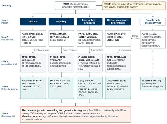
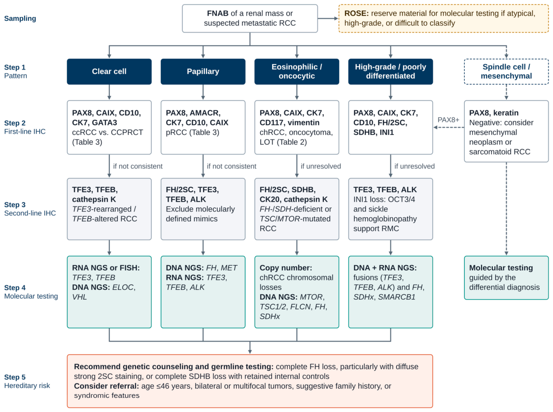
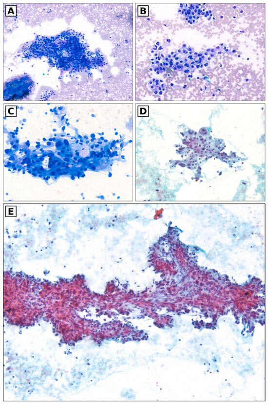
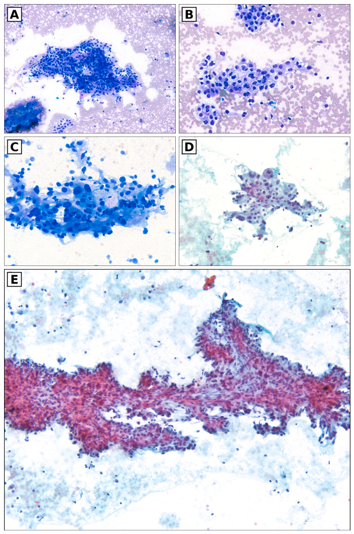
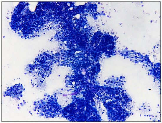
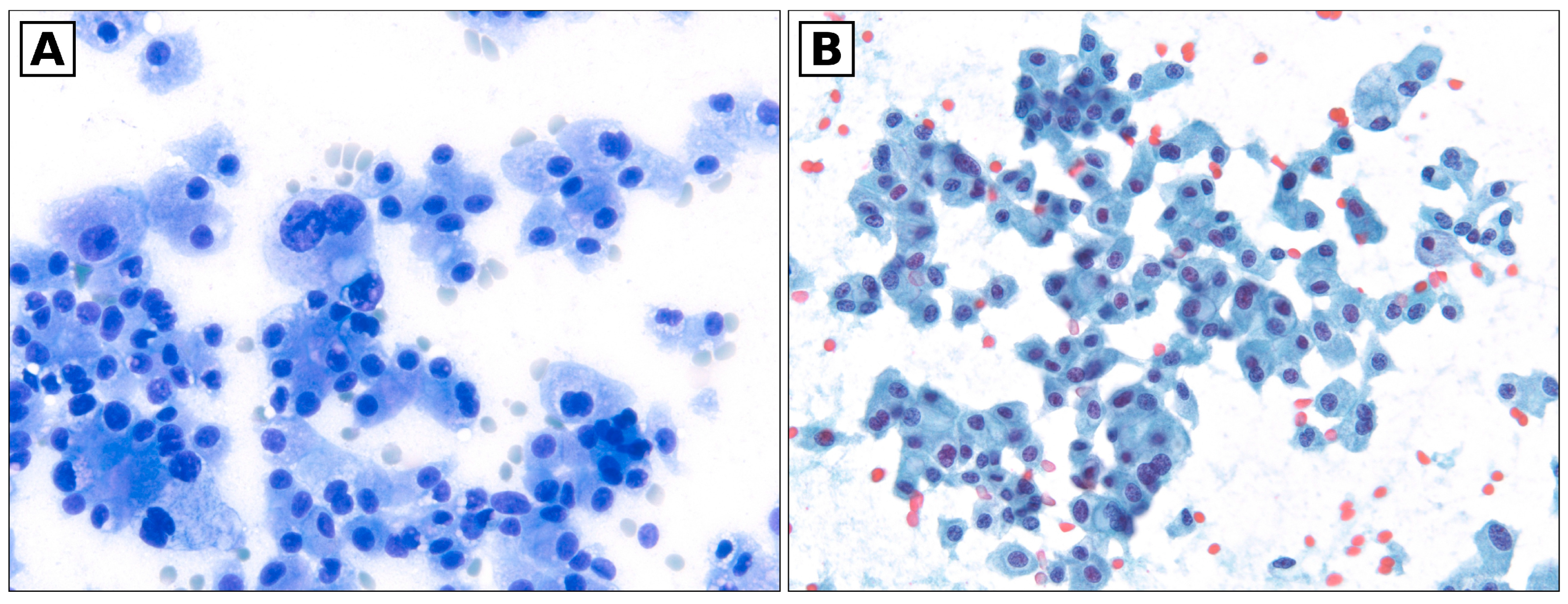
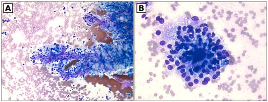
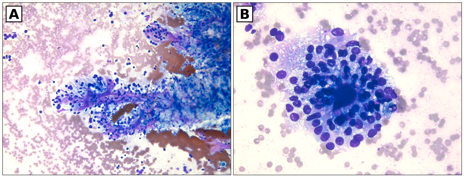
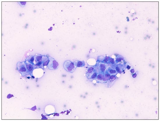
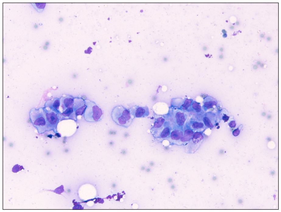
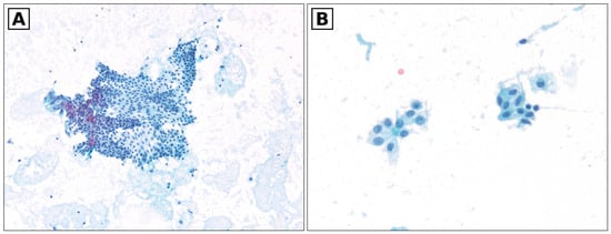
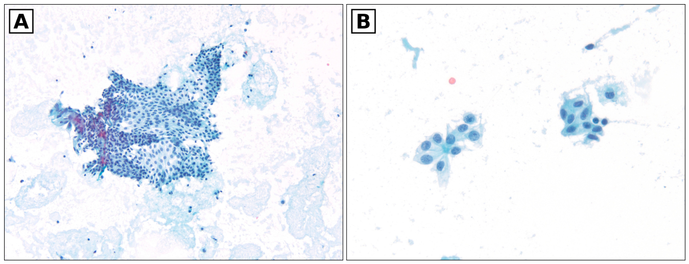

Disclaimer/Publisher’s Note: The statements, opinions and data contained in all publications are solely those of the individual author(s) and contributor(s) and not of MDPI and/or the editor(s). MDPI and/or the editor(s) disclaim responsibility for any injury to people or property resulting from any ideas, methods, instructions or products referred to in the content. |
© 2026 by the authors. Licensee MDPI, Basel, Switzerland. This article is an open access article distributed under the terms and conditions of the Creative Commons Attribution (CC BY) license.