


Abstract
Dengue virus (DENV) is classically regarded as a non-neurotropic systemic pathogen, yet severe neurological complications, most notably meningoencephalitis, are increasingly recognized. This review synthesizes the available evidence on the molecular determinants of DENV neuroinvasion and central nervous system (CNS) injury, indicating which steps are established and which remain hypothetical. Crossing of the blood–brain barrier is thought to involve secreted non-structural protein 1 (NS1), which degrades the endothelial glycocalyx, and together with matrix metalloproteinase-9 impairs endothelial junctions (the role of TLR4 is disputed), as well as monocyte-mediated trans-endothelial migration. Disease severity may be shaped by antibody-dependent enhancement (ADE) during secondary heterologous infection through extrinsic (enhanced viral entry) and intrinsic (suppressed antiviral signaling) mechanisms documented in peripheral myeloid cells and proposed here, by extrapolation, to operate in microglia, possibly counterbalanced by natural killer cell-mediated cytotoxicity. Elevated cerebrospinal fluid IL-6 and TNF-α indicate neuroinflammation; by extrapolation from other flaviviruses, microglial activation and impaired astrocytic glutamate clearance may converge on NMDA receptor-mediated excitotoxicity and neuronal apoptosis, a step not directly examined in dengue. As much of this model rests on peripheral, in vitro, animal, or other-flavivirus data, CNS-focused mechanistic studies are needed.
1. Introduction
Dengue has been recognized as a distinct clinical entity for centuries, historically described under names such as “break-bone fever”, “kidinga pepo”, and “dandy fever”, reflecting the severe myalgia and postural stiffness it produces [1]. The global spread of dengue has been closely tied to the expansion of maritime trade, which facilitated the dissemination of Aedes mosquitoes along shipping routes [1,2]. Its viral etiology, however, was established only in the 1940s, when the causative agent was independently isolated in Japan and Hawaii, formally establishing DENV within the genus Orthoflavivirus and enabling recognition, by the mid-20th century, of the four antigenically distinct serotypes that define its immunopathogenesis [1]. Today, the World Health Organization reports a dramatic rise in dengue incidence in recent decades, with about half of the global population now at risk and an estimated 100–400 million infections each year in more than 100 endemic countries [1,3]; a modeling estimate puts the number of infections at 390 million per year, of which 96 million are clinically apparent [4]. Dengue is the second most common mosquito-borne disease affecting human beings after malaria [5].
Dengue virus (DENV) (species name: Orthoflavivirus dengue) is a single-stranded, positive-sense RNA virus of the family Flaviviridae, genus Orthoflavivirus [6,7]. DENV circulates as four antigenically distinct serotypes (DENV-1 to DENV-4) that confer only partial and transient cross-protection against one another, a feature with important pathogenic consequences that are central to the biology of dengue infection [8,9]. A first DENV infection confers lifelong immunity against that same serotype. In contrast, a subsequent infection with one of the other three serotypes can paradoxically result in more severe illness, driven by antibody-dependent enhancement (ADE) [10]. The virus is transmitted to humans through the bite of infected Aedes mosquitoes—chiefly Aedes aegypti and Aedes albopictus. In the laboratory, DENV is generally handled at Biosafety Level 2 (BSL-2) in the United States, although biosafety levels vary across countries [11]. DENV shares this arthropod-borne transmission strategy with several other flaviviruses of major clinical importance, including Zika virus, Japanese encephalitis virus (JEV), West Nile virus (WNV), yellow fever virus, and the less common but still consequential Murray Valley, St. Louis, and tick-borne encephalitis (TBEV) viruses [12]. This shared ancestry also underlies the serological cross-reactivity these viruses can display in diagnostic assays [13,14].
Historically confined to tropical and subtropical regions across Southeast Asia, the Western Pacific, and Latin America, dengue transmission has expanded considerably in recent decades; dengue is now documented in at least 128 countries [15]. Its expansion has been attributed to urbanization and climate change [16]. Human mobility contributes to introductions into new areas [13]. Autochthonous outbreaks are now increasingly reported outside traditional endemic areas, including southern Europe, where local transmission had been essentially absent for more than 80 years before re-emerging over the past decade [13].
Despite the licensure of two vaccines, CYD–TDV and TAK-003, neither confers uniform protection against all four serotypes, as efficacy varies by serotype [9]. No universally protective vaccine or specific antiviral therapy is yet available [16]. Clinically, DENV infection ranges from a self-limiting febrile illness to severe, life-threatening disease, classified by the WHO into dengue without warning signs, dengue with warning signs, and severe dengue [17]. Among the less common but most serious manifestations are neurological complications, including encephalopathy (the most frequently reported), encephalitis, myelitis, myositis, Guillain-Barré syndrome, and mononeuropathies [18]. Encephalitis and meningoencephalitis are among the most severe [9].
Although increasingly reported, dengue meningoencephalitis remains an underrecognized and incompletely characterized complication, whose precise molecular pathogenesis has yet to be fully elucidated and for which therapeutic options remain limited once neurological disease is established [9]. Direct evidence has challenged the earlier view of DENV as a strictly non-neurotropic pathogen: postmortem brain tissue from fatal dengue encephalopathy cases has stained positive for viral antigen by immunohistochemistry, while cerebrospinal fluid from patients with dengue encephalitis has tested positive for both viral RNA by PCR, and anti-DENV IgM antibodies [5]. Previous reviews have addressed dengue neuroinvasion and neurovirulence [19], the infection of blood–brain barrier cells [20], the routes of flaviviral entry into the central nervous system (CNS) [21], and the mechanisms of neurologic damage across mosquito-borne flaviviruses, mainly in the post-acute phase [22]; the clinical and neuroimaging aspects of dengue encephalitis have recently been updated [9], and the neurological spectrum of dengue (“neurodengue”) has been summarized in a narrative review [23]. Individual components of the pathogenic process—NS1-driven vascular dysfunction, antibody-dependent enhancement, microglial neuroinflammation, and glutamate-mediated excitotoxicity—have each been characterized to varying degrees, largely in systemic severe dengue or in vitro and animal models. However, an account of how they interact within the CNS, from disruption of the blood–brain barrier to neuronal injury, and of the strength of the evidence supporting each step, has been lacking. The aim of this review is therefore to synthesize current biomolecular evidence on the pathogenesis of DENV-associated meningoencephalitis, linking blood–brain barrier dysfunction, viral and host responses, neuroinflammation, and neuronal injury, and distinguishing established steps from working hypotheses.
2. Virus Genome, Structure, and Serotypes
2.1. Genome Organization and Viral Proteins
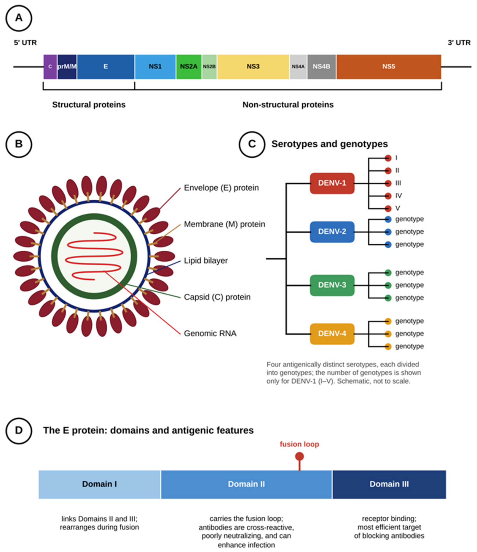
DENV is a small (~50 nm) [24,25], enveloped, single-stranded positive-sense RNA virus with a genome of roughly 11 kb, encoding a single open reading frame, flanked by untranslated regions (UTRs), that is translated into one long polyprotein [6,16]. This polyprotein is cleaved into three structural proteins—capsid (C), membrane (M, from its precursor prM), and envelope (E)—and seven non-structural proteins (NS1, NS2A, NS2B, NS3, NS4A, NS4B, and NS5) (Figure 1A), several of which—notably NS1, NS4, and NS5—together with overall genome variation, influence dengue severity by shaping immune evasion, viral replication, and disease progression [14]. Unlike the other non-structural proteins, NS1 is not confined to the intracellular replication complex: it is also secreted from infected cells into the bloodstream, where it acts on host tissues independently of ongoing viral replication. The E protein, organized into three functionally distinct domains (I, II, and III), plays a central role in host range, tropism, and virulence. Rather than encoding protein, the flanking UTRs contain complementary sequences at the two ends of the genome that base-pair to circularize it, a conformation shown to be required for viral RNA replication [26]; the 5′ and 3′ UTRs bind each other with high affinity and adopt distinct conformations [27].
2.2. Virion Structure
Structurally, the mature virion is an icosahedral particle built from three concentric layers [24,25]: a nucleocapsid core, in which the genomic RNA is complexed with the capsid (C) protein [28]; a lipid envelope derived from the host endoplasmic reticulum membrane [28]; and an outer protein shell of 180 copies each of the envelope (E) and membrane (M) proteins [29], in which E mediates attachment to the host cell [28] and pH-triggered membrane fusion [25] (Figure 1B).
Figure 1.
Genome organization and structure of the dengue virus (DENV). (A) The DENV genome is a single open reading frame flanked by 5′ and 3′ untranslated regions (UTRs), encoding a polyprotein that is cleaved into three structural proteins (C, capsid; prM/M, precursor membrane/membrane; E, envelope) and seven non-structural proteins (NS1, NS2A, NS2B, NS3, NS4A, NS4B, NS5). (B) The mature virion is a spherical particle: an inner nucleocapsid, formed by the capsid protein bound to the genomic RNA, is surrounded by a host-derived lipid membrane studded with M protein and covered by a shell of E-protein dimers. (C) Four antigenically distinct serotypes of DENV (DENV-1 to DENV-4) are distinguished by sequence differences in the E protein, and each serotype is further divided into genotypes; the five genotypes (I–V) of DENV-1 are shown. (D) Domains of the E protein: Domain I links Domains II and III, Domain II carries the fusion loop, and Domain III mediates receptor binding (Section 2.3). All panels are schematic and not to scale. Original schematic drawn by the authors, based on [24,25,30].
2.3. Serotypes, Genotypes, E-Protein Domains, and Neurovirulence
Amino acid sequence comparison of the E protein—the outer shell’s principal antigenic component—distinguishes four antigenically related but distinct serotypes, DENV-1 through DENV-4 [12]. Phylogenetic analysis of the E gene further resolves each serotype into several genotypes (Figure 1C) [31,32]: for example, five genotypes (I–V) within DENV-1, differing by up to about 6% in nucleotide sequence [32], and several genotypes within DENV-2 [31]. Genotype and lineage replacement has been documented over time and across regions [33]. The diversity of the four serotypes is estimated to have arisen within the last century, in step with human population growth, urbanization, and transport [34]. Genotype-level variation—rather than serotype identity alone—may influence the clinical expression of infection, although this is difficult to establish from the available data [35].
Beyond this sequence diversity, the E protein itself is organized into functionally distinct domains (Figure 1D). Domain III, exposed on the protein’s outer surface, is implicated in receptor binding: anti-domain III antibodies are the most efficient blockers of virus adsorption to Vero cells in vitro [36]. Domain II carries the fusion loop, the structural element responsible for merging the viral and host-cell membranes after receptor binding [37,38]. Domain I links Domains II and III, and the rearrangement of E from the prefusion dimer to the postfusion trimer drives membrane fusion [38]. Antibodies directed against the fusion-loop epitope are cross-reactive but generally neutralize poorly and can mediate potent enhancement [30].
Whether such genetic and structural differences translate into differences in neurological outcome is still unclear. Neurological dengue has been reported with all four serotypes [39]. Severe neurological presentations, however, cluster most consistently with DENV-2 and DENV-3 [9] (Table 1). In a Cambodian pediatric cohort of 1160 children with acute meningoencephalitis, DENV was identified in 4.6% of cases, a minority compared with Japanese encephalitis virus (24.4%) [40]. This serotype-linked pattern is consistent with the broader clinical picture: in a single Brazilian cross-sectional study of 485 confirmed cases, which included few DENV-2 and DENV-3 cases, DENV-2 infection was associated with an approximately sevenfold higher risk of severe disease compared with the other serotypes—across dengue severity generally, not limited to neurological presentations [41]. Beyond this serotype-level picture, molecular correlates of neurovirulence are beginning to emerge at the level of individual strains: a distinct NS2A substitution has been identified in a CSF (cerebrospinal fluid)-derived DENV-3 genotype-III strain that grew better in human neuronal cell lines [42], and specific envelope and NS3 substitutions have been associated with a neurovirulent DENV-1 phenotype in mice—suggesting that strain-specific mutations may contribute to neurovirulence, although these are isolated observations [39].
Table 1.
Serotype- and strain-specific determinants of DENV neurovirulence.
3. Epidemiology and Transmission
3.1. Global Burden and Geographic Spread
Dengue transmission is hyperendemic across Southeast Asia, the Western Pacific, and Latin America, with urbanization and international travel facilitating its spread [43]; in the Americas, reported cases rose from about 1.0 million in the 1980s to 4.8 million in 2000–2007 [44]. A recent study estimates that 5.6 billion people are at risk of infection with dengue and other arboviruses [3,13]. This burden has risen markedly: WHO case counts grew roughly tenfold between 2000 and 2019, from about 500,000 to 5.2 million annually [39]. Reported cases reached 14.6 million, with more than 12,000 deaths, in 2024 [3]. An independent analysis of WHO surveillance data also described 2024 as a record year, with about 14 million cases worldwide [45]. Asia carries the largest share, approximately 70% of global cases [9]. In 2024, however, more than 90% of the cases reported to WHO came from the region of the Americas, with Brazil alone reporting over 10 million cases and 6321 deaths [46]. In the Americas, about 4.1 million cases were reported in 2023, the highest figure in the region’s history and approximately 80% of the cases reported worldwide that year [39], and WHO recorded more than 13 million cases in the region in 2024 [3]. From January to July 2025, over 4 million cases and more than 3000 deaths were reported to the WHO from 97 countries worldwide [3]. All four serotypes still co-circulate in the Americas, though unevenly across countries, a heterogeneity that shapes local rates of secondary heterologous infection [2]. These totals likely understate true transmission, given widespread asymptomatic infection and non-mandatory reporting in several endemic countries [39]. Global environmental suitability for dengue in 2015 is mapped in Figure 2 [47].
Figure 2.
Environmental suitability for dengue in 2015. Maps of the modeled probability of dengue occurrence, ranging from 0 (grey, unsuitable environment) to 1 (red, suitable environment). (a) Global map; (b) North America, Central America, and the Caribbean; (c) North Africa, the Middle East, and Europe. The maps show environmental suitability, not reported cases or documented transmission. Reproduced from Messina et al. [47], published under a Creative Commons Attribution 4.0 International (CC BY 4.0) license (https://creativecommons.org/licenses/by/4.0/); the panels were cropped and the color saturation was increased by the authors.
3.2. Autochthonous Transmission in Europe
Climate models project a poleward expansion of climatic suitability for Aedes-borne virus transmission, including most of Europe [48]. In the 2015 map, environmental suitability was low across most of Europe, with higher values confined to limited Mediterranean coastal areas (Figure 2c); such estimates may nevertheless understate future risk if warming continues, since summer 2026 was the warmest on record for western Europe, surpassing the previous record set in 2003 [49]. Autochthonous outbreaks are now documented in southern Europe—Italy, France, Spain, Croatia, and Portugal (Madeira) [13]. This resurgence is recent but not unprecedented: the last European epidemic occurred in 1927, and all diagnosed cases were imported until autochthonous cases were reported in France (two cases) and Croatia (15 cases) in 2010 [13]. In 2024, 308 cases were reported to the WHO from three European countries (France, Italy and Spain) [3]. Aedes albopictus (the “tiger mosquito”) is now the principal European vector, while the more efficient Ae. aegypti remains largely confined to outermost European Union (EU) regions such as Madeira and Cyprus; local transmission requires the co-occurrence of a competent vector, viremic travelers, and permissive climate, typically May–October/November in southern Europe [13].
Madeira illustrates this climatic dependence: the 2012 autochthonous DENV-1 outbreak was attributed to a warmer-than-usual summer that shortened the extrinsic incubation period (EIP), and transmission collapsed when autumn cooling pushed the EIP beyond the lifespan of Ae. aegypti; the risk persists whenever vector, travelers, and temperature coincide, as shown by the detection of DENV-2 in Ae. aegypti collected in Funchal in January 2025 [13].
In 2024, DENV-2 caused multiple transmission foci in four Italian regions, with 296 locally acquired cases, the largest number of autochthonous cases ever recorded in mainland Europe [50]. Autochthonous cases continued to be notified in Italy in 2025 and 2026 [51], and repeated autochthonous transmission has been detected in Spain [52]. Comparable patterns of Aedes albopictus establishment and recurrent autochthonous transmission have also been documented in France, Germany, and Portugal, for which a detailed country-by-country account is provided by Bononad-Brugger et al. [13].
3.3. Dengue in Pakistan
Pakistan illustrates a markedly different trajectory: dengue there is a long-established burden rather than a recent, climate-driven emergence, with peak case years in 2010, 2011, and 2013 [53]. National surveillance shows marked year-to-year volatility, and the 2022 floods were followed by a marked surge in dengue cases [54]. All four serotypes have been recorded nationally [53]. DENV-3 is currently the most prevalent [55]; DENV-4 is notably present alongside the others, unlike in other countries of the wider Middle East/Red Sea region [2]. Targeted larval surveillance and control have reduced dengue cases locally [56,57].
3.4. Serotype Co-Circulation and Disease Severity
Virulence is known to vary among DENV genotypes [58], although for neurological disease, the evidence is limited (see Table 1). Co-circulation of multiple serotypes in hyperendemic regions increases the incidence of secondary heterologous infection, a recognized risk factor for severe dengue [59]; consistently, in CYD–TDV trials, dengue-seronegative vaccine recipients had a higher risk of hospitalization for dengue than seronegative controls (hazard ratio [HR] 1.75, ages 2–16 years), whereas seropositive recipients were protected [60].
3.5. Transmission Cycles and Incubation Periods
DENV is maintained through two transmission cycles: an ancestral sylvatic cycle between non-human primates and forest-dwelling Aedes species, thought to be the virus’s evolutionary origin in Asian and African forests [5]. The urban (or epidemic) cycle now dominates global transmission, sustained by human-to-human spread via the anthropophilic vectors Ae. aegypti and Ae. albopictus [16] (Figure 3). Spillover from the sylvatic cycle to humans, i.e., the occasional transmission of the virus from non-human primates to people through the bite of an infected forest-dwelling mosquito, can seed new transmission chains in the human population. In laboratory experiments with Aedes aegypti, DENV-2 was transmitted vertically (transovarially) to a fraction of the offspring, and infected males transmitted the virus venereally to females, potentially providing an additional route for viral maintenance between epidemic seasons [61].
Figure 3.
Schematic representation of the DENV human–mosquito transmission cycle. The extrinsic incubation period (EIP) is the time required, following an infective blood meal, for DENV to replicate within the mosquito and disseminate to its salivary glands, rendering the vector infectious to the next person it bites; the intrinsic incubation period (IIP) is the corresponding interval in the human host, from the infective mosquito bite to the onset of clinical symptoms. Dengue is not transmitted through casual person-to-person contact; mosquito-borne transmission accounts for the large majority of cases, while rare non-vector routes (vertical, and less commonly, sexual transmission) are discussed in the text. The upper panel shows the sylvatic cycle between non-human primates and forest-dwelling Aedes mosquitoes. The blue arrow indicates spillover, i.e., the occasional transmission of the virus from this sylvatic cycle to humans (through the bite of an infected forest mosquito), which can then seed the human (epidemic) cycle shown below. Red boxes denote infected hosts and infectious mosquitoes (the mosquito becomes infected by feeding on a viremic host); the white box denotes an uninfected, susceptible person. An infected mosquito can continue transmitting the virus for the remainder of its life. Original schematic drawn by the authors based on information from [5,16,30,62].
Following a viremic blood meal, the virus crosses the mosquito midgut barrier, infects midgut epithelial cells, and disseminates via the hemolymph to the salivary glands [62]. This process defines the extrinsic incubation period (EIP, typically 8–10 days [2]; about 8–12 days at 25–28 °C according to WHO [3]). Once this barrier is crossed, the mosquito remains infectious for the remainder of its life [2]. In the human host, infection first targets dendritic (Langerhans) cells and macrophages in the skin; the intrinsic incubation period (IIP) preceding symptom onset averages 4–7 days [2] (4–10 days according to the WHO [3]) (Figure 3).
Vector-borne transmission via Aedes mosquitoes accounts for the large majority of DENV spread; infected individuals are not directly contagious to close contacts in the absence of a mosquito vector [63]. Non-vector routes exist and are relevant to this review’s CNS-focused scope since they define additional populations at risk of early-life or occult exposure: DENV can cross the placenta, has been detected in breast milk, and limited evidence suggests possible sexual transmission [63].
3.6. Drivers of Expansion
The recent geographic expansion of dengue reflects several converging drivers. Rising temperatures shorten the EIP and accelerate mosquito reproduction, while altered rainfall—including extreme events such as the 2023 El Niño—creates additional larval breeding sites, directly increasing vector populations and their transmission potential [9]. These climatic pressures compound with rapid, often unplanned urbanization, which increases human-vector contact and generates the water-storage practices that favor Aedes breeding [1]. Non-climatic factors add further pressure: healthcare systems weakened by the COVID-19 pandemic have struggled to sustain vector-control and surveillance programs in several endemic regions, while political instability, economic crisis, and population displacement further erode containment capacity [9]. Underlying all of this is the adaptability of Ae. aegypti and Ae. albopictus themselves: both readily exploit small artificial water containers for breeding and insecticide resistance complicates their control [13,16]. The result is a transmission landscape expanding both geographically, into new temperate regions, and intensively, through larger and more frequent outbreaks within already-endemic areas.
4. Clinical Presentation and WHO Classification
4.1. Clinical Course and WHO Classification
Most DENV infections are asymptomatic or present as a self-limiting febrile illness characterized by high fever, retro-orbital headache, and severe myalgia/arthralgia—the classic “break-bone fever”—often accompanied by rash and mild hemorrhagic manifestations [1,64]. Only about one in four infected individuals develops symptomatic disease [63]; the febrile phase presents as an acute illness with two or more of nausea, vomiting, headache, rash, retro-orbital pain, myalgia, arthralgia, petechiae, leukopenia, and a positive tourniquet test [39]. Symptoms generally last 2–7 days [63].
The WHO classification stratifies disease into three categories: dengue without warning signs, dengue with warning signs, and severe dengue [17] (Figure 4). Warning signs—abdominal pain or tenderness, persistent vomiting, clinical fluid accumulation (ascites, pleural or pericardial effusion), mucosal bleeding, lethargy or restlessness, hepatomegaly >2 cm, and a rising hematocrit with concurrent falling platelet count—mark the transition toward the critical phase and warrant closer monitoring [17,39]. Severe dengue is defined by severe plasma leakage causing shock or respiratory distress, severe hemorrhage as clinically assessed, or severe organ involvement (liver AST/ALT ≥ 1000, impaired consciousness, cardiac or other organ dysfunction) [17,39].
Figure 4.
WHO clinical classification of dengue, showing the progression from disease without warning signs to severe dengue. Arrows indicate progression between categories; box colors (blue, orange, red) denote increasing severity. * Warning signs require strict observation and medical intervention. AST, aspartate aminotransferase; ALT, alanine aminotransferase; CNS, central nervous system. Original schematic drawn by the authors, based on the WHO classification [17].
Defervescence, occurring 3–7 days into illness, marks either recovery or the onset of the critical phase, during which clinically significant plasma leakage typically lasts 24–48 h; progressive leukopenia followed by a rapid platelet decline often, though not always, precedes this leakage [39]. Untreated, hypoperfusion from prolonged shock can produce hemorrhage, metabolic acidosis, and disseminated intravascular coagulation, and severe hepatitis, encephalitis, and myocarditis may occur with or without overt shock—consistent with the 2009 WHO classification, which counts severe organ involvement, including neurological involvement, among the criteria of severe dengue [17,39]. Case fatality in untreated severe dengue can exceed 20% but falls below 1% with appropriate management [17,65], and it still varies by setting depending on health-system capacity for early recognition.
Infection confers durable homotypic immunity to the infecting serotype, but not reliable cross-protection against the other three, allowing repeat infections over a lifetime [63].
4.2. Neurological Complications of Dengue
Neurological complications, though comparatively uncommon, represent some of the most severe manifestations of DENV infection. Their exact incidence is uncertain [66]: encephalopathy and encephalitis have been reported in about 0.5–6.2% of cases [5], whereas CNS involvement has been reported in 4–13% of cases, with marked geographic variation (~20% in Thailand, 15% in India, 5–6% in Vietnam) [9]. Risk is further shaped by host factors, including age extremes, comorbidities such as diabetes and obesity, and genetic polymorphisms in human leukocyte antigen (HLA) and cytokine genes (TNF-α, IL-10) [9]. Beyond meningoencephalitis, the neurological spectrum includes cranial nerve palsies, ischemic and hemorrhagic stroke, retinal and choroidal vascular involvement, and posterior reversible encephalopathy syndrome, in addition to the post-infectious, immune-mediated syndromes described below [5]. A recent systematic review and meta-analysis has specifically addressed intracerebral hemorrhage in patients with dengue [67]. This spectrum is commonly grouped into three categories based on pathogenesis [39,68]:
- Direct CNS invasion, confirmed by viral RNA or antigen in the CSF—encephalitis, meningitis, myelitis [69].
- Post-infectious syndromes, presumed to be immune-mediated—acute disseminated encephalomyelitis (ADEM), Guillain-Barré syndrome, transverse myelitis [5,70,71]; molecular mimicry has been proposed but not demonstrated.
- Systemic dengue encephalopathy—indirect CNS dysfunction secondary to hepatic failure, metabolic acidosis, or hemorrhagic shock, without direct neuroinvasion [39].
Neurological dengue is not a single entity. In a systematic review of 162 published cases, dengue-associated acute necrotizing encephalopathy (DANE) accounted for 38.3% of cases, acute encephalitis for 34.6%, ADEM for 16.7%, and other syndromes for 10.5%; DANE was characterized by bilateral thalamic lesions on imaging in all cases, and overall mortality was 11.7% [72]. Because these data derive from published case reports, which are biased toward severe and radiologically abnormal cases, they do not estimate true incidence or case fatality. In a prospective series of 17 patients with neurological dengue, encephalopathy carried the worst outcome (3 out of 11 patients died), whereas all 6 patients with motor weakness recovered [73]. DENV-associated meningoencephalitis is thought to combine direct viral injury with an aberrant host immune response [5]. The molecular mechanisms proposed to underlie this dual injury—from viral neuroinvasion to the immune dysregulation that may drive severe neurological disease—are examined in the remainder of this review.
5. Molecular Pathogenesis of Dengue Meningoencephalitis
Having established the clinical and nosological context, this section addresses the central aim of this review: the biomolecular cascade through which DENV crosses the blood–brain barrier, subverts resident immune cells, and ultimately drives neuronal death in meningoencephalitis. This cascade is the exception rather than the rule. DENV belongs to the same genus as several classically encephalitic flaviviruses—West Nile virus (WNV), Japanese encephalitis virus (JEV), and tick-borne encephalitis virus (TBEV) among them—which reach the central nervous system via defined, if still incompletely characterized, hematogenous and transneural routes; DENV itself, however, is classified primarily as a systemic, hemorrhagic pathogen and is only occasionally associated with neurological disease [39,74]. Following the infective mosquito bite, most infections therefore remain confined to the periphery, and only in a small minority of patients does the virus go on to invade the central nervous system. The subsections that follow trace, step by step, precisely this minority neuroinvasive pathway—from the initial breach of the blood–brain barrier to the neuronal death that defines meningoencephalitis—rather than the far more common outcome of contained systemic infection. Because the global dengue burden is large and still expanding (Section 3), even a small neuroinvasive fraction is likely to correspond to a growing absolute number of cases, which is why the mechanisms of this minority pathway deserve attention.
5.1. Innate Antiviral Sensing and Its Viral Subversion
Before considering CNS-specific mechanisms, it is useful to establish how DENV is normally sensed by the innate immune system. This baseline proves directly relevant, since antibody-dependent enhancement (ADE)—a mechanism this review considers a possible contributor to meningoencephalitis severity—dismantles this same pathway in infected myeloid cells. Viral RNA generated during replication is recognized as a pathogen-associated molecular pattern (PAMP) by cytoplasmic pattern recognition receptors, such as retinoic acid-inducible gene I (RIG-I) and melanoma differentiation-associated protein 5 (MDA5), and by endosomal Toll-like receptors (TLR) such as TLR3. In cultured fibroblasts, RIG-I and MDA5 were each individually dispensable for dengue-triggered IRF-3 signaling, which required the adaptor interferon-β promoter stimulator 1 (IPS-1) [75]; in hepatoma cells and macrophages, RIG-I, MDA5, and TLR3 were each required for effective interferon production and the restriction of DENV [76]; these data do not come from neural cells. Downstream, interferon regulatory factors 3 and 7 (IRF-3/IRF-7) drive type I interferon (IFN-I) production; in mice, IRF-3 and IRF-7 act redundantly, with IRF-7 more important for the initial IFN-α/β response, and an IRF-3/IRF-7-independent late pathway contributes to the control of DENV [77]. Sensor signaling also activates nuclear factor kappa-light-chain-enhancer of activated B cells (NF-κB), inducing pro-inflammatory cytokine transcription.
It is worth emphasizing what this interferon-signaling cascade ultimately achieves: secreted IFN-I reaches infected and neighboring cells via the interferon-α/β receptor (IFNAR), activates the Janus kinase/signal transducer and activator of transcription (JAK-STAT) pathway, and switches on a broad panel of antiviral genes [14]. This places the cell on alert and mobilizes natural killer (NK) cells for action [78]. However, DENV subverts this pathway at its source: NS2A and NS4B inhibit the phosphorylation of TANK-binding kinase 1 (TBK1) and IRF3 and the induction of IFN-β in cell-based assays, and DENV-1 NS4A has a similar effect [79], whereas the NS2B/3 protease interacts with IκB kinase ε and blocks IRF3 phosphorylation and nuclear translocation in human cells [80]; these findings come from cultured cells. This attenuated interferon response may contribute to a permissive stage for the CNS-specific mechanisms detailed below: blood–brain barrier (BBB) breakdown, ADE in Fcγ receptor-bearing myeloid cells (proposed here to include resident microglia), and the counter-regulatory role of NK cells, including DENV’s HLA-mediated evasion of NK surveillance.
5.2. Neuroinvasion and Blood–Brain Barrier (BBB) Breakdown
Several routes of CNS entry have been hypothesized for flaviviruses, including disruption of the BBB, passage across it without disruption, and axonal transport [81]. The BBB is composed of brain microvascular endothelial cells (BMECs), pericytes, and astrocytic end-feet; whether its destabilization is required for DENV entry, and whether circulating viral factors or endothelial activation drive it, is proposed but has not been demonstrated in humans [82]. For West Nile virus, the evidence that barrier disruption is required for neuroinvasion has itself been critically appraised and found to be less secure than often assumed [82]. Pericytes wrap the abluminal surface of brain capillaries and regulate endothelial tight-junction expression, but their role in dengue is unknown.
Secreted hexameric NS1 (sNS1) is the best-studied viral candidate for disruption of the endothelial barrier, but the evidence is experimental and partly contested. In human pulmonary microvascular endothelial cells, recombinant DENV NS1 degrades the endothelial glycocalyx—sialic acid is lost and heparan sulfate proteoglycans are shed—through sialidases and heparanase, with cathepsin L activating heparanase, and increases permeability; this effect is specific to NS1 from DENV-1–4 and is not seen with West Nile virus NS1 [83]; whether the same occurs in brain endothelium was not shown. Macrophage migration inhibitory factor (MIF) contributes to this glycocalyx degradation and to NS1-induced leakage in mice, and serum heparanase, MMP-9, and syndecan-1 are increased in patients with dengue [84]. Circulating NS1 alone induced vascular leak and cytokine production in mice and increased the permeability of human endothelial monolayers [85]. NS1 was also reported to activate cells through Toll-like receptor 4 (TLR4) and to disrupt endothelial monolayer integrity [86], but this has been challenged: in a recent study, NS1 prepared from several sources did not activate TLR4 and had only a modest effect on endothelial monolayer integrity compared with TNF, and medium from DENV-infected macrophages disrupted the monolayer mainly through TNF [87]. In mouse dermis and human dermal endothelial cells, NS1-induced leak was independent of inflammatory cytokines and of TLR4 or TNF receptor signaling but dependent on glycocalyx components [88], so endothelium-intrinsic and cytokine-mediated routes are not mutually exclusive, and their relative weight in the human blood–brain barrier is unresolved. The NS1–TLR4–MMP-9–BBB axis therefore remains a working hypothesis.
Whether this endothelial action reaches the brain is less certain. In the most direct comparison available, NS1 from DENV, ZIKV, WNV, and JEV each increased the permeability of cultured human brain microvascular endothelial cells; DENV NS1 acted on endothelial cells from all five tissues tested, with the strongest effect on pulmonary cells, whereas WNV and JEV NS1 acted only on brain endothelium, consistent with the systemic versus neurotropic character of the corresponding diseases [89]. The effect required binding to the endothelial cell surface and was accompanied by the loss of sialic acid and heparan sulfate; in mice, intravenous DENV, ZIKV, or WNV NS1 increased brain accumulation of a fluorescent dextran tracer [89]; these mouse data derive from recombinant NS1 injected intravenously (10 mg/kg), not from infection, and do not show passage of the virus. These data indicate that DENV NS1 can act on brain endothelium in experimental systems. However, they come from recombinant NS1 applied to single-cell-type monolayers lacking astrocytes and pericytes and from small groups of mice, a dextran tracer was used for the brain readout, and the authors themselves present the implications for pathogenesis as hypotheses; they do not show that NS1 opens the blood–brain barrier during human dengue.
Proteolytic injury to junctional proteins provides a second, partly independent mechanism. NS1 binds MMP-9 directly, and together, they degrade β-catenin, ZO-1, ZO-2, and N-cadherin in endothelial cells and lower transendothelial resistance; NS1-induced leakage in lung, spleen, and liver was absent in MMP-9-deficient mice [90]. More recently, flavivirus NS1 was found to trigger tissue-specific disassembly of intercellular junctions, with barrier dysfunction and vascular leak depending on glycogen synthase kinase-3β (GSK-3β) [91]. MMP-9 in this setting appears to derive mainly from leukocytes rather than from endothelial cells: NS1 induced MMP-9 release from leukocytes in a MIF-dependent manner but not from endothelial cells [84,90]. Neither study examined the brain. In mice, DENV-2 infection increases BBB permeability to tracers, even after intraperitoneal inoculation when no virus is detected in the brain, through a virus-induced circulating cytokine acting via histamine [92]. In a mouse model of West Nile virus infection, MMP-9 deficiency preserved BBB integrity and protected against lethal infection, and MMP-9 was elevated in the CSF of patients with West Nile encephalitis [93]—evidence from another flavivirus that can only be extrapolated to dengue. MMP-2 and MMP-9 are increased in patients with dengue in correlation with disease severity, and immune cells and their mediators are thought to contribute to MMP-mediated plasma leakage, although the underlying mechanism is not fully understood [94]. A direct effect of NS1 and MMP-9 on the human blood–brain barrier, however, has not been demonstrated. Activated microglia and infiltrating monocytes may contribute further proteolytic enzymes once infection is established within the parenchyma; barrier breakdown is therefore best understood as a process that may be initiated peripherally and subsequently reinforced by the neuroinflammatory processes it enables, rather than completed in a single step [92]. Three non-exclusive routes of CNS entry have been proposed, whose relative contribution in human disease is unknown: (i) paracellular leakage, in which free virions extravasate through inter-endothelial gaps—in a mouse brain endothelial monolayer, DENV infection displaced ZO-1 and claudin-1, lowered transendothelial electrical resistance, and increased the paracellular passage of free virus [95]; (ii) endothelial infection with possible transcytosis, in which virions could transit the cytoplasm of infected BMECs without immediate lysis—shown in vitro in mouse brain endothelial cells [95] and in neonatal mice infected with a neuroadapted DENV-4 [96], whereas transcytosis across brain endothelium has not been demonstrated in humans [19,20], and in 13 autopsies of dengue hemorrhagic fever, no viral antigen was detected in endothelial cells [97]; in primary human endothelial cells of non-cerebral origin, productive infection has been reported, directed by heparan sulfate proteoglycan receptors [98]; and (iii) monocyte-mediated (“Trojan horse”) transmigration, in which infected monocytes/macrophages are proposed to undergo diapedesis across the weakened neurovascular unit [21], consistent with parenchymal infection: DENV replication was shown in neurons, microglia, and endothelial cells in brain tissue from three fatal cases, although the route of entry cannot be inferred from these findings [99]. In vitro, DENV infection of mouse brain endothelial cells upregulated vascular cell adhesion molecule-1 (VCAM-1), platelet endothelial cell adhesion molecule-1 (PECAM-1), monocyte chemoattractant protein-1 (MCP-1), and TNF-α transcripts [95]; whether such signals recruit circulating monocytes and convert a passive breach into a self-sustaining portal of entry is a working hypothesis. Host factors may also restrict infection at this interface: in human brain microvascular endothelial cells and in suckling mice challenged intracranially, vimentin knockout increased the DENV-2 viral load and the severity of disease [100]; because the mice were inoculated intracranially, this model does not test passage across the barrier itself.
The NS1–glycocalyx–MMP axis and monocyte diapedesis described above may operate alongside a broader repertoire of vasoactive mediators whose relative contributions remain incompletely resolved. Mast cell proteases—chymase and tryptase—together with inflammatory lipid mediators including platelet-activating factor, leukotrienes, prostaglandins, and phospholipase A2 products, are consistently elevated in patients who develop plasma leakage. Angiopoietin-2 and vascular endothelial growth factor amplify this response by stimulating phospholipase activity and platelet-activating factor generation in a self-reinforcing loop. Activated platelets contribute in parallel, releasing IL-1β and promoting monocyte-derived inflammatory cytokine output, thereby linking coagulation and inflammation at the endothelial surface [5]. A counter-regulatory, barrier-protective role has separately been proposed for sphingosine-1-phosphate signaling, although its weight against these injurious mediators in the CNS-adjacent vasculature specifically remains unestablished [5,65].
At the structural level, this convergence of mediators is thought to culminate in breakdown of the endothelial glycocalyx layer (EGL), the proteoglycan- and glycoprotein-rich coating that normally lines the luminal endothelial surface. NS1 is the best-characterized trigger of EGL degradation [83,84], but its weight relative to the other mediators listed above has not been established in dengue; loss of the glycocalyx exposes the underlying endothelium to disturbed coagulation and increased leukocyte adhesion, compounding the paracellular and transcellular entry routes outlined above [5].
Circulating NS1 has been reported to activate complement pathways [1]. Soluble NS1 has also been reported to interact with the complement regulator clusterin [30]; complement activation is thought to contribute to vascular permeability independently of antibody-dependent enhancement. Antibodies raised against NS1 itself introduce a separate complication: although protective in animal models, the same antibodies have been implicated in severe dengue pathogenesis, since endothelial cells respond to anti-NS1 antibody stimulation by secreting additional cytokines and chemokines that amplify, rather than resolve, local vascular inflammation [14]. Endothelial injury in dengue meningoencephalitis is therefore plausibly the product of several converging insults—glycocalyx and tight-junction breakdown, complement activation, and antibody-driven cytokine release—that together set the stage for downstream excessive cytokine release, rather than the result of a single pathway [1,14].
Receptor usage determines which neural cells can be infected once the virus has reached the parenchyma. Dendritic cell-specific intercellular adhesion molecule-3-grabbing non-integrin (DC-SIGN; CD209) and the other C-type lectin receptors implicated in the infection of peripheral myeloid cells, as well as the T-cell immunoglobulin and mucin domain (TIM) phosphatidylserine receptors, are not expressed by neurons [101]. Heparan sulfate proteoglycans (HSPG) act as attachment factors that concentrate the virus on the cell surface and are thought to facilitate binding to other receptors [101], and members of the TIM and Tyro3, Axl and Mer (TAM) families of phosphatidylserine receptors mediate DENV entry in cell-culture systems [102]; whether these molecules support the infection of neurons or microglia in patients has not been shown. In cultured human neural cells, prohibitin 1/2 was identified as a putative receptor for DENV-3 in neuroblastoma (SH-SY5Y) and microglial (CHME-3) cells, but not in astroglial U-87 MG cells [103]. Fcγ receptor-mediated uptake of antibody-bound virus, documented in peripheral myeloid cells, is the proposed route of ADE-mediated infection of microglia (Section 5.3) and remains an extrapolation. No receptor has been shown to mediate neuronal infection in dengue meningoencephalitis, so the molecular link between viral entry into the parenchyma and the neuronal apoptosis discussed in Section 5.7 remains incomplete.
5.3. Antibody-Dependent Enhancement: Extrinsic and Intrinsic Mechanisms and Their Proposed Extension to Resident Microglia
Antibody-dependent enhancement was first characterized in peripheral monocytes, macrophages, and dendritic cells, and is extended here, by analogy, to resident microglia—the CNS’s own myeloid population, which shares the same Fcγ receptor biology (see the caveats on this extrapolation in Section 5.11). To the best of our knowledge, direct demonstration of FcγRIIa-mediated ADE in human resident microglia during dengue is currently lacking; the mechanisms described in this section are therefore presented as a hypothesis supported by evidence from peripheral myeloid cells, in vitro systems, and patients with systemic disease.
Within the CNS, the outcome of DENV infection is thought to be shaped by the host’s immunological history. During a secondary infection with a heterologous serotype, pre-existing IgG antibodies generated against the primary infecting serotype recognize the new virus through partially conserved envelope epitopes, but bind with insufficient avidity to neutralize it, a mechanism first shown as antibody-enhanced infection of primate leukocytes in vitro [104]. This generates virus–antibody complexes—a process termed opsonization—that remain infectious yet are still recognized, via their Fc region, by FcγRIIa receptors expressed on myeloid cells [105]—and, as proposed here, on resident microglia—conceptually, the antibody functions as a molecular passport that is accepted at a cellular checkpoint without corresponding to a validated, neutralizing identity, so the virus is admitted rather than intercepted.
Rather than being neutral, this admission actively increases infection and disease severity—a phenomenon known as antibody-dependent enhancement (ADE), which follows a distinctive dose–response relationship: low pre-existing antibody titers fail to enhance severity, intermediate sub-neutralizing titers exacerbate it, and high titers instead protect against severe disease, a relationship demonstrated in a long-term pediatric cohort in Nicaragua, in which the risk of severe dengue was highest within a narrow range of pre-existing antibody titers and high titers were protective [106]; whether ADE operates in the CNS has not been shown. ADE itself operates through two complementary mechanisms: (i) extrinsic ADE, in which FcγRIIa engagement triggers receptor-mediated endocytosis of the immune complex, increasing both the proportion of infected cells and the viral output relative to infection without antibody, as shown in primary human monocytes, macrophages and mature dendritic cells (but not in immature dendritic cells) [105]; and (ii) intrinsic ADE, in which entry of the immune complex through Fcγ receptors triggers inhibitory intracellular signaling: DENV–antibody complexes activated negative regulators (dihydroxyacetone kinase and ATG5–ATG12) that disrupt RIG-I/MDA5 signaling and IFN-I production, while interleukin-10 (IL-10) induced suppressor of cytokine signaling 3 (SOCS3), which inactivates JAK-STAT signaling; both loops were found in severe but not in mild secondary dengue [107]. The concept is reviewed in [108], and for viral infections in general in [109].
Intrinsic ADE extends beyond this interferon blockade: in monocytic THP-1 cells, engagement of FcγRI and FcγRIIa downregulated TLR gene expression and upregulated negative regulators of NF-κB signaling, including SARM (sterile alpha and armadillo motif-containing protein) and TANK (TRAF family member-associated NF-κB activator), and the same pattern was seen in peripheral blood mononuclear cells from patients with severe secondary dengue [110]. Co-ligation of the inhibitory leukocyte immunoglobulin-like receptor B1 (LILRB1) has also been implicated in dampening the interferon-stimulated gene response [111]. The intrinsic pathway has been characterized mainly in continuous cell lines and a limited number of primary cells, its contribution relative to extrinsic enhancement has not been directly quantified, and in primary macrophages, enhancement can occur at the level of endosomal fusion without increased viral entry, so cell type matters [111].
The combined effect—increased viral entry coupled with active suppression of the antiviral response—is supported by in vitro studies in peripheral myeloid cells and by epidemiological associations, although its in vivo contribution is hard to quantify; should the same mechanism operate in resident microglia, it could convert these cells from immune sentinels into productive viral reservoirs and contribute to the disproportionate severity of secondary heterologous DENV infection in the CNS, although this remains to be demonstrated [14,107,108]. Not all Fcγ receptors promote this outcome, however: FcγRIIb, an inhibitory receptor sharing overlapping ligand specificity with FcγRIIa, can oppose enhancement when co-engaged in monocytes [112]; whether the balance between activating and inhibitory Fcγ receptors on microglia contributes to variability in ADE susceptibility has not been studied and is a working hypothesis.
The persistence of sub-neutralizing antibody that drives this cycle has been linked to a phenomenon termed “original antigenic sin”: the humoral response to secondary infection is thought to be dominated by memory B cells recalling low-affinity epitopes from the primary serotype, rather than mounting a de novo, high-affinity response against the new strain; the concept remains debated. Antibodies directed against the E-protein fusion-loop epitope and other epitopes shared among serotypes are cross-reactive but poorly neutralizing, and they can mediate potent enhancement [30].
Antibody concentration, however, is not the only determinant of enhancement: the functional quality of the antibody itself—a separate property from how much of it is present—also shapes ADE risk. In acute dengue, higher proportions of afucosylated DENV-specific IgG1—an antibody lacking core fucosylation of the Fc glycan, which increases its affinity for the activating Fc gamma receptor IIIa (FcγRIIIa)—associate with increased disease severity at any given neutralizing-antibody titer, identifying Fc glycosylation as an additional risk factor that acts on top of, rather than in place of, the concentration-dependent mechanism described above [65]. Increased binding to FcγRIIIa is well-documented; an equivalent effect on FcγRIIa, the receptor invoked here for ADE in myeloid cells, and by extrapolation, in microglia, cannot be assumed and has not been shown. The possible contribution of afucosylated IgG1 to ADE-mediated severe dengue has also been discussed in a recent perspective [113].
A structurally distinct route to enhancement runs through the immature virion. Particles that leave the cell before completing maturation still carry uncleaved prM (precursor Membrane protein) rather than fully processed M protein; because prM locks the E protein in a fusion-incompetent conformation, these immature particles are intrinsically poor at infecting cells on their own. However, DENV infection also elicits antibodies directed against prM itself, and when these anti-prM antibodies bind the immature particle, the resulting immune complex can still be recognized and internalized by Fcγ receptors on target cells—rescuing the infectivity that the immature particle otherwise lacks. This constitutes a second ADE pathway, mechanistically parallel to the E-protein/fusion-loop route described above but operating independently of it, since here it is prM, rather than E, that supplies the enhancing epitope on the same immature virion surface [65].
Enhancement-like phenomena have also been suggested after vaccination. In an open-label phase 1 human challenge study, six volunteers who had received a tetravalent inactivated prime followed by a live-attenuated boost and four flavivirus-naïve controls were inoculated with an attenuated DENV-1 strain 27–65 months after the booster dose. Although all vaccinees had detectable DENV-1 neutralizing antibody before challenge, they were not protected: RNAemia (detectable viral RNA in the blood) appeared earlier than in the controls (mean day 5 versus day 8), peak viral load did not differ significantly, severe adverse events occurred only in vaccinees, and transcriptomic analysis showed a more rapid and more robust inflammatory response, with post hoc in vitro evidence of ADE [114]. With only ten participants, the study is hypothesis-generating and the interpretation in terms of enhancement is correlative; it nevertheless illustrates that non-protective vaccine-induced antibody may behave like enhancing antibody [65,114]. At the population level, the 4.5-year follow-up of the phase 3 TAK-003 trial showed efficacy against all four serotypes in the baseline-seropositive participants, but against DENV-1 and DENV-2 only in the baseline-seronegative participants, with no efficacy observed against DENV-3 [115].
5.4. Possible Counter-Regulatory Role of Natural Killer Cells
Natural killer (NK) cells were originally defined as innate effector lymphocytes with constitutive cytolytic functions, able to recognize and eliminate virus-infected or otherwise abnormal cells without prior priming; they can, however, adapt to their environment and mount antigen-specific memory, blurring the distinction between innate and adaptive immunity [116]. T and B cells each rely on a single, specific antigen receptor to recognize their target. NK cells work differently: they read a combination of signals from many activating and inhibitory receptors on their surface, and attack a target cell only when this balance tips toward activation. This principle is known as the “missing-self” hypothesis: a healthy cell normally displays enough self major histocompatibility complex (MHC) class I molecules on its surface to keep NK cells in check, so an NK cell is more likely to attack cells—such as those infected by a virus—that have lost enough of these molecules to fall below this threshold. Beyond this direct cytotoxicity, NK cells play an important role in innate responses to viral infections, including flavivirus infections, mainly as shown in human studies [117].
One of the principal mechanisms through which NK cells engage their targets is antibody-dependent cellular cytotoxicity (ADCC): rather than sensing the infected cell directly, the NK cell recognizes antibodies already bound to it. This recognition is mediated by FcγRIIIa (also known as CD16), an activating receptor on the NK-cell surface that binds the Fc portion—the antibody’s constant, non-antigen-binding region—of IgG molecules coating the target cell. Engagement of FcγRIIIa activates intracellular signaling cascades that culminate in degranulation—release of the pore-forming protein perforin and the protease granzyme, which together induce apoptosis of the bound target cell. In parallel, this engagement triggers the secretion of interferon-gamma (IFN-γ), which further recruits and amplifies the surrounding immune response; this mechanism has been characterized mainly in cancer immunotherapy [118]. Its operation in dengue is addressed below [78].
Within DENV infection, this general mechanism has been proposed to play a counter-regulatory role relative to antibody-dependent enhancement. Unlike ADE, which depends on the receptor FcγRIIa, ADCC depends instead on FcγRIIIa; when this receptor recognizes an antibody-coated (opsonized), DENV-infected cell, it promotes elimination of the infected cell and restrains further viral spread, rather than facilitating viral entry [78]. Experimental removal of NK cells increases viral production from opsonized cell cultures, and this protective effect disappears when IFN-γ is neutralized—evidence, from cell-culture systems, that ADCC can counteract enhancement rather than merely coinciding with it [78].
Current evidence on this NK response in dengue, however, comes almost exclusively from peripheral blood, so the specific contribution of tissue-resident NK populations—which differ from circulating NK cells in phenotype and activation requirements—remains essentially uncharacterized within the central nervous system (CNS), the very context central to this review [78]. DENV also evades NK surveillance at the level of antigen presentation by adjusting HLA expression to favor inhibitory receptor engagement and reduce NK degranulation—a mechanism distinct from, but complementary to, its ADE- and interferon-suppressive strategies [78].
Anti-NS1 antibodies offer a further, correlative line of evidence. In pre-infection plasma from 33 Thai schoolchildren, binding to NS1 was higher in children who subsequently had a subclinical secondary infection than in those with symptomatic infection, and this plasma also activated NK cells and mediated more ADCC against NS1-expressing target cells [119]. The groups were small (five to seven children each), the analyses were not corrected for multiple comparisons, and the data are associations rather than proof of protection; they also concern the periphery, not the CNS.
Clinical outcome in secondary DENV infection may therefore reflect the net balance between ADCC and ADE, rather than the isolated action of either mechanism—a balance that could be particularly consequential in the poorly regenerative, immunologically specialized environment of the CNS. It should be noted, however, that this interrelationship is not fully resolved even at the systemic level: whether ADCC can reliably counteract ADE in vivo remains genuinely unclear, a complexity attributed to variability in antibody quality, concentration, and Fc receptor engagement, compounding the broader CNS-specific uncertainty surrounding this evidence base [78].
5.5. T-Cell-Mediated Immunopathology
T lymphocytes are the second arm of the adaptive immune system, complementing the B-lymphocyte and antibody responses already discussed. Unlike B cells, which secrete antibodies that act at a distance, T cells act mostly through direct, cell-to-cell mechanisms, and fall into two broad classes. Cytotoxic CD8+ T cells recognize infected cells directly—via viral peptides displayed on HLA class I molecules—and kill them by releasing cytotoxic granules and producing effector cytokines; distinct CD8+ subsets have been described [120]. Helper CD4+ T cells instead recognize peptides presented on HLA class II molecules, and rather than killing directly, orchestrate the rest of the immune response: depending on the surrounding cytokine environment, they can polarize into distinct functional subsets, including Th1 (supporting cell-mediated, antiviral immunity), Th2 (supporting antibody-mediated immunity), Th17, and induced regulatory T (iTreg) cells [121]. Regulatory T cells (Tregs), a further subset, maintain tolerance to self-antigens and suppress excessive immune responses [122]. In a well-functioning immune response, these populations are activated in a coordinated, self-limiting sequence. DENV infection disrupts this coordination at several points simultaneously [30].
In DENV infection, CD8+ T-cell responses are directed chiefly against the non-structural proteins NS3 and NS5, whereas CD4+ and CD8+ T cells recognize HLA-restricted epitopes on both structural and non-structural proteins [14]. This response may, however, be compromised upstream of the T cells themselves. DENV replication in B cells downregulates B-cell receptor signaling, interferon-stimulated genes, and antigen processing, most markedly in patients who progress to severe disease; plasmablasts are expanded in severe disease, and it has been speculated—but not shown in dengue—that extrafollicular plasmablasts produce short-lived, lower-quality antibody [65]. Such an upstream defect could limit how effectively DENV-specific T cells are subsequently activated, which remains hypothetical [65].
Compounding this problem, during a secondary infection with a different serotype, some of the responding T cells may be memory T cells left over from the earlier infection that recognize the new serotype only weakly. In a small observational study of Vietnamese patients, cross-reactive cytotoxic T cells of higher avidity secreted higher levels of inflammatory cytokines and were not expanded from convalescent samples [123]. The T-cell counterpart of the antibody-level “original antigenic sin” was described in Thai children with dengue: during acute infection, few dengue-responsive CD8+ T cells were recovered, most were activated and undergoing programmed cell death, and many had low affinity for the infecting virus but higher affinity for other, probably previously encountered strains [124]; the authors suggested that this could suppress or delay viral elimination, which remains a hypothesis. Markers of immune activation are measurable in the blood: IL-6, soluble TNF receptors I and II, and soluble ST2 were higher in patients with warning signs or severe dengue, whereas TNF-α and IL-17 were increased irrespective of severity [125]. T-cell “exhaustion”—a progressive loss of function under chronic or repeated antigen stimulation—has been characterized mainly in chronic infections and cancer [126]. It has been reported in severe dengue [65], although dengue is an acute infection.
Regulatory T cells may add a further layer of dysfunction: mild cases may show higher ratios of Tregs to effector T cells, and impaired Treg function has been associated with severe dengue [14]; Tregs expanded in progressing patients express inhibitory molecules and are a possible source of IL-10, an anti-inflammatory cytokine that could further suppress the T-cell response against DENV, which is a hypothesis [65]. Together with the B-cell and T-cell features described in this section, these are coexisting features of severe dengue rather than a demonstrated causal circuit.
DENV infection has also been associated with apoptosis in several non-neuronal cell types: CD137 signaling mediated DENV-induced apoptosis in HepG2 hepatoma cells [127]; apoptosis was increased in monocyte/macrophage cultures from patients [125]; and in post-mortem tissue from 13 children who died of dengue hemorrhagic fever or dengue shock syndrome, DENV antigens were found in hepatocytes, Kupffer cells, and macrophage-like cells of the spleen and lymph nodes, together with germinal-center lymphocyte depletion and complement deposition [97]. These observations are non-neuronal and do not by themselves show a neuron-specific apoptotic cascade; they indicate only that virus- and inflammation-associated cell death occurs in dengue.
The degree to which any individual patient experiences this cascade of ADE, T-cell hyperactivation, and excessive cytokine release varies considerably, and host genetic background is increasingly recognized as a major source of this variability. Polymorphisms in HLA genes shape which viral fragments are presented to T cells, and specific alleles carry opposing associations with outcome: in a case–control study of Vietnamese children, HLA-A*24 was more frequent in patients with dengue hemorrhagic fever and dengue shock syndrome, whereas HLA-DRB1*0901 was less frequent in secondary-infection shock syndrome in one subcohort; these are associations in a single population and do not concern neurological disease [128]. Polymorphisms in FCGR2A, the gene encoding FcγRIIa, have been associated with the clinical presentation of dengue in a Mexican population, but with the histidine variant linked to milder disease, in contrast to earlier reports from other populations [129]; the mechanism is unknown and neurological outcomes were not studied. Mutations affecting the innate sensors IFNAR1 or TLR3 have been reported to weaken antiviral responses and to strengthen disease severity [78]. Host genetic variation could therefore plausibly contribute to why only a minority of patients with secondary heterologous DENV infection develop meningoencephalitis, although this has not been tested.
5.6. Microglial Polarization and the Neuroinflammatory Cascade
In mouse models, DENV infection has been associated with a type 1 (“M1-like”) macrophage/microglial signature in the brain, partly dependent on C-C chemokine receptor 2 (CCR2) and TNF-α [130]; “M1” is a simplified nomenclature that does not capture the full range of microglial activation states, and whether human microglia adopt this state is unknown. Microglia may also be protective: in a mouse model of DENV encephalitis, pharmacological depletion of microglia increased viral replication, neuropathology, and mortality and abolished antiviral cytokine expression and CD8+ T-cell infiltration [131]; the role of microglia is therefore probably dual, and an M1-like contribution to injury remains a working hypothesis. In human monocyte-derived inflammatory macrophages, DENV triggers C-type lectin domain family 5 member A (CLEC5A)-dependent activation of the NOD-, LRR- and pyrin domain-containing protein 3 (NLRP3) inflammasome, an intracellular sensor complex that activates caspase-1 and releases interleukin-1β (IL-1β) and IL-18 [132]; whether this occurs in microglia has not been shown, but if it did, it would add IL-1β to the TNF-α detected in microglia in fatal cases [99]. DENV NS1 also activates the inflammasome in mouse and human macrophages, but through caspase-1 and CD14 rather than NLRP3, and caspase-1/11-deficient mice are more susceptible to lethal infection, which points to a protective role of this pathway [133]. In brain tissue from three fatal human cases, TNF-α and IFN-γ were detected in microglia and RANTES/CCL5 in endothelial cells with CD8+ infiltrates [99]; in neonatal mice infected with a neuroadapted virus, microglia stained positive for viral antigen [96]. This model does not establish the situation in humans. The chemokines CXCL9, CXCL10, and CXCL11, collectively C-X-C motif chemokine ligands, are elevated systemically during T-cell-driven inflammation [14]. These chemokines could promote leukocyte trafficking across the BBB, but this remains a hypothesis. An intrathecal inflammatory response is clinically detectable: cerebrospinal fluid concentrations of IL-6 and TNF-α are elevated in patients with dengue meningoencephalitis [9]. A systematic review of DANE likewise describes an IL-6-driven innate response, with a neutrophil reaction in brain lesions at autopsy [72]. Outside dengue, a meta-analysis of encephalitis of mixed etiology likewise reported higher CSF concentrations of IL-6, IL-8, IL-10, CXCL10, and TNF-α than in the controls [134]. These findings show that neuroinflammation is present in the human CNS; they do not by themselves establish the upstream steps proposed here, such as NLRP3 inflammasome activation or microglial polarization in patients. In suckling mice infected with DENV-2, the Chinese patent medicine Tanreqing reduced microglial and NLRP3 activation, inflammatory cytokines, and neuronal death [135]; this is indirect, pharmacological evidence from a mouse model. We hypothesize that dying and damaged cells may release damage-associated molecular patterns that re-prime the NLRP3 inflammasome, creating a self-sustaining loop in which inflammation generates further tissue injury; this has not been tested in dengue.
Glutamate is the principal excitatory neurotransmitter in the brain—the chemical signal that neurons normally use to communicate with one another across synapses, released in small, tightly controlled amounts and cleared away almost immediately afterward. Astrocytes normally perform the bulk of extracellular glutamate uptake at the tripartite synapse through the transporter GLT-1/EAAT2 (glutamate transporter 1/excitatory amino acid transporter 2), so impaired astrocytic uptake could in principle convert a contained inflammatory response into excitotoxic damage. In other flavivirus infections, however, the evidence is mixed: GLT-1/EAAT2 was downregulated in the brains of West Nile virus-infected mice, whereas EAAT expression increased in JEV-infected astrocytes, yet TNF-α inhibited glutamate uptake in the same cultures [136]; a role in dengue has never been directly addressed [136]. Astrocytic end-feet are also integral structural components of the BBB itself and separately regulate extracellular potassium buffering at the synapse; whether cytokine-driven astrocyte dysfunction compromises barrier integrity and synaptic homeostasis simultaneously, connecting the vascular and excitotoxic arms of this cascade, is a hypothesis.
5.7. Hypothesized Glutamate-Mediated Excitotoxicity and Neuronal Apoptosis
Evidence that glutamate dysregulation operates in flavivirus encephalitis comes mainly from Japanese encephalitis virus (JEV). In primary cultures, JEV-infected microglia—but not neurons or astrocytes—released glutamate through TNF-α-dependent induction of glutaminase, via gap-junction hemichannels and the cystine/glutamate antiporter, and TNF-α almost completely blocked astrocytic glutamate uptake even though expression of the transporters EAAT1 and EAAT2 was increased; conditioned medium from infected microglia killed neurons, partly through NMDA receptors [137]. In a more recent JEV study, blockade of NMDA receptors attenuated JEV-induced microglial activation [138]. A recent review concludes that glutamatergic dysfunction is a recurrent feature of JEV, WNV, and ZIKV neuropathology in cell and animal models, but it expressly leaves dengue aside for lack of in-depth studies and notes that a role in dengue “has never been directly addressed” [136]. Even for JEV, glutamate alone “might not be the main determinant” of neuronal death [137]. Whether the same cascade operates in dengue is therefore a hypothesis. More broadly, a recent review of virus-induced neurodegeneration lists chronic inflammation, oxidative stress, impaired mitophagy, altered mitochondrial dynamics, and altered neurotransmitter systems among the mechanisms of neuronal injury by neurotropic viruses [139].
If glutamate does accumulate, the classical excitotoxic sequence might be expected to follow: in non-infectious neurodegeneration and acute brain injury, overactivation of glutamate receptors, including N-methyl-D-aspartate (NMDA) receptors, leads to excessive Ca2+ influx into the postsynaptic neuron [140]. Excessive glutamatergic input together with dysregulated mitochondrial calcium handling contributes to neuronal injury and death [141]; NMDA-receptor antagonists have not provided clinical neuroprotection [140]. Whether this sequence applies to dengue has not been shown, and the downstream cascade (opening of the mitochondrial permeability transition pore, cytochrome c release, caspase activation, and production of reactive oxygen species) is extrapolated from non-dengue models and from other flaviviruses [136]. Experimental data on neuronal death in dengue point instead to direct virus-induced mechanisms: in neonatal mice inoculated intracerebrally with neuroadapted DENV, apoptosis was restricted to infected cortical and hippocampal neurons [142]. Single-nucleus RNA sequencing of brains from DENV-infected immunocompromised mice identified neurons, particularly inhibitory GABAergic subtypes, as the main viral targets, together with immune-cell infiltration and loss of axonal and synaptic markers [143]. In mice inoculated intracranially with DENV-3, hippocampal TNF-α and IL-6 mRNA, caspase-3-positive neurons, and loss of neurons in the cornu ammonis (CA) region were observed at day 5, together with anxiety-like behavior [144]; this model bypasses crossing of the blood–brain barrier and does not show the involvement of glutamate or of the mitochondrial pore. This apoptotic endpoint is not purely inferential: postmortem brain-cortex examination from fatal dengue cases has directly demonstrated apoptotic signals, and a large Brazilian necropsy series documented histopathological meningitis/encephalitis with cerebral congestion, edema, hemorrhage, or necrosis in virus-positive cases—limited human tissue support for neuronal injury as an endpoint, although not for its glutamatergic cause [39]. Diffuse bilateral thalamic and cortical involvement seen on imaging in severe cases [9,72] is compatible with, but does not demonstrate, an excitotoxic mechanism.
5.8. Comparative Neurotropism Among Flaviviruses
The mechanistic cascade described above is best understood as a DENV-specific instance of a broader flaviviral capacity for neuroinvasion, rather than a wholly unique pathway. WNV, JEV, and TBEV—the other clinically significant neurotropic members of the genus Orthoflavivirus—likewise traverse the BBB and cause direct CNS infection. Antibody-dependent enhancement of infectivity has similarly been demonstrated across multiple flaviviruses beyond DENV: convalescent plasma from DENV- or West Nile virus-infected individuals enhanced Zika virus infection in vitro through IgG engagement of Fcγ receptors, and its transfer into susceptible mice increased morbidity, viral loads, and mortality [145]. Cross-enhancement may also run in the opposite direction: prior Zika virus exposure enhanced peak DENV-2 viremia in rhesus macaques, suggesting that antibody cross-reactivity and enhancement may operate in both directions [146]; these are peripheral viremia data and do not address CNS infection. The tissue-specific endothelial effects of NS1 from DENV, ZIKV, WNV, JEV, and yellow fever virus provide one of the few direct comparisons across these viruses [89]. This shared architecture suggests that the NS1–endothelium and FcγR-mediated mechanisms detailed here may have counterparts, of varying importance, across the genus. Insights gained from DENV meningoencephalitis pathogenesis could therefore plausibly inform the understanding of neuroinvasion by related flaviviruses, and vice versa—though direct head-to-head mechanistic comparisons across viruses remain limited, and DENV is notably absent from existing comparative surveys of flaviviral neuroinvasion routes [74].
The four-serotype structure of DENV nonetheless distinguishes it even within this family. ADE requires pre-existing, cross-reactive but non-neutralizing antibody. In DENV, such antibody typically derives from a prior infection with a heterologous serotype, which is common where the four antigenically distinct serotypes co-circulate; cross-reactive antibodies against other flaviviruses may also enhance infection in experimental systems, as described above. ADE-related amplification of neuroinvasion is therefore plausibly a more prominent feature of DENV neuropathogenesis than of single-serotype flaviviruses such as JEV or TBEV. This divergence in mechanism is mirrored by a divergence in outcome that is directly relevant to the broader host-versus-virus balance underlying dengue meningoencephalitis. JEV produces markedly more extensive cellular apoptosis—involving neurons, microglia, and endothelial cells, with accompanying brain edema—than is typically observed in DENV infection [5]. Correspondingly, up to one-third of hospitalized JEV-associated encephalitis cases die, with roughly half of survivors left with permanent neurological sequelae, whereas many dengue patients recover from neurological involvement [5]. Mortality is nevertheless not negligible in published series—about 12% overall and roughly 15% for DANE (14.5–16.1%, depending on whether the table or the abstract of the source is used) among 162 reported cases, which are biased toward severe disease [72]. The less extensive cellular destruction described in DENV neuroinvasion is compatible with, but does not demonstrate, a larger relative contribution of host-derived immunopathology than of direct viral cytopathic destruction on the scale seen in JEV.
5.9. Overview of the Pathogenic Cascade
Figure 5 summarizes the proposed sequence of molecular triggers and downstream consequences described in the preceding sections, from initial BBB breakdown to terminal neuronal apoptosis, including the counter-regulatory role of NK-mediated ADCC.
Figure 5.
Proposed molecular cascade of DENV meningoencephalitis. Secreted non-structural protein 1 (sNS1) degrades the endothelial glycocalyx, and together with matrix metalloproteinase-9 (MMP-9), is proposed to impair blood–brain barrier (BBB) junctions; the participation of Toll-like receptor 4 (TLR4) is disputed (Section 5.2). The breach would allow monocyte-mediated (“Trojan horse”) seeding of the CNS with virus. Sub-neutralizing antibodies bound to the virus may engage FcγRIIa on resident microglia by extrapolation from peripheral myeloid cells, driving antibody-dependent enhancement (ADE) through increased viral entry (“extrinsic” ADE) and suppressed type I interferon (IFN-I) signaling (“intrinsic” ADE). In parallel, natural killer (NK) cells may engage antibody-coated targets through FcγRIIIa and mediate antibody-dependent cellular cytotoxicity (ADCC) through perforin, granzyme, and interferon-γ (IFN-γ). The balance between ADE and ADCC is proposed to determine whether infection is contained or amplified. When ADE prevails, microglia are proposed to adopt a pro-inflammatory (“M1-like”) state through NF-κB and NLRP3 inflammasome signaling, releasing tumor necrosis factor-α (TNF-α), interleukin-1β (IL-1β), and interleukin-6 (IL-6), which are proposed to impair astrocytic glutamate clearance (GLT-1/EAAT2). The resulting glutamate accumulation would overactivate N-methyl-D-aspartate (NMDA) receptors, causing calcium (Ca2+) overload, opening of the mitochondrial permeability transition pore (mPTP), and neuronal death through caspase activation. Dashed outlines indicate steps extrapolated from peripheral cells or other flaviviruses, which have not been directly demonstrated in the human CNS in dengue (Section 5.11). Arrows indicate the proposed sequence of events; red denotes ADE-related steps and the terminal neuronal apoptosis, green the NK cell/ADCC arm, purple the net ADE–ADCC balance, and blue the remaining steps. Ab, antibody. Original schematic; the underlying mechanisms are detailed in Section 5.1, Section 5.2, Section 5.3, Section 5.4, Section 5.5, Section 5.6 and Section 5.7.
The diagram is best read as a proposed chronological sequence with a single putative branch point: once monocyte-mediated seeding has occurred, the same FcγR-bearing target population is engaged in parallel by two opposing antibody-mediated mechanisms—pathogenic ADE via FcγRIIa and protective ADCC via FcγRIIIa—whose net balance, rather than either mechanism alone, determines whether the infection is contained or amplified. In this scheme, every downstream node (microglial activation, glutamate accumulation, NMDA-driven apoptosis) would be contingent on the outcome of this upstream balance, which is itself shaped by host-genetic and T-cell factors already discussed. Table 2 maps each step of this cascade to the strongest available evidence and to the level at which it was obtained.
Table 2.
Evidence map for the proposed cascade of DENV meningoencephalitis. Direct evidence from the human central nervous system exists only for cerebrospinal fluid cytokines, microglial cytokines in three fatal cases, and descriptively, postmortem neuronal apoptosis. ADE, antibody-dependent enhancement; ADCC, antibody-dependent cellular cytotoxicity; IFN, interferon; MMP-9, matrix metalloproteinase-9; NK, natural killer; PBMC, peripheral blood mononuclear cells; WNV, West Nile virus; JEV, Japanese encephalitis virus.
5.10. Implications
Two broader implications follow from the pathogenic cascade reconstructed above. First, because ADE—proposed here as a possible contributor to severity—is fundamentally a property of the host’s pre-existing antibody repertoire rather than of the infecting virus itself, individual susceptibility to meningoencephalitis may not be predictable from viral serotype or genotype alone, and would require the consideration of prior infection history and host genetic background in combination. Second, several of the mechanisms reviewed here—barrier disruption involving host proteases and cytokines, Fcγ receptor-mediated enhancement, cross-reactive T-cell responses, and, hypothetically, excitotoxic neuronal death—involve the host response, which suggests that host-mediated amplification may contribute substantially to neuronal injury alongside direct viral effects. The relative weight of the two cannot be established from current evidence: viral antigen and RNA have been detected in brain tissue and CSF (Section 1), and no study has directly compared viral and host contributions in dengue meningoencephalitis. If host amplification does prove important, neuroprotective research could usefully extend beyond direct antiviral targeting to the host response, at the points identified in Figure 5.
5.11. Limitations of the Current Evidence Base
The mechanistic picture synthesized above rests unevenly on direct human evidence. Much of the molecular detail underlying Section 5.1, Section 5.2, Section 5.3, Section 5.4, Section 5.5, Section 5.6 and Section 5.7 derives from peripheral blood, in vitro, or animal-model studies of systemic severe dengue rather than from CNS tissue or CSF sampled during active meningoencephalitis, which remains scarce for ethical and practical reasons. Direct biochemical confirmation within the CNS compartment is strongest for the neuroinflammatory cytokine response (Section 5.6, elevated CSF IL-6 and TNF-α [9]) and descriptively for the terminal apoptotic step (Section 5.7, postmortem brain tissue [39]). The upstream ADE, NK-ADCC, and T-cell mechanisms of Section 5.3, Section 5.4 and Section 5.5—including the B-cell dysfunction and T-cell exhaustion components—remain, in contrast, extrapolations from systemic immunology rather than CNS-confirmed findings, and should be treated as hypotheses rather than established mechanisms. The same applies to the NLRP3 and M1-like steps of Section 5.6, which rest on human peripheral macrophages and mouse models, and to the glutamate-mediated excitotoxic steps of Section 5.6 and Section 5.7, which are extrapolated from Japanese encephalitis, West Nile, and Zika virus models in rodent cultures and animals; a role for glutamatergic dysfunction in dengue has never been directly addressed [136]. Finally, neurological dengue is heterogeneous (Section 4), and most mechanistic studies do not stratify by syndrome—DANE, acute encephalitis, ADEM, and others [72]—so the mechanisms discussed here may apply differently to different clinical entities.
A further, more specific caveat applies to NS1: its capacity to independently drive vascular leak (Section 2 and Section 5.2) was shown in mice largely by injecting recombinant protein, and whether it does so during natural infection is not established, so its contribution to human BBB injury remains genuinely open. Moreover, NS1 permeability data derive mainly from recombinant protein in single-cell-type monolayers or from systemic mouse leak assays, the TLR4 mechanism is disputed [87], and none of these studies tested the effect of NS1 on the human blood–brain barrier in vivo. A final gap concerns the very framework in which DENV neuroinvasion has been studied: comparative reviews of flaviviral CNS entry distinguish hematogenous routes, such as the BBB breakdown detailed in Section 5.2, from transneural routes via peripheral or olfactory nerves—well-documented for WNV, JEV, and Zika virus, whereas for DENV, the evidence is limited to experimental animals and they have not been established in humans [74]. The cascade reconstructed in this review therefore reflects only the hematogenous half of a potentially more complex picture [74].
Three more specific gaps merit attention. The relative contribution of extrinsic versus intrinsic ADE to clinical severity has not been reliably quantified in vivo; antibody Fc glycosylation has recently emerged as one candidate determinant, with afucosylated IgG1 linked to increased severity independently of neutralizing titer (Section 5.3), but whether this finding—established for systemic FcγRIIIa-mediated effects—extends to the FcγRIIa-mediated pathway proposed to operate in resident microglia remains untested [65]. Similarly, the balance between protective NK-mediated ADCC and pathogenic ADE has been characterized mainly in peripheral blood; whether resident or infiltrating CNS NK cells play an analogous counter-regulatory role within the meningoencephalitic parenchyma itself remains essentially unexplored [78]. Finally, the mechanistic link between metabolic comorbidities—obesity, diabetes, cardiovascular disease—and neurological severity specifically has not been examined, despite the disproportionate systemic risk these conditions confer on severe dengue more broadly [9] and their rising prevalence in the newly endemic regions of southern Europe discussed in Section 3 and Section 4. More broadly, human brain organoids are increasingly used to study neurotropic viruses, but most lack microglia and a blood–brain barrier [147]; newer microglia- and barrier-containing systems [148] could provide a human platform to test the hypotheses proposed here.
6. Conclusions
The evidence reviewed here is compatible with a model in which DENV-associated meningoencephalitis arises from the convergence of two interconnected processes: viral neuroinvasion, probably involving blood–brain barrier disruption, and an aberrant, antibody-enhanced immune response—documented in peripheral myeloid cells and proposed to extend to resident microglia—possibly counterbalanced by NK-mediated ADCC and complicated by cross-reactive, low-avidity T-cell activation—that may culminate in neuronal death, possibly through excitotoxic mechanisms extrapolated from other flaviviruses. This host-mediated component, in which intrinsic and extrinsic ADE are proposed to play a major role, may account for much of the severity observed during secondary heterologous infection of the CNS, a pattern that plausibly extends, in modified form, to other neurotropic flaviviruses.
Taken as a whole, the picture that emerges from this review is one of convergence rather than a single decisive lesion: no single mechanism discussed in Section 5—not NS1-driven barrier breakdown, not ADE, not the neuroinflammatory cytokine response, not glutamate-mediated excitotoxicity—is sufficient on its own to account for the clinical severity of dengue meningoencephalitis. It is instead plausibly the sequential, mutually reinforcing combination of these mechanisms, each documented to a varying degree—mostly outside the CNS—but rarely considered together within a single explanatory framework, that is proposed to produce the distinctive severity of this complication relative to other neurological manifestations of dengue infection. This integrative framing is itself a modest contribution of the present review, distinct from any single new experimental finding.
Clinical management currently remains largely supportive, since no specific antiviral therapy exists: hydration is the mainstay of dengue treatment, and aspirin, other salicylates, and NSAIDs are avoided because of their anticoagulant properties and the hemorrhagic potential of dengue; for neurological involvement, supportive measures such as seizure control and reduction in intracranial pressure are used where indicated. Immunomodulatory approaches such as corticosteroids and intravenous immunoglobulin have shown anecdotal benefit in small observational series, but their efficacy remains unproven and is not directly grounded in the mechanistic cascade reviewed here.
Addressing the specific gaps outlined in Section 5.11 through longitudinal, tissue-resolved, and mechanistically anchored studies represents a necessary next step. As dengue transmission continues to expand into new geographic and demographic territory, as detailed in Section 3 and Section 4, closing these mechanistic gaps will only grow more urgent, and the integrated framework proposed in this review is intended as a starting point rather than a final account.
Author Contributions
Original idea and first draft of the manuscript, G.S.; literature search, V.F., S.U.K. and S.K.; figure design and preparation, A.F.; review and editing, S.U.K.; manuscript development, major rewriting, editing and finalization, S.M. All authors have read and agreed to the published version of the manuscript.
Funding
This research received no external funding.
Institutional Review Board Statement
Not applicable.
Informed Consent Statement
Not applicable.
Data Availability Statement
No new data were created or analyzed in this study. Data sharing is not applicable to this article.
Acknowledgments
The authors wish to thank the anonymous reviewers for their careful reading of the manuscript and for their valuable comments and suggestions, which have substantially improved the quality and clarity of this review.
Conflicts of Interest
The authors declare no conflicts of interest.
Abbreviations
| ADCC | Antibody-Dependent Cellular Cytotoxicity |
| ADE | Antibody-Dependent Enhancement |
| ADEM | Acute Disseminated Encephalomyelitis |
| ALT | Alanine Aminotransferase |
| AST | Aspartate Aminotransferase |
| BBB | Blood–Brain Barrier |
| BMECs | Brain Microvascular Endothelial Cells |
| BSL-2 | Biosafety Level 2 |
| CA | Cornu Ammonis |
| CCR2 | C-C Chemokine Receptor 2 |
| CD4/CD8 | Cluster of Differentiation 4/8 |
| CLEC5A | C-Type Lectin Domain Family 5 Member A |
| CNS | Central Nervous System |
| CSF | Cerebrospinal Fluid |
| CXCL | C-X-C Motif Chemokine Ligand |
| CYD–TDV | Chimeric Yellow Fever–Dengue Tetravalent Dengue Vaccine |
| DANE | Dengue-Associated Acute Necrotizing Encephalopathy |
| DC-SIGN | Dendritic Cell-Specific Intercellular Adhesion Molecule-3-Grabbing Non-Integrin |
| DENV | Dengue Virus |
| EGL | Endothelial Glycocalyx Layer |
| EIP | Extrinsic Incubation Period |
| EU | European Union |
| FCGR2A | Fc Gamma Receptor IIA (gene) |
| FcγRIIa/FcγRIIIa | Fc Gamma Receptor IIa/IIIa |
| GLT-1/EAAT2 | Glutamate Transporter 1/Excitatory Amino Acid Transporter 2 |
| GSK-3β | Glycogen Synthase Kinase-3β |
| HLA | Human Leukocyte Antigen |
| HR | Hazard Ratio |
| HSPG | Heparan Sulfate Proteoglycans |
| IFN/IFN-I | Interferon/Type I Interferon |
| IFN-γ | Interferon-Gamma |
| IFNAR/IFNAR1 | Interferon-α/β Receptor (gene: IFNAR1) |
| IgG/IgM | Immunoglobulin G/M |
| IIP | Intrinsic Incubation Period |
| IL | Interleukin |
| IPS-1 | Interferon-β Promoter Stimulator 1 |
| IRF | Interferon Regulatory Factor |
| iTreg | Induced Regulatory T Cell |
| JAK-STAT | Janus Kinase/Signal Transducer and Activator of Transcription |
| JEV | Japanese Encephalitis Virus |
| LILRB1 | Leukocyte Immunoglobulin-Like Receptor B1 |
| MCP-1 | Monocyte Chemoattractant Protein-1 |
| MDA5 | Melanoma Differentiation-Associated Protein 5 |
| MHC | Major Histocompatibility Complex |
| MIF | Macrophage Migration Inhibitory Factor |
| MMP/MMP-2/MMP-9 | Matrix Metalloproteinase/2/9 |
| mPTP | Mitochondrial Permeability Transition Pore |
| NF-κB | Nuclear Factor Kappa-Light-Chain-Enhancer of Activated B Cells |
| NK cell | Natural Killer Cell |
| NLRP3 | NOD-, LRR- and Pyrin Domain-Containing Protein 3 |
| NMDA | N-Methyl-D-Aspartate (receptor) |
| NS1–NS5 | Non-Structural Protein 1–5 |
| NSAIDs | Non-Steroidal Anti-Inflammatory Drugs |
| PAMPs | Pathogen-Associated Molecular Patterns |
| PBMC | Peripheral Blood Mononuclear Cells |
| PCR | Polymerase Chain Reaction |
| PECAM-1 | Platelet Endothelial Cell Adhesion Molecule-1 |
| RIG-I | Retinoic Acid-Inducible Gene I |
| SARM | Sterile Alpha and Armadillo Motif-Containing Protein |
| sNS1 | Secreted (hexameric) NS1 |
| SOCS3 | Suppressor of Cytokine Signaling 3 |
| TAK-003 | Takeda Tetravalent Dengue Vaccine |
| TAM | Tyro3, Axl and Mer (receptor family) |
| TANK | TRAF Family Member-Associated NF-κB Activator |
| TBEV | Tick-Borne Encephalitis Virus |
| TBK1 | TANK-Binding Kinase 1 |
| Th1/Th2/Th17 | T Helper Cell Type 1/Type 2/Type 17 |
| TIM | T-Cell Immunoglobulin and Mucin Domain (receptor family) |
| TLR | Toll-Like Receptor |
| TNF-α | Tumor Necrosis Factor alpha |
| Treg | Regulatory T Cell |
| UTR | Untranslated Region |
| VCAM-1 | Vascular Cell Adhesion Molecule-1 |
| WHO | World Health Organization |
| WNV | West Nile Virus |
| ZIKV | Zika Virus |
| ZO-1 | Zonula Occludens-1 |
References
- Shinde, R.V.; Mohite, S.T.; Durgawale, P.P.; Patil, S.R.; Shinde, A.R.; Shinde, S.B. History, virology, pathogenesis, epidemiology, and laboratory diagnosis of dengue virus: A focused review. Cureus 2026, 18, e109151. [Google Scholar] [CrossRef] [Scilit] [PubMed]
- Ulgheri, F.M.; Bernardes, B.G.; Lancellotti, M. Decoding dengue: A global perspective, history, role, and challenges. Pathogens 2025, 14, 954. [Google Scholar] [CrossRef] [Scilit] [PubMed]
- World Health Organization. Dengue [Fact Sheet]; World Health Organization: Geneva, Switzerland, 2025; Available online: https://www.who.int/news-room/fact-sheets/detail/dengue-and-severe-dengue (accessed on 6 October 2026).
- Bhatt, S.; Gething, P.W.; Brady, O.J.; Messina, J.P.; Farlow, A.W.; Moyes, C.L.; Drake, J.M.; Brownstein, J.S.; Hoen, A.G.; Sankoh, O.; et al. The global distribution and burden of dengue. Nature 2013, 496, 504–507. [Google Scholar] [CrossRef] [Scilit] [PubMed]
- Trivedi, S.; Chakravarty, A. Neurological complications of dengue fever. Curr. Neurol. Neurosci. Rep. 2022, 22, 515–529. [Google Scholar] [CrossRef] [Scilit] [PubMed]
- Chambers, T.J.; Hahn, C.S.; Galler, R.; Rice, C.M. Flavivirus genome organization, expression, and replication. Annu. Rev. Microbiol. 1990, 44, 649–688. [Google Scholar] [CrossRef]
- Kuhn, R.J.; Barrett, A.D.T.; Desilva, A.M.; Harris, E.; Kramer, L.D.; Montgomery, R.R.; Pierson, T.C.; Sette, A.; Diamond, M.S. A prototype-pathogen approach for the development of flavivirus countermeasures. J. Infect. Dis. 2023, 228, S398–S413. [Google Scholar] [CrossRef] [Scilit] [PubMed]
- Guzman, M.G.; Harris, E. Dengue. Lancet 2015, 385, 453–465. [Google Scholar] [CrossRef] [Scilit] [PubMed]
- Noronha, A.K.; Miraclin, T.A.; Rupali, P. Dengue encephalitis: What’s new? Curr. Opin. Infect. Dis. 2025, 38, 364–371. [Google Scholar] [CrossRef] [Scilit] [PubMed]
- Aynekulu Mersha, D.G.; van der Sterren, I.; van Leeuwen, L.P.M.; Langerak, T.; Hakim, M.S.; Martina, B.; van Lelyveld, S.F.L.; van Gorp, E.C.M. The role of antibody-dependent enhancement in dengue vaccination. Trop. Dis. Travel Med. Vaccines 2024, 10, 22. [Google Scholar] [CrossRef] [Scilit] [PubMed]
- Kim Le, K.; Blacksell, S.D. Dengue virus biosafety: An analysis of evidence, global inconsistencies, and risk gaps. Appl. Biosaf. 2025, 30, 217–228. [Google Scholar] [CrossRef] [Scilit] [PubMed]
- Thomas, S.J.; Endy, T.P.; Rothman, A.L.; Barrett, A.D. Flaviviruses (Dengue, Yellow Fever, Japanese Encephalitis, West Nile Encephalitis, St. Louis Encephalitis, Tick-Borne Encephalitis, Kyasanur Forest Disease, Alkhurma Hemorrhagic Fever, Zika). In Mandell, Douglas, and Bennett’s Principles and Practice of Infectious Diseases, 8th ed.; Bennett, J.E., Dolin, R., Blaser, M.J., Eds.; Elsevier: Philadelphia, PA, USA, 2014; Volume 2, pp. 1881–1903.e6. [Google Scholar]
- Bononad-Brugger, M.; Valero, M.A.; Morales-Suárez-Varela, M. Autochthonous transmission of dengue in Europe, with special attention to the Spanish context. Epidemiol. Infect. 2026, 154, e38. [Google Scholar] [CrossRef] [Scilit] [PubMed]
- Yang, Z.S.; Baua, A.D.; Hemdan, M.S.; Assavalapsakul, W.; Wang, W.H.; Lin, C.Y.; Chao, D.Y.; Chen, Y.H.; Wang, S.F. Dengue virus infection: A systematic review of pathogenesis, diagnosis and management. J. Infect. Public Health 2025, 18, 102982. [Google Scholar] [CrossRef] [Scilit] [PubMed]
- Brady, O.J.; Gething, P.W.; Bhatt, S.; Messina, J.P.; Brownstein, J.S.; Hoen, A.G.; Moyes, C.L.; Farlow, A.W.; Scott, T.W.; Hay, S.I. Refining the global spatial limits of dengue virus transmission by evidence-based consensus. PLoS Negl. Trop. Dis. 2012, 6, e1760. [Google Scholar] [CrossRef] [Scilit] [PubMed]
- Umar, K.; Sutradhar, T.; Prakash, P.; Bavanilatha, M.; Hemamalani, A.U.; Prakashini, R.S.; Thangam, T.; Parthasarathy, K. Dengue virus: Structure, genome, evolution and challenges to control and prevent transmission. Antonie Leeuwenhoek 2025, 118, 139. [Google Scholar] [CrossRef] [Scilit] [PubMed]
- World Health Organization; Special Programme for Research and Training in Tropical Diseases. Dengue: Guidelines for Diagnosis, Treatment, Prevention and Control, New ed.; World Health Organization: Geneva, Switzerland, 2009; Available online: https://www.who.int/publications/i/item/9789241547871 (accessed on 28 July 2026).
- Verma, R.; Sahu, R.; Holla, V. Neurological manifestations of dengue infection: A review. J. Neurol. Sci. 2014, 346, 26–34. [Google Scholar] [CrossRef] [Scilit] [PubMed]
- Puccioni-Sohler, M.; Rosadas, C. Advances and new insights in the neuropathogenesis of dengue infection. Arq. Neuropsiquiatr. 2015, 73, 698–703. [Google Scholar] [CrossRef] [Scilit] [PubMed]
- Calderón-Peláez, M.-A.; Velandia-Romero, M.L.; Bastidas-Legarda, L.Y.; Beltrán, E.O.; Camacho-Ortega, S.J.; Castellanos, J.E. Dengue virus infection of blood–brain barrier cells: Consequences of severe disease. Front. Microbiol. 2019, 10, 1435. [Google Scholar] [CrossRef] [Scilit] [PubMed]
- Mustafá, Y.M.; Meuren, L.M.; Coelho, S.V.A.; de Arruda, L.B. Pathways exploited by flaviviruses to counteract the blood-brain barrier and invade the central nervous system. Front. Microbiol. 2019, 10, 525. [Google Scholar] [CrossRef] [Scilit] [PubMed]
- Blackhurst, B.M.; Funk, K.E. Molecular and cellular mechanisms underlying neurologic manifestations of mosquito-borne flavivirus infections. Viruses 2023, 15, 2200. [Google Scholar] [CrossRef] [Scilit] [PubMed]
- Araujo, A.Q.C.; Lima, M.A.; Silva, M.T.T. Neurodengue, a narrative review of the literature. Arq. Neuropsiquiatr. 2024, 82, 1–11. [Google Scholar] [CrossRef] [Scilit] [PubMed]
- Apte-Sengupta, S.; Sirohi, D.; Kuhn, R.J. Coupling of replication and assembly in flaviviruses. Curr. Opin. Virol. 2014, 9, 134–142. [Google Scholar] [CrossRef] [Scilit] [PubMed]
- Kuhn, R.J.; Zhang, W.; Rossmann, M.G.; Pletnev, S.V.; Corver, J.; Lenches, E.; Jones, C.T.; Mukhopadhyay, S.; Chipman, P.R.; Strauss, E.G.; et al. Structure of dengue virus: Implications for flavivirus organization, maturation, and fusion. Cell 2002, 108, 717–725. [Google Scholar] [CrossRef] [Scilit] [PubMed]
- Alvarez, D.E.; Lodeiro, M.F.; Ludueña, S.J.; Pietrasanta, L.I.; Gamarnik, A.V. Long-range RNA-RNA interactions circularize the dengue virus genome. J. Virol. 2005, 79, 6631–6643. [Google Scholar] [CrossRef] [Scilit] [PubMed]
- Robinson, Z.E.; Pereira, H.S.; D’Souza, M.H.; Patel, T.R. Structural dynamics of dengue virus UTRs and their cyclization. Biophys. J. 2025, 124, 3610–3625. [Google Scholar] [CrossRef] [Scilit] [PubMed]
- Clyde, K.; Kyle, J.L.; Harris, E. Recent advances in deciphering viral and host determinants of dengue virus replication and pathogenesis. J. Virol. 2006, 80, 11418–11431. [Google Scholar] [CrossRef] [Scilit] [PubMed]
- Zhang, W.; Chipman, P.R.; Corver, J.; Johnson, P.R.; Zhang, Y.; Mukhopadhyay, S.; Baker, T.S.; Strauss, J.H.; Rossmann, M.G.; Kuhn, R.J. Visualization of membrane protein domains by cryo-electron microscopy of dengue virus. Nat. Struct. Biol. 2003, 10, 907–912. [Google Scholar] [CrossRef] [Scilit] [PubMed]
- Dhole, P.; Zaidi, A.; Nariya, H.K.; Sinha, S.; Jinesh, S.; Srivastava, S. Host immune response to dengue virus infection: Friend or foe? Immuno 2024, 4, 549–577. [Google Scholar] [CrossRef] [Scilit]
- Twiddy, S.S.; Farrar, J.J.; Vinh Chau, N.; Wills, B.; Gould, E.A.; Gritsun, T.; Lloyd, G.; Holmes, E.C. Phylogenetic relationships and differential selection pressures among genotypes of dengue-2 virus. Virology 2002, 298, 63–72. [Google Scholar] [CrossRef] [Scilit] [PubMed]
- Goncalvez, A.P.; Escalante, A.A.; Pujol, F.H.; Ludert, J.E.; Tovar, D.; Salas, R.A.; Liprandi, F. Diversity and evolution of the envelope gene of dengue virus type 1. Virology 2002, 303, 110–119. [Google Scholar] [CrossRef] [Scilit] [PubMed]
- Carrillo-Valenzo, E.; Danis-Lozano, R.; Velasco-Hernández, J.X.; Sánchez-Burgos, G.; Alpuche, C.; López, I.; Rosales, C.; Baronti, C.; de Lamballerie, X.; Holmes, E.C.; et al. Evolution of dengue virus in Mexico is characterized by frequent lineage replacement. Arch. Virol. 2010, 155, 1401–1412. [Google Scholar] [CrossRef] [Scilit] [PubMed]
- Twiddy, S.S.; Holmes, E.C.; Rambaut, A. Inferring the rate and time-scale of dengue virus evolution. Mol. Biol. Evol. 2003, 20, 122–129. [Google Scholar] [CrossRef] [Scilit] [PubMed]
- Kularatne, S.A.; Dalugama, C. Dengue infection: Global importance, immunopathology and management. Clin. Med. 2022, 22, 9–13. [Google Scholar] [CrossRef] [Scilit] [PubMed]
- Crill, W.D.; Roehrig, J.T. Monoclonal antibodies that bind to domain III of dengue virus E glycoprotein are the most efficient blockers of virus adsorption to Vero cells. J. Virol. 2001, 75, 7769–7773. [Google Scholar] [CrossRef] [Scilit] [PubMed]
- Modis, Y. Relating structure to evolution in class II viral membrane fusion proteins. Curr. Opin. Virol. 2014, 5, 34–41. [Google Scholar] [CrossRef] [Scilit] [PubMed]
- Modis, Y.; Ogata, S.; Clements, D.; Harrison, S.C. Structure of the dengue virus envelope protein after membrane fusion. Nature 2004, 427, 313–319. [Google Scholar] [CrossRef] [Scilit] [PubMed]
- Guzman, M.G.; Martinez, E. Central and peripheral nervous system manifestations associated with dengue illness. Viruses 2024, 16, 1367. [Google Scholar] [CrossRef] [Scilit] [PubMed]
- Horwood, P.F.; Duong, V.; Laurent, D.; Mey, C.; Sothy, H.; Santy, K.; Richner, B.; Heng, S.; Hem, S.; Cheval, J.; et al. Aetiology of acute meningoencephalitis in Cambodian children, 2010–2013. Emerg. Microbes Infect. 2017, 6, e35. [Google Scholar] [CrossRef] [Scilit] [PubMed]
- Vicente, C.R.; Herbinger, K.-H.; Fröschl, G.; Romano, C.M.; de Souza Areias Cabidelle, A.; Cerutti Junior, C. Serotype influences on dengue severity: A cross-sectional study on 485 confirmed dengue cases in Vitória, Brazil. BMC Infect. Dis. 2016, 16, 320. [Google Scholar] [CrossRef] [Scilit] [PubMed]
- Ly, M.H.P.; Nguyen, C.T.; Nguyen, T.V.; Nabeshima, T.; Adungo, F.; Takamatsu, Y.; Huy, N.T.; Le, T.Q.M.; Morita, K.; Hasebe, F.; et al. Differential infectivity of human neural cell lines by a dengue virus serotype-3 genotype-III with a distinct nonstructural protein 2A (NS2A) amino acid substitution isolated from the cerebrospinal fluid of a dengue encephalitis patient. Can. J. Infect. Dis. Med. Microbiol. 2023, 2023, 2635383. [Google Scholar] [CrossRef] [Scilit] [PubMed]
- Messina, J.P.; Brady, O.J.; Scott, T.W.; Zou, C.; Pigott, D.M.; Duda, K.A.; Bhatt, S.; Katzelnick, L.; Howes, R.E.; Battle, K.E.; et al. Global spread of dengue virus types: Mapping the 70 year history. Trends Microbiol. 2014, 22, 138–146. [Google Scholar] [CrossRef] [Scilit] [PubMed]
- San Martín, J.L.; Brathwaite, O.; Zambrano, B.; Solórzano, J.O.; Bouckenooghe, A.; Dayan, G.H.; Guzmán, M.G. The epidemiology of dengue in the Americas over the last three decades: A worrisome reality. Am. J. Trop. Med. Hyg. 2010, 82, 128–135. [Google Scholar] [CrossRef] [Scilit] [PubMed]
- Haider, N.; Hasan, M.N.; Onyango, J.; Billah, M.; Khan, S.; Papakonstantinou, D.; Paudyal, P.; Asaduzzaman, M. Global dengue epidemic worsens with record 14 million cases and 9000 deaths reported in 2024. Int. J. Infect. Dis. 2025, 158, 107940. [Google Scholar] [CrossRef] [Scilit] [PubMed]
- World Health Organization. Dengue: Global situation, surveillance and progress—2024 update. Wkly. Epidemiol. Rec. 2025, 100, 665–678. [Google Scholar]
- Messina, J.P.; Brady, O.J.; Golding, N.; Kraemer, M.U.G.; Wint, G.R.W.; Ray, S.E.; Pigott, D.M.; Shearer, F.M.; Johnson, K.; Earl, L.; et al. The current and future global distribution and population at risk of dengue. Nat. Microbiol. 2019, 4, 1508–1515. [Google Scholar] [CrossRef] [Scilit] [PubMed]
- Ryan, S.J.; Carlson, C.J.; Mordecai, E.A.; Johnson, L.R. Global expansion and redistribution of Aedes-borne virus transmission risk with climate change. PLoS Negl. Trop. Dis. 2019, 13, e0007213. [Google Scholar] [CrossRef] [Scilit] [PubMed]
- Copernicus Climate Change Service (C3S). European Summer 2026: Records, Extremes and Context; European Centre for Medium-Range Weather Forecasts: Reading, UK, 2026; Available online: https://climate.copernicus.eu/sites/default/files/custom-uploads/summer-review-2026/C3S_Summer_Review_2026.pdf (accessed on 6 November 2026).
- Molina Grané, C.; Del Manso, M.; Manica, M.; Sacco, C.; Menegale, F.; Bella, A.; Riccardo, F.; Liverani, A.; Pesaresi, A.; Mattei, G.; et al. Autochthonous transmission patterns of dengue virus serotype 2 in Italy: Evidence from outbreaks in 2024. Eurosurveillance 2026, 31, 2600035. [Google Scholar] [CrossRef] [Scilit] [PubMed]
- Istituto Superiore di Sanità (ISS). Febbre Dengue. EpiCentro. 2026. Available online: https://www.epicentro.iss.it/febbre-dengue/aggiornamenti (accessed on 5 October 2026).
- Centro Nacional de Epidemiología (CNE); Instituto de Salud Carlos III (ISCIII); Red Nacional de Vigilancia Epidemiológica (RENAVE). Informe Epidemiológico Sobre la Situación de Dengue en España. Año 2024; RENAVE: Madrid, Spain, 2025; Available online: https://cne.isciii.es/documents/d/cne/informe_renave_dengue-2024 (accessed on 5 October 2026).
- Khan, J.; Khan, I.; Ghaffar, A.; Khalid, B. Epidemiological trends and risk factors associated with dengue disease in Pakistan (1980–2014): A systematic literature search and analysis. BMC Public Health 2018, 18, 745. [Google Scholar] [CrossRef] [Scilit] [PubMed]
- Saleem, M.; Sheikh, A.; Nawaz, H.; Ara, G. Strengthening dengue control in Pakistan. East. Mediterr. Health J. 2023, 29, 921–923. [Google Scholar] [CrossRef] [Scilit] [PubMed]
- Nisar, L.; Jameel, N.; Mustafa, M.S.; Fiaz, E.; Tariq, N.; Mustafa, M.; Riaz, M.A.M.; Iqbal, N.; Jahangir, A.; Bibi, K. Insight into Dengue Virus: Epidemiology, Molecular Structure, and Disease Pathogenesis. In Emerging Infectious Diseases in the 21st Century: Zoonoses, Vectors and Global Health Challenges; Abbas, R.Z., Akhtar, T., Farooqi, S.H., Aziz, S., Eds.; Unique Scientific Publishers: Faisalabad, Pakistan, 2026; pp. 188–195. [Google Scholar] [CrossRef] [Scilit]
- Abbas, S.; Abbas, M.; Alam, A.; Hussain, N.; Irshad, M.; Khaliq, M.; Han, X.; Hafeez, F.; Romano, D.; Chen, R.Z. Mitigating dengue incidence through advanced Aedes larval surveillance and control: A successful experience from Pakistan. Bull. Entomol. Res. 2024, 114, 444–453. [Google Scholar] [CrossRef] [Scilit] [PubMed]
- Ebrahimi, R.; Nejadghaderi, S.A.; Khalili, M.; Haghdoost, A.; Aghaei-Afshar, A.; Sharifi, H. Managing and controlling diseases transmitted by Aedes mosquitoes: A review on best practices. Trop. Med. Health 2026, 54, 12. [Google Scholar] [CrossRef] [Scilit] [PubMed]
- Rico-Hesse, R. Dengue virus virulence and transmission determinants. Curr. Top. Microbiol. Immunol. 2010, 338, 45–55. [Google Scholar] [CrossRef] [Scilit] [PubMed]
- OhAinle, M.; Balmaseda, A.; Macalalad, A.R.; Tellez, Y.; Zody, M.C.; Saborío, S.; Nuñez, A.; Lennon, N.J.; Birren, B.W.; Gordon, A.; et al. Dynamics of dengue disease severity determined by the interplay between viral genetics and serotype-specific immunity. Sci. Transl. Med. 2011, 3, 114ra128. [Google Scholar] [CrossRef] [Scilit] [PubMed]
- Sridhar, S.; Luedtke, A.; Langevin, E.; Zhu, M.; Bonaparte, M.; Machabert, T.; Savarino, S.; Zambrano, B.; Moureau, A.; Khromava, A.; et al. Effect of dengue serostatus on dengue vaccine safety and efficacy. N. Engl. J. Med. 2018, 379, 327–340. [Google Scholar] [CrossRef] [Scilit] [PubMed]
- Sánchez-Vargas, I.; Harrington, L.C.; Doty, J.B.; Black, W.C., IV; Olson, K.E. Demonstration of efficient vertical and venereal transmission of dengue virus type-2 in a genetically diverse laboratory strain of Aedes aegypti. PLoS Negl. Trop. Dis. 2018, 12, e0006754. [Google Scholar] [CrossRef] [Scilit] [PubMed]
- Abduljalil, J.M.; Abd Al Galil, F.M. Molecular pathogenesis of dengue virus infection in Aedes mosquitoes. J. Insect Physiol. 2022, 138, 104367. [Google Scholar] [CrossRef] [Scilit] [PubMed]
- American Medical Association. Dengue Resources. AMA Public Health, Infectious Diseases. Updated 29 April 2025. Available online: https://www.ama-assn.org/public-health/infectious-diseases/dengue-resources (accessed on 6 October 2026).
- Wilder-Smith, A.; Ooi, E.E.; Horstick, O.; Wills, B. Dengue. Lancet 2019, 393, 350–363. [Google Scholar] [CrossRef] [Scilit] [PubMed]
- Malavige, G.N.; Ogg, G.S. Immune responses and severe dengue: What have we learned? Curr. Opin. Infect. Dis. 2024, 37, 349–356. [Google Scholar] [CrossRef] [Scilit] [PubMed]
- Murthy, J.M.K. Neurological complications of dengue infection. Neurol. India 2010, 58, 581–584. [Google Scholar] [CrossRef] [Scilit] [PubMed]
- Xu, M.; Dong, M. The intracerebral haemorrhage in patients with dengue fever: A systematic review and meta-analysis. Rev. Med. Virol. 2025, 35, e70060. [Google Scholar] [CrossRef] [Scilit] [PubMed]
- Li, G.-H.; Ning, Z.-J.; Liu, Y.-M.; Li, X.-H. Neurological manifestations of dengue infection. Front. Cell. Infect. Microbiol. 2017, 7, 449. [Google Scholar] [CrossRef] [Scilit] [PubMed]
- Puccioni-Sohler, M.; Rosadas, C.; Cabral-Castro, M.J. Neurological complications in dengue infection: A review for clinical practice. Arq. Neuropsiquiatr. 2013, 71, 667–671. [Google Scholar] [CrossRef] [Scilit] [PubMed]
- Seet, R.C.; Lim, E.C.; Wilder-Smith, E.P. Acute transverse myelitis following dengue virus infection. J. Clin. Virol. 2006, 35, 310–312. [Google Scholar] [CrossRef] [Scilit] [PubMed]
- Soares, C.N.; Faria, L.C.; Peralta, J.M.; de Freitas, M.R.; Puccioni-Sohler, M. Dengue infection: Neurological manifestations and cerebrospinal fluid (CSF) analysis. J. Neurol. Sci. 2006, 249, 19–24. [Google Scholar] [CrossRef] [Scilit] [PubMed]
- Barron, S.; Han, V.X.; Gupta, J.; Lingappa, L.; Sankhyan, N.; Thomas, T. Dengue-associated acute necrotizing encephalopathy is an acute necrotizing encephalopathy variant rather than a mimic: Evidence from a systematic review. Pediatr. Neurol. 2024, 161, 208–215. [Google Scholar] [CrossRef] [Scilit] [PubMed]
- Misra, U.K.; Kalita, J.; Syam, U.K.; Dhole, T.N. Neurological manifestations of dengue virus infection. J. Neurol. Sci. 2006, 244, 117–122. [Google Scholar] [CrossRef] [Scilit] [PubMed]
- Marshall, E.M.; Koopmans, M.P.G.; Rockx, B. A Journey to the Central Nervous System: Routes of Flaviviral Neuroinvasion in Human Disease. Viruses 2022, 14, 2096. [Google Scholar] [CrossRef] [Scilit] [PubMed]
- Loo, Y.M.; Fornek, J.; Crochet, N.; Bajwa, G.; Perwitasari, O.; Martinez-Sobrido, L.; Akira, S.; Gill, M.A.; García-Sastre, A.; Katze, M.G.; et al. Distinct RIG-I and MDA5 signaling by RNA viruses in innate immunity. J. Virol. 2008, 82, 335–345. [Google Scholar] [CrossRef] [Scilit] [PubMed]
- Nasirudeen, A.M.; Wong, H.H.; Thien, P.; Xu, S.; Lam, K.P.; Liu, D.X. RIG-I, MDA5 and TLR3 synergistically play an important role in restriction of dengue virus infection. PLoS Negl. Trop. Dis. 2011, 5, e926. [Google Scholar] [CrossRef] [Scilit] [PubMed]
- Chen, H.W.; King, K.; Tu, J.; Sanchez, M.; Luster, A.D.; Shresta, S. The roles of IRF-3 and IRF-7 in innate antiviral immunity against dengue virus. J. Immunol. 2013, 191, 4194–4201. [Google Scholar] [CrossRef] [Scilit] [PubMed]
- Islam, F. The role of natural killer (NK) cells in dengue virus infection: A narrative review. Health Sci. Rep. 2026, 9, e72783. [Google Scholar] [CrossRef] [Scilit] [PubMed]
- Dalrymple, N.A.; Cimica, V.; Mackow, E.R. Dengue virus NS proteins inhibit RIG-I/MAVS signaling by blocking TBK1/IRF3 phosphorylation: Dengue virus serotype 1 NS4A is a unique interferon-regulating virulence determinant. mBio 2015, 6, e00553-15. [Google Scholar] [CrossRef] [Scilit] [PubMed]
- Angleró-Rodríguez, Y.I.; Pantoja, P.; Sariol, C.A. Dengue virus subverts the interferon induction pathway via NS2B/3 protease-IκB kinase epsilon interaction. Clin. Vaccine Immunol. 2014, 21, 29–38. [Google Scholar] [CrossRef] [Scilit] [PubMed]
- de Vries, L.; Harding, A.T. Mechanisms of neuroinvasion and neuropathogenesis by pathologic flaviviruses. Viruses 2023, 15, 261. [Google Scholar] [CrossRef] [Scilit] [PubMed]
- Suen, W.W.; Prow, N.A.; Hall, R.A.; Bielefeldt-Ohmann, H. Mechanism of West Nile virus neuroinvasion: A critical appraisal. Viruses 2014, 6, 2796–2825. [Google Scholar] [CrossRef] [Scilit] [PubMed]
- Puerta-Guardo, H.; Glasner, D.R.; Harris, E. Dengue virus NS1 disrupts the endothelial glycocalyx, leading to hyperpermeability. PLoS Pathog. 2016, 12, e1005738. [Google Scholar] [CrossRef] [Scilit] [PubMed]
- Chen, H.-R.; Chao, C.-H.; Liu, C.-C.; Ho, T.-S.; Tsai, H.-P.; Perng, G.C.; Lin, Y.-S.; Wang, J.-R.; Yeh, T.-M. Macrophage migration inhibitory factor is critical for dengue NS1-induced endothelial glycocalyx degradation and hyperpermeability. PLoS Pathog. 2018, 14, e1007033. [Google Scholar] [CrossRef] [Scilit] [PubMed]
- Beatty, P.R.; Puerta-Guardo, H.; Killingbeck, S.S.; Glasner, D.R.; Hopkins, K.; Harris, E. Dengue virus NS1 triggers endothelial permeability and vascular leak that is prevented by NS1 vaccination. Sci. Transl. Med. 2015, 7, 304ra141. [Google Scholar] [CrossRef] [Scilit] [PubMed]
- Modhiran, N.; Watterson, D.; Muller, D.A.; Panetta, A.K.; Sester, D.P.; Liu, L.; Hume, D.A.; Stacey, K.J.; Young, P.R. Dengue virus NS1 protein activates cells via Toll-like receptor 4 and disrupts endothelial cell monolayer integrity. Sci. Transl. Med. 2015, 7, 304ra142. [Google Scholar] [CrossRef] [Scilit] [PubMed]
- Vajjhala, P.R.; Kim, J.; Pliego Zamora, A.; Mostafavi, H.; Labzin, L.I.; Stacey, K.J. Dengue virus NS1 protein does not activate TLR4 and has a modest effect on endothelial monolayer integrity compared to TNF. PLoS Pathog. 2025, 21, e1013695. [Google Scholar] [CrossRef] [Scilit] [PubMed]
- Glasner, D.R.; Ratnasiri, K.; Puerta-Guardo, H.; Espinosa, D.A.; Beatty, P.R.; Harris, E. Dengue virus NS1 cytokine-independent vascular leak is dependent on endothelial glycocalyx components. PLoS Pathog. 2017, 13, e1006673. [Google Scholar] [CrossRef] [Scilit] [PubMed]
- Puerta-Guardo, H.; Glasner, D.R.; Espinosa, D.A.; Biering, S.B.; Patana, M.; Ratnasiri, K.; Wang, C.; Beatty, P.R.; Harris, E. Flavivirus NS1 triggers tissue-specific vascular endothelial dysfunction reflecting disease tropism. Cell Rep. 2019, 26, 1598–1613.e8. [Google Scholar] [CrossRef] [Scilit] [PubMed]
- Pan, P.; Li, G.; Shen, M.; Yu, Z.; Ge, W.; Lao, Z.; Fan, Y.; Chen, K.; Ding, Z.; Wang, W.; et al. DENV NS1 and MMP-9 cooperate to induce vascular leakage by altering endothelial cell adhesion and tight junction. PLoS Pathog. 2021, 17, e1008603. [Google Scholar] [CrossRef] [Scilit] [PubMed]
- Puerta-Guardo, H.; Biering, S.B.; de Sousa, F.T.G.; Shu, J.; Glasner, D.R.; Li, J.; Blanc, S.F.; Beatty, P.R.; Harris, E. Flavivirus NS1 triggers tissue-specific disassembly of intercellular junctions leading to barrier dysfunction and vascular leak in a GSK-3β-dependent manner. Pathogens 2022, 11, 615. [Google Scholar] [CrossRef] [Scilit] [PubMed]
- Chaturvedi, U.C.; Dhawan, R.; Khanna, M.; Mathur, A. Breakdown of the blood-brain barrier during dengue virus infection of mice. J. Gen. Virol. 1991, 72, 859–866. [Google Scholar] [CrossRef] [Scilit] [PubMed]
- Wang, P.; Dai, J.; Bai, F.; Kong, K.-F.; Wong, S.J.; Montgomery, R.R.; Madri, J.A.; Fikrig, E. Matrix metalloproteinase 9 facilitates West Nile virus entry into the brain. J. Virol. 2008, 82, 8978–8985. [Google Scholar] [CrossRef] [Scilit] [PubMed]
- Niranjan, R.; Kishor, S.; Kumar, A. Matrix metalloproteinases in the pathogenesis of dengue viral disease: Involvement of immune system and newer therapeutic strategies. J. Med. Virol. 2021, 93, 4629–4637. [Google Scholar] [CrossRef] [Scilit] [PubMed]
- Velandia-Romero, M.L.; Calderón-Peláez, M.-A.; Castellanos, J.E. In vitro infection with dengue virus induces changes in the structure and function of the mouse brain endothelium. PLoS ONE 2016, 11, e0157786. [Google Scholar] [CrossRef] [Scilit] [PubMed]
- Velandia-Romero, M.L.; Acosta-Losada, O.; Castellanos, J.E. In vivo infection by a neuroinvasive neurovirulent dengue virus. J. Neurovirol. 2012, 18, 374–387. [Google Scholar] [CrossRef] [Scilit] [PubMed]
- Aye, K.S.; Charngkaew, K.; Win, N.; Wai, K.Z.; Moe, K.; Punyadee, N.; Thiemmeca, S.; Suttitheptumrong, A.; Sukpanichnant, S.; Prida, M.; et al. Pathologic highlights of dengue hemorrhagic fever in 13 autopsy cases from Myanmar. Hum. Pathol. 2014, 45, 1221–1233. [Google Scholar] [CrossRef] [Scilit] [PubMed]
- Dalrymple, N.; Mackow, E.R. Productive dengue virus infection of human endothelial cells is directed by heparan sulfate-containing proteoglycan receptors. J. Virol. 2011, 85, 9478–9485. [Google Scholar] [CrossRef] [Scilit] [PubMed]
- Salomão, N.; Rabelo, K.; Basílio-de-Oliveira, C.; Basílio-de-Oliveira, R.; Geraldo, L.; Lima, F.; dos Santos, F.; Nuovo, G.; Oliveira, E.R.A.; Paes, M. Fatal dengue cases reveal brain injury and viral replication in brain-resident cells associated with the local production of pro-inflammatory mediators. Viruses 2020, 12, 603. [Google Scholar] [CrossRef] [Scilit] [PubMed]
- Yu, J.; Li, X.; Zhou, D.; Liu, X.; He, X.; Huang, S.-H.; Wu, Q.; Zhu, L.; Yu, L.; Yao, J.; et al. Vimentin inhibits dengue virus type 2 invasion of the blood-brain barrier. Front. Cell. Infect. Microbiol. 2022, 12, 868407. [Google Scholar] [CrossRef] [Scilit] [PubMed]
- Laureti, M.; Narayanan, D.; Rodriguez-Andres, J.; Fazakerley, J.K.; Kedzierski, L. Flavivirus receptors: Diversity, identity, and cell entry. Front. Immunol. 2018, 9, 2180. [Google Scholar] [CrossRef] [Scilit] [PubMed]
- Meertens, L.; Carnec, X.; Lecoin, M.P.; Ramdasi, R.; Guivel-Benhassine, F.; Lew, E.; Lemke, G.; Schwartz, O.; Amara, A. The TIM and TAM families of phosphatidylserine receptors mediate dengue virus entry. Cell Host Microbe 2012, 12, 544–557. [Google Scholar] [CrossRef] [Scilit] [PubMed]
- Sharma, A.; Vasanthapuram, R.; Venkataswamy, M.M.; Desai, A. Prohibitin 1/2 mediates Dengue-3 entry into human neuroblastoma (SH-SY5Y) and microglia (CHME-3) cells. J. Biomed. Sci. 2020, 27, 55. [Google Scholar] [CrossRef] [Scilit] [PubMed]
- Halstead, S.B.; O’Rourke, E.J. Antibody-enhanced dengue virus infection in primate leukocytes. Nature 1977, 265, 739–741. [Google Scholar] [CrossRef] [Scilit] [PubMed]
- Boonnak, K.; Dambach, K.M.; Donofrio, G.C.; Tassaneetrithep, B.; Marovich, M.A. Cell type specificity and host genetic polymorphisms influence antibody-dependent enhancement of dengue virus infection. J. Virol. 2011, 85, 1671–1683. [Google Scholar] [CrossRef] [Scilit] [PubMed]
- Katzelnick, L.C.; Gresh, L.; Halloran, M.E.; Mercado, J.C.; Kuan, G.; Gordon, A.; Balmaseda, A.; Harris, E. Antibody-dependent enhancement of severe dengue disease in humans. Science 2017, 358, 929–932. [Google Scholar] [CrossRef] [Scilit] [PubMed]
- Ubol, S.; Phuklia, W.; Kalayanarooj, S.; Modhiran, N. Mechanisms of immune evasion induced by a complex of dengue virus and preexisting enhancing antibodies. J. Infect. Dis. 2010, 201, 923–935. [Google Scholar] [CrossRef] [Scilit] [PubMed]
- Ubol, S.; Halstead, S.B. How innate immune mechanisms contribute to antibody-enhanced viral infections. Clin. Vaccine Immunol. 2010, 17, 1829–1835. [Google Scholar] [CrossRef] [Scilit] [PubMed]
- Sawant, J.; Patil, A.; Kurle, S. A review: Understanding molecular mechanisms of antibody-dependent enhancement in viral infections. Vaccines 2023, 11, 1240. [Google Scholar] [CrossRef] [Scilit] [PubMed]
- Modhiran, N.; Kalayanarooj, S.; Ubol, S. Subversion of innate defenses by the interplay between DENV and pre-existing enhancing antibodies: TLRs signaling collapse. PLoS Negl. Trop. Dis. 2010, 4, e924. [Google Scholar] [CrossRef] [Scilit] [PubMed]
- Narayan, R.; Tripathi, S. Intrinsic ADE: The dark side of antibody dependent enhancement during dengue infection. Front. Cell. Infect. Microbiol. 2020, 10, 580096. [Google Scholar] [CrossRef] [Scilit] [PubMed]
- Chan, K.R.; Zhang, S.L.; Tan, H.C.; Chan, Y.K.; Chow, A.; Lim, A.P.; Vasudevan, S.G.; Hanson, B.J.; Ooi, E.E. Ligation of Fc gamma receptor IIB inhibits antibody-dependent enhancement of dengue virus infection. Proc. Natl. Acad. Sci. USA 2011, 108, 12479–12484. [Google Scholar] [CrossRef] [Scilit] [PubMed]
- Teo, A.; Tan, H.D.; Loy, T.; Chia, P.Y.; Chua, C.L.L. Understanding antibody-dependent enhancement in dengue: Are afucosylated IgG1s a concern? PLoS Pathog. 2023, 19, e1011223. [Google Scholar] [CrossRef] [Scilit] [PubMed]
- Lyke, K.E.; Chua, J.V.; Koren, M.; Friberg, H.; Gromowski, G.D.; Rapaka, R.R.; Waickman, A.T.; Joshi, S.; Strauss, K.; McCracken, M.K.; et al. Efficacy and immunogenicity following dengue virus-1 human challenge after a tetravalent prime-boost dengue vaccine regimen: An open-label, phase 1 trial. Lancet Infect. Dis. 2024, 24, 896–908. [Google Scholar] [CrossRef] [Scilit] [PubMed]
- Tricou, V.; Yu, D.; Reynales, H.; Biswal, S.; Saez-Llorens, X.; Sirivichayakul, C.; Lopez, P.; Borja-Tabora, C.; Bravo, L.; Kosalaraksa, P.; et al. Long-term efficacy and safety of a tetravalent dengue vaccine (TAK-003): 4.5-year results from a phase 3, randomised, double-blind, placebo-controlled trial. Lancet Glob. Health 2024, 12, e257–e270. [Google Scholar] [CrossRef] [Scilit] [PubMed]
- Vivier, E.; Raulet, D.H.; Moretta, A.; Caligiuri, M.A.; Zitvogel, L.; Lanier, L.L.; Yokoyama, W.M.; Ugolini, S. Innate or adaptive immunity? The example of natural killer cells. Science 2011, 331, 44–49. [Google Scholar] [CrossRef] [Scilit] [PubMed]
- Björkström, N.K.; Strunz, B.; Ljunggren, H.G. Natural killer cells in antiviral immunity. Nat. Rev. Immunol. 2022, 22, 112–123. [Google Scholar] [CrossRef] [Scilit] [PubMed]
- Wang, W.; Erbe, A.K.; Hank, J.A.; Morris, Z.S.; Sondel, P.M. NK cell-mediated antibody-dependent cellular cytotoxicity in cancer immunotherapy. Front. Immunol. 2015, 6, 368. [Google Scholar] [CrossRef] [Scilit] [PubMed]
- Sanchez-Vargas, L.A.; Mathew, A.; Salje, H.; Sousa, D.; Casale, N.A.; Farmer, A.; Buddhari, D.; Anderson, K.; Iamsirithaworn, S.; Kaewhiran, S.; et al. Protective role of NS1-specific antibodies in the immune response to dengue virus through antibody-dependent cellular cytotoxicity. J. Infect. Dis. 2024, 230, 1147–1156. [Google Scholar] [CrossRef] [Scilit] [PubMed]
- Koh, C.H.; Lee, S.; Kwak, M.; Kim, B.S.; Chung, Y. CD8 T-cell subsets: Heterogeneity, functions, and therapeutic potential. Exp. Mol. Med. 2023, 55, 2287–2299. [Google Scholar] [CrossRef] [Scilit] [PubMed]
- Zhu, J.; Paul, W.E. CD4 T cells: Fates, functions, and faults. Blood 2008, 112, 1557–1569. [Google Scholar] [CrossRef] [Scilit] [PubMed]
- Sakaguchi, S.; Yamaguchi, T.; Nomura, T.; Ono, M. Regulatory T cells and immune tolerance. Cell 2008, 133, 775–787. [Google Scholar] [CrossRef] [Scilit] [PubMed]
- Dong, T.; Moran, E.; Vinh Chau, N.; Simmons, C.; Luhn, K.; Peng, Y.; Wills, B.; Phuong Dung, N.; Thi Thu Thao, L.; Hien, T.T.; et al. High pro-inflammatory cytokine secretion and loss of high avidity cross-reactive cytotoxic T-cells during the course of secondary dengue virus infection. PLoS ONE 2007, 2, e1192. [Google Scholar] [CrossRef] [Scilit] [PubMed]
- Mongkolsapaya, J.; Dejnirattisai, W.; Xu, X.-N.; Vasanawathana, S.; Tangthawornchaikul, N.; Chairunsri, A.; Sawasdivorn, S.; Duangchinda, T.; Dong, T.; Rowland-Jones, S.; et al. Original antigenic sin and apoptosis in the pathogenesis of dengue hemorrhagic fever. Nat. Med. 2003, 9, 921–927. [Google Scholar] [CrossRef] [Scilit] [PubMed]
- Arias, J.; Valero, N.; Mosquera, J.; Montiel, M.; Reyes, E.; Larreal, Y.; Alvarez-Mon, M. Increased expression of cytokines, soluble cytokine receptors, soluble apoptosis ligand and apoptosis in dengue. Virology 2014, 452–453, 42–51. [Google Scholar] [CrossRef] [Scilit] [PubMed]
- Wherry, E.J.; Kurachi, M. Molecular and cellular insights into T cell exhaustion. Nat. Rev. Immunol. 2015, 15, 486–499. [Google Scholar] [CrossRef] [Scilit] [PubMed]
- Nagila, A.; Netsawang, J.; Srisawat, C.; Noisakran, S.; Morchang, A.; Yasamut, U.; Puttikhunt, C.; Kasinrerk, W.; Malasit, P.; Yenchitsomanus, P.T.; et al. Role of CD137 signaling in dengue virus-mediated apoptosis. Biochem. Biophys. Res. Commun. 2011, 410, 428–433. [Google Scholar] [CrossRef] [Scilit] [PubMed]
- Nguyen, T.P.L.; Kikuchi, M.; Vu, T.Q.H.; Do, Q.H.; Tran, T.T.; Vo, D.T.; Ha, M.T.; Vo, V.T.; Cao, T.P.N.; Tran, V.D.; et al. Protective and enhancing HLA alleles, HLA-DRB1*0901 and HLA-A*24, for severe forms of dengue virus infection, dengue hemorrhagic fever and dengue shock syndrome. PLoS Negl. Trop. Dis. 2008, 2, e304. [Google Scholar] [CrossRef] [Scilit] [PubMed]
- Noecker, C.A.; Amaya-Larios, I.Y.; Galeana-Hernández, M.; Ramos-Castañeda, J.; Martínez-Vega, R.A. Contrasting associations of polymorphisms in FcγRIIa and DC-SIGN with the clinical presentation of dengue infection in a Mexican population. Acta Trop. 2014, 138, 15–22. [Google Scholar] [CrossRef] [Scilit] [PubMed]
- Jhan, M.-K.; Chen, C.-L.; Shen, T.-J.; Tseng, P.-C.; Wang, Y.-T.; Satria, R.D.; Yu, C.-Y.; Lin, C.-F. Polarization of type 1 macrophages is associated with the severity of viral encephalitis caused by Japanese encephalitis virus and dengue virus. Cells 2021, 10, 3181. [Google Scholar] [CrossRef] [Scilit] [PubMed]
- Tsai, T.-T.; Chen, C.-L.; Lin, Y.-S.; Chang, C.-P.; Tsai, C.-C.; Cheng, Y.-L.; Huang, C.-C.; Ho, C.-J.; Lee, Y.-C.; Lin, L.-T.; et al. Microglia retard dengue virus-induced acute viral encephalitis. Sci. Rep. 2016, 6, 27670. [Google Scholar] [CrossRef] [Scilit] [PubMed]
- Wu, M.-F.; Chen, S.-T.; Hsieh, S.-L. Distinct regulation of dengue virus-induced inflammasome activation in human macrophage subsets. J. Biomed. Sci. 2013, 20, 36. [Google Scholar] [CrossRef] [Scilit] [PubMed]
- Wong, M.P.; Juan, E.Y.W.; Pahmeier, F.; Chelluri, S.S.; Wang, P.; Castillo-Rojas, B.; Blanc, S.F.; Biering, S.B.; Vance, R.E.; Harris, E. The inflammasome pathway is activated by dengue virus non-structural protein 1 and is protective during dengue virus infection. PLoS Pathog. 2024, 20, e1012167. [Google Scholar] [CrossRef] [Scilit] [PubMed]
- Soltani Khaboushan, A.; Pahlevan-Fallahy, M.-T.; Shobeiri, P.; Teixeira, A.L.; Rezaei, N. Cytokines and chemokines profile in encephalitis patients: A meta-analysis. PLoS ONE 2022, 17, e0273920. [Google Scholar] [CrossRef] [Scilit] [PubMed]
- Huang, H.; He, X.; Shi, L.; Yu, J.; Lu, Z.; Cao, H.; Ou, J.; Chen, X.; Yan, L.; Yang, J.; et al. Tanreqing injection inhibits dengue virus encephalitis by suppressing the activation of NLRP3 inflammasome. Chin. Med. 2024, 19, 24. [Google Scholar] [CrossRef] [Scilit] [PubMed]
- Nogueira, C.O.; Rocha, T.; Messor, D.F.; Souza, I.N.O.; Clarke, J.R. Fundamental neurochemistry review: Glutamatergic dysfunction as a central mechanism underlying flavivirus-induced neurological damage. J. Neurochem. 2023, 166, 915–927. [Google Scholar] [CrossRef] [Scilit] [PubMed]
- Chen, C.-J.; Ou, Y.-C.; Chang, C.-Y.; Pan, H.-C.; Liao, S.-L.; Chen, S.-Y.; Raung, S.-L.; Lai, C.-Y. Glutamate released by Japanese encephalitis virus-infected microglia involves TNF-α signaling and contributes to neuronal death. Glia 2012, 60, 487–501. [Google Scholar] [CrossRef] [Scilit] [PubMed]
- Chang, C.-Y.; Wu, C.-C.; Tzeng, C.-Y.; Li, J.-R.; Chen, Y.-F.; Chen, W.-Y.; Kuan, Y.-H.; Liao, S.-L.; Chen, C.-J. NMDA receptor blockade attenuates Japanese encephalitis virus infection-induced microglia activation. J. Neuroinflamm. 2024, 21, 291. [Google Scholar] [CrossRef] [Scilit] [PubMed]
- Wongchitrat, P.; Chanmee, T.; Govitrapong, P. Molecular mechanisms associated with neurodegeneration of neurotropic viral infection. Mol. Neurobiol. 2024, 61, 2881–2903. [Google Scholar] [CrossRef] [Scilit] [PubMed]
- Lau, A.; Tymianski, M. Glutamate receptors, neurotoxicity and neurodegeneration. Pflügers Arch. 2010, 460, 525–542. [Google Scholar] [CrossRef] [Scilit] [PubMed]
- Verma, M.; Lizama, B.N.; Chu, C.T. Excitotoxicity, calcium and mitochondria: A triad in synaptic neurodegeneration. Transl. Neurodegener. 2022, 11, 3. [Google Scholar] [CrossRef] [Scilit] [PubMed]
- Desprès, P.; Frenkiel, M.-P.; Ceccaldi, P.-E.; Duarte Dos Santos, C.; Deubel, V. Apoptosis in the mouse central nervous system in response to infection with mouse-neurovirulent dengue viruses. J. Virol. 1998, 72, 823–829. [Google Scholar] [CrossRef] [Scilit] [PubMed]
- Qiu, M.; Zhao, L.; Li, X.; Fan, Y.; Liu, M.; Hua, D.; Zhu, Y.; Liang, Y.; Zhang, Y.; Xiao, W.; et al. Decoding dengue’s neurological assault: Insights from single-cell CNS analysis in an immunocompromised mouse model. J. Neuroinflamm. 2025, 22, 62. [Google Scholar] [CrossRef] [Scilit] [PubMed]
- de Miranda, A.S.; Rodrigues, D.H.; Amaral, D.C.; de Lima Campos, R.D.; Cisalpino, D.; Vilela, M.C.; Lacerda-Queiroz, N.; de Souza, K.P.; Vago, J.P.; Campos, M.A.; et al. Dengue-3 encephalitis promotes anxiety-like behavior in mice. Behav. Brain Res. 2012, 230, 237–242. [Google Scholar] [CrossRef] [Scilit] [PubMed]
- Bardina, S.V.; Bunduc, P.; Tripathi, S.; Duehr, J.; Frere, J.J.; Brown, J.A.; Nachbagauer, R.; Foster, G.A.; Krysztof, D.; Tortorella, D.; et al. Enhancement of Zika virus pathogenesis by preexisting antiflavivirus immunity. Science 2017, 356, 175–180. [Google Scholar] [CrossRef] [Scilit] [PubMed]
- George, J.; Valiant, W.G.; Mattapallil, M.J.; Walker, M.; Huang, Y.-J.S.; Vanlandingham, D.L.; Misamore, J.; Greenhouse, J.; Weiss, D.E.; Verthelyi, D.; et al. Prior exposure to Zika virus significantly enhances peak dengue-2 viremia in rhesus macaques. Sci. Rep. 2017, 7, 10498. [Google Scholar] [CrossRef] [Scilit] [PubMed]
- Depla, J.A.; Mulder, L.A.; de Sá, R.V.; Wartel, M.; Sridhar, A.; Evers, M.M.; Wolthers, K.C.; Pajkrt, D. Human brain organoids as models for central nervous system viral infection. Viruses 2022, 14, 634. [Google Scholar] [CrossRef] [Scilit] [PubMed]
- Barreras, P.; Pamies, D.; Hartung, T.; Pardo, C.A. Human brain microphysiological systems in the study of neuroinfectious disorders. Exp. Neurol. 2023, 365, 114409. [Google Scholar] [CrossRef] [Scilit] [PubMed]
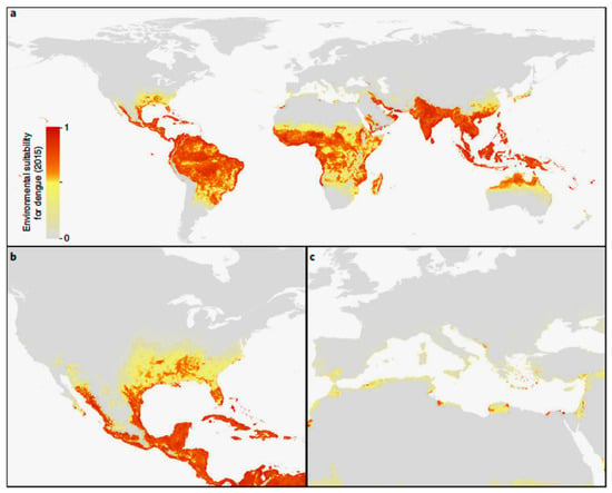
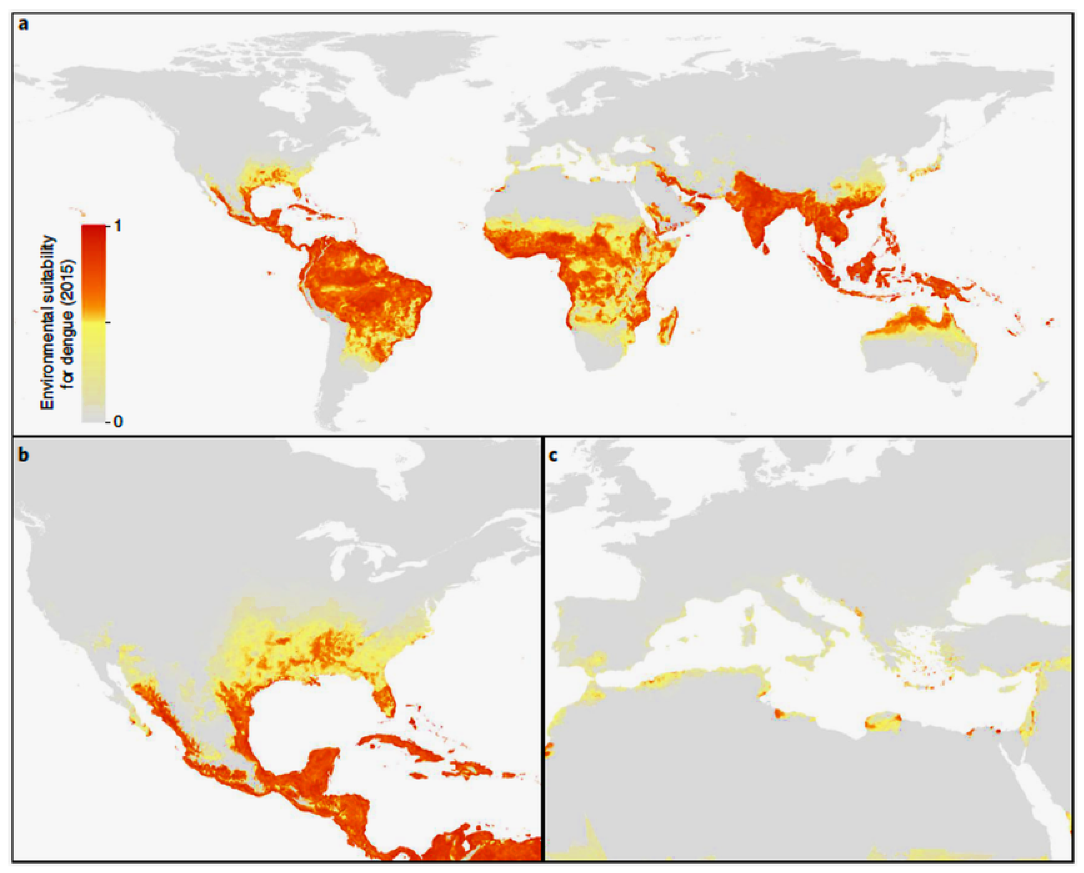
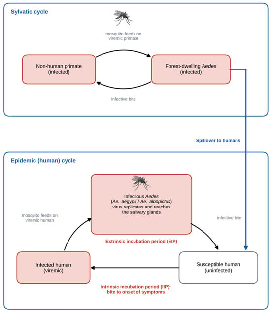
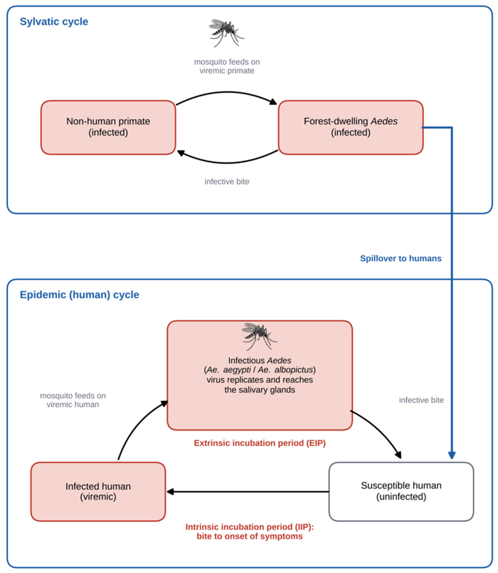
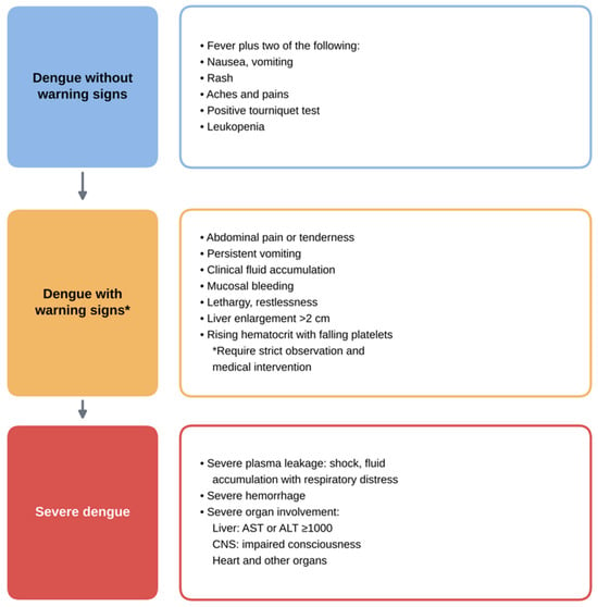
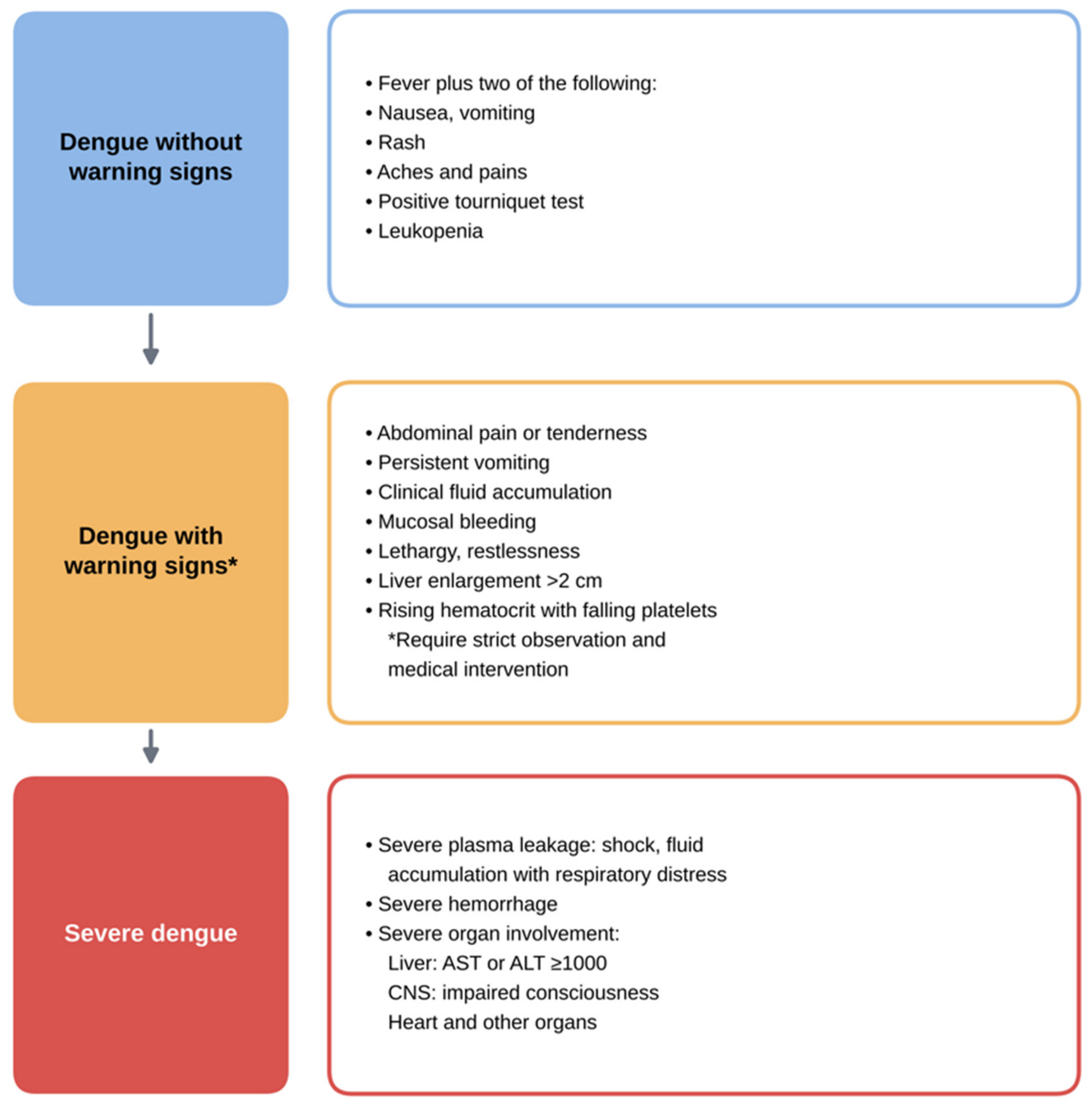
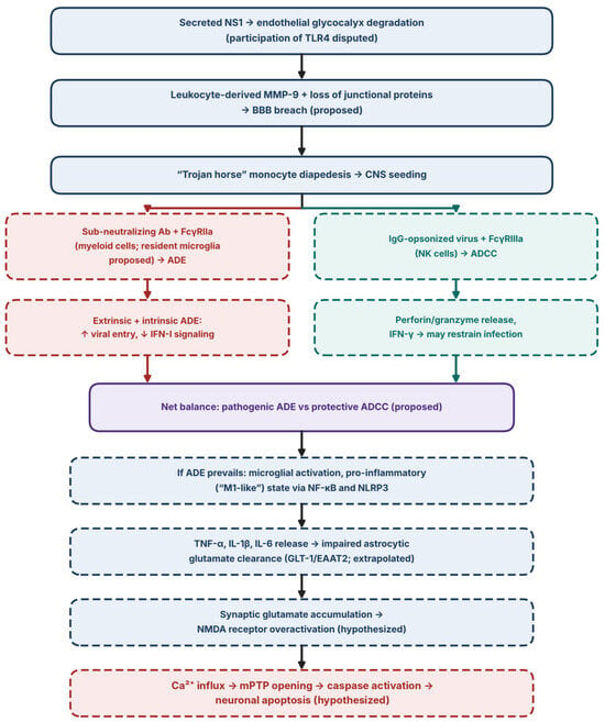
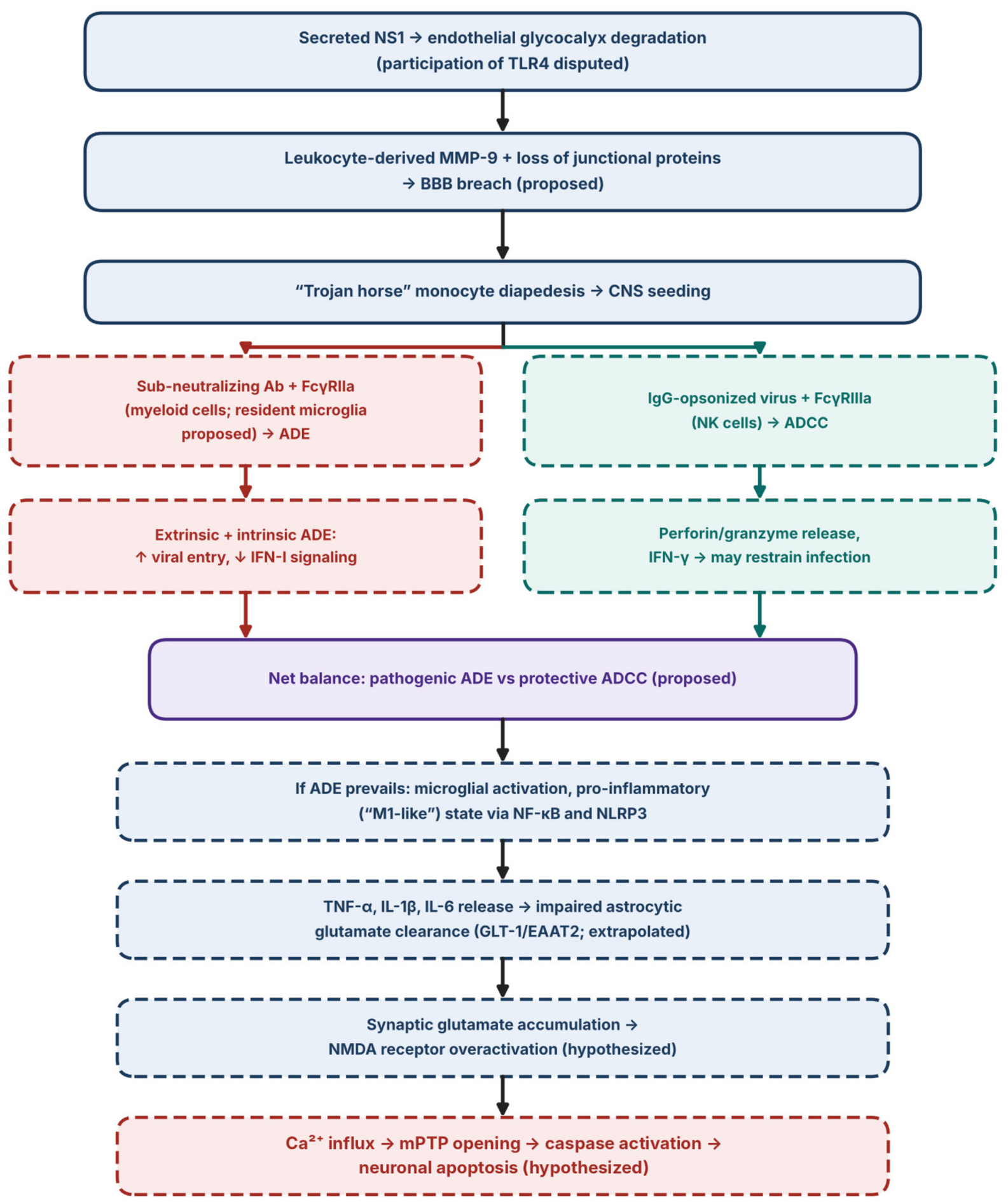

Disclaimer/Publisher’s Note: The statements, opinions and data contained in all publications are solely those of the individual author(s) and contributor(s) and not of MDPI and/or the editor(s). MDPI and/or the editor(s) disclaim responsibility for any injury to people or property resulting from any ideas, methods, instructions or products referred to in the content. |
© 2026 by the authors. Licensee MDPI, Basel, Switzerland. This article is an open access article distributed under the terms and conditions of the Creative Commons Attribution (CC BY) license.