


Abstract
Background/Objectives: The adoption of electronic cigarettes and vaping devices worldwide as healthier alternatives to smoking tobacco has led to a new clinical condition termed EVALI—E-cigarette or vaping-associated lung injury. The symptoms of EVALI present large overlaps with other lung pathologies and injuries, making diagnosis, treatment, and prevention difficult. Specific biomarkers that can distinguish lung cell exposure to E-liquids, and subsequently EVALI, from other forms of toxin exposure are thus an emerging requirement. Small extracellular vesicle (sEV)-associated microRNAs represent a promising source of non-invasive molecular biomarkers of lung injury. In particular, the large, imprinted microRNA cluster members on human chromosome 14 (C14), released via sEVs, were shown to be potentially important biomarkers in lung cancer. We investigated the effects of nicotine-containing E-liquid on sEV-associated C14 miRNA profiles released from human lung adenocarcinoma (A549) and bronchial epithelial (BEAS-2B) cells. Methods: Cells were exposed to different doses of nicotine or nicotine-containing E-liquid for 72 h and sEVs were isolated using size-exclusion chromatography, characterized, and their small RNA cargo was analyzed by targeted evaluation of selected microRNAs using qRT-PCR. Results: Cellular assays reproducibly showed that nicotine containing E-liquid was significantly more toxic than pure nicotine at >100 uM for both cell lines (p < 0.001). Nanoparticle tracking analysis (NTA) confirmed the expected size distribution (<200 nm) and concentration of sEVs across all the samples while f-NTA and Western blot confirmed the presence of canonical tetraspanin surface markers. Quantitative analysis of candidate microRNAs from sEVs revealed differential expression in A549 and BEAS-2B cells after treatment, especially upon exposure to E-liquid. C14 members released from untreated A549 cells were consistently less abundant than those from untreated BEAS-2B cells. Treatment with E-liquid and nicotine reversed the pattern, showing an increase in C14MC miRNAs released via sEVs from A549 cells compared to BEAS2B cells. Conclusions: Our work is the first report to show that extracellular microRNAs secreted as sEV cargo from human lung cells can be robust molecular responses to E-liquid exposure and can lead to future non-invasive biomarkers for EVALI.
1. Introduction
The use of e-cigarettes and other electronic nicotine delivery systems (ENDSs) has swiftly found widespread use among individuals seeking alternatives to conventional tobacco smoking globally, for their lower carcinogenicity to the user, especially among the youth. Instead of the smoke of cigarettes, these devices heat a liquid to generate aerosols that are rapidly inhaled and delivered into the airways and lungs. E-liquids are typically composed of propylene glycol, vegetable glycerine, flavouring agents, a range of additional constituents, and may include nicotine. According to the 2023 report by Action on Smoking and Health (ASH), ~5.6 million adults in Britain (10.7% of the adult population) are currently using E-liquids [1].
ENDSs are frequently promoted and prescribed as lower-risk alternatives to conventional cigarettes; for example, the NHS (National Health Service) in the UK has been formally advertising vaping as a tool to quit smoking since 2022. However, growing evidence indicates that vaping aerosols contain numerous potentially harmful substances, including carbonyl compounds, volatile organic compounds (VOCs), heavy metals and other chemical by-products, albeit in lower concentrations than cigarette smoke. Thus, ENDSs are not without potential harm and may cause adverse health effects. Consequently, concerns remain regarding the respiratory ill effects of vaping. E-cigarette- or vaping-associated lung injury (EVALI) has been recently identified as a clinical syndrome characterized by severe respiratory symptoms, with many patients requiring critical care intervention [2]. Alarmingly, the Centre for Disease control and Prevention (CDC), USA, reported 2558 non-fatal hospitalizations and 60 fatal cases caused by EVALI in 2020 alone.
Clinically, EVALI manifests as persistent cough, shortness of breath, chest pain, dyspnea and hypoxemia. Radiographically, EVALI-affected individuals show pulmonary infiltrates on chest X-rays and bilateral ground glass infiltrates on thoracic CT, which also appear in lung infections and even cancer. Histopathological findings range from acute eosinophilic pneumonia and lipoid pneumonia to organizing pneumonia and respiratory bronchiolitis-associated interstitial lung disease (RB-ILD) [3]. Moreover, symptoms presented by EVALI patients may vary due to pre-existing lung conditions and a definitive etiology of EVALI remains evasive. The diversity of clinical and pathological presentations complicates the diagnosis of EVALI, which presently relies on excluding other causes of acute lung injury. An accurate clinical differentiation of EVALI from other pulmonary illnesses and injuries (including traditional cigarette smoking) is crucial for effective patient management and warrants a precise diagnostic approach aided by reliable, and preferably non-invasive, biomarkers.
Small extracellular vesicles (sEVs) are lipid bilayer membrane-bound, <200 nm in diameter, and secreted by most living cells. Small EVs are known to carry a variety of biomolecules including nucleic acids as cargo [4]. They exhibit the potential for intercellular communication by transporting functional cellular components between cells and can traverse biological barriers (e.g., the blood–brain barrier). Thus, sEVs provide a relatively less invasive (or non-invasive) means to monitor cellular environments through bodily fluids [5]. Recent reviews have emphasized the growing importance of extracellular vesicles in pulmonary disease biology both as a source of biomarkers [6] and potential therapeutics [7]. Specifically, sEV-associated microRNAs are emerging as minimally invasive biomarkers of pulmonary disease and environmental exposure [8].
MicroRNAs (miRNAs) are a class of small non-coding RNA species that regulate gene expression post-transcriptionally [9] by binding to target mRNA, and modulating processes like mRNA degradation and translational repression. To date, more than 2600 different miRNAs have been identified in the human genome [10]. Studies show that about 30% of human genes are regulated by miRNAs [11] and nearly half of the miRNAs are located within fragile loci, many of which are tumour-associated [12]. Moreover, miRNAs are also found in the extracellular space, circulating in body fluids freely or encapsulated within vesicles—where they play a role in intercellular communication modulating phenotypes, including cancer [13]. Given their stability in the extracellular environment and their utility as biomarkers, circulating miRNAs are increasingly explored as potential circulating biomarkers for lung conditions like COPD [14], asthma [15], and idiopathic lung fibrosis [16] for diagnosis and therapeutics [7,8].
MicroRNAs are frequently organized in clusters, where multiple miRNAs are transcribed from a single primary transcript; these clusters are also known as “polycistronic miRNAs” [17]. As per the latest miRbase (version 22.1) database, the human genome comprises 159 miRNA clusters with 481 precursor miRNAs giving rise to 717 mature miRNAs [18]. Interestingly, ~30% of all known miRNAs in the human genome are organized within such clusters [19], which are highly conserved across species suggesting evolutionary and functional significance [20]. Many miRNA clusters have been shown to be important as diagnostic and prognostic biomarkers in multiple cancer studies including miR-17/miR-92, miR-106b/miR-25, miR-106b/miR-363 and miR-379/miR-656 [21].
Among these, the chromosome 14 miRNA cluster (miR-379/miR-656; hereafter C14MC) represents the second-largest cluster in the human genome accounting for over 70 mature miRNAs located within the DLK1-DIO3 imprinted domain [22]. C14MC is unique to placental mammals and is reported to have crucial roles in development and disease [23]. Downregulation of C14MC is associated with tumorigenesis, suggestive of a tumour-suppressive role particularly in different forms of glioma [24,25,26] and breast [27], liver and gastric cancers [23,24,25]. While the miRNAs from C14MC were found to be downregulated in most human cancers, the entire cluster was upregulated in lung cancer, suggesting oncogenic activity [28]. Members of C14MC were identified in the extracellular space via small extracellular vesicles (sEVs) [29] and has been proposed as a powerful source for liquid biopsy for lung diseases [30]. Previous studies have highlighted the potential of sEV-associated microRNAs as minimally invasive biomarkers of lung disease [31].
Given the unmet need for biomarkers for E-liquid exposure including EVALI and the emerging role of vesicle-associated miRNAs in pulmonary pathologies, we investigated the effect of nicotine and E-liquid exposure on sEVs derived from two human lung epithelium cell lines, one with a malignant origin and the other from a healthy donor. The miRNA content of the sEVs was then quantified by a candidate-based investigation for C14MC members. Through this ex vivo study we present a proof-of-concept for the identification of sEV-based microRNA biomarkers of E-liquid exposure that can be extended to EVALI and associated lung conditions.
2. Methods
2.1. Cell Culture
Human adenocarcinoma basal epithelial cells (A549; ATCC) and human bronchial epithelial cells (BEAS-2B; ATCC) were cultured under standard conditions at 37 °C in a humidified atmosphere containing 5% CO2. Briefly, BEAS-2B and A549 cell lines were maintained in RPMI 1640 medium (LabTech, Rotherham, UK) supplemented with 10% exosome-depleted Fetal Bovine Serum (FBS One Shot; Gibco, ThermoFischer, Loughborough, UK Cat. # A27208-03) and 1% penicillin–streptomycin (LabTech, Rotherham, UK). Cells were cultured for 72 h prior to downstream experimental procedures to ensure optimal confluency and sEV collection. The overall study was approved by the institutional ethics committee at the University of Salford.
2.2. Nicotine Treatment
Both cell lines, A549 and BEAS-2B, were treated with pure nicotine (Sigma-Aldrich, Gillingham, UK; 1236-50 MG) and E-liquid (locally procured from retailers). The specific E-liquid used for this study was strawberry-flavoured and contained 18 mg/mL nicotine (https://vape-jucce.com/product/strawberry-e-liquid/ first accessed on 12 August 2019.
A stock solution was created with a concentration of 2 mM. Subsequent dilutions for treatment were prepared ranging from 1 mM to 1 µM (specifically 1 mM, 500 µM, 100 µM, 10 µM, and 1 µM). The cells were treated for 72 h at six different concentrations ranging from 1 mM to 1 µM for both the E-liquid and nicotine—alongside untreated controls.
2.3. Cell Viability upon Treatment
Cell viability was assessed using the MTT [3-(4,5-dimethylthiazol-2-yl)-2,5-diphenyltetrazolium bromide] (Sigma-Aldrich, Gillingham, UK) assay. A549 and BEAS-2B cells were seeded in triplicate at a density of 10,000 cells per well in a 96-well plate and incubated for 24 h at 37 °C in a humidified 5% CO2 atmosphere. Cells were subsequently treated with pure nicotine and E-liquid. After 72 h of incubation under the same conditions, 50 µL MTT (Merck, Gillingham, UK) reagent was added to each well and incubated for an additional 3 h to allow for formazan crystal formation. The formazan crystals were dissolved by adding 200 µL of Dimethyl sulfoxide (DMSO) (Sigma–Aldrich, Gillingham, UK), and the absorbance was measured at 540 nm using a microplate reader. Cell viability was expressed as a percentage relative to untreated control cells and results were presented as mean ± SD (standard deviation). Each experiment was replicated three times. Based on the data we chose the 100 µM nicotine concentration for further experiments to minimize sEVs released from dying or dead cells, also supported by the literature [32].
2.4. Small EV Isolation and Purification from Cell Culture Supernatant
Small EV isolation and characterization were carried out following the MiSEV guidelines [33,34,35]. Conditioned medium (~50 mL) harvested from two 175 cm2 flasks of cultured cells was processed for sEV isolation. Initial centrifugation steps were carried out at 1000× g for 10 min and then 2000× g for 10 min to remove cells and cellular debris. The resulting cell-free supernatant was collected and centrifuged at 16,000× g for 48 min to eliminate platelets and larger extracellular vesicles.
Subsequently, sEVs were isolated using size exclusion chromatography (SEC) with Exo-Spin mini columns (Cell Guidance Systems; Cambridge, UK). The supernatant was mixed with Exo-Spin buffer at a 2:1 (buffer: supernatant) ratio, gently inverted to mix, and incubated overnight at 4 °C. The following day, the sample was centrifuged at 16,000× g for 96 min to pellet the sEV.
The pellet was then re-suspended in 100–200 μL Phosphate buffer saline (PBS)(Scientific laboratory suppliers, Nottingham, UK) and further purified using the 30 nm pore size Exo-Spin mini column according to the manufacturer’s instructions. The eluted sEV fraction (100–200 μL PBS) was supplemented with 2 µL of proteinase inhibitor (Thermo Fisher Scientific, Loughborough, UK) and stored for downstream processing.
2.5. Characterization of sEVs
2.5.1. Transmission Electron Microscopy
Small extracellular vesicles (sEVs) were visualized using transmission electron microscopy (TEM) to confirm their morphology and integrity. Approximately 10 µL of isolated sEVs were pipetted onto a parafilm. A glow-discharged grid was placed onto the sample and incubated at RT for 10 min. The grid was then gently washed twice with 100 µL of Phosphate-buffer saline (PBS), followed by fixation with 1% glutaraldehyde (Sigma-Aldrich, Gillingham, UK) for 5 min. Subsequently, the grid was rinsed twice with 100 mL of ddH2O.
Negative staining was performed by incubating the grid with 1% aqueous uranyl acetate (UA) (Fisher Scientifics, Loughborough, UK) for 30–90 secs. For enhanced contrast, the grid was then placed on a mixture of 4% UA and 2% Methyl Cellulose (MC) (Sigma-Aldrich, Gillingham, UK) at a 1:9 ratio on ice for 10 min. Following incubation, excess stain was removed by gently touching the edge of the grid to filter paper at a 45° angle using a 5 mm wire loop. The grid was air-dried for approximately 20 min.
Imaging was conducted using an FEI Tecnai G2 Spirit transmission electron microscope (Thermo Fisher Scientific, Loughborough, UK) operating at 120 kV, equipped with a Gatan RIO16 digital camera. TEM was performed on sEV samples isolated from conditioned media of both untreated and E-liquid-treated A549 and BEAS-2B cells.
2.5.2. Nanoparticle Tracking Analysis (NTA)
Prior to nanoparticle tracking analysis, all small extracellular vesicle (sEV) samples were tested for mycoplasma using the Mycoplasma PCR detection kit (Applied biological materials Inc., Richmond, BC, Canada) in accordance with the manufacturer’s instructions.
Nanoparticle tracking analysis (NTA) was performed using ZetaView systems (Particle Matrix, Inning am Ammersee, Germany), which is equipped with a 520 nm laser, a high-sensitivity camera, and a 550 nm long-pass manual fluorescence filter. For each experimental condition, sEV samples isolated from six biological replicates per cell line were analyzed. The suspension was diluted using Phosphate-buffer saline (PBS) (Scientific laboratory suppliers, Nottingham, UK) at a 1:1000 ratio to ensure optimal particle concentration within the recommended detection range. The Zetaview software version 8.04.02 (Particle Metrix, Inning am Ammersee, Germany) was used to process and analyze the recorded data to determine particle size distribution and concentration. Measurements were conducted at room temperature, and all setting were maintained consistently across all samples.
2.5.3. Fluorescent NTA (f-NTA)
Fluorescent nanoparticle tracking analysis was performed to characterize and differentiate small EVs based on the presence of tetraspanin contents—namely CD9, CD63 and CD81. Prior to measurement, the instrument was calibrated using standard polystyrene beads (100 nm) (Analytik, Cambridge, UK) to ensure optimal performance in scatter mode.
For scatter measurements, isolated sEVs were diluted 1:1000 in particle-free PBS and measured at room temperature. For fNTA, sEVs were stained with the lipophilic membrane dye Cell Mask Orange (Invitrogen, Scotland, UK) and fluorescently conjugated antibodies against CD9 (BioLegend UK Ltd., London, UK 312118), CD63 (BioLegend UK Ltd., London, UK, 353022) and CD81 (BioLegend UK Ltd., London, UK, 349520). The CMO dye (Thermo Fisher Scientific, Loughborough, UK, C10045) was pre-diluted at a ratio of 1:250 in PBS. Antibodies were diluted in PBS according to the manufacturer’s instructions.
A total of 9 µL isolated sEVs were mixed with 1µL of CMO or antibody solution in separate tubes. Samples were incubated in the dark at room temperature for 60 min. Post incubation, stained sEV samples were further diluted in PBS prior to fNTA analysis. The particle count was optimized to approximately 200 particles per frame to ensure accurate quantification.
All analyses were conducted using ZetaView software version 8.04.02 (Particle Metrix, Inning am Ammersee, Germany), and data were processed to evaluate particle concentrations and tetraspanin-specific fluorescence profiles.
2.5.4. Western Blot
Small EV samples were lysed using radioimmunoprecipitation (RIPA) buffer (Sigma –Aldrich, Gillingham, UK) and protein concentration was measured using the Bicinchoninic (BCA) assay (Thermo Fisher Scientific, Loughborough, UK). A total of 40 µg of sEV protein, normalized against 20 µg of total protein from the corresponding cell lysate, was used for electrophoresis. Samples were reduced in Laemmli buffer(Thermo Fisher Scientific, Loughborough, UK), with heating at 70 °C for 10 min. Proteins were separated on 10% gradient SDS—PAGE gels and transferred onto 0.25 mm nitrocellulose membranes (Cytiva, (Fisher Scientifics, Loughborough, UK). Membranes were blocked with 5% bovine serum albumin (BSA) (Fisher Scientifics, Loughborough, UK) for 1 h at room temperature and incubated overnight at 4 °C with specific primary antibodies diluted at 1:1000. The following primary antibodies were used: CD9 (Fisher Scientifics, Loughborough, UK), CD81 (Fisher Scientifics, Loughborough, UK), CD63 (Fisher Scientifics, Loughborough, UK), TSG-101 (sEV markers) (Abcam, Cambridge, UK) and calnexin (negative control for EV contamination) (Proteintech, Manchester, UK). After three 10 min washes, membranes were incubated with horseradish peroxidase (HRP)-conjugated secondary anti-mouse antibody (1:5000 dilution) (Cell Signalling, Leiden, The Netherlands) for 1 h at room temperature. Protein bands were visualized using enhanced chemiluminescence (ECL) reagents (Bio-Rad, Hertfordshire, UK) and imaged (Image studio version 5.2) using the Licor Imaging system (Lincoln, NE, USA).
2.6. Isolation of sEV RNA and Quantification
2.6.1. Proteinase K and RNase A Treatment
A total of 50 μL of isolated sEVs was treated with Proteinase K (20 μg/mL) by gentle pipetting to ensure uniform mixing. The mixture was incubated at 37 °C for 30 min to degrade any protein contaminants. Next, a protease inhibitor (Halt, 100X Thermo Fisher Scientific, Loughborough, UK) was added, and the sample was incubated on ice for 10 min.
Later, RNase A (Thermo Fisher Scientific, Loughborough, UK) was added to the mixture and incubated at 37 °C for an additional 30 min to digest the extracellular RNA. To terminate the activity of RNase A, 700 μL of Trizol/QIAzol (Qiagen, Manchester, UK) was added immediately to the tube and vortexed. The lysate was then left at room temperature for 20 min to ensure complete dissociation of nucleoprotein complexes. This treatment prior to sEV RNA isolation ensures the absence of any other extracellular RNA that was not encapsulated within the sEVs. Total RNA was isolated using the Rneasy MinElute spin column (Qiagen, Manchester, UK) with a combination of triazole-based lysis and membrane-based affinity purification. The RNA was eluted in RNA-free water and quantified using Qubit 3.0 Fluorometer (Thermo Fisher, Loughborough, UK) for downstream applications.
2.6.2. cDNA and qRT-PCR
Quantitative PCR (qPCR) analysis was performed to assess the expression of selected candidate miRNAs from the C14 miRNA cluster (C14MC). For this we selected 13 microRNAs covering the entire cluster as present on the genome. MicroRNA-specific primers were procured from Eurofins Genomics (Eurofin Genomics, Wolverhampton, UK). For reverse transcription, 50 ng of total RNA from cellular samples and 3 ng from sEV-derived RNA were used. The cDNA synthesis was conducted using the QuantiMir kit (System Biosciences, Cambridge, UK), which incorporates Poly(A) polymerase tailing for miRNA-specific reverse transcription, following the manufacturer’s protocol.
The resulting cDNA was diluted at a ratio of 1:10 and 1 µL of diluted cDNA was used as a template in qPCR reactions. Reactions were performed using the SYBR Green-based PCR master mix (no ROX) (Meridian Bioscience, London, UK) on a Rotor-Gene Q real-time PCR cycler (Qiagen, Manchester, UK). Differential expression of miRNAs in nicotine-treated A549 or BEAS-2B cells, compared to untreated controls, was conducted using the 2^Delta Delta Ct. All reactions were normalized to spike-in Cel-39, which served as an internal control. A no-template control (NTC) served as a negative control. Melting curve analysis was used to confirm the specificity of the amplification reactions.
2.7. Statistical Analysis
All experiments were performed in triplicate unless stated otherwise. All results are presented as mean ± SEM. An unpaired Student′s t-test was performed using GraphPad Prism Version 9, and the Holm–Sidak correction method [36] for multiple comparisons was used to ascertain the p-values (corrected p < 0.05).
3. Results
3.1. E-Liquid Is More Toxic than Pure Nicotine for Human Lung Cells
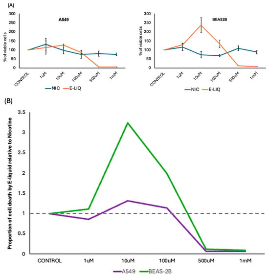
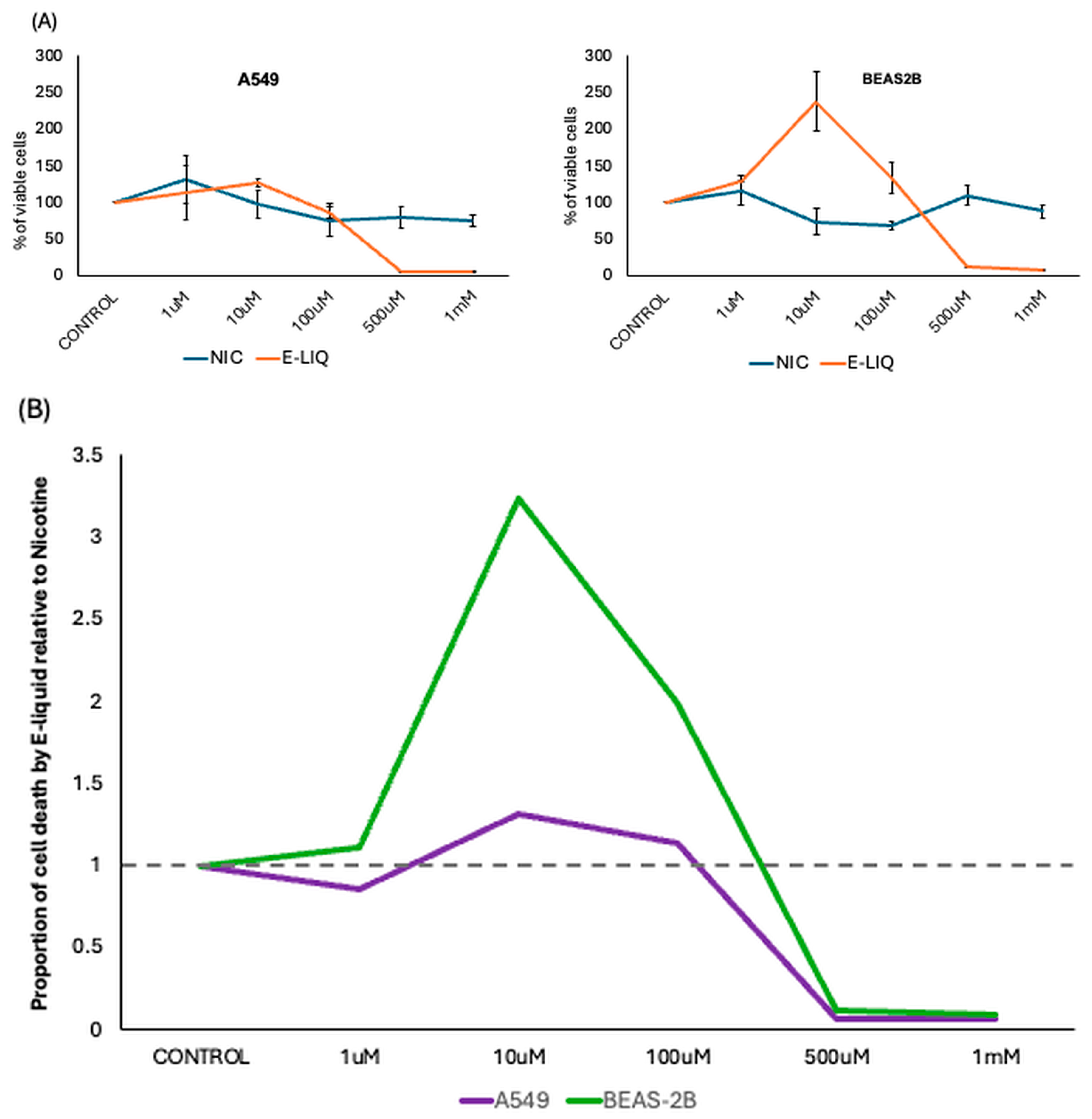
E-liquid (containing nicotine) treatment on both A549 and BEAS2-B cells showed significantly increased toxicity above the 100 µM concentration, compared to treatment with nicotine alone (Figure 1A and Supplementary Figure S1). To ensure the downstream sEVs isolation were not substantially derived from dead cells, a concentration of 100 µM of nicotine was selected for all further experiments, which was also supported by the literature [32]. For both cell lines and for either of the treatments, the cell viability drastically reduced at concentrations above 100 µM (Figure 1A). A derivative of the data presented in Figure 1B reveals that, compared to nicotine, the effect of E-liquid is much more drastic in both cell lines—and more pronounced in BEAS-2B (Figure 1B). Additional experiments (Supplementary Figure S2) using the same E-liquid composition but without the nicotine content also showed that E-liquid toxicity in both cell lines is comparable with or without nicotine, highlighting the potential harm caused by the flavouring and delivery agents present in E-liquids.
Figure 1.
Cellular toxicity upon treatment. (A) Effect of nicotine and E-Liquid on A549 (left) and BEAS-2B cells (right). Each experiment was performed in triplicate. (B) The effect of E-liquid on cell death relative to the effect of nicotine for both cell lines. The dotted horizontal line indicates no difference in toxicity between the two cell lines. Note the drastic cell death beyond the 100 µM concentration for both cell lines, especially for BEAS-2B (green line).
3.2. E-Liquid Treatment Shows Enrichment of sEV Surface Markers for Human Lung Cells
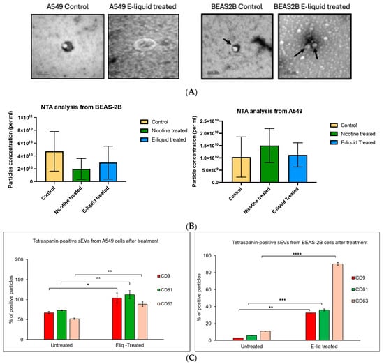
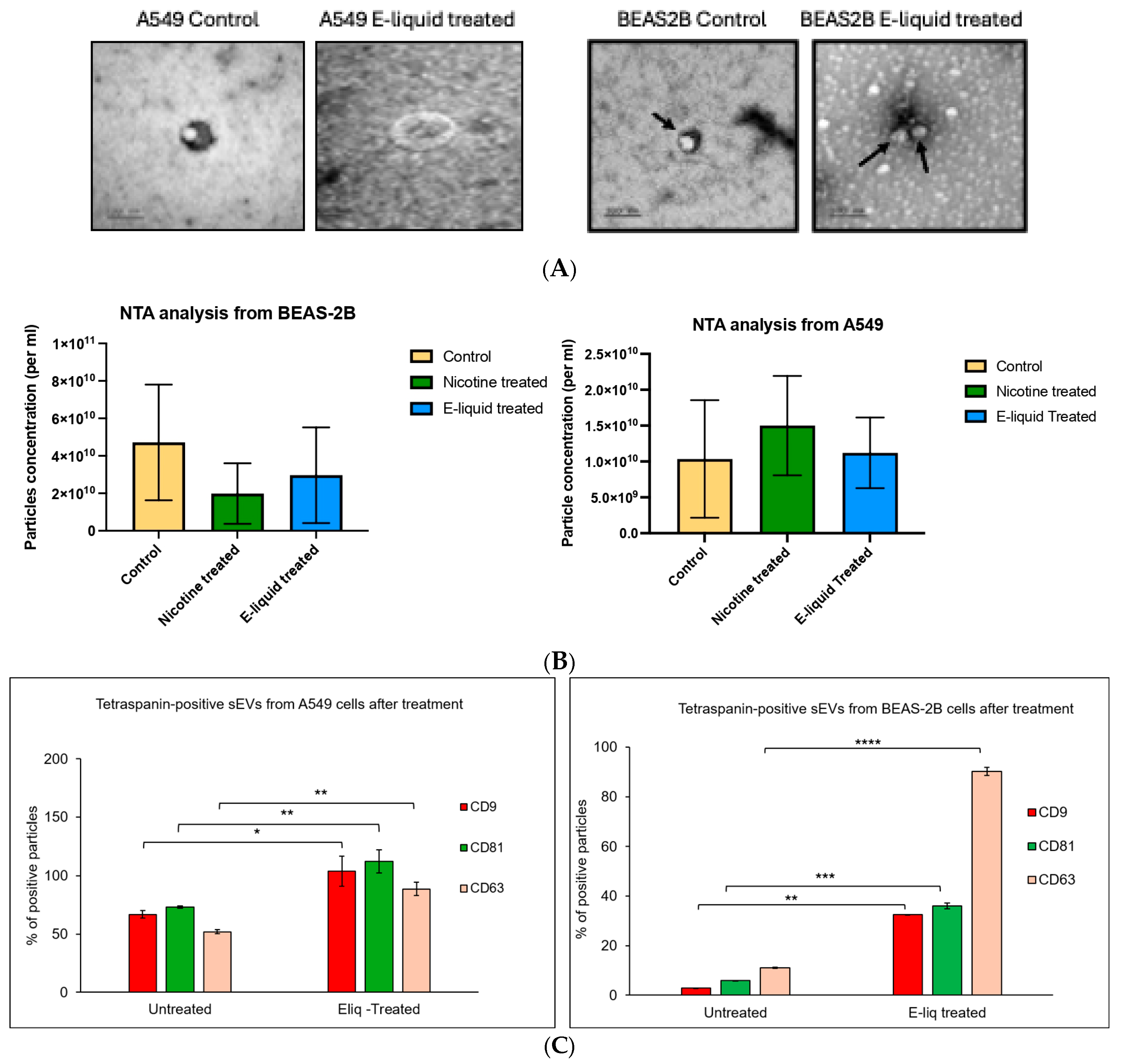
The morphology, size distribution, and molecular markers of the isolated sEVs were characterized according to the MiSEV guidelines. The TEM analysis did not show any discernible differences in sEV morphology between the cell lines or due to the treatment (Figure 2A, Supplementary Figure S3A). The size distributions for all isolations were within the expected size range of <200 nm (Supplementary Figure S3B) and nicotine or E-liquid treatments did not affect the number of particles released from the cells (Figure 2B). Fluorescence NTA revealed a consistent increase in all three canonical tetraspanin markers CD9, CD63, and CD81 upon E-liquid treatment for both cell lines (Figure 2C). This effect was not observed in nicotine-treated cells (Supplementary Figure S3C). Immunoblotting qualitatively confirmed the presence of sEV surface markers CD9, CD63, and CD81, and the absence of Calnexin as the negative marker (Supplementary Figure S3D). Supplementary Figures S4 and S5 show the uncropped western blots and the particle-to-protein ratios, respectively.
Figure 2.
Small EV characterization. (A) Four representative TEM images showing sEV morphology isolated from different cell lines and treatment as indicated. Arrows pointing to sEVs. (B) NTA performed on sEVs isolated from the A549 (left) and BEAS-2B (right) cell cultures showed no significant difference in the number of particles released after treatment. (C) Fluorescence NTA data for the 3 canonical tetraspanin sEV surface markers for A549 (left) and BEAS-2B (right) upon E-liquid treatment (all data in triplicates). The p-value indicates * p < 0.05, ** p < 0.01, *** p < 0.001, and **** p < 0.0001 respectively.
3.3. E-Liquid and Nicotine Treatment Reverses Expression Pattern in sEV-C14 miRNAs
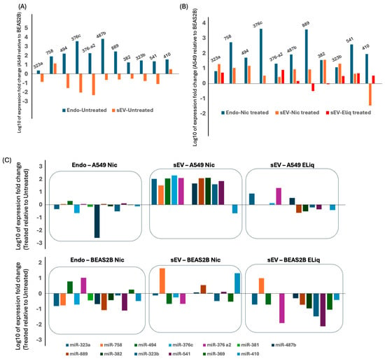
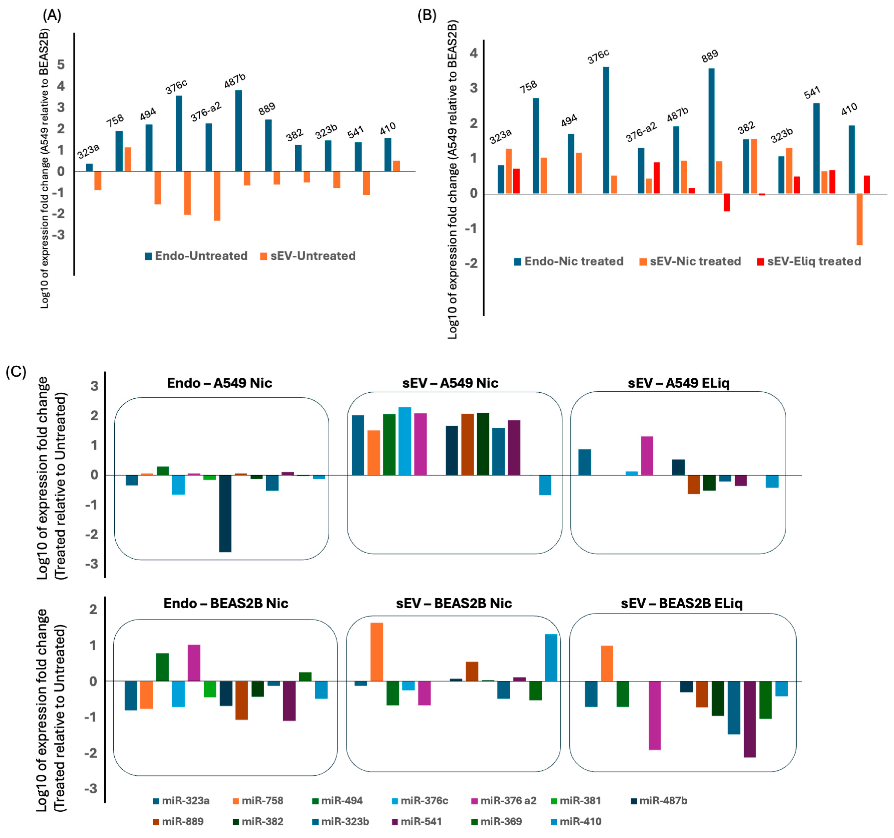
Under untreated conditions, the endogenous C14 miRNAs are upregulated in A549 (cancer) cells compared to BEAS-2B (healthy) cells (Figure 3A)—supporting earlier studies. The sEVs released from the same cells showed the opposite pattern where C14 miRNAs in A549 sEVs were downregulated compared to BEAS-2B cells (Figure 3A). A treatment with nicotine maintains the same trend for endogenous C14 miRNAs—but reverses the trend in sEVs released from the treated cells (Figure 3B). The E-liquid treatment showed similar trend reversal, albeit with some of the miRNAs being below the detectable level of expression following the treatment (Figure 3B). In a derivative of the data, when analyzed for each cell line and when treatment was compared against the untreated condition, this cluster-wide change was observed with a higher resolution (Figure 3C; also see later in the discussion).
Figure 3.
C14MC members as candidate biomarkers. (A) Relative expression of the candidates in A549 compared to BEAS-2B in untreated cells. (B) Similar data representation as panel A for the microRNA expressions in treated cells. For both panels A and B, zero on the vertical axis represents no change in expression for A549 relative to BEAS-2B. (C) Effect of treatment on C14MC candidates compared to untreated equivalents in A549 (upper) and BEAS-2B (lower) cells. Here, zero on the vertical axis represents no change in expression upon treatment. For all three panels, the C14MC members are arranged as they are ordered on the genome encompassing the entire length of the cluster. For all panels ‘Endo’ indicates RNA expression in the cellular compartment (Endogenous). The raw data for qPCR is provided in the Supplementary Files.
4. Discussion
Patients with E-liquid- or vaping-associated lung injury (EVALI) often require critical care [37], challenging the limited resources of healthcare systems. There is a need for stable, easily accessible, and reproducible biomarkers to distinguish EVALI from similar lung injuries resulting from other pathological conditions. Although exposure biomarkers like Tetrahydrocannabinol (THC) metabolites and vitamin E acetate have been reported at elevated levels in urine and plasma, these are not specific to E-liquid exposure [38]. Similarly, systemic inflammatory and oxidative stress markers like CRP, ESR, and interleukins found to be increased in EVALI patient plasma samples [39] are also only indicative of injury in general. The strategy and findings reported in our study are aimed at identifying biomarker candidates that are specific to E-liquid exposure and can potentially distinguish EVALI from other lung conditions .
Contrary to the popular narrative that promotes the E-liquids used in vaping as safer alternatives to traditional tobacco products, our findings reveal that E-liquids exhibit consistently higher toxicity than pure nicotine when applied to human lung cells. Although the experimental condition used (100 microM nicotine) is much higher than the physiological concentration of nicotine found in smokers, it was surprising to note that, at the same concentration of nicotine, E-liquid was much more toxic to lung cells than nicotine alone. This was consistent even when E-liquids were tested without having any nicotine. As shown in the results section, we selected a treatment condition of 100 microM nicotine (and an equivalent amount in nicotine-containing E-liquid), which was optimal for the survival of 50% of the cells (an approximate IC50 value). This was done to ensure that the extracellular vesicles released into the culture medium after treatment did not primarily originate from dying cells, such as apoptotic bodies, which could have confounded our observations. As we have shown for both treatments, the quantity of nicotine was very similar—suggesting the increased toxicity in E-liquid treatment is not due to nicotine, and is most likely due to the other chemicals present in the mixture. Moreover, the BEAS-2B (healthy lung epithelium) cells show a much higher sensitivity to E-liquids than the A549 cells, which are from a cancerous source. Extrapolating this to human subjects, this will imply that healthy lung epithelial cells (in people without cancer) will suffer much higher toxicity from E-liquids than an exposure to nicotine alone. While it is true that the diversity of toxic chemicals and their carcinogenic potential in traditional cigarettes far exceeds the chemical composition in E-liquids, the latter are not completely free from harm [40]. Currently, the lower carcinogenicity of ENDSs is translated to mean ‘less harmful’ in public discourse and perception, disregarding the possibility that they may become the source of new health problems. Thus, the recent public health guidelines in the UK and many other countries suggesting the regulated use or ban of E-liquid-based vaping products is a welcome step in public health [41].
Small EVs are hailed as a potent source of biomarkers for many health conditions and as a powerful vehicle for drug delivery [42]. EV-associated miRNA profiling has successfully identified distinct molecular signatures associated with smoking and chronic obstructive pulmonary disease, supporting the translational potential of extracellular miRNAs as respiratory biomarkers [43]. Freely circulating RNA is prone to degradation in the bloodstream and exists mostly in a complex with RNA-binding proteins that are difficult to dissociate. But when encapsulated in sEVs, RNA molecules remain protected and can therefore travel longer distances, potentially affecting tissues and organs distant from the site of origin. The morphology, size, and particle concentration of sEVs showed no difference between untreated and nicotine/E-liquid-treated cells, suggesting that the nicotine and E-liquid concentration chosen for the treatments did not interfere with sEV purification. While there was no major difference in the size and quantity of sEVs upon treatment, fluorescent NTA (f-NTA) revealed that the distributions of the canonical surface markers CD9, CD63, and CD81 were increased in both cell lines under E-liquid treatment but not under nicotine treatment. Since the particle concentration remains unchanged after treatment, the cause of this increase demands further investigation. It also remains to be seen if the increased tetraspanins contribute to the altered cargo packaging of the sEVs released from the treated cells.
From a biomarker discovery perspective, molecules exhibiting increased abundance under disease or exposure conditions are often more amenable to analytical detection and therapeutic strategies than those showing reduced expression. Previous studies have reported upregulation of C14MC members in lung cancer [44], which is counter-intuitive given their global downregulation in most other human cancers [24]. Across 13 different C14MC members analyzed in this study, the untreated cancerous A549 cells show a higher expression than the untreated healthy BEAS-2B cells for the endogenous (cellular) microRNAs, corroborating the reported upregulation of this cluster in lung cancer. Interestingly, for the extracellular component of sEVs, this pattern reversed, with the same C14MC members showing an increased expression in sEVs from untreated BEAS-2B cells compared to those from A549 cells. Our interpretation of this observation is limited by the lack of a mechanistic investigation such as an inhibition of the EV secretion pathways. Therefore, although the mechanism behind this observation requires further investigation, this difference could theoretically be argued to arise from differential sEV cargo packaging between the two cell lines. Alternatively, it could reflect the disease state of the two kinds of cells. We also acknowledge other limitations of the study where the treatment concentrations were much higher than physiological levels of nicotine in smokers, and the microRNA repertoire we tested is limited given the rest of the microRNAs in the genome, which could potentially be even better biomarkers for E-liquid exposure.
Surprisingly, upon treatment with nicotine and E-liquid, the expression pattern of sEV-C14MC members is reversed again—now matching with the overall profile of untreated cellular RNA as well as cellular RNA after nicotine treatment. Thus, while untreated BEAS-2B cells seem to transport more of these microRNAs as sEV cargo, neither treatment shows a higher export from A549 cells. It will be interesting to study whether the increased export translates to increased intercellular communication and uptake within lung tissue as well as across other organs (e.g., lung to brain)—potentially suggesting addictive mechanisms.
Further analysis of the data presented in Figure 3 revealed that three C14MC microRNAs—miR-889, miR-382, and miR-541—exhibited reversed expression patterns in response to nicotine-versus-E-liquid treatment in sEV-associated microRNAs released from both cell lines. All three microRNAs were upregulated upon nicotine treatment, while E-liquid treatment resulted in downregulation. These microRNAs exemplify our primary objective, demonstrating that sEV-associated microRNAs could serve as discriminatory, minimally invasive biomarkers for E-liquid exposure, and highlighting the potential of C14MC members as valuable candidates for such biomarkers.
5. Conclusions
The candidate microRNA-based approach shows that expression of the members of the chromosome 14 microRNA cluster might be sensitive and distinct between exposure of human lung cell lines to nicotine and E-liquid. The data also indicates that the packaging and release of these microRNAs via small extracellular vesicles might depend on the toxicant exposure of the human lung cells tested. Overall, this is one of the first reports on extracellular microRNA as biomarkers for human lung cell exposure to E-liquid and can aid future discoveries of novel actionable biomarkers for clinical conditions such as EVALI.
Supplementary Materials
The following supporting information can be downloaded at: https://www.mdpi.com/article/10.3390/jmp7040036/s1, Figure S1: Cell viability assay–Treatment with100uM of pure nicotine & E-liquid (Strawberry flavoured with nicotine) on A549 and BEAS2B for 72 hrs; Figure S2: Cell viability assay to determine the compare the effect of e-liquid without nicotine with other toxicants; Figure S3: Characterization of sEV; Figure S4: Uncropped western blots; Figure S5: Particle to Protein ratio for sEVs; Table S1: raw qPCR data.
Author Contributions
S.C. performed most of the experiments and analysis, and also wrote the first draft of the manuscript. N.B. provided expert technical helps in protein isolation, quantitation and western blot experiments. N.B. also helped with new analysis and revisions in the revised version. A.M. conceptualized the project, organized the resources, led the project and wrote the final manuscript with major inputs from S.C. and N.B. All authors have read and agreed to the published version of the manuscript.
Funding
This work was partially supported by the Innovative Strategy Fund of the University of Salford (SEFA-39). The funders had no role in study design, data analysis and the data presented in the manuscript.
Institutional Review Board Statement
The cell line-based study was approved by the Ethics Committee at the University of Salford (STR1819-38 dated 1 May 2019). The study does not use any material from any living human subjects.
Informed Consent Statement
Not applicable.
Data Availability Statement
All relevant data and protocols are available and can be shared upon request, if not already provided.
Acknowledgments
We acknowledge the imaging facility at the Liverpool University for the technical service regarding the TEM analysis. The technical team at the translational medicine laboratory, especially Rumana Rafiq and Muna Abubaker, are acknowledged for their expert technical support and maintenance of the facility. During the preparation of this manuscript, the authors used ChatGPT (GPT-5.6 Luna, OpenAI) for the purposes of grammar and spelling checks of responses to reviewers’ comments and for the cover letter. The tool was also used to inspire ideas for the graphical abstract. The authors have reviewed and edited the output and take full responsibility for the content of this publication.
Conflicts of Interest
All authors declare that there are no conflicts of interest.
References
- Taylor, E.; Arnott, D.; Cheeseman, H.; Hammond, D.; Reid, J.L.; McNeill, A.; Driezen, P.; East, K. Association of Fully Branded and Standardized E-Cigarette Packaging With Interest in Trying Products Among Youths and Adults in Great Britain. JAMA Netw. Open 2023, 6, e231799. [Google Scholar] [CrossRef] [Scilit] [PubMed]
- Amjad, M.A.; Ocazionez Trujillo, D.; Estrada-Y-Martin, R.M.; Cherian, S.V. E-Cigarette or Vaping Product Use-Associated Lung Injury: A Comprehensive Review. Int. J. Environ. Res. Public Health 2025, 22, 792. [Google Scholar] [CrossRef] [Scilit] [PubMed]
- Singh, A. Adolescent E-Cigarette or Vaping Product Use-Associated Lung Injury: A Case Series and Review of the Literature. Pediatr. Pulmonol. 2022, 57, 1076–1084. [Google Scholar] [CrossRef] [Scilit] [PubMed]
- Chen, Y.; Zhao, Y.; Yin, Y.; Jia, X.; Mao, L. Mechanism of Cargo Sorting into Small Extracellular Vesicles. Bioengineered 2021, 12, 8186–8201. [Google Scholar] [CrossRef] [Scilit] [PubMed]
- Morgan, J.; Aarons, T.; Lace, G.; Mukhopadhyay, A. Harnessing the Power of MicroRNA Cargoes in Small Extracellular Vesicles Released from Fresh-Frozen Human Brain Sections. JoVE 2024, 67379. [Google Scholar] [CrossRef] [Scilit] [PubMed]
- Mohan, A.; Agarwal, S.; Clauss, M.; Britt, N.S.; Dhillon, N.K. Extracellular Vesicles: Novel Communicators in Lung Diseases. Respir. Res. 2020, 21, 175. [Google Scholar] [CrossRef] [Scilit] [PubMed]
- Coward-Smith, M.; Zakarya, R.; Oliver, B.G.; Kim, R.Y.; Donovan, C. Extracellular Vesicles in Pulmonary Diseases: Roles and Therapeutic Potential. BMC Pulm. Med. 2025, 25, 516. [Google Scholar] [CrossRef] [Scilit] [PubMed]
- Eckhardt, C.M.; Gambazza, S.; Bloomquist, T.R.; De Hoff, P.; Vuppala, A.; Vokonas, P.S.; Litonjua, A.A.; Sparrow, D.; Parvez, F.; Laurent, L.C.; et al. Extracellular Vesicle-Encapsulated microRNAs as Novel Biomarkers of Lung Health. Am. J. Respir. Crit. Care Med. 2023, 207, 50–59. [Google Scholar] [CrossRef] [Scilit] [PubMed]
- Saliminejad, K.; Khorram Khorshid, H.R.; Soleymani Fard, S.; Ghaffari, S.H. An Overview of microRNAs: Biology, Functions, Therapeutics, and Analysis Methods. J. Cell Physiol. 2019, 234, 5451–5465. [Google Scholar] [CrossRef] [Scilit] [PubMed]
- Kozomara, A.; Birgaoanu, M.; Griffiths-Jones, S. miRBase: From microRNA Sequences to Function. Nucleic Acids Res. 2019, 47, D155–D162. [Google Scholar] [CrossRef] [Scilit] [PubMed]
- Wu, H.H.; Leng, S.; Sergi, C.; Leng, R. How MicroRNAs Command the Battle against Cancer. Int. J. Mol. Sci. 2024, 25, 5865. [Google Scholar] [CrossRef] [Scilit] [PubMed]
- Taefehshokr, S.; Taefehshokr, N.; Derakhshani, A.; Baghbanzadeh, A.; Astamal, R.V.; Safaei, S.; Abbasi, S.; Hajazimian, S.; Maroufi, N.F.; Isazadeh, A.; et al. The Regulatory Role of Pivotal MicroRNAs in the AKT Signaling Pathway in Breast Cancer. Curr. Mol. Med. 2022, 22, 263–273. [Google Scholar] [CrossRef] [Scilit] [PubMed]
- Sepúlveda, F.; Mayorga-Lobos, C.; Guzmán, K.; Durán-Jara, E.; Lobos-González, L. EV-miRNA-Mediated Intercellular Communication in the Breast Tumor Microenvironment. Int. J. Mol. Sci. 2023, 24, 13085. [Google Scholar] [CrossRef] [Scilit] [PubMed]
- Gomez, N.; James, V.; Onion, D.; Fairclough, L.C. Extracellular Vesicles and Chronic Obstructive Pulmonary Disease (COPD): A Systematic Review. Respir. Res. 2022, 23, 82. [Google Scholar] [CrossRef] [Scilit] [PubMed]
- Prasad, C.; Griffard, R.; Pei, D.; Haworth, S.; Dhillon, N.K.; Castro, M.; Sundar, I.K. Extracellular Vesicle miRNAs as Biomarkers of Asthma Severity. Allergy 2026, 81, 271–276. [Google Scholar] [CrossRef] [Scilit] [PubMed]
- Ramírez-Hernández, A.A.; Velázquez-Enríquez, J.M.; Santos-Álvarez, J.C.; López-Martínez, A.; Reyes-Jiménez, E.; Carrasco-Torres, G.; González-García, K.; Vásquez-Garzón, V.R.; Baltierrez-Hoyos, R. The Role of Extracellular Vesicles in Idiopathic Pulmonary Fibrosis Progression: An Approach on Their Therapeutics Potential. Cells 2022, 11, 630. [Google Scholar] [CrossRef] [Scilit] [PubMed]
- Vilimova, M.; Pfeffer, S. Post-Transcriptional Regulation of Polycistronic microRNAs. Wiley Interdiscip. Rev. RNA 2023, 14, e1749. [Google Scholar] [CrossRef] [Scilit] [PubMed]
- Ware, A.P.; Satyamoorthy, K.; Paul, B. CmirC: An Integrated Database of Clustered miRNAs Co-Localized with Copy Number Variations in Cancer. Funct. Integr. Genom. 2022, 22, 1229–1241. [Google Scholar] [CrossRef] [Scilit] [PubMed]
- Chambers, C.; Quan, L.; Yi, G.; Esquela-Kerscher, A. CRISPR Gene Editing Tool for MicroRNA Cluster Network Analysis. J. Vis. Exp. 2022. [Google Scholar] [CrossRef] [Scilit] [PubMed]
- Simkin, A.; Geissler, R.; McIntyre, A.B.R.; Grimson, A. Evolutionary Dynamics of microRNA Target Sites across Vertebrate Evolution. PLoS Genet 2020, 16, e1008285. [Google Scholar] [CrossRef] [Scilit] [PubMed]
- Puła, A.; Robak, P.; Robak, T. MicroRNA in Multiple Myeloma - A Role in Pathogenesis and Prognostic Significance. Curr. Med. Chem. 2021, 28, 6753–6772. [Google Scholar] [CrossRef] [Scilit] [PubMed]
- Valbuena, G.N.; Apostolidou, S.; Roberts, R.; Barnes, J.; Alderton, W.; Harper, L.; Jacobs, I.; Menon, U.; Keun, H.C. The 14q32 Maternally Imprinted Locus Is a Major Source of Longitudinally Stable Circulating microRNAs as Measured by Small RNA Sequencing. Sci. Rep. 2019, 9, 15787. [Google Scholar] [CrossRef] [Scilit] [PubMed]
- Guo, L.-H.; Li, H.; Wang, F.; Yu, J.; He, J.-S. The Tumor Suppressor Roles of miR-433 and miR-127 in Gastric Cancer. IJMS 2013, 14, 14171–14184. [Google Scholar] [CrossRef] [Scilit] [PubMed]
- Laddha, S.V.; Nayak, S.; Paul, D.; Reddy, R.; Sharma, C.; Jha, P.; Hariharan, M.; Agrawal, A.; Chowdhury, S.; Sarkar, C.; et al. Genome-Wide Analysis Reveals Downregulation of miR-379/miR-656 Cluster in Human Cancers. Biol. Direct 2013, 8, 10. [Google Scholar] [CrossRef] [Scilit] [PubMed]
- Nayak, S.; Aich, M.; Kumar, A.; Sengupta, S.; Bajad, P.; Dhapola, P.; Paul, D.; Narta, K.; Purkrait, S.; Mehani, B.; et al. Novel Internal Regulators and Candidate miRNAs within miR-379/miR-656 miRNA Cluster Can Alter Cellular Phenotype of Human Glioblastoma. Sci. Rep. 2018, 8, 7673. [Google Scholar] [CrossRef] [Scilit] [PubMed]
- Kumar, A.; Nayak, S.; Pathak, P.; Purkait, S.; Malgulawar, P.B.; Sharma, M.C.; Suri, V.; Mukhopadhyay, A.; Suri, A.; Sarkar, C. Identification of miR-379/miR-656 (C14MC) Cluster Downregulation and Associated Epigenetic and Transcription Regulatory Mechanism in Oligodendrogliomas. J. Neurooncol 2018, 139, 23–31. [Google Scholar] [CrossRef] [Scilit] [PubMed]
- Lal, M.; Ansari, A.H.; Agrawal, A.; Mukhopadhyay, A. Diagnostic and Prognostic Potential of MiR-379/656 MicroRNA Cluster in Molecular Subtypes of Breast Cancer. J. Clin. Med. 2021, 10, 4071. [Google Scholar] [CrossRef] [Scilit] [PubMed]
- González-Vallinas, M.; Rodríguez-Paredes, M.; Albrecht, M.; Sticht, C.; Stichel, D.; Gutekunst, J.; Pitea, A.; Sass, S.; Sánchez-Rivera, F.J.; Lorenzo-Bermejo, J.; et al. Epigenetically Regulated Chromosome 14q32 miRNA Cluster Induces Metastasis and Predicts Poor Prognosis in Lung Adenocarcinoma Patients. Mol. Cancer Res. 2018, 16, 390–402. [Google Scholar] [CrossRef] [Scilit] [PubMed]
- Tsang, E.K.; Abell, N.S.; Li, X.; Anaya, V.; Karczewski, K.J.; Knowles, D.A.; Sierra, R.G.; Smith, K.S.; Montgomery, S.B. Small RNA Sequencing in Cells and Exosomes Identifies eQTLs and 14q32 as a Region of Active Export. G3 Genes|Genomes|Genetics 2017, 7, 31–39. [Google Scholar] [CrossRef] [Scilit] [PubMed]
- Martínez-Espinosa, I.; Serrato, J.A.; Cabello-Gutiérrez, C.; Carlos-Reyes, Á.; Ortiz-Quintero, B. Exosome-Derived miRNAs in Liquid Biopsy for Lung Cancer. Life 2024, 14, 1608. [Google Scholar] [CrossRef] [Scilit] [PubMed]
- Sinha, A.; Yadav, A.K.; Chakraborty, S.; Kabra, S.K.; Lodha, R.; Kumar, M.; Kulshreshtha, A.; Sethi, T.; Pandey, R.; Malik, G.; et al. Exosome-Enclosed microRNAs in Exhaled Breath Hold Potential for Biomarker Discovery in Patients with Pulmonary Diseases. J. Allergy Clin. Immunol. 2013, 132, 219–222.e7. [Google Scholar] [CrossRef] [Scilit] [PubMed]
- Du, X.; Qi, F.; Lu, S.; Li, Y.; Han, W. Nicotine Upregulates FGFR3 and RB1 Expression and Promotes Non-Small Cell Lung Cancer Cell Proliferation and Epithelial-to-Mesenchymal Transition via Downregulation of miR-99b and miR-192. BioMed Pharmacother. 2018, 101, 656–662. [Google Scholar] [CrossRef] [Scilit] [PubMed]
- Théry, C.; Witwer, K.W.; Aikawa, E.; Alcaraz, M.J.; Anderson, J.D.; Andriantsitohaina, R.; Antoniou, A.; Arab, T.; Archer, F.; Atkin-Smith, G.K.; et al. Minimal Information for Studies of Extracellular Vesicles 2018 (MISEV2018): A Position Statement of the International Society for Extracellular Vesicles and Update of the MISEV2014 Guidelines. J. Extracell. Vesicles 2018, 7, 1535750. [Google Scholar] [CrossRef] [Scilit] [PubMed]
- Witwer, K.W.; Goberdhan, D.C.; O’Driscoll, L.; Théry, C.; Welsh, J.A.; Blenkiron, C.; Buzás, E.I.; Di Vizio, D.; Erdbrügger, U.; Falcón-Pérez, J.M.; et al. Updating MISEV: Evolving the Minimal Requirements for Studies of Extracellular Vesicles. J. Extracell. Vesicles 2021, 10, e12182. [Google Scholar] [CrossRef] [Scilit] [PubMed]
- Welsh, J.A.; Goberdhan, D.C.I.; O’Driscoll, L.; Buzas, E.I.; Blenkiron, C.; Bussolati, B.; Cai, H.; Di Vizio, D.; Driedonks, T.A.P.; Erdbrügger, U.; et al. Minimal Information for Studies of Extracellular Vesicles (MISEV2023): From Basic to Advanced Approaches. J. Extracell. Vesicles 2024, 13, e12404. [Google Scholar] [CrossRef] [Scilit] [PubMed]
- Aickin, M.; Gensler, H. Adjusting for Multiple Testing When Reporting Research Results: The Bonferroni vs Holm Methods. Am. J. Public Health 1996, 86, 726–728. [Google Scholar] [CrossRef] [Scilit] [PubMed]
- Hayes, D.; Board, A.; Calfee, C.S.; Ellington, S.; Pollack, L.A.; Kathuria, H.; Eakin, M.N.; Weissman, D.N.; Callahan, S.J.; Esper, A.M.; et al. Pulmonary and Critical Care Considerations for E-Cigarette, or Vaping, Product Use-Associated Lung Injury. CHEST 2022, 162, 256–264. [Google Scholar] [CrossRef] [Scilit] [PubMed]
- Podguski, S.; Kaur, G.; Muthumalage, T.; McGraw, M.D.; Rahman, I. Noninvasive Systemic Biomarkers of E-Cigarette or Vaping Use-Associated Lung Injury: A Pilot Study. ERJ Open Res. 2022, 8, 00639–02021. [Google Scholar] [CrossRef] [Scilit] [PubMed]
- Singh, K.P.; Lawyer, G.; Muthumalage, T.; Maremanda, K.P.; Khan, N.A.; McDonough, S.R.; Ye, D.; McIntosh, S.; Rahman, I. Systemic Biomarkers in Electronic Cigarette Users: Implications for Noninvasive Assessment of Vaping-Associated Pulmonary Injuries. ERJ Open Res. 2019, 5, 00182–02019. [Google Scholar] [CrossRef] [Scilit] [PubMed]
- Bin Abdulrahman, K.A.; Bin Abdulrahman, A.K.; Anwer, R. Inflammatory and Carcinogenic Biomarker Signatures in E-Cigarette Users: A Comprehensive Meta-Analysis of ∼24,000 Adults. Int. J. Public Health 2025, 70, 1608885. [Google Scholar] [CrossRef] [Scilit] [PubMed]
- Drazen, J.M.; Morrissey, S.; Campion, E.W. The Dangerous Flavors of E-Cigarettes. N Engl. J. Med. 2019, 380, 679–680. [Google Scholar] [CrossRef] [Scilit] [PubMed]
- Zhang, J.; Wu, J.; Wang, G.; He, L.; Zheng, Z.; Wu, M.; Zhang, Y. Extracellular Vesicles: Techniques and Biomedical Applications Related to Single Vesicle Analysis. ACS Nano 2023, 17, 17668–17698. [Google Scholar] [CrossRef] [Scilit] [PubMed]
- Sundar, I.K.; Li, D.; Rahman, I. Small RNA-Sequence Analysis of Plasma-Derived Extracellular Vesicle miRNAs in Smokers and Patients with Chronic Obstructive Pulmonary Disease as Circulating Biomarkers. J. Extracell. Vesicles 2019, 8, 1684816. [Google Scholar] [CrossRef] [Scilit] [PubMed]
- Nadal, E.; Zhong, J.; Lin, J.; Reddy, R.M.; Ramnath, N.; Orringer, M.B.; Chang, A.C.; Beer, D.G.; Chen, G. A MicroRNA Cluster at 14q32 Drives Aggressive Lung Adenocarcinoma. Clin. Cancer Res. 2014, 20, 3107–3117. [Google Scholar] [CrossRef] [Scilit] [PubMed]
Disclaimer/Publisher’s Note: The statements, opinions and data contained in all publications are solely those of the individual author(s) and contributor(s) and not of MDPI and/or the editor(s). MDPI and/or the editor(s) disclaim responsibility for any injury to people or property resulting from any ideas, methods, instructions or products referred to in the content. |
© 2026 by the authors. Licensee MDPI, Basel, Switzerland. This article is an open access article distributed under the terms and conditions of the Creative Commons Attribution (CC BY) license.