


Abstract
We design and construct a prone positioning device for postural drainage therapy. The device uses a linear motor control system to extend and retract its structure, allowing controlled elevation and lowering of the device. The mechanism maintains an adjustable angle between 30° and 45° relative to the horizontal plane to facilitate effective sputum drainage. Functional testing and performance evaluation demonstrated that the device operates according to the design specifications, operational safety, and structural stability. The integrated structural and control system successfully lifted and supported loads ranging from 40 to 80 kg and operated continuously for 30 min without structural failure or mechanical malfunction. The results indicate that the proposed device provides sufficient stability, operational safety, and reliability for postural drainage applications.
1. Introduction
Management of bronchial secretions remains a practical challenge in patients with respiratory disorders, especially in those who have limited ability to cough or clear the airway by themselves. In clinical practice, airway clearance techniques (ACTs) are commonly used in clinical practice to help remove mucus, reduce airway obstruction, and improve ventilation [1,2,3,4].
Conventional ACTs commonly rely on manual methods and patient positioning to assist mucus removal in patients with respiratory disorders. One widely used approach is postural drainage, which applies gravity to facilitate mucus movement from the lower lung regions toward the upper airways [5,6]. Prone positioning represents a form of postural drainage in which the patient lies face-down and has been shown to enhance secretion clearance from dependent lung regions. From an anatomical perspective, positioning the patient prone reduces compressive forces on the dorsal lung areas by shifting the weight of the heart toward the sternum, thereby promoting more uniform ventilation [7]. As a result, prone positioning has been associated with improved breathing mechanics, better ventilation distribution, increased lung expansion, and more effective airway secretion clearance [7,8,9,10]. These physiological benefits support the use of prone positioning as an effective approach for airway clearance across various clinical contexts.
In surgical and anesthetic applications, prone positioning has prompted the development of specialized support devices. Singh et al. [11] reported a low-cost headrest designed for prone positioning during anesthesia. Despite such innovations, prone positioning remains technically demanding, requiring careful management to maintain hemodynamic stability and minimize pressure-related complications. Visual complications, including central retinal artery occlusion and ischemic optic neuropathy, have been identified as among the most frequently reported adverse outcomes associated with this position.
Device-based solutions have been proposed to address these challenges. Ding et al. [12] designed a body-shaped plaster bed combined with skull traction for posterior cervical spine fracture surgery. Rahman et al. [13] developed an orthopedic prone positioning device for intubated patients with COVID-19. Although prone positioning offers well-recognized clinical benefits, its application must consider associated risks such as visual impairment, neurological injury, and the development of pressure ulcers over bony prominences. In intensive care units, intubated COVID-19 patients were often maintained in the prone position for prolonged durations of 16–18 h, exceeding typical surgical timeframes and potentially increasing the incidence of complications. Kolb et al. [14] proposed an advanced prone positioning technique to accommodate patients with complex spinal deformities and increased frailty during spine surgery.
Severe kyphosis resulting from ankylosing spondylitis (AS) presents additional difficulties for prone positioning, particularly when the chin–brow angle approaches or exceeds 90°. To overcome these limitations, patient-specific positioning devices fabricated using three-dimensional printing have been developed to closely conform to individual anatomy, thereby reducing skin injury and excessive contact pressure. Zhang et al. [15] introduced the adjustable prone positioning frame, which enables patients with severe AS-related kyphosis to be positioned safely and comfortably during two-level osteotomy procedures. While previous studies have reviewed smart medical bed technologies [16], these systems are designed for alternative clinical applications, underscoring the need for task-specific positioning devices.
Beyond hospital settings, physical therapists routinely provide community-based care for bedridden patients in their homes. However, commercially available medical positioning beds are often large and heavy, limiting their practicality for home visits. As a result, therapists frequently rely on pillows, blankets, foam rolls, or wedges to improve comfort and relieve pressure during therapy sessions. These materials, however, lack sufficient stability for prone positioning and may collapse under load. Furthermore, manually maintaining patients in the prone position can be physically demanding for healthcare providers and uncomfortable for patients.
To address these challenges, we developed a portable prone positioning device with several key design requirements. First, the structure must ensure patient safety by providing stable support throughout a typical treatment duration of approximately 30 min. Second, the device should be constructed using waterproof materials to allow for effective cleaning and disinfection. Third, portability is essential to facilitate use during community-based rehabilitation visits. In response to these clinical and practical needs, this study proposes the design and construction of an adjustable prone positioning device that complies with established treatment protocols.
2. Materials and Methods
The prone positioning device is configured to support the prone posture in accordance with user-centered design principles and clinical guidelines for airway clearance. The system consists of four main parts: hardware, a mechanical system, software, and circuitry.
2.1. Materials and System Components
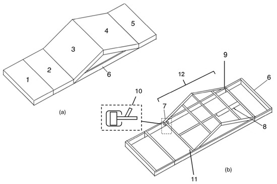
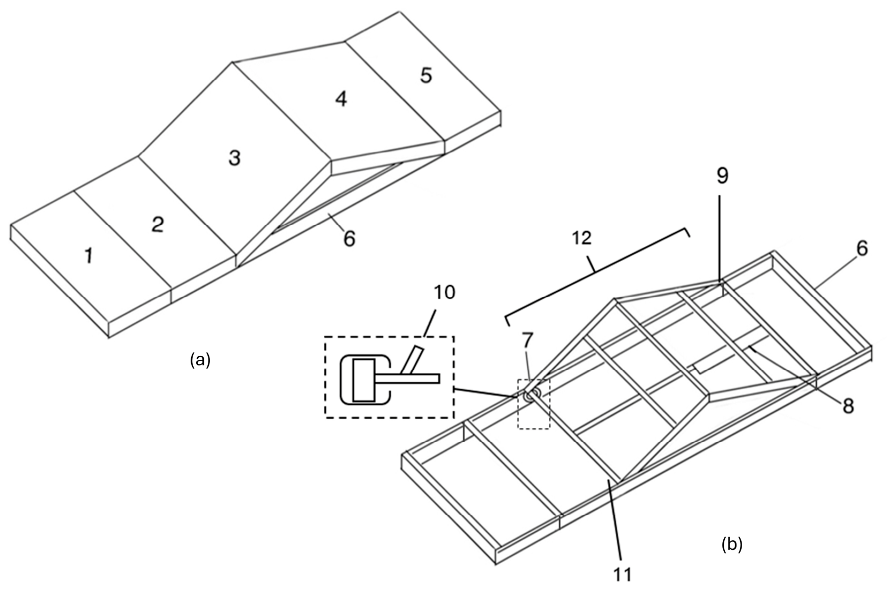
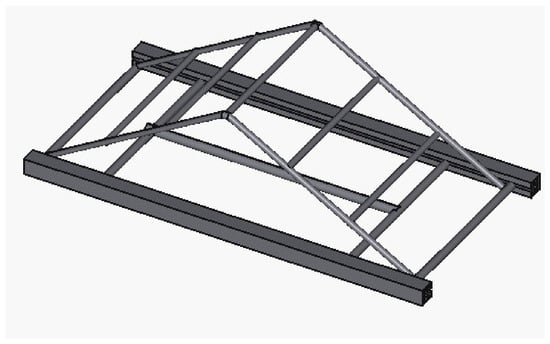
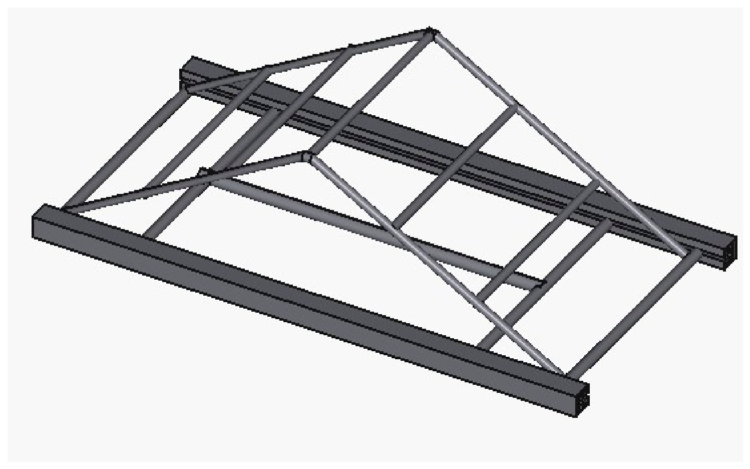
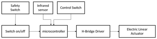
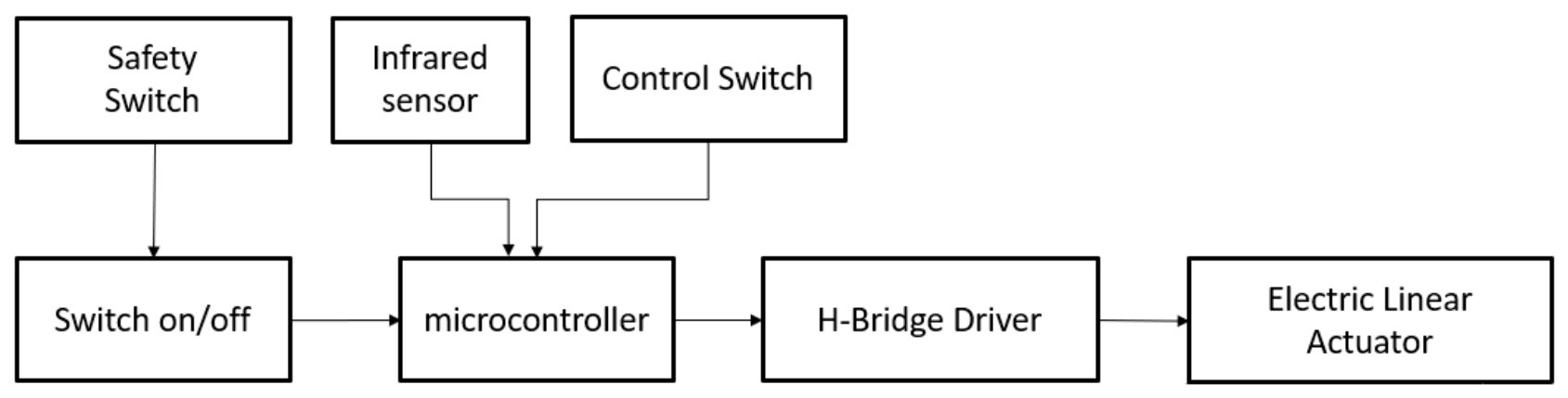
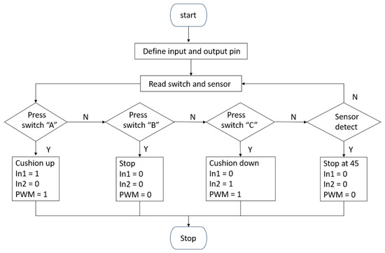
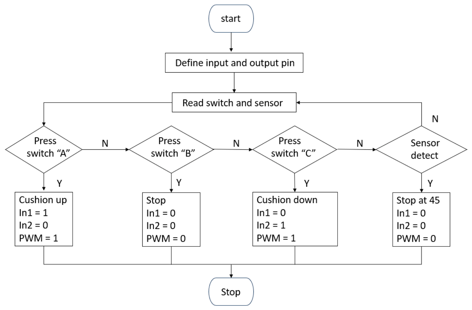
The structural support system consists of a main frame, a prone cushion upholstered with PVC leather, and a head cushion (Figure 1). The elevating mechanism is constructed from stainless steel, enabling vertical adjustment of the structure, as shown in Figure 2. The circuit system includes a microcontroller that reads input signals from push-button switches and sensors to control the linear motor, allowing the mechanical structure to move upward or downward (Figure 3). The control program, developed in the Arduino IDE, regulates motor rotation and adjusts the inclination to achieve the desired angle via push-button operation, as illustrated in Figure 4.
Figure 1.
(a) Hardware framework supporting the PVC-leather-upholstered structure with head and tail cushions; (b) design of the mechanical structure. (1, 2: head and arm cushions; 3, 4: main torso mattress; 5: leg and foot cushion; 6: cushion support structure; 7: wheel rails; 8: linear actuator motor; 9: actuator fixation point; 10: internal casters/bearings; 11: wheel assembly; 12: patient support frame).
Figure 2.
Mechanical design of the developed system.
Figure 3.
A Block Diagram of the operation for the designed device.
Figure 4.
Flowchart of the developed system.
2.2. Hardware Mechanism and Movement
As shown in Figure 1, the steel frame incorporates a hinge joint system that allows for a wide range of motion from 0° to 45°. The standard clinical range of inclination angles for prone positioning is defined to support both patient comfort and therapeutic effectiveness. A neutral prone position at 0° (horizontal alignment) is used for resting. The Trendelenburg prone position, with the head positioned lower than the body at an angle between 15° and 30°, is the most commonly applied range in postural drainage therapy. This utilizes gravitational force to facilitate the drainage of airway secretions from the lower lung lobes, thereby enhancing sputum clearance and supporting airway clearance techniques. According to clinical guidelines, the maximum safety limit should not exceed 45° in the head-down position, as steep Trendelenburg angles are associated with a significant increase in intraocular pressure (IOP) and intracranial pressure (ICP), potentially leading to neurological and ocular complications during prolonged therapy [17,18,19].
The base is constructed with a sliding rail system, allowing the individual sections to remain properly aligned during adjustment. This configuration supports the upholstery structure, which is covered with PVC leather for durability and hygiene, including the header and tail cushions. As shown in Figure 1, the retractable prone positioning device is designed to assist patients in maintaining a prone posture, particularly when independent sputum drainage is difficult. The device utilizes gravity, in combination with other ACTs to support effective airway secretion clearance.
The system consists of a main mattress (Section 3 and Section 4) that supports the patient’s torso and can be adjusted to the desired inclination angle. Section 1 and Section 2 are designated for supporting the patient’s head and arms, while Section 5 is dedicated to supporting the leg and feet.
The system consists of a main mattress (components 3 and 4 in Figure 1a) that supports the patient’s torso and can be adjusted to the desired inclination angle. Components 1 and 2 (Figure 1a) are designated for supporting the patient’s head and arms, while component 5 (Figure 1a) is dedicated to supporting the leg and feet. The cushion support structure (part 6 in Figure 1b) is constructed from C-shaped metal steel. The wheel rails (part 7 in Figure 1b) are integrated with bearings that function as internal casters (part 10 in Figure 1b). This configuration ensures smooth, precise, and stable movement of the structure along the rail system. All supporting components and upholstery surfaces are covered with PVC material to facilitate hygiene and cleaning.
The patient support frame (part 12 in Figure 1b) is responsible for lifting and supporting the cushion assembly and operates under motorized control. A linear actuator motor (part 8 in Figure 1b) serves as the primary leveling mechanism by extending or retracting along a straight axis connected to the wheel assembly (part 11 in Figure 1b), thereby adjusting the mattress angle. The actuator is secured to the main structure via a fixation point (part 9 in Figure 1b) to ensure stable and accurate operation.
As illustrated in Figure 1 and Figure 2, the structural design focuses on a modular support system constructed to facilitate prone positioning for therapy. The assembly is divided into two functional layers: a rigid steel frame that enables mechanical movement and a segmented support structure that distributes the patient’s weight. The base structure and sliding rail system are designed with specific anatomical support zones.
Component 3 (Figure 1a) provides support for the patient’s torso and is the primary movable component driven by the linear actuator. Section 4 is connected to Section 5 with hinges. The primary load-bearing structure for Section 3 and Section 4 has dimensions of 60 × 127 cm. Meanwhile, the frame supporting the sliding wheel rails measures 60 × 130 cm to allow for proper clearance and movement. The third section is the dynamic component of the design, utilizing a linear motor to achieve precise angular adjustments. This allows the equipment to safely transition the patient into the therapeutic prone position while maintaining structural stability through the integrated rail system. The structure was simulated at an angle of 30 degrees as shown in Figure 2, with a steel diameter of 2.5 inches and a thickness of 1 mm.
2.3. Control System Architecture
The mechanical movement is driven by an electronic control system, as detailed in the block diagram in Figure 3. The system integrates a microcontroller that processes inputs from user-operated control switches, an infrared (IR) object detection sensor, an on/off switch, and a safety switch. The operational workflow is as follows.
- Microcontroller logic: The controller regulates the leveling of the cushion by sending signals to an H-Bridge driver;
- Actuation: The H-Bridge driver manages the electric linear actuators located beneath the mattress, providing the necessary torque to push the equipment up or lower it down;
- Positioning: This allows the mattress to achieve precise therapy angles in prone positions. Once the desired level is reached, the user can stop the motor or release the system to lock the current position using the control switch;
- Safety Protection: The safety switch was designed to remove all power from the system.
The microcontroller gets the control signal from the control switches to control the linear motor under the mattress, allowing the mattress to adjust the prone position up and down to achieve a therapy angle, and a stop switch to maintain the desired position. A safety switch that immediately halts system operation in emergencies.
2.4. Workflow
The operation starts by receiving a command from the push button to raise the seat upwards. An infrared object detection sensor is installed inside the structure for detection at a maximum angle of 45 degrees according to the clinical guideline. As shown in Figure 4.
The H-Bridge is an electronic circuit that controls the linear motor in either direction. By toggling the digital inputs (In1, In2) and the pulse width modulation (PWM) signal, the microcontroller controls the actuator’s state. The digital pins In1 and In2 function as directional switches.
- 5.
- When In1 = 1 and In2 = 0, the H-bridge allows current to flow in a direction that extends the linear actuator, raising the cushion;
- 6.
- When In1 = 0 and In2 = 1, the current flow is reversed, causing the actuator to retract and lower the cushion;
- 7.
- When both In1 = 0 and In2 = 0, the circuit is disconnected or shorted to ground, causing the motor to stop immediately.
A comprehensive view of the system’s operational logic is shown in Table 1.
Table 1.
Operational logic for h-bridge directional control and actuator state.
2.5. Test Design
The functional performance of the prone positioning device was evaluated through physical testing using simulated patient loads. The objective of testing was to assess the mechanical stability, angle maintenance capability, and operational reliability of the device. Testing on a conventional bed may introduce errors due to mattress compliance and surface deformation. To minimize this effect and ensure accurate and repeatable angle measurements, the experiments were conducted by placing the prone positioning device on a rigid, solid surface.
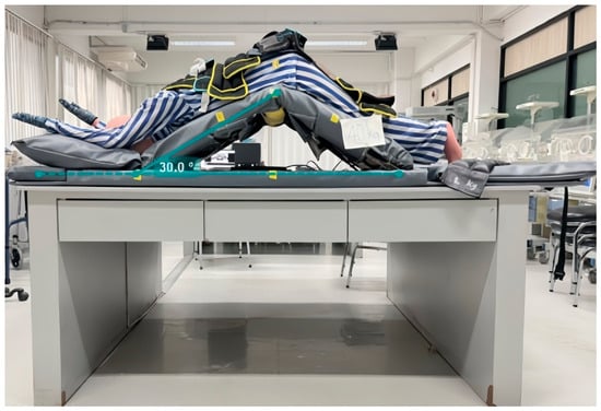
We applied a full-body mannequin in a prone position with sandbags distributed evenly across the torso to represent body weight. The applied weights ranged from 40 kg to 80 kg, covering the typical weight range of adult patients. During testing, the device was adjusted to a target inclination angle of 30°. The inclination angle was measured using an angle ruler, and the entire test procedure was recorded on video for a continuous duration of 30 min. The recorded videos were subsequently analyzed using Kinovea software (GPL v2 license) to extract angle and height measurements over time.
The evaluation focused on the device’s ability to maintain the target inclination angle without observable collapse, drift, or mechanical instability throughout the test duration. The measured results are shown in Table 2.
Table 2.
Test results measure the angle at the fuselage below the reference level at the weight value and duration difference.
3. Results
The functional performance of the prone positioning device was evaluated through physical testing under simulated patient loads, as described in Section 2.5. The evaluation focused on the device’s ability to maintain a stable therapeutic inclination angle during continuous operation under different weight conditions.
Functional Performance Under Simulated Patient Loads
The functional performance of the lifting mechanism and support structure was assessed using a full-body mannequin with sandbags distributed across the torso to simulate patient body weight. The applied loads ranged from 40 to 80 kg. During each test, the device was adjusted to an inclination angle of 30° relative to the horizontal plane and operated continuously for 30 min. Throughout the testing period, the inclination angle remained constant for all tested load conditions. No observable angular drift, structural deformation, or mechanical instability was detected during the 30 min operation. The lifting mechanism maintained the target position without back-driving or collapse after the actuator was stopped.
Figure 5 shows the experimental setup with the mannequin positioned in the prone posture on the device. The measured inclination angles at different time points and load conditions are summarized in Table 2. As shown in the results, the inclination angle remained at 30° for all loads and throughout the entire test duration The overall operation and movement of the prone positioning device are demonstrated in the supplementary video (see Supplementary Material Video S1).
Figure 5.
The arrangement in a prone position.
4. Discussion
The functional performance evaluation demonstrated that the developed prone positioning device is capable of maintaining a stable therapeutic inclination angle under simulated patient loads ranging from 40 to 80 kg. During a continuous 30 min operation at an inclination angle of 30°, no observable angular drift, mechanical instability, or structural deformation was detected. These results indicate that the mechanical design and the lifting mechanism provide stability for a short duration of therapy.
The ability to hold the setting angle without collapse is important for clinical practice to ensure patient safety and minimize the need for manual adjustment by physical therapists. Stable angle maintenance reduces the risk of positional changes that may compromise therapeutic effectiveness or increase physiological risks associated with excessive inclination angles. Compared with conventional postural drainage approaches that rely on pillows, foam wedges, or manual support, the proposed device offers improved mechanical reliability and consistency of positioning. This is especially relevant for community-based and home-care rehabilitation settings, where portable and stable equipment is essential and where therapists often work without additional clinical support. In addition, the use of waterproof PVC-leather upholstery supports infection control and hygiene requirements. The total device weight of approximately 25 kg provides adequate structural robustness while being able to be transported during home visits.
Although the present result demonstrates promising functional performance, the evaluation was limited to intermediate inclination angles between 20° and 45° and simulated patient loads representative of typical adult patients. Future work should include usability assessment with healthy volunteers, ergonomic evaluation for therapists, and clinical studies to assess patient comfort and therapeutic effectiveness during actual postural drainage sessions.
5. Conclusions
We designed and fabricated an adjustable prone positioning device that supports postural drainage therapy through controlled inclination and stable mechanical support. The proposed hardware integrates a modular steel frame and a linear actuator system to enable precise angular adjustment over a 0–45 ° range. Function testing results under simulated patient loads demonstrated that the device can safely support weights ranging from 40 to 80 kg while maintaining a stable therapeutic inclination angle of 30° during continuous 30 min operation. No structural deformation, angular drift, or mechanical failure was observed, confirming the reliability of the mechanical and control systems. The total device weight of approximately 25 kg provides a balance between structural robustness and portability, making the system suitable for both clinical and community-based rehabilitation settings. Overall, the developed prone positioning device represents a practical alternative to conventional manual positioning methods for postural drainage therapy. In the following studies, clinical evaluation will be conducted with healthy volunteers to assess user comfort, therapist usability, and therapeutic effectiveness in real-world applications.
Supplementary Materials
The following supporting information can be downloaded at: https://drive.google.com/drive/folders/1xEVicBAm-oKcOdKkq594dTf_Ckf1q3fq?usp=sharing (accessed on 30 September 2026); Video S1: Demonstration of the operation of the prone positioning device.
Author Contributions
Conceptualization, T.Y. and J.P.; methodology, T.Y. and J.P.; software, T.Y.; validation, T.Y.; formal analysis, T.Y.; resources, T.Y.; data curation, J.P.; writing—original draft preparation, J.P.; writing—review and editing, T.Y. and J.P.; visualization, J.P.; supervision, T.Y. and J.P.; project administration, J.P. All authors have read and agreed to the published version of the manuscript.
Funding
This research received no external funding.
Institutional Review Board Statement
The study was conducted in accordance with the Declaration of Helsinki and approved by the Ethics Review Board of Rangsit University (RSUERB2025-004; approval date: 27 February 2025). Since this research did not involve human subjects.
Informed Consent Statement
Not applicable.
Data Availability Statement
No new datasets were generated or analyzed in this study. Data supporting the reported results is available from the corresponding author upon reasonable request.
Acknowledgments
The authors sincerely thank Benjarat Saengthong, for Administration, Faculty of Physical Therapy and Sports Science, Rangsit University, for her technical guidance related to pulmonary patient care. In addition, the authors gratefully acknowledge Nuntachai Thongpance, of the College of Biomedical Engineering, Rangsit University, for his ongoing support of this research.
Conflicts of Interest
The authors declare no conflicts of interest.
Abbreviations
The following abbreviations are used in this manuscript:
| ACTs | airway clearance techniques |
| APPF | adjustable prone positioning frame |
| AS | ankylosing spondylitis |
| ICP | intracranial pressure |
| IOP | intraocular pressure |
| IR | infrared |
| PWM | pulse width modulation |
| PVC | polyvinyl chloride |
References
- Nici, L.; Donner, C.F.; Wouters, E.; ZuWallack, R.; Ambrosino, N.; Bourbeau, J.; Carone, M.; Celli, B.; Engelen, M.; Fahy, B.; et al. American Thoracic Society/European Respiratory Society Statement on Pulmonary Rehabilitation. Am. J. Respir. Crit. Care Med. 2006, 173, 1390–1413. [Google Scholar] [CrossRef] [Scilit]
- Wanner, A.; Salathé, M.; O’Riordan, T.G. Mucociliary clearance in the airways. Am. J. Respir. Crit. Care Med. 1996, 154, 1868–1902. [Google Scholar] [CrossRef] [Scilit]
- Randell, S.H.; Boucher, R.C. Effective Mucus Clearance Is Essential for Respiratory Health. Am. J. Respir. Cell Mol. Biol. 2006, 35, 20–28. [Google Scholar] [CrossRef] [Scilit]
- Fahy, J.V.; Dickey, B.F. Airway Mucus Function and Dysfunction. N. Engl. J. Med. 2010, 363, 2233–2247. [Google Scholar] [CrossRef] [Scilit]
- McIlwaine, M.; Bradley, J.; Elborn, J.S.; Moran, F. Personalising airway clearance in chronic lung disease. Eur. Respir. Rev. 2017, 26, 160086. [Google Scholar] [CrossRef] [Scilit]
- Belli, S.; Prince, I.; Savio, G.; Paracchini, E.; Cattaneo, D.; Bianchi, M.; Masocco, F.; Bellanti, M.; Balbi, B. Airway Clearance Techniques: The Right Choice for the Right Patient. Front. Med. 2021, 8, 544826. [Google Scholar] [CrossRef] [Scilit]
- Albert, R.K.; Hubmayr, R.D. The Prone Position Eliminates Compression of the Lungs by the Heart. Am. J. Respir. Crit. Care Med. 2000, 161, 1660–1665. [Google Scholar] [CrossRef] [Scilit]
- Messerole, E.; Peine, P.; Wittkopp, S.; Marini, J.J.; Albert, R.K. The Pragmatics of Prone Positioning. Am. J. Respir. Crit. Care Med. 2002, 165, 1359–1363. [Google Scholar] [CrossRef] [Scilit]
- Gattinoni, L.; Busana, M.; Giosa, L.; Macrì, M.; Quintel, M. Prone Positioning in Acute Respiratory Distress Syndrome. Semin. Respir. Crit. Care Med. 2019, 40, 94–100. [Google Scholar] [CrossRef] [Scilit]
- Scholten, E.L.; Beitler, J.R.; Prisk, G.K.; Malhotra, A. Treatment of ARDS With Prone Positioning. Chest 2017, 151, 215–224. [Google Scholar] [CrossRef] [Scilit]
- Singh, N.; Rao, P.B.; Ambesh, S.P.; Gupta, D. An innovative low-cost head-rest for anesthesia in prone position. J. Neurosci. Rural Pract. 2014, 5, S96–S97. [Google Scholar] [CrossRef] [Scilit]
- Ding, Z.; Ren, Y.; Cao, H.; Li, Y.; Chen, S.; Miao, J.; Li, J. An Innovative Prone Position Using a Body-Shape Plaster Bed and Skull Traction for Posterior Cervical Spine Fracture Surgeries. Front. Surg. 2022, 9, 649421. [Google Scholar] [CrossRef] [Scilit]
- Rahman, O.; Zbeda, R.M.; Romeo, A.A. The Development of an Orthopaedic Prone Positioning Tool in the COVID-19 Era. J. Orthop. Exp. Innov. 2021, 2, 21441. [Google Scholar] [CrossRef] [Scilit]
- Kolb, B.; Large, J.; Watson, S.; Smurthwaite, G. An innovative prone positioning system for advanced deformity and frailty in complex spine surgery. J. Neurosurg. Spine 2020, 32, 229–234. [Google Scholar] [CrossRef] [Scilit]
- Zhang, W.; Yu, H.Y.; Wang, H.L.; Zheng, G.H.; Zhai, Y.L.; Cui, X.L.; Jiang, J.S.; Zhang, J.X.; Shen, C.L.; Wang, Y. An innovative adjustable prone positioning frame for treatment of severe kyphosis secondary to ankylosing spondylitis with two-level osteotomy. Eur. Spine J. 2021, 30, 3209–3215. [Google Scholar] [CrossRef] [Scilit]
- Ghersi, I.; Mariño, M.; Miralles, M.T. Smart medical beds in patient-care environments of the twenty-first century: A state-of-art survey. BMC Med. Inform. Decis. Mak. 2018, 18, 63. [Google Scholar] [CrossRef] [Scilit]
- Awad, H.; Santilli, S.; Ohr, M.; Roth, A.; Yan, W.; Fernandez, S.; Roth, S.; Patel, V. The effects of steep trendelenburg positioning on intraocular pressure during robotic radical prostatectomy. Anesth. Analg. 2009, 109, 473–478. [Google Scholar] [CrossRef] [Scilit]
- Ripa, M.; Schipa, C.; Kopsacheilis, N.; Nomikarios, M.; Perrotta, G.; De Rosa, C.; Aceto, P. The Impact of Steep Trendelenburg Position on Intraocular Pressure. J. Clin. Med. 2022, 11, 2844. [Google Scholar] [CrossRef] [Scilit]
- Hoshikawa, Y.; Tsutsumi, N.; Ohkoshi, K.; Serizawa, S.; Hamada, M.; Inagaki, K.; Tsuzuki, K.; Koshimizu, J.; Echizen, N.; Fujitani, S.; et al. The effect of steep Trendelenburg positioning on intraocular pressure and visual function during robotic-assisted radical prostatectomy. Br. J. Ophthalmol. 2013, 98, 305–308. [Google Scholar] [CrossRef] [Scilit]

Disclaimer/Publisher’s Note: The statements, opinions and data contained in all publications are solely those of the individual author(s) and contributor(s) and not of MDPI and/or the editor(s). MDPI and/or the editor(s) disclaim responsibility for any injury to people or property resulting from any ideas, methods, instructions or products referred to in the content. |
© 2026 by the authors. Licensee MDPI, Basel, Switzerland. This article is an open access article distributed under the terms and conditions of the Creative Commons Attribution (CC BY) license.