


Abstract
Fetal bovine serum (FBS) is the most widely used supplement in cell culture; however, ethical and environmental concerns have driven the search for alternatives. Adult bovine serum (ABS) is one of the promising substitutes. Here, we evaluated complete and gradual replacement of FBS with ABS together with the small molecule MBF 3 from the Maybridge fragment library, which was previously shown to enhance the viability of pancreatic carcinoma cell line Panc-1. Complete substitution with 10% ABS reduced cell viability by 30% and decreased cell numbers. Partial replacement of FBS with ABS, from 8% FBS + 2% ABS to 4% FBS + 6% ABS, maintained comparable cell viability to that observed with 10% FBS, whereas 2% FBS + 8% ABS and 10% ABS significantly reduced viability. G2/M accumulation and decreased cell numbers were observed among 10% ABS-supplemented conditions, indicating attenuated proliferation. Under 6% ABS conditions, MBF 3 modestly increased cell viability but failed to restore cell numbers, suggesting that it promotes cellular survival and adaptation under growth factor-limited conditions rather than rescuing proliferative capacity. Overall, this study demonstrates that ABS induces G2/M-phase accumulation in Panc-1 cells, highlighting its potential utility for cell-cycle synchronization and suggesting a potential role in cell-cycle transition and longer-term cell culture studies. Future studies should evaluate the applicability of ABS and MBF 3 across a broader range of cell types and explore co-supplementing with chemically defined additives to promote cellular proliferation and facilitate the transition away from FBS.
1. Introduction
Fetal bovine serum (FBS) is one of the most widely used supplements in mammalian cell culture owing to its rich content of growth factors, hormones, attachment factors, proteins, lipids, vitamins, and trace elements that support cell survival, proliferation, and metabolism [1]. Despite its widespread use, the inhumane harvesting process of FBS collection raises significant ethical concerns [2]. FBS is collected from bovine fetuses during the slaughter of pregnant cows, which has been the subject of ongoing animal welfare debates [3]. Regulatory authorities, research institutions, and funding organizations increasingly advocate for the reduction or replacement of FBS to promote more ethical and sustainable research practices [4]. While numerous FBS alternatives, including human platelet lysates (hPLs), chemically defined media and recombinant protein supplements, have been developed, adult bovine serum (ABS) has emerged as an ethically sustainable and cost-effective option due to its non-fetal origin and lower production cost [5]. Previous studies have demonstrated that ABS can support the growth and maintenance of various cell types, although its performance is often cell-type dependent and may be inferior to that of FBS because of differences in composition [6]. Expressions of representative serum growth factors, including vascular endothelial growth factor A (VEGF-A), basic fibroblast growth factor (bFGF), insulin-like growth factor 1 (IGF-1), transforming growth factor-β (TGF-β), and total protein in FBS and ABS are shown in Table 1. ABS has a higher total protein content due to the elevated immunoglobulin G (IgG) associated with immune maturation, but lower growth factor levels compared to FBS as fetal circulation is enriched with mitogenic and differentiation signals that are required for embryonic growth. Consequently, strategies that overcome the limited growth-promoting capacity of ABS may facilitate its use as a viable alternative to FBS. For optimizing ABS culture systems, chemically defined supplements are generally considered to be a more practical approach than recombinant growth factors as they balance cost, ethical considerations, and cell growth performance. For example, supplementation of ABS with insulin–transferrin–selenium (ITS) utilizes the residual growth factors that are present in ABS. Insulin enhances glucose and amino acid uptake via the PI3K/AKT pathways, transferrin supplies iron required for cellular metabolism and proliferation, and selenium maintains redox homeostasis, thereby preserving signaling competence [7]. Previously, a study about Maybridge Library screening identified several Maybridge Library fragments (MBFs) at a concentration of 10 μM that increased the viability of pancreatic carcinoma Panc-1 cells by more than 10%, with MBF 3 exhibiting the strongest effect [8]. In this study, we explored the use of MBF 3 to improve the cell growth in ABS-containing culture media and thereby reduce the requirement for FBS in Panc-1 cell culture. By assessing cell viability, cell number and cell-cycle progression, we aimed to determine whether MBF 3 could support Panc-1 cell viability and proliferation under ABS conditions and provide a practical approach for decreasing dependence on FBS.
Table 1.
Concentrations of components in FBS and ABS (μg/L unless stated otherwise).
2. Methods and Materials
2.1. Cell Subculture
The human pancreatic duct epithelioid carcinoma cell line Panc-1 was cultured in Dulbecco’s modified Eagle’s medium (DMEM) (Sigma-Aldrich, Darmstadt, Germany, D6429) containing 10% heat-inactivated fetal bovine serum (FBS; Gibco, Grand Island, NY, USA, A5256801, Lot B2741832RP, Brazil origin,) and 1% penicillin–streptomycin (Thermo Fisher Scientific, Waltham, WA, USA, 15140-122). Cells were routinely cultured at 37 °C in a humidified 5% CO2 atmosphere with continuous exponential growth at a density ranging between 1 × 10⁵ and 1 × 10⁶ cells/mL by passaging every three days. The medium was discarded and washed with 5 mL 1× phosphate-buffered saline (PBS) (Sigma-Aldrich, Darmstadt, Germany, D8537) to remove all traces of serum from the medium. 1 mL of 0.1% trypsin (Sigma-Aldrich, Darmstadt, Germany T3924) was added, followed by incubation for 5 min at 37 °C. The flask was gently tapped several times, and cell detachment was examined under a light microscope (Nikon, Tokyo, Japan). Then, 4 mL of medium was added to neutralize the trypsin, and the cell suspension was transferred to a 15 mL tube (Starlab, Hamburg, Germany, S1615-5515) for centrifugation at 1000 rpm for 5 min. The supernatant was then discarded, and cells were resuspended in 1 mL medium by gentle pipetting until pellets were resuspended. The desired number of cells was then transferred back into a T75 TC flask (Starlab, CC7682-4175, Hamburg, Germany) containing 15 mL of pre-warmed media.
2.2. Treatment
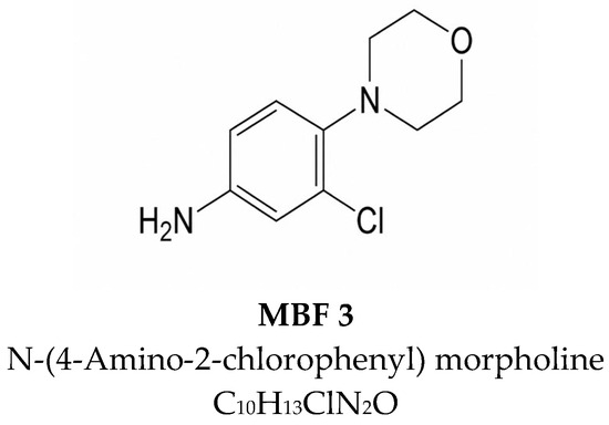
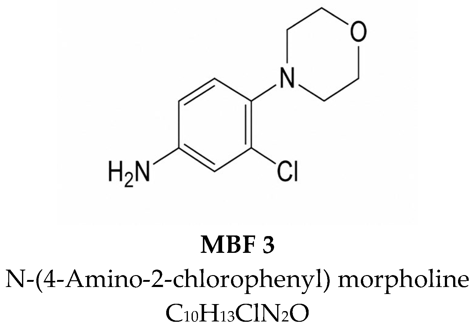
Cells were cultured in medium supplemented with FBS (Gibco, Grand Island, NY, USA, heat-inactivated, A5256801, Lot B2741832RP, Brazil origin) and adult bovine serum (ABS; Gibco, Grand Island, NY, USA, 16170078; Lot 2742078, New Zealand origin). The sera were stored at −20 °C and thawed in DMEM before being stored at 4 °C for cell culture. A single serum lot was used throughout the study to eliminate lot-to-lot variation. Figure 1 showed the chemical structure of MBF 3 from Maybridge Library (https://www.thermofisher.com/uk/en/home/industrial/pharma-biopharma/drug-discovery-development/screening-compounds-libraries-hit-identification/maybridge-fragment-libraries.html (accessed on 1 July 2026)). MBF 3 was dissolved in dimethyl sulfoxide (DMSO) to prepare a 50 mM stock solution and stored at −20 °C. MBF 3 was diluted 5000-fold in freshly prepared culture medium prior to treatment to achieve a final concentration of 10 μM. The control group was treated with 0.02% DMSO. After 120 h incubation, endpoint measurements were performed as described below.
Figure 1.
Chemical structure of MBF 3.
2.3. Cell Number Counting
Cell counting was performed manually by using a hemocytometer (Marienfeld, Lauda-Königshofen, Germany, 0640010) and the trypan blue staining technique. After harvesting the cells from a 6-well plate into centrifuge tubes (Eppendorf, Hamburg, Germany, 0030120086), a 1:1 mixture of the cell suspension and trypan blue solution (Sigma-Aldrich, Darmstadt, Germany, T8154) was prepared. Then, 10 μL of the stained cell suspension was loaded into the sample chamber of the hemocytometer (Marienfeld, Lauda-Königshofen, Germany, 0640010). The light microscope was used to count the number of viable (unstained) cells from the four large corner quadrants. The number of viable cells was calculated using the average count of cells from the four corner quadrants, multiplying this value by the dilution factor and 1 × 10⁴ to get the total number of cells per mL of culture media.
2.4. Cell Viability
Cell viability was assessed using the 3-(4,5-dimethylthiazol-2-yl)-2,5-diphenyltetrazolium bromide (MTT) assay following the manufacturer’s instructions. At the endpoint, the medium in the 96-well plate was replaced with 50 μL of fresh media containing reduced FBS (1.5%) and 10 μL of the MTT reagent (APExBIO, Houston, TX, USA, B7777). The plate was then incubated at 37 °C for 3 h until purple precipitate was visible. This was followed by the addition of 100 μL of a solution comprising isopropanol (Sigma-Aldrich, Darmstadt, Germany, 190764) with 37% hydrochloric acid (Sigma-Aldrich, Darmstadt, Germany, 258148) in a ratio of 250:1 to all the wells. Absorbance in each well was measured by Polarstar Omega Plate Reader at 570 nm (BMG LABTECH, Ortenberg, Germany, 0415B001G). Cell viability in the 10% FBS control group was set to 100% for each passage and experiment. All the data were normalized to the corresponding 10% FBS control.
2.5. Cell-Cycle Progression
One million cells per sample were harvested and fixed in 2 mL of 70% ethanol overnight at 4 °C. Cells were then washed thoroughly using PBS to remove all residual fixative. Then, 0.5 mL of FxCycle™ PI/RNase Staining Solution (Invitrogen, Waltham, MA, USA, F10797) was added into each sample followed by a 15 min incubation at room temperature in the dark to prevent photobleaching. Finally, samples were analyzed directly by flow cytometry (BD Science, Franklin Lakes, NJ, USA) using 488 nm excitation and collecting emission with a 610/20 bandpass filter. Histograms were generated using Flowjo software V10.10.2 (Ashland, OR, USA) and the Dean–Jett–Fox model.
2.6. Statistical Analysis
All the data were analyzed as mean ± SEM using GraphPad Prism 11.1.0 (San Diego, CA, USA). Comparisons among multiple treatment groups were assessed using one-way ANOVA with Tukey correction. A p value < 0.05 was considered significant.
3. Results
3.1. The 10% ABS Reduces Panc-1 Cell Viability and Cell Number
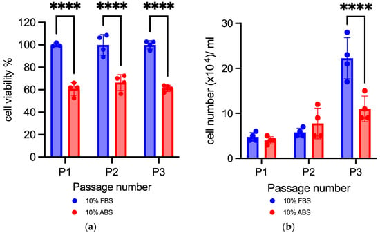
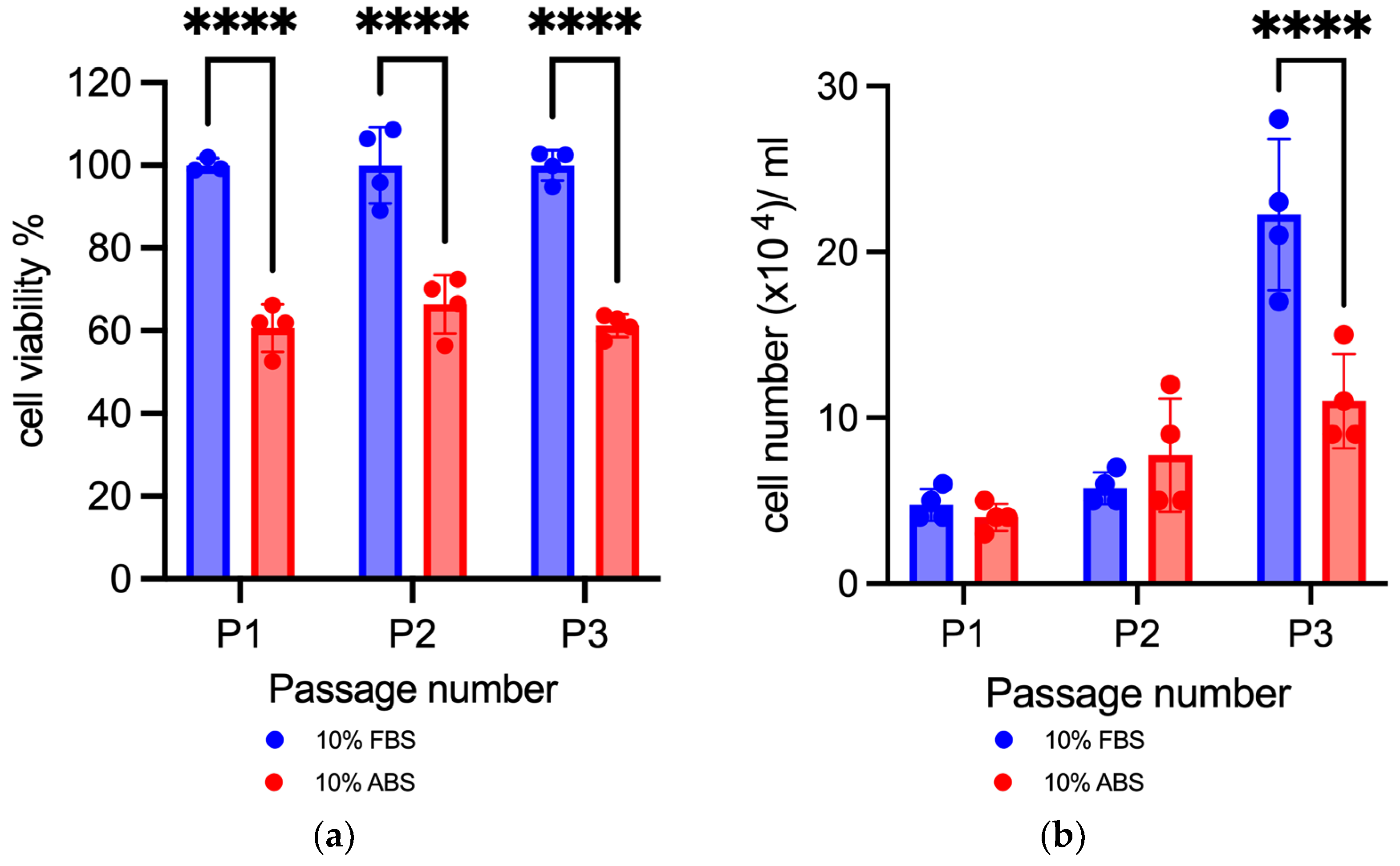
Culturing Panc-1 cells in DMEM medium supplemented with 10% ABS resulted in a reduction in cell viability of more than 30% compared with 10% FBS across three successive passages (Figure 2a). The cell numbers in 10% ABS-containing media were numerically higher than those in 10% FBS after the second passage but declined markedly by approximately 50% following the third passage (Figure 2b).
Figure 2.
Effects of 10% FBS and 10% ABS on Panc-1 (a) cell viability (n = 4 biological replicates); (b) cell number (n = 4 biological replicates). Data are presented as the mean ± SD. **** p < 0.0001. The blue bar represents 10% FBS and red 10% ABS.
3.2. Partially Substituting FBS with ABS
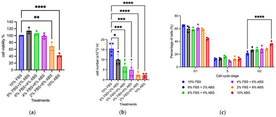
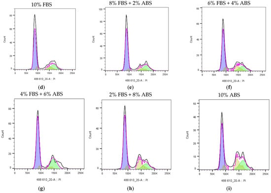
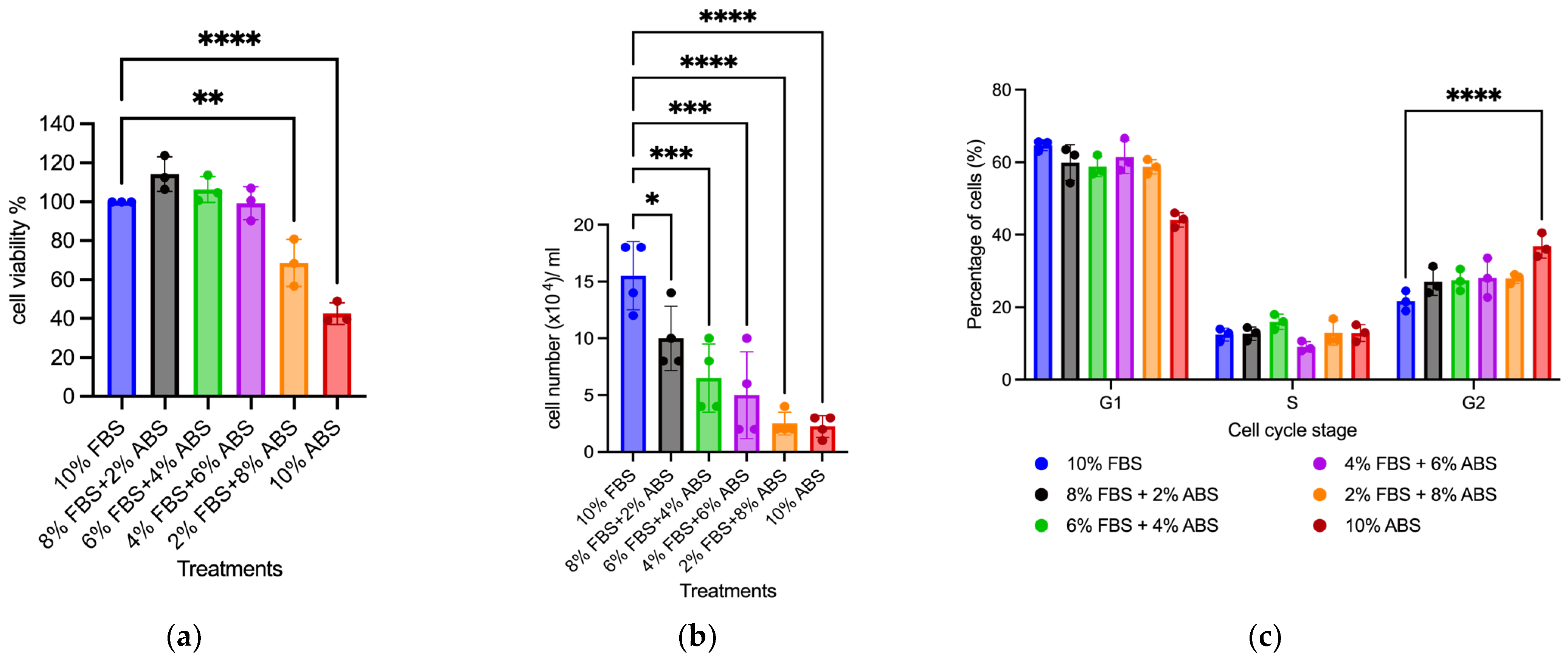
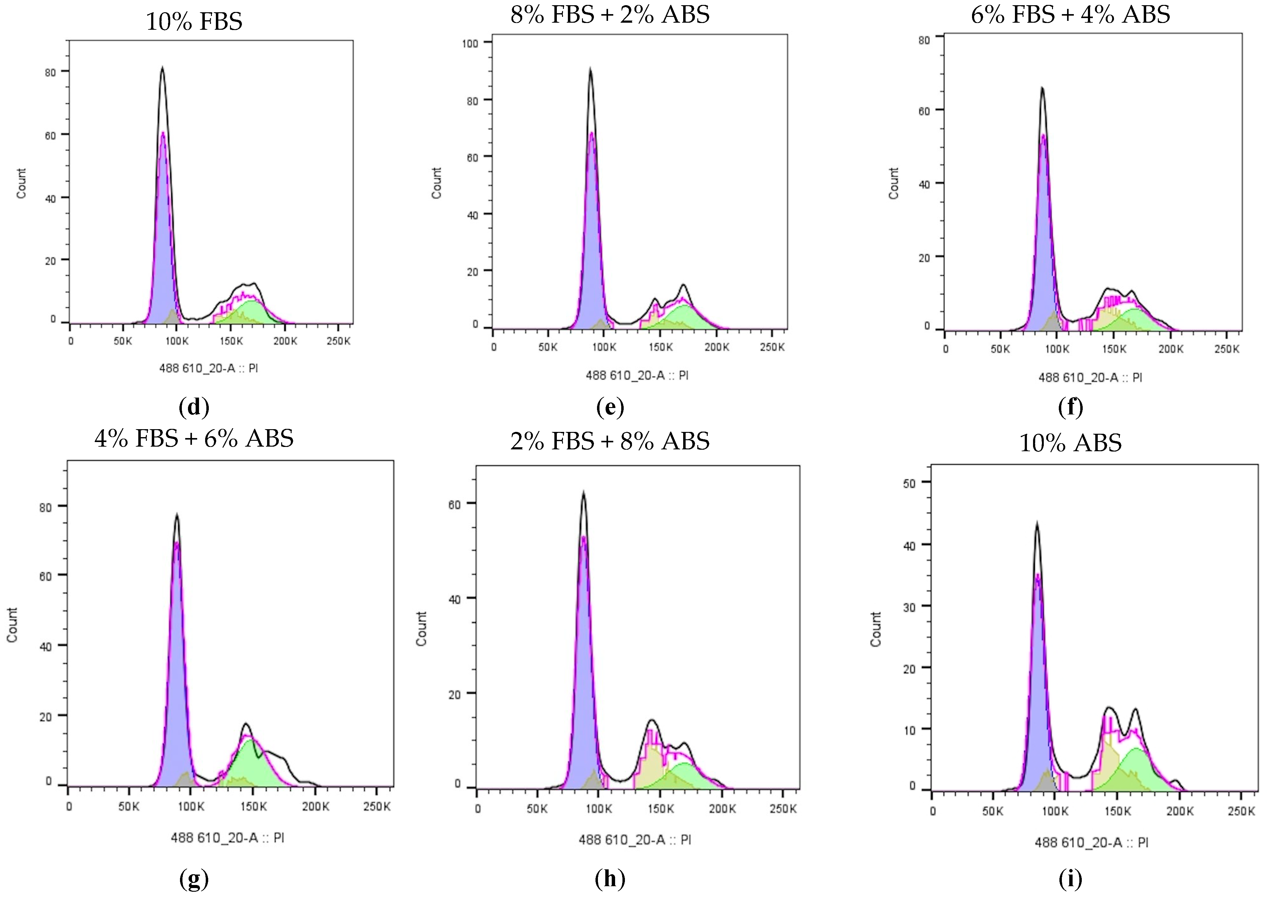
To further assess the potential for partially reducing FBS with ABS, Panc-1 cells were co-cultured with varying proportions of FBS and ABS (maintaining a total serum concentration of 10%) for 5 days. As illustrated in Figure 3a,b, supplementing ABS between 2% and 6% produced a slight upward trend in viability but significantly reduced cell number. Further substituting FBS with ABS at concentrations exceeding 8% led to a statistically significant decline in both cell viability and cell number. Notably, treatment with 10% ABS alone resulted in an approximately 50% reduction in cell viability compared with 10% FBS, consistent with our previous findings, and was associated with G2/M-phase accumulation (Figure 3).
Figure 3.
Effects of partially replacing FBS with ABS on Panc-1 (a) cell viability (n = 3 biological replicates); (b) cell number (n = 4 biological replicates); (c) cell cycle was analyzed as the percentage of cells at each stage (n = 3 biological replicates); (d–i) the cell-cycle progression was analyzed by Flowjo software and depicted using histograms. Data are presented as the mean ± SD. * p < 0.05, ** p < 0.01, *** p < 0.001, and **** p < 0.0001. The blue bar represents 10% FBS, black 8% FBS + 2% ABS, green 6% FBS + 4% ABS, purple 4% FBS + 6% ABS, yellow 2% FBS + 8% ABS, and red 10% ABS.
3.3. Replacing FBS with 6% ABS and 10 μM MBF 3
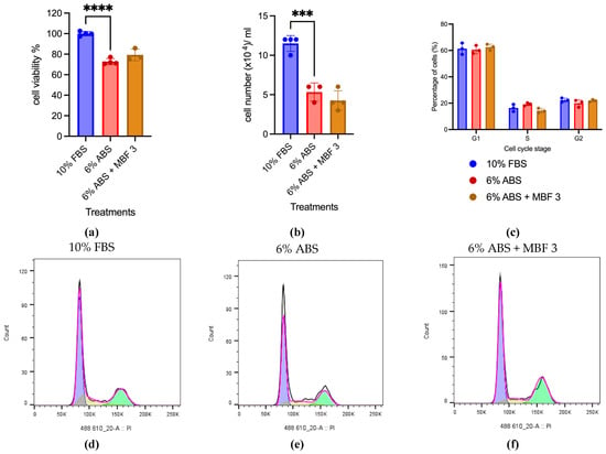
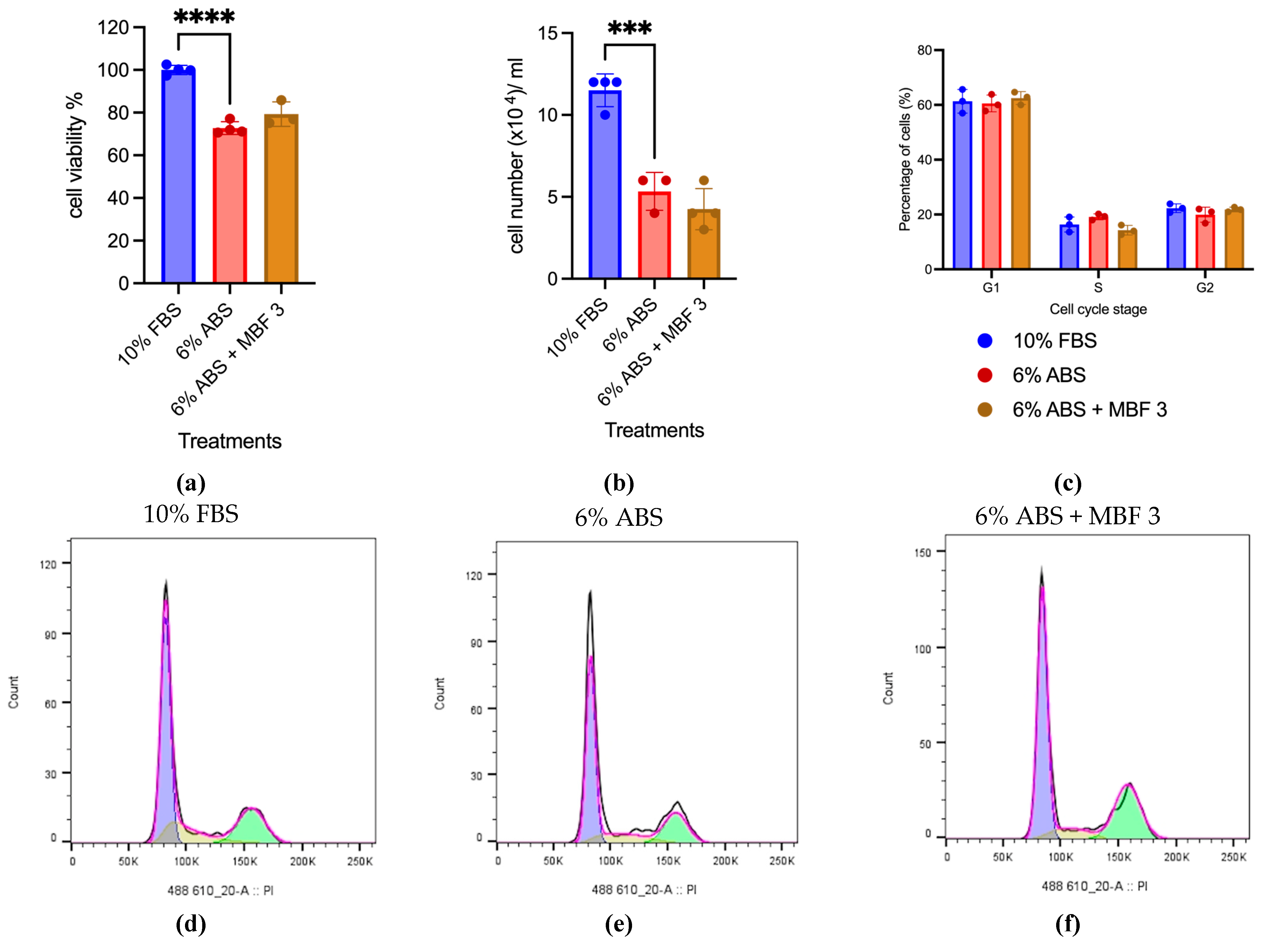
Figure 3a showed that 4% FBS + 6% ABS achieved comparable Panc-1 viability to 10% FBS. Given that, 6% FBS was replaced with ABS. Then, 10 μM MBF 3, previously shown to enhance Panc-1 cell viability, was added to the 6% ABS. As shown in Figure 4a,b, culturing in 6% ABS significantly reduced Panc-1 cell viability and cell number compared with 10% FBS (p < 0.001). However, treatment with MBF 3 resulted in a non-significant upward trend in cell viability, with no statistical difference compared with 6% ABS alone (Figure 4a). No significant improvement in cell number or effect on cell-cycle progression was observed (Figure 4b–f).
Figure 4.
Effects of 6% ABS and 10 μM MBF 3 on Panc-1 (a) cell viability (n = 4 biological replicates); (b) cell number (n = 4 biological replicates); (c) cell cycle was analyzed as the percentage of cells at each stage (n = 3 biological replicates); (d–f) the cell-cycle progression was analyzed by Flowjo software and depicted using histograms. Data are presented as the mean ± SD. *** p < 0.001, and **** p < 0.0001. The blue bar represents 10% FBS, red 6% ABS, and brown 6% ABS + MBF 3.
4. Discussion
FBS is widely used in cell culture due to its complex mixture of growth factors, hormones, proteins and lipids. However, its continued use raises ethical and sustainability concerns associated with fetal serum collection [1]. In line with the 3Rs principles (replacement, reduction, and refinement), considerable efforts have been directed toward developing alternatives to animal-derived culture supplements, including chemically defined media, hPLs and alternative animal sera [16]. Among these, ABS has emerged as an ethically favourable and cost-effective option, since it is obtained from cattle aged 12 months or older as a byproduct of the meat industry (ABS, approx. £85/500 mL; FBS, £400/500 mL) [5]. ABS contains higher levels of total proteins and endocrine-associated hormones that have been shown to support the differentiation of several cell types, including primary human fibroblasts and endothelial cells [5,17]. However, its lower abundance of embryonic growth factors may limit its capacity to sustain long-term cell growth and proliferation [6]. Previous studies have reported that, while 10% ABS maintained comparable cell viability and cell numbers to 10% FBS in C2BBe1 endothelial cells during 5-day incubation, proliferative capacity declined after serial passaging, as observed in both C2BBe1 and mesenchymal stem cells [6,17]. These findings highlight the cell-type specificity and reinforce that no universal serum replacement is optimal for all cell types. In our study, Panc-1 cell viability was significantly decreased in 10% ABS across all three passages, whereas a significant reduction in cell number was observed only at the third passage. The underlying reason for this discrepancy remains unclear. Nevertheless, these findings indicate that complete replacement of ABS is insufficient to support the long-term culture of Panc-1 cells. Partial replacement of FBS with ABS, however, maintained cell viability when up to 6% FBS was replaced with ABS (i.e., 8% FBS + 2% ABS to 4% FBS + 6% ABS) while maintaining a total serum concentration of 10%. Nevertheless, ABS-containing conditions were associated with reduced cell numbers and a trend towards G2/M-phase accumulation. G2/M arrest occurs when cells have completed DNA replication but fail to progress through mitosis, suggesting that ABS may not fully support the proliferative capacity provided by FBS. To our knowledge, this is the first study to demonstrate ABS-associated G2/M-phase accumulation and impaired proliferation in Panc-1 cells, highlighting a potential limitation of ABS as a complete substitute for FBS. Interestingly, G2/M-phase accumulation is reported to result in synchronized cell populations, which are widely used to investigate cell-cycle transitions, DNA replication, and long-term cell culture by improving population homogeneity [6]. Therefore, ABS supplementation may be advantageous for cell-cycle studies and stable long-term live-cell imaging under low-mitogenic conditions.
Recent studies have demonstrated that small molecules can partially compensate for the loss of serum-derived growth signals by modulating key pathways involved in cell survival, proliferation and metabolism [6,18]. Compared with recombinant growth factors, small molecules offer greater stability, lower cost, and improved suitability for cell culture applications [19,20]. For instance, several approaches have been developed to promote cell growth under serum-free or reduced-serum conditions, including inhibition of TGF-β signaling with SB431542 to restore limbal epithelial cell growth [6], the use of synthetic fibroblast growth factor receptor 1 (FGFR1) agonists to mimic the mitogenic activity of bFGF [18], and the combination of forskolin and p38 inhibitors to enhance bovine muscle stem cell expansion in the absence of FBS [21]. In addition, our previous study identified several MBF candidates that increased Panc-1 cell viability by more than 10%, with 10 μM MBF 3 showing the largest effect [8]. MBF 3 is a morpholine-substituted derivative. Morpholine-containing scaffolds are widely employed in chemistry because they improve aqueous solubility and the ability to participate in protein-ligand interactions [22]. Since the combination of 4% FBS + 6% ABS maintained comparable cell viability to 10% FBS, MBF 3 was subsequently evaluated as a supplement to maintain Panc-1 cell viability under reduced serum conditions. MBF 3 showed an increasing trend towards promoting Panc-1 cell viability without significantly altering cell number or cell-cycle progression under 6% ABS, suggesting that it may promote cellular survival rather than directly stimulating mitogenic signaling under ABS culture conditions. Thus, the pro-viability effect of MBF 3 appears to be evident for FBS, and does not exert a comparable effect with ABS.
Several study limitations should be acknowledged. Firstly, the study was conducted exclusively in the Panc-1 cell line, so the applicability of ABS and MBF 3 to other cell types remains unknown. Secondly, complete FBS replacement may require the combination of MBF 3 with chemically defined supplements, e.g., ITS, to stimulate mitogenic pathways and enhance cell proliferation [7]. In addition, prolonged culture may induce cellular adaptation and metabolic reprogramming; therefore, further studies should evaluate the long-term stability, safety, and biological effects of MBF 3 supplementation over multiple passages.
5. Conclusions
This study evaluated the potential of MBF 3 to maintain Panc-1 cell viability and proliferation under ABS conditions as a strategy to reduce reliance on FBS and promote more ethical and sustainable cell culture. Although complete replacement of 10% FBS with ABS impaired Panc-1 cell viability and resulted in a G2/M-phase accumulation, partial substitution with up to 6% ABS maintained short-term cell viability while inducing decreased cell counts, revealing a previously unreported effect of ABS on cell cycle regulation that may be exploited for cell synchronization studies. The 10 μM MBF 3 supplementation under 6% ABS modestly improved cell viability but failed to restore cell number and cell-cycle progression. Collectively, these findings suggest that MBF 3 may act as a metabolic modulator that supports cell survival and adaptation under growth factor-limited conditions. Ongoing studies are investigating the combination of MBF 3 with chemically defined compounds that stimulate proliferation pathways and enhance cell growth [23] to further facilitate the development of complete FBS-free culture systems.
Author Contributions
Conceptualization, P.J.R.D.; writing—original draft preparation, H.M.; visualization, H.M.; writing—review and editing, P.J.R.D. All authors have read and agreed to the published version of the manuscript.
Funding
The study was facilitated by the EPSRC doctoral training partnership grant EP/R513131/1 made to the University of Manchester. This work was also supported by the University of Manchester FBMH Sustainability Fund, the Biotechnology and Biological Sciences Research Council (BBSRC) grant BB/K019783/1 and BB/M017702/1.
Institutional Review Board Statement
Ethical review and approval were not applicable for this study because only commercially available established cell lines were used.
Informed Consent Statement
Not applicable.
Data Availability Statement
The raw data supporting the conclusions of this article will be made available by the authors on request.
Conflicts of Interest
The authors declare no conflicts of interest. The funders had no role in the design of the study; in the collection, analyses, or interpretation of data; in the writing of the manuscript; or in the decision to publish the results.
Abbreviations
The following abbreviations are used in this manuscript:
| ABS | Adult bovine serum |
| bFGF | Basic fibroblast growth factor |
| FBS | Fetal bovine serum |
| FGFR1 | Fibroblast growth factor receptor 1 |
| hPL | Human platelet lysate |
| IgG | Immunoglobulin |
| IGF-1 | Insulin-like growth factor 1 |
| TGF-β | Transforming growth factor-beta |
| MBF | Maybridge Library fragment |
| VEGF-A | Vascular epidermal growth factor-A |
References
- van der Valk, J.; Bieback, K.; Buta, C.; Cochrane, B.; Dirks, W.G.; Fu, J.; Hickman, J.J.; Hohensee, C.; Kolar, R.; Pistollato, F.; et al. Fetal bovine serum (FBS): Past—Present—Future. Altern. Anim. Exp. 2018, 35, 99–118. [Google Scholar] [CrossRef] [Scilit]
- Meng, H.; Day, P.J.R. Exploring ethical, sustainable and effective foetal bovine serum alternatives for in vitro mammalian cell culture. Front. Toxicol. 2026, 8, 1776815. [Google Scholar] [CrossRef] [Scilit]
- van der Valk, J. Fetal bovine serum—A cell culture dilemma. Science 2022, 375, 143–144. [Google Scholar] [CrossRef] [Scilit]
- Prater, D. People for the Ethical Treatment of Animals. 2022. Available online: https://www.peta.org/blog/replacing-fetal-bovine-serum-fbs-with-animal-free-media/ (accessed on 14 January 2026).
- Yu, J.E.; Kim, M.; Pokharel, S.; Kim, J.; Choi, I.; Choe, N.-H.; Nahm, S.-S. Potential use of adult bovine serum obtained during the slaughtering process as a biological reagent. Anim. Cells Syst. 2013, 17, 106–112. [Google Scholar] [CrossRef] [Scilit]
- Shehzadi, S.; Javed, M.; Ullah, A.; Bilal Waqar, A.; Iftikhar Shah, F.; Ullah, S. In-vitro Augmentation of Mesenchymal Stem Cells by Using Adult Bovine Serum. Curr. Stem Cell Res. Ther. 2024, 19, 1286–1292. [Google Scholar] [CrossRef] [Scilit]
- Mainzer, C.; Barrichello, C.; Debret, R.; Remoué, N.; Sigaudo-Roussel, D.; Sommer, P. Insulin–transferrin–selenium as an alternative to foetal serum for epidermal equivalents. Int. J. Cosmet. Sci. 2014, 36, 427–435. [Google Scholar] [CrossRef] [Scilit]
- Grixti, J. A Fragment Based Binary Weapon Approach to Cellular Drug Targeting; University of Manchester: Manchester, UK, 2017. [Google Scholar]
- Stival, A.C.S.; da Silva, A.C.G.; Valadares, M.C. Qualitative and quantitative evaluation of Fetal Bovine Serum composition: Toward ethical and best quality in vitro science. NAM J. 2025, 1, 100047. [Google Scholar] [CrossRef] [Scilit]
- Cytiva. HyClone Sera: Growth Factors in Serum Products; Cytiva: Marlborough, MA, USA, 2020. [Google Scholar]
- Mohamed, H.; Asker, M.; Kotb, N.; Mohamed, A. Human platelet lysate efficiency, stability, and optimal heparin concentration required in culture of mammalian cells. Blood Res. 2020, 55, 35–43. [Google Scholar] [CrossRef] [Scilit]
- Pagano, G.T.; Lazzaroni, C.; Pagano, P.G.; Galeazzi, D.; Biagini, D. A study on muscular hypertrophy in cattle: Serum concentrations of IGF-1, cortisol, insulin and testosterone. Livest. Prod. Sci. 2001, 70, 235–239. [Google Scholar] [CrossRef] [Scilit]
- Oida, T.; Weiner, H.L. Depletion of TGF-β from fetal bovine serum. J. Immunol. Methods 2010, 362, 195–198. [Google Scholar] [CrossRef] [Scilit]
- Lot B2741832RP; Certificate of Analysis: Fetal Bovine Serum, Product 5256801. Gibco: Grand Island, NY, USA, 2025.
- Lot 2742078; Certificate of Analysis: Bovine Serum, Product 16170078. Gibco: Grand Island, NY, USA, 2025.
- Pfeifer, L.M.; Sensbach, J.; Pipp, F.; Werkmann, D.; Hewitt, P. Increasing sustainability and reproducibility of in vitro toxicology applications: Serum-free cultivation of HepG2 cells. Front. Toxicol. 2024, 6, 1439031. [Google Scholar] [CrossRef] [Scilit]
- Piletz, J.E.; Drivon, J.; Eisenga, J.; Buck, W.; Yen, S.; McLin, M.; Meruvia, W.; Amaral, C.; Brue, K. Human Cells Grown With or Without Substitutes for Fetal Bovine Serum. Cell Med. 2018, 10, 2155179018755140. [Google Scholar] [CrossRef] [Scilit]
- Feofanov, M.; Daubner, G.M.; Saltalamacchia, A.; Köhler, K.; Schulz, C.; Henry, C.E.; Ziegler, M.J.; Benabderrahmane, M.; Hiault, F.A.; Decker, T.-M.; et al. Discovery and optimization of a guanylhydrazone-based small molecule to replace bFGF for cell culture applications. Biochem. Biophys. Rep. 2025, 43, 102167. [Google Scholar] [CrossRef] [Scilit]
- Chen, G.; Guo, Y.; Li, C.; Li, S.; Wan, X. Small Molecules that Promote Self-Renewal of Stem Cells and Somatic Cell Reprogramming. Stem Cell Rev. Rep. 2020, 16, 511–523. [Google Scholar] [CrossRef] [Scilit]
- Li, W.; Li, K.; Wei, W.; Ding, S. Chemical approaches to stem cell biology and therapeutics. Cell Stem Cell 2013, 13, 270–283. [Google Scholar] [CrossRef] [Scilit]
- Lu, H.; Liu, Z.; Liu, X.; Pan, Y.; Yan, D.; Cheng, X.; Zhang, L.; Ding, S.; Li, C.; Zhou, G.; et al. A small molecule strategy with forskolin and p38 inhibitor for serum-free muscle stem cell expansion. npj Sci. Food 2026, 10, 81. [Google Scholar] [CrossRef] [Scilit]
- Meanwell, N.A. Improving drug candidates by design: A focus on physicochemical properties as a means of improving compound disposition and safety. Chem. Res. Toxicol. 2011, 24, 1420–1456. [Google Scholar] [CrossRef] [Scilit]
- Li, Q.; Li, Z.; Luo, T.; Shi, H. Targeting the PI3K/AKT/mTOR and RAF/MEK/ERK pathways for cancer therapy. Mol. Biomed. 2022, 3, 47. [Google Scholar] [CrossRef] [Scilit]

Disclaimer/Publisher’s Note: The statements, opinions and data contained in all publications are solely those of the individual author(s) and contributor(s) and not of MDPI and/or the editor(s). MDPI and/or the editor(s) disclaim responsibility for any injury to people or property resulting from any ideas, methods, instructions or products referred to in the content. |
© 2026 by the authors. Licensee MDPI, Basel, Switzerland. This article is an open access article distributed under the terms and conditions of the Creative Commons Attribution (CC BY) license.