


Abstract
Angiogenesis is a fundamental process in wound healing that facilitates tissue repair and regeneration by forming new blood vessels. Natural polyphenols have attracted considerable attention as potential therapeutic agents owing to their antioxidant, anti-inflammatory, and tissue-protective properties. In the present study, an in silico approach was employed to investigate the angiogenic potential of 28 natural polyphenols targeting the vascular endothelial growth factor receptor-2 (VEGFR-2; PDB ID: 3VHE), a key regulator of angiogenesis and vascular remodelling. Molecular docking was performed using AutoDock, and Dexpanthenol, a clinically used wound-healing agent, was selected as the reference standard. The investigated compounds exhibited binding affinities ranging from −5.3 to −10.6 kcal/mol. Among them, kaempferol and fisetin demonstrated the highest binding affinity (−10.6 kcal/mol), followed by quercetin (−10.3 kcal/mol), while apigenin, luteolin, chrysin, naringenin, and hesperetin each exhibited docking scores of −10.2 kcal/mol. Notably, several polyphenols displayed stronger binding affinities than Dexpanthenol (−5.1 kcal/mol). Molecular interaction analysis revealed stable ligand–receptor complex formation through hydrogen bonding, π–π stacking, and hydrophobic interactions involving key amino acid residues, including CYS919, ASP1046, VAL848, LEU1035, PHE1047, and CYS1045. Furthermore, ADMET prediction studies demonstrated favourable pharmacokinetic properties, acceptable toxicity profiles, and compliance with drug-likeness criteria for the top-performing compounds. These findings suggest that kaempferol and fisetin are promising natural VEGFR-2 modulators with potential applications in angiogenesis-mediated wound healing. Further molecular dynamics simulations and experimental validation are warranted to confirm their therapeutic potential and underlying mechanisms of action.
1. Introduction
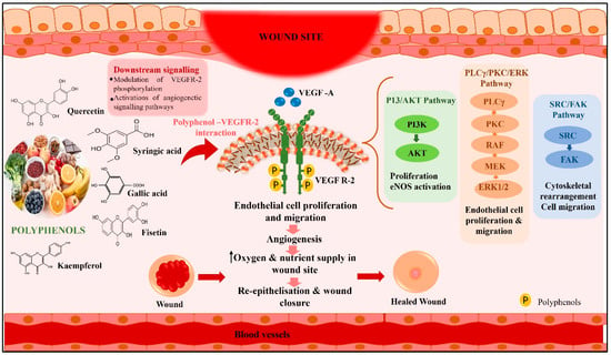
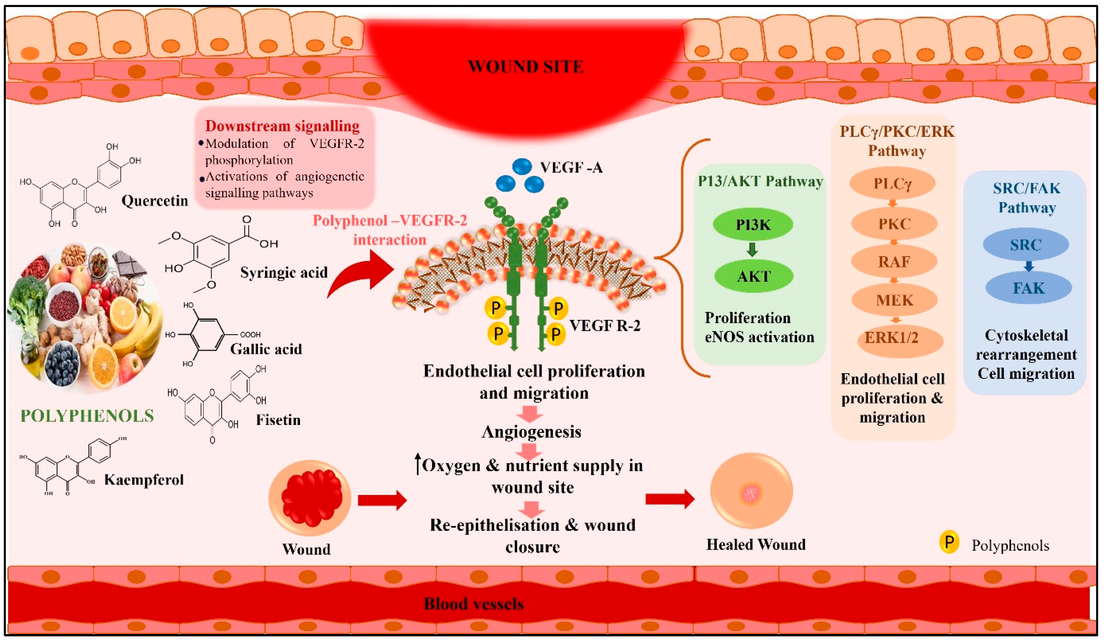
Wound healing is a complex biological process comprising four phases: hemostasis, inflammation, proliferation, and tissue remodelling. The key to successful healing is the regulation of activities such as keratinocyte migration, fibroblast proliferation, and angiogenesis, which involves forming new blood vessels [1]. Angiogenesis is crucial during the proliferative phase, ensuring proper oxygen and nutrient supply to the healing tissue. Insufficient angiogenesis can lead to chronic wounds and delayed tissue repair [2]. Chronic wounds persist for more than 3 months, primarily due to an extended inflammatory phase that impairs the response of dermal and epidermal cells. The rising elderly population, along with comorbidities such as diabetes and vascular issues, contributes significantly to chronic wounds, leading to increased health costs and morbidity. Impaired blood flow in these cases results in non-healing wounds, which are susceptible to infection and septicemia [3]. The incidence and burden of wounds are major issues in healthcare, with more than 8.2 million people in the U.S. alone and a cost of $28.1 to $96.8 billion. The global wound care market was valued at $18.4 billion in 2018 and is expected to grow at a CAGR of 3.9% till 2026. Chronic wounds are a major problem, costing the U.S. more than $25 billion per year and affecting more than 6.5 million people [4]. Polyphenols, natural compounds with anti-inflammatory and antimicrobial properties, have shown promise in promoting wound healing through various mechanisms, such as enhancing antioxidant defences, modulating inflammatory responses, and improving healing times. Polyphenols, derived from plants and marine organisms, have gained attention in wound treatment due to their antimicrobial, regenerative, and antioxidant properties [5]. Over 8000 polyphenols exist, classified into flavonoids and non-flavonoid compounds. They are particularly effective for hard-to-heal wounds in diabetic and elderly populations, promoting angiogenesis and collagen synthesis. They also exhibit high antioxidant activity, neutralising reactive oxygen species (ROS) and providing protection by donating electrons or hydrogen atoms. Some also demonstrate antimicrobial activity against bacteria in infected chronic wounds, potentially disrupting bacterial cell walls or modifying intracellular functions. Enhanced lipophilicity of polyphenols is associated with increased antimicrobial efficacy, particularly against antibiotic-resistant strains such as methicillin-resistant S. aureus [6]. Vascular endothelial growth factor (VEGF) is crucial for wound repair as the main pro-angiogenic cytokine. It activates vascular endothelial growth factor receptor-2 (VEGFR-2) on endothelial cells, triggering multiple signalling pathways that regulate cell proliferation, migration, survival, and vascular permeability. VEGFR-2 may interact with polyphenols causing downstream PI3K/AKT, PLCγ/PKC/ERK, and SRC/FAK signalling cascades, promoting wound healing [7]. The possible mechanism of cations of natural polyphenols in wound healing is through the VEGFR-2 pathway (Figure 1).
Figure 1.
Possible mechanism of action of natural polyphenols in wound healing through the VEGFR-2 pathway. Note: here, P: Natural polyphenols, VEGF-A: Vascular Endothelial Growth Factor A, VEGFR-2: Vascular Endothelial Growth Factor Receptor-2, PI3K: Phosphoinositide 3-Kinase, AKT: Protein Kinase B, eNOS: Endothelial Nitric Oxide, PLCγ: Phospholipase C Gamma, PKC: Protein Kinase C, RAF: Rapidly Accelarated Fibrosarcoma, MEK: Mitogen-Activated Protein Kinase, ERK1/2: Extracellular Signal Regulated Kinase ½, SRC: SRC proto-oncogene, FAK: Focal Adhesion Kinase.
Recent advancements in computational biology have enhanced drug discovery through in silico methods, including molecular docking and pharmacokinetic prediction. This study evaluated natural polyphenolic compounds for their binding affinity to VEGFR-2 using molecular docking, aiming to identify compounds that may influence angiogenesis in wound repair. Additionally, in silico ADMET predictions were used to assess the drug-likeness and safety of top candidates. The research underscores the therapeutic potential of natural polyphenols as angiogenic modulators, establishing a computational basis for future experimental validation in wound healing.
2. Materials and Methods
2.1. Selection of Natural Polyphenol
Natural polyphenols are plant-based bioactive compounds known for their antioxidant and anti-inflammatory benefits, categorised into three classes: flavonoids, phenolic acids, and lignans. An extensive analysis of the published literature and several databases was conducted to identify natural polyphenols effective in wound healing. The 2D structure was obtained from PubChem (https://pubchem.ncbi.nlm.nih.gov/) [8].
2.2. Molecular Docking Stimulation
Molecular docking is a powerful computational tool for studying protein–ligand interactions. The 2D structures of the phytochemicals were converted into 3D conformation using Chem3D Pro (v.12.0) and saved in Protein Data Bank (PDB) format. For this study, 3VHE (human VEGF2 kinase) was chosen [9]. The vascular endothelial growth factor (VEGF) and its receptors, particularly VEGFR-1 and VEGFR-2, play essential roles in wound healing by stimulating angiogenesis, which provides oxygen and nutrients. VEGF production increases shortly after injury, primarily from macrophages, platelets, and endothelial cells. It activates Type III tyrosine kinases to promote endothelial cell migration, tissue regeneration via fibroblast–endothelial cell interactions, and faster wound closure, which is crucial for non-healing or diabetic wounds [10]. The protein was prepared in AutoDock Tools v1.5.6. Preparation steps include deleting water, adding polar hydrogens, adding AD4 atoms, and, lastly, adding Gasteiger charges, followed by conversion to the PDBQT format. Molecular docking simulation was done in AutoDock Vina (version 1.2.0) to assess binding affinity [11]. The grid box dimensions of the protein–ligand interactions were given in Table 1. Docking results were visualised in BIOVIA Discovery Studio 2025, providing 2D and 3D diagrams. This analysis assessed the protein with the highest binding affinity for the ligand.
Table 1.
Detailed grid details employed in this study.
2.3. In Silico Drug-Likeness and ADME Predictions
The top 3 polyphenols were further analysed for in silico pharmacokinetics using SwissADME (https://www.swissadme.ch/). The toxicity was evaluated using ProTox 3.0 (https://tox.charite.de/protox3/).
2.4. BOILED-Egg Method
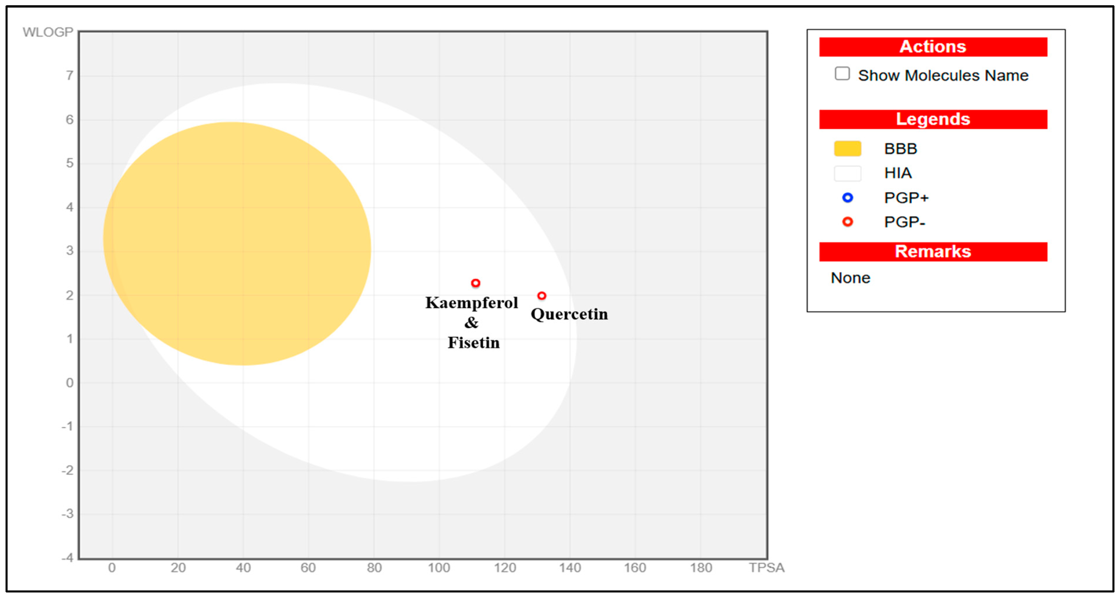
The BOILED-Egg (Brain Or IntestinaL EstimateD permeation) method in SwissADME predicts passive gastrointestinal absorption (HIA) and blood–brain barrier (BBB) permeation using a graphical model based on lipophilicity (WLogP) and polarity (tPSA). The diagram consists of three regions: the white region (Egg White) signifies a high probability of gastrointestinal absorption, indicating good oral bioavailability; the yellow region (yolk) suggests a high likelihood of BBB permeation, allowing entry to the central nervous system (CNS); and the grey zone (outside the egg) denotes molecules that are poorly absorbed and unlikely to cross the BBB. In this model, molecules are represented as dots, with blue indicating P-glycoprotein (P-gp) substrates and red identifying non-substrates. The top 3 compounds were assessed [12].
3. Result and Discussions
3.1. Molecular Docking Stimulation Results
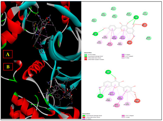
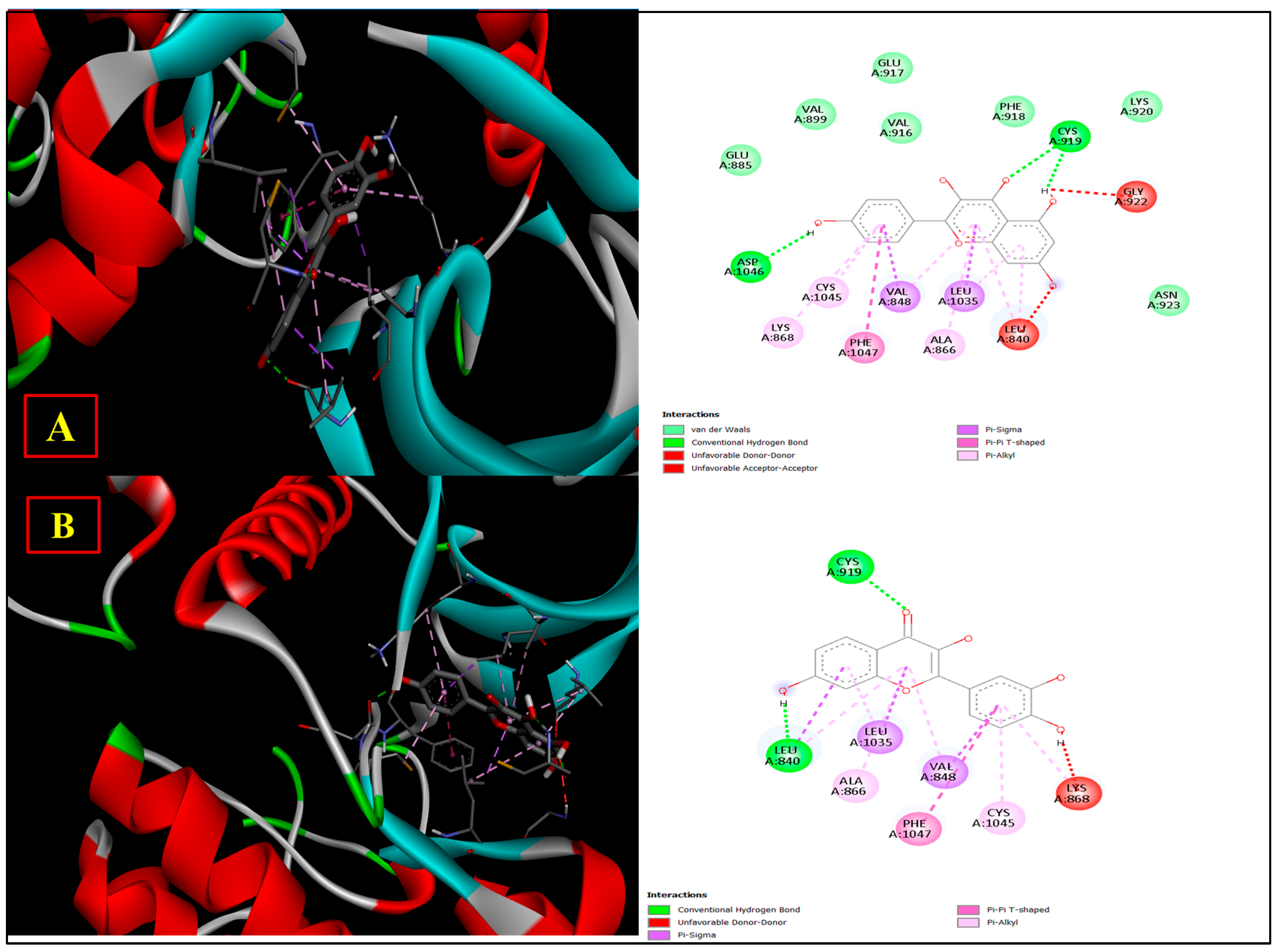
The 28 polyphenolic compounds were analysed against human VEGF2 kinase (3VHE). Among all phytochemicals, Kaempferol and fisetin show strong interactions with the protein molecule, with a docking score of −10.6. Quercetin shows a docking score of −10.3, and apigenin, luteolin, chrysin, naringin and hesperetin showed a good docking score of −10.2. The standard drug, Dexpanthenol, shows a docking score of −5.1. The docking score results are presented in Table 2. Kaempferol formed a conventional hydrogen bond with Cys919 and Leu840. It also formed several hydrophobic bond interactions with Leu1035, Val848, Ala866, Phe1047, Cys1045 and Lys868. It showed some Van der Waals contacts with protein molecules, including Glu885, Val899, Val916, Glu917, Phe918, and Lys920 (Figure 2A). Fisetin formed conventional hydrogen bonds with Cys919 and Leu840. Additional hydrophobic bonds are Leu1035, Val848, Ala866, Phe1047, and Cys1045 (Figure 2B). Overall, kaempferol and fisetin can form stable complexes with VEGFR-2 through hydrogen bonding and hydrophobic interactions, indicating their potential to inhibit VEGFR-2-mediated angiogenic signalling. Further validation through molecular dynamics simulations and experimental assays is suggested.
Table 2.
Molecular docking score of all phytochemicals.
Figure 2.
Three-dimensional binding poses and two-dimensional protein–ligand interaction maps of (A) kaempferol and (B) fisetin docked within the active site of VEGFR-2 (PDB ID: 3VHE).
3.2. ADMET Studies
The top three best docked polyphenol drugs against VEGF2 kinases were analysed by ADMET studies (Table 3). All three compounds showed high gastrointestinal absorption, with no blood–brain barrier (BBB) permeation, and no p-glycoprotein substrate activity. All three show the same skin permeability activity (log kp = −2.735). They satisfied the Lipinski, Ghose, Veber, and Egan drug-likeness criteria. Kaempferol showed the highest LD50 value of 3919 mg/kg, whereas fisetin and quercetin had LD50 values of 159 mg/kg.
Table 3.
ADMET profile of the top three best docked polyphenols.
3.3. BOILED-Egg Method Analysis
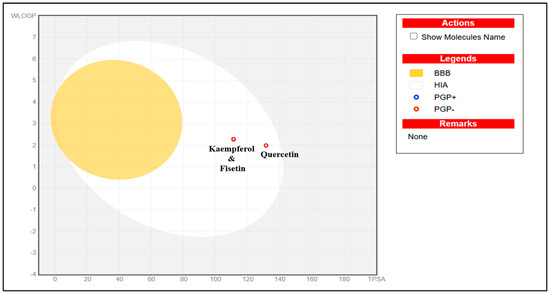
The pharmacokinetic evaluation of kaempferol, fisetin and quercetin was performed by the BOILED-Egg model in SwissADME (Figure 3) which predicts passive human intestinal absorption (HIA) and blood–brain barrier (BBB) permeability based on lipophilicity (WLOGP) and topological polar surface area (TPSA). Compounds in the white region of this model have high gastrointestinal absorption, while those in the yellow yolk are good candidates for BBB penetration. The model also displays interactions with P-glycoprotein (P-gp) where red circles are non-substrates (PGP-) and blue circles are substrates (PGP+). BOILED-Egg analysis shows that all three flavonoids, kaempferol, fisetin and quercetin are in the white region but outside the yellow yolk indicating high passive intestinal absorption and limited BBB permeability. This profile is advantageous for targeting peripheral sites such as VEGFR-2, thereby permitting good oral absorption without potential CNS exposure. Moreover, all compounds are PGP, which means that they are not substrates of P-glycoprotein, hence reducing the possibility of active efflux and improving oral bioavailability and intracellular retention. Fisetin and kaempferol are very close to each other in the BOILED-Egg plot which is indicative of a similar physicochemical property and pharmacokinetic profile. Quercetin has a slightly higher topological polar surface area (TPSA) due to an additional hydroxyl group and is also slightly further to the right, but still within the human intestinal absorption region. Quercetin is more polar but also predicted to be well absorbed in the intestine and not to cross the BBB, which indicates that the extra hydroxyl group of quercetin does not have a large impact on its oral absorption. Overall, these compounds demonstrate favourable pharmacokinetic properties, making them potential orally active natural wound healing molecules for further research.
Figure 3.
BOILED-Egg model to analyse the top 3 polyphenols for wound healing.
4. Conclusions
The in silico study highlights kaempferol and fisetin as leading polyphenolic compounds targeting VEGFR-2, with binding affinities of −10.6 kcal/mol and −10.3 kcal/mol, respectively. These flavonoids interact stably with critical amino acid residues in the VEGFR-2 active site, indicating their potential to influence VEGFR-2-driven signalling. ADMET and BOILED-Egg analyses show promising drug-like properties, including high gastrointestinal absorption and suitable pharmacokinetics, suggesting their viability as oral therapies. Given the importance of VEGF/VEGFR-2 signalling in wound repair, these compounds may enhance wound healing. However, further validation through molecular dynamics simulations and in vitro/in vivo studies is necessary to confirm their biological efficacy.
Author Contributions
Conceptualisation, S.D. and A.P.; methodology, S.D.; writing—original draft preparation, S.D. and A.P.; writing—review and editing, A.P. All authors have read and agreed to the published version of the manuscript.
Funding
This research received no external funding.
Institutional Review Board Statement
Not applicable.
Informed Consent Statement
Not applicable.
Data Availability Statement
No new data were created or analysed in this study. Data sharing is not applicable to this article.
Conflicts of Interest
The authors declare no conflicts of interest.
References
- Jin, C.; Jin, Y.; Ding, Z.; Nuch, K.S.; Han, M.; Shim, J.; Chien, P.N.; Heo, C.Y. Cellular and Molecular Mechanisms of Wound Repair: From Biology to Therapeutic Innovation. Cells 2025, 14, 1850. [Google Scholar] [CrossRef] [Scilit] [PubMed]
- Shi, Z.; Yao, C.; Shui, Y.; Li, S.; Yan, H. Research progress on the mechanism of angiogenesis in wound repair and regeneration. Front. Physiol. 2023, 14, 1284981. [Google Scholar] [CrossRef] [Scilit] [PubMed]
- Pignet, A.L.; Schellnegger, M.; Hecker, A.; Kamolz, L.P.; Kotzbeck, P. Modeling Wound Chronicity In Vivo: The Translational Challenge to Capture the Complexity of Chronic Wounds. J. Investig. Dermatol. 2024, 144, 1454–1470. [Google Scholar] [CrossRef] [Scilit] [PubMed]
- Sen, C.K. Human Wounds and Its Burden: An Updated Compendium of Estimates. Adv. Wound Care 2019, 8, 39–48. [Google Scholar] [CrossRef] [Scilit] [PubMed]
- Utpal, B.K.; Sutradhar, B.; Zehravi, M.; Sweilam, S.H.; Panigrahy, U.P.; Urs, D.; Fatima, A.F.; Nallasivan, P.K.; Chhabra, G.S.; Sayeed, M.; et al. Polyphenols in wound healing: Unlocking prospects with clinical applications. Naunyn-Schmiedeberg’s Arch. Pharmacol. 2025, 398, 2459–2485. [Google Scholar] [CrossRef] [Scilit] [PubMed]
- Rudrapal, M.; Khairnar, S.J.; Khan, J.; Bin Dukhyil, A.; Ansari, M.A.; Alomary, M.N.; Alshabrmi, F.M.; Palai, S.; Deb, P.K.; Devi, R. Dietary Polyphenols and Their Role in Oxidative Stress-Induced Human Diseases: Insights Into Protective Effects, Antioxidant Potentials and Mechanism(s) of Action. Front. Pharmacol. 2022, 13, 806470. [Google Scholar] [CrossRef] [Scilit] [PubMed]
- Wise, L.M.; Stuart, G.S.; Real, N.C.; Fleming, S.B.; Mercer, A.A. VEGF Receptor-2 Activation Mediated by VEGF-E Limits Scar Tissue Formation Following Cutaneous Injury. Adv. Wound Care 2018, 7, 283–297. [Google Scholar] [CrossRef] [Scilit] [PubMed]
- V.S., S.; Singh, S. Natural Polyphenols as Multi-Targeted Agents in Diabetic Foot Ulcer: A Comprehensive Systematic Review. Curr. Chem. Biol. 2025, 20, 66–84. [Google Scholar] [CrossRef] [Scilit]
- Mishra, A.; Sabikhi, Y.; Singh, A.; Santoshi, S.; Akhtar, S. Ligand-based pharmacophore modelling, molecular docking, and molecular dynamic studies of VEGFR2 inhibitors. Lett. Drug Des. Discov. 2026, 23, 100419. [Google Scholar] [CrossRef] [Scilit]
- Bao, P.; Kodra, A.; Tomic-Canic, M.; Golinko, M.S.; Ehrlich, H.P.; Brem, H. The Role of Vascular Endothelial Growth Factor in Wound Healing. J. Surg. Res. 2009, 153, 347–358. [Google Scholar] [CrossRef] [Scilit] [PubMed]
- Das, S.; Pandey, A. Computational Exploration of Skin-Protective Potential of Oxalis corniculata Phytoconstituents Using Network Pharmacology and Docking Approaches. J. Comput. Biophys. Chem. 2026, 26, 247–268. [Google Scholar] [CrossRef] [Scilit]
- Yadav, S.; Das, S.; Pandey, A. Computational Insights into the Antimalarial Potential of Phytochemicals from Centella asiatica: A Molecular Docking Approach. Chem. Proc. 2025, 18, 123. [Google Scholar] [CrossRef] [Scilit]

Disclaimer/Publisher’s Note: The statements, opinions and data contained in all publications are solely those of the individual author(s) and contributor(s) and not of MDPI and/or the editor(s). MDPI and/or the editor(s) disclaim responsibility for any injury to people or property resulting from any ideas, methods, instructions or products referred to in the content. |
© 2026 by the authors. Licensee MDPI, Basel, Switzerland. This article is an open access article distributed under the terms and conditions of the Creative Commons Attribution (CC BY) license.