


Abstract
Physarum polycephalum is a member of macroscopic myxomycetes, a phylum of fungus-like organisms within the protista kingdom, commonly known as slime molds. As a unicellular eukaryote, Physarum polycephalum has attracted considerable attention due to its sophisticated life cycle and remarkable complexity in its communication behavior and adaptation to its environment. Recent studies have suggested slime molds as a valuable source of biologically active substances, however, their metabolome remains poorly known. In the present work, intracellular and extracellular metabolomes of Physarum polycephalum were deeply investigated using nuclear magnetic resonance (NMR) in combination with liquid chromatography coupled with high resolution mass spectrometry (LC/HRMS). Many metabolites were rapidly and unambiguously identified by NMR including constituents of the central carbon metabolism, free and acetylated amino acids, sterols, polyunsaturated fatty acids, nucleotides and nucleosides, small lactones, phenolic acids, pyranones, and methylfurane derivatives. Several metabolites, including nicotinamide, nicotinic acid, mevalonolactone, p-coumaric acid, phenylacetic acid, p-hydroxyphenylacetic acid, p-hydroxyhippuric acid, and p-hydroxybenzoic acid are identified for the first time in Physarum polycephalum. Some of these compounds, such as p-coumaric acid, niacinamide, linoleic acid, and kojic acid, are known for their potential applications in skincare formulations. A diversity of peptides was also annotated by LC/HRMS. Such detailed overview of Physarum polycephalum metabolite diversity opens up perspectives to better understand slime mold metabolism and consider new applications of Physarum polycephalum extracts, including their potential as a source of novel cosmetic ingredients.
1. Introduction
Physarum polycephalum Schweinitz is an unusual free-living organism belonging to macroscopic myxomycetes, a phylum of fungus-like organisms within the kingdom Protista, commonly known as slime molds. Recent data from genomic and transcriptomic studies have positioned Physarum polycephalum close to animals in the eukaryotic tree [1], but it also displays characteristics specific to the plant kingdom, making its classification still controversial.
Slime molds are commonly found on decaying wood or leaf debris in the forest, occurring as large and thin layers, yellow or cream in colour, amorphous, and gelatinous in appearance. They can take many forms, reaching from a few centimeters to several square meters in size. As a unicellular eukaryote, Physarum polycephalum has attracted considerable attention because it displays a sophisticated life cycle and a remarkable complexity in its behaviour [2]. Its life cycle starts with the germination of spores that become amoeba-like cells. The unicellular amoebae undergo fusion to form zygotes that grow into a plasmodium. The plasmodium corresponds to a single cell containing millions of nuclei flowing freely inside the cytoplasm [3]. At this stage, the cell is not surrounded by a cell wall. Despite absence of a nervous system, Physarum polycephalum is capable of amazing actions, such as finding the shortest route between different food sources using information from previous experiences to adjust its behaviour. Recent studies further showed that Physarum polycephalum can adapt to ultraviolet stress through behavioural avoidance, damage compartmentalization, and dormancy-based survival strategies, thus highlighting the remarkable adaptive capabilities of this unicellular organism [4]. The precise mechanisms by which it perceives and responds to its environment remain unknown, but recent data showed that the plasmodium is equipped with a range of surface receptors and that communication takes place across its entire body with signals propagating via electric waves, molecular, and electric stimuli [3,5].
All these characteristics have made Physarum polycephalum a valuable model to study problem-solving in abneural biological systems and to better understand eukaryotic cell functions and biochemical processes.
From a chemical point of view, more than 120 substances have been described so far in myxomycetes, including a diversity of original secondary metabolites with potent biological activities [6,7].
Indeed, slime molds have been reported to produce a wide variety of pigments, lipids, aromatic compounds, glycosides, lactones, and exopolysaccharides. Several of these metabolites exhibit biological properties relevant to skin care, such as antioxidant, antimicrobial, anti-inflammatory, UV-protective, or pigmentation-related activities, highlighting slime molds as an emerging and still largely underexplored source of natural cosmetic ingredients [6,8,9].
Regarding Physarum polycephalum, only a few sterols, lipids, and colouring substances with polyenoic unsaturated chains, such as chrysophysarins, physarochromes, polycephalins, or physarorubinic acids, have been reported [10,11,12]. Therefore, it is certain that Physarum polycephalum still has many secrets to deliver regarding its metabolome.
As part of continuing efforts to explore the chemical diversity of Physarum polycephalum, a robust analytical strategy was applied here to deeply investigate intracellular and extracellular metabolites of Physarum polycephalum when cultivated in a liquid medium. The workflow combines liquid–liquid fractionation of cell extract and culture supernatant, NMR analyses, Hierarchical Clustering Analysis (HCA) of 13C NMR data, and metabolite identification using an in-house NMR natural metabolite database reinforced by manual scrutinization of 2D NMR spectra. This approach was initially developed to accelerate the chemical profiling of complex natural extracts [13], and many concrete examples of natural extract characterization have been published in the last years [14,15,16]. High-resolution liquid chromatography–mass spectrometry (LC-MS) was also used to complete the identification process. Our objective was to demonstrate that liquid–liquid chromatography combined with NMR is an efficient approach to chemically profile slime mold samples, providing a valuable technical support to decipher the metabolome of these fascinating organisms without discrimination between chemical classes, to investigate their primary and secondary metabolism, and to better understand their biological functions and properties.
2. Materials and Methods
2.1. Chemicals
Agar, citric acid, hemin, iron (II) chloride tetrahydrate, potassium dihydrogen phosphate, and deuterated dimethyl sulfoxide (DMSO-d6) were purchased from Sigma Aldrich (Buchs SG, Switzerland). Calcium chloride dihydrate, magnesium sulfate heptahydrate, and zinc sulphate heptahydrate were purchased from Duchefa Biochemie (Haarlem, The Netherlands). Glucose, tryptone, acetic acid, and formic acid were purchased from VWR (Dietikon, Switzerland). Yeast extract was purchased from ThermoFisherScientific (Zürich, Switzerland). Ethyl acetate, acetonitrile, and methanol were purchased from Carlo Erba (Val de Reuil, France). Deionized water was used to prepare all aqueous solutions.
2.2. Slime Mold Culture
The Physarum polycephalum strain 96951 was obtained from the American Type Culture Collection (ATCC). It was cultivated for several weeks on a semi-solid complex medium with agar as described by ATCC (Medium 1288: N plus C, ATCC.org). Liquid cultures were established from plasmodium pieces (approximately 1 cm2) of the semi-solid culture. A 14-day liquid pre-culture was grown in ATCC 1288: N plus C medium without agar to gain enough starting material. This pre-culture was used to start a 500 mL culture in defined medium adapted from ATCC 1288: N plus C medium in 1 L Erlenmeyer flask with narrow neck (DURAN®). In this defined medium tryptone and yeast extract were substituted by their corresponding amino acids and vitamins. The culture was incubated for 14 days at 25 °C in the dark and shaken at 100 rpm in a Multitron Standard shaker (Infors HT, Bottmingen, Switzerland). The exudate was then separated from the biomass by centrifugation for 45 min at 4700 rpm in a Multifuge® 1 S-R centrifuge (Heraeus, Hanau, Germany). Subsequently, the supernatant, as well as a not cultivated medium mixture, were lyophilized in an Alpha 2-4 LSCplus (CHRIST). The biomass was stored frozen.
2.3. Extracellular Metabolome Extraction and Fractionation
In addition, 46,170 mg of the lyophilized supernatant were solubilized in 500 mL of the lower phase of the biphasic solvent system ethyl acetate/acetonitrile/water (3/3/4, v/v). Three successive extractions were performed with 500 mL of fresh upper phase. The three upper phases were combined and evaporated to dryness at 50 °C with a rotary evaporator, resulting in a dry fraction of 1693 mg (BLO-UP). The remaining lower phase was also evaporated to dryness, resulting in a dry fraction of 40,818 mg (BLO-LOW). The remaining biomass was thrown over with an emulsion, which was formed at the interface between the two phases.
BLO-UP was then fractionated by Centrifugal Partition Chromatography (CPC) using a column of 231 mL (CPC-250-PRO from Gilson, Villiers Le Bel, France) connected to a Blueshadow pump 80 P (Knauer, Berlin, Germany) and a Labocol Vario 4000 collector (Knauer, Berlin, Germany). The column was made of 12 partition disks engraved with 288 twin cells of ≈ 1 mL each. The biphasic solvent system ethyl acetate/acetonitrile/water (3/3/4, v/v) was prepared in a separatory funnel. After equilibration and decantation, the two phases were separated, and the column was filled with the lower phase (stationary phase) at 500 rpm and 50 mL/min. Then, the rotation was set at 1600 rpm. BLO-UP (1488 mg) was dissolved in a mixture of 10 mL of stationary phase and 8 mL of mobile phase and injected into the CPC column using a 35 mL sample loop. The upper phase (mobile phase) was pumped at 20 mL/min in the ascending mode for 60 min. Then, all compounds retained inside the column were recovered by extrusion in the descending mode for 8 min, still at 20 mL/min. Fractions of 20 mL were collected over the whole experiment (elution and extrusion) and combined according to their high-performance thin-layer chromatography profiles (HPTLC). HPTLC was performed on a CAMAG® system equipped with an automatic sampler ATS4, a developing chamber ADC2, and a visualizer 2. Fractions were spotted on pre-coated silica gel 60 F254 Merck plates, developed with the migration solvent system ethyl acetate/water/formic acid/acetic acid (10/2/1/1, v/v), and visualized under visible light after spraying the dried plates with 50% H2SO4 and vanillin followed by heating.
2.4. Intracellular Metabolome Extraction and Fractionation
The cells were completely dried under vacuum, and the dry biomass (1441 mg) was extracted with methanol under sonication for 1 hour, then with methanol/water (1/1, v/v) under sonication for 1 hour, resulting in a cell extract of 572 mg (BLO-Ext) after solvent evaporation. BLO-Ext was then fractionated by CPC using the same system and in the same conditions as described above. Fractions were spotted on pre-coated silica gel 60 F254 plates (Merck, Darmstadt, Germany), developed with the migration solvent system ethyl acetate/water/formic acid/acetic acid (10/2/1/1, v/v), and visualized under visible light after spraying the dried plates with 50% H2SO4 and vanillin followed by heating. A series of seven fractions was obtained.
2.5. NMR Chemical Profiling
An aliquot of each fraction from F01 to F12 from the supernatant and from F01 to F07 from the cells (up to 15 mg when possible) was dissolved in 600 µL of DMSO-d6. NMR analyses were performed at 298 K on a Bruker Avance AVIII-600 spectrometer (Karlsruhe, Germany) equipped with a TCI cryoprobe optimized for 1H detection and with cooled 1H and 13C coils and preamplifiers. The spectrometer operated at a proton frequency of 600.13 MHz and a carbon frequency of 150.91 MHz (B0 = 14.1 T). Prior to NMR data acquisition, the probe was automatically tuned and matched for each sample at these resonance frequencies. An udeft pulse program from Bruker was used with an acquisition time of 0.3599 s. For each sample, 512 scans were coadded to obtain a satisfactory signal-to-noise ratio. The spectral width was 238.9070 ppm, and the receiver gain was set to 2050. Detailed acquisition parameters of the UDEFT sequence are given in Supplementary Data S4. All spectra were manually phased and baseline corrected using the TOPSPIN 4.0.5 software (Bruker, Fällanden, Switzerland) and calibrated on the central resonance of DMSO-d6 (δ 39.80 ppm). The absolute intensities of all 13C NMR signals were automatically collected in all spectra of the fraction series and transferred into a table using a locally developed computer script dedicated to NMR signal bucketing with a bucket size of 0.2 ppm. The resulting table was imported into the Permut Matrix version 1.9.3 software (LIRMM, Montpellier, France) for hierarchical clustering analysis (HCA) on peak intensity values. The classification was performed on the rows only, i.e., on the chemical shift buckets. The Euclidian distance was used to measure the proximity between samples, and the Ward’s method was applied to agglomerate the data. In this way, 13C NMR chemical shift clusters were visualized as dendrograms on a two-dimensional map. The right-hand vertical axis corresponds to the chemical shift buckets reorganized after hierarchical clustering analysis. The higher the intensity of 13C NMR peaks, the brighter the yellow colour on the map. For metabolite identification, each 13C NMR chemical shift cluster obtained from HCA was manually submitted to the structure search engine of a 13C NMR chemical shift database (ACD/NMR Workbook Suite 2012 software, ACD/Laboratories, Toronto, ON, Canada) comprising the predicted chemical shifts and structures of natural molecules (≈12,000 records in August 2026). Additional homonuclear correlation spectroscopy (COSY), heteronuclear single-quantum coherence (HSQC), and heteronuclear multiple bond correlation (HMBC) spectra were also acquired for all fractions to confirm, correct or further elucidate the chemical structures proposed by the database at the end of the dereplication process. All 13C and HSQC spectra of the CPC fractions obtained from the cell extract and from the supernatant are also given in Supplementary Data S4.
2.6. Liquid Chromatography Coupled to High Resolution Mass Spectrometry (LC/MS)
BLO-Ext, BLO-UP, and BLO-LOW from the cells and from the culture supernatant, and the free culture medium were analyzed by high-resolution liquid chromatography coupled to mass spectrometry on an Acquity UPLC H-Class system (Waters, Manchester, UK) coupled to a Synapt G2-Si (Waters) equipped with an electrospray (ESI) ion source. Samples were diluted at 5 g/L in methanol/water (50/50, v/v). Chromatographic separation was achieved on a Uptisphere C-18 ODB column (150 × 4.6 mm, 5 μm, Interchim, Montluçon, France). The column temperature was set at 35 °C. The sample temperature was maintained at 20 °C. Elution was performed using a gradient of water (Eluent A) and acetonitrile (Eluent B) with 0.1% formic acid in each mobile phase, as followed: t = 0 min, 100% A; t = 4 min, 74% A; t = 18.5 min, 35% A; t = 18.7 min, 0% A; t = 28.7 min, 0% A; t = 28.9 min, 100% A, t = 31.0 min, 100% A. The flow rate was maintained at 0.7 mL/min. The injection volume was 0.5 μL. MS analyses were performed in positive and negative ion modes. The capillary voltage was 3 kV for positive mode and 2 kV for negative mode, the desolvation temperature was 650 °C, the desolvation gas flow was 750 L/h, the source temperature was 150 °C, the cone voltage was 40 V, and cone gas flow 50 L/h. The mass range for scanning was m/z 50–2000. Mass was corrected during acquisition using external reference (Lock-Spray) consisting of a 1 ng/µL solution of leucine encephalin at a flow rate of 5 µL/min, in order to make sure the accuracy and reproducibility during the MS analysis. All data collected were acquired using MassLynx™ (V4.1) software in continuum mode.
3. Results
3.1. Physarum Polycephalum Growth
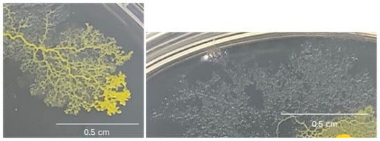
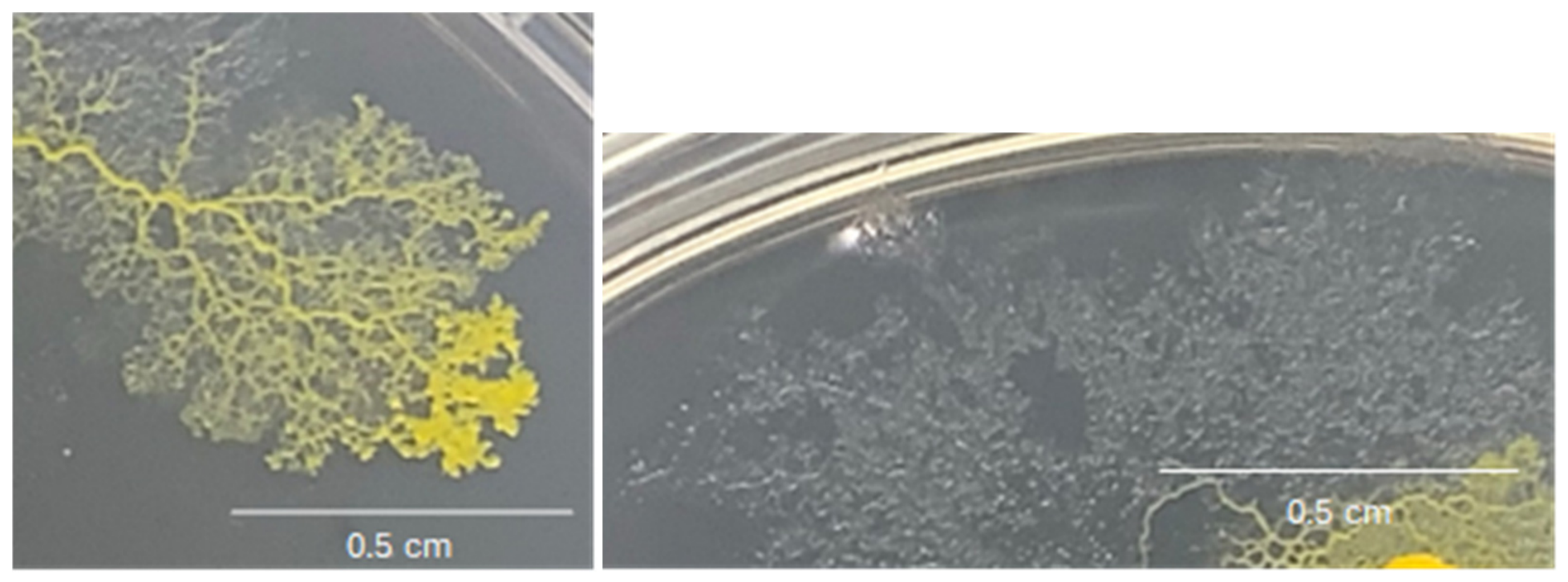
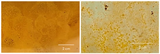
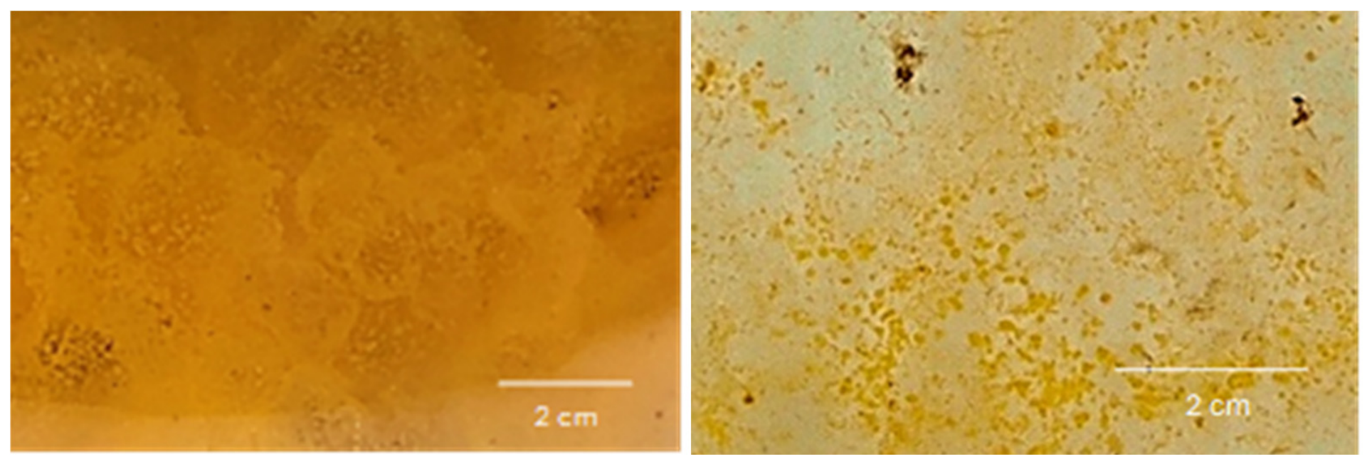
Physarum polycephalum was grown on a semi-solid medium containing agar and various nutrients. In these culture conditions, plasmodia migrated over the agar plate, and produced extracellular mucus (Figure 1). A piece of the organism was transferred to a liquid medium, and after approximately 1 week, macroplasmodia were formed. They could be described as small, irregularly shaped, and multinucleate pieces of plasmodia. A healthy growth of plasmodia was indicated by a bright yellow to orange colour, a gelatinous, and homogenous texture, and by observation of plasmodia network development. On the contrary, abiotic stress, e.g., poor nutrition led to a discoloration of plasmodia and fragmentation into small pieces. Figure 2 illustrates the morphology of plasmodia in a liquid culture medium under standard growth conditions or under abiotic stress.
Figure 1.
Physarum polycephalum plasmodia (left) and mucus (right) on semi-solid medium.
Figure 2.
Physarum polycephalum plasmodia in liquid culture under standard growth conditions (left) or under abiotic stress (right).
To access both the intracellular and extracellular metabolome of Physarum polycephalum, cell culture in liquid medium was the most adapted because it allowed a uniform distribution of nutrients, making it easier for the organism to feed. This promoted a more homogenous and quicker growth [17]. Furthermore, shaken liquid culture allowed better aeration and oxygen intake by the organism. Scalability was possible in liquid cultures, and parameters such as temperature, pH, and nutrient concentration could be easily controlled. Finally, secreted metabolites were homogenously distributed in the liquid medium, and this type of culture enabled the cells to be quickly separated from their culture medium by centrifugation. The cells were first grown as a liquid pre-culture in a complex culture medium composed of different nutrients required by the organism to grow. This medium contained glucose as a carbon source [18], citric acid, and different salts, as well as tryptone and hematin. Tryptone is a pancreatic digest of casein from animal origin, naturally rich in amino acid nitrogen (Oxoid™ Tryptone, Thermofischer.com Catalog number LP0042T). It was described as essential for the slime mold to grow [19] and able to influence lipid and biomass production of plasmodia [2]. Hematin was used as growth factor for Physarum polycephalum since its omission showed considerable growth inhibition [20,21]. The growth was also evaluated in an animal product-free culture medium containing vegetable tryptone (M-Gredient® Vegetable Tryptone, sigmaaldrich.com Catalog number 16922-500G-F) and synthetic hemin as a source of hematin. Cultures in the animal product-free medium led to similar growth characteristics (color, texture, growth rate) to those in the medium containing animal-derived components. To reduce chemical composition variability, Physarum polycephalum was subsequently transferred to a defined medium; where tryptone was substituted by its corresponding vitamins and amino acids. In this main culture, Physarum polycephalum displayed a growth profile comparable to that observed in the complex medium, demonstrating that the replacement of tryptone did not adversely affect growth. The results presented in this publication were obtained using cultures grown in the defined medium with hematin of animal origin as a growth factor. However, residual traces of complex medium components were also present due to carryover during the transfer of the slime mold from the preculture to the main culture. After the organism was grown for 2 weeks, plasmodia cells were separated from the growth medium supernatant by centrifugation. The supernatant contained a complex mixture of molecules, some of which coming from the medium, other excreted by the myxomycete. The chemical composition of the free liquid culture medium was also investigated as a reference to be compared with the supernatant.
3.2. Intracellular and Extracellular Metabolite Extraction and Fractionation
Physarum polycephalum cells were sonicated successively with methanol, then with methanol/water (1/1, v/v) to ensure the recovery of intracellular metabolites, and the resulting extract (BLO-Ext) was fractionated by centrifugal partition chromatography (CPC) in a total of seven fractions of simplified chemical composition. In parallel, the supernatant was partitioned in the biphasic solvent system ethyl acetate/acetonitrile/water (3/3/4, v/v) to separate the abundant hydrophilic constituents, mainly salts and simple sugars of the culture medium, from the other more specialized metabolites of lower polarity. Most of the biomass (≈88%) was recovered in the aqueous lower phase (BLO-LOW), but a higher chemical diversity was observed in the upper phase (BLO-UP). Therefore, this fraction was further fractionated by CPC. A total of 12 fractions were obtained by CPC in an increasing order of polarity and without loss of biomass during separation. The high-performance thin layer chromatography (HPTLC) profiles of all fractions obtained from the cells and from the supernatant are given in Supplementary Data S1 and S2, respectively, revealing the presence of metabolites from various chemical classes over a wide polarity range.
3.3. Metabolite Identification
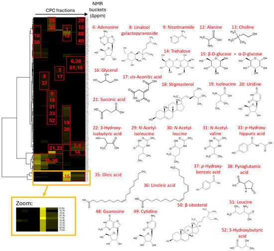
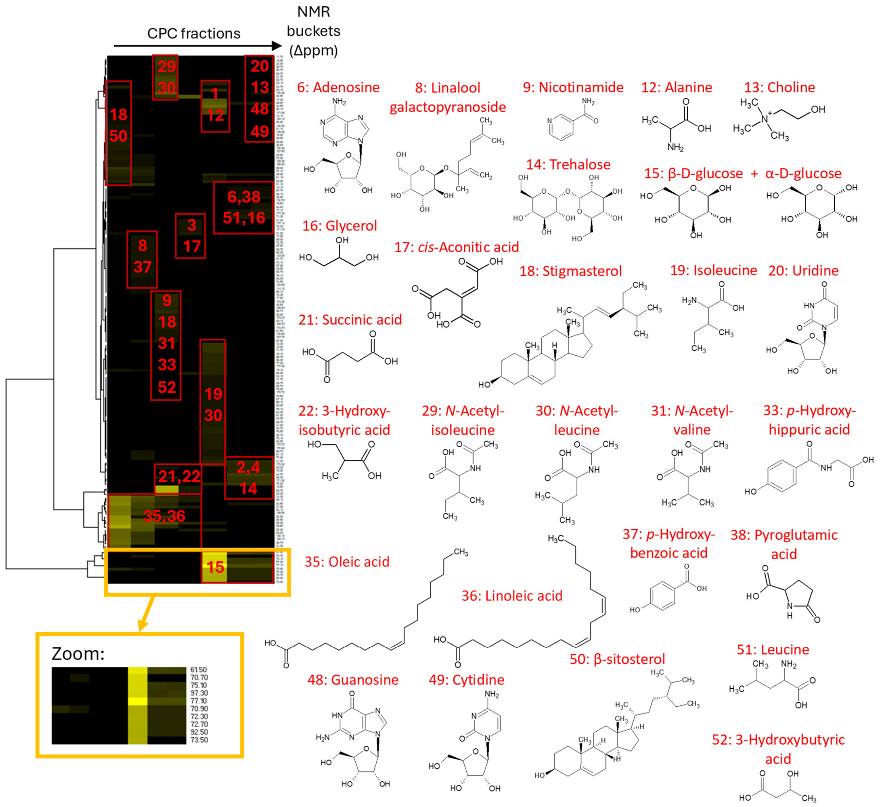
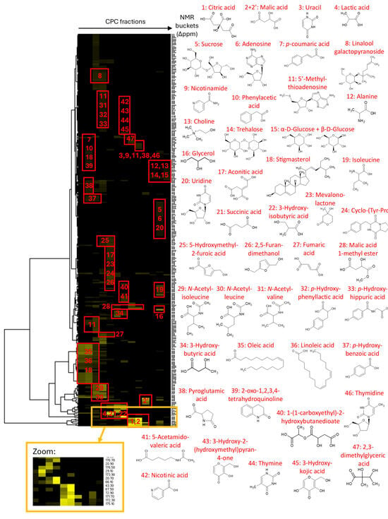
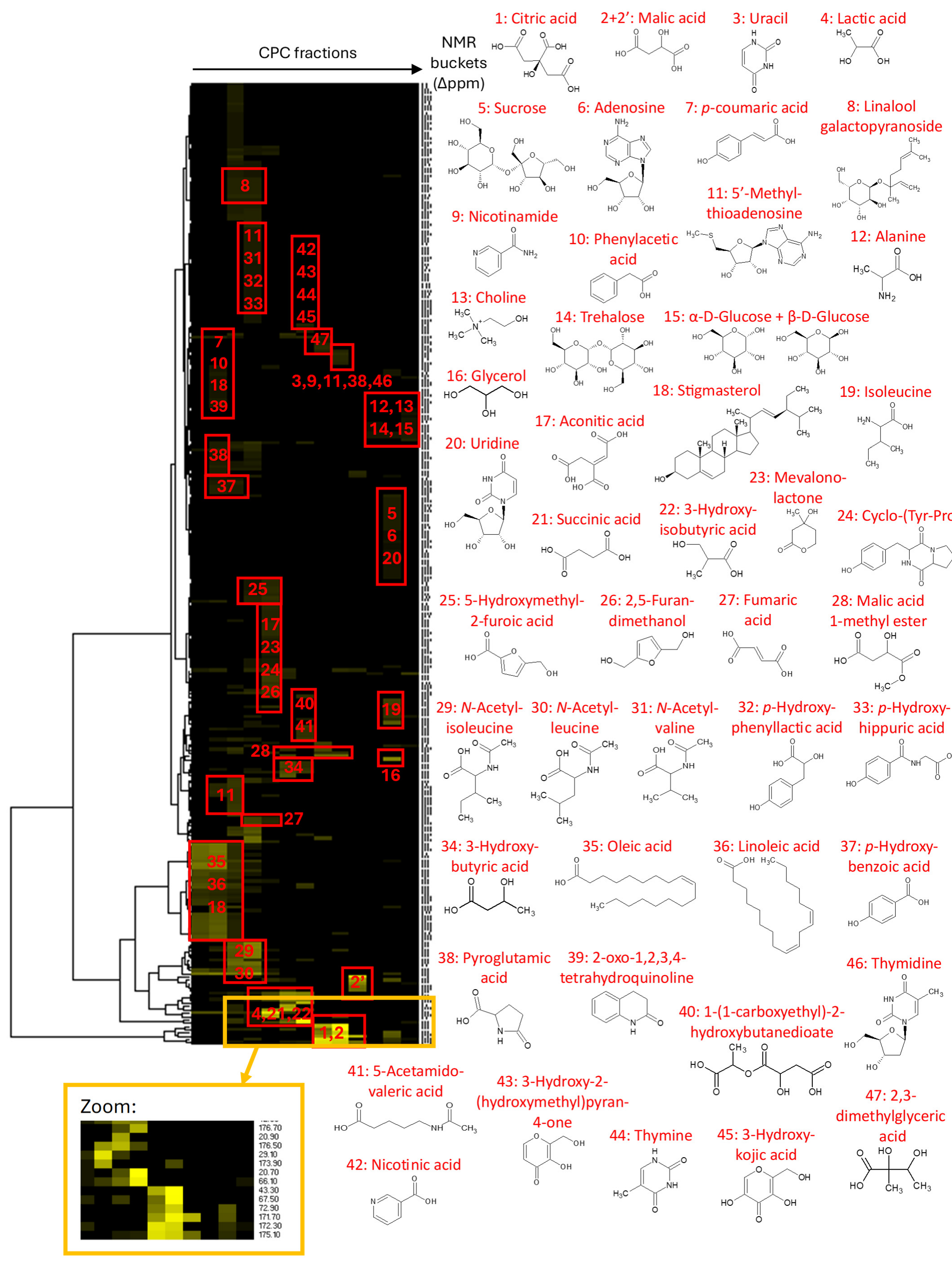
All fractions were directly analyzed by 1D and 2D NMR for metabolite identification. 13C NMR spectra were firstly exploited, the peaks detected from 0 to 200 ppm were aligned across the series using buckets of 0.2 ppm in width. As a result, one table was obtained for the cell extract with seven columns corresponding to the CPC fractions and 162 rows corresponding to the chemical shift buckets for which a 13C NMR peak was detected in at least one fraction. A second table was obtained for the supernatant, made of 13 columns corresponding to the CPC fractions F01–F12 and BLO-LOW at the end, and 292 rows corresponding to the chemical shift buckets for which a 13C NMR peak was detected in at least one fraction. Hierarchical Clustering Analysis (HCA) was applied to the rows only. In this way, 13C NMR peaks evolving with the same intensity in successive CPC fractions were aggregated as “NMR chemical shift clusters”, corresponding to the carbon skeleton of individual constituents of the extract. These chemical shift clusters are presented in the heatmaps in Figure 3 for the cells and Figure 4 for the supernatant. The deeper the yellow color on the map, the higher the intensity of 13C NMR peaks in the fractions, providing qualitative information about relative abundance of metabolites.
Figure 3.
HCA correlation heatmap of 13C NMR signals with the identified metabolites obtained for Physarum polycephalum cells (intracellular metabolites).
Figure 4.
HCA correlation heatmap of 13C NMR signals with the identified metabolites obtained for the supernatant (extracellular metabolites).
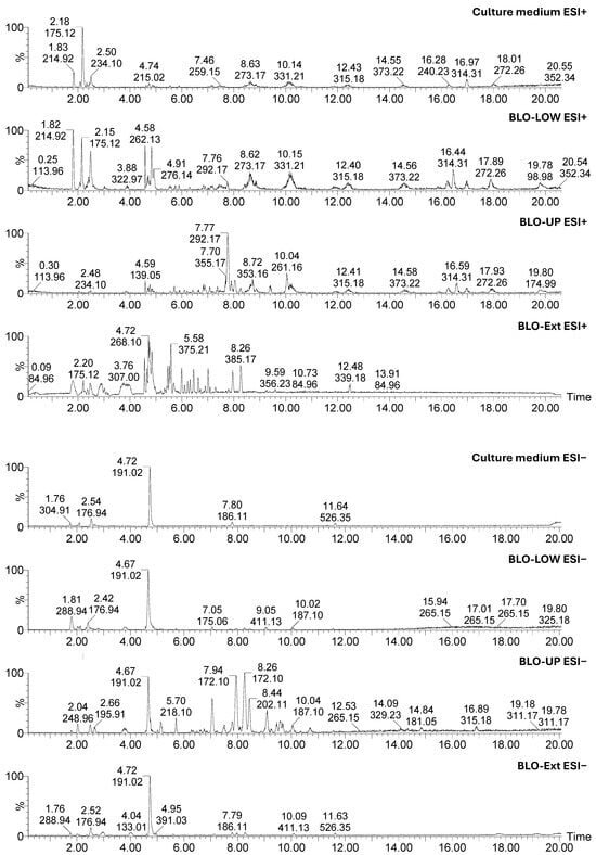
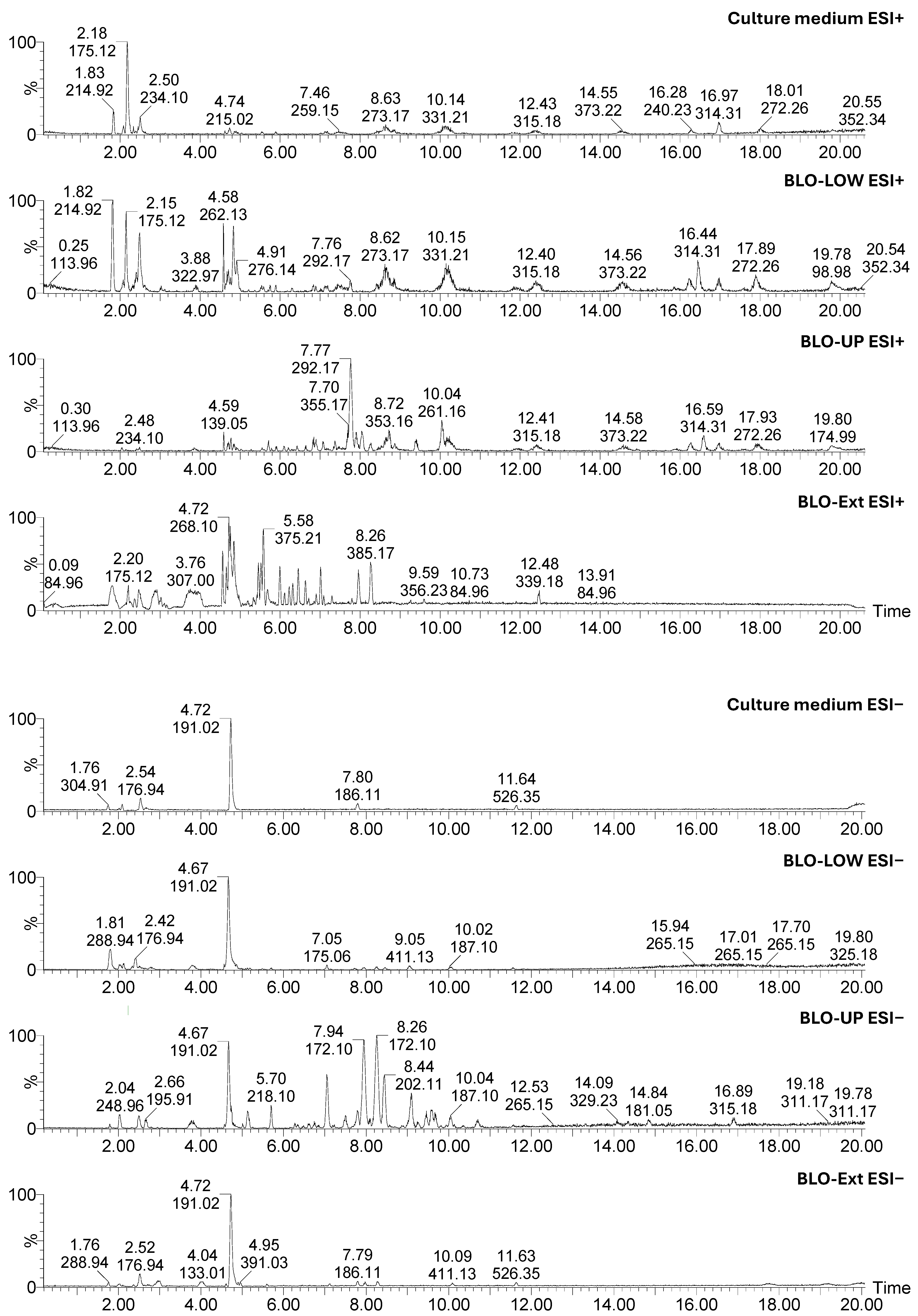
Each cluster was assigned to a metabolite using an in-house database of predicted NMR assignments for natural products. Database proposals were confirmed or re-oriented to the right chemical structures by rigorously scrutinizing 1H–13C and 1H–1H correlations in HSQC, HMBC, and COSY spectra. To complete the identification process, the cell extract BLO-Ext and the two supernatant sub-extracts BLO-UP and BLO-LOW were analyzed by liquid chromatography coupled to high-resolution mass spectrometry (LC/MS), which allowed us to annotate some additional minor metabolites having good ionization capabilities. LC/MS analysis of the free culture medium also enabled the distinction between metabolites arising from Physarum polycephalum and medium nutrients. The chromatograms and summary of LC/MS data obtained in positive and negative ionization modes are presented in Figure 5 and Table 1, respectively.
Figure 5.
LC/MS chromatograms of BLO-Ext, BLO-UP, BLO-LOW, and free culture medium.
Table 1.
Summary of LC/MS data.
4. Discussion
A total of 52 intracellular and extracellular metabolites from various chemical classes were unambiguously identified by NMR (Supplementary Data S3). In addition, 32 metabolites were annotated by high-resolution LC/MS, six of which overlapped with those identified by NMR. Among the remaining LC/MS annotations, four matched entries in our internal LC/MS database, whereas 22 were putatively annotated at a lower confidence level (Table 1).
Among primary metabolites, a significant amount of glucose was detected in all polar fractions from the cell extract and supernatant. Glucose was a major constituent of the culture medium. Therefore, its presence in the supernatant is normal. Glucose is also one of the main chemical units of exopolysaccharides from Physarum polycephalum [9], which explains its presence in the cell extract. Disaccharides such as sucrose and trehalose were also identified by NMR, but in much lower levels.
A range of small organic acids, including lactic acid, malic acid, fumaric acid, and succinic acid, were also identified by NMR as significant constituents of Physarum polycephalum culture supernatant, and to a lesser extent, of the cell extract. Citric acid was a significant constituent of the free culture medium, but altogether, these compounds are also intermediates of the Krebs Cycle or end products of glycolysis, a series of biochemical reactions involved in the central carbon metabolism of most living cells. Therefore, they also undoubtedly arise from Physarum polycephalum energy metabolism. Malic acid is also possibly a degradation product of poly-β-L-malate, a water-soluble polymer which is synthesized by plasmodia of Physarum polycephalum during growth and released from the nuclei to the cytoplasm, and finally to the culture medium [22]. The two metabolites, 3-hydroxybutyric acid and 3-hydroxyisobutyric acid, elucidated by NMR are also probably degradation products of the reduced form of poly-β-L-malate. Aconitic acid, identified by NMR as a minor constituent of the cell extract and culture supernatant, is a tricarboxylic acid derivative also involved in energy metabolism and in the regulation of mitochondrial functions. It was also reported to have anti-inflammatory and potential nematocidal properties [23]. Citric acid was detected in high levels both in several fractions of the cell extract and supernatant, but it was a major constituent of the culture medium and thus was not directly excreted by Physarum polycephalum. Interestingly, several of the organic acids identified in Physarum polycephalum are relevant to cosmetic applications. Lactic acid and malic acid are members of the α-hydroxy acid (AHA) family commonly used for skin exfoliation and renewal [24], while citric acid serves as a pH-adjusting ingredient in numerous formulations [25]. These findings highlight the cosmetic relevance of metabolite classes produced by slime molds.
Several nucleotides and nucleosides, including uracil, uridine, thymine, thymidine, cytidine, and adenosine, were also identified by NMR in Physarum polycephalum cell extract. These metabolites play important roles in energy metabolism and cellular signalling activities. Nucleoside metabolism occurs primarily in the cytoplasm of the cells, but they can be released outside by various mechanisms, which can explain their presence in the culture supernatant. One sulphur-containing nucleoside, namely 5′-methyl-thioadenosine, was also elucidated by NMR, but as a very minor compound of the supernatant.
Oleic acid and linoleic acid were the two major fatty acids detected by NMR in the cell extract and supernatant, in accordance with a previous study describing these two molecules as the major fatty acids in Physarum polycephalum, with palmitic acid [7]. Stigmasterol and β-sitosterol were the major sterols identified by NMR, also in agreement with a previous study conducted on an axenic strain of Physarum polycephalum [26]. Linoleic acid is widely recognized for its contribution to skin barrier function and skin conditioning [27], while phytosterols such as β-sitosterol and stigmasterol are established cosmetic ingredients used for skin conditioning and protection (https://www.cir-safety.org/).
A range of amino acids, including alanine, leucine and isoleucine, and pyroglutamic acid, were identified together with several N-acetylated derivatives such as N-acetylleucine, its isomer N-acetylisoleucine, and N-acetylvaline. Most of these amino acids were constituents of the culture medium, but the acetylated forms were undoubtedly produced by the plasmodia. Acetylation is a biochemical reaction playing an important role in cellular processes. It is catalysed by acetyltransferase enzymes involved in protein stabilization, activation, or deactivation, and in the transformation of small molecules like amino acids or alcohols [28]. In addition to their multiple biological functions in living cells, some acetylated amino acids exhibit properties that are exploited in the pharmaceutical sector. Acetylleucine, for instance, is used as a psychotropic drug to treat episodes of vertigo, although its effectiveness was recently called into question [29]. The cyclopeptide cyclo-(Tyr-Pro) was unambiguously identified by NMR in the supernatant, and many other peptides, mainly dipeptides and tripeptides, were putatively annotated by LC/MS (Table 1).
Interestingly, the glycosylated monoterpene linalool galactopyranoside was identified in the cell extract and supernatant. Its chemical structure was elucidated by NMR and confirmed by LC/MS. The biosynthesis and emission of volatile monoterpenoids and sesquiterpenoids like linalool, α-muurolene, and β-caryophyllene by Physarum polycephalum was reported previously [30]. Such metabolites are used by many living systems for communication.
Nicotinamide and nicotinic acid were identified by NMR as minor but confidently detected constituents of Physarum polycephalum cells and culture supernatant. Nicotinamide (niacinamide) is widely used in cosmetic formulations due to its beneficial effects on skin barrier function, pigmentation, and photoaged skin, making its identification in Physarum polycephalum of particular interest [31]. These molecules were not detected in the free culture medium. Nicotinic acid is related to vitamin B3, which is synthesized from the amino acid tryptophan. Nicotinamide corresponds to its amide form, the pyridine skeleton being substituted by a carboxamide group instead of a carboxyl group at position 3. Nicotinic acid and nicotinamide are reported here in Physarum polycephalum for the first time. Mevalonolactone is a metabolite typically occurring in plants. It is also reported here for the first time in Physarum polycephalum. Its chemical structure was unambiguously elucidated by NMR (Supplementary data S3).
Interestingly, several phenolic compounds, including p-coumaric acid, phenylacetic acid, p-hydroxyphenyllactic acid, p-hydroxyhippuric acid, and p-hydroxybenzoic acid were identified by NMR in Physarum polycephalum culture supernatant. These secondary metabolites are non-essential but play probably a role in defense or signalling processes. To our knowledge, they are described here for the first time in Physarum polycephalum. Among these metabolites, p-coumaric acid is of particular interest for cosmetic applications. This phenolic acid has been reported to possess antioxidant and anti-inflammatory activities and to inhibit tyrosinase, a key enzyme involved in melanin biosynthesis, making it a promising skin-brightening ingredient for cosmetic formulations [32]. It can be underlined that, apart from p-hydroxybenzoic acid, all these phenolic acids were not detected in the cell extract; they were mainly extracellular metabolites released in the supernatant. The same applies for 2-oxo-1,2,3,4-tetrahydroquinoline, which is also described for the first time in a slime mold. Tetrahydroquinolines are found in numerous biologically active natural products and are pharmacologically relevant therapeutic agents [33]. The presence of tetrahydroquinolines in slime molds would deserve further attention. Two pyran-4-one derivatives, including 3-hydroxy-2-(hydroxymethyl)pyran-4-one and kojic acid, were identified by NMR in several fractions of the supernatant, not in the cell extract. These heterocyclic compounds are known to occur in some plants and microorganisms. Kojic acid, for instance, is a chelation agent produced by several species of fungi. It is widely used in cosmetic formulations for the management of hyperpigmentation and skin color disorders through inhibition of tyrosinase, a key enzyme involved in melanin synthesis [34]. In addition, 5-Hydroxymethyl-2-furoic acid and 2,5-furandimethanol are known as fungal metabolites with antibacterial activity [35]. The presence of such metabolites in Physarum polycephalum is therefore of high interest.
5. Conclusions
This work provided a detailed profile of Physarum polycephalum intracellular and extracellular metabolites. Biotechnological culture of the myxomycete from semi-solid to liquid culture provided a suitable material for chemical analyses. Centrifugal Partition Chromatography combined with NMR enabled us to determine all the chemical classes of major slime mold metabolites produced by biotechnology. Such an approach, which is, to the best of our knowledge, one of the first approaches to give a comprehensive and complete profile of all chemical classes, provides interesting perspectives to better understand the metabolism of Physarum polycephalum. A number of metabolites, including nicotinamide and nicotinic acid, mevalonolactone, p-coumaric acid, phenylacetic acid, p-hydroxyphenylacetic acid, p-hydroxyhippuric acid, and p-hydroxybenzoic acid, were reported here for the first time in Physarum polycephalum. Some of the detected compounds, such as p-coumaric acid, niacinamide, linoleic acid, and kojic acid, are known for their potential applications in skincare formulations. Notably, several metabolites identified in this study are already recognized for cosmetic applications, including skin-conditioning, skin-brightening, and photoprotective agents, supporting the potential of Physarum polycephalum as a valuable and largely untapped source of novel active ingredients. More globally, this reservoir of chemical substances may provide valuable natural ingredients with various biological activities.
Supplementary Materials
The following supporting information can be downloaded at: https://www.mdpi.com/article/10.3390/analytica7040099/s1, Supplementary Data S1 presents the HPTLC profile of Physarum polycephalum cell extract (BLOC-Ext) and CPC fractions. Supplementary Data S2 presents the HPTLC profile of BLO-UP and BLO-LOW obtained after liquid-liquid partitioning of the lyophilized supernatant and CPC fractionation of BLO-UP. Supplementary Data S3 presents the Experimental NMR data validated after interpretation of 1D and 2D NMR spectra (1H, 13C, HSQC, HMBC and COSY). Supplementary Data S4 presents 13C and HSQC NMR spectra of all CPC fractions obtained from the cells and culture supernatant of Physarum polycephalum.
Author Contributions
Conceptualization, S.P., K.S., C.B., J.B. and J.H.; Methodology, S.P., K.S., C.B., D.N., J.B. and J.H.; Experiments, J.T., C.B., D.N., J.H. and A.A.D.; Data curation, C.B., A.K., D.N., J.B. and J.H.; Writing/Manuscript preparation, S.P., K.S., C.B., D.N., J.B. and J.H.; Supervision, S.P., K.S., J.H., C.B. and J.B. All authors have read and agreed to the published version of the manuscript.
Funding
This research was financially supported by Mibelle Biochemistry—Solabia Biochemistry AG and NatExplore. This work received no other external funding.
Data Availability Statement
The original contributions presented in this study are included in the article/supplementary material. Further inquiries can be directed to the corresponding authors.
Acknowledgments
The authors thank the University of Reims Champagne-Ardenne for access to the analytical platform PlAneT.
Conflicts of Interest
The authors declare no competing financial interest. Jane Hubert, Alexis Kotland, and Joseph Thiery are affiliated with NatExplore, and Coralie Boutte, Daniel Nägeli, Julia Baumann, Kirsten Schröder, and Stéphane Poigny are affiliated with Mibelle Biochemistry, Solabia Biochemistry and the companies had no role inthe design of the study; in the collection, analyses, or interpretation of data; in the writing of the manuscript; or in the decision to publish.
References
- Poulet, A.; Mishra, L.N.; Téletchéa, S.; Hayes, J.J.; Jacob, Y.; Thiriet, C.; Duc, C. Identification and characterization of histones in Physarum polycephalum evidence a phylogenetic vicinity of Mycetozoans to the animal kingdom. NAR Genom. Bioinform. 2021, 3, 107. [Google Scholar] [CrossRef] [Scilit] [PubMed]
- Truong, K.A.; Stephenson, S.L.; Phung, T.V.; Tran, H.T.M. Effects of medium composition on the growth and lipid production of microplasmodia of Physarum polycephalum. Biotechnol. Prog. 2019, 35, e2873. [Google Scholar] [CrossRef] [Scilit] [PubMed]
- Reid, C.R. Thoughts from the forest floor: A review of cognition in the slime mould Physarum polycephalum. Anim. Cogn. 2023, 26, 1783–1797. [Google Scholar] [CrossRef] [Scilit] [PubMed]
- Saint-Lager, M.; Eloy, O.; Rodriguez-Rada, V.; Sumpter, E.; Guérin, C.; Martin, N.; Baumann, J.; Poigny, S.; Dussutour, A. Natural strategies for ultraviolet resilience: Lessons from slime moulds. R. Soc. Open Sci. 2026, 13, 260092. [Google Scholar] [CrossRef] [Scilit]
- Boussard, A.; Fessel, A.; Oettmeier, C.; Briard, L.; Döbereiner, H.G.; Dussutour, A. Adaptive behaviour and learning in slime moulds: The role of oscillations. Phil. Trans. R. Soc. B 2021, 376, 20190757. [Google Scholar] [CrossRef] [Scilit] [PubMed]
- Dembitsky, V.M.; Rezanka, T.; Spízek, J.; Hanus, L.O. Secondary metabolites of slime molds (myxomycetes). Phytochemistry 2005, 66, 747–769. [Google Scholar] [CrossRef] [Scilit] [PubMed]
- Stoyneva-Gärtner, M.; Uzunov, B.; Androv, M.; Ivanov, K.; Gärtner, G. Potential of Slime Molds as a Novel Source for the Cosmetics Industry. Cosmetics 2023, 10, 3. [Google Scholar] [CrossRef] [Scilit]
- Tafakori, V. Slime molds as a valuable source of antimicrobial agents. AMB Express 2021, 11, 92. [Google Scholar] [CrossRef] [Scilit] [PubMed]
- Huynh, T.T.M.; Phung, T.V.; Stephenson, S.L.; Tran, H.T.M. Biological activities and chemical compositions of slime tracks and crude exopolysaccharides isolated from plasmodia of Physarum polycephalum and Physarella oblonga. BMC Biotechnol. 2017, 17, 76. [Google Scholar] [CrossRef] [Scilit] [PubMed]
- Bullock, E.; Dawson, C.J. Sterol content of the Myxomycetes Physarum polycephalum and P. flavicomum. J. Lipid Res. 1976, 17, 565–571. [Google Scholar] [CrossRef] [Scilit]
- Eisenbarth, S.; Steffan, B. Structure and Biosynthesis of Chrysophysarin A, a Plasmodial Pigment from the Slime Mould Physarum polycephalum (Myxomycetes). Tetrahedron 2000, 56, 363–365. [Google Scholar] [CrossRef] [Scilit]
- Rezanka, T. Glycosides of polyenoic branched fatty acids from myxomycetes. Phytochemistry 2002, 60, 639–646. [Google Scholar] [CrossRef] [Scilit] [PubMed]
- Hubert, J.; Nuzillard, J.M.; Purson, S.; Hamzaoui, M.; Borie, N.; Reynaud, R.; Renault, J.H. Identification of natural metabolites in mixture: A pattern recognition strategy based on 13C NMR. Anal. Chem. 2014, 86, 2955–2962. [Google Scholar] [CrossRef] [Scilit] [PubMed]
- Grigolon, G.; Nowak, K.; Poigny, S.; Hubert, J.; Kotland, A.; Waldschütz, L.; Wandrey, F. From Coffee Waste to Active Ingredient for Cosmetic Applications. Int. J. Mol. Sci. 2023, 24, 8516. [Google Scholar] [CrossRef] [Scilit] [PubMed]
- Hubert, J.; Kotland, A.; Henes, B.; Poigny, S.; Wandrey, F. Deciphering the Phytochemical Profile of an Alpine Rose (Rhododendron ferrugineum L.) Leaf Extract for a Better Understanding of Its Senolytic and Skin-Rejuvenation Effects. Cosmetics 2022, 9, 37. [Google Scholar] [CrossRef] [Scilit]
- Pannakal, S.T.; Eilstein, J.; Hubert, J.; Kotland, A.; Prasad, A.; Gueguiniat-Prevot, A.; Juchaux, F.; Beaumard, F.; Seru, G.; John, S.; et al. Rapid Chemical Profiling of Filipendula ulmaria Using CPC Fractionation, 2-D Mapping of 13C NMR Data, and High-Resolution LC-MS. Molecules 2023, 28, 6349. [Google Scholar] [CrossRef] [Scilit] [PubMed]
- Taylor, R.L.; Mallette, M.F. Growth of Physarum gyrosum on agar plates and in liquid culture. Antimicrob. Agents Chemother. 1976, 10, 613–617. [Google Scholar] [CrossRef] [Scilit] [PubMed]
- Nguyen, T.N.N.; Huynh, T.T.M.; Stephenson, S.L.; Tran, H.T.M. Study of the biological activities of Physarum polycephalum and Physarella oblonga plasmodial extracts. Mycosphere 2017, 8, 968–976. [Google Scholar] [CrossRef] [Scilit]
- Chet, I.; Naveh, A.; Henis, Y. Chemotaxis of Physarum polycephalum towards Carbohydrates, Amino Acids and Nucleotides. J. Gen. Microbiol. 1977, 102, 145–148. [Google Scholar] [CrossRef] [Scilit]
- Daniel, J.W.; Kelley, J.; Rusch, H.P. Hematin-requiring plasmodial myxomycete. J. Bacteriol. 1962, 84, 1104–1110. [Google Scholar] [CrossRef] [Scilit] [PubMed]
- Bong-Seop, L.; Eggehard, H. β-Poly(L-malate) production by non-growing microplasmodia of Physarum polycephalum: Effects of metabolic intermediates and inhibitors. FEMS Microbiol. Lett. 2000, 193, 69–74. [Google Scholar] [CrossRef] [Scilit]
- Holler, E.; Achhammer, G.; Angerer, B.; Gantz, B.; Hambach, C.; Reisner, H.; Seidel, B.; Weber, C.; Windisch, C.; Braud, C. Specific inhibition of Physarum polycephalum DNA-polymerase-alpha-primase by poly(L-malate) and related polyanions. Eur. J. Biochem. 1992, 206, 1–6. [Google Scholar] [CrossRef] [Scilit] [PubMed]
- Klasson, K.T.; Qi, Y.; Bruni, G.O.; Watson, T.T.; Pancio, B.T.; Terrell, E. Recovery of Aconitic Acid from Sweet Sorghum Plant Extract Using a Solvent Mixture, and Its Potential Use as a Nematicide. Life 2023, 13, 724. [Google Scholar] [CrossRef] [Scilit] [PubMed]
- Almeman, A.A. Evaluating the Efficacy and Safety of Alpha-Hydroxy Acids in Dermatological Practice: A Comprehensive Clinical and Legal Review. Clin. Cosmet. Investig. Dermatol. 2024, 17, 1661–1685. [Google Scholar] [CrossRef] [Scilit] [PubMed]
- Nangare, S.; Vispute, Y.; Tade, R.; Dugam, S.; Patil, P. Pharmaceutical applications of citric acid. Future J. Pharm. Sci. 2021, 7, 54. [Google Scholar] [CrossRef] [Scilit]
- Lenfant, M.; Lecompte, M.F.; Farrugia, G. Identification des stérols de Physarum polycephalum. Phytochemistry 1970, 9, 2529–2535. [Google Scholar] [CrossRef] [Scilit]
- Wang, X.; Jia, Y.; He, H. The Role of Linoleic Acid in Skin and Hair Health: A Review. Int. J. Mol. Sci. 2024, 26, 246. [Google Scholar] [CrossRef] [Scilit] [PubMed]
- Ramunaidu, A.; Pavankumar, P.; Ragi, N.; Ramesh, R.; Jagannatham, M.V.; Sripadi, P. Characterization of isomeric acetyl amino acids and di-acetyl amino acids by LC/MS/MS. J. Mass Spec. 2023, 58, e4982. [Google Scholar] [CrossRef] [Scilit] [PubMed]
- Vanderkam, P.; Blanchard, C.; Naudet, F.; Pouchain, D.; Vaillant Roussel, H.; Perault-Pochat, M.C.; Jaafari, N.; Boussageon, R. Efficacy of acetylleucine in vertigo and dizziness: A systematic review of randomised controlled trials. Eur. J. Clin. Pharmacol. 2019, 75, 603–607. [Google Scholar] [CrossRef] [Scilit] [PubMed]
- Chen, X.; Köllner, T.G.; Xiong, W.; Wei, G.; Chen, F. Emission and biosynthesis of volatile terpenoids from the plasmodial slime mold Physarum polycephalum. Beilstein J. Org. Chem. 2019, 15, 2872–2880. [Google Scholar] [CrossRef] [Scilit] [PubMed]
- Chen, A.C.; Damian, D.L. Nicotinamide and the skin. Aust. J. Dermatol. 2014, 55, 169–175. [Google Scholar] [CrossRef] [Scilit] [PubMed]
- Song, K.; An, S.M.; Kim, M.; Koh, J.S.; Boo, Y.C. Comparison of the antimelanogenic effects of p-coumaric acid and its methyl ester and their skin permeabilities. J. Dermatol. Sci. 2011, 63, 17–22. [Google Scholar] [CrossRef] [Scilit] [PubMed]
- Sridharan, V.; Suryavanshi, P.A.; Menéndez, J.C. Advances in the chemistry of tetrahydroquinolines. Chem. Rev. 2011, 111, 7157–7259. [Google Scholar] [CrossRef] [Scilit] [PubMed]
- Cabanes, J.; Chazarra, S.; Garcia-Carmona, F. Kojic acid, a cosmetic skin whitening agent, is a slow-binding inhibitor of catecholase activity of tyrosinase. J. Pharm. Pharmacol. 1994, 46, 982–985. [Google Scholar] [CrossRef] [Scilit] [PubMed]
- Xu, L.; Meng, W.; Cao, C.; Wang, J.; Shan, W.; Wang, Q. Antibacterial and antifungal compounds from marine fungi. Mar. Drugs 2015, 13, 3479–3513. [Google Scholar] [CrossRef] [Scilit] [PubMed]

Disclaimer/Publisher’s Note: The statements, opinions and data contained in all publications are solely those of the individual author(s) and contributor(s) and not of MDPI and/or the editor(s). MDPI and/or the editor(s) disclaim responsibility for any injury to people or property resulting from any ideas, methods, instructions or products referred to in the content. |
© 2026 by the authors. Licensee MDPI, Basel, Switzerland. This article is an open access article distributed under the terms and conditions of the Creative Commons Attribution (CC BY) license.