


Abstract
Background/Objectives: Post-hepatectomy biliary leakage (BL) remains a major source of postoperative morbidity. While traditional risk models treat BL as a single clinical entity, its chronological presentation follows distinct patterns that raise the hypothesis of distinct underlying pathophysiological mechanisms. This study aimed to stratify post-hepatectomy biliary fistulas into early and late onset to identify unique independent risk factors and clarify their separate risk profiles. Methods: This retrospective cohort study evaluated 370 patients who underwent hepatic resection between 2020 and 2024. BL was defined according to the International Study Group of Liver Surgery criteria. Based on the bimodal distribution of diagnostic timing within the cohort, patients were stratified by the postoperative day (POD) of diagnosis into Early Biliary Leakage (EBL, diagnosed on POD ≤ 7) and Late Biliary Leakage (LBL, diagnosed on POD > 7). Independent predictors were identified using binary and multinomial logistic regression models. Results: The overall incidence of BL was 9.7% (36/370), with diagnostic peaks occurring at PODs 3 and 11. EBL accounted for 55.6% of leaks, while LBL accounted for 44.4%. In multivariable analysis, an operative time exceeding 5 h was the sole independent global predictor for overall leakage (OR = 6.9; 95% CI 2.4–19.4; p < 0.001). For EBL, independent predictors were tumor proximity to major intrahepatic vessels (OR = 4.1; 95% CI 1.5–11.6; p = 0.007) and an operative time exceeding 5 h (OR = 5.2; 95% CI 1.4–19.2; p = 0.012). For LBL, an operative time exceeding 5 h was the single independent predictor, carrying a substantial risk (OR = 12.2; 95% CI 2.6–56.7; p = 0.001). Histologically, cholangiocarcinoma was significantly overrepresented in the leakage cohort compared to non-leak patients (41.7% vs. 11.7%; p = 0.05). Conclusions: Stratifying post-hepatectomy biliary leakage by diagnostic timing highlights critical differences in patient pathways and hospital readmission rates. While EBL are identified during the initial stay, LBL present insidiously after an uneventful first week, causing a higher 30-day readmission rate (62.5% vs. 30.0%, p = 0.091). Clinical surveillance and patient education at discharge are essential to identify late biliary complications early.
1. Introduction
Modern liver surgery has undergone a profound evolution over the last few decades, driven by significant advancements in parenchymal transection technologies, improved perioperative care pathways, and the safe expansion of minimally invasive laparoscopic and robotic approaches [1,2,3]. Despite a corresponding and substantial decline in perioperative mortality, postoperative morbidity remains a major clinical challenge. Among procedure-specific complications, biliary leakage (BL), also widely referred to as post-hepatectomy biliary fistula, remains one of the most frequent and troublesome adverse events, severely burdening the postoperative course [4].
To standardize clinical reporting and comparative research, the International Study Group of Liver Surgery (ISGLS) defined a biliary leak as a bilirubin concentration in the drainage fluid at least three times greater than the concurrent serum bilirubin level, measured on or after the third postoperative day (POD), or as the structural necessity for subsequent radiological or surgical intervention to manage localized biliary collections or bile peritonitis. The severity of BL is stratified by the ISGLS classification into three distinct grades based on the escalating impact on clinical management [5].
While the ISGLS consensus is globally accepted, alternative diagnostic cut-offs have historically been reported in the surgical literature. These include the macroscopic visual presence of bile in the surgical drains persisting for more than 7 days, a daily bile output exceeding 50 mL for 3 or more consecutive days, or a drain fluid bilirubin concentration surpassing 10 mg/dL (171 μmol/L) on two or more consecutive measurements taken on PODs 3, 5, and 7 [6,7,8].
The incidence of BL reported in the modern era typically ranges from 3.6% to 12% following standard hepatic resections without biliary reconstruction [9,10,11]. However, these rates escalate dramatically, reaching peaks between 26% and 33%, in complex hepatobiliary procedures that require a companion bilioenteric anastomosis [12,13]. Regardless of the baseline anatomy, the clinical and economic consequences of a biliary fistula are profound. It invariably results in a prolonged hospital length of stay, escalated healthcare costs, a heightened susceptibility to intra-abdominal sepsis, and the physical trauma of sequential invasive secondary interventions [10,11,12,14]. Furthermore, in the landscape of oncological liver surgery, such as resections for colorectal liver metastases (CRLM) or cholangiocarcinoma, the development of a clinically significant BL often delays the timely initiation of mandatory adjuvant oncological therapies, thereby potentially compromising long-term disease-free and overall survival [15,16].
While an extensive body of literature has evaluated global risk factors for post-hepatectomy BL, including common culprits such as major hepatectomy, extensive blood loss, and prolonged operative time, the vast majority of predictive models treat BL as a single, homogeneous clinical entity [9,17,18]. However, emerging clinical evidence suggests that the chronological presentation of a leak is not uniform, but rather follows a distinct distribution that implies separate underlying pathophysiological mechanisms [19]. Early-onset leaks (manifesting within the immediate post-operative window) are hypothesized to stem predominantly from direct mechanical disruption, such as the incomplete clipping or ligation of exposed subsegmental bile ducts along the transection plane or inadvertent traction injury close to major vascular structures [20]. By comparison, late-onset leaks (developing after the first post-operative week) may reflect delayed thermal or ischemic injury of the bile duct walls, potentially exacerbated by the prolonged local application of advanced energy devices or focal microvascular devascularization [19]. Granular data differentiating the specific perioperative and clinicopathological predictors between early and late BL onset remain remarkably scarce.
To address this clinical challenge, this study aimed to evaluate post-hepatectomy biliary leakage stratified by diagnostic timing (early vs. late onset). By identifying specific clinical risk factors and highlighting the post-discharge burden of late leaks, particularly regarding 30-day readmission rates, we seek to optimize perioperative surveillance protocols and refine post-discharge patient education.
2. Materials and Methods
2.1. Study Design
This retrospective, single-center cohort study evaluated patients who underwent hepatic resection for benign or malignant liver diseases at Careggi University Hospital (Florence, Italy) from January 2020 to December 2024. The study was conducted in accordance with the ethical principles outlined in the Declaration of Helsinki, and the study protocol was formally approved by the Institutional Review Board and Ethics Committee.
The primary objective of this study was to determine the overall incidence of BL following hepatic resection and to identify independent preoperative and perioperative predictive factors. The secondary endpoint was to isolate specific clinical, anatomical, and surgical predictors uniquely associated with the chronological timing of fistula onset, specifically comparing early-onset versus late-onset BL.
2.2. Study Population
Eligible participants included patients who underwent any surgical liver resection involving parenchymal transection. The study cohort encompassed both minor and major hepatectomies, including cases requiring concomitant bilioenteric reconstruction (such as hepaticojejunostomy) or concurrent vascular resection and reconstruction. The exclusion criteria consisted of hepatic procedures performed without formal parenchymal transection (such as unroofing of simple hepatic cysts), exploratory laparoscopies or laparotomies, and isolated liver biopsies.
2.3. Surgical Technique and Perioperative Management
All surgical procedures were performed by senior, dedicated hepatobiliary surgeons. The choice of surgical approach (open surgery, conventional laparoscopy, or robotic surgery) was determined based on tumor characteristics, anatomical location, and patient-specific factors.
For patients undergoing open hepatic resections, parenchymal transection was conducted using either the Cavitron Ultrasonic Surgical Aspirator (CUSA; Integra LifeSciences, Princeton, NJ, USA) or the Kelly clamp-crushing technique, depending on parenchymal texture and surgeon preference. For the robotic approach, parenchymal transection was performed utilizing standard bipolar electrocautery or an advanced, multifunctional tissue-sealing radiofrequency-based energy device.
Hemostasis and biliostasis along the exposed liver raw surface were systematically achieved through a combination of precision bipolar electrocoagulation, non-absorbable surgical sutures, and the application of metallic or polymer clips on visible subsegmental structures.
Regarding the perioperative drainage policy, abdominal silicone drains were not placed routinely in all patients. Instead, a selective strategy was adopted, placing drains mainly in patients presenting high-risk features, such as major hepatic resections, concomitant bilioenteric anastomoses, or intraoperative evidence of biliary leakage and ultimately determined by surgeon discretion.
2.4. Data Collection, Definitions and Patient Follow-Up
Clinical, clinicopathological, and intraoperative variables were retrospectively extracted from the institutional electronic database. To ensure a structured analysis, data were categorized into four primary domains: baseline demographics and comorbidities, preoperative clinical and laboratory parameters, radiological and histopathological tumor characteristics, and intraoperative and perioperative variables. Proximity to major vessels was defined on preoperative contrast-enhanced computed tomography (CT) as a distance of <1 cm between the outer border of the tumor and major vascular structures, specifically the main hepatic veins or 1st- and 2nd-order Glissonian pedicles. Long-term postoperative outcomes, including the occurrence of complications, length of hospital stay, and 30-day readmission rates, were monitored through electronic medical records and structured outpatient clinical follow-up visits.
2.5. Definition and Classification of Biliary Leakage
In this study, the consensus definition established by the ISGLS was applied as the reference standard to identify all biliary leak events.
Fistula-related morbidity was further stratified using the Clavien–Dindo classification. To evaluate the clinical profiles of biliary leakage in the absence of a standardized temporal threshold, leaks were stratified according to the POD of clinical diagnosis. The POD 7 threshold was empirically derived from the nadir between diagnostic peaks among the 36 leakage events in this cohort, defining two diagnostic groups: Early Biliary Leakage (EBL, diagnosed on POD ≤ 7) and Late Biliary Leakage (LBL, diagnosed on POD > 7).
2.6. Statistical Analysis
Continuous variables were tested for normality using the Shapiro–Wilk test. Normally distributed variables were expressed as mean ± standard deviation (SD) and compared using the independent Student’s t-test. Non-normally distributed variables were expressed as median and interquartile range (IQR) and compared using the Mann–Whitney U test. Categorical variables were reported as absolute frequencies and percentages and analyzed using the Chi-square test or Fisher’s exact test, as appropriate.
To identify independent predictors of BL, two distinct multivariable regression approaches were utilized. Binary logistic regression was performed to evaluate independent risk factors for the overall occurrence of postoperative BL. Subsequently, multinomial logistic regression was executed to identify unique predictors associated with the timing of fistula onset (No Leak vs. EBL vs. LBL). Candidate variables were selected based on univariable significance (p < 0.10) and biological plausibility. A complete-case analysis approach was adopted after excluding individuals with insufficient clinical or laboratory data. Variables lacking statistical significance in univariable analysis, such as bilioenteric anastomosis, caudate lobe resection, or cholangiocarcinoma histology, were excluded to avoid parameter inflation. To prevent model overfitting and over-parameterization given the event frequency (36 total cases: 20 EBL, 16 LBL), candidate covariates were strictly limited according to events-per-variable ratios (EPV = 9.0 for binary regression; EPV = 6.7 for EBL and 5.3 for LBL in multinomial regression). Due to the limited subgroup sample sizes, direct head-to-head multivariable comparisons between early and late leakage groups were avoided to prevent model over-parameterization. Instead, exploratory direct comparisons between EBL and LBL cohorts were performed using univariable cross-tabulation. Internal model validation and robustness checks were conducted using 1000 bootstrap resamples with bias-corrected 95% confidence intervals. All covariates selected for the multivariable models had 100% complete data across the entire cohort, ensuring that multivariable analyses were conducted on the full dataset without loss of sample size. Results were reported as Odds Ratios (OR) with corresponding 95% Confidence Intervals (CI).
The optimal diagnostic cut-off values for continuous variables were determined by calculating the Receiver Operating Characteristic (ROC) curve and maximizing the Youden Index, with the performance evaluated via the Area Under the Curve (AUC). For all statistical analyses, significance was set at a two-tailed p-value < 0.05. Statistical computations were performed using IBM SPSS Statistics version 29.0 (IBM Corporation, Armonk, NY, USA), and JASP version 0.95.4 (University of Amsterdam, Amsterdam, The Netherlands) was utilized for data presentation and visualization.
3. Results
3.1. Patient Characteristics and Comparative Analysis of Biliary Leakage Factors
3.1.1. Incidence and Severity of Biliary Leakage
A total of 370 patients were included in the final analysis, of whom 36 (9.7%) developed BL, whereas 334 (90.3%) did not experience this complication (nBL). The clinical severity and subsequent management of the 36 identified biliary leaks were stratified according to ISGLS. Grade B was the most frequent presentation, occurring in 77.8% (n = 28) of cases. When assessed using the Clavien–Dindo classification, the distribution of fistula-related complications was primarily represented by Grade 3a (52.8%, n = 19) and Grade 3b (25.0%, n = 9). No Grade 4 events were recorded, while one case of postoperative mortality (Grade 5) directly related to septic complications of the leak was observed (2.8%).
3.1.2. Demographic and Anatomical Variables
Demographic and baseline characteristics, including mean age (66.9 ± 10.4 years in the BL group vs. 65.0 ± 14.4 years in the nBL group; p = 0.56), male sex distribution (58.3% vs. 64.1%; p = 0.50), and body mass index (25.3 ± 4.8 vs. 25.2 ± 4.4 kg/m2; p = 0.93), were similarly distributed between the two cohorts. Clinical history variables, such as diabetes mellitus (20.0% vs. 18.0%; p = 0.74), liver cirrhosis (27.3% vs. 41.3%; p = 0.21), and the administration of preoperative chemotherapy (8.3% vs. 16.2%; p = 0.22), also demonstrated no statistically significant differences in distribution.
In contrast, the distribution of BL showed significant variations based on anatomical and tumor-specific parameters. Lesions involving Segment 5 were significantly more frequent in the BL group compared to the nBL group (47.2% vs. 26.0%; p = 0.007). By comparison, diseases involving the anterior hepatic segments were more frequently represented in the nBL group (81.7% vs. 63.0%; p = 0.01). Tumor proximity to major intrahepatic vascular structures was identified in 52.8% (n = 19) of patients who developed a BL compared to only 25.4% (n = 85) of those who did not (p < 0.001). The baseline demographic, clinical, and anatomical characteristics of the overall study population, stratified by the development of postoperative biliary leakage, are summarized in Table 1.
Table 1.
Demographic, baseline clinical, and anatomical characteristics of the study population, stratified by the occurrence of postoperative biliary leakage.
3.1.3. Surgical and Intraoperative Outcomes
Regarding the surgical approach, the distribution of techniques showed no significant differences between the two groups (p = 0.98). Open surgery was performed in 77.8% (n = 28) of the BL group compared to 59.6% (n = 199) of the nBL group, while minimally invasive approaches (laparoscopy and robotics) accounted for 22.2% (n = 8) of BL cases and 40.5% (n = 135) of nBL cases. Similarly, the conversion rate among minimally invasive procedures did not differ significantly, occurring in 50.0% (n = 4) of BL cases and 19.3% (n = 26) of nBL cases (p = 0.60).
Significant variations were observed in the distribution of the procedure type (p = 0.007). Lobectomies and extended lobectomies were more frequent in the BL group, representing 30.6% (n = 11) and 11.1% (n = 4) of cases, respectively, compared to 14.7% (n = 49) and 2.4% (n = 8) in the nBL group. By comparison, wedge resections showed a higher frequency in the nBL group (51.5%, n = 172) versus the BL group (33.3%, n = 12). Formal anatomical procedures were performed in 66.7% (n = 24) of BL patients compared to 48.2% (n = 161) of nBL patients (p = 0.03). Furthermore, major hepatectomies showed a markedly higher frequency in the BL group (47.2%, n = 17) than in the nBL group (19.5%, n = 65; p < 0.001). The association of an additional anatomical procedure with the primary hepatectomy differed significantly, being present in 13.9% (n = 5) of BL cases compared to 4.8% (n = 16) of nBL cases (p = 0.04). Additionally, caudate lobe resections were more frequent in the BL group (16.7%, n = 6) than in the nBL group (4.8%, n = 16; p = 0.01). Finally, the distribution of bilioenteric anastomoses showed a significant difference, with a higher prevalence in the BL group (16.7%, n = 6) compared to the nBL group (3.6%, n = 12; p = 0.004).
Regarding the intraoperative management of liver inflow and outflow, the distribution of vascular clamping techniques and vascular resections showed no significant differences between the groups. Specifically, the use of the Pringle maneuver (50.0% BL vs. 56.8% nBL; p = 0.28), hemi-Pringle maneuvers (11.1% vs. 4.9%), and hepatic vein clamping (p = 0.34) was similar across both cohorts. Quantitative parameters, including the number of maneuvers (p = 0.62), maximum duration (p = 0.86), and total cumulative clamping time (22.2 ± 26.1 min vs. 20.5 ± 24.8 min; p = 0.69), were also evenly distributed. Finally, no significant difference was observed in the frequency of concurrent vascular resections (5.6% BL vs. 3.0% nBL; p = 0.33).
Regarding intraoperative technical equipment, the distribution of tools used for parenchymal transection was homogeneous between the two groups. Specifically, no significant differences were observed in the use of advanced energy devices (65.7% BL vs. 71.0% nBL; p = 0.65) or CUSA (42.9% vs. 45.6%; p = 0.76).
A significant difference was found in the total operative time, which was markedly longer in the BL group (407.1 ± 117.5 min) compared to the nBL group (298.2 ± 119.2 min; p < 0.001). Other intraoperative parameters, such as the frequency of general complications (13.9% vs. 8.1%; p = 0.24) and estimated blood loss (470.8 ± 276.7 mL vs. 347.0 ± 388.9 mL; p = 0.29), showed a similar distribution across both cohorts. The surgical and intraoperative characteristics of the overall study population, stratified by the development of postoperative biliary leakage, are summarized in Table 2.
Table 2.
Surgical and intraoperative characteristics of the study population, stratified by the occurrence of postoperative biliary leakage.
3.1.4. Histopathological Characteristics
Regarding the histological classification of the lesions, the distribution of the different types showed a significant difference between the two groups (p = 0.05). Primary liver tumors represented 58.3% (n = 21) of cases in the BL group compared to 41.0% (n = 137) in the nBL group. By comparison, metastatic disease accounted for 38.9% (n = 14) of the BL cohort versus 44.9% (n = 150) of the nBL cohort. No biliary leaks were observed in patients with benign tumors, who represented 12.9% (n = 43) of the nBL group. Specifically, Colorectal Liver Metastases (CRLM) were identified in 25.0% (n = 9) of the BL group compared to 34.1% (n = 114) of the nBL group, while Hepatocellular Carcinoma (HCC) accounted for 16.7% (n = 6) of the BL cohort versus 29.3% (n = 98) of the nBL group. Notably, Cholangiocarcinoma showed a substantially higher frequency in the BL group, representing 41.7% (n = 15) of cases, compared to 11.7% (n = 39) in the nBL group. Histopathological characteristics and background liver disease of the overall study population, stratified by the development of postoperative biliary leakage, are summarized in Table 3.
Table 3.
Histopathological characteristics and background liver disease of the study population, stratified by the occurrence of postoperative biliary leakage.
3.2. Temporal Distribution and Comparative Analysis of Early Versus Late Biliary Leakage
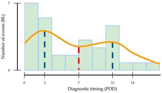
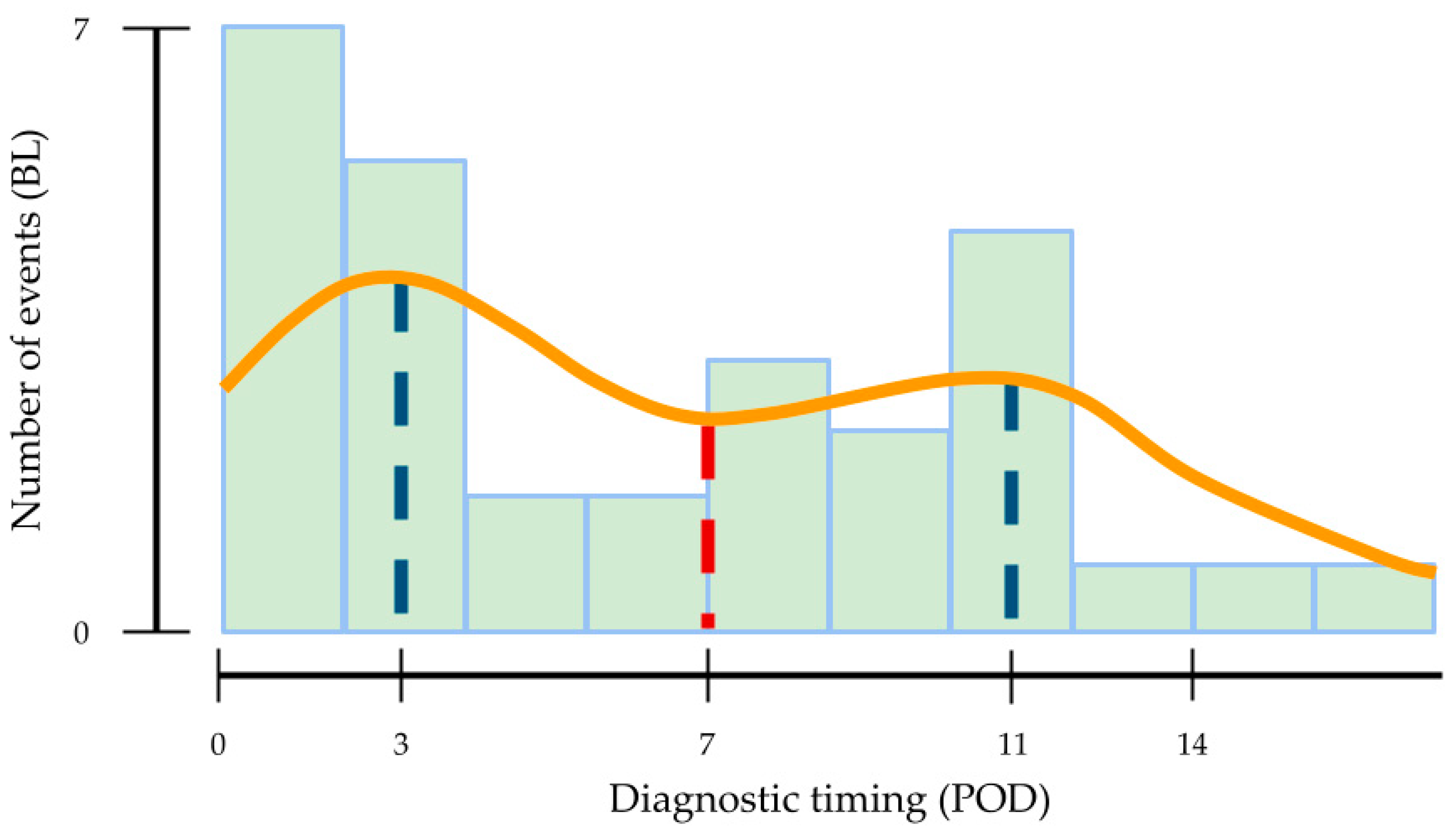
The distribution of biliary leak diagnosis exhibited a clear bimodal pattern (Figure 1). Two distinct peaks in diagnosis were observed at PODs 3 and 11, separated by a distinct trough at POD 7. Based on this diagnostic distribution, POD 7 was selected as the empirical cutoff for day of diagnosis.
Figure 1.
Frequency distribution of biliary leakage diagnosis during the postoperative period. The distribution highlights a distinct bimodal pattern, with diagnostic peaks at PODs 3 and 11 (blue dash lines) and a nadir at POD 7 (red dash line).
Following this classification, 55.6% (n = 20) of the leaks were categorized as EBL (diagnosed on or before POD 7), while 44.4% (n = 16) were classified as LBL (diagnosed after POD 7). The mean time to diagnosis differed significantly between the two cohorts, with EBL cases identified at a mean of 3.0 ± 2.0 days compared to 12.8 ± 3.1 days for the LBL group (p < 0.001).
A detailed cross-tabulation comparing baseline, anatomical, and operative characteristics between EBL and LBL is provided in Table S1. Briefly, EBL was significantly associated with left hepatic segment involvement (40.0% vs. 6.3%, p = 0.023), formal anatomical resections (85.0% vs. 43.8%, p = 0.012), and a longer mean Pringle maneuver duration per clamping (15.9 ± 3.0 vs. 12.6 ± 4.0 min, p = 0.04). In detail, the EBL cohort was characterized by a higher frequency of major resections, such as lobectomies (40.0% vs. 18.8%) and sectionectomies (30.0% vs. 0%), as well as a higher rate of associated secondary anatomical procedures (55.5% vs. 0%, p = 0.005). Conversely, LBL presented a higher proportion of wedge resections (56.3% vs. 15.0%, p = 0.015). Definitive histological diagnosis showed a distribution approaching statistical significance (p = 0.06), with primary malignancies identified in 75.0% (n = 15) of EBL cases compared to 37.5% (n = 6) of LBL cases, whereas metastatic disease was more prevalent in late presentations (56.3% vs. 25.0%). Direct head-to-head comparison between EBL and LBL showed no statistically significant differences in operative duration (417 ± 121 min vs. 393 ± 115 min, p = 0.57) or proximity to major vessels (65% vs. 37.5%, p = 0.09). Regarding clinical outcomes (Table S2), while ISGLS severity grades (p = 0.89) and Clavien–Dindo classifications (p = 0.56) were comparable between groups, patients with LBL experienced a higher rate of 30-day readmission compared to EBL patients (62.5% vs. 30.0%, p = 0.091; OR 3.89, 95% CI 0.97–15.65), although this comparison did not reach statistical significance.
3.3. Univariable and Multivariable Regression Analysis of Risk Factors for Biliary Leakage
The evaluation of potential predictive factors for the development of BL was performed using a binary logistic regression model (Table 4). In the univariable analysis, several clinical and operative variables showed a significant association with the occurrence of the complication. An operative time exceeding 5 h was associated with the highest probability of leakage (OR = 9.4; 95% CI 3.6–25.0; p < 0.001). Other significant univariable predictors included tumor proximity to major vessels (OR = 3.3; 95% CI 1.6–6.6; p = 0.002) and the execution of a major hepatectomy (OR = 3.7; 95% CI 1.8–7.5; p < 0.001). By comparison, a total Pringle maneuver duration exceeding 40 min did not show a significant univariable effect (p = 0.21). In the multivariable analysis, an operative time greater than 5 h was the only variable that remained a significant independent predictor for overall BL (OR = 6.9; 95% CI 2.4–19.4; p < 0.001). The other variables included in the model, such as proximity to major vessels (p = 0.14), major hepatectomy (p = 0.41), and a total Pringle duration over 40 min (p = 0.69), did not maintain statistical significance.
Table 4.
Univariable and multivariable binary logistic regression analyses of risk factors associated with the development of overall postoperative biliary leakage.
To further differentiate the risk profiles, a multinomial logistic regression was performed to identify specific independent predictors for early leakage (EBL, ≤7 days) and late leakage (LBL, >7 days), using the absence of leakage (nBL) as the reference category (Table 5).
Table 5.
Multinomial logistic regression analysis of independent risk factors predicting early versus late post-hepatectomy biliary leakage.
Regarding EBL, the univariable analysis identified proximity to major vessels (OR = 5.4; 95% CI 2.1–14.4; p = 0.01), an operative time > 5 h (OR = 8.7; 95% CI 2.5–30.3; p < 0.001), and a total Pringle duration > 40 min (OR = 2.7; 95% CI 1.0–7.1; p = 0.042) as significant risk factors. In the final multivariable model, both proximity to major vessels (OR = 4.1; 95% CI 1.5–11.6; p = 0.007) and an operative time > 5 h (OR = 5.2; 95% CI 1.4–19.2; p = 0.012) were confirmed as statistically significant independent predictors for EBL, while a total Pringle duration over 40 min did not maintain statistical significance (p = 0.213).
For LBL, an operative time exceeding 5 h was the single significant predictor identified in the univariable analysis (OR = 10.5; 95% CI 2.3–47.5; p = 0.002). In the multivariable model, an operative time > 5 h was confirmed as a notable independent predictor, with a notably high risk (OR = 12.2; 95% CI 2.6–56.7; p = 0.001). By comparison, neither tumor proximity to major vessels (p = 0.75) nor a total Pringle duration over 40 min (p = 0.31) showed any significant association with the occurrence of late postoperative biliary complications.
4. Discussion
The identification of predictive determinants for BL after hepatic resection remains a cornerstone in modern hepatobiliary surgery, as this complication profoundly impacts postoperative morbidity, length of hospital stay, and healthcare costs. In the present study, the overall incidence of BL was 9.7%, a figure that aligns with the current international literature, which reports rates ranging between 4% and 12% depending on center volume and case-mix complexity. In terms of clinical severity, the vast majority of leaks in our cohort were classified as ISGLS Grade B (77.8%) and Clavien–Dindo Grade 3a or 3b, requiring active radiological or endoscopic interventions but successfully avoiding surgical re-exploration.
In our study, an operative time longer than five hours was the only factor independently associated with the development of postoperative BL (OR = 6.9; 95% CI 2.4–19.4; p < 0.001). This variable should not be interpreted merely as a chronological measurement. Instead, a prolonged operative duration serves as a reliable indirect indicator and surrogate marker for subclinical technical challenges and high surgical complexity.
The weight of an extended surgical duration as an independent predictor of BL is strongly consolidated in the current international literature. The multicenter study by Sakamoto et al. confirmed that operative time is an independent predictive factor for biliary fistula, identifying an optimal cut-off value of 384 min beyond which the risk of developing a fistula increases significantly (OR = 4.2) [21]. Furthermore, the continuous impact of procedural duration underscores the structural validity of the Biliary Leakage Risk Score proposed by Riediger et al. based on 844 resections, where each additional minute of surgery was reported to contribute incrementally to the risk of tissue injury and subsequent leakage [22].
In highly complex surgical settings involving extensive dissection of the hepatic hilum, such as resections for hilar cholangiocarcinoma, prolonged operation has been consistently classified among the key independent predictors of severe ISGLS Grade B/C leaks [23,24]. While some authors emphasize specific intraoperative maneuvers, these often function as direct surrogates for overall case complexity and prolonged duration; for instance, Donadon et al. found that an extended Pringle maneuver duration ≥ 90 min was independently associated with BL (OR 3.4, p < 0.001), representing a direct consequence of a challenging parenchymal transection [6]. A large-scale meta-analysis on 43,824 patients definitively validated that a prolonged operative time, combined with extended exposure of the liver raw surface to thermal energy devices, represents a definitive multi-institutional predictor for postoperative biliary complications [9]. In our series, the increase in surgical complexity related to the intrinsic characteristics of the procedure (such as tumor site, proximity to major vessels, and the type of intervention) outweighed intraoperative hemodynamic variables or clamping duration alone, aligning with previous surgical series [21,25]. Finally, the progressive fatigue of the surgical team during complex, hours-long procedures may subtly influence precision during parenchymal transection or biliostasis, further explaining why a five-hour threshold represents a critical turning point for postoperative biliary morbidity.
The core finding and clinical novelty of this study lies in the distinct clinical profiles observed when stratifying post-hepatectomy biliary leaks by day of diagnosis into early-diagnosed (EBL, POD of diagnosis ≤ 7) and late-diagnosed (LBL, POD of diagnosis > 7) events. The temporal distribution of biliary leak diagnosis in our cohort exhibited a clear bimodal pattern, characterized by two distinct diagnostic peaks at PODs 3 and 11, separated by a trough at POD 7. This biological bimodal distribution suggests potential differences in surgical and pathogenic mechanisms are at play depending on the timing of fistula manifestation, suggesting a potential transition from an immediate mechanical etiology in early leaks toward a delayed ischemic or thermal pathway in late presentations, a hypothesis that requires prospective validation.
While sample size constraints precluded direct multivariable modeling between EBL and LBL, univariable exploratory comparisons provide valuable clinical insights. Direct head-to-head comparison showed no statistically significant differences in operative duration or vessel proximity between groups, whereas the higher concentration of anatomical resections and left-segment involvement in EBL supports an immediate mechanical mechanism, in contrast to LBL presentations which appeared more frequently after non-anatomical or less extensive resections.
EBL was independently predicted in our multivariable model by both tumor proximity to major intrahepatic vessels (OR = 4.1; 95% CI 1.5–11.6; p = 0.007) and an operative time exceeding 5 h (OR = 5.2; 95% CI 1.4–19.2; p = 0.012). We hypothesize that early leaks may primarily represent immediate mechanical or structural disruptions along the parenchymal transection plane. When a lesion originates close to the central hepatic compartment or major intrahepatic vascular structures, surgeons must perform deep, meticulous, and demanding manipulations around the Glissonian pedicles or the hepatic hilum. This clinical observation is highly consistent with the landmark concepts of Nagano et al., who identified extensive incision surfaces and direct exposure of the major Glissonian sheath as prime risk factors for mechanical bile duct disruption [20]. The technical failure of a poorly positioned metallic or polymer clip, or a suture line acutely disrupted by early postoperative pressure changes, leads to an immediate macroscopic extravasation of bile, causing the leak to manifest rapidly within the first 48 to 72 h, as also noted by Xue et al. [9]. Furthermore, this is supported by Sakamoto et al., who observed that central-type fistulas communicating directly with the main biliary tree display a remarkably rapid median time to diagnosis [21].
Although LBL was predicted solely by an operative time > 5 h in multivariable modeling relative to non-leak controls (OR = 12.2; 95% CI 2.6–56.7; p = 0.001), direct comparison between EBL and LBL revealed no statistically significant difference in operative duration (417 ± 121 min vs. 393 ± 115 min, p = 0.57) or vessel proximity (65% vs. 37.5%, p = 0.09). Therefore, a prolonged operative duration reflects a shared marker of high procedural complexity across all biliary leaks rather than a distinct discriminator separating late from early presentations. Nevertheless, late leaks might still be driven by progressive subacute ischemic or thermal changes along the exposed liver raw surface, rather than an immediate macroscopic technical disruption. Prolonged parenchymal transection routinely involves the extensive, repetitive, and cumulative application of thermal energy devices (including standard monopolar/bipolar electrocautery or advanced tissue-sealers) to secure intraoperative hemostasis. Paradoxically, while the appropriate use of modern energy devices has been shown by Gotohda et al. in a multicenter randomized trial to significantly decrease overall and early mechanical biliary leaks by optimizing immediate tissue sealing, their prolonged and repetitive application in highly complex procedures carries a severe drawback [26]. The critical role of ischemia in the pathogenesis of severe post-hepatectomy leaks is further corroborated by Mohkam et al., who identified a liver remnant ischemia time ≥ 45 min as a powerful independent predictor of Grade B/C leaks in a large multicenter French cohort [17].
This delayed ischemic hypothesis aligns with the multi-institutional study by Kaibori et al., who demonstrated that late-onset leaks are significantly associated with intra-abdominal infections and require more invasive radiological or endoscopic interventions than early ones. Alternatively, these events can arise from an unrecognized subclinical biloma that becomes secondarily infected, leading to localized inflammatory erosion and subsequent rupture into an adjacent intrahepatic bile duct [19].
A key clinical finding of this study is that an uneventful first postoperative week does not guarantee freedom from biliary complications. Although the higher 30-day readmission rate observed in LBL patients compared to EBL (62.5% vs. 30.0%) did not achieve formal statistical significance, it underscores a clinically relevant trend. Unremarkable early post-operative recovery in LBL patients should not obscure the potential for delayed biliary morbidity requiring outpatient surveillance. Because late leaks often develop after initial discharge following seemingly less extensive resections, clinicians may be misled into a false sense of security. Recognizing that prolonged operative time (>5 h) is a major indicator of overall risk for both early and late presentations allows surgical teams to target these complex patients for enhanced post-discharge monitoring.
While biologically plausible, these pathophysiological pathways remain working hypotheses. Direct operative verification of leak sites and objective tissue ischemia parameters were beyond the observational scope of this study. Given that intraoperative energy device usage and clamping durations were comparable between groups, these clinical data identify strong temporal associations rather than direct causality.
Regarding histopathology, Cholangiocarcinoma (CCA) was markedly overrepresented in our BL group, accounting for 41.7% of cases compared to 11.7% in the non-leak population (p = 0.05). This correlation is well-documented, as CCA resections routinely dictate aggressive parenchymal transections and radical hilar dissections [25].
The unparalleled procedural complexity of CCA surgery introduces three distinct pathophysiological triggers for leakage. First, the frequently required resection of Segment 1 exposes delicate subsegmental biliary branches directly on the central raw surface [6]. Second, extended lymphadenectomy requires complete skeletonization of the portal pedicle, stripping away the peribiliary vascular plexus and inducing subacute ischemic devascularization of the remaining bile duct stumps. Third, the necessity of a definitive bilioenteric anastomosis introduces a major mechanical and biological vulnerability, which is often further compromised by pre-existing tissue inflammation and bacterial colonization from mandatory preoperative biliary drainage [11,12,18].
These findings highlight the potential value of objective intraoperative detection methods in high-risk scenarios; although not directly evaluated in our study cohort, techniques such as lipid emulsion leak testing (white test) or indocyanine green (ICG) fluorescence cholangiography may provide useful real-time guidance to identify active biliary extravasation before abdominal closure [27,28,29,30,31].
Several limitations of the present study must be acknowledged. First, its retrospective design inherently limits the ability to establish direct causal relationships, allowing us to identify strong statistical associations rather than definitive etiologic mechanisms. Second, the POD 7 cutoff used to distinguish early from late leaks was empirically derived from the temporal distribution within our cohort in the absence of standardized literature criteria; while biologically intuitive, this threshold requires external validation. Furthermore, all time-to-event analyses strictly reflect the POD of clinical diagnosis rather than the exact pathophysiological moment of leak onset, which cannot be retrospectively determined. Third, selective abdominal drainage practice may have introduced a degree of diagnostic timing bias. In patients with high-risk features and surgical drains, early low-grade leaks were readily identified via visual drain inspection, whereas in non-drained patients or following early drain removal, diagnosis relied on clinical symptoms triggering secondary imaging, potentially shifting the documented timing of late-onset events. Fourth, despite a representative total cohort, the absolute number of leakage events constrained multivariable modeling capacity and yielded wider confidence intervals; consequently, these multivariable findings should be viewed as exploratory clinical insights. Fifth, direct intraoperative cholangiography and objective parameters evaluating peribiliary devascularization or tissue ischemia were beyond the observational scope of this real-world dataset. Given that energy device utilization rates were comparable across groups, our proposed pathophysiological pathways represent plausible working frameworks rather than direct procedural outcomes. Finally, while conducting this study at a single specialized institution ensured high uniformity in surgical techniques, transection protocols, and perioperative care, prospective multi-center studies are essential to confirm the external generalizability of this temporal risk stratification.
5. Conclusions
In conclusion, diagnostic timing significantly impacts the clinical trajectory and healthcare burden of post-hepatectomy biliary leakage. The primary clinical takeaway of this study is that a smooth postoperative course during the first week should not lead to early complacency. While early leaks are predominantly identified during the index hospitalization and are linked to technical or anatomical complexity near major vessels, LBL demonstrated a trend toward higher 30-day readmissions, highlighting that an uneventful early postoperative course does not preclude late biliary morbidity and warrants targeted post-discharge monitoring.
Clinicians must maintain high vigilance for subtle clinical signs of late-onset leakage, such as low-grade fever, unexplained abdominal discomfort, or delayed leukocytosis, around the second postoperative week. To mitigate these post-discharge risks, implementing structured patient discharge education, establishing dedicated outpatient surveillance strategies, and maintaining a low threshold for diagnostic imaging are paramount. This is particularly crucial for patients undergoing prolonged resections exceeding 5 h or resections for high-risk pathologies such as cholangiocarcinoma.
Given the modest sample size of leakage events in this single-center cohort, these findings should be interpreted as hypothesis-generating. Prospective, multi-center studies are required to provide external validation and to establish standardized post-discharge surveillance guidelines for patients undergoing complex liver surgery.
Supplementary Materials
The following supporting information can be downloaded at https://www.mdpi.com/article/10.3390/livers6050088/s1. Table S1: Comprehensive comparison of demographic, anatomical, intraoperative, and histopathological characteristics between patients with early versus late post-hepatectomy biliary leakage; Table S2: Clinical outcomes, severity, and healthcare utilization associated with postoperative biliary leakage, stratified by early versus late onset.
Author Contributions
Conceptualization, G.P., L.T., I.B., A.T. and G.L.G.; methodology, G.P. and L.T.; validation, G.P., L.T., I.B., A.T. and G.L.G.; formal analysis, G.P. and L.T.; investigation, G.P., L.T., M.R., N.N., A.M. and M.N.R.; data curation, G.P. and L.T.; writing—original draft preparation, G.P. and L.T.; writing—review and editing, I.B., M.R., N.N., A.M., M.N.R., A.T. and G.L.G.; visualization, G.P. and L.T.; supervision, A.T. and G.L.G. All authors have read and agreed to the published version of the manuscript.
Funding
This research received no external funding.
Institutional Review Board Statement
The study was conducted in accordance with the Declaration of Helsinki, and approved by the Ethics Committee of Regione Toscana—Area Vasta Centro (protocol code 27975_BIO, date of approval 29 April 2025).
Informed Consent Statement
Informed consent for participation was obtained from all subjects involved in the study.
Data Availability Statement
The data that support the findings of this study are available from the corresponding author, L.T., upon reasonable request.
Conflicts of Interest
The authors declare no conflicts of interest.
Abbreviations
The following abbreviations are used in this manuscript:
| BL | Bile leakage |
| EBL | Early Bile leakage |
| LBL | Late Bile leakage |
| CCA | Cholangiocarcinoma |
| POD | Postoperative day |
References
- Ceccarelli, G.; Avella, P.; Muttillo, E.M.; Conticchio, M.; Tebala, G.D.; Piccolo, G.; Romano, L.; Memeo, R.; Rocca, A.; Risi, L.; et al. Devices for Minimally Invasive Liver Parenchyma Transection: The SICE (Italian Society of Endoscopic Surgery) Italian and International Survey. Surg. Endosc. 2025, 39, 4822–4838. [Google Scholar] [CrossRef] [Scilit] [PubMed]
- Kamarajah, S.K.; Wilson, C.H.; Bundred, J.R.; Lin, A.; Sen, G.; Hammond, J.S.; French, J.J.; Manas, D.M.; White, S.A. A Systematic Review and Network Meta-Analysis of Parenchymal Transection Techniques during Hepatectomy: An Appraisal of Current Randomised Controlled Trials. HPB 2020, 22, 204–214. [Google Scholar] [CrossRef] [Scilit] [PubMed]
- Haugen, C.; Noriega, M.; Andy, C.; Waite, C.; Carpenter, D.; Halazun, K.; Samstein, B.; Rocca, J.P. Complete Transition from Laparoscopic to Robotic Liver Surgery Achieves Superior Outcomes in Difficult Hepatectomies: A Seven-Year Retrospective Study. Surg. Endosc. 2025, 39, 1600–1608. [Google Scholar] [CrossRef] [Scilit] [PubMed]
- Vining, C.C.; Kuchta, K.; Al Abbas, A.I.; Hsu, P.J.; Paterakos, P.; Schuitevoerder, D.; Sood, D.; Roggin, K.K.; Talamonti, M.S.; Hogg, M.E. Bile Leak Incidence, Risk Factors and Associated Outcomes in Patients Undergoing Hepatectomy: A Contemporary NSQIP Propensity Matched Analysis. Surg. Endosc. 2022, 36, 5710–5723. [Google Scholar] [CrossRef] [Scilit] [PubMed]
- Koch, M.; Garden, O.J.; Padbury, R.; Rahbari, N.N.; Adam, R.; Capussotti, L.; Fan, S.T.; Yokoyama, Y.; Crawford, M.; Makuuchi, M.; et al. Bile Leakage after Hepatobiliary and Pancreatic Surgery: A Definition and Grading of Severity by the International Study Group of Liver Surgery. Surgery 2011, 149, 680–688. [Google Scholar] [CrossRef] [Scilit] [PubMed]
- Donadon, M.; Costa, G.; Cimino, M.; Procopio, F.; Del Fabbro, D.; Palmisano, A.; Torzilli, G. Diagnosis and Management of Bile Leaks After Hepatectomy: Results of a Prospective Analysis of 475 Hepatectomies. World J. Surg. 2016, 40, 172–181. [Google Scholar] [CrossRef] [Scilit] [PubMed]
- Sliwinski, S.; Heil, J.; Franz, J.; El Youzouri, H.; Heise, M.; Bechstein, W.O.; Schnitzbauer, A.A. A Critical Appraisal of the ISGLS Definition of Biliary Leakage after Liver Resection. Langenbecks Arch. Surg. 2023, 408, 77. [Google Scholar] [CrossRef] [Scilit] [PubMed]
- Rahbari, N.N.; Elbers, H.; Koch, M.; Kirchberg, J.; Dutlu, M.; Mehrabi, A.; Büchler, M.W.; Weitz, J. Bilirubin Level in the Drainage Fluid Is an Early and Independent Predictor of Clinically Relevant Bile Leakage after Hepatic Resection. Surgery 2012, 152, 821–831. [Google Scholar] [CrossRef] [Scilit] [PubMed]
- Xue, S.; Wang, H.; Chen, X.; Zeng, Y. Risk Factors of Postoperative Bile Leakage after Liver Resection: A Systematic Review and Meta-Analysis. Cancer Med. 2023, 12, 14922–14936. [Google Scholar] [CrossRef] [Scilit] [PubMed]
- Brooke-Smith, M.; Figueras, J.; Ullah, S.; Rees, M.; Vauthey, J.N.; Hugh, T.J.; Garden, O.J.; Fan, S.T.; Crawford, M.; Makuuchi, M.; et al. Prospective Evaluation of the International Study Group for Liver Surgery Definition of Bile Leak after a Liver Resection and the Role of Routine Operative Drainage: An International Multicentre Study. HPB 2015, 17, 46–51. [Google Scholar] [CrossRef] [Scilit] [PubMed]
- Vienet, J.; Labgaa, I.; Duran, R.; Godat, S.; Blanc, C.; Uldry, E.; Melloul, E.; Fuks, D.; Joliat, G.R. Incidence and Risk Factors of Biliary Leaks after Partial Hepatectomy within an Enhanced Recovery Perioperative Pathway: A Single-Center Retrospective Cohort Study. Langenbecks Arch. Surg. 2025, 410, 104. [Google Scholar] [CrossRef] [Scilit] [PubMed]
- Martin, A.N.; Narayanan, S.; Turrentine, F.E.; Bauer, T.W.; Adams, R.B.; Stukenborg, G.J.; Zaydfudim, V.M. Clinical Factors and Postoperative Impact of Bile Leak After Liver Resection. J. Gastrointest. Surg. 2018, 22, 661–667. [Google Scholar] [CrossRef] [Scilit] [PubMed]
- Braunwarth, E.; Ratti, F.; Aldrighetti, L.; Al-Saffar, H.A.; D’Souza, M.A.; Sturesson, C.; Linke, R.; Schnitzbauer, A.; Bodingbauer, M.; Kaczirek, K.; et al. Incidence and Risk Factors for Anastomotic Bile Leakage in Hepatic Resection with Bilioenteric Reconstruction—A International Multicenter Study. HPB 2023, 25, 54–62. [Google Scholar] [CrossRef] [Scilit] [PubMed]
- Guillaud, A.; Pery, C.; Campillo, B.; Lourdais, A.; Laurent, S.; Boudjema, K. Incidence and Predictive Factors of Clinically Relevant Bile Leakage in the Modern Era of Liver Resections. HPB 2013, 15, 224–229. [Google Scholar] [CrossRef] [Scilit] [PubMed]
- Dorcaratto, D.; Mazzinari, G.; Fernandez, M.; Muñoz, E.; Garcés-Albir, M.; Ortega, J.; Sabater, L. Impact of Postoperative Complications on Survival and Recurrence After Resection of Colorectal Liver Metastases: Systematic Review and Meta-Analysis. Ann. Surg. 2019, 270, 1018–1027. [Google Scholar] [CrossRef] [Scilit] [PubMed]
- Fernández-Moreno, M.C.; Dorcaratto, D.; Garcés-Albir, M.; Muñoz, E.; Arvizu, R.; Ortega, J.; Sabater, L. Impact of Type and Severity of Postoperative Complications on Long-Term Outcomes after Colorectal Liver Metastases Resection. J. Surg. Oncol. 2020, 122, 212–225. [Google Scholar] [CrossRef] [Scilit] [PubMed]
- Mohkam, K.; Farges, O.; Vibert, E.; Soubrane, O.; Adam, R.; Pruvot, F.R.; Regimbeau, J.M.; Adham, M.; Boleslawski, E.; Mabrut, J.Y.; et al. Risk Score to Predict Biliary Leakage after Elective Liver Resection. Br. J. Surg. 2018, 105, 128–139. [Google Scholar] [CrossRef] [Scilit] [PubMed]
- Patkar, S.; Kunte, A.; Sundaram, S.; Goel, M. Post-Hepatectomy Biliary Leaks: Analysis of Risk Factors and Development of a Simplified Predictive Scoring System. Langenbecks Arch. Surg. 2023, 408, 63. [Google Scholar] [CrossRef] [Scilit] [PubMed]
- Kaibori, M.; Shimizu, J.; Hayashi, M.; Nakai, T.; Ishizaki, M.; Matsui, K.; Kim, Y.K.; Hirokawa, F.; Nakata, Y.; Noda, T.; et al. Late-Onset Bile Leakage after Hepatic Resection. Surgery 2015, 157, 37–44. [Google Scholar] [CrossRef] [Scilit] [PubMed]
- Nagano, Y.; Togo, S.; Tanaka, K.; Masui, H.; Endo, I.; Sekido, H.; Nagahori, K.; Shimada, H. Risk Factors and Management of Bile Leakage after Hepatic Resection. World J. Surg. 2003, 27, 695–698. [Google Scholar] [CrossRef] [Scilit] [PubMed]
- Sakamoto, K.; Tamesa, T.; Yukio, T.; Tokuhisa, Y.; Maeda, Y.; Oka, M. Risk Factors and Managements of Bile Leakage After Hepatectomy. World J. Surg. 2016, 40, 182–189. [Google Scholar] [CrossRef] [Scilit] [PubMed]
- Riediger, C.; Hoffmann, R.; Löck, S.; Giehl-Brown, E.; Dennler, S.; Kahlert, C.; Weitz, J. Novel Personalized Score Predicts Risk for Postoperative Biliary Leak in Liver Surgery—A Retrospective Database Analysis. J. Gastrointest. Surg. 2022, 26, 2101–2110. [Google Scholar] [CrossRef] [Scilit] [PubMed]
- Nakagawa, K.; Tanaka, K.; Nojiri, K.; Sawada, Y.; Kumamoto, T.; Ueda, M.; Minami, Y.; Mochizuki, Y.; Morioka, D.; Kubota, T.; et al. Predictive Factors for Bile Leakage after Hepatectomy for Hepatic Tumors: A Retrospective Multicenter Study with 631 Cases at Yokohama Clinical Oncology Group (YCOG). J. Hepato-Biliary-Pancreat. Sci. 2017, 24, 33–41. [Google Scholar] [CrossRef] [Scilit] [PubMed]
- Taguchi, Y.; Ebata, T.; Yokoyama, Y.; Igami, T.; Sugawara, G.; Kokuryo, T.; Wakai, K.; Nagino, M. The Determination of Bile Leakage in Complex Hepatectomy Based on the Guidelines of the International Study Group of Liver Surgery. World J. Surg. 2014, 38, 168–176. [Google Scholar] [CrossRef] [Scilit] [PubMed]
- Capussotti, L.; Ferrero, A.; Viganò, L.; Sgotto, E.; Muratore, A.; Polastri, R. Bile Leakage and Liver Resection: Where Is the Risk? Arch. Surg. 2006, 141, 690–694. [Google Scholar] [CrossRef] [Scilit] [PubMed]
- Gotohda, N.; Yamanaka, T.; Saiura, A.; Uesaka, K.; Hashimoto, M.; Konishi, M.; Shimada, K. Impact of Energy Devices during Liver Parenchymal Transection: A Multicenter Randomized Controlled Trial. World J. Surg. 2015, 39, 1543–1549. [Google Scholar] [CrossRef] [Scilit] [PubMed]
- Wang, H.Q.; Yang, J.; Yang, J.Y.; Yan, L.N. Bile Leakage Test in Liver Resection: A Systematic Review and Meta-Analysis. World J. Gastroenterol. 2013, 19, 8420–8426. [Google Scholar] [CrossRef] [Scilit] [PubMed]
- Vaska, A.I.; Abbas, S. The Role of Bile Leak Testing in Liver Resection: A Systematic Review and Meta-Analysis. HPB 2019, 21, 148–156. [Google Scholar] [CrossRef] [Scilit] [PubMed]
- Cristaudi, A.; Tarantino, I.; Scheiwiller, A.; Wiencierz, A.; Majno-Hurst, P.; Schmied, B.M.; Metzger, J.; Hartel, M.; Kremer, M.; Manzini, G. Diagnostic Role of the “white Test” with Lipidic Solution in the Early Intraoperative Identification of Open Bile Ducts for the Prevention of Bile Leakage after Liver Resection: Study Protocol for a Randomised Controlled Multicentric Superiority Trial (BiLe-Trial). BMJ Open 2021, 11, e049449. [Google Scholar] [CrossRef] [Scilit] [PubMed]
- Hanaki, T.; Goto, K.; Tokuyasu, N.; Endo, Y.; Sunada, H.; Noma, H.; Kishino, M.; Yagyu, T.; Uchinaka, E.; Murakami, Y.; et al. Systemic Indocyanine Green Administration to Detect Bile Leakage after Liver Surgery: A Prospective Clinical Trial, Using Historical Controls. BMJ Open 2025, 15, e097205. [Google Scholar] [CrossRef] [Scilit] [PubMed]
- Gon, H.; Omiya, S.; Komatsu, S.; Yamasaki, N.; Murakami, S.; Fukushima, K.; Urade, T.; Tsugawa, D.; Yanagimoto, H.; Toyama, H.; et al. Efficacy and Safety of Indocyanine Green-Fluorescence Imaging Guided Liver Resection: A Single-Arm Prospective Cohort Study. Langenbecks Arch. Surg. 2025, 410, 34. [Google Scholar] [CrossRef] [Scilit] [PubMed]

Disclaimer/Publisher’s Note: The statements, opinions and data contained in all publications are solely those of the individual author(s) and contributor(s) and not of MDPI and/or the editor(s). MDPI and/or the editor(s) disclaim responsibility for any injury to people or property resulting from any ideas, methods, instructions or products referred to in the content. |
© 2026 by the authors. Licensee MDPI, Basel, Switzerland. This article is an open access article distributed under the terms and conditions of the Creative Commons Attribution (CC BY) license.