


Abstract
The menstrual cycle is classically understood as the endocrine program governing reproduction. However, its effects beyond the reproductive tract are generally investigated within separate disciplines. Consequently, the cycle continues to be viewed predominantly as a collection of tissue-specific hormonal effects rather than as a coordinated systems-level physiological program. This review evaluates an alternative interpretation based on the Integrated Human System Framework (IHSF): the menstrual cycle as a recurring systemic biological rhythm that conveys temporal endocrine information throughout the body. We conducted a multidisciplinary narrative synthesis of evidence from reproductive endocrinology, dermatology, neuroimmunology, mucosal biology, microbiology, and systems physiology. Evidence was organized around three complementary models—the skin, the neuroendocrine–immune network, and the vulvovaginal ecosystem—and graded qualitatively. The IHSF was compared with related frameworks, and five testable predictions and potential refuting observations were specified. Across the three models, the literature describes hormonal effects and cycle-associated physiological changes involving barrier regulation, immune homeostasis, epithelial renewal, vascular adaptation, extracellular matrix remodeling, neural communication, and host–microbial ecology. Their expression varies with tissue characteristics and local biological context, and the strength of supporting evidence differs among outcomes. Evidence is comparatively consistent for vaginal microbial dynamics, whereas reproducible menstrual-phase rhythmicity of the cutaneous microbiome remains hypothesized. Within the IHSF, these findings are brought together to propose that a common endocrine rhythm is interpreted through tissue-specific biological processes, potentially linking physiological responses across the body. This integrative interpretation is distinct from direct demonstration of coordinated multisystem regulation. This synthesis broadens the biological significance of the menstrual cycle beyond reproduction and raises the possibility that its recurring endocrine rhythm coordinates adaptive responses across interconnected tissues. Through the IHSF, the menstrual cycle is reframed as a potential multisystem organizer that links a shared temporal endocrine signal to specialized, context-dependent responses throughout the body. The IHSF is a conceptual framework requiring further empirical validation through longitudinal, hormone-verified multisystem research that accounts for behavioral, therapeutic, product-related, and environmental modifiers.
1. Introduction—The Menstrual Cycle as a Recurring Systemic Biological Rhythm
Life is organized through biological rhythms that coordinate physiological processes across multiple temporal scales. Circadian rhythms synchronize metabolism, endocrine secretion, immune activity, and behavior with the 24-h light-dark cycle, whereas ultradian, infradian, and seasonal rhythms regulate additional aspects of organismal function [1,2,3,4]. These recurring biological oscillations are not simply periodic events; they provide temporal information that enables physiological systems to anticipate changing internal and external conditions while maintaining homeostasis. Increasing evidence from systems physiology indicates that biological rhythms are fundamental organizational principles governing communication among interconnected organ systems rather than isolated regulatory processes. Consequently, human physiology is increasingly recognized as a dynamic network in which temporal coordination is essential for adaptive function [5].
The menstrual cycle is considered here within a broader temporal context that includes daily physiological rhythms and the emergence, modification, and cessation of menstrual cyclicity across reproductive life. The present review concentrates on the recurring menstrual rhythm, while recognizing shorter-timescale rhythms and longer-term reproductive transitions as relevant context for interpreting tissue responses.
Among these biological rhythms, the menstrual cycle represents one of the most complex recurring physiological programs in humans. Traditionally, the menstrual cycle has been viewed primarily through the lens of reproductive endocrinology, emphasizing ovarian follicular development, ovulation, endometrial preparation, and reproductive potential. These reproductive events are orchestrated by the hypothalamic–pituitary–ovarian (HPO) axis through highly regulated oscillations in gonadotropin-releasing hormone, luteinizing hormone, follicle-stimulating hormone, estradiol, and progesterone. Although reproduction remains its primary biological purpose, accumulating evidence demonstrates that these endocrine oscillations extend well beyond the reproductive tract, influencing multiple organ systems through widespread hormone receptor expression and coordinated physiological adaptation [6,7,8].
Over the past two decades, studies from dermatology, immunology, neuroscience, cardiovascular biology, and mucosal physiology have described cyclical changes in epithelial biology, immune regulation, vascular function, extracellular matrix remodeling, microbial ecology, and neural communication. These adaptations have been reported in the skin [9,10], the immune system [11,12,13], the brain [14,15,16], and the vulvovaginal ecosystem [17,18,19,20,21,22], among other hormone-responsive tissues [23,24,25,26,27,28]. Their significance may extend beyond the organs in which they were first described: considered together, they raise the possibility that the menstrual cycle synchronizes adaptive responses across interconnected tissues. The magnitude and consistency of these adaptations nevertheless vary among systems and among individuals, and much of the supporting evidence comes from small studies with heterogeneous methods and variable verification of cycle phase.
Despite these advances, findings on cycle-associated physiology remain largely organized within individual disciplines. The gap addressed here is the limited integration of these observations into a comparative framework that considers tissue-specific responses alongside possible relationships among systems.
This review proposes an alternative interpretation based on the Integrated Human System Framework (IHSF), a conceptual model that views physiological function as emerging from interactions among endocrine, immune, neural, epithelial, vascular, connective tissue, and microbial systems [5,29,30,31,32,33]. Its clinical relevance lies in considering how tissue responsiveness and local biological context, alongside hormone concentrations, may help explain why individuals experience different physiological responses and clinical manifestations under similar endocrine conditions. Within this perspective, the menstrual cycle is viewed as a recurring systemic biological rhythm that conveys temporal endocrine information throughout the body. Individual tissues interpret this common rhythm according to their cellular composition, receptor repertoire, extracellular environment, and local biological context. Conserved processes, including barrier regulation, immune homeostasis, extracellular matrix remodeling, vascular adaptation, neural communication, epithelial renewal, and host–microbial ecology, convert that information into tissue-specific physiology. This perspective complements rather than replaces classical reproductive endocrinology.
Accordingly, this review applies the IHSF to three complementary hormone-responsive models: the skin, the neuroendocrine–immune network, and the vulvovaginal ecosystem. Its objective is to integrate evidence from these systems to examine how a recurring endocrine rhythm is expressed through tissue-specific biological processes and to identify proposed relationships for future investigation. The review combines qualitative evidence appraisal, comparison with related conceptual frameworks and testable predictions. Together, these elements provide an integrative basis for examining the menstrual cycle as a systemic biological rhythm (Figure 1).
Figure 1.
The Integrated Human System Framework and Multisystem Decoding of the Menstrual Cycle. Note: This conceptual diagram is provided for illustrative purposes only. Menstrual-cycle curves and other visual elements are schematic, are not drawn to scale, and do not represent exact hormone concentrations, phase durations, anatomical proportions, or interindividual variability. The diagram summarizes proposed information flow and testable relationships; arrows indicate conceptual associations and do not establish causal direction or universal clinical outcomes.
By integrating evidence across these three model systems, this review aims to broaden understanding of the menstrual cycle as a whole-body biological rhythm whose significance extends beyond reproduction. For clinicians, this perspective encourages consideration of cycle-associated changes within an interconnected physiological context and supports dialogue across specialties. Its broader societal purpose is to support menstrual health literacy, helping women better understand, anticipate, and navigate their own cycle-associated experiences and participate more confidently in discussions about their health. The intention is to move beyond a view of the menstrual cycle defined primarily by reproduction or monthly discomfort toward one of physiological understanding and informed participation, without equating normal variation with dysfunction or dismissing clinically significant symptoms.
Scope, Literature Search, and Appraisal of Evidence
This is a narrative, conceptual review rather than a systematic review, and it is reported as such. This review focuses primarily on women with naturally occurring menstrual cycles, using the regular ovulatory cycle as the physiological reference model for examining the menstrual cycle as a systemic biological rhythm within the IHSF. Detailed examination of physiological organization before menstrual cyclicity begins, when it is altered or interrupted, and after it ceases is beyond the central scope of the present review, although these states are acknowledged as important contexts for future investigation. Literature was identified by searching PubMed and Web of Science for English-language human studies and reviews, combining terms for the menstrual cycle and its phases with terms for each model system. Exact dates of the original database searches were not recorded, limiting reproducibility of the literature-identification process.
Studies were selected for discussion when they reported human data with a stated method of cycle-phase assignment, when they addressed one of the three model systems, or when they established a mechanism relevant to the interpretation of endocrine signals. Literature selection was led by M.A.F. and J.D.S., with additional input from H.I.M., and was informed by the authors’ subject-matter expertise and familiarity with the relevant published literature. Evidence appraisal was qualitative, with authors contributing within their respective areas of expertise; this process did not involve independent duplicate assessment of every included study. The evidence labels used throughout this review are qualitative descriptors: “consistent” denotes findings reproduced across independent human studies; “mixed” denotes disagreement among human studies in magnitude or direction; “preliminary” denotes support from few or small studies; and “hypothesized” denotes biological plausibility without direct testing in humans. Representative studies are summarized in Supplementary Table S1–S3, distinguishing human observational studies, human endocrine-intervention studies, and preclinical experiments.
2. The Menstrual Cycle Generates a Recurring Systemic Endocrine Rhythm
2.1. Beyond Reproduction and the Menstrual Cycle as a Systems-Level Physiological Rhythm
The menstrual cycle has traditionally been interpreted as the endocrine program responsible for ovulation, fertilization, and preparation of the endometrium for implantation. These reproductive events are coordinated through the hypothalamic–pituitary–ovarian (HPO) axis, which generates tightly regulated oscillations in gonadotropin-releasing hormone, luteinizing hormone, follicle-stimulating hormone, estradiol, and progesterone. Although reproduction remains the primary evolutionary function of this endocrine system, the physiological influence of these recurring hormonal oscillations extends well beyond the reproductive tract. Estrogen and progesterone receptors are widely distributed throughout epithelial tissues, connective tissue, vascular endothelium, immune cells, the nervous system, and other organs, allowing the same endocrine rhythm to influence diverse biological processes across the body [7,8,27,28,34,35,36,37,38,39,40,41,42,43,44,45]. These coordinated responses suggest that the menstrual cycle functions as a recurring systemic biological rhythm capable of synchronizing adaptive physiology across multiple interconnected organ systems [5,33].
2.2. Recurring Endocrine Oscillations Convey Temporal Biological Information
Unlike endocrine systems that maintain relatively stable circulating hormone concentrations, the menstrual cycle is characterized by predictable oscillations in estradiol and progesterone that recur approximately every month during the reproductive years [6,7,8]. These endocrine oscillations provide more than fluctuations in hormone concentration. They represent a recurring temporal signal that is delivered simultaneously to virtually every hormone-responsive tissue. Consequently, all organs experience the same endocrine rhythm, but they do not respond identically. Instead, physiological responses depend on each tissue’s receptor repertoire, cellular composition, extracellular environment, metabolic state, and local signaling networks. Within this framework, the menstrual cycle functions as a source of temporal biological information rather than simply a sequence of endocrine events. The biological diversity observed among organs therefore reflects differences in tissue interpretation rather than differences in endocrine exposure.
2.3. Tissue-Specific Decoding of a Common Endocrine Rhythm
Although estradiol and progesterone circulate systemically, individual tissues decode these endocrine oscillations according to their intrinsic biological architecture. Hormone receptor expression differs among tissues and even among individual cell populations within the same tissue. Furthermore, endocrine signaling is modified by receptor density, co-regulatory proteins, intracellular signaling pathways, epigenetic regulation, local metabolism, immune mediators, neural inputs, vascular perfusion, extracellular matrix composition, and interactions with resident microbial communities [5,12,13,14,23,24,25,26,27,28,34,35,36,37,38,39,40,41,43,44,45,46,47,48]. Consequently, identical endocrine signals can produce distinct biological responses in different organs without requiring tissue-specific endocrine secretion.
2.4. Conserved Biological Translation Mechanisms Across Systems
Despite their structural and functional diversity, hormone-responsive tissues share several highly conserved biological processes that translate recurring endocrine information into tissue-specific physiology. These include epithelial barrier regulation, immune homeostasis, extracellular matrix remodeling, vascular adaptation, neural communication, epithelial renewal, and host–microbial ecology. The relative contribution of each process varies among organs according to their physiological role, but the underlying biological principles remain remarkably consistent. For example, epithelial tissues continuously adjust barrier integrity and renewal in response to endocrine cues, immune cells modify inflammatory responsiveness according to hormonal conditions, vascular networks regulate tissue perfusion and permeability, extracellular matrix remodeling maintains structural integrity, neural signaling coordinates local physiological responses, and microbial communities adapt to hormone-dependent changes in their surrounding microenvironment [12,13,34,49,50]. Collectively, these processes provide the interface through which a common endocrine rhythm is converted into organ-specific physiology (Table 1 and Table 2).
Table 1.
Menstrual-Cycle Phases and Systems-Level Biological Translation.
Table 2.
Conserved Biological Translation Mechanisms Across Organ Systems.
2.5. Coordinated Adaptive Physiology Across Organ Systems
Within the IHSF, the physiological consequences of the menstrual cycle are interpreted through interactions among multiple biological systems rather than through isolated endocrine actions alone. Individual tissues respond to a common recurring endocrine rhythm according to their local biological context, generating tissue-specific physiological responses. Established hormonal effects and observed cycle-associated changes provide the biological foundation for this interpretation. The further proposal that these responses constitute coordinated adaptation across organ systems remains a testable systems-level interpretation, because shared exposure to circulating hormones does not, by itself, establish coordination. The IHSF brings these observations together within a conceptual model in which temporal endocrine information is translated through interacting biological processes into specialized tissue physiology (Figure 2).
Figure 2.
The Menstrual Cycle as a Recurring Systemic Biological Rhythm. Note: This conceptual diagram is provided for illustrative purposes only. Menstrual-cycle curves and other visual elements are schematic, are not drawn to scale, and do not represent exact hormone concentrations, phase durations, anatomical proportions, or interindividual variability. The diagram summarizes proposed information flow and testable relationships; arrows indicate conceptual associations and do not establish causal direction or universal clinical outcomes.
2.6. Relationship to Existing Conceptual Frameworks
The proposal advanced here overlaps with several established frameworks, and its contribution is best judged against them. Chronobiology established that recurring oscillations organize physiology and allow anticipation of predictable change [1,2,3,4]. Allostasis describes stability achieved through change and the cumulative cost of adaptation [51]. Network physiology and network medicine describe organ systems as dynamically coupled, and disease as disruption of network behavior rather than dysfunction of isolated organs [5,31,32,52,53]. Hormone-sensitivity models of premenstrual disorder attribute symptoms to differential central nervous system responsiveness to normal ovarian steroid fluctuation rather than to abnormal hormone concentrations [54,55]. The framework proposed here borrows from each. What it adds is the treatment of one recurring, predictable and measurable endocrine signal as a common input to tissues that are not usually studied together, together with the claim that most of the variability in outcome resides in tissue-level interpretation of that input rather than in the signal itself (Table 3). That claim is empirical, and Section 6.6 states how it could fail.
Table 3.
The Integrated Human System Framework in Relation to Established Conceptual Frameworks.
The specific contribution of this review is the application of the IHSF to the menstrual cycle as a recurring physiological model linking the skin, the neuroendocrine–immune network, and the vulvovaginal ecosystem within a common temporal framework. The contribution lies not in proposing a previously unknown receptor pathway or tissue process, but in examining how a shared endocrine rhythm is interpreted through conserved biological processes to generate distinct, potentially interrelated physiological responses. This shifts the organizing question from whether individual tissues respond to ovarian hormones to whether relationships among their responses reveal multisystem adaptation beyond independent hormonal effects. That proposed relationship is the testable systems-level interpretation advanced here; it is not established simply by bringing separate observations together. Table 3 identifies the conceptual contribution, while Table 11 specifies predictions through which it can be evaluated.
2.7. Terminology
Several terms used throughout this review carry established meanings in other contexts and are therefore defined explicitly (Table 4). Two important clarifications matter: first, biological translation as used here refers to the tissue-level conversion of a systemic signal into a local response, and has no relationship to translation in the molecular sense. Second, temporal endocrine information is used descriptively rather than in a formal information-theoretic sense: no quantitative measure of information transmission is proposed, and none is implied.
Table 4.
Glossary of Core Terms.
“Coordination” is used with its biological level made explicit, distinguishing interactions within a tissue or network from the proposed coordination across the three physiological models. The latter remains a testable IHSF interpretation rather than an inference from shared endocrine exposure alone.
3. The Skin as a Biological Interface That Decodes the Menstrual Cycle into Tissue-Specific Adaptation
3.1. The Menstrual Cycle Provides Temporal Biological Information to the Skin
Among the numerous tissues that respond to ovarian hormones, the skin represents one of the most accessible and biologically integrated models through which to understand the systemic effects of the menstrual cycle. Far from serving as a passive target of endocrine signaling, the skin functions as a highly dynamic organ that continuously integrates endocrine, immune, neural, vascular, connective tissue, and microbial inputs to maintain barrier integrity and tissue homeostasis. Because these regulatory systems are present simultaneously within the skin, it provides an ideal physiological model for examining how a recurring systemic endocrine rhythm is translated into coordinated tissue-specific adaptation [6,9,10]. Within the Integrated Human System Framework (IHSF), the skin therefore represents more than a hormone-responsive organ; it functions as a biological interface that continuously decodes the temporal information conveyed by the menstrual cycle into integrated physiological responses [5,33].
The HPO axis generates predictable oscillations in estradiol and progesterone that are delivered simultaneously to all hormone-responsive tissues. The skin experiences this common endocrine rhythm through widespread expression of estrogen and progesterone receptors in keratinocytes, fibroblasts, sebocytes, endothelial cells, immune cells, hair follicles, and melanocytes. Rather than producing identical responses in every cellular compartment, the same endocrine signals are interpreted according to each cell population’s receptor repertoire, intracellular signaling pathways, metabolic activity, and interactions with neighboring cells [7,8,34,46]. Consequently, the physiological state of the skin changes throughout the menstrual cycle, not because different hormonal environments exist within the tissue, but because individual skin cells continuously interpret a common systemic endocrine rhythm according to their specialized biological functions.
The biological consequences of this endocrine interpretation extend well beyond classical hormone-responsive processes such as sebum production or collagen synthesis. Contemporary evidence demonstrates that multiple physiological characteristics of the skin vary across the menstrual cycle, including elasticity, temperature, blood flow, sweating, and, in some studies, hydration and barrier-related parameters. Although the magnitude of these changes varies among individuals and between experimental methodologies, their collective presence indicates that the skin exhibits measurable physiological adaptation throughout the menstrual cycle [6,9,10].
Within the IHSF, these cyclical changes are interpreted as the consequence of recurring temporal biological information delivered by the menstrual cycle. The endocrine rhythm generated by the HPO axis provides a common physiological signal, whereas the skin determines how that signal is translated through interactions among epithelial cells, connective tissue, immune cells, vascular networks, peripheral nerves, and the resident microbiome. The resulting phenotype therefore reflects coordinated biological interpretation rather than direct hormonal action alone. This distinction is fundamental because it shifts the biological focus from endocrine exposure to adaptive tissue responses and provides the conceptual basis for understanding why the same endocrine rhythm can generate highly specialized physiology in different organs (Figure 3).
Figure 3.
The Skin as a Dynamic Biological Interface Decoding the Menstrual Cycle. Note: This conceptual diagram is provided for illustrative purposes only. Menstrual-cycle curves and other visual elements are schematic, are not drawn to scale, and do not represent exact hormone concentrations, phase durations, anatomical proportions, or interindividual variability. The diagram summarizes proposed information flow and testable relationships; arrows indicate conceptual associations and do not establish causal direction or universal clinical outcomes.
3.2. Cellular Decoding of the Menstrual Cycle
The ability of the skin to respond dynamically to the menstrual cycle depends on the coordinated activity of multiple specialized cell populations rather than on any single endocrine-responsive cell type. Each population receives the same circulating endocrine information but interprets it according to its receptor expression, intracellular signaling networks, developmental state, and interactions with the surrounding tissue microenvironment [34,46,56]. Consequently, adaptive cutaneous physiology emerges from the integration of multiple cellular responses operating simultaneously across the epidermis, dermis, adnexal structures, vascular system, immune compartment, and microbial ecosystem.
Keratinocytes constitute the principal epithelial cell population of the epidermis and are central to barrier formation, epidermal renewal, and innate immune defense. Estrogen signaling influences keratinocyte proliferation, differentiation, lipid metabolism, and production of structural proteins that contribute to epidermal integrity. As endocrine conditions change across the menstrual cycle, keratinocytes continuously adjust barrier function and epithelial renewal while simultaneously responding to mechanical stress, ultraviolet radiation, inflammatory mediators, and microbial interactions. These responses integrate endocrine and environmental information to preserve epidermal homeostasis [9,34,46].
Fibroblasts perform a complementary role within the dermis by maintaining extracellular matrix composition and supporting tissue repair. Through estrogen- and progesterone-responsive pathways, fibroblasts regulate collagen synthesis, elastin organization, matrix turnover, and wound healing. These processes influence dermal architecture and mechanical properties of the skin, contributing to the dynamic structural adaptation observed throughout the menstrual cycle. Importantly, fibroblast activity is coordinated with epithelial renewal, vascular function, and immune regulation, emphasizing that connective tissue remodeling is part of a broader adaptive program rather than an isolated endocrine response [34,46,47,57,58,59,60].
Sebocytes provide another example of tissue-specific endocrine interpretation. These specialized cells integrate estrogen, progesterone, and androgen signaling to regulate sebaceous lipid production, thereby influencing barrier lipids, antimicrobial defense, and the ecological environment of the skin microbiome. Hormone-dependent changes in sebaceous activity contribute to well-recognized clinical phenomena such as premenstrual acne, yet their biological significance extends beyond disease. By continuously modifying the biochemical composition of the skin surface, sebocytes participate in the regulation of barrier function, microbial ecology, and cutaneous immune homeostasis [9,10,34,46,61,62,63,64].
Endothelial cells, resident immune cells, melanocytes, peripheral sensory neurons, and hair follicle cells further expand the diversity of endocrine-responsive cell populations within the skin. Endothelial cells regulate tissue perfusion and vascular permeability; immune cells coordinate inflammatory surveillance and tolerance; melanocytes modulate pigment production; peripheral nerves integrate sensory and neuroimmune signaling; and hair follicles maintain cyclic regeneration. Although each cell type fulfills distinct physiological functions, all participate in interpreting the recurring endocrine rhythm generated by the menstrual cycle. Their coordinated responses ultimately converge to produce integrated cutaneous adaptation rather than independent cellular events [11,28,56,65,66,67,68].
Together, these cell populations illustrate a central principle of the framework: cutaneous adaptation emerges from coordinated interactions among specialized cells rather than from isolated hormonal effects on any one of them [6,9] (Table 5 and Table 6).
Table 5.
Cutaneous Adaptive Responses and Representative Clinical Phenotypes.
Table 6.
Menstrual Phase-Specific Cutaneous Adaptation.
3.3. Biological Translation Mechanisms in the Skin
Within the skin, the biological processes considered here center on barrier regulation, immune homeostasis, extracellular matrix remodeling, vascular adaptation, neural communication, epithelial renewal, and host–microbial ecology. These processes integrate endocrine signaling with local environmental information and provide the tissue-specific basis for examining cycle-associated cutaneous adaptation within the IHSF.
3.3.1. Barrier Regulation
Barrier function represents one of the most fundamental adaptive responsibilities of the skin. Keratinocyte differentiation, lipid synthesis, epidermal renewal, and tight-junction organization collectively maintain the physical and biochemical interface between the organism and its environment. Throughout the menstrual cycle, these processes respond to changing endocrine conditions while simultaneously integrating environmental stressors, microbial interactions, ultraviolet exposure, and mechanical injury. Human studies have reported menstrual-phase variation in hydration, transepidermal water loss, and other barrier-related parameters, although findings differ by anatomical site, phase definition, and study methodology [69,70,71]. Rather than indicating instability, this physiological variability reflects the capacity of the skin to dynamically regulate barrier function while maintaining overall tissue homeostasis [6,9,34,72,73,74].
3.3.2. Cutaneous Immune Homeostasis
The skin contains one of the largest immune networks in the human body, comprising resident macrophages, dendritic cells, Langerhans cells, mast cells, innate lymphoid cells, and adaptive immune populations that continuously balance defense against pathogens with tolerance toward commensal microorganisms and host tissues. Estrogen and progesterone influence numerous aspects of innate and adaptive immunity, including cytokine production, leukocyte trafficking, antigen presentation, and inflammatory responsiveness. However, immune adaptation during the menstrual cycle should not be viewed as alternating states of immune activation and suppression. Instead, endocrine oscillations contribute to continuous recalibration of immune homeostasis, enabling the skin to maintain effective host defense while limiting unnecessary inflammation [11,12,13,34,56,75].
3.3.3. Extracellular Matrix Remodeling
The extracellular matrix provides structural support and a dynamic signaling environment. Fibroblasts regulate collagen synthesis, elastin organization, proteoglycan turnover and matrix remodeling through endocrine-responsive pathways, and these adaptations are coordinated with epithelial renewal, immune regulation and vascular remodeling rather than driven by hormonal stimulation of fibroblasts alone [34,46,47,57,58,59,60]. The menstrual cycle therefore influences extracellular matrix dynamics through integrated tissue communication rather than isolated hormonal stimulation of fibroblasts.
3.3.4. Vascular Adaptation
Cutaneous blood vessels continuously adjust tissue perfusion, oxygen delivery, nutrient exchange, and inflammatory cell trafficking in response to local physiological demands. Estrogen-responsive endothelial signaling contributes to vascular tone and microcirculatory regulation, while hormonal variation across the menstrual cycle has been associated with measurable changes in skin blood flow and tissue water content in human studies. Importantly, vascular adaptation also supports barrier repair, immune surveillance, extracellular matrix turnover, and thermoregulation, illustrating how vascular physiology contributes to multiple adaptive processes simultaneously rather than functioning as an isolated circulatory response [7,46].
3.3.5. Cutaneous Neural Communication
The skin is densely innervated by sensory, autonomic, and neuroimmune networks that regulate sensation, vascular tone, immune responses, barrier repair, and wound healing. Peripheral nerves communicate bidirectionally with immune cells, keratinocytes, fibroblasts, and vascular structures through neuropeptides, neurotransmitters, and inflammatory mediators. Ovarian hormones influence many of these signaling pathways either directly through receptor-mediated mechanisms or indirectly through modulation of central nervous system activity. Consequently, neural communication represents another conserved biological translation mechanism through which the recurring endocrine rhythm generated by the menstrual cycle contributes to integrated cutaneous adaptation [56,61,65,76,77,78].
3.3.6. Cutaneous Host–Microbial Ecology
The skin microbiome forms an integral component of cutaneous physiology rather than an independent microbial community. Resident microorganisms participate in barrier maintenance, immune education, pathogen resistance, and metabolic homeostasis through continuous interactions with epithelial and immune cells. Changes in endocrine conditions can influence the biochemical environment of the skin by modifying lipid production, hydration, immune activity, and barrier function, thereby altering the ecological conditions experienced by resident microorganisms. Although direct evidence for reproducible menstrual-phase oscillations of the cutaneous microbiome remains limited, endocrine-associated changes in the local tissue environment provide biologically plausible mechanisms through which microbial ecology may contribute to adaptive physiology [9,49,50,79,80,81]. On the grading used in this review, cycle-associated rhythmicity of the cutaneous microbiome is best classified as hypothesized rather than established, and it is labelled accordingly in Table 2.
Collectively, these conserved biological processes function as interconnected translation mechanisms rather than independent physiological pathways. Barrier regulation influences microbial ecology and immune function; immune mediators regulate connective tissue remodeling and vascular adaptation; vascular physiology supports epithelial renewal and inflammatory surveillance; neural signaling coordinates immune and vascular responses; and microbial communities reciprocally influence epithelial differentiation and host defense. Adaptive physiology therefore emerges from interactions among multiple biological systems operating simultaneously rather than from isolated responses to endocrine stimulation [34,46,56]. Within the Integrated Human System Framework, these conserved translation mechanisms provide the biological interface through which a common endocrine rhythm is translated into tissue-specific cutaneous physiology. Figure 3 summarizes these relationships.
3.4. Integrated Cutaneous Adaptation Across the Menstrual Cycle
The physiological state of the skin at any point during the menstrual cycle reflects the integrated activity of multiple biological systems rather than the action of individual hormones. Barrier integrity, immune homeostasis, vascular regulation, extracellular matrix remodeling, neural communication, sebaceous activity, and microbial ecology continuously interact as endocrine conditions evolve across the cycle. Consequently, the skin should be viewed as occupying a series of adaptive physiological states rather than progressing through isolated hormone-dependent phases.
During the menstrual and early follicular phases, circulating concentrations of estradiol and progesterone are relatively low. Some studies have reported changes in hydration, sensitivity, and barrier-related parameters during this interval, although findings vary according to anatomical site and methodology. Within the IHSF, this phase represents one adaptive physiological state in which epithelial renewal, immune recalibration, and restoration of tissue homeostasis occur under low ovarian hormone conditions rather than a period of diminished physiological function [6,9].
As estradiol concentrations rise during the follicular phase, estrogen-responsive pathways influence epidermal renewal, dermal extracellular matrix biology, vascular function, and epithelial differentiation. Several investigations have reported improvements in selected measures of hydration, elasticity, or vascular responsiveness during this interval, although the magnitude of these changes is not uniform across studies.
Around ovulation, peak estradiol concentrations coincide with continued coordination of epithelial, vascular, immune, and connective tissue processes. Rather than describing this interval as one of “optimal” skin function, the available evidence is better interpreted as demonstrating an adaptive physiological state characterized by coordinated endocrine responsiveness. Human studies suggest that selected skin parameters, including hydration and microvascular function, may differ during the periovulatory period, but considerable interindividual variability remains.
During the luteal phase, progesterone becomes the dominant ovarian hormone while sebaceous activity, immune regulation, vascular function, and barrier physiology continue to adjust in response to the evolving endocrine environment. Clinically, this interval is associated with increased susceptibility to premenstrual acne in many individuals, reflecting altered sebaceous activity and inflammatory responsiveness rather than pathological endocrine function. As progesterone concentrations decline before menstruation, the skin transitions toward the next adaptive physiological state, illustrating the continuous and cyclical nature of cutaneous adaptation throughout the menstrual cycle [9,10,63,82,83]. Table 6 summarizes phase-specific cutaneous adaptation together with the strength of evidence for each claim.
4. The Neuroendocrine–Immune Network and Coordinated Decoding of the Menstrual Cycle
4.1. The Menstrual Cycle Provides Temporal Biological Information to the Neuroendocrine–Immune Network
The neuroendocrine and immune systems are among the most extensively interconnected regulatory networks in human physiology. Together, they coordinate organismal responses to internal and external stimuli through continuous bidirectional communication involving hormones, neurotransmitters, cytokines, chemokines, neuropeptides, and autonomic pathways. They operate as an integrated adaptive network that maintains homeostasis across changing environmental and physiological conditions. Within this network, the menstrual cycle represents a recurring systemic biological rhythm that provides temporal endocrine information capable of simultaneously influencing neural activity, immune regulation, vascular physiology, metabolism, and behavior [6,7,12,13].
Although ovarian hormones are traditionally discussed in relation to reproduction, estradiol and progesterone receptors are widely distributed throughout the central nervous system, peripheral nervous system, endocrine organs, and both innate and adaptive immune cells. These receptors allow the same endocrine rhythm generated by the hypothalamic–pituitary–ovarian axis to influence multiple biological systems simultaneously. Importantly, endocrine exposure is essentially common to all tissues; what differs is how individual components of the neuroendocrine–immune network interprets that information according to their receptor repertoire, intracellular signaling pathways, developmental state, and local microenvironment [12,15,34].
Human studies increasingly demonstrate that menstrual-cycle-associated changes extend beyond reproductive physiology to include modulation of immune responsiveness, pain perception, emotional processing, stress reactivity, sleep physiology, and selected aspects of cognition. However, these responses should not be interpreted as isolated hormone-dependent phenomena [6,15,84]. Rather, they reflect coordinated adaptation within an integrated regulatory network in which endocrine, neural, and immune signaling continuously influence one another. The magnitude and direction of these physiological changes vary among individuals and experimental methodologies, but their collective occurrence supports the concept that the menstrual cycle functions as a recurring systems-level physiological rhythm rather than a series of independent endocrine events [5,33].
Within the Integrated Human System Framework, the neuroendocrine–immune network serves as a second physiological model of how recurring endocrine information is translated into tissue-specific adaptation [5,33]. Unlike the skin, where adaptive responses are expressed primarily through epithelial, vascular, connective tissue, and microbial biology, this network integrates endocrine signaling through coordinated changes in neural communication, immune homeostasis, autonomic regulation, neurovascular function, and behavioral physiology. Despite these different biological outputs, both systems operate according to the same organizational principle: a common endocrine rhythm is interpreted through conserved biological translation mechanisms to generate adaptive physiology appropriate for the tissue or network involved [6,12,13].
4.2. Cellular and Systems-Level Decoding of the Menstrual Cycle
The neuroendocrine–immune network does not rely on a single cell type to interpret endocrine information. Instead, recurring ovarian hormone oscillations are decoded simultaneously by neurons, astrocytes, microglia, oligodendrocytes, endothelial cells, autonomic neurons, peripheral immune cells, and neuroendocrine tissues. Each cellular population responds according to its receptor expression and local signaling environment, while maintaining continuous communication with neighboring cells through neurotransmitters, cytokines, growth factors, and extracellular signaling molecules [12,13,15].
Neurons are the principal information-processing cells of this network. Estrogen and progesterone influence synaptic plasticity, dendritic remodeling, neurotransmitter systems and functional connectivity in regions involved in emotional regulation, cognition, sensory processing and autonomic control, although neuroimaging findings vary considerably across individuals and study designs [15,84,85].
Microglia and astrocytes further illustrate the integrated nature of endocrine decoding within the central nervous system. These glial populations participate in synaptic remodeling, neuroinflammation, metabolic support, and regulation of neuronal excitability while responding to endocrine, immune, and environmental signals. Peripheral immune cells simultaneously receive the same endocrine information through estrogen and progesterone receptors, adjusting cytokine production, leukocyte trafficking, antigen presentation, and inflammatory responsiveness according to changing physiological conditions [12,13,56,61,78]. These parallel responses highlight that neural and immune adaptation are inseparable components of a unified biological network rather than independent hormone-responsive systems.
Endothelial cells and autonomic pathways provide an additional level of integration by coupling endocrine signaling with cerebral perfusion, vascular tone, stress responsiveness, and inflammatory regulation. Through continuous interactions among neural, vascular, endocrine, and immune systems, the neuroendocrine–immune network coordinates adaptive responses that extend well beyond the central nervous system itself [7,12,15]. The resulting physiological phenotype therefore reflects distributed systems-level decoding of the menstrual cycle rather than localized endocrine effects.
Together, these observations illustrate a central principle of the framework: adaptive physiology is an emergent property of interacting networks rather than of any single hormone or cell type [6,12,15] (Figure 4 and Table 7 and Table 8).
Figure 4.
The Neuroendocrine–Immune Network as a Central Integrative System Decoding the Menstrual Cycle. Note: This conceptual diagram is provided for illustrative purposes only. Menstrual-cycle curves and other visual elements are schematic, are not drawn to scale, and do not represent exact hormone concentrations, phase durations, anatomical proportions, or interindividual variability. The diagram summarizes proposed information flow and testable relationships; arrows indicate conceptual associations and do not establish causal direction or universal clinical outcomes.
Table 7.
Biological Translation Mechanisms in the Neuroendocrine–Immune Network.
Table 8.
Systems-Level Functions of the Neuroendocrine–Immune Network.
4.3. Biological Translation Mechanisms in the Neuroendocrine–Immune Network
Within the neuroendocrine–immune network, the processes considered here center on communication among neural, immune, vascular, and endocrine components. The following subsections examine neural communication and neuroplasticity, immune homeostasis, neuroimmune communication, neurovascular regulation, and stress adaptation. Within the IHSF, these processes provide the basis for examining how recurring endocrine changes are expressed through interacting regulatory networks, with attention to the evidence supporting each response.
4.3.1. Neural Communication and Neuroplasticity
Neural communication forms the central information-processing component of this adaptive network. Throughout the menstrual cycle, endogenous fluctuations in estradiol and progesterone influence synaptic transmission, dendritic remodeling, neurotransmitter systems, and functional connectivity within cortical and subcortical brain regions involved in emotional regulation, cognition, sensory processing, and autonomic control. Estrogen has been associated with modulation of glutamatergic, dopaminergic, serotonergic, and cholinergic pathways, whereas progesterone and its neuroactive metabolites influence γ-aminobutyric acid (GABA)-mediated inhibitory signaling. Together, these endocrine-responsive pathways contribute to continuous modulation of neural activity rather than discrete changes in brain function [15,84,85]. Current neuroimaging studies support hormone-associated variation in functional connectivity and regional activation, although substantial interindividual variability emphasizes that neural adaptation should be viewed as dynamic and context-dependent rather than deterministic.
4.3.2. Immune Homeostasis in the Neuroendocrine–Immune Network
The immune system continuously balances host defense, immune tolerance, tissue repair, and inflammatory regulation through coordinated interactions among innate and adaptive immune cells. Estradiol and progesterone influence cytokine production, leukocyte trafficking, antigen presentation, macrophage polarization, and lymphocyte function, but these effects are highly dependent on hormone concentration, receptor distribution, tissue context, and concurrent physiological signals. The menstrual cycle appears to promote ongoing recalibration of immune homeostasis according to changing physiological requirements [12,13,86]. This interpretation better reflects the complexity of endocrine-immune interactions and explains why different immune compartments may respond differently during the same phase of the menstrual cycle.
4.3.3. Neuroimmune Communication
Neural and immune systems are linked through continuous bidirectional communication involving cytokines, neuropeptides, neurotransmitters, autonomic pathways, and endocrine mediators. Peripheral immune activation influences central neural activity, while the brain regulates inflammatory responses through autonomic and neuroendocrine pathways. These reciprocal interactions contribute to pain perception, stress responsiveness, behavioral adaptation, and recovery following injury or infection. Because ovarian hormones modulate multiple components of these communication pathways, the menstrual cycle influences neuroimmune integration as a whole rather than affecting neural and immune physiology independently. Consequently, neuroimmune communication represents a conserved biological translation mechanism through which recurring endocrine information is converted into coordinated adaptive physiology [12,15,54].
4.3.4. Neurovascular Regulation
Neural and immune function are closely coupled to vascular physiology. Endothelial cells regulate cerebral perfusion, blood–brain barrier integrity, leukocyte trafficking, and nutrient delivery while responding to endocrine and inflammatory signals. Changes in vascular responsiveness across the menstrual cycle have been documented in several tissues, supporting the concept that vascular adaptation contributes to systems-level physiological coordination. Vascular networks participate actively in translating endocrine information into tissue-specific function by integrating metabolic demand, inflammatory status, and neural activity. Their role illustrates that adaptive physiology emerges through coordinated interactions among multiple biological systems rather than isolated endocrine responses [7,15,85].
4.3.5. Stress Adaptation
Stress responsiveness provides another example of integrated biological translation. The hypothalamic–pituitary–adrenal (HPA) axis communicates continuously with the HPO axis through reciprocal endocrine regulation, while neural and immune systems simultaneously influence both pathways. Consequently, responses to physical and psychological stress vary according to endocrine context, autonomic activity, inflammatory signaling, and previous physiological experience. The menstrual cycle therefore contributes to adaptive regulation of stress physiology by modifying the biological environment in which stress responses occur rather than directly determining behavioral outcomes [12,15,51,54,77,87,88]. This interpretation aligns with current concepts of network physiology in which resilience emerges from coordinated interactions among multiple regulatory systems.
Collectively, these conserved biological translation mechanisms illustrate that adaptive neuroendocrine physiology emerges from continuous communication among neurons, glial cells, immune cells, vascular structures, and endocrine pathways. None of these systems functions independently. Instead, they cooperate to interpret the recurring endocrine rhythm generated by the menstrual cycle according to local biological conditions, producing integrated physiological responses that maintain homeostasis while accommodating changing endocrine environments [12,13,15]. This systems-level organization parallels the biological architecture described in the skin and reinforces the concept that conserved translation mechanisms underlie tissue-specific adaptation throughout the body.
4.4. Integrated Neuroendocrine–Immune Adaptation Across the Menstrual Cycle
The physiological state of the neuroendocrine–immune network reflects the integrated activity of multiple interacting biological systems rather than the isolated action of individual hormones. Neural communication, immune homeostasis, neurovascular regulation, autonomic activity, and stress physiology continuously adapt as endocrine conditions evolve throughout the menstrual cycle. Consequently, the network occupies a continuum of adaptive physiological states rather than progressing through discrete hormone-dependent phenotypes.
During the menstrual and early follicular phases, circulating estradiol and progesterone concentrations are relatively low. Immune recalibration, neuroendocrine feedback, and vascular adaptation continue within this endocrine environment, while some individuals report changes in pain sensitivity, fatigue, or mood. The magnitude of these changes varies considerably among individuals and is influenced by genetic, environmental, psychological, and behavioral factors [6,15,55,89,90,91,92,93,94].
As estradiol concentrations rise during the follicular phase, multiple estrogen-responsive pathways influence synaptic plasticity, vascular regulation, immune signaling, and autonomic function. Several studies have reported phase-associated differences in selected measures of emotional processing, functional brain connectivity, or inflammatory responsiveness, although results remain heterogeneous [12,15,84].
Around ovulation, peak estradiol concentrations coincide with continued integration of neural, vascular, endocrine, and immune signaling. Current evidence is better interpreted as demonstrating another adaptive physiological state in which endocrine-responsive pathways are coordinated differently from other phases of the menstrual cycle. Human neuroimaging studies support hormone-associated changes in functional connectivity and regional activation during this period, but substantial interindividual variability remains [15,84,85].
During the luteal phase, progesterone becomes the dominant ovarian hormone while neuroactive progesterone metabolites influence inhibitory neurotransmission through GABAergic pathways. Concurrent adaptations occur within immune regulation, autonomic function, vascular physiology, and stress responsiveness. For most women, these coordinated changes represent normal adaptive physiology. In susceptible individuals, however, altered sensitivity to otherwise normal endocrine oscillations may contribute to conditions such as premenstrual dysphoric disorder or menstrual migraine [12,15,55,90,93,94]. This distinction is central to the Integrated Human System Framework because it emphasizes that disease may arise from altered biological interpretation of recurring endocrine information rather than from abnormal hormone concentrations alone.
Table 9 summarizes phase-associated neuroendocrine–immune adaptation together with the strength of evidence for each claim [6,12,84].
Table 9.
Integrated Neuroendocrine–Immune Adaptation Across Menstrual-Cycle Phases.
4.5. Clinical Translation When Adaptive Neuroendocrine Physiology Becomes Dysregulated
The coordinated physiological adaptations generated by the menstrual cycle are remarkably effective in maintaining homeostasis throughout the reproductive years. For most women, recurring endocrine oscillations are translated into adaptive neural, immune, vascular, and behavioral responses without producing clinically significant symptoms. However, considerable interindividual variability exists in the way biological systems interpret the menstrual cycle. In some individuals, altered sensitivity to otherwise normal endocrine oscillations may contribute to the development or exacerbation of clinical disorders [6,12,95]. Within the Integrated Human System Framework, these conditions are interpreted not primarily as disorders of hormone production, but as disorders of biological translation in which normal endocrine information is decoded differently because of genetic susceptibility, receptor sensitivity, immune regulation, neural plasticity, environmental exposures, or other modifying factors [5,33].
Premenstrual dysphoric disorder (PMDD) represents one of the clearest examples of altered biological interpretation of the menstrual cycle. Women with PMDD generally exhibit normal ovarian hormone concentrations and normal menstrual cyclicity, yet experience severe mood, affective, and behavioral symptoms during the luteal phase. Experimental and clinical studies indicate that these symptoms are more closely related to altered central nervous system sensitivity to physiological fluctuations in estradiol and progesterone, particularly through modulation of GABAergic signaling and corticolimbic networks, than to abnormal endocrine production itself. Within the IHSF, PMDD therefore illustrates how disease may arise from altered tissue responsiveness to recurring endocrine information rather than from abnormalities in the endocrine rhythm itself [15,54,55,86,96].
Menstrual migraine provides another example of context-dependent physiological adaptation. Migraine attacks frequently occur during the late luteal and early menstrual phases, coinciding with the decline in circulating estradiol. Although changes in estrogen availability contribute to this phenomenon, current evidence indicates that migraine susceptibility also depends on neurovascular regulation, trigeminovascular activation, inflammatory signaling, and individual genetic predisposition. Thus, endocrine change alone is insufficient to explain disease expression. Instead, menstrual migraine reflects interactions among endocrine, neural, vascular, and immune systems that together determine clinical phenotype [90,91,92,93,94,97].
Similar principles may apply to chronic pain disorders, autoimmune diseases, and selected inflammatory conditions in which symptom severity fluctuates during the menstrual cycle. Changes in pain perception, immune responsiveness, and inflammatory activity have been reported across menstrual phases, although considerable variability exists among disorders and individuals. These observations should therefore not be interpreted as evidence that the menstrual cycle directly causes disease. Rather, they suggest that recurring endocrine oscillations may modify the biological context within which underlying pathological processes are expressed. The clinical phenotype reflects interactions between endocrine physiology and disease-specific mechanisms rather than endocrine signaling alone [12,55,90,91,92,93,94].
Psychodermatology provides an instructive example of systems-level integration because the skin and nervous system communicate continuously through neuroimmune pathways. Emotional stress can modify skin barrier function, inflammation, wound healing, and immune responses, while cutaneous inflammation reciprocally influences neural signaling and psychological well-being. Because both systems simultaneously interpret the recurring endocrine rhythm generated by the menstrual cycle, alterations in one network may influence adaptive physiology in the other. This reciprocal communication illustrates that hormone-responsive tissues should not be considered independently but rather as components of an integrated physiological network [56,61,76,77].
Collectively, these clinical examples reinforce an important principle of the Integrated Human System Framework: disease frequently emerges not because the endocrine rhythm is abnormal, but because the biological interpretation of that rhythm differs among individuals or becomes maladaptive in the presence of genetic, immunological, neural, or environmental susceptibility [5,33].
4.6. The Neuroendocrine–Immune Network as a Model Supporting the Integrated Human System Framework
The neuroendocrine–immune network provides a second physiological model supporting the Integrated Human System Framework. Like the skin, this network receives the same recurring endocrine rhythm generated by the hypothalamic–pituitary–ovarian axis, yet translates that information through biological processes that are unique to its physiological function. Neural communication, immune homeostasis, neurovascular regulation, stress adaptation, and autonomic control collectively determine how endocrine information is interpreted and expressed as adaptive physiology [6,12,15].
Together with the skin, the neuroendocrine–immune network demonstrates that tissue-specific physiology emerges from coordinated biological translation rather than direct hormonal action alone. These convergent observations provide additional evidence that the menstrual cycle functions as a recurring systemic biological rhythm capable of synchronizing adaptive physiology across multiple interconnected organ systems [6,9,12]. The following section extends this concept to the vulvovaginal ecosystem, where epithelial biology, immunity, vascular regulation, neural communication, and microbial ecology converge within another highly integrated hormone-responsive tissue [33].
5. The Vulvovaginal Ecosystem as an Integrated Biological Interface for Mucosal Adaptation
5.1. The Menstrual Cycle Provides Temporal Biological Information to the Vulvovaginal Ecosystem
The vulvovaginal ecosystem represents one of the most biologically integrated hormone-responsive tissues in the human body. Unlike the skin or isolated reproductive organs, it functions as a dynamic mucosal interface in which epithelial biology, innate and adaptive immunity, vascular physiology, extracellular matrix remodeling, peripheral innervation, and resident microbial communities continuously interact to preserve tissue homeostasis. Throughout the reproductive years, these biological systems adapt to the recurring endocrine rhythm generated by the menstrual cycle while simultaneously responding to environmental exposures, sexual activity, local microorganisms, and host defense requirements [6,98,99].
Although the menstrual cycle is traditionally viewed as regulating reproductive function, its endocrine oscillations simultaneously influence multiple components of the vulvovaginal ecosystem. Estradiol and progesterone regulate epithelial maturation, glycogen accumulation, mucus production, vascular permeability, immune-cell activity, extracellular matrix turnover, and neural signaling throughout the lower female reproductive tract. These endocrine effects also modify the biochemical environment in which resident microbial communities develop. Importantly, the vulvovaginal ecosystem should not be viewed as a passive endocrine target. Instead, it continuously interprets the recurring endocrine rhythm through coordinated interactions among epithelial cells, immune populations, stromal cells, vascular networks, peripheral nerves, and the microbiota, producing adaptive physiological states that maintain mucosal homeostasis [99,100,101]. The relationship between ovarian hormones, glycogen availability, and microbial community structure is biologically important but not strictly linear, reflecting the influence of multiple host and environmental factors [33]. Notably, vaginal glycogen rather than circulating estradiol has been most closely associated with bacterial community composition in at least one adolescent cohort [102], indicating that the endocrine contribution to microbial ecology is indirect and mediated by the epithelial environment.
Human studies have consistently demonstrated that the vulvovaginal environment changes throughout the menstrual cycle. Epithelial thickness, mucus composition, glycogen content, local immunoglobulins, cytokine profiles, microbial community structure, and vaginal pH may all vary according to endocrine conditions, although the magnitude and stability of these changes differ among individuals. Menstruation is commonly associated with increased microbial diversity in some women, whereas higher estradiol concentrations during the proliferative phase favor epithelial maturation and modify the biochemical environment that often supports Lactobacillus-dominated communities [18,19,100,103]. These observations should not be interpreted as isolated endocrine effects but rather as coordinated physiological adaptation within an integrated mucosal ecosystem [33].
Within the Integrated Human System Framework, the vulvovaginal ecosystem provides a third physiological model of how recurring endocrine information is translated into tissue-specific adaptation. Unlike the skin, where adaptation is expressed primarily through barrier integrity and connective tissue remodeling, or the neuroendocrine–immune network, where adaptation is expressed through neural and immune communication, the vulvovaginal ecosystem integrates epithelial biology, mucosal immunity, microbial ecology, vascular regulation, extracellular matrix remodeling, and peripheral innervation simultaneously [6,17,19,95]. Despite these distinct physiological outputs, all three systems operate according to the same organizational principle: a common endocrine rhythm is interpreted locally through conserved biological translation mechanisms to maintain homeostasis while accommodating changing physiological conditions.
5.2. Cellular and Systems-Level Decoding of the Menstrual Cycle
The adaptive physiology of the vulvovaginal ecosystem emerges through coordinated interactions among multiple specialized cell populations rather than through endocrine responses of a single tissue compartment. Vaginal epithelial cells, fibroblasts, endothelial cells, smooth muscle cells, resident and infiltrating immune cells, peripheral sensory neurons, and resident microbial communities all receive the same recurring endocrine information generated by the menstrual cycle [98,99,104]. However, each interprets that information according to its receptor expression, intracellular signaling pathways, metabolic activity, and local microenvironment. The resulting physiology therefore reflects distributed biological decoding across an interconnected mucosal ecosystem rather than isolated hormonal responses [5,33].
Vaginal epithelial cells form the primary physical and biochemical barrier separating the host from the external environment. Under the influence of estradiol, the epithelium undergoes cyclical maturation characterized by proliferation, differentiation, glycogen accumulation, and changes in mucus production. These adaptations influence barrier integrity, hydration, nutrient availability, and antimicrobial defense while also shaping the ecological environment encountered by resident microorganisms [98,101]. Rather than functioning solely as a structural barrier, epithelial cells actively participate in immune signaling through production of cytokines, chemokines, antimicrobial peptides, and pattern-recognition receptors, integrating endocrine, microbial, and inflammatory information into coordinated mucosal responses.
Resident immune cells further expand the complexity of endocrine decoding within the mucosa. Macrophages, dendritic cells, neutrophils, natural killer cells, T lymphocytes, and plasma cells continuously balance antimicrobial defense with tolerance toward commensal microorganisms and reproductive physiology. Their activity is influenced by endocrine conditions, epithelial signaling, microbial metabolites, and local cytokine networks. Rather than cycling between generalized activation and suppression, mucosal immunity undergoes continuous functional recalibration that preserves host defense while minimizing excessive inflammation and maintaining reproductive function [98,99]. This dynamic immune adaptation distinguishes the female reproductive tract from many other mucosal tissues.
Fibroblasts, endothelial cells, and peripheral nerves provide additional layers of biological integration. Stromal fibroblasts regulate extracellular matrix organization and tissue repair, endothelial cells coordinate perfusion, vascular permeability, and leukocyte trafficking, while sensory and autonomic nerves communicate bidirectionally with epithelial and immune cells to influence vascular tone, nociception, inflammation, and epithelial repair. These interactions ensure that structural integrity, immune homeostasis, vascular regulation, and sensory function adapt together as endocrine conditions evolve throughout the menstrual cycle.
The resident microbiota constitutes the final component of this integrated biological network. Lactobacillus species and other members of the vaginal microbial community interact continuously with epithelial cells and immune populations through metabolic exchange, production of lactic acid and other metabolites, regulation of local pH, and modulation of innate immune signaling. Microbial communities are therefore active participants in mucosal physiology rather than passive colonizers. Because endocrine conditions influence epithelial maturation, glycogen availability, mucus composition, and immune regulation, the menstrual cycle indirectly shapes the ecological landscape within which microbial communities establish, fluctuate, and contribute to host homeostasis [19,98,99,101,105,106]. This reciprocal host–microbe communication represents one of the clearest examples of systems-level biological integration within the Integrated Human System Framework (Figure 5 summarizes these relationships).
Figure 5.
The Vulvovaginal Ecosystem as an Integrated Biological Interface for Mucosal Adaptation. Note: This conceptual diagram is provided for illustrative purposes only. Menstrual-cycle curves and other visual elements are schematic, are not drawn to scale, and do not represent exact hormone concentrations, phase durations, anatomical proportions, or interindividual variability. The diagram summarizes proposed information flow and testable relationships; arrows indicate conceptual associations and do not establish causal direction or universal clinical outcomes.
5.3. Biological Translation Mechanisms in the Vulvovaginal Ecosystem
Within the vulvovaginal ecosystem, the processes considered here center on barrier regulation, immune homeostasis, host–microbial ecology, vascular adaptation, extracellular matrix remodeling, and neural communication. These mechanisms operate within a single mucosal environment and provide the tissue-specific basis for examining cycle-associated vulvovaginal adaptation within the IHSF.
The stratified squamous epithelium of the vulvovaginal tract provides the first line of defense against mechanical injury, microbial invasion, and environmental stress while simultaneously supporting reproductive function. Throughout the menstrual cycle, epithelial proliferation, differentiation, glycogen accumulation, mucus production, and cell turnover respond to changing endocrine conditions. These adaptations preserve barrier integrity while modifying the biochemical environment experienced by resident microorganisms and immune cells. Rather than functioning as a static physical barrier, the epithelium acts as an active biological interface that integrates endocrine, microbial, and inflammatory information through secretion of cytokines, antimicrobial peptides, and pattern-recognition receptors [98,99,100]. Consequently, barrier biology represents a dynamic translation mechanism through which recurring endocrine information is converted into coordinated mucosal adaptation.
5.3.1. Mucosal Immune Homeostasis
The female lower reproductive tract contains a highly specialized immune system that must balance antimicrobial defense with immune tolerance required for normal reproductive physiology. Resident macrophages, dendritic cells, neutrophils, natural killer cells, T lymphocytes, plasma cells, and epithelial-derived immune mediators continuously interact to regulate inflammation, tissue repair, and host defense. Estradiol and progesterone influence multiple components of these immune pathways, but their effects depend on hormone concentration, receptor distribution, local cytokine networks, microbial signals, and tissue context [98,99,100,101]. The menstrual cycle promotes continuous recalibration of mucosal immunity that preserves homeostasis while accommodating the changing physiological requirements of the reproductive tract.
5.3.2. Vaginal Host–Microbial Ecology
One of the defining characteristics of the vulvovaginal ecosystem is the intimate relationship between host tissues and resident microorganisms. Lactobacillus species frequently dominate healthy vaginal communities during the reproductive years by producing lactic acid and other metabolites that contribute to low vaginal pH, pathogen resistance, and immune regulation. However, microbial community structure is determined by far more than circulating estrogen concentrations alone. Endocrine conditions influence epithelial maturation, glycogen availability, mucus composition, immune activity, and tissue metabolism, collectively modifying the ecological environment in which microbial communities develop. Conversely, microbial metabolites influence epithelial differentiation, cytokine production, immune function, and barrier integrity [19,98,99,101,106]. This reciprocal communication illustrates that host and microbiota function as components of a single adaptive biological system rather than independent physiological entities.
5.3.3. Vascular Adaptation and Extracellular Matrix Remodeling
Vascular physiology and connective tissue remodeling are essential for maintaining the structural and functional integrity of the vulvovaginal mucosa. Endothelial cells regulate tissue perfusion, vascular permeability, leukocyte trafficking, and nutrient delivery, while stromal fibroblasts continuously remodel collagen, elastin, and extracellular matrix components that support tissue elasticity and repair. These processes respond to changing endocrine conditions while interacting closely with epithelial cells, immune populations, and neural pathways [7,35]. Consequently, vascular adaptation and connective tissue remodeling should be viewed as integrated components of mucosal physiology rather than isolated endocrine responses.
5.3.4. Neural Communication in the Vulvovaginal Ecosystem
The vulvovaginal mucosa is richly innervated by sensory and autonomic nerves that regulate vascular tone, nociception, epithelial repair, and immune function. Peripheral nerves communicate bidirectionally with epithelial cells, immune populations, and stromal tissues through neurotransmitters, neuropeptides, and inflammatory mediators. Ovarian hormones influence many components of these pathways either directly through receptor-mediated signaling or indirectly through central neuroendocrine regulation. As a result, neural communication represents another conserved biological translation mechanism through which the menstrual cycle contributes to coordinated mucosal adaptation. Neural regulation is integrated with barrier function, immune homeostasis, vascular physiology, and microbial ecology to preserve tissue resilience [98,106,107].
Collectively, these biological processes demonstrate that adaptive mucosal physiology emerges through continuous interactions among epithelial, immune, vascular, neural, connective tissue, and microbial systems. Barrier integrity influences microbial ecology and immune function; microbial communities regulate epithelial differentiation and inflammation; immune mediators affect vascular remodeling and extracellular matrix turnover; vascular physiology supports tissue repair and leukocyte trafficking; and neural pathways coordinate inflammatory responses and epithelial homeostasis [19,95,97,98,100]. These systems function together as conserved biological translation mechanisms that decode the recurring endocrine rhythm generated by the menstrual cycle into integrated tissue-specific adaptation.
5.4. Integrated Vulvovaginal Adaptation Across the Menstrual Cycle
The physiological state of the vulvovaginal ecosystem at any point during the menstrual cycle reflects the coordinated activity of multiple interacting biological systems rather than the isolated action of ovarian hormones. Epithelial renewal, immune homeostasis, microbial ecology, vascular regulation, extracellular matrix remodeling, and neural communication continuously adapt as endocrine conditions evolve throughout the cycle. Consequently, the vulvovaginal ecosystem occupies a continuum of adaptive physiological states rather than progressing through discrete hormone-dependent phenotypes.
During menstruation and the early follicular phase, circulating estradiol and progesterone concentrations are relatively low while menstrual bleeding temporarily alters the local biochemical environment. Changes in pH, nutrient availability, epithelial turnover, and microbial composition have been reported during this interval, with several longitudinal studies demonstrating transient increases in microbial diversity. Restoration of epithelial renewal and immune recalibration during this phase prepares the mucosa for the subsequent endocrine environment [19,99] rather than evidence of ecosystem instability.
As estradiol concentrations rise during the follicular phase, epithelial proliferation and differentiation increase, glycogen accumulation expands, and the biochemical environment of the vaginal lumen changes accordingly. These adaptations often coincide with the re-establishment of Lactobacillus-dominated microbial communities in many women, although considerable individual variation exists. Simultaneously, mucosal immune function, vascular regulation, and extracellular matrix remodeling continue to adjust in parallel. Rather than representing isolated effects of estrogen, these coordinated biological changes illustrate integrated ecosystem adaptation to the evolving endocrine rhythm [98,99,100,101].
Around ovulation, peak estradiol concentrations are associated with continued integration of epithelial biology, immune regulation, vascular physiology, neural communication, and microbial ecology. Although relatively few human studies have examined this phase specifically, available evidence suggests that endocrine-responsive pathways continue to coordinate tissue homeostasis rather than producing a single “optimal” physiological state. Within the Integrated Human System Framework, ovulation represents another adaptive configuration of the vulvovaginal ecosystem in which multiple biological systems respond simultaneously to the recurring endocrine rhythm [33].
During the luteal phase, progesterone becomes the dominant ovarian hormone while epithelial maintenance, immune regulation, vascular physiology, and microbial ecology continue to adapt. In most women, these coordinated responses preserve mucosal homeostasis throughout the late luteal phase. In susceptible individuals, however, alterations in microbial resilience, immune regulation, epithelial barrier integrity, or neuroimmune signaling may contribute to recurrent vulvovaginal symptoms. Importantly, these conditions should not be interpreted simply as consequences of hormonal change but rather as examples of altered biological interpretation of the recurring endocrine rhythm within a complex mucosal ecosystem [98,99,100,101,105,108].
The menstrual cycle continuously modulates the biological organization of the vulvovaginal ecosystem through coordinated interactions among epithelial, immune, vascular, neural, connective tissue, and microbial systems [6,95,97]. Adaptive physiology therefore emerges from conserved biological translation mechanisms that maintain mucosal homeostasis while accommodating changing endocrine conditions. This interpretation parallels the adaptive organization observed in the skin and neuroendocrine–immune network and provides a third physiological model supporting the Integrated Human System Framework.
Table 10 summarizes phase-associated vulvovaginal adaptation together with the strength of evidence for each claim [6,98,99].
Table 10.
Integrated Vulvovaginal Adaptation Across Menstrual-Cycle Phases.
5.5. Clinical Translation When Mucosal Adaptation Becomes Dysregulated
The coordinated biological adaptations of the vulvovaginal ecosystem normally preserve barrier integrity, microbial homeostasis, immune balance, and reproductive function throughout the menstrual cycle. For most women, the recurring endocrine rhythm generated by the hypothalamic–pituitary–ovarian axis is translated into adaptive mucosal physiology without producing clinically significant symptoms [6,98,99,101]. However, substantial interindividual variation exists in how this endocrine information is interpreted. Genetic susceptibility, immune responsiveness, epithelial barrier function, microbial resilience, neural sensitivity, environmental exposures, and behavioral factors may all modify adaptive capacity. Within the Integrated Human System Framework (IHSF), vulvovaginal disorders can therefore be understood as conditions in which the biological interpretation of a normal endocrine rhythm becomes dysregulated rather than as disorders caused solely by hormonal fluctuations [5,33].
One of the clearest examples of altered adaptive physiology is recurrent bacterial vaginosis (BV). Although menstruation is frequently associated with transient alterations in vaginal microbial communities, most women rapidly re-establish a stable mucosal ecosystem. In contrast, women with recurrent BV often exhibit reduced microbial resilience, diminished Lactobacillus dominance, altered biofilm formation, and persistent inflammatory dysregulation [99,105,108,109,110,111]. Current evidence suggests that endocrine changes alone do not explain disease recurrence. Instead, disease expression reflects interactions among epithelial barrier function, microbial ecology, host immunity, sexual behavior, and environmental influences that together determine ecosystem stability. Within the IHSF, recurrent BV illustrates how disruption of coordinated biological translation may produce clinical disease despite normal endocrine cycling.
Recurrent vulvovaginal candidiasis (RVVC) provides another example of altered biological adaptation. Candida species commonly colonize the lower reproductive tract without producing symptoms, indicating that disease depends on host–microbial interactions rather than fungal presence alone. Hormonal conditions may influence epithelial biology and local immunity, but susceptibility to RVVC also depends on epithelial barrier integrity, innate immune responses, microbial community composition, and genetic factors. Consequently, recurrent candidiasis should be viewed as a disorder of ecosystem regulation in which endocrine conditions modify, rather than solely determine, disease expression [105,108,111,112,113,114].
Vulvodynia and other chronic vulvovaginal pain disorders further illustrate the complexity of biological translation within the mucosal ecosystem. These conditions are characterized by interactions among peripheral sensory nerves, epithelial barrier function, immune activation, connective tissue remodeling, and central pain processing. Although symptom intensity may fluctuate during the menstrual cycle in some individuals, current evidence does not support a simple endocrine explanation. Instead, recurring endocrine oscillations appear to modify an already complex neuroimmune environment in which altered sensory processing and inflammatory signaling contribute to persistent pain. Within the IHSF, vulvodynia exemplifies dysregulation of neural, immune, and epithelial communication rather than abnormal endocrine physiology [93,94,115].
Collectively, these disorders illustrate that clinical phenotypes emerge from the interaction between a recurring endocrine rhythm and tissue-specific biological susceptibility. The menstrual cycle provides the temporal endocrine signal, but the resulting physiological or pathological outcome depends on how epithelial, immune, neural, vascular, connective tissue, and microbial systems interpret that information [6,99,101]. This systems-level perspective expands traditional reproductive endocrinology by emphasizing adaptive biological translation rather than endocrine exposure alone as a determinant of health and disease.
5.6. The Vulvovaginal Ecosystem as a Model Supporting the Integrated Human System Framework
Among the organ systems examined in this review, the vulvovaginal ecosystem provides an integrated physiological model supporting the Integrated Human System Framework [5,33]. Within a single anatomical environment, endocrine signaling is integrated with epithelial biology, mucosal immunity, vascular regulation, extracellular matrix remodeling, neural communication, and host–microbial ecology [6,98,99,100,101]. These biological systems do not respond independently to the menstrual cycle. Rather, they cooperate continuously to interpret the recurring endocrine rhythm generated by the hypothalamic–pituitary–ovarian axis and translate it into coordinated adaptive physiology.
The vulvovaginal ecosystem demonstrates how the menstrual cycle functions as a recurring systemic biological rhythm that coordinates adaptive physiology within one of the most biologically integrated mucosal environments in the human body. Through coordinated interactions among epithelial, immune, vascular, neural, connective tissue, and microbial systems, recurring endocrine information is continuously translated into tissue-specific adaptation that preserves barrier integrity, reproductive function, microbial homeostasis, and mucosal resilience. Disorders such as recurrent bacterial vaginosis, recurrent vulvovaginal candidiasis, and vulvodynia illustrate how clinical disease may emerge when this coordinated biological translation becomes dysregulated rather than when endocrine physiology itself is abnormal [5,6,95,97,103,106,109].
Viewed together with the skin and the neuroendocrine–immune network, the vulvovaginal ecosystem provides convergent evidence supporting the central premise of the Integrated Human System Framework [33]. Three biologically distinct systems receive the same recurring endocrine rhythm generated by the menstrual cycle, yet each translates that common signal through conserved biological mechanisms into specialized adaptive physiology appropriate for its local biological context [6,9,12,99]. This convergence is consistent with the proposal that the menstrual cycle functions as a systems-level biological organizer rather than solely as a reproductive endocrine program, although convergence of description across systems is not by itself evidence of a shared mechanism [5,33].
Together with the skin and the neuroendocrine–immune network, the vulvovaginal ecosystem completes a series of complementary physiological models demonstrating that diverse hormone-responsive tissues operate according to a shared organizational principle. These observations provide convergent support for the Integrated Human System Framework and support viewing the menstrual cycle as a recurring systemic biological rhythm that conveys temporal biological information throughout the body. The following section synthesizes these observations into a unified systems biology perspective and explores their implications for precision women’s health, translational research, and future investigations of female physiology across the reproductive lifespan [33].
6. The Integrated Human System Framework as a Systems Biology Perspective on the Menstrual Cycle
6.1. Convergent Physiological Evidence Across Organ Systems
The traditional view of the menstrual cycle has focused primarily on its role in regulating ovulation, endometrial receptivity, and reproductive function. Although this perspective has been fundamental to reproductive endocrinology, the evidence reviewed throughout this manuscript demonstrates that the physiological influence of the menstrual cycle extends far beyond reproduction. The recurring endocrine oscillations generated by the hypothalamic–pituitary–ovarian (HPO) axis are associated with coordinated adaptive responses across multiple biological systems [6,7,8].
The three model systems contribute complementary rather than interchangeable evidence. The skin provides an accessible epithelial interface in which barrier-related measures can be considered alongside immune, vascular, neural, and sebaceous responses. However, the magnitude and direction of reported cycle-associated barrier changes vary, and reproducible menstrual-phase rhythmicity of the cutaneous microbiome remains hypothetical within this synthesis. The neuroendocrine–immune network illustrates hormone-sensitive neural and immune regulation, including differences in responsiveness to otherwise normal endocrine fluctuations. Its evidence requires outcome-specific interpretation: findings in susceptible clinical populations do not establish a uniform neurological or immune phenotype for each menstrual phase. The vulvovaginal ecosystem brings epithelial maturation, local immunity, and microbial ecology together within one mucosal environment, with comparatively stronger longitudinal human evidence for vaginal microbial dynamics but substantial variation among individuals.
These differences are central to the comparison rather than exceptions to it. Barrier regulation, immune homeostasis, vascular adaptation, neural communication, and other processes recur across the models, but their cellular substrates, local interactions, and physiological outputs are tissue-specific (Table 2). Considering the systems together allows the IHSF to ask whether these distinct responses share temporal relationships or patterns of susceptibility that would not be apparent from separate tissue studies. The contribution is therefore not that every tissue behaves similarly, but that a common rhythm can be examined through different biological interfaces within one comparative framework. This convergence motivates investigation of coordinated multisystem adaptation; it does not, by itself, demonstrate coordination among the three systems.
6.2. Integrating Tissue-Specific Responses Within a Common Endocrine Rhythm
The concept of biological rhythms has traditionally been applied to circadian, ultradian, infradian, and seasonal oscillations that synchronize physiological processes with predictable environmental or internal cycles. These rhythms provide temporal information that enables biological systems to anticipate changing conditions and coordinate adaptive responses before homeostasis is disrupted. The menstrual cycle shares these defining characteristics. It is a recurring infradian rhythm generated through coordinated interactions within the HPO axis, producing predictable endocrine oscillations that are distributed simultaneously throughout the body [6,7,8]. However, unlike most discussions of menstrual physiology, the evidence reviewed in this manuscript suggests that the biological significance of this rhythm extends beyond reproductive regulation to include systems-level coordination of adaptive physiology across multiple organ systems [1,2,3,4,5,33].
Within this perspective, endocrine oscillations are not viewed simply as fluctuations in circulating hormone concentrations but as recurring carriers of temporal biological information. Every hormone-responsive tissue receives essentially the same endocrine rhythm, yet the physiological responses differ according to receptor expression, cellular composition, tissue architecture, extracellular environment, metabolic activity, and interactions with neighboring biological systems [10,12,34,99]. Consequently, tissue diversity arises not because different endocrine signals are delivered to different organs, but because each tissue interprets a common systemic rhythm according to its own biological context.
The same conserved processes, namely barrier biology, immune regulation, vascular physiology, neural communication, extracellular matrix remodeling, epithelial renewal and host–microbial ecology, recur across the skin, the neuroendocrine–immune network and the vulvovaginal ecosystem despite their differing anatomy [9,12,98].
Importantly, this interpretation does not diminish the reproductive significance of the menstrual cycle. Instead, it expands the questions that can be asked about its physiological effects beyond reproduction. Within the IHSF, ovulation, endometrial remodeling, skin adaptation, neuroimmune regulation, and vulvovaginal homeostasis are considered within a common temporal framework, with coordination across the three physiological models proposed as a relationship to be tested. Viewing the menstrual cycle in this way bridges reproductive endocrinology with systems physiology and provides a conceptual framework through which female biology can be investigated as an integrated, dynamic network of interacting tissues rather than as a collection of isolated hormone-responsive organs (Figure 6).
Figure 6.
The Integrated Human System Framework as a Multi-Organ Model of Menstrual-Cycle Decoding. Note: This conceptual diagram is provided for illustrative purposes only. Menstrual-cycle curves and other visual elements are schematic, are not drawn to scale, and do not represent exact hormone concentrations, phase durations, anatomical proportions, or interindividual variability. The diagram summarizes proposed information flow and testable relationships; arrows indicate conceptual associations and do not establish causal direction or universal clinical outcomes.
6.3. The Integrated Human System Framework as an Integrative Model of Menstrual Physiology
The evidence brought throughout this review supports a broader interpretation of menstrual physiology than that provided by classical reproductive endocrinology alone. Traditionally, the hypothalamic–pituitary–ovarian (HPO) axis has been understood as the endocrine regulator of ovulation, corpus luteum formation, and endometrial preparation for implantation. While these reproductive functions remain central to female physiology, the coordinated adaptive responses observed across the skin, neuroendocrine–immune network, and vulvovaginal ecosystem indicate that the physiological significance of the menstrual cycle extends well beyond reproduction [6,7,8]. Collectively, these observations support an integrative systems biology perspective in which the menstrual cycle functions as a recurring systemic biological rhythm that coordinates adaptive physiology across multiple interconnected organ systems [5,33].
The Integrated Human System Framework proposes that the recurring endocrine oscillations generated by the HPO axis should be viewed as carriers of temporal biological information rather than solely as fluctuating hormone concentrations. This endocrine information is distributed simultaneously throughout the body, where it is interpreted by hormone-responsive tissues according to their cellular composition, receptor expression, extracellular environment, metabolic activity, and interactions with neighboring biological systems. Consequently, endocrine signaling provides a common physiological input, whereas tissue-specific biology determines the adaptive output [7,12,34,99]. The diversity of physiological responses observed throughout the menstrual cycle therefore reflects differences in biological interpretation rather than differences in endocrine exposure [5,33].
A central claim of the framework is that adaptation is mediated by conserved processes recurring across anatomically distinct systems. Barrier regulation, immune homeostasis, extracellular matrix remodeling, vascular adaptation, neural communication, epithelial renewal and host–microbial ecology appeared repeatedly in Section 3, Section 4 and Section 5, with their relative contribution differing by tissue [5,33].
Perhaps the most compelling observation emerging from this review is that three biologically distinct systems independently support the same organizational model. The skin, neuroendocrine–immune network, and vulvovaginal ecosystem differ profoundly in structure, function, developmental origin, and clinical manifestations [9,12,99,111]. Nevertheless, each receives the same recurring endocrine rhythm, each interprets that rhythm through conserved biological translation mechanisms, and each produces tissue-specific adaptive physiology while maintaining homeostasis. Convergence across independent systems is consistent with the organizational model proposed here, but it does not by itself establish it; Section 6.6 sets out the observations that would distinguish the framework from the alternative that each tissue simply responds to the same circulating hormones independently.
6.4. Health and Disease as Adaptive Biological Translation
One of the major implications of the Integrated Human System Framework is a shift in how health and disease are conceptualized during the reproductive years. Classical endocrine models frequently interpret clinical manifestations as direct consequences of hormonal fluctuations or hormone deficiency. While endocrine signaling is unquestionably important, the evidence reviewed here suggests that physiological outcomes depend equally on how tissues interpret recurring endocrine information. Within this perspective, health is associated with responses that keep tissue function within its usual operating range across the cycle, whereas symptoms may emerge when that range is exceeded or when one or more responses become persistently dysregulated. Defined in this way, adaptive capacity is measurable in principle, through the magnitude of a tissue-level response to a defined endocrine change and the time taken to return to baseline, rather than inferred from the outcome it is intended to explain [6,12,33].
This concept helps reconcile a longstanding clinical observation: women with similar circulating concentrations of estradiol and progesterone often experience markedly different physiological responses during the menstrual cycle. Some individuals remain asymptomatic throughout reproductive life, whereas others develop premenstrual dysphoric disorder, menstrual migraine, acne flares, recurrent bacterial vaginosis, recurrent vulvovaginal candidiasis, dysmenorrhea, or chronic pelvic pain [9,48,55,91,92,96,99]. These differences cannot be explained solely by endocrine concentrations. Rather, they likely reflect variation in tissue sensitivity, receptor signaling, immune regulation, neural communication, microbial ecology, extracellular matrix remodeling, genetic background, and environmental exposures. Within the Integrated Human System Framework, these factors collectively determine how recurring endocrine information is translated into physiological or pathological outcomes [5,33].
This systems perspective also emphasizes that health and disease exist along a continuum rather than as binary physiological states. Throughout the reproductive years, tissues continuously adapt to recurring endocrine information while simultaneously responding to aging, nutrition, psychosocial stress, infection, environmental exposures, circadian influences, and genetic variation. Adaptive physiology therefore reflects the dynamic integration of multiple biological signals rather than endocrine regulation alone. Clinical phenotypes emerge when the capacity for coordinated biological adaptation is exceeded or when one or more translation mechanisms become persistently dysregulated (see Section 3, Section 4 and Section 5) [5,12]. This interpretation aligns with contemporary concepts of systems medicine, in which disease is increasingly viewed as disruption of biological networks rather than dysfunction of isolated organs [5,32,33,52].
Importantly, the Integrated Human System Framework does not imply that every hormone-responsive disorder originates from altered biological translation. Rather, it provides a conceptual model for understanding why normal endocrine oscillations can produce markedly different physiological outcomes among individuals despite comparable hormonal environments. This distinction has important implications for future research because it redirects attention from endocrine concentrations alone toward the mechanisms that determine tissue responsiveness, resilience, and adaptive capacity [6,7,12]. Such an approach complements precision medicine by recognizing that biological context is as important as endocrine exposure in determining physiological outcomes.
Taken together, these observations suggest that adaptive biological translation may represent the central organizing principle linking the diverse physiological phenomena described throughout this review. The menstrual cycle provides the recurring systemic rhythm, endocrine oscillations convey temporal biological information, conserved biological translation mechanisms decode that information, and tissues generate adaptive physiology according to their local biological context (Section 3, Section 4 and Section 5). Health is maintained when this translation remains coordinated and resilient, whereas disease may arise when adaptive mechanisms become dysregulated. Framed in this way, the Integrated Human System Framework extends classical reproductive endocrinology into systems biology while providing a unifying conceptual foundation for understanding female physiology across multiple organ systems [5,33].
6.5. Implications for Precision Women’s Health
The recognition of the menstrual cycle as a recurring systemic biological rhythm has important implications for precision women’s health. Historically, the menstrual cycle has often been regarded as a source of biological variability that complicates experimental design or clinical interpretation. Consequently, many physiological studies have either excluded menstruating women or failed to account adequately for menstrual phase, limiting our understanding of female biology [6,7,116,117,118,119,120]. The evidence synthesized in this review supports an alternative perspective: menstrual-cycle-dependent variation should not be viewed simply as biological “noise,” but rather as organized physiological information that reflects coordinated adaptive regulation across multiple organ systems. Recognizing this temporal organization provides an opportunity to incorporate menstrual physiology into personalized approaches to health, disease prevention, and therapeutic intervention [5,33].
Future approaches to precision medicine may benefit from integrating tissue responsiveness, biological timing, and adaptive capacity into clinical assessment. Individuals exposed to similar endocrine environments frequently exhibit markedly different physiological responses because tissues vary in receptor signaling, immune regulation, vascular function, microbial ecology, neural communication, and extracellular matrix remodeling (studies presented in Section 3, Section 4 and Section 5). These differences suggest that biological resilience and tissue-specific interpretation may represent important determinants of health alongside endocrine status. Within the Integrated Human System Framework, precision medicine therefore extends beyond hormone measurement to include evaluation of the mechanisms through which endocrine information is translated into adaptive physiology [5,33].
The clinical observations discussed in Section 4.5 and Section 5.5 provide examples of cycle-associated symptom expression and individual differences in responsiveness. These observations should be distinguished from the proposed clinical value of examining several physiological systems together. The IHSF encourages dialogue across specialties and provides a rationale for investigating relationships among symptoms and tissue responses that are usually considered separately. However, a cycle-associated physiological change is not, by itself, evidence of dysfunction, and the present synthesis does not establish the IHSF as a diagnostic tool or treatment strategy.
Specific translational questions include whether combined measurements from more than one physiological system improve prediction of symptom timing or severity beyond hormone profiles or organ-specific assessment alone, and whether tissue-specific responses help explain differences in symptom burden among individuals with comparable endocrine profiles. These questions require prospective evaluation before informing clinical recommendations. Such studies should account for age, cycle regularity, hormonal contraception, other medications, underlying health conditions, sleep, stress, and relevant environmental exposures, as discussed in Section 6.7 and Section 6.9. The practical aim is to determine whether an integrated assessment adds useful information, rather than to assume that broader monitoring is necessarily beneficial.
6.6. Testable Predictions and Potential Falsification
A framework of this kind is useful only if it can be shown to be wrong. The predictions in Table 11 are therefore stated together with the observations that would refute them. Each contrasts the central claim made here, that a shared temporal signal is interpreted through conserved tissue-level mechanisms to produce coordinated adaptation, with the simpler alternative that each tissue responds independently to the same circulating hormones. In every case, the most informative evidence would be within-person, cross-system, and hormone-verified, a design that is uncommon in the literature synthesized in this review [53,116,117,118].
Table 11.
Predictions Generated by the Framework and the Observations That Would Refute Them.
A longitudinal test of the framework would repeatedly assess endocrine exposure and tissue responses within the same participants across at least two menstrual cycles. Circulating estradiol and progesterone measurements would characterize endocrine exposure, while their sequence, direction, and rate of change would represent temporal endocrine information. Tissue responsiveness would be assessed through the timing, magnitude, and duration of within-person changes relative to these hormonal trajectories. Representative outcomes could include transepidermal water loss and hydration in the skin, circulating cytokines and selected functional-connectivity measures in the neuroendocrine–immune network, and vaginal pH and microbial community composition in the vulvovaginal ecosystem. Comparisons would use tissue-specific outcomes and individual reference states rather than assume that adaptation has the same direction or numerical scale across systems.
The biological translation mechanisms listed in Table 2 refer to tissue processes discussed throughout this review; their proposed role in coordinated multisystem adaptation is the conceptual interpretation to be tested. Analyses would examine whether relationships among tissue responses persist after accounting for measured hormonal trajectories and relevant modifiers. Such findings would help evaluate the framework against independent tissue responses to shared endocrine exposure, without treating observational associations alone as proof of causal coordination.
6.7. Hormonal Contraception, Anovulatory Cycles, and Other Modifiers of the Rhythm
A framework built on a recurring endocrine rhythm must account for the circumstances in which that rhythm is absent, altered, or pharmacologically replaced. Combined and progestin-only hormonal contraceptives suppress or replace endogenous ovarian oscillation in a substantial proportion of the population of reproductive age, and they do so in a way that is predictable and therefore experimentally useful. Anovulatory and irregular cycles, luteal phase deficiency, polycystic ovary syndrome, lactational amenorrhea, relative energy deficiency in sport, and the perimenopausal transition each alter the amplitude, regularity or presence of the rhythm [8,121]. Cycle length and variability in the general population are also considerably wider than the conventional 28-day model implies [121].
These states are not merely confounders to be excluded from study. Within the framework proposed here they are the most direct available test of it, because they dissociate hormone concentration from rhythmic structure. If coordinated multisystem adaptation persists when ovulation is pharmacologically suppressed while exogenous steroid concentrations are held steady, the emphasis placed here on rhythmic temporal information would require revision. Studies that report contraceptive status, ovulatory status and cycle regularity, rather than excluding participants who use hormonal contraception, are therefore a priority.
6.8. Research Priorities
The framework proposed in this review generates several testable research questions that can guide future investigations. First, longitudinal studies are needed to characterize coordinated physiological changes across multiple organ systems within the same individuals throughout the menstrual cycle. Most existing studies examine single tissues or isolated biological pathways, making it difficult to determine how adaptive responses are synchronized at the systems level [53,116,117,118,122,123]. Simultaneous assessment of endocrine profiles, skin physiology, immune biomarkers, neuroimaging, microbiome composition, vascular function, and clinical phenotypes would provide a more comprehensive understanding of temporal biological adaptation.
Second, future studies should prioritize objective verification of menstrual phase through hormonal assessment rather than relying solely on calendar-based estimates. Although self-reported cycle timing remains useful in many settings, direct measurement of estradiol, progesterone, luteinizing hormone, or validated ovulation markers will improve biological precision and facilitate comparison among studies. Standardized reporting of menstrual phase, hormonal status, contraceptive use, and reproductive stage will also strengthen reproducibility across disciplines [8,121].
Third, advances in systems biology, computational modeling, artificial intelligence, and multi-omics technologies provide unprecedented opportunities to investigate coordinated physiological adaptation across interconnected biological networks. Integration of transcriptomics, proteomics, metabolomics, microbiomics, imaging, digital phenotyping, and longitudinal clinical data may identify conserved biological translation mechanisms that cannot be detected using reductionist approaches alone.
Finally, future investigations should expand beyond healthy reproductive physiology to examine how adaptive biological translation changes across different reproductive states, including adolescence, pregnancy, lactation, perimenopause, menopause, and endocrine disorders [121,124]. Determining how the loss, modification, or restoration of the recurring menstrual rhythm influences tissue adaptation may provide new insights into female health across the lifespan and identify opportunities for preventive and personalized interventions [33].
6.9. Limitations of the Current Evidence Base
Several limitations qualify this narrative synthesis. Studies were selected for relevance rather than through a predefined systematic-review protocol, so selection bias cannot be excluded. Differences in participant age, cycle regularity, hormonal contraception, medication use, and underlying conditions may limit comparisons among studies. Anatomical sampling sites, measurement methods, and definitions of menstrual-cycle phases also vary, while calendar-based phase assignment may not identify equivalent endocrine states across participants. Small samples and inconsistent hormonal verification further limit confidence in phase-specific patterns. These concerns are particularly relevant where reported changes differ in direction or magnitude, as discussed for cutaneous barrier measures. Publication bias is an additional possibility: studies reporting statistically significant cycle-associated effects may be more likely to be published than studies reporting little or no variation. The published literature may therefore overrepresent the frequency or consistency of such changes.
An additional limitation is the inconsistent assessment of external modifiers across the literature. Behavioral habits, including diet, physical activity, sleep, stress, smoking, and alcohol use; exposure to topical, menstrual, intimate-care, and other personal products; prescription and nonprescription medications, including hormonal therapies; and environmental or occupational exposures may influence endocrine signaling, tissue responsiveness, barrier function, immune activity, and microbial ecology. These factors may confound, amplify, attenuate, or otherwise modify observed cycle-associated changes, yet they are not consistently measured or controlled across studies. Consequently, some findings attributed to menstrual phase may reflect interactions between endogenous endocrine rhythms and external exposures. Future longitudinal studies should incorporate hormone-verified cycle staging, within-person measurements, and standardized documentation of relevant behavioral, therapeutic, product-related, and environmental factors.
The principal limitation of the proposed framework concerns the inference from tissue-specific findings to multisystem coordination. The conserved processes discussed here are general features of tissue biology, and their involvement in several hormone-responsive systems does not establish a unified menstrual-cycle regulatory mechanism. Some observations may reflect independent tissue responses to shared endocrine exposure; shared behavioral or environmental influences may also contribute to apparently related changes. These explanations are not necessarily mutually exclusive. The studies synthesized here do not directly establish coordinated adaptation across all three model systems within the same individuals. Confidence in individual hormonal effects or cycle-associated observations must therefore be distinguished from confidence in the broader systems-level interpretation. The available evidence supports the plausibility of applying the IHSF to menstrual physiology and provides a rationale for the comparative tests in Section 6.6, rather than demonstrating the proposed multisystem relationship.
6.10. Future Directions Across the Female Lifespan
One of the most important implications of the Integrated Human System Framework is that it provides a conceptual foundation extending across the female lifespan, including the years before, during, and after reproductive maturity. Although this review focuses on the menstrual cycle during the reproductive years, the framework raises a broader question: how is multisystem physiology organized before the recurring menstrual rhythm emerges, how is it reorganized as cyclical endocrine signaling develops during puberty, how does it respond when menstrual cyclicity is suspended during pregnancy, and how does it adapt when the rhythm becomes disrupted and ultimately ceases during perimenopause and menopause?
The prepubertal period provides an important developmental reference state in which barrier biology, immune homeostasis, neural communication, vascular adaptation, extracellular matrix remodeling, microbial ecology, and tissue repair operate without mature cyclical ovarian signaling. The transition through puberty then represents the progressive establishment of a new systemic endocrine rhythm. Examining this transition may help distinguish biological translation mechanisms that are already present before puberty from those that are activated, modified, or reorganized as hypothalamic–pituitary–ovarian signaling matures and menstrual cyclicity emerges.
Pregnancy provides a complementary and particularly informative physiological state. Ovulatory and menstrual cyclicity is suspended, while maternal and placental hormones create a sustained and evolving endocrine environment that differs fundamentally from the recurring rhythm of the nonpregnant menstrual cycle. The abrupt endocrine transition after delivery, followed in some individuals by lactation-associated suppression of ovarian cycling, introduces additional states of biological adaptation. Comparing cycling, pregnant, postpartum, and lactating physiology may help distinguish the effects of absolute hormone concentrations from those of temporal endocrine rhythmicity and reveal how interconnected tissues recalibrate their barrier, immune, neural, vascular, metabolic, and microbial functions.
At the other end of the reproductive lifespan, perimenopause and menopause provide a transition in which cyclical endocrine signaling becomes increasingly variable and ultimately ceases. Understanding how tissues and organ systems respond to the disruption and loss of this temporal signal may provide important insights into healthy aging, physiological resilience, and hormone-responsive disease [125,126,127].
Future studies must also determine how endogenous endocrine rhythms interact with external modifiers. Behavioral factors, personal habits, topical and intimate-care products, medications and hormonal therapies, and environmental or occupational exposures may alter endocrine signaling, tissue responsiveness, microbial ecology, or the clinical expression of cycle-associated changes. Longitudinal studies should therefore combine hormone-verified cycle staging with systematic assessment of these exposures.
Comparative investigation across prepuberty, puberty, reproductive maturity, pregnancy, postpartum recovery, lactation, perimenopause, and menopause may reveal which biological translation mechanisms are conserved across the lifespan, which depend on cyclical signaling, and how adaptive physiology is reorganized during major reproductive transitions. These questions provide the rationale for future reviews examining the IHSF across the female lifespan.
7. Conclusions
For more than a century, the menstrual cycle has been understood primarily as the endocrine program governing female reproduction. The evidence reviewed in this manuscript supports a broader physiological perspective. Beyond regulating ovulation and endometrial preparation, the menstrual cycle functions as a recurring systemic biological rhythm that conveys temporal endocrine information throughout the body. Diverse hormone-responsive tissues receive this common endocrine rhythm and translate it through conserved biological translation mechanisms into tissue-specific adaptive physiology according to their local biological context.
The skin, neuroendocrine–immune network, and vulvovaginal ecosystem provide three complementary physiological models for examining the systemic significance of the menstrual cycle. Across these systems, the literature describes hormonal effects and cycle-associated responses that differ in their expression and strength of supporting evidence. Applying the IHSF brings these observations into a common framework linking recurring endocrine signals, tissue-specific biological processes, and physiological responses. This comparative synthesis supports a testable systems-level interpretation, but does not establish coordinated adaptation across all three models within the same individuals.
This framework does not replace classical reproductive endocrinology. It extends it by drawing on systems biology, network physiology, immunology, microbiology, neuroscience and epithelial biology, and by treating tissue responsiveness alongside hormone concentration as a determinant of physiological and clinical outcome.
Ultimately, the menstrual cycle should be understood not solely as a reproductive process, but as a normal, dynamic biological rhythm with effects across multiple physiological systems. By integrating these effects, this review aims to broaden dialogue across specialties and support menstrual health literacy, helping women understand their own cycle-associated experiences within a wider physiological context. The IHSF provides a framework for investigating the relationships among biological timing, tissue responsiveness, and physiological outcomes, with broader questions about altered or absent cyclicity reserved for future investigation. Its proposed cross-system relationships and potential clinical applications require prospective validation through longitudinal, hormone-verified studies that assess multiple systems within the same individuals. The promise of the framework lies in connecting evidence usually considered separately and generating explicit predictions through which the proposed relationships can be tested.
Supplementary Materials
The following supporting information can be downloaded at: https://www.mdpi.com/article/10.3390/women6040078/s1, Table S1. Human observational studies; Table S2. Human endocrine-intervention studies; Table S3. Preclinical mechanisms.
Author Contributions
All authors have read and agreed to the published version of the manuscript.
Funding
This research received no external funding.
Institutional Review Board Statement
Not applicable.
Informed Consent Statement
Not applicable.
Data Availability Statement
Data availability via the references cited.
Acknowledgments
Figures were developed by the authors with assistance from ChatGPT using OpenAI’s (OpenAI, San Francisco, CA, USA). ChatGPT also assisted in reference-sequence auditing. The authors directed the scientific content and design and reviewed, corrected, and approved all labels and visual representations. The authors take full responsibility for the accuracy and interpretation of the final content.
Conflicts of Interest
The authors declare no conflicts of interest.
Abbreviations
The following abbreviations are used in this manuscript:
| TEWL | transepidermal water loss; |
| LH | luteinizing hormone |
| IHSF | Integrated Human System Framework |
| HPO | hypothalamic–pituitary–ovarian |
| FSH | Follicle-Stimulating Hormone |
| HPA | hypothalamic–pituitary–adrenal |
| GABA | γ-aminobutyric acid |
| PMDD | Premenstrual dysphoric disorder |
References
- Halberg, F. Chronobiology. Annu. Rev. Physiol. 1969, 31, 675–726. [Google Scholar] [CrossRef] [Scilit] [PubMed]
- Refinetti, R. Circadian Physiology, 3rd ed.; CRC Press/Taylor & Francis: Boca Raton, FL, USA, 2016. [Google Scholar]
- Takahashi, J.S. Transcriptional architecture of the mammalian circadian clock. Nat. Rev. Genet. 2017, 18, 164–179. [Google Scholar] [CrossRef] [Scilit] [PubMed]
- Panda, S. Circadian physiology of metabolism. Science 2016, 354, 1008–1015. [Google Scholar] [CrossRef] [Scilit] [PubMed]
- Bartsch, R.P.; Liu, K.K.L.; Bashan, A.; Ivanov, P.C. Network physiology: How organ systems dynamically interact. PLoS ONE 2015, 10, e0142143. [Google Scholar] [CrossRef] [Scilit] [PubMed]
- Farage, M.A.; Neill, S.; MacLean, A.B. Physiological changes associated with the menstrual cycle: A review. Obstet. Gynecol. Surv. 2009, 64, 58–72. [Google Scholar] [CrossRef] [Scilit] [PubMed]
- Hall, J.E. Neuroendocrine control of the menstrual cycle. In Yen & Jaffe’s Reproductive Endocrinology, 8th ed.; Strauss, J.F., III, Barbieri, R.L., Eds.; Elsevier: Philadelphia, PA, USA, 2019; pp. 149–166.e5. [Google Scholar]
- Reed, B.G.; Carr, B.R. The normal menstrual cycle and the control of ovulation. In Endotext; Feingold, K.R., Anawalt, B., Blackman, M.R., Chrousos, G., Corpas, E., de Herder, W.W., Dhatariya, K., Dungan, K., Hamilton, E., Hofland, J., et al., Eds.; MDText.com, Inc.: South Dartmouth, MA, USA, 2018. [Google Scholar]
- Nguyen, M.L.; Nguyen, S.; Sood, N.; Marivada, S.; Magaldino, A.; Mayrovitz, H.N. Physiological changes in women’s skin during the menstrual cycle: A scoping review. Cureus 2024, 16, e75286. [Google Scholar] [CrossRef] [Scilit] [PubMed]
- Raghunath, R.S.; Venables, Z.C.; Millington, G.W.M. The menstrual cycle and the skin. Clin. Exp. Dermatol. 2015, 40, 111–115. [Google Scholar] [CrossRef] [Scilit] [PubMed]
- Klein, S.L.; Flanagan, K.L. Sex differences in immune responses. Nat. Rev. Immunol. 2016, 16, 626–638. [Google Scholar] [CrossRef] [Scilit] [PubMed]
- Straub, R.H. The complex role of estrogens in inflammation. Endocr. Rev. 2007, 28, 521–574. [Google Scholar] [CrossRef] [Scilit] [PubMed]
- Bouman, A.; Heineman, M.J.; Faas, M.M. Sex hormones and the immune response in humans. Hum. Reprod. Update 2005, 11, 411–423. [Google Scholar] [CrossRef] [Scilit] [PubMed]
- Brinton, R.D.; Thompson, R.F.; Foy, M.R.; Baudry, M.; Wang, J.; Finch, C.E.; Morgan, T.E.; Pike, C.J.; Mack, W.J.; Stanczyk, F.Z.; et al. Progesterone receptors: Form and function in brain. Front. Neuroendocrinol. 2008, 29, 313–339. [Google Scholar] [CrossRef] [Scilit] [PubMed]
- Toffoletto, S.; Lanzenberger, R.; Gingnell, M.; Sundström-Poromaa, I.; Comasco, E. Emotional and cognitive functional imaging of estrogen and progesterone effects in the female human brain: A systematic review. Psychoneuroendocrinology 2014, 50, 28–52. [Google Scholar] [CrossRef] [Scilit] [PubMed]
- Brinton, R.D. The healthy cell bias of estrogen action: Mitochondrial bioenergetics and neurological implications. Trends Neurosci. 2008, 31, 529–537. [Google Scholar] [CrossRef] [Scilit] [PubMed]
- Ravel, J.; Gajer, P.; Abdo, Z.; Schneider, G.M.; Koenig, S.S.K.; McCulle, S.L.; Karlebach, S.; Gorle, R.; Russell, J.; Tacket, C.O.; et al. Vaginal microbiome of reproductive-age women. Proc. Natl. Acad. Sci. USA 2011, 108, 4680–4687. [Google Scholar] [CrossRef] [Scilit] [PubMed]
- Gajer, P.; Brotman, R.M.; Bai, G.; Sakamoto, J.; Schütte, U.M.E.; Zhong, X.; Koenig, S.S.K.; Fu, L.; Ma, Z.S.; Zhou, X.; et al. Temporal dynamics of the human vaginal microbiota. Sci. Transl. Med. 2012, 4, 132ra52. [Google Scholar] [CrossRef] [Scilit] [PubMed]
- Ma, B.; Forney, L.J.; Ravel, J. Vaginal microbiome: Rethinking health and disease. Annu. Rev. Microbiol. 2012, 66, 371–389. [Google Scholar] [CrossRef] [Scilit] [PubMed]
- Wira, C.R.; Fahey, J.V.; Ghosh, M.; Patel, M.V.; Hickey, D.K.; Ochiel, D.O. Sex hormone regulation of innate immunity in the female reproductive tract: The role of epithelial cells in balancing reproductive potential with protection against sexually transmitted pathogens. Am. J. Reprod. Immunol. 2010, 63, 544–565. [Google Scholar] [CrossRef] [Scilit] [PubMed]
- Vitali, D.; Wessels, J.M.; Kaushic, C. Role of sex hormones and the vaginal microbiome in susceptibility and mucosal immunity to HIV-1 in the female genital tract. AIDS Res. Ther. 2017, 14, 39. [Google Scholar] [CrossRef] [Scilit] [PubMed]
- Hickey, R.J.; Zhou, X.; Pierson, J.D.; Ravel, J.; Forney, L.J. Understanding vaginal microbiome complexity from an ecological perspective. Transl. Res. 2012, 160, 267–282. [Google Scholar] [CrossRef] [Scilit] [PubMed]
- Nilsson, S.; Mäkelä, S.; Treuter, E.; Tujague, M.; Thomsen, J.; Andersson, G.; Enmark, E.; Pettersson, K.; Warner, M.; Gustafsson, J.A. Mechanisms of estrogen action. Physiol. Rev. 2001, 81, 1535–1565. [Google Scholar] [CrossRef] [Scilit] [PubMed]
- Björnström, L.; Sjöberg, M. Mechanisms of estrogen receptor signaling: Convergence of genomic and nongenomic actions on target genes. Mol. Endocrinol. 2005, 19, 833–842. [Google Scholar] [CrossRef] [Scilit] [PubMed]
- Levin, E.R. Plasma membrane estrogen receptors. Trends Endocrinol. Metab. 2009, 20, 477–482. [Google Scholar] [CrossRef] [Scilit] [PubMed]
- Heldring, N.; Pike, A.; Andersson, S.; Matthews, J.; Cheng, G.; Hartman, J.; Tujague, M.; Ström, A.; Treuter, E.; Warner, M.; et al. Estrogen receptors: How do they signal and what are their targets? Physiol. Rev. 2007, 87, 905–931. [Google Scholar] [CrossRef] [Scilit] [PubMed]
- Fuentes, N.; Silveyra, P. Estrogen receptor signaling mechanisms. Adv. Protein Chem. Struct. Biol. 2019, 116, 135–170. [Google Scholar] [CrossRef] [Scilit] [PubMed]
- Asavasupreechar, T.; Saito, R.; Miki, Y.; Edwards, D.P.; Boonyaratanakornkit, V.; Sasano, H. Systemic distribution of progesterone receptor subtypes in human tissues. J. Steroid Biochem. Mol. Biol. 2020, 199, 105599. [Google Scholar] [CrossRef] [Scilit] [PubMed]
- Kitano, H. Systems biology: A brief overview. Science 2002, 295, 1662–1664. [Google Scholar] [CrossRef] [Scilit] [PubMed]
- Noble, D. Claude Bernard, the first systems biologist, and the future of physiology. Exp. Physiol. 2008, 93, 16–26. [Google Scholar] [CrossRef] [Scilit] [PubMed]
- Bashan, A.; Bartsch, R.P.; Kantelhardt, J.W.; Havlin, S.; Ivanov, P.C. Network physiology reveals relations between network topology and physiological function. Nat. Commun. 2012, 3, 702. [Google Scholar] [CrossRef] [Scilit] [PubMed]
- Barabási, A.L.; Gulbahce, N.; Loscalzo, J. Network medicine: A network-based approach to human disease. Nat. Rev. Genet. 2011, 12, 56–68. [Google Scholar] [CrossRef] [Scilit] [PubMed]
- Farage, M.A.; Sobel, J.D.; Tzeghai, G.; Maibach, H.I. The Integrated Human System Framework: A conceptual model of skin as a biological interface across health and well-being. Clin. Cosmet. Investig. Dermatol. 2026, 19, 617083. [Google Scholar] [CrossRef] [Scilit] [PubMed] [PubMed Central]
- Kanda, N.; Watanabe, S. Regulatory roles of sex hormones in cutaneous biology and immunology. J. Dermatol. Sci. 2005, 38, 1–7. [Google Scholar] [CrossRef] [Scilit] [PubMed]
- Strauss, J.F., III; Barbieri, R.L. (Eds.) Yen & Jaffe’s Reproductive Endocrinology: Physiology, Pathophysiology, and Clinical Management, 8th ed.; Elsevier: Philadelphia, PA, USA, 2019. [Google Scholar]
- Graham, J.D.; Clarke, C.L. Physiological action of progesterone in target tissues. Endocr. Rev. 1997, 18, 502–519. [Google Scholar] [CrossRef] [Scilit] [PubMed]
- Conneely, O.M.; Mulac-Jericevic, B.; DeMayo, F.; Lydon, J.P.; O’Malley, B.W. Reproductive functions of progesterone receptors. Recent. Prog. Horm. Res. 2002, 57, 339–355. [Google Scholar] [CrossRef] [Scilit] [PubMed]
- Gellersen, B.; Fernandes, M.S.; Brosens, J.J. Non-genomic progesterone actions in female reproduction. Hum. Reprod. Update 2009, 15, 119–138. [Google Scholar] [CrossRef] [Scilit] [PubMed]
- Critchley, H.O.D.; Maybin, J.A.; Armstrong, G.M.; Williams, A.R.W. Physiology of the endometrium and regulation of menstruation. Physiol. Rev. 2020, 100, 1149–1179. [Google Scholar] [CrossRef] [Scilit] [PubMed]
- Maybin, J.A.; Critchley, H.O.D. Menstrual physiology: Implications for endometrial pathology and beyond. Hum. Reprod. Update 2015, 21, 748–761. [Google Scholar] [CrossRef] [Scilit] [PubMed]
- Jabbour, H.N.; Kelly, R.W.; Fraser, H.M.; Critchley, H.O.D. Endocrine regulation of menstruation. Endocr. Rev. 2006, 27, 17–46. [Google Scholar] [CrossRef] [Scilit] [PubMed]
- Evans, J.; Salamonsen, L.A.; Winship, A.; Menkhorst, E.; Nie, G.; Gargett, C.E.; Dimitriadis, E. Fertile ground: Human endometrial programming and lessons in health and disease. Nat. Rev. Endocrinol. 2016, 12, 654–667. [Google Scholar] [CrossRef] [Scilit] [PubMed]
- Moussatche, P.; Lyons, T.J. Non-genomic progesterone signalling and its non-canonical receptor. Biochem. Soc. Trans. 2012, 40, 200–204. [Google Scholar] [CrossRef] [Scilit] [PubMed]
- Scarpin, K.M.; Graham, J.D.; Mote, P.A.; Clarke, C.L. Progesterone action in human tissues: Regulation by progesterone receptor (PR) isoform expression, nuclear positioning and coregulator expression. Nucl. Recept. Signal 2009, 7, e009. [Google Scholar] [CrossRef] [Scilit] [PubMed]
- Katzenellenbogen, B.S. Estrogen receptors: Bioactivities and interactions with cell signaling pathways. Biol. Reprod. 1996, 54, 287–293. [Google Scholar] [CrossRef] [Scilit] [PubMed]
- Böhm, M.; Stegemann, A.; Paus, R.; Kleszczyński, K.; Maity, P.; Wlaschek, M.; Scharffetter-Kochanek, K. Endocrine controls of skin aging. Endocr. Rev. 2025, 46, 349–375. [Google Scholar] [CrossRef] [Scilit] [PubMed]
- Ashcroft, G.S.; Dodsworth, J.; van Boxtel, E.; Tarnuzzer, R.W.; Horan, M.A.; Schultz, G.S.; Ferguson, M.W. Estrogen accelerates cutaneous wound healing associated with an increase in TGF-beta1 levels. Nat. Med. 1997, 3, 1209–1215. [Google Scholar] [CrossRef] [Scilit] [PubMed]
- Jain, V.; Chodankar, R.R.; Maybin, J.A.; Critchley, H.O.D. Uterine bleeding: How understanding endometrial physiology underpins menstrual health. Nat. Rev. Endocrinol. 2022, 18, 290–308. [Google Scholar] [CrossRef] [Scilit] [PubMed]
- Grice, E.A.; Segre, J.A. The skin microbiome. Nat. Rev. Microbiol. 2011, 9, 244–253. [Google Scholar] [CrossRef] [Scilit] [PubMed]
- Byrd, A.L.; Belkaid, Y.; Segre, J.A. The human skin microbiome. Nat. Rev. Microbiol. 2018, 16, 143–155. [Google Scholar] [CrossRef] [Scilit] [PubMed]
- McEwen, B.S. Protective and damaging effects of stress mediators. N. Engl. J. Med. 1998, 338, 171–179. [Google Scholar] [CrossRef] [Scilit] [PubMed]
- Loscalzo, J.; Kohane, I.; Barabási, A.L. Human disease classification in the postgenomic era: A complex systems approach to human pathobiology. Mol. Syst. Biol. 2007, 3, 124. [Google Scholar] [CrossRef] [Scilit] [PubMed]
- Ivanov, P.C. The new field of network physiology: Building the human physiolome. Front. Netw. Physiol. 2021, 1, 711778. [Google Scholar] [CrossRef] [Scilit] [PubMed]
- Peters, J.R.; Schmalenberger, K.M.; Eng, A.G.; Stumper, A.; Martel, M.M.; Eisenlohr-Moul, T.A. Dimensional affective sensitivity to hormones across the menstrual cycle (DASH-MC): A transdiagnostic framework for ovarian steroid influences on psychopathology. Mol. Psychiatry. 2025, 30, 251–262. [Google Scholar] [CrossRef] [Scilit] [PubMed]
- Hantsoo, L.; Epperson, C.N. Allopregnanolone in premenstrual dysphoric disorder (PMDD): Evidence for dysregulated sensitivity to GABA-A receptor modulating neuroactive steroids across the menstrual cycle. Neurobiol. Stress. 2020, 12, 100213. [Google Scholar] [CrossRef] [Scilit] [PubMed]
- Slominski, R.M.; Raman, C.; Jetten, A.M.; Slominski, A.T. Neuro-immuno-endocrinology of the skin: How environment regulates body homeostasis. Nat. Rev. Endocrinol. 2025, 21, 495–509. [Google Scholar] [CrossRef] [Scilit] [PubMed]
- Brincat, M.P. Hormone replacement therapy and the skin. Maturitas 2000, 35, 107–117. [Google Scholar] [CrossRef] [Scilit] [PubMed]
- Eming, S.A.; Martin, P.; Tomic-Canic, M. Wound repair and regeneration: Mechanisms, signaling, and translation. Sci. Transl. Med. 2014, 6, 265sr6. [Google Scholar] [CrossRef] [Scilit] [PubMed]
- Gurtner, G.C.; Werner, S.; Barrandon, Y.; Longaker, M.T. Wound repair and regeneration. Nature 2008, 453, 314–321. [Google Scholar] [CrossRef] [Scilit] [PubMed]
- Nyström, A.; Bruckner-Tuderman, L. Matrix molecules and skin biology. Semin. Cell Dev. Biol. 2019, 89, 136–146. [Google Scholar] [CrossRef] [Scilit] [PubMed]
- Slominski, A.; Wortsman, J. Neuroendocrinology of the skin. Endocr. Rev. 2000, 21, 457–487. [Google Scholar] [CrossRef] [Scilit]
- Zouboulis, C.C. Frontiers in sebaceous gland biology and pathology. Exp. Dermatol. 2008, 17, 542–551. [Google Scholar] [CrossRef] [Scilit] [PubMed]
- Thiboutot, D. Hormones and acne: Pathophysiology, clinical evaluation, and therapies. Semin. Cutan. Med. Surg. 2001, 20, 144–153. [Google Scholar] [CrossRef] [Scilit] [PubMed]
- Thiboutot, D. Regulation of human sebaceous glands. J. Investig. Dermatol. 2004, 123, 1–12. [Google Scholar] [CrossRef] [Scilit] [PubMed]
- Chiu, I.M.; von Hehn, C.A.; Woolf, C.J. Neurogenic inflammation and the peripheral nervous system in host defense and immunopathology. Nat. Neurosci. 2012, 15, 1063–1067. [Google Scholar] [CrossRef] [Scilit] [PubMed]
- Paus, R.; Cotsarelis, G. The biology of hair follicles. N. Engl. J. Med. 1999, 341, 491–497. [Google Scholar] [CrossRef] [Scilit] [PubMed]
- Randall, V.A. Androgens and hair growth. Dermatol. Ther. 2008, 21, 314–328. [Google Scholar] [CrossRef] [Scilit] [PubMed]
- Slominski, A.; Tobin, D.J.; Shibahara, S.; Wortsman, J. Melanin pigmentation in mammalian skin and its hormonal regulation. Physiol. Rev. 2004, 84, 1155–1228. [Google Scholar] [CrossRef] [Scilit] [PubMed]
- Harvell, J.; Hussona-Saeed, I.; Maibach, H.I. Changes in transepidermal water loss and cutaneous blood flow during the menstrual cycle. Contact Dermat. 1992, 27, 294–301. [Google Scholar] [CrossRef] [Scilit] [PubMed]
- Berardesca, E.; Distante, F.; Vignoli, G.P.; Rabbiosi, G. Effects of site and menstrual cycle on barrier function and stratum corneum water-holding capacity. Skin. Res. Technol. 1996, 2, 88–90. [Google Scholar] [CrossRef] [Scilit] [PubMed]
- Nikoletić, Đ.C.; Ivanov, D.; Levakov, O.; Bulajić, J.; Lukač, S.; Karan Rakić, V.; Ivkov-Simić, M. Menopause, Menstrual Cycle, and Skin Barrier Function. Skin. Res. Technol. 2025, 31, e70203, Correction in Skin. Res. Technol. 2025, 31, e70274. https://doi.org/10.1111/srt.70274.. [Google Scholar] [CrossRef] [Scilit] [PubMed]
- Elias, P.M. Stratum corneum defensive functions: An integrated view. J. Investig. Dermatol. 2005, 125, 183–200. [Google Scholar] [CrossRef] [Scilit] [PubMed]
- Proksch, E.; Brandner, J.M.; Jensen, J.M. The skin: An indispensable barrier. Exp. Dermatol. 2008, 17, 1063–1072. [Google Scholar] [CrossRef] [Scilit] [PubMed]
- Madison, K.C. Barrier function of the skin: ‘la raison d’être’ of the epidermis. J. Investig. Dermatol. 2003, 121, 231–241. [Google Scholar] [CrossRef] [Scilit] [PubMed]
- Medzhitov, R. Origin and physiological roles of inflammation. Nature 2008, 454, 428–435. [Google Scholar] [CrossRef] [Scilit] [PubMed]
- Slominski, A. Neuroendocrine system of the skin. Dermatology 2005, 211, 199–208. [Google Scholar] [CrossRef] [Scilit] [PubMed]
- Arck, P.C.; Slominski, A.; Theoharides, T.C.; Peters, E.M.J.; Paus, R. Neuroimmunology of stress: Skin takes center stage. J. Investig. Dermatol. 2006, 126, 1697–1704. [Google Scholar] [CrossRef] [Scilit] [PubMed]
- Trier, A.M.; Mack, M.R.; Kim, B.S. The neuroimmune axis in skin sensation, inflammation, and immunity. J. Immunol. 2019, 202, 2829–2835. [Google Scholar] [CrossRef] [Scilit] [PubMed]
- Belkaid, Y.; Hand, T.W. Role of the microbiota in immunity and inflammation. Cell 2014, 157, 121–141. [Google Scholar] [CrossRef] [Scilit] [PubMed]
- Hooper, L.V.; Littman, D.R.; Macpherson, A.J. Interactions between the microbiota and the immune system. Science 2012, 336, 1268–1273. [Google Scholar] [CrossRef] [Scilit] [PubMed]
- Liu, H.; Wang, J.; Li, F.; Yang, W.; Zheng, Y.; Yin, Y.; Wei, Q.; Hong, G.; Liu, Z. Dynamics of facial skin microbiota in female college students during menstruation: Comparative analysis between regular and irregular menstrual cycles with identification of phase-specific microbiota. BMC Womens Health 2026, 26, 376. [Google Scholar] [CrossRef] [Scilit] [PubMed]
- Thornton, M.J. The biological actions of estrogens on skin. Exp. Dermatol. 2002, 11, 487–502. [Google Scholar] [CrossRef] [Scilit] [PubMed]
- Zaenglein, A.L.; Pathy, A.L.; Schlosser, B.J.; Alikhan, A.; Baldwin, H.E.; Berson, D.S.; Bowe, W.P.; Graber, E.M.; Harper, J.C.; Kang, S.; et al. Guidelines of care for the management of acne vulgaris. J. Am. Acad. Dermatol. 2016, 74, 945–973.e33. [Google Scholar] [CrossRef] [Scilit] [PubMed]
- Le, J.; Thomas, N.; Gurvich, C. Cognition, the menstrual cycle, and premenstrual disorders: A review. Brain Sci. 2020, 10, 198. [Google Scholar] [CrossRef] [Scilit] [PubMed]
- Dubol, M.; Epperson, C.N.; Sacher, J.; Pletzer, B.; Derntl, B.; Lanzenberger, R.; Sundström-Poromaa, I.; Comasco, E. Neuroimaging the menstrual cycle: A multimodal systematic review. Front. Neuroendocrinol. 2021, 60, 100878. [Google Scholar] [CrossRef] [Scilit] [PubMed]
- Bäckström, T.; Bixo, M.; Johansson, M.; Nyberg, S.; Ossewaarde, L.; Ragagnin, G.; Savic, I.; Strömberg, J.; Timby, E.; van Broekhoven, F.; et al. Allopregnanolone and mood disorders. Prog. Neurobiol. 2014, 113, 88–94. [Google Scholar] [CrossRef] [Scilit] [PubMed]
- Mar, K.; Rivers, J.K. The mind body connection in dermatologic conditions: A literature review. J. Cutan. Med. Surg. 2023, 27, 628–640. [Google Scholar] [CrossRef] [Scilit] [PubMed]
- Yadav, S.; Narang, T.; Kumaran, M.S. Psychodermatology: A comprehensive review. Indian. J. Dermatol. Venereol. Leprol. 2013, 79, 176–192. [Google Scholar] [CrossRef] [Scilit] [PubMed]
- Tracey, K.J. The inflammatory reflex. Nature 2002, 420, 853–859. [Google Scholar] [CrossRef] [Scilit] [PubMed]
- Sundström-Poromaa, I.; Comasco, E.; Sumner, R.; Luders, E. Progesterone—Friend or foe? Front. Neuroendocrinol. 2020, 59, 100856. [Google Scholar] [PubMed]
- MacGregor, E.A.; Frith, A.; Ellis, J.; Aspinall, L.; Hackshaw, A. Incidence of migraine relative to menstrual cycle phases of rising and falling estrogen. Neurology 2006, 67, 2154–2158. [Google Scholar] [CrossRef] [Scilit] [PubMed]
- Raffaelli, B.; Do, T.P.; Chaudhry, B.A.; Ashina, M.; Amin, F.M.; Ashina, H. Menstrual migraine is caused by estrogen withdrawal: Revisiting the evidence. J. Headache Pain. 2023, 24, 131. [Google Scholar] [CrossRef] [Scilit] [PubMed]
- Iacovides, S.; Avidon, I.; Baker, F.C. Does pain vary across the menstrual cycle? A review. Eur. J. Pain. 2015, 19, 1389–1405. [Google Scholar] [CrossRef] [Scilit] [PubMed]
- Iacovides, S.; Avidon, I.; Baker, F.C. What we know about primary dysmenorrhea today: A critical review. Hum. Reprod. Update 2015, 21, 762–778. [Google Scholar] [CrossRef] [Scilit] [PubMed]
- Oertelt-Prigione, S. The influence of sex and gender on the immune response. Autoimmun. Rev. 2012, 11, A479–A485. [Google Scholar] [CrossRef] [Scilit] [PubMed]
- Schmidt, P.J.; Martinez, P.E.; Nieman, L.K.; Koziol, D.E.; Thompson, K.D.; Schenkel, L.; Wakim, P.G.; Rubinow, D.R. Premenstrual Dysphoric Disorder Symptoms Following Ovarian Suppression: Triggered by Change in Ovarian Steroid Levels But Not Continuous Stable Levels. Am. J. Psychiatry 2017, 174, 980–989. [Google Scholar] [CrossRef] [Scilit] [PubMed]
- Vetvik, K.G.; MacGregor, E.A. Menstrual migraine: A distinct disorder needing greater recognition. Lancet Neurol. 2021, 20, 304–315. [Google Scholar] [CrossRef] [Scilit] [PubMed]
- Mirmonsef, P.; Gilbert, D.; Zariffard, M.R.; Hamaker, B.R.; Kaur, A.; Landay, A.L.; Spear, G.T. The effects of commensal bacteria on innate immune responses in the female genital tract. Am. J. Reprod. Immunol. 2011, 65, 190–195. [Google Scholar] [CrossRef] [Scilit] [PubMed]
- Brotman, R.M. Vaginal microbiome and sexually transmitted infections: An epidemiologic perspective. J. Clin. Investig. 2011, 121, 4610–4617. [Google Scholar] [CrossRef] [Scilit]
- Anahtar, M.N.; Byrne, E.H.; Doherty, K.E.; Bowman, B.A.; Yamamoto, H.S.; Soumillon, M.; Padavattan, N.; Ismail, N.; Moodley, A.; Sabatini, M.E.; et al. Cervicovaginal bacteria are a major modulator of host inflammatory responses in the female genital tract. Immunity 2015, 42, 965–976. [Google Scholar] [CrossRef] [Scilit] [PubMed]
- Mirmonsef, P.; Modur, S.; Burgad, D.; Gilbert, D.; Golub, E.T.; French, A.L.; McCotter, K.; Landay, A.L.; Spear, G.T. Exploratory comparison of vaginal glycogen and Lactobacillus levels in premenopausal and postmenopausal women. Menopause 2015, 22, 702–709. [Google Scholar] [CrossRef] [Scilit] [PubMed]
- Nunn, K.L.; Ridenhour, B.J.; Chester, E.M.; Vitzthum, V.J.; Fortenberry, J.D.; Forney, L.J. Vaginal glycogen, not estradiol, is associated with vaginal bacterial community composition in Black adolescent women. J. Adolesc. Health 2019, 65, 130–138. [Google Scholar] [CrossRef] [Scilit] [PubMed]
- Landolt, E.F.; da Conceição Mendonça, J.; Behler, A.E.; Lumsdaine, S.W.; Jafar, T.; Burcham, L.R. Exploring the vaginal ecosystem: Insights into host-microbe interactions and microbial community dynamics. Infect. Immun. 2025, 93, e0049924. [Google Scholar] [CrossRef] [Scilit] [PubMed]
- Zhou, J.Z.; Way, S.S.; Chen, K. Immunology of the uterine and vaginal mucosae. Trends Immunol. 2018, 39, 302–314. [Google Scholar] [CrossRef] [Scilit] [PubMed]
- Sobel, J.D. Bacterial vaginosis. Annu. Rev. Med. 2000, 51, 349–356. [Google Scholar] [CrossRef] [Scilit] [PubMed]
- Willard, F.H.; Schuenke, M.D. The neuroanatomy of female pelvic pain. In Pain in Women; Bailey, A., Bernstein, C., Eds.; Springer: New York, NY, USA, 2013. [Google Scholar] [CrossRef] [Scilit]
- Wheeler, M.A.; Quintana, F.J. The neuroimmune connectome in health and disease. Nature 2025, 638, 333–342. [Google Scholar] [CrossRef] [Scilit]
- Muzny, C.A.; Sobel, J.D. Understanding and preventing recurring bacterial vaginosis: Important considerations for clinicians. Int. J. Womens Health 2023, 15, 1317–1325. [Google Scholar] [CrossRef] [Scilit] [PubMed]
- Muzny, C.A.; Schwebke, J.R. Pathogenesis of bacterial vaginosis: Discussion of current hypotheses. J. Infect. Dis. 2016, 214, S1–S5. [Google Scholar] [CrossRef] [Scilit] [PubMed]
- Srinivasan, S.; Morgan, M.T.; Fiedler, T.L.; Djukovic, D.; Hoffman, N.G.; Raftery, D.; Marrazzo, J.M.; Fredricks, D.N. Metabolic signatures of bacterial vaginosis. mBio 2015, 6, e00204–e00215. [Google Scholar] [CrossRef] [Scilit] [PubMed]
- Sobel, J.D.; Vempati, Y.S. Bacterial vaginosis and vulvovaginal candidiasis pathophysiologic interrelationship. Microorganisms 2024, 12, 108. [Google Scholar] [CrossRef] [Scilit] [PubMed]
- Sobel, J.D. Vulvovaginal candidosis. Lancet 2007, 369, 1961–1971. [Google Scholar] [CrossRef] [Scilit] [PubMed]
- Sobel, J.D. Recurrent vulvovaginal candidiasis. Am. J. Obstet. Gynecol. 2016, 214, 15–21. [Google Scholar] [CrossRef] [Scilit] [PubMed]
- Jafarzadeh, L.; Ranjbar, M.; Nazari, T.; Naeimi Eshkaleti, M.; Aghaei Gharehbolagh, S.; Sobel, J.D.; Mahmoudi, S. Vulvovaginal candidiasis: An overview of mycological, clinical, and immunological aspects. J. Obstet. Gynaecol. Res. 2022, 48, 1546–1560. [Google Scholar] [CrossRef] [Scilit] [PubMed]
- Bornstein, J.; Goldstein, A.T.; Stockdale, C.K.; Bergeron, S.; Pukall, C.; Zolnoun, D.; Coady, D.; ISSVD; ISSWSH; IPPS. 2015 ISSVD, ISSWSH, and IPPS consensus terminology and classification of persistent vulvar pain and vulvodynia. J. Low. Genit. Tract. Dis. 2016, 20, 126–130. [Google Scholar] [CrossRef] [Scilit] [PubMed]
- Clayton, J.A. Applying the new SABV (Sex as a Biological Variable) policy to research and clinical care. Physiol. Behav. 2018, 187, 2–5. [Google Scholar] [CrossRef] [Scilit] [PubMed]
- Mazure, C.M.; Jones, D.P. Twenty years and still counting: Including women as participants and studying sex and gender in biomedical research. BMC Womens Health 2015, 15, 94. [Google Scholar] [CrossRef] [Scilit] [PubMed]
- National Academies of Sciences, Engineering, and Medicine. A New Vision for Women’s Health Research: Transformative Change at the National Institutes of Health; National Academies Press: Washington, DC, USA, 2025. [Google Scholar] [CrossRef] [Scilit]
- Clayton, J.A.; Collins, F.S. Policy: NIH to balance sex in cell and animal studies. Nature 2014, 509, 282–283. [Google Scholar] [CrossRef] [Scilit] [PubMed]
- National Institutes of Health. Consideration of Sex as a Biological Variable in NIH-Funded Research. Notice NOT-OD-15-102. Available online: https://grants.nih.gov/grants/guide/notice-files/not-od-15-102.html (accessed on 7 October 2026).
- Bull, J.R.; Rowland, S.P.; Scherwitzl, E.B.; Scherwitzl, R.; Danielsson, K.G.; Harper, J. Real-world menstrual cycle characteristics of more than 600,000 menstrual cycles. NPJ Digit. Med. 2019, 2, 83. [Google Scholar] [CrossRef] [Scilit] [PubMed]
- Hennegan, J.; Winkler, I.T.; Bobel, C.; Keiser, D.; Hampton, J.; Larsson, G.; Chandra-Mouli, V.; Plesons, M.; Mahon, T. Menstrual health: A definition for policy, practice, and research. Sex. Reprod. Health Matters. 2021, 29, 1911618. [Google Scholar] [CrossRef] [Scilit] [PubMed]
- Stachenfeld, N.S.; Mazure, C.M. Precision medicine requires understanding how both sex and gender influence health. Cell 2022, 185, 1619–1622. [Google Scholar] [CrossRef] [Scilit] [PubMed]
- Mishra, G.D.; Cooper, R.; Kuh, D. A life course approach to reproductive health: Theory and methods. Maturitas 2010, 65, 92–97. [Google Scholar] [CrossRef] [Scilit] [PubMed]
- Panay, N.; Briggs, P.; Kovacs, G. (Eds.) Managing the Menopause: 21st Century Solutions; Cambridge University Press: Cambridge, UK, 2015. [Google Scholar] [CrossRef] [Scilit]
- Phillips, N.A.; Bachmann, G.A. The genitourinary syndrome of menopause. Menopause 2021, 28, 579–588. [Google Scholar] [CrossRef] [Scilit] [PubMed]
- Farage, M.A.; Miller, K.W.; Woods, N.F.; Maibach, H.I. (Eds.) Skin, Mucosa and Menopause: Management of Clinical Issues; Springer: Berlin, Germany, 2015. [Google Scholar] [CrossRef] [Scilit]
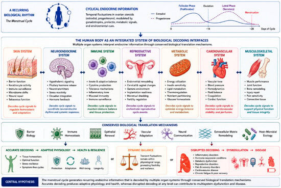
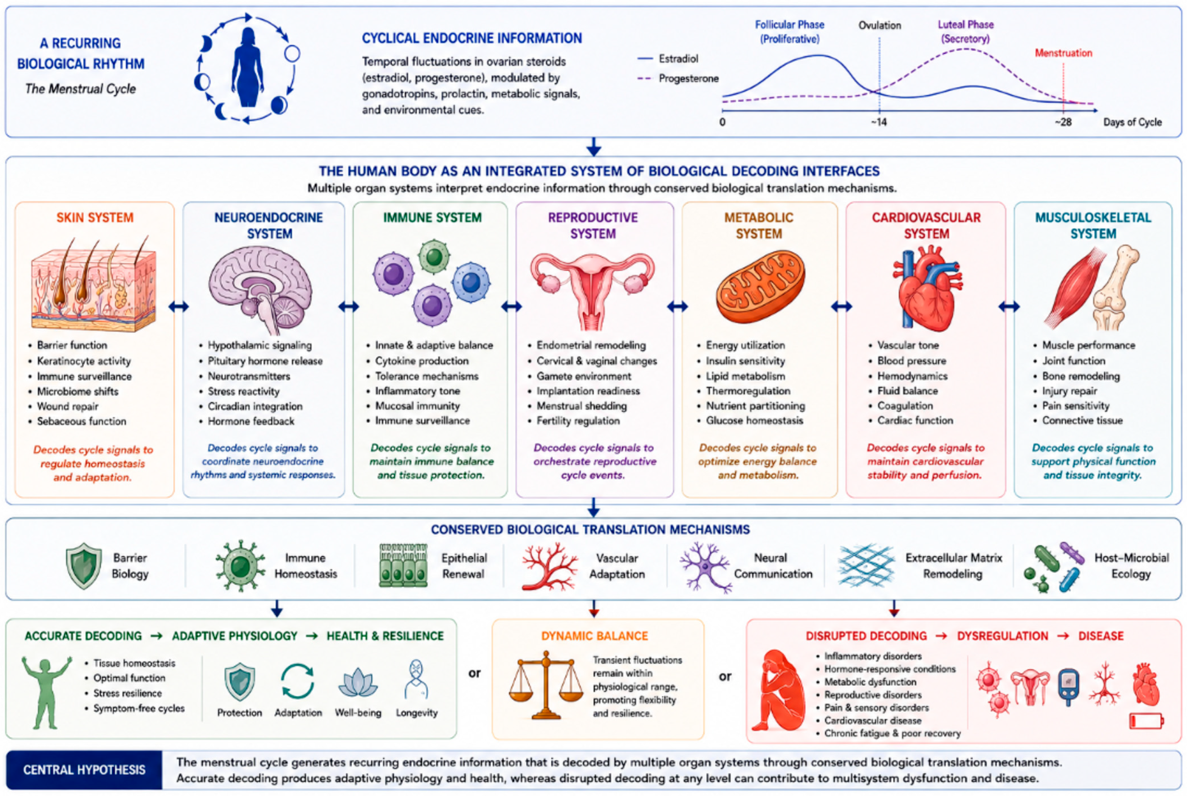
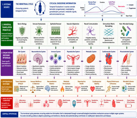
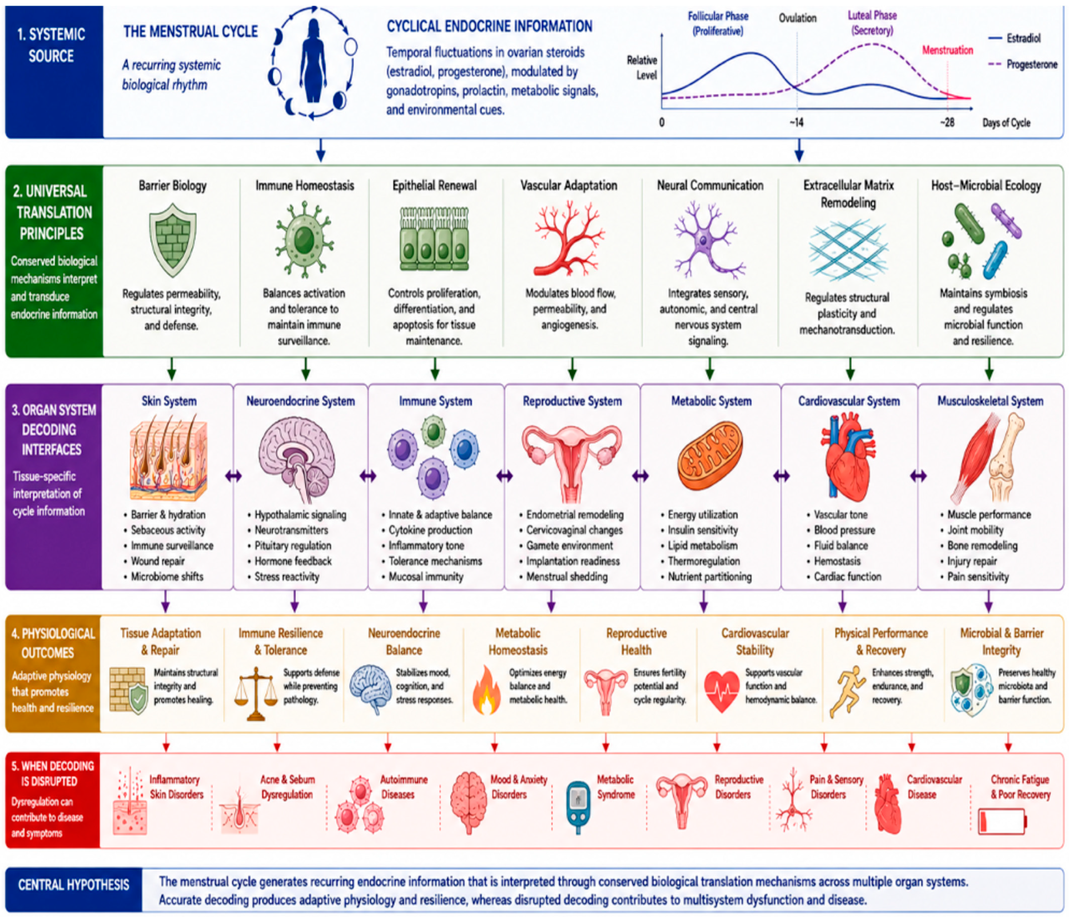
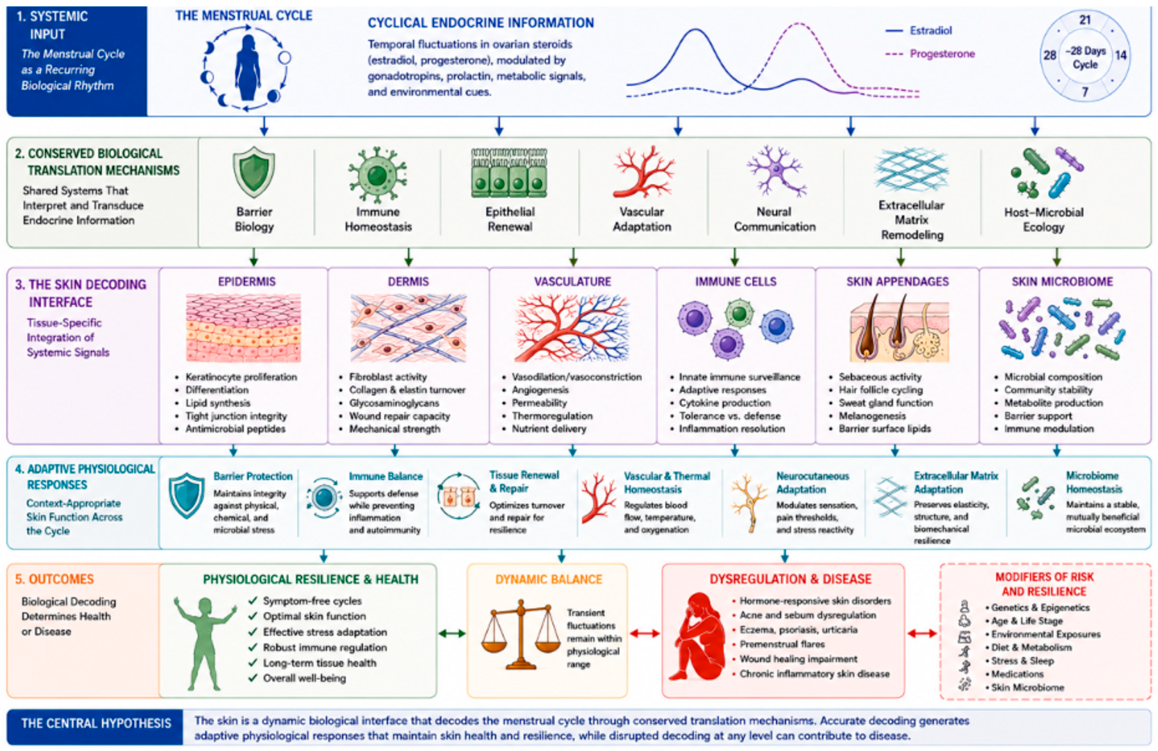
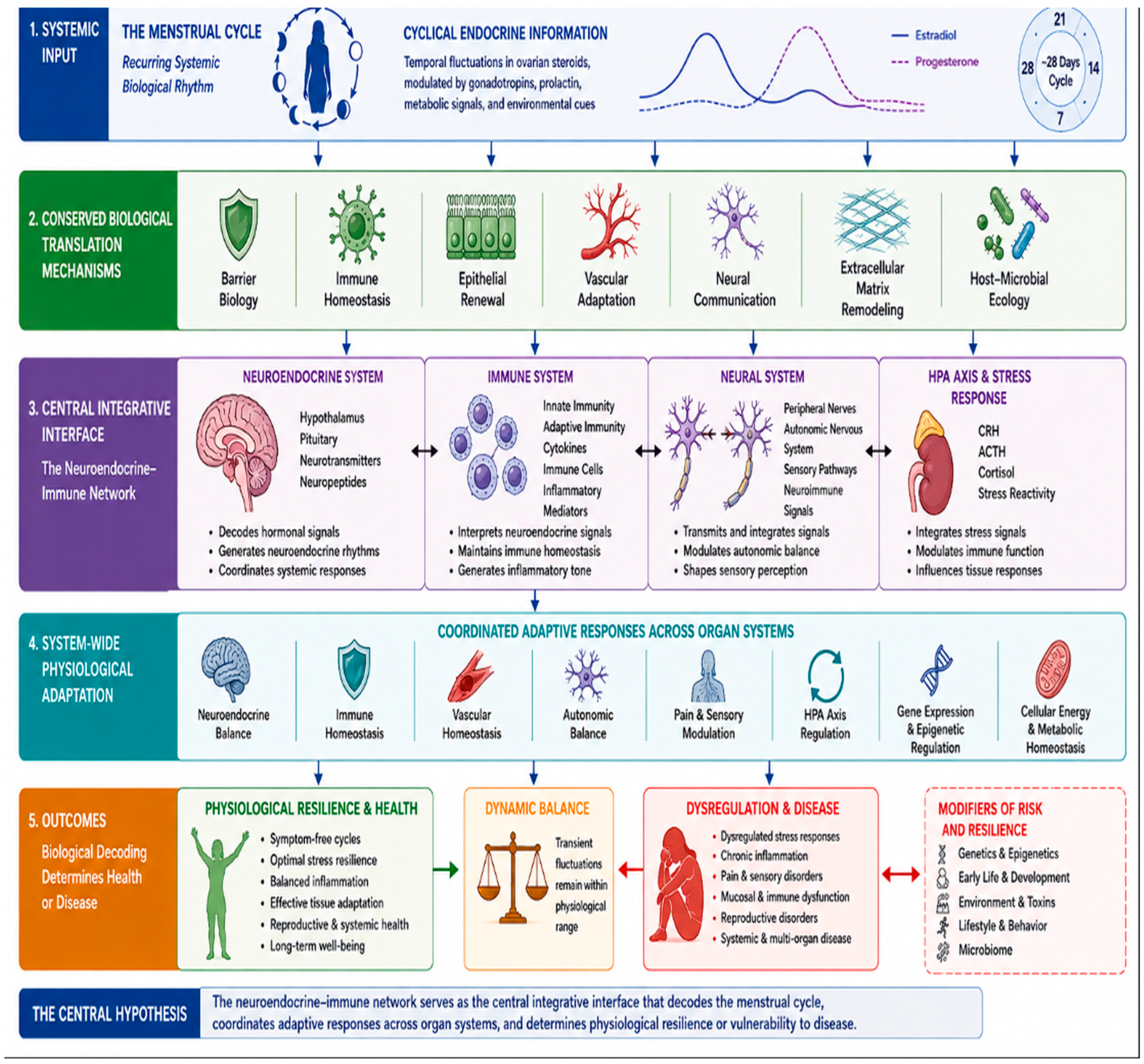
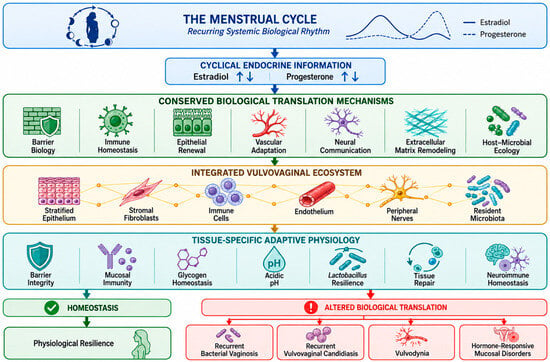
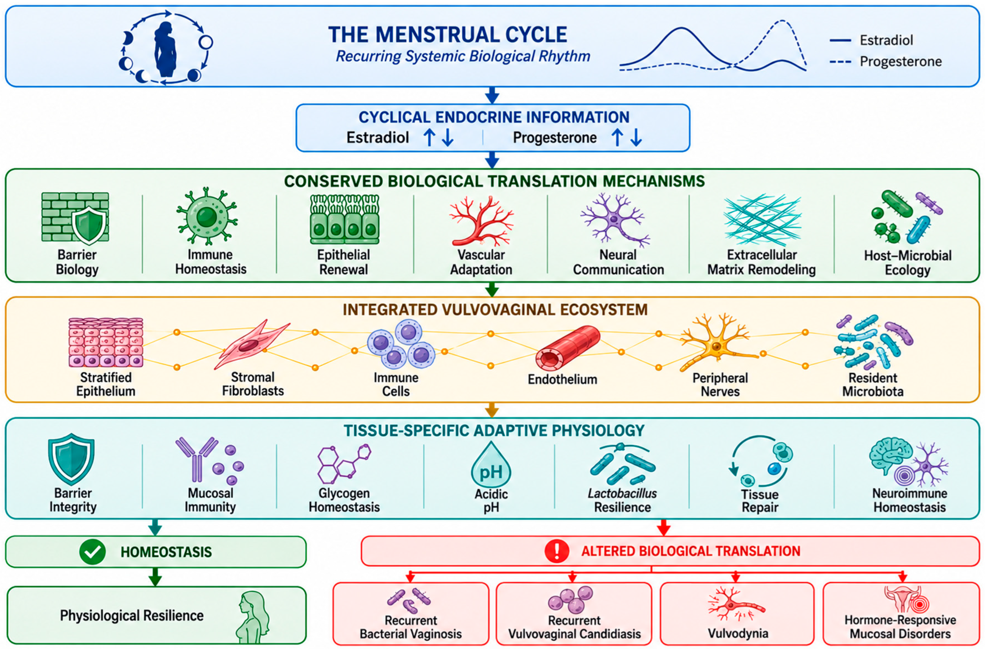
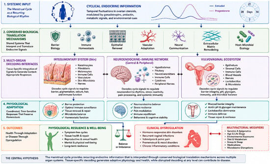
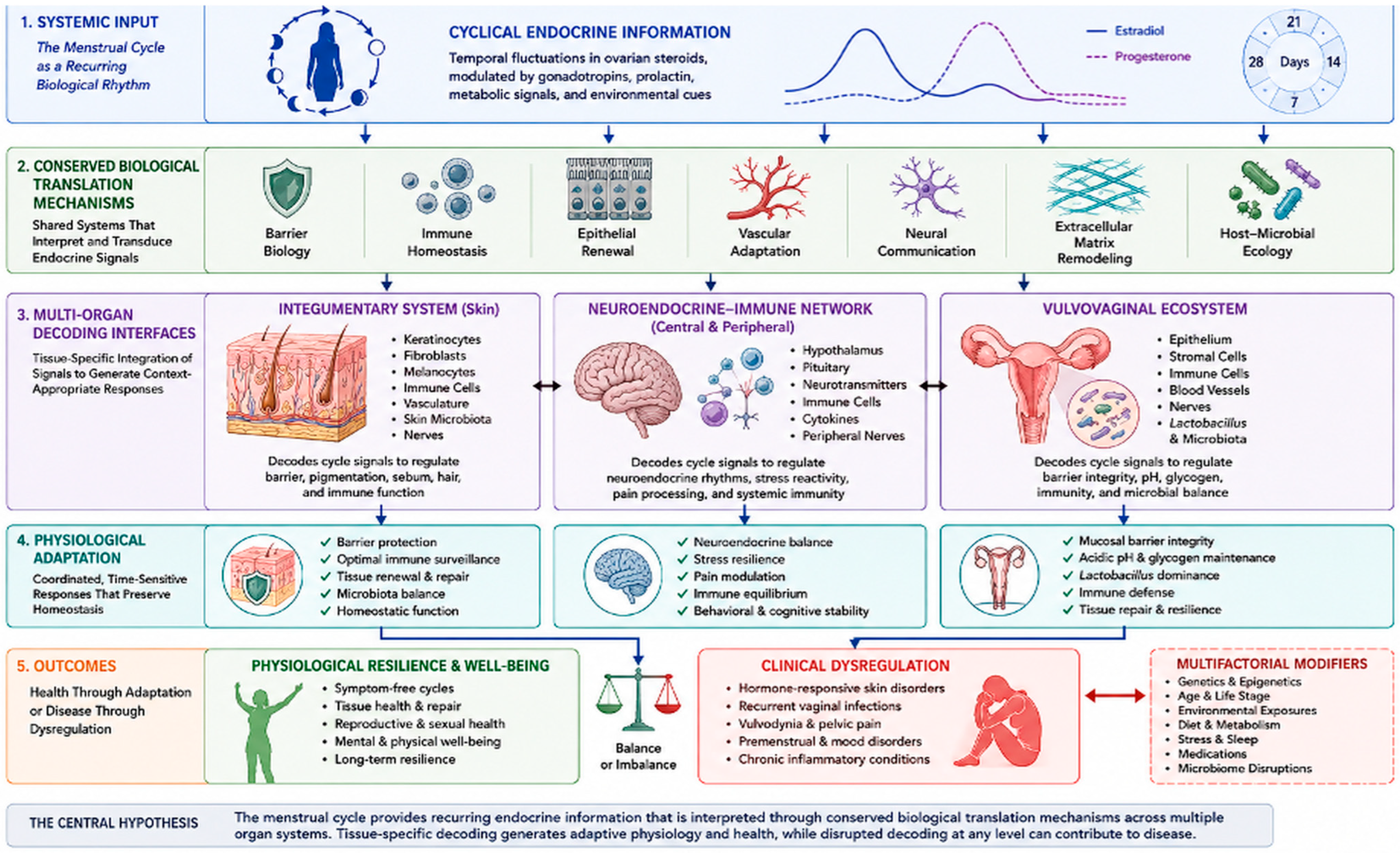

Disclaimer/Publisher’s Note: The statements, opinions and data contained in all publications are solely those of the individual author(s) and contributor(s) and not of MDPI and/or the editor(s). MDPI and/or the editor(s) disclaim responsibility for any injury to people or property resulting from any ideas, methods, instructions or products referred to in the content. |
© 2026 by the authors. Licensee MDPI, Basel, Switzerland. This article is an open access article distributed under the terms and conditions of the Creative Commons Attribution (CC BY) license.