


Abstract
Background: Pregnancy is a period of major brain adaptation, involving structural and functional changes that help women adjust to motherhood and support caregiving. Objective: To review current evidence on neuroplasticity during pregnancy and the postpartum period, focusing on brain changes, their biological basis, and their relevance for maternal mental health and nursing care. Methods: A systematic review with narrative synthesis was conducted in accordance with the PRISMA 2020 statement. The protocol was prospectively registered in PROSPERO (CRD420261376422). Searches were performed in PubMed, BVS, IBECS, CINAHL, and the Cochrane Library for studies published between 2016 and 2026 in English or Spanish. Results: Of the 1722 records initially identified, 15 studies were included in the narrative synthesis after screening and quality assessment using the CASP checklists and SANRA scale. The evidence showed that pregnancy is associated with changes in grey and white matter, as well as in brain connectivity, particularly in regions involved in social cognition, emotional regulation, and maternal behavior. These changes, largely driven by hormonal fluctuations, may persist after childbirth and are associated with greater maternal sensitivity, stronger mother–infant bonding, and more effective caregiving. However, they may also increase vulnerability to perinatal mental health disorders. Conclusions: Pregnancy represents a critical period of neuroplasticity. A better understanding of these changes may improve perinatal care, support maternal mental health, and guide nursing interventions during the transition to motherhood.
1. Introduction
Pregnancy is a universal biological phenomenon of great relevance to public health, with approximately 140 million births worldwide each year [1]. Beyond its reproductive dimension, it represents a period of profound physiological, psychological, and social transformations for women [2]. In Europe, declining fertility rates and the progressive delay in childbearing have altered the clinical and sociodemographic profile of pregnant women [3,4]. In Spain, this trend is reflected in a sustained decline in the birth rate and an increase in the average maternal age at first birth [5,6].
The somatic adaptations of pregnancy, such as the endocrine, circulatory, metabolic, and anatomical alterations required to promote fetal growth, have historically been the focus of research and therapeutic practice [7]. Nonetheless, there has been an increase in interest in comprehending the changes that the mother’s brain experiences during pregnancy in recent decades. Although less obvious, these alterations might be crucial for adjusting to parenthood and regulating behavioral, cognitive, and emotional processes [8,9,10].
It should be noted that pregnancy is marked by great hormonal variability, with increases in hormones such as estrogen, progesterone, cortisol, prolactin, and placental hormones. All these substances directly influence the functioning of certain areas of the brain related to processes such as memory, emotionality, social interactions, and motherly behavior [8].
According to available scientific data, pregnancy is linked to quantifiable neuroanatomical alterations. Numerous neuroimaging studies have found changes in areas related to social cognition, empathy, and emotional processing, especially in regions linked to the formation of mother-infant attachment [11,12]. Additionally, longitudinal studies have demonstrated dynamic changes in white and grey matter over the course of pregnancy [13]. These results point to adaptive reorganization aimed at maximizing the capacities needed for the care and protection of the infant, rather than a pathogenic condition. Additionally, some research suggests that some alterations may last for years following childbirth, leaving observable traces in the structure of the mother’s brain [14].
These changes can be interpreted within the framework of neuroplasticity, understood as the nervous system’s capacity to reorganize itself structurally and functionally in response to internal or external stimuli [15,16]. Although it has traditionally been associated with early stages of development, current evidence shows that mechanisms such as synaptic remodeling and changes in neuronal connectivity can also be activated in adulthood under specific conditions. In this regard, pregnancy can be considered a stage of high brain plasticity, comparable to other periods of intense neurobiological reorganization, such as adolescence [17].
Attachment theory provides a useful conceptual framework for understanding the functional significance of these adaptations. Current research suggests that the brain changes associated with pregnancy and the postpartum period promote maternal sensitivity, an emotional response to the baby’s needs, and the formation of a secure attachment [18,19]. From a neurobiological perspective, the parental brain constitutes an adaptive system that facilitates protection, care, and social bonding by activating neural networks modulated by neuroendocrine systems such as oxytocin and dopamine [19].
In addition to its scientific interest, understanding the physiological, psychological, and neurobiological changes associated with pregnancy has significant clinical and care-related relevance. Understanding these adaptations enables us to better identify the needs of pregnant women, facilitate early detection of mental health problems, and design interventions to support maternal-fetal well-being and strengthen the early mother-child bond [10,20]. Furthermore, the World Health Organization emphasizes the importance of providing woman-centered antenatal care that considers the physical, emotional, and psychosocial dimensions of pregnancy [21]. From a nursing perspective, this knowledge contributes to the development of more comprehensive, humanized, and evidence-based care.
Despite the growing volume of research, significant limitations persist, including methodological heterogeneity across studies and a lack of integration between neurobiological findings and their clinical applicability. Consequently, it is necessary to synthesize the available evidence to better understand the brain changes associated with pregnancy and their relevance to clinical practice. Therefore, this review aims to analyze the available scientific evidence on structural and functional changes in the maternal brain during pregnancy and to explore their relationship with emotional adaptation and maternal mental health from a nursing perspective.
2. Methodology
A systematic review with narrative synthesis was conducted to examine the available evidence on structural and functional changes in the maternal brain during pregnancy and related reproductive stages, and their associations with maternal emotional adaptation and mental health. This type of review allows studies with diverse methodological designs to be integrated and analyzed together, promoting a broad, in-depth understanding of complex phenomena related to human health and behavior [22].
The methodological process was carried out in accordance with the PRISMA (Preferred Reporting Items for Systematic Reviews and Meta-Analyses) statement to ensure transparency, reproducibility, and rigor in the identification, selection, and synthesis of scientific evidence [23]. The review protocol was registered in PROSPERO (CRD420261376422) in advance.
2.1. Research Question
The research question was formulated using the PIO (Population, Intervention, and Outcome) model. The population of interest consisted of pregnant women; the intervention corresponded to brain changes associated with pregnancy; and the outcome focused on neurobiological adaptations and their relationship with maternal mental and emotional health. Based on this framework, the following research question was formulated: What structural and functional changes occur in the brain during pregnancy, and how are these related to maternal emotional adaptation and mental health?
2.2. Search Strategy
The literature search was conducted between February and May 2026 in PubMed, the Virtual Health Library (BVS), the Spanish Bibliographic Index in Health Sciences (IBECS), CINAHL (Cumulative Index to Nursing and Allied Health Literature), and the Cochrane Library.
Natural language terms and controlled vocabulary from the MeSH and DeCS thesauri were used to retrieve information, combined using the Boolean operators AND and OR. The main descriptors used were ‘pregnancy’, ‘neuronal plasticity’, ‘brain’, ‘neuroanatomy’, ‘maternal behavior’, and ‘maternal attachment’. The search strategies were tailored to each database’s specific characteristics to maximize the sensitivity and specificity of the results. The complete search strategy for each database, including the search platform, date, controlled vocabulary and free-text terms, Boolean operators, filters applied, and number of records retrieved, is provided in Supplementary Table S1.
2.3. Selection Criteria
Studies analyzing structural and/or functional changes in the brain during pregnancy were included, as were investigations of the neurobiological mechanisms underlying these changes and studies linking them to maternal behavior, emotional adaptation, or mental health. Only human studies published in English or Spanish between 2016 and 2026 were eligible. This publication period was predefined in the review protocol to provide a focused synthesis of the most recent decade of evidence, during which longitudinal neuroimaging and multimodal approaches to maternal brain plasticity underwent substantial development.
Duplicate studies, research not involving a pregnant population, publications unrelated to the research question, and studies that, following critical appraisal, were found to be of insufficient methodological quality were excluded.
2.4. Study Selection Process
The selection process was carried out in accordance with the PRISMA guidelines and comprised several phases. The literature search was conducted in PubMed, Virtual Health Library (BVS), IBECS, CINAHL, and Cochrane Library databases. Records retrieved from the different databases were combined, and duplicates were removed before screening. In total, 1722 potentially relevant records were identified. After removing 19 duplicate records and excluding 1390 records using the predefined search filters, 313 records underwent title and abstract screening.
Titles and abstracts were screened independently by two reviewers using the predefined eligibility criteria. Records considered potentially relevant by at least one reviewer underwent further assessment before full-text retrieval. Full-text reports were subsequently assessed independently by the same reviewers. Disagreements regarding eligibility were resolved through discussion and consensus, with the involvement of a third reviewer when necessary. The study-selection process was documented using a flow diagram in accordance with the PRISMA 2020 guidelines.
2.5. Data Extraction
Data extraction was carried out systematically using a template designed specifically for this review. Variables relating to authorship, year of publication, country of origin, methodological design, sample characteristics, objectives, main results, conclusions, and methodological quality were collected. The review of titles and abstracts was carried out independently by two researchers. To assess the reliability of the selection process, Cohen’s Kappa coefficient was calculated, yielding κ = 0.79, indicating substantial agreement between reviewers according to the criteria of Landis and Koch [24]. Any discrepancies identified during the screening process were jointly analyzed and resolved by consensus, with the involvement of a third researcher. Articles considered potentially relevant were subsequently assessed in full text using the same procedure.
2.6. Assessment of Methodological Quality
The methodological quality of the included studies was assessed using validated tools appropriate to each type of publication. Original human studies were evaluated using the relevant Critical Appraisal Skills Programme (CASP) checklist according to their study design [25]. Narrative reviews were assessed using the Scale for the Assessment of Narrative Review Articles (SANRA) [26]. No overall numerical CASP score was calculated; instead, the checklist criteria were used to identify methodological strengths and limitations relevant to the interpretation of each original study.
2.7. Interpretive Scope and Limitations of Neuroimaging and Neurophysiological Techniques
The techniques described in the included publications provide information at different levels of brain organization and should not be considered equivalent measures of neuroplasticity. Structural MRI measures macroscopic features like grey matter volume, cortical thickness, surface area, and subcortical volume, but it cannot directly detect the cellular mechanisms that underlie these changes. Synaptic pruning, dendritic remodeling, altered glial structure, or myelination are therefore plausible biological explanations and not direct structural MRI findings [27,28,29].
Functional MRI uses the hemodynamic blood-oxygen-level-dependent signal to examine task-related activity or statistical functional connectivity and does not measure neuronal activity directly. Diffusion MRI provides indirect measures sensitive to the microstructural organization of white matter but cannot directly demonstrate axonal change or adaptive myelination [27,30]. EEG and MEG provide complementary information about rapid temporal and oscillatory dynamics, while PET and magnetic resonance spectroscopy may provide information about metabolic, molecular, or neurochemical processes [31,32,33]. However, these latter modalities were minimally represented, or not represented at all, in the original studies included in the systematic synthesis. These techniques should therefore be considered complementary but non-equivalent, and none can independently establish the cellular or molecular mechanisms underlying the observed changes.
2.8. Synthesis of Information
Due to the methodological heterogeneity of the included studies, characterized by differences in research designs, sample characteristics, and the variables analyzed, it was not possible to conduct a quantitative meta-analysis. Consequently, a narrative synthesis of the findings was chosen as the most appropriate strategy for integrating the available evidence.
The results were organized into four thematic categories: structural and neuroanatomical changes; functional connectivity and brain–behaviour associations; hormonal and neurobiological mechanisms; and behavioural and mental-health-related findings. This organization enabled the identification of common patterns across studies, comparison of results from different methodological approaches, and an overall interpretation of the phenomenon under study in relation to the review’s objectives.
The narrative synthesis integrated available findings and provided a comprehensive overview of brain changes during pregnancy and their potential influence on emotional adaptation, maternal behavior, and the mental health of pregnant women.
3. Results
3.1. Study Selection
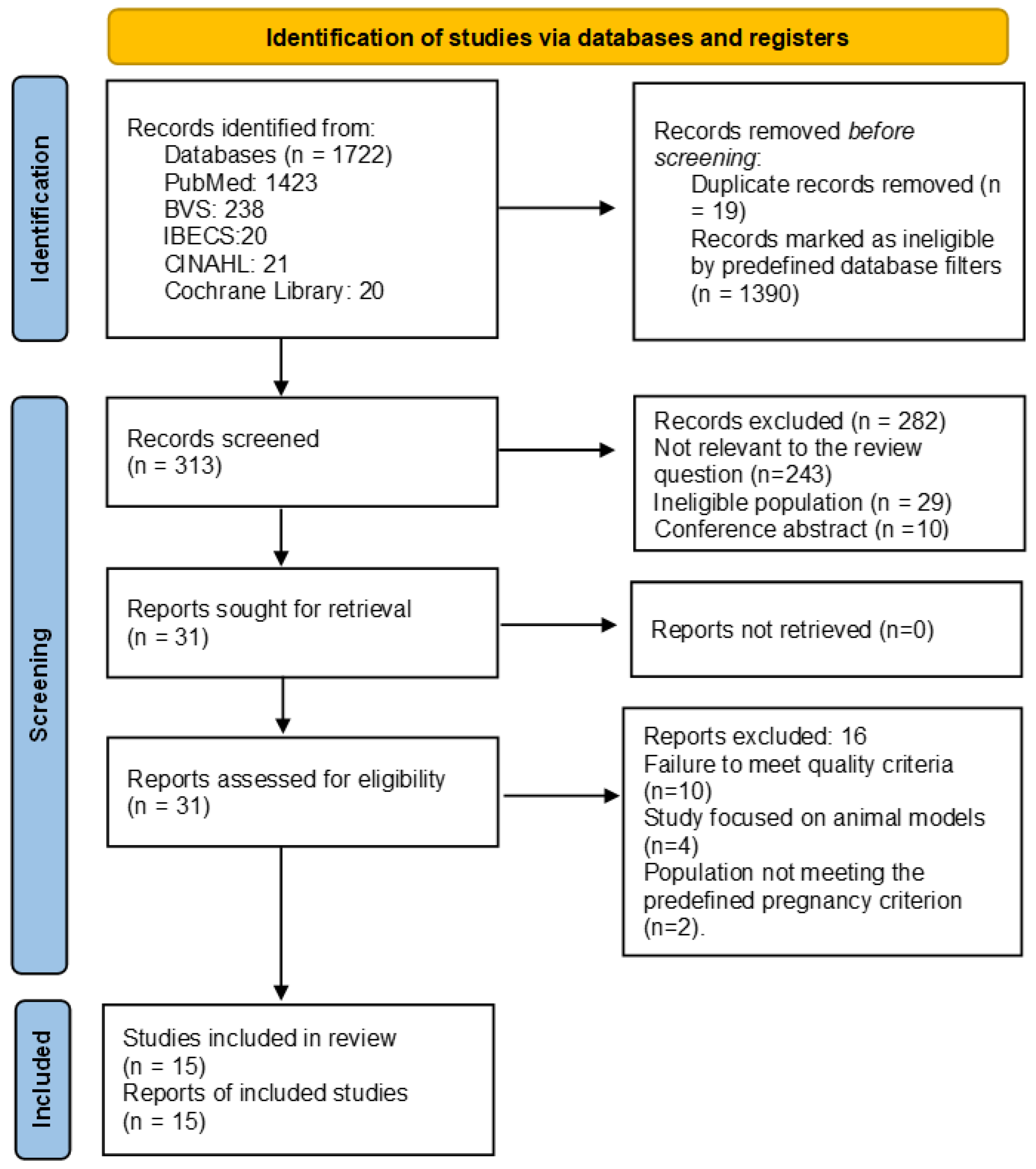
The selection process was carried out in accordance with the PRISMA 2020 guidelines and comprised several phases. The literature search was conducted in PubMed, Virtual Health Library (BVS), IBECS, CINAHL, and Cochrane Library databases. Through database searches, 1722 articles were identified. Applying predefined database filters to exclude duplicate articles reduced the number of unique articles screened in titles and abstracts to 313. Of these, 282 articles were excluded after screening titles and abstracts; 29 did not meet the population criterion, and 10 were conference abstracts. As a result, 31 articles were reviewed in terms of full text. Of these, 16 were found ineligible: 10 failed the predefined methodological quality criteria, four were animal models only, and two excluded pregnant women. Fifteen studies formed part of the systematic review. The selection process was reliable and consistent with substantial agreement between reviewers (κ = 0.79).
The entire selection process is shown in Figure 1.
Figure 1.
PRISMA 2020 flow diagram.
3.2. Characteristics and Methodological Quality of the Included Publications
The 15 included publications comprised nine original human studies and six narrative reviews. Among the original studies, six used controlled longitudinal cohorts or secondary analyses of longitudinal cohort data [10,12,17,34,35,36], one was a prospective longitudinal observational study without a non-pregnant comparison group [37], one was an intensive single-participant longitudinal study [13], and one used a cross-sectional comparative design [38]. The six narrative reviews were published between 2016 and 2025 [10,39,40,41,42,43]. The included publications varied considerably in sample size, assessment timing, neuroimaging and endocrine measures, and behavioural or clinical outcomes.
Nine original human studies and six narrative reviews have been discussed individually because of their different evidentiary purposes. The characteristics and findings of the original studies that form the basis of the synthesis are listed in Table 1 below. The narrative reviews, which are meant to provide mechanistic and contextual synthesis and are not considered to be primary empirical evidence, are described in Table 2 below.
Table 1.
Characteristics and main findings of the original human studies included in the review.
Table 2.
Characteristics and interpretive contribution of the narrative reviews included in the review.
The CASP appraisal indicated that the original studies generally addressed clearly focused research questions and used measures appropriate to their objectives. However, recurrent limitations included small or highly selected samples, incomplete follow-up, limited control of potential confounding factors, absence of an independent comparison group in some studies, and the restricted generalisability of cross-sectional or single-participant designs. Several publications also involved secondary analyses or partially overlapping cohorts and therefore should not be interpreted as providing fully independent evidence.
The six narrative reviews obtained SANRA scores ranging from 9 to 10 out of 12. They generally provided a clear justification of the topic, stated their objectives, referenced the relevant evidence appropriately, and presented coherent scientific reasoning. However, none reported a reproducible literature search or study-selection procedure. In addition, some reviews provided limited endpoint data and relied substantially on mechanistic findings from animal models. These considerations were taken into account when interpreting their contribution to the narrative synthesis.
3.3. Structural and Neuroanatomical Changes
Evidence from structural neuroimaging was found in seven of the nine initial human studies. Reductions in grey matter volume during pregnancy or during the transition from preconception to the early postpartum period were consistently seen in original human investigations. These changes affected widespread cortical regions, with areas related to social cognition and the default mode and frontoparietal networks showing the most noticeable changes [13,17,34,36,44]. Changes in cortical thickness, surface area, gyrification, and sulcal morphology coincided with reductions in grey matter volume, although precise measurements and evaluation intervals differed between studies [13,17,34,36].
Region-specific analyses showed reductions outside the cortical regions studied in the whole-brain studies. A secondary analysis of the Hoekzema cohort revealed a decrease in right ventral striatal volume and a statistical trend toward a decrease in the left ventral striatum. No significant change in comparison was observed in the dorsal striatum [44]. A secondary analysis of two combined longitudinal cohorts also showed reductions in several hypothalamic subregions as well as total hypothalamic volume [35]. However, the studies by Hoekzema et al., Carmona et al., and a subset of the sample analyzed by Spalek et al. had overlapping participants and thus do not represent fully independent replications [12,17,35,44].
Longitudinal data showed that the structural trajectory persisted throughout the postpartum period. Cortical volume, thickness, and surface area increased between late pregnancy and early postpartum in the primary and replication cohorts examined by Paternina-Die et al. [34]. In frontoparietal and default mode regions, however, recovery was less apparent. Cerebral grey matter volume decreased by approximately 4.9% between preconception and late pregnancy and increased by around 3.4% by six months postpartum; according to Servin-Barthet et al. [36], preconception levels were not fully recovered, following a U-shaped trajectory. The left hippocampus showed modest recovery, but the majority of the grey matter reductions in the smaller follow-up sample reported by Hoekzema et al. [12] were still visible about two years after delivering a child. Instead of either total reversal or uniform persistence, our results point to partial and regionally varied postpartum recovery.
White matter changes were characterized in detail in the intensive single-participant study by Pritschet et al. [13]. Quantitative anisotropy of white matter increased mainly during the first and second trimester. It returned to baseline postpartum. Global grey matter volume and cortical thickness were only partially recovered and remained below preconception levels at the 2-year assessment. Ventricular and CSF volume increased later in pregnancy. These observations were from one participant and, while providing detailed temporal information, did not allow any conclusion to be drawn about the typical population-level trajectory.
Narrative reviews have interpreted pregnancy-related decreases in cortical measurements and brain volume as potential signs of neural specialization or adaptive “fine-tuning” rather than signs of brain decline [10,42,43,45]. Postpartum increases may represent experience-dependent plasticity related to caregiving, according to certain reviews. These explanations are still speculative, though: whether the underlying mechanisms entail synaptic pruning, changes in dendritic or glial structure, neurogenesis, myelination, fluid shifts, or a mix of events cannot be determined from MRI-derived changes in grey matter volume. Animal models provide much of the cellular evidence for these hypothesized processes.
3.4. Functional Connectivity and Brain–Behaviour Associations
Compared to structural data, functional neuroimaging evidence from the initial human research was significantly more limited. According to Hoekzema et al., when mothers watched pictures of their own babies during a postpartum fMRI task, some of the brain regions exhibiting grey matter reductions during the transition to pregnancy corresponded with those activated. Grey matter alteration patterns were also linked to the absence of animosity and postpartum maternal connection, but not to enjoyment of social engagement [12]. This study did not directly show longitudinal functional changes throughout pregnancy since task-based fMRI was only conducted after deliveries.
A subsequent analysis of the same cohort showed that greater reduction in right ventral striatal volume was associated with a stronger postpartum functional response to images of the mother’s own infant [44]. However, this association was observed only in the right ventral striatum, and no direct behavioural measure of caregiving was assessed. Servin-Barthet et al. examined resting-state functional connectivity before pregnancy and at approximately one and six months postpartum but found no significant longitudinal changes in the network-segregation measures investigated [36]. Resting-state fMRI was not obtained during pregnancy; therefore, potential gestational changes and their postpartum evolution could not be evaluated directly.
The original investigations found different correlations between behavioral measurements and anatomical alterations in the brain. Higher prenatal attachment scores and certain nesting and postnatal bonding measures were linked to greater decreases in specific hypothalamic subregions [35]. In a statistical mediation analysis, Servin-Barthet et al. found that greater postpartum grey matter recovery was linked to reduced mother animosity toward the infant [36]. This association was partially explained by maternal well-being. In contrast, Paternina-Die et al. did not find any significant correlations between the neuropsychological tests evaluated and global cortical alterations [34]. When combined, these results show correlations between certain structural markers and characteristics of maternal behavior or attachment. However, they do not prove that pregnancy-related brain alterations lead to more effective or sensitive caring.
Narrative reviews have proposed that maternal adaptation involves coordinated activity across networks supporting social cognition, salience detection, reward, motivation, empathy, and emotion regulation [10,42,43,45]. They also describe functional responses to infant cues as potentially relevant to maternal sensitivity and caregiving. However, these propositions integrate heterogeneous human studies, many of which were observational or conducted only during the postpartum period, together with mechanistic evidence from animal models. Consequently, the functional significance of the structural changes observed during pregnancy remains plausible but not conclusively established.
3.5. Hormonal and Neurobiological Mechanisms
Three original human neuroimaging studies directly examined associations between endocrine measurements and brain structural changes [13,35,36]. In the intensive single-participant study by Pritschet et al., extensive reductions in cortical and subcortical grey matter measures occurred alongside increasing concentrations of oestradiol and progesterone and advancing gestational week [13]. However, these variables changed concurrently throughout pregnancy, preventing the independent effects of specific hormones from being determined.
Spalek et al. reported that higher second-trimester oestradiol levels were associated with greater reductions in both posterior and total hypothalamic volume [35]. The analyses of hormonal and behavioural indicators, apart from the few exceptions involving test samples, were also less than 1–2% for most trivial associations. Servin-Barthet et al. described associations of changes in oestriol and oestrone sulfate with the U-shaped time course of measures of cortical grey matter throughout pregnancy and postpartum [36]. These longitudinal relationships are consistent with temporal covariation between endocrine and neuroanatomical changes, but do not demonstrate that specific hormonal fluctuations drove the observed brain trajectories.
Although they did not evaluate brain structure or function, Côté et al. offered further longitudinal endocrine evidence [37]. While peripheral oxytocin concentrations stayed statistically constant during pregnancy, human placental lactogen rose. Maternal-fetal attachment was not significantly correlated with either hormone. As a result, this study adds to our understanding of the endocrine and psychological changes that occur during pregnancy. However, it does not directly demonstrate how hormones affect the plasticity of the mother’s brain. The remaining original research either described hormonal mechanisms without explicitly measuring them or did not gather endocrine data [12,17,34,38,44].
Narrative reviews have proposed that fluctuations in oestradiol, progesterone, prolactin, oxytocin, glucocorticoids, and other endocrine signals contribute to structural and functional adaptation of the maternal brain [10,40,41,42,43,45]. Oxytocinergic and dopaminergic systems have been implicated in maternal motivation, attachment, reward processing, and responsiveness to infant cues. However, human studies frequently rely on plasma, salivary, or urinary hormone concentrations as indirect indicators of central neuroendocrine activity, and their correspondence with brain hormone concentrations or signaling remains uncertain.
As possible mechanisms of maternal brain plasticity, the reviews also discuss alterations in neurogenesis, dendritic complexity, synaptic density, glial cells, neuroimmune signaling, and reward-related and hypothalamic circuits [10,40,42,43]. Animal models have provided most of the comprehensive cellular and circuit-level evidence supporting these claims. Therefore, rather than being mechanisms that the original human studies included in this review directly established, processes including synaptic pruning, altered neurogenesis, dendritic remodeling, and glial plasticity should be considered as potential mechanistic explanations.
3.6. Behavioural and Mental-Health-Related Findings
Heterogeneous data regarding cognitive, affective, and behavioral consequences were found in original human investigations. Pregnant women did worse than mothers and nulliparous women in the cross-sectional study by Bakay et al. on tests of verbal fluency, digit span, processing speed and attention, and verbal learning and memory [38]. After statistical adjustment, pregnancy status was still linked to worse performance on a few cognitive tasks, but there were no discernible group differences in Stroop completion times. Additionally, anxiety and depressed symptom scores were higher among pregnant participants. Nevertheless, these results cannot be interpreted as cognitive changes brought on by pregnancy-related brain plasticity due to the study’s cross-sectional design, lack of neuroimaging and endocrine data, and variations across the comparison groups.
Côté et al. found that higher average human placental lactogen concentrations were associated with higher anxiety and depressive symptom scores, while the association between within-person variation in peripheral oxytocin and depressive symptoms differed according to pregnancy stage [37]. Neither hormone was significantly associated with maternal–fetal attachment. As this study did not assess brain structure or function, its findings indicate endocrine–psychological associations but do not establish a relationship between maternal brain changes and mental health.
Among the neuroimaging studies, associations with behavioural and psychological outcomes were inconsistent. Selected grey matter and hypothalamic volume changes were associated with maternal attachment, nesting, postnatal bonding, or lower hostility towards the infant in some studies [12,35,36]. However, Paternina-Die et al. [34] found no significant associations between global cortical changes and the neuropsychological measures assessed, and Spalek et al. [35] found no significant association between hypothalamic volume changes and postnatal depressive symptoms. In the single-participant study by Pritschet et al. [13], most neuroanatomical changes were not associated with the repeatedly assessed mood or lifestyle measures. Overall, the original studies were predominantly conducted in women without diagnosed perinatal mental disorders and were not designed to determine whether pregnancy-related brain changes cause maternal psychopathology.
According to narrative reviews, adaptation to caregiving may be supported by changes in reward, salience, social cognition, and emotion-regulation systems, as well as heightened sensitivity to newborn cues and enhanced alertness to possible hazards [10,39,42,43]. However, several evaluations indicate that some women may be more susceptible to anxiety, depression, or other perinatal mental problems due to the considerable endocrine and neuronal plasticity that occurs throughout pregnancy and the postpartum period [10,40,45,46]. Maternal neuroplasticity is thus presented in these evaluations as a possible context for both susceptibility and adaptation. However, the suggested connection with psychopathology is predicated on mechanical extrapolation and a variety of observational data. It should not be taken as proof that perinatal mental disorders are caused by normal pregnancy-related brain changes.
4. Discussion
This systematic review with narrative synthesis examined structural and functional changes in the maternal brain during pregnancy and the postpartum period and their possible relationships with endocrine variation, maternal behaviour, cognition, and mental health. The nine original human studies provided the most consistent evidence for structural change, particularly reductions in cortical grey matter volume and thickness during pregnancy, followed by partial and regionally variable recovery after childbirth [12,17,34,36,47]. Region-specific changes were also identified in the ventral striatum and hypothalamus [35,44]. By contrast, evidence concerning functional connectivity, cognition, maternal behaviour, and mental health was more limited and heterogeneous, and the associations observed did not establish causal relationships.
Pregnancy-related neural changes may help some women adapt to caregiving. However, they may also coincide with increased vulnerability, according to the six narrative reviews that interpreted these findings within larger models of maternal and parental brain plasticity [10,40,41,42,43,45]. Nonetheless, postpartum research and mechanistic data from animal models were often included in these views. The time and methodology of the underlying research must therefore be taken into account when interpreting the results: evaluations carried out during pregnancy, soon after childbirth, or a few years later may not accurately reflect sequential observations of a single continuous biological trajectory [10,12].
The most consistent empirical finding was a reduction in grey matter volume and cortical thickness during pregnancy, affecting widespread cortical regions and showing particular involvement of the default mode and frontoparietal networks, as well as areas associated with social cognition [12,17,34,36,47].
Region-specific analyses extended these observations to the ventral striatum and hypothalamus [12,35]. Nevertheless, the apparent consistency of these findings should be interpreted cautiously because several publications involved secondary analyses or partially overlapping cohorts, particularly those derived from the Barcelona and Leiden samples. The number of fully independent pregnancy cohorts is therefore smaller than the number of publications might suggest.
The longitudinal research indicates that these changes do not follow a clear pattern of either permanent reduction or complete postpartum reversal. Studies that included tests throughout late pregnancy and after births found that cortical measures partially recovered during the early postpartum period, whereas some regions recovered more slowly or remained below preconception values at six months [34,36]. Grey matter reductions were still seen about two years after delivery in the short follow-up sample described by Hoekzema et al. [12], and partial persistence was also observed in the extensive single-participant trajectory reported by Pritschet et al. [13]. These findings support a dynamic and geographically varied trajectory. However, the tiny follow-up samples do not demonstrate that the same trend persists over time or is prevalent in a wider population.
The cellular basis and functional significance of reductions in MRI-derived grey matter measures are currently unknown. Narrative studies suggest that these changes could be caused by brain specialization or adaptive “fine-tuning” of circuits involved in social cognition, emotional regulation, and caregiving [10,39,42,43]. Carmona et al. discovered parallels between pregnancy-related and adolescent brain morphometric alterations, which lends credence to the idea that pregnancy is an important stage of adult neuroplasticity [17]. However, similarities in MRI-derived patterns do not indicate that the biological processes underlying the two stages of development are the same. Grey matter volume can be affected by variations in glial structure, myelination, vascular or fluid-related factors, dendritic or synaptic organization, or combinations of these mechanisms. Changes in grey matter volume may reflect multiple processes, including alterations in dendritic or synaptic organization, glial structure, myelination, vascular or fluid-related factors, or combinations of these mechanisms. Consequently, neuronal specialization and synaptic pruning should be presented as plausible explanations rather than processes directly demonstrated by the included human studies.
The coincidence of fluctuations in reproductive hormone levels with neuroanatomical development makes it plausible that hormones could influence brain plasticity in mothers. In particular, Pritschet et al. [13] found an increase in the levels of oestradiol and progesterone in association with neuroanatomical changes occurring across pregnancy. In contrast, Spalek et al. [35] found correlations between second-trimester oestradiol levels and decreases in the volumes of some hypothalamic structures. Servin-Barthet et al. [36] found correlations between the levels of oestriol and oestrone sulphate and the U-shaped curve in the development of measures of the cortex. Nevertheless, hormone concentration, gestational age, and other physiological factors developed concurrently in the observational studies; their separate influences cannot be distinguished.
The endocrine evidence was also dependent on the biological samples and outcomes examined. Côté et al. [37] found increasing concentrations of human placental lactogen but statistically stable peripheral oxytocin concentrations across pregnancy; this study did not include neuroimaging. More broadly, peripheral measurements in serum, plasma, saliva, or urine may not accurately represent hormone concentrations or signaling within the brain. This limitation is particularly relevant to oxytocin, whose proposed role in maternal attachment and caregiving is prominent in the narrative literature but was not directly demonstrated through central measurements in the original human studies included in this review [10,40].
Animal models have provided critical evidence for identifying potential cellular and circuit-level mechanisms, including modifications in hypothalamic, limbic, reward and sensory-processing circuits, and changes in neurogenesis, dendritic morphology, synaptic organization and glial or neuroimmune processes [10,40,42,43]. Therefore, oxytocinergic, prolactin-related and dopaminergic pathways have been proposed to contribute to maternal motivation, infant-cue processing and caregiving behaviour. However, these mechanisms have not yet been demonstrated to work in the same way in humans, and none of the original studies included in this review directly measured central oxytocinergic or dopaminergic activity. Animal data provide plausibility of mechanisms, but should not be taken as direct explanations of the MRI-derived changes in pregnant women.
Complementary studies conducted in non-pregnant women indicate that functional brain organization may vary across physiological hormonal transitions. Dense-sampling fMRI has shown rhythmic reorganization of functional brain networks across the menstrual cycle [47]. MEG studies have also identified cycle-related differences in spontaneous neural oscillations and have reported greater repertoire diversity and brain flexibility during the peri-ovulatory phase than during the early follicular phase [48,49]. In addition, individual patterns of functional connectivity remained highly identifiable across menstrual-cycle phases. However, modest phase-related variations in posterior connectivity were associated with mood, self-esteem, and psychological well-being [50]. These studies provide physiological context showing that hormonal variation may coincide with rapid functional brain dynamics that structural MRI does not capture. However, they involved non-pregnant women, generally included small samples, and were not eligible for the systematic synthesis. Their findings should therefore not be extrapolated directly to the more extensive endocrine and physiological changes occurring during pregnancy.
The adaptive parental brain provides a useful conceptual framework for interpreting pregnancy-related structural and functional changes. Associations between selected brain measures and responses to the mother’s own infant, prenatal or postnatal attachment, nesting behaviour, and lower maternal hostility are compatible with a potential role in the transition to caregiving [12,35,36,44]. However, these associations were region- and outcome-specific, and some studies found no significant relationships between cortical changes and the behavioural or neuropsychological measures examined [34]. Furthermore, statistical associations and mediation models cannot demonstrate that neuroanatomical changes improve caregiving or produce stronger attachment.
These findings are placed within more comprehensive models that include reward, salience, social cognition, motivation, empathy, and emotion-regulation systems [10,45,47,50]. In these ideas, caregiving may be aided by heightened attention to socially or ecologically important information and enhanced responsiveness to newborn cues. However, the more intricate circuit-level mechanisms are primarily derived from animal research, and much of the supporting functional evidence was acquired postpartum. Therefore, the evidence that is provided supports the idea of functional adaptation, but it does not show that the structural changes that occur during pregnancy have a planned caregiving purpose.
Evidence concerning cognition and mental health was more limited. Bakay et al. identified poorer performance on selected cognitive tests and higher anxiety and depressive symptom scores among pregnant women [38]. However, the cross-sectional design, absence of neuroimaging, and differences between groups prevent these outcomes from being attributed to pregnancy-related brain changes. Similarly, Côté et al. [37] reported associations between endocrine measures and anxiety or depressive symptoms without assessing the brain. Conversely, several neuroimaging studies found no significant relationships between structural changes and depressive symptoms, global neuropsychological outcomes, or most repeatedly assessed mood measures [13,34,35].
Narrative reviews propose that the extensive plasticity associated with pregnancy and postpartum may create a context in which adaptation and vulnerability coexist [10,40,45,46]. This is a clinically relevant hypothesis, but the original studies were conducted predominantly in women without diagnosed perinatal mental disorders. They were not designed to test whether normative brain changes predict the onset of anxiety, depression, or postpartum psychosis. The available evidence therefore does not establish that pregnancy-related neuroplasticity independently increases the risk of perinatal mental illness. Rather, its clinical significance is likely to depend on interactions with pre-existing psychological vulnerability, obstetric factors, stress, social support, and the wider caregiving environment.
The longest follow-up provided by the original studies included in the systematic synthesis was approximately two years postpartum. Persistent grey matter differences were reported in the small follow-up sample of Hoekzema et al. [12] and in the single-participant trajectory described by Pritschet et al. [13]. These findings indicate that some structural changes may remain detectable beyond the early postpartum period. However, the very limited follow-up samples do not establish their persistence at the population level or over subsequent decades.
Population-based studies by de Lange et al. [14] and Aleknavičiūtė et al. [51] offer complex contextual data about potential longer-term relationships between the female brain and reproductive history. These studies found correlations between parity and estimated brain age or later-life brain structure. However, because they did not evaluate pregnant people or follow them prospectively from pregnancy into later life, they were excluded from the systematic synthesis. Their cross-sectional or retrospective reproductive-history measures cannot ascertain whether the observed differences developed later as a result of interactions between age, repeated pregnancies, caregiving experience, health, lifestyle, and socioeconomic factors, or whether they started during pregnancy and continued into the perinatal period. Therefore, rather than proving that pregnancy-related brain alterations last for decades, these data should be regarded as evidence of later-life correlations with reproductive history.
Immune-related pathways may be one possible, if unproven, avenue connecting reproductive history with later-life brain abnormalities. Due to its documented connections with neurogenesis, synaptic function, and cognitive functions, circulating β2-microglobulin has recently been suggested as a factor in brain aging [52]. However, this evidence is not specific to pregnancy or motherhood, and none of the research included in the systematic synthesis investigated β2-microglobulin. Therefore, its relevance to the aging of the mother’s brain should be considered a hypothesis that must be investigated in long-term research combining immunological biomarkers, neuroimaging, and reproductive history.
4.1. Implications for Nursing Practice
The findings of this review may help nurses and other perinatal healthcare professionals understand pregnancy as a period involving substantial biological, psychological, and social adaptation. Awareness of maternal neuroplasticity may contribute to a non-pathologizing explanation of some emotional or cognitive experiences reported during pregnancy, while avoiding the assumption that such experiences are universal or directly caused by measurable brain changes. Nursing assessment should remain person-centered and consider psychological well-being, previous mental health conditions, perceived maternal self-efficacy, social support, stressful life circumstances, and concerns regarding prenatal or postnatal bonding.
Current clinical guidelines advise screening for perinatal depression and anxiety using standardized, validated instruments at the first prenatal visit, later in pregnancy, and during postpartum care, even though the studies included in this review did not assess the efficacy of mental health screening or nursing interventions [53]. Tools including the Generalized Anxiety Disorder-7, Patient Health Questionnaire-9, and Edinburgh Postnatal Depression Scale can help identify women who need more evaluation. Instead of being viewed as a stand-alone process, screening should be integrated into systems that guarantee prompt clinical evaluation, suitable referral or treatment, and subsequent monitoring.
Nurses are well placed to offer emotional support, active listening, health education and continuity of care throughout pregnancy and the postpartum period. These interventions may help women recognize when emotional or cognitive difficulties require professional support, reduce stigma surrounding perinatal mental health, and facilitate access to specialist services. Assessment should also consider the wider caregiving environment: social vulnerability, mental load, chronic stress, and access to practical and relational support [54]. However, current evidence does not demonstrate that nursing interventions directly modify maternal brain structure or neuroendocrine pathways. The clinical value of neuroscientific knowledge therefore lies primarily in informing comprehensive and individualised care, not in attributing specific psychological outcomes to presumed neural mechanisms.
4.2. Strengths and Limitations
This review has several methodological strengths. The protocol was prospectively registered in PROSPERO, the review process followed PRISMA 2020 guidance, study selection was conducted independently by two reviewers, and agreement between reviewers was assessed. The distinction between nine original human studies and six narrative reviews enabled primary empirical findings to be separated from contextual and mechanistic interpretation. The characteristics, assessment timing, techniques, outcomes, and evidence considerations of the original studies were also presented separately from the scope, propositions, and SANRA assessments of the narrative reviews. Methodological appraisal using CASP and SANRA further supported a design-sensitive interpretation of the evidence.
The principal limitation of the available empirical evidence was its substantial methodological heterogeneity. Studies differed in design, sample size, comparison groups, parity, assessment timing, neuroimaging and endocrine methods, and behavioural or psychological outcomes. Several studies involved small or highly selected samples, and some analyses were based on a single participant or small follow-up subsamples. In addition, several publications involved secondary analyses or partially overlapping cohorts, meaning that the number of publications overestimates the number of independent pregnancy samples. These differences precluded meta-analysis and prevented the calculation of pooled effect estimates [12,17,35,44].
Most original studies were observational and therefore could identify temporal changes or statistical associations but not establish causality. Structural MRI findings cannot determine the cellular processes underlying changes in volume or cortical morphology, and direct functional evidence during pregnancy was particularly limited. Endocrine measurements were obtained from different peripheral biological samples and at different assessment points, while central hormonal or neurotransmitter activity was not directly measured. Evidence concerning maternal behaviour, cognition, and mental health was also inconsistent, and most neuroimaging samples consisted of women without diagnosed perinatal mental disorders. Longer-term follow-up was limited to small samples, restricting conclusions about the persistence and clinical significance of the observed changes.
The generalisability of the findings is limited by the predominance of participants from Western, industrialized, and relatively socioeconomically advantaged populations [10,40,54]. Cultural context, ethnicity, social support, caregiving arrangements, chronic stress, and socioeconomic conditions may influence both maternal experience and its relationships with biological outcomes. However, these factors were incompletely represented or controlled. More diverse samples are therefore required to determine whether the observed trajectories are consistent across populations and caregiving contexts.
The review process itself also has limitations. Restricting eligibility to publications from 2016 to 2026 may have excluded earlier relevant primary evidence and limits the historical comprehensiveness of the synthesis. Only studies published in English or Spanish were considered, and the selected databases may not capture all relevant neuroscience, psychology, or reproductive-health literature. The exclusion of conference abstracts and other grey literature may also have increased the possibility of publication bias. Furthermore, the inclusion of narrative reviews introduced indirect evidence and potential overlap with the original studies. However, this was addressed by presenting the two publication types separately and treating the narrative reviews as contextual and mechanistic evidence rather than as independent empirical findings. Finally, heterogeneity prevented formal quantitative synthesis and limits the certainty with which common structural patterns can be linked to functional, behavioural, or clinical outcomes.
4.3. Future Lines of Research
Future research will require large multicenter cohorts representing a variety of socio-economic and cultural backgrounds, studied prospectively from before conception through all three trimesters of pregnancy, at delivery, and at various postpartum stages. Repetition of measurements is required to distinguish changes due to pregnancy from those due to labor, birthing method, breastfeeding, parenting experience, sleep deprivation, and other aspects of the postpartum period. Relevant comparison groups, such as nulliparous women and, where applicable, non-gestational mothers, will be useful in distinguishing changes due to pregnancy from those due to parenting.
Multimodal designs combining structural, diffusion, resting-state, and task-based functional neuroimaging with repeated endocrine, immune, cognitive, behavioural, and mental-health assessments could clarify the temporal relationships among biological and psychological changes. Standardized acquisition and analysis protocols, preregistered hypotheses, transparent reporting, and data sharing would improve comparability and reproducibility [55,56]. Studies incorporating immune biomarkers, including β2-microglobulin, may also help evaluate the hypothesis that reproductive experience interacts with immune-related pathways associated with later-life brain ageing [52].
Further research should include women with and without perinatal mental health disorders. It should test whether specific brain or endocrine trajectories predict clinically meaningful outcomes rather than assuming that normative neuroplasticity is either uniformly adaptive or intrinsically pathological. Longitudinal studies should also examine maternal attachment, caregiving behaviour, social support, stress, and child outcomes using validated measures and sufficiently powered analyses. Finally, the clinical value of maternal-neuroplasticity research should be evaluated directly. Studies are needed to determine whether incorporating this knowledge into nursing assessment, health education, mental-health screening, or supportive interventions improves maternal or infant outcomes before specific neuroplasticity-informed interventions can be recommended.
5. Conclusions
The available evidence indicates that pregnancy is associated with measurable structural and functional changes in the maternal brain, particularly in regions involved in social cognition, emotional processing, and maternal behavior. These findings support the characterization of pregnancy as a period of substantial adult neuroplasticity. However, the cellular and molecular mechanisms underlying the neuroimaging findings remain uncertain and cannot be inferred directly from macroscopic changes in brain volume, cortical thickness, or connectivity.
Some original human studies reported associations between pregnancy-related brain or endocrine changes and maternal attachment, behavioral adaptation, cognitive performance, anxiety, or depressive symptoms. Nevertheless, this evidence remains limited and predominantly observational and does not establish that maternal brain remodeling improves caregiving or causes vulnerability to perinatal mental health disorders.
From a nursing perspective, complete, woman-centered care can be supported by a clearer understanding of the emotional and cognitive changes that may precede pregnancy. These results highlight the significance of evaluating psychosocial risk factors and recognizing depressive and anxious symptoms during pregnancy and the postpartum phase. However, before maternal neuroplasticity findings may directly guide certain nursing approaches, more research is needed to determine their practical application.
There is still much to learn about the long-term course and clinical importance of pregnancy-related brain alterations. To understand the duration, variability, processes, and therapeutic implications of maternal brain plasticity, more long-term studies that track women from preconception or pregnancy through the postpartum period and later life are required.
Supplementary Materials
The following supporting information can be downloaded at: https://www.mdpi.com/article/10.3390/women6030056/s1, Table S1. Search strategies used in each database.
Author Contributions
Conceptualization, E.C.-M., R.M.G.-S., P.S.-P., A.M.-S. and E.C.-S.; methodology, E.C.-M., R.M.G.-S., N.N.-E., P.d.P.-H. and E.C.-S.; software, R.J.-V. and M.C.; validation, E.C.-M., R.M.G.-S., P.S.-P., A.M.-S., R.J.-V., M.C. and E.C.-S.; formal analysis, E.C.-M., R.M.G.-S., P.S.-P., P.d.P.-H., R.J.-V., A.M.-S., R.M.M.-P. and E.C.-S.; investigation, E.C.-M., R.M.G.-S., P.S.-P., A.M.-S., R.M.M.-P. and E.C.-S.; resources, A.M.-S., M.C. and R.J.-V.; data curation, E.C.-M., R.M.G.-S., P.S.-P., A.M.-S. and E.C.-S.; writing—original draft preparation, E.C.-M., R.M.G.-S., P.S.-P., A.M.-S., R.J.-V., M.C. and E.C.-S.; writing—review and editing, E.C.-M., R.M.G.-S., P.S.-P., P.d.P.-H., R.J.-V., A.M.-S., R.M.M.-P. and E.C.-S.; visualization, E.C.-M., R.M.G.-S., P.S.-P., P.d.P.-H., R.J.-V., N.N.-E., A.M.-S., R.M.M.-P. and E.C.-S.; supervision, A.M.-S. and R.M.G.-S.; project administration, A.M.-S.; funding acquisition, A.M.-S. All authors have read and agreed to the published version of the manuscript.
Funding
This research received no external funding.
Institutional Review Board Statement
Not applicable.
Data Availability Statement
The data that support the findings of this study are available from the corresponding author upon reasonable request.
Acknowledgments
During the preparation of this manuscript, the authors used Grammarly to assist with English language editing, grammar, and spelling. The tool was used solely to improve language quality and did not contribute to the scientific content, data analysis, interpretation, or conclusions. The authors reviewed and edited all suggestions and accept full responsibility for the final version of the manuscript.
Conflicts of Interest
The authors declare no conflicts of interest.
Abbreviations
The following abbreviations are used in this manuscript:
| BVS | Virtual Health Library |
| CASP | Critical Appraisal Skills Programme |
| CINAHL | Cumulative Index to Nursing and Allied Health Literature |
| DeCS | Descriptores en Ciencias de la Salud |
| IBECS | Indice Bibliográfico Español en Ciencias de la Salud |
| JBI | Joanna Briggs Institute |
| MeSH | Medical Subject Headings |
| PRISMA | Preferred Reporting Items for Systematic Reviews and Meta-Analyses. |
| PROSPERO | PROspective REGISter of SYSTEmatic REviews |
| SANRA | Scale for the Assessment of Narrative Review Articles |
References
- World Health Organization. WHO Recommendations on Intrapartum Care for a Positive Childbirth Experience, 1st ed.; World Health Organization: Geneva, Switzerland, 2018. [Google Scholar]
- Bjelica, A.; Cetkovic, N.; Trninic-Pjevic, A.; Mladenovic-Segedi, L. The phenomenon of pregnancy—A psychological view. Ginekol. Pol. 2018, 89, 102–106. [Google Scholar] [CrossRef] [Scilit]
- Department of Economic and Social Affairs. World Population Prospects 2024. In Statistical Papers—United Nations (Ser. A), Population and Vital Statistics Report; United Nations Publications: Bloomfield, NJ, USA, 2024. [Google Scholar]
- Mills, M.; Rindfuss, R.R.; McDonald, P.; te Velde, E. Why do people postpone parenthood? Reasons and social policy incentives. Hum. Reprod. Update 2011, 17, 848–860. [Google Scholar] [CrossRef] [Scilit]
- Instituto Nacional de Estadística. Movimiento Natural de la Población: Nacimientos por Lugar de Residencia de la Madre y Sexo. Total Nacional y Provincias (6506); Instituto Nacional de Estadística: Madrid, Spain, 2026. Available online: https://www.ine.es/jaxiT3/Tabla.htm?t=6506 (accessed on 17 June 2026).
- Eurostat. Demography of Europe—2026 Edition; Interactive Publications: Carindale, QLD, Australia; Eurostat: Luxembourg, 2026; Available online: https://ec.europa.eu/eurostat/web/interactive-publications/demography-2026.
- Soma-Pillay, P.; Nelson-Piercy, C.; Tolppanen, H.; Mebazaa, A. Physiological changes in pregnancy. Cardiovasc. J. Afr. 2016, 27, 89–94. [Google Scholar] [CrossRef] [Scilit]
- Grattan, D.R.; Ladyman, S.R. Neurophysiological and cognitive changes in pregnancy. In Handbook of Clinical Neurology; Elsevier Health Sciences: Amsterdam, The Netherlands, 2020; Volume 171, pp. 25–55. [Google Scholar]
- Russell, J.A.; Douglas, A.J.; Ingram, C.D. Brain preparations for maternity-adaptive changes in behavioral and neuroendocrine systems during pregnancy and lactation. An overview. Prog. Brain Res. 2001, 133, 1–38. [Google Scholar] [CrossRef] [Scilit]
- Barba-Müller, E.; Craddock, S.; Carmona, S.; Hoekzema, E. Brain plasticity in pregnancy and the postpartum period: Links to maternal caregiving and mental health. Arch. Women’s Ment. Health 2019, 22, 289–299. [Google Scholar] [CrossRef] [Scilit]
- Rehbein, E.; Kogler, L.; Kotikalapudi, R.; Sattler, A.; Krylova, M.; Kagan, K.O.; Sundström-Poromaa, I.; Derntl, B. Pregnancy and brain architecture: Associations with hormones, cognition and affect. J. Neuroendocrinol. 2022, 34, e13066. [Google Scholar] [CrossRef] [Scilit]
- Hoekzema, E.; Barba-Müller, E.; Pozzobon, C.; Picado, M.; Lucco, F.; García-García, D.; Soliva, J.C.; Tobeña, A.; Desco, M.; Crone, E.A.; et al. Pregnancy leads to long-lasting changes in human brain structure. Nat. Neurosci. 2017, 20, 287–296. [Google Scholar] [CrossRef] [Scilit]
- Pritschet, L.; Taylor, C.M.; Cossio, D.; Faskowitz, J.; Santander, T.; Handwerker, D.A.; Grotzinger, H.; Layher, E.; Chrastil, E.R.; Jacobs, E.G. Neuroanatomical changes observed over the course of a human pregnancy. Nat. Neurosci. 2024, 27, 2253–2260. [Google Scholar] [CrossRef] [Scilit]
- de Lange, A.G.; Kaufmann, T.; van der Meer, D.; Maglanoc, L.A.; Alnæs, D.; Moberget, T.; Douaud, G.; Andreassen, O.A.; Westlye, L.T. Population-based neuroimaging reveals traces of childbirth in the maternal brain. Proc. Natl. Acad. Sci. USA 2019, 116, 22341–22346. [Google Scholar] [CrossRef] [Scilit]
- Förster, J.; López, I. Neurodesarrollo humano: Un proceso de cambio continuo de un sistema abierto y sensible al contexto. Rev. Méd. Clín. Las Condes 2022, 33, 338–346. [Google Scholar] [CrossRef] [Scilit]
- Garcés-Vieira, M.V.; Suárez-Escudero, J.C. Neuroplasticidad: Aspectos bioquimicos y neurofisiologicos. CES Med. 2014, 28, 119–132. [Google Scholar]
- Carmona, S.; Martínez-García, M.; Paternina-Die, M.; Barba-Müller, E.; Wierenga, L.M.; Alemán-Gómez, Y.; Pretus, C.; Marcos-Vidal, L.; Beumala, L.; Cortizo, R.; et al. Pregnancy and adolescence entail similar neuroanatomical adaptations: A comparative analysis of cerebral morphometric changes. Hum. Brain Mapp. 2019, 40, 2143–2152. [Google Scholar] [CrossRef] [Scilit]
- Nehls, S.; Losse, E.; Enzensberger, C.; Frodl, T.; Chechko, N. Time-sensitive changes in the maternal brain and their influence on mother-child attachment. Transl. Psychiatry 2024, 14, 84–89. [Google Scholar] [CrossRef] [Scilit]
- Feldman, R. The adaptive human parental brain: Implications for children’s social development. Trends Neurosci. 2015, 38, 387–399. [Google Scholar] [CrossRef] [Scilit]
- Meltzer-Brody, S.; Howard, L.M.; Bergink, V.; Vigod, S.; Jones, I.; Munk-Olsen, T.; Honikman, S.; Milgrom, J. Postpartum psychiatric disorders. Nat. Rev. Dis. Primers 2018, 4, 18022. [Google Scholar] [CrossRef] [Scilit]
- Tunçalp, Ö.; Pena-Rosas, J.P.; Lawrie, T.; Bucagu, M.; Oladapo, O.T.; Portela, A.; Metin Gülmezoglu, A. WHO recommendations on antenatal care for a positive pregnancy experience—Going beyond survival. BJOG 2017, 124, 860–862. [Google Scholar] [CrossRef] [Scilit]
- Whittemore, R.; Knafl, K. The integrative review: Updated methodology. J. Adv. Nurs. 2005, 52, 546–553. [Google Scholar] [CrossRef] [Scilit]
- Page, M.J.; McKenzie, J.E.; Bossuyt, P.M.; Boutron, I.; Hoffmann, T.C.; Mulrow, C.D.; Shamseer, L.; Tetzlaff, J.M.; Akl, E.A.; Brennan, S.E.; et al. The PRISMA 2020 statement: An updated guideline for reporting systematic reviews. BMJ 2021, 372, n71. [Google Scholar] [CrossRef] [Scilit]
- Landis, J.R.; Koch, G.G. The Measurement of Observer Agreement for Categorical Data. Biometrics 1977, 33, 159–174. [Google Scholar] [CrossRef] [Scilit]
- CASP Checklists. Available online: https://casp-uk.net/casp-tools-checklists/ (accessed on 17 June 2026).
- Baethge, C.; Goldbeck-Wood, S.; Mertens, S. SANRA—A scale for the quality assessment of narrative review articles. Res. Integr. Peer Rev. 2019, 4, 5–7. [Google Scholar] [CrossRef] [Scilit]
- Logothetis, N.K. What we can do and what we cannot do with fMRI. Nature 2008, 453, 869–878. [Google Scholar] [CrossRef] [Scilit]
- Poldrack, R.A. Can cognitive processes be inferred from neuroimaging data? Trends Cogn. Sci. 2006, 10, 59–63. [Google Scholar] [CrossRef] [Scilit]
- Walhovd, K.B.; Fjell, A.M.; Giedd, J.; Dale, A.M.; Brown, T.T. Through Thick and Thin: A Need to Reconcile Contradictory Results on Trajectories in Human Cortical Development. Cereb. Cortex 2017, 27, bhv301. [Google Scholar] [CrossRef] [Scilit]
- Jones, D.K.; Knösche, T.R.; Turner, R. White matter integrity, fiber count, and other fallacies: The do’s and don’ts of diffusion MRI. NeuroImage 2013, 73, 239–254. [Google Scholar] [CrossRef] [Scilit]
- Baillet, S. Magnetoencephalography for brain electrophysiology and imaging. Nat. Neurosci. 2017, 20, 327–339. [Google Scholar] [CrossRef] [Scilit]
- Cherry, S.R.; Sorenson, J.A.; Phelps, M.E. Physics in Nuclear Medicine, 4th ed.; Saunders Elsevier: Philadelphia, PA, USA, 2012. [Google Scholar]
- Öz, G.; Alger, J.R.; Barker, P.B.; Bartha, R.; Bizzi, A.; Boesch, C.; Bolan, P.J.; Brindle, K.M.; Cudalbu, C.; Dinçer, A. Clinical Proton MR Spectroscopy in Central Nervous System Disorders. Radiology 2014, 270, 658–679. [Google Scholar] [CrossRef] [Scilit]
- Paternina-Die, M.; Martínez-García, M.; Martín de Blas, D.; Noguero, I.; Servin-Barthet, C.; Pretus, C.; Soler, A.; López-Montoya, G.; Desco, M.; Carmona, S. Women’s neuroplasticity during gestation, childbirth and postpartum. Nat. Neurosci. 2024, 27, 319–327. [Google Scholar] [CrossRef] [Scilit]
- Spalek, K.; Straathof, M.; Koyuncu, L.; Grydeland, H.; van der Geest, A.; van‘t Hof, S.R.; Crone, E.A.; Barba-Müller, E.; Carmona, S.; Denys, D.; et al. Pregnancy renders anatomical changes in hypothalamic substructures of the human brain that relate to aspects of maternal behavior. Psychoneuroendocrinology 2024, 164, 107021. [Google Scholar] [CrossRef] [Scilit]
- Servin-Barthet, C.; Martínez-García, M.; Paternina-Die, M.; Marcos-Vidal, L.; Martín de Blas, D.; Soler, A.; Khymenets, O.; Bergé, D.; Casals, G.; Prats, P.; et al. Pregnancy entails a U-shaped trajectory in human brain structure linked to hormones and maternal attachment. Nat. Commun. 2025, 16, 730. [Google Scholar] [CrossRef] [Scilit]
- Coté, J.J.; Coté, R.D.; Dilsaver, D.B.; Stessman, H.A.F.; Watson, C.; Handelzalts, J.; Doehrman, P.; Walters, R.W.; Badura-Brack, A.S. Human placental lactogen (human chorionic somatomammotropin) and oxytocin during pregnancy: Individual patterns and associations with maternal-fetal attachment, anxiety, and depression. Horm. Behav. 2024, 163, 105560. [Google Scholar] [CrossRef] [Scilit]
- Bakay, B.; Bakay, H.; Kırpınar, İ. Do pregnancy and motherhood have an impact on cognitive functions in women. Women Health 2024, 64, 486–500. [Google Scholar] [CrossRef] [Scilit]
- Kim, S.; Strathearn, L. Oxytocin and Maternal Brain Plasticity. New Dir. Child Adolesc. Dev. 2016, 2016, 59–72. [Google Scholar] [CrossRef] [Scilit]
- Kim, P.; Strathearn, L.; Swain, J.E. The maternal brain and its plasticity in humans. Horm. Behav. 2016, 77, 113–123. [Google Scholar] [CrossRef] [Scilit]
- Glynn, L.M.; Howland, M.A.; Fox, M. Maternal programming: Application of a developmental psychopathology perspective. Dev. Psychopathol. 2018, 30, 905–919. [Google Scholar] [CrossRef] [Scilit]
- Pawluski, J.L.; Hoekzema, E.; Leuner, B.; Lonstein, J.S. Less can be more: Fine tuning the maternal brain. Neurosci. Biobehav. Rev. 2022, 133, 104475. [Google Scholar] [CrossRef] [Scilit]
- Husain, B.F.A.; Kohl, J. Plasticity of the parental brain. Curr. Opin. Neurobiol. 2025, 95, 103129. [Google Scholar] [CrossRef] [Scilit]
- Hoekzema, E.; Tamnes, C.K.; Berns, P.; Barba-Müller, E.; Pozzobon, C.; Picado, M.; Lucco, F.; Martínez-García, M.; Desco, M.; Ballesteros, A.; et al. Becoming a mother entails anatomical changes in the ventral striatum of the human brain that facilitate its responsiveness to offspring cues. Psychoneuroendocrinology 2020, 112, 104507. [Google Scholar] [CrossRef] [Scilit]
- Glynn, L.M.; Howland, M.A.; Sandman, C.A.; Davis, E.P.; Phelan, M.; Baram, T.Z.; Stern, H.S. Prenatal maternal mood patterns predict child temperament and adolescent mental health. J. Affect. Disord. 2018, 228, 83–90. [Google Scholar] [CrossRef] [Scilit]
- Pritschet, L.; Santander, T.; Taylor, C.M.; Layher, E.; Yu, S.; Miller, M.B.; Grafton, S.T.; Jacobs, E.G. Functional reorganization of brain networks across the human menstrual cycle. NeuroImage 2020, 220, 117091. [Google Scholar] [CrossRef] [Scilit]
- Kim, P. Human Maternal Brain Plasticity: Adaptation to Parenting. New Dir. Child Adolesc. Dev. 2016, 2016, 47–58. [Google Scholar] [CrossRef] [Scilit]
- Haraguchi, R.; Hoshi, H.; Ichikawa, S.; Hanyu, M.; Nakamura, K.; Fukasawa, K.; Poza, J.; Rodríguez-González, V.; Gómez, C.; Shigihara, Y. The Menstrual Cycle Alters Resting-State Cortical Activity: A Magnetoencephalography Study. Front. Hum. Neurosci. 2021, 15, 652789. [Google Scholar] [CrossRef] [Scilit]
- Liparoti, M.; Cipriano, L.; Troisi Lopez, E.; Polverino, A.; Minino, R.; Sarno, L.; Sorrentino, G.; Lucidi, F.; Sorrentino, P. Brain flexibility increases during the peri-ovulatory phase as compared to early follicular phase of the menstrual cycle. Sci. Rep. 2024, 14, 1976. [Google Scholar] [CrossRef] [Scilit]
- Cipriano, L.; Liparoti, M.; Troisi Lopez, E.; Romano, A.; Sarno, L.; Mazzara, C.; Alivernini, F.; Lucidi, F.; Sorrentino, G.; Sorrentino, P. Brain fingerprint and subjective mood state across the menstrual cycle. Front. Neurosci. 2024, 18, 1432218. [Google Scholar] [CrossRef] [Scilit]
- Aleknaviciute, J.; Evans, T.E.; Aribas, E.; de Vries, M.W.; Steegers, E.A.P.; Ikram, M.A.; Tiemeier, H.; Kavousi, M.; Vernooij, M.W.; Kushner, S.A. Long-term association of pregnancy and maternal brain structure. Eur. J. Epidemiol. 2022, 37, 271–281. [Google Scholar] [CrossRef] [Scilit]
- You, Y. β2-Microglobulin and the ageing brain. Transl. Res. J. Lab. Clin. Med. 2026, 293, 25–28. [Google Scholar] [CrossRef] [Scilit]
- American College of Obstetricians and Gynecologists (ACOG). Screening and Diagnosis of Mental Health Conditions During Pregnancy and Postpartum. Obstet. Gynecol. 2023, 141, 1232–1261. [Google Scholar] [CrossRef] [Scilit]
- Callaghan, B.L.; McCormack, C.; Kim, P.; Pawluski, J.L. Understanding the maternal brain in the context of the mental load of motherhood. Nat. Ment. Health 2024, 2, 764–772. [Google Scholar] [CrossRef] [Scilit]
- Poldrack, R.A.; Baker, C.I.; Durnez, J.; Gorgolewski, K.J.; Matthews, P.M.; Munafò, M.R.; Nichols, T.E.; Poline, J.; Vul, E.; Yarkoni, T. Scanning the horizon: Towards transparent and reproducible neuroimaging research. Nat. Rev. Neurosci. 2017, 18, 115–126. [Google Scholar] [CrossRef] [Scilit]
- Marek, S.; Tervo-Clemmens, B.; Calabro, F.J.; Montez, D.F.; Kay, B.P.; Hatoum, A.S.; Donohue, M.R.; Foran, W.; Miller, R.L.; Hendrickson, T.J.; et al. Reproducible brain-wide association studies require thousands of individuals. Nature 2022, 603, 654–660. [Google Scholar] [CrossRef] [Scilit]

Disclaimer/Publisher’s Note: The statements, opinions and data contained in all publications are solely those of the individual author(s) and contributor(s) and not of MDPI and/or the editor(s). MDPI and/or the editor(s) disclaim responsibility for any injury to people or property resulting from any ideas, methods, instructions or products referred to in the content. |
© 2026 by the authors. Licensee MDPI, Basel, Switzerland. This article is an open access article distributed under the terms and conditions of the Creative Commons Attribution (CC BY) license.