


Abstract
Synthetic materials used to enhance the productivity of agricultural crops have, in turn, begun to exert negative impacts on soil ecology. This has led to an increased interest in environmentally safe biomaterials. In this context, an antibacterial biofilm was developed based on oxidized starch and microcrystalline cellulose (MCC) as well as cellulose nanofibers (CNFs) derived from corn husk (CH). According to the obtained results, the film containing 3% CNFs exhibited the highest mechanical strength, reaching 3.87 MPa. To impart antibacterial properties to the resulting biofilm, different volumetric amounts of copper nanoparticles (CuNPs) synthesized via a green method were incorporated. As a result, the biofilm containing 1 mL of CuNPs demonstrated the highest antibacterial activity. It was also found that, compared to the pristine film, the mechanical strength of the CuNPs-immobilized biofilm decreased by threefold, while its flexibility increased. The antibacterial biofilm was comparatively characterized using FTIR, XRD, SEM, and TGA techniques, and its physicochemical properties were determined. The biodegradation behavior of the biofilm in soil was also investigated, revealing that 57% of its total mass degraded within 80 days. In this context, it was determined that the degradation of the biofilm did not significantly affect soil pH or the levels of macro- and microelements. Based on its physicochemical properties, the obtained biofilm demonstrates high potential for application in the agro-industrial sector as a mulching film, as well as in the production of food packaging materials and bioplastics.
1. Introduction
Due to rapid climate change and population growth, food scarcity has begun to exert significant pressure on the agricultural sector [1]. This, in turn, highlights the need to increase crop productivity, emphasizing the relevance of developing and implementing modern innovative technologies. At present, conventional agriculture, particularly crop production, is one of the largest consumers of synthetic chemicals and plastic products. Globally, approximately 10 million tons of plastics and about 2 million tons of pesticides and herbicides are used annually [2,3]. Among these, plastic materials intended for soil surface applications account for approximately 7.4 million tons [3]. According to Food and Agriculture Organization data, Asian countries represent a major consumption region for agricultural synthetic products, with an annual usage of around 6 million tons, accounting for nearly half of global consumption [4]. This not only exacerbates environmental problems but also increases the dependence of agriculture on synthetic materials.
Plastic mulching films are widely used in agriculture to enhance crop productivity. They help reduce water evaporation from the soil surface, conserve soil moisture, and regulate soil temperature [5,6]. In addition, this approach is particularly effective in regions with limited irrigation capacity, hot climates, or low soil temperatures [7].
The majority of fruit production in Kazakhstan is concentrated in the eastern and southern regions [8]. In these areas, high temperatures and water scarcity create challenges in maintaining soil moisture and controlling weeds. Consequently, producers have increasingly relied on synthetic mulching films [7].
Most currently used mulching films are produced from polyethylene and polypropylene [9]. However, as these materials are non-biodegradable, their recycling and reuse lead to additional economic costs. Moreover, they can accelerate the biological degradation of soil organic carbon, contribute to the deterioration of soil structure, reduce its porosity and water permeability, and result in the accumulation of micro- and nanoplastics in the soil [9,10].
In this regard, the development of next-generation materials for mulching films with high economic and environmental efficiency is of great importance. Recent studies have demonstrated the significant potential of nanomaterials in addressing this challenge. Nanomaterials can serve as alternatives to conventional nutrient sources, act as carriers for fertilizers and pesticides, reduce nutrient losses, enhance crop productivity, and minimize pesticide consumption through controlled and targeted delivery systems [11,12,13]. In addition, studies [14,15,16,17,18] have shown that nanocomposite films obtained by combining biopolymers (chitosan, alginate, polylactic acid, and starch) with metal nanoparticles exhibit high antibacterial activity and are of significant importance in food preservation, biomedicine, and pest control in agriculture. Furthermore, considering the biodegradability of these composite materials, they have been proposed as promising alternatives to synthetic polymers for addressing complex environmental challenges. However, the polymers used in these studies, such as chitosan and alginate, are relatively expensive [19], while films based on polylactic acid and starch are known to exhibit low thermal stability and poor mechanical strength [20,21]. In this regard, studies have demonstrated that the incorporation of nanocellulose into biofilms can enhance their thermal stability and mechanical properties [22], as well as improve biocompatibility and enable control over the biodegradation process [23,24,25].
In studies [26,27], mulching films based on nanocellulose and gelatinized starch were prepared. The results showed that the film exhibited a slight antibacterial effect due to the presence of nanocellulose and biodegradation in soil within 30 days. However, such rapid biodegradation may limit the film’s ability to maintain sufficient moisture and temperature for plants during the initial growth stage. At the same time, its antibacterial activity is beneficial, as it may improve protection against soil-borne pests and microorganisms. Therefore, oxidation of native starch followed by its combination with nanocellulose is expected to positively affect the film’s flexibility, light transmittance, and biodegradation period [28].
Based on the aforementioned studies, the use of nanocomposite films as mulching materials in agriculture can promote the development of organic farming, reduce soil, water, and air pollution, and minimize the excessive use of agrochemicals. In this regard, the aim of this study was to synthesize an antibacterial nanocomposite film based on oxidized starch and cellulose and to investigate its potential application as a mulching material.
2. Experimental Section
2.1. Materials
Acetic acid (≥55%, CH3COOH Alita, Oskemen, Kazakhstan), hydrogen peroxide (15%, H2O2 Alita, Oskemen, Kazakhstan), starch (Alita, Oskemen, Kazakhstan), glycerol (Alita, Oskemen, Kazakhstan), hexane (Alita, Oskemen, Kazakhstan), sulfuric acid (98%, H2SO4, Alita, Oskemen, Kazakhstan), sodium hydroxide (97%, NaOH, Alita, Oskemen, Kazakhstan), ethanol (80%, C2H5OH, Alita, Oskemen, Kazakhstan), monochloroacetic acid (99%, ClCH2COOH, Sigma-Aldrich, Bangalore, India), ethyl chloride (98%, Sigma-Aldrich, Bangalore, India), copper nitrate (99%, Cu(NO3)2, Alita, Oskemen, Kazakhstan), hydrochloric acid (35%, HCl, Alita, Oskemen, Kazakhstan), and hydroxylamine hydrochloride (99%, Sigma-Aldrich, Bangalore, India). All other reagents were of analytical grade and were used without further purification.
2.2. Methods
2.2.1. Extraction of Microcrystalline Cellulose (MCC)
Corn husk (CH) obtained from an early-maturing maize variety cultivated in the eastern region of Kazakhstan was used as the raw material for the production of microcrystalline cellulose (MCC). The extraction process was carried out based on the method described in our previous study [28]. Briefly, 10 g of CH was mixed with peroxyacetic acid at a solid-to-liquid ratio of 1:18 (g/mL). Delignification was carried out in a round-bottom flask equipped with a reflux condenser at 90 ± 2 °C under continuous stirring for 40 min. After completion of the delignification process, the suspension was cooled to 25 ± 2 °C. The MCC was then separated from the suspension using filter paper and neutralized with distilled water to pH = 7. The neutralized MCC was dried at 70 ± 2 °C for 4 h until a constant weight was achieved. In this study, the MCC obtained from CH was denoted as MCCCH. Under these conditions, the optimal extraction ratio of SFH/PAA was determined to be 1/18 g/mL. The yield of the obtained MCC was 52%.
2.2.2. Synthesis of Carboxymethylcellulose (CMC) from MCCCL
The synthesis of carboxymethyl cellulose (CMC) from MCCCL was carried out based on our previous study [29]. Into a flask, 5 g of cellulose, 100 mL of 95% ethanol, and 10 mL of a 45% NaOH solution were added and stirred using a magnetic stirrer at 750 rpm at room temperature for 60 min. Subsequently, 5 mL of trichloroacetic acid was added to the mixture, and the reaction mixture was heated in a water bath at 60 °C for 60 min with continuous stirring. The obtained mixture was cooled to 25 ± 2 °C and neutralized with glacial acetic acid to pH 6–7. The resulting product was filtered using filter paper and washed in a Soxhlet apparatus with 500 mL of 80% ethanol for 3 h. The prepared CMC was then dried at room temperature (25 °C) and ground into powder form. The yield of the obtained CMC was 32%.
2.2.3. Oxidation of Starch
The oxidation of starch was carried out according to the method described in our previous study [30]. For this purpose, a suspension of corn starch and distilled water at a ratio of 1:10 g/mL was continuously and intensively stirred in a flask equipped with a reflux condenser at 80 ± 2 °C for 1 h until a homogeneous solution was obtained. The resulting thick solution was cooled to a temperature of 25 ± 2 °C. Then, 100 mL of 8% hydrogen peroxide solution was added dropwise to the prepared starch thick solution. The resulting mixture was continuously stirred at room temperature for 24 h, and the prepared suspension was neutralized with distilled water until pH = 7 using a centrifuge. The neutralized suspension was dried to a constant mass in a vacuum cabinet (Grodtorgmash DC-80, Grodno, Belarus) at a temperature of 50 ± 2 °C. The dried mass was ground to obtain a film. As a result, the yield of oxidized starch was 76.45%. The content of carboxyl groups formed during oxidation was determined to be 0.0675%, while the carbonyl group content was 0.3192%.
2.2.4. Extraction of Cellulose Nanofibers (CNFs) from MCCCL
The extraction of cellulose nanofibers (CNFs) from MCCCL was carried out according to our previous study [31] using the formic acid (FA) hydrolysis method. The obtained CNF suspension was cooled to 25 ± 2 °C, separated by centrifugation at 3000 rpm, and neutralized with distilled water to pH = 7. The neutralized CNF suspension was then treated using an ultrasonic homogenizer (SCIENTZ-3000F, Ningbo Xinzhi Biotechnology Co., Ltd., Ningbo, China) at 40 kHz for 15 min. Finally, the suspension was freeze-dried in a lyophilizer (LGJ-12S, Beijing Songyuan Huaxing Biotechnology, Beijing, China) at −40 °C for 3 h. The yield of the obtained CNFs was 71%.
2.2.5. Starch/CMC/CNF Biofilm Preparation
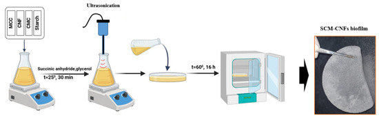
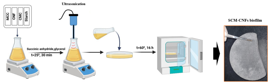
To prepare the starch/CMC/CNF biofilm, a pre-prepared 3% CMC solution and a 2.5% MCC suspension were mixed in a 3:7 ratio in a 50 mL flask. Subsequently, CNF suspension was added at concentrations of 1%, 3%, and 5% of the total volume, and the mixture was intensively stirred for 8 h until a homogeneous system was obtained. Thereafter, 0.875 g of oxidized corn starch was added to the mixture, and the system was further stirred using a magnetic stirrer for 2 h until a homogeneous composition was achieved. To the prepared suspension, 0.75 g of succinic anhydride was added as a crosslinking agent, and 3.8 mL of glycerol was introduced as a plasticizer, followed by stirring at room temperature for 30 min. The resulting viscous mixture was then treated using an ultrasonic generator (SCIENTZ-3000F, Ningbo Xinzhi Biotechnology Co., Ltd., Ningbo, China) at 45 kHz for 30 min. Subsequently, to facilitate the crosslinking process, the mixture was dried in a vacuum oven at 60 ± 2 °C for 16 h (Figure 1). In this study, the biofilm composed of starch/CMC/MCC–CNFs was conventionally denoted as the SCM–CNFs biofilm.
Figure 1.
The synthesis scheme of SCM-CNFs biofilm.
The synthesis of CuNPs via a green method was carried out according to the procedure described in study [32], using an aqueous extract of hibiscus as a reducing agent. For this purpose, 50 mL of 0.1 M aqueous solution of copper nitrate was prepared and heated to 70 °C on a hot plate equipped with a magnetic stirrer. Subsequently, 50 mL of aqueous hibiscus extract was added as a reducing agent, and the mixture was continuously stirred for 6 h until the solution turned light reddish in color. The synthesized CuNPs were characterized by UV–Vis spectroscopy in the wavelength range of 300–800 nm using a spectrophotometer (PE5400-UV, ECROSKHIM Ltd., Saint-Petersburg, Russia) with a 10 mm quartz cuvette. The particle size distribution was determined according to the method described in [33] using a particle size analyzer (Zetasizer Nano ZS90, Malvern Instruments, Worcestershire, UK).
2.2.6. Synthesis of Antibacterial Biofilm
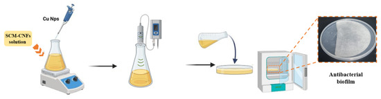
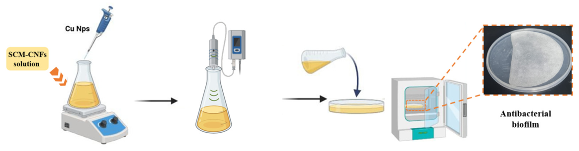
Based on the SCM–CNFs biofilm preparation procedure, a film-forming mixture was prepared, to which 0.5 mL (1.59 mg/mL), 0.75 mL (2.38 mg/mL), and 1 mL (3.18 mg/mL) of CuNPs suspension were added. The mixture was then stirred using a magnetic stirrer for 1 h until complete homogenization was achieved. The resulting mixture was treated with an ultrasonic generator (SCIENTZ-3000F, Ningbo Xinzhi Biotechnology Co., Ltd., Ningbo, China) at 45 kHz and subsequently cast into disk-shaped plastic molds. The samples were then dried in a vacuum oven at 60 ± 2 °C for 16 h to facilitate the crosslinking process (Figure 2).
Figure 2.
The synthesis scheme of SCM-3%CNFs/CuNPs biofilm.
2.2.7. The Antibacterial Properties of Biofilms
The antibacterial activity of the biofilms was evaluated using the agar diffusion method with the soil bacterial strain Bacillus mojavensis. The bacterial suspension was prepared by culturing Bacillus mojavensis to a standard turbidity of 0.5 McFarland, corresponding to an optical density of 0.08–0.12 at a wavelength of 600 nm, as measured using a spectrophotometer. Additionally, the suspension density was verified using a McFarland densitometer. The agar-based nutrient medium was sterilized by standard autoclaving, cooled to 45–50 °C, and uniformly inoculated with the bacterial suspension. After solidification in sterile Petri dishes, biofilm samples cut into disks of identical diameter were placed on the surface of the agar.
Erythromycin, ground into a fine powder and applied in an equivalent amount onto the surface of the agar medium, was used as a positive control. Samples without active components were used as a negative control. The Petri dishes were incubated at 37 °C for 24 h. The antibacterial activity was evaluated based on the presence and diameter of inhibition zones formed around the tested samples and control disks.
2.2.8. SEM Analysis
The surface morphology of the samples was analyzed using a Quanta 200i 3D scanning electron microscope (FEI™, Eindhoven, The Netherlands). Surface morphology was visualized under high vacuum conditions using a secondary electron detector at an accelerating voltage of 15 kV. For sample preparation, 1 g of each MCC and CNF powder was dispersed separately in 50 mL of deionized water containing silver nanoparticles at a concentration of 10−3 mol/L The suspensions were subjected to mechanical stirring for 2 h, then filtered using standard filter paper and dried in an oven at 50 ± 2 °C for 1 h. The dried samples were mounted on aluminum stubs using carbon adhesive tape prior to imaging.
2.2.9. Mechanical Characterization
The mechanical characteristics of the samples were studied using a texture analyzer (TA-3000, LabSol, Beijing, China) with a measured load range of 0.01–60 N and a loading speed range of 0.0005–500 mm/min. The data registration was performed automatically using a computer. During the testing of the samples under a load of 0.01 N, the movement speed was 0.1 mm/min. The samples were tested until the maximum tensile force was reached or before the sample became deformed.
2.2.10. Fourier-Transform Infrared Spectroscopy (FTIR)
FTIR analysis was performed on a spectrometer FTIR FT-801 (Simex, Novosibirsk, Russia), with a resolution of 1 cm−1 and a wavelength of 4500–4700 cm−1, by placing the sample on the surface of the attachment with the method of internal and variable-diffuse reflection, at a temperature of 25 °C and a number of scans of 100.
2.2.11. XRD Analysis
The crystalline structure of the samples was analyzed using an X’Pert PRO diffractometer (Malvern Panalytical Empyrean, Almelo, The Netherlands) equipped with monochromatized CuKα radiation (λ = 0.1542 Å). XRD measurements were carried out in the 2θ range of 10–40° with a step size of 0.02°. The diffractometer was operated at an accelerating voltage of 40 kV and a tube current of 30–45 mA. The measurement time per step was set to 0.5 s to ensure sufficient resolution and intensity of the diffraction peaks.
2.2.12. UV Analysis of CuNPs
The optical absorption spectra of the CuNPs aqueous solutions obtained were recorded on a PE-5400 UV spectrophotometer (Promecolab, Saint Petersburg, Russia), with a scanning speed of 240 nm/min and a wavelength of 190–1000 nm.
2.2.13. Thermogravimetric Analysis
The thermal stability and degradation behavior of the samples were evaluated using a thermogravimetric analyzer (STA200 Synchronous Thermal Analyzer, Wuhan Bonnin Technology Ltd., Wuhan, China) under an argon atmosphere to prevent oxidative degradation. Approximately 25 ± 2 mg of each sample was placed in the crucible for analysis. The temperature range was set from 30 ± 5 °C to 800 ± 5 °C, with a heating rate of 10 ± 1 °C/min. The mass loss profiles were recorded to assess the decomposition stages and thermal resistance of the biofilms.
3. Results and Discussion
3.1. Preparation and Surface Morphology of MCC, CNFs and CMC
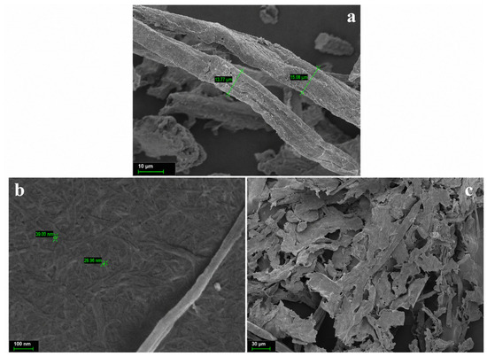
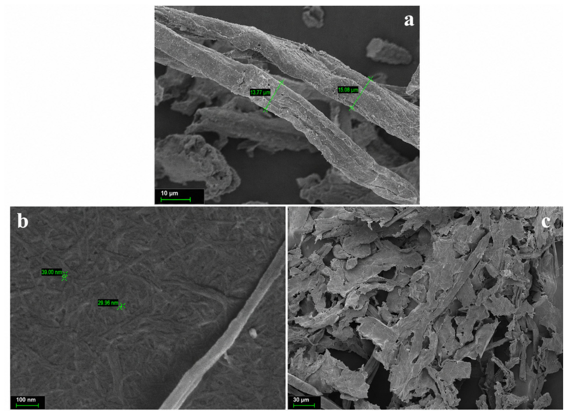
The scanning electron microscopy (SEM) method was employed to determine the characteristic surface morphological features of the studied samples. Figure 3 presents the comparative surface morphology of MCC, CNFs, and the synthesized CMC. The surface morphology of MCC consists of ribbon-like fibers with a rough texture. The average length of the fibers ranges from 150 to 200 µm, while their width is approximately 13–20 µm (Figure 3a). As shown in Figure 3b, CNFs exhibit an interwoven, thread-like fibrillar morphology. The diameter of the fibrils was determined to be in the range of approximately 30–40 nm. This indicates that the fibrillation of microfibers into nanofibers during formic acid hydrolysis proceeded successfully. In contrast, the morphological structure of CMC synthesized from MCC was observed to transform into a different form (Figure 3c). During the synthesis process, the fibrous structure of MCC was converted into irregular flake- and granule-like morphologies, with fragmented fibers also present. This suggests that the crystalline structure was completely transformed into an amorphous state, which is clearly evidenced by the diffractogram shown in Figure 9c. The obtained results are in full agreement with previous studies [33].
Figure 3.
The SEM image of (a)—MCC, (b)—CNFs and (c)—CMC.
3.2. Oxidation, Surface Morphology and Oxidized Functional Groups of Starch
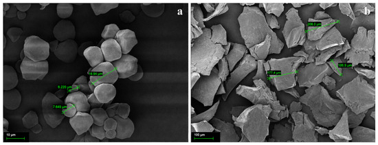
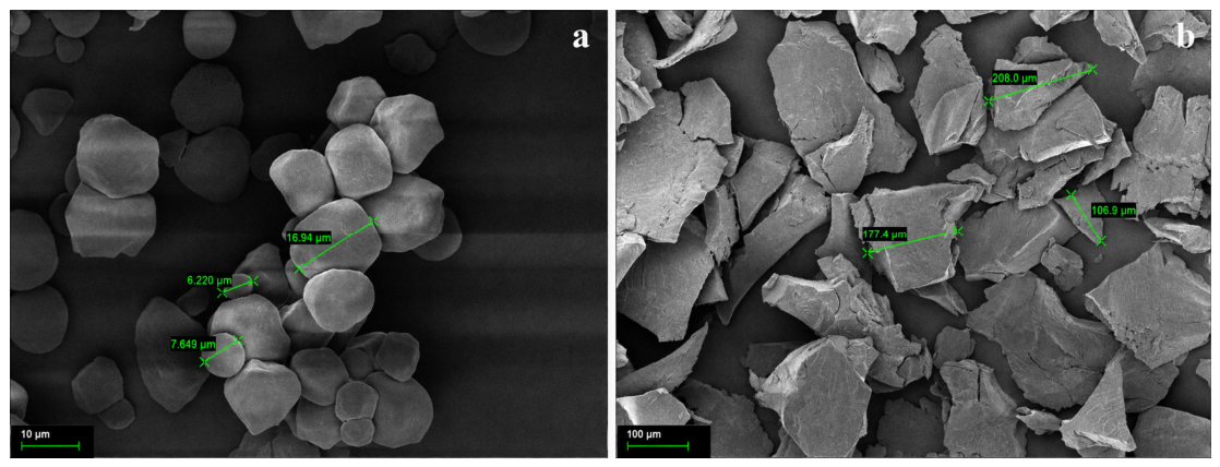
Figure 4 presents a comparative analysis of the surface morphology of native corn starch and corn starch oxidized with hydrogen peroxide, which were used for the preparation of nanocomposite films. The native corn starch appears slightly agglomerated and consists of dense, compact granules with a polygonal shape, lacking pores and cracks (Figure 4a). The average particle size of the granules was determined to be in the range of approximately 6–16 µm. In contrast, the intact granular structure of starch oxidized with hydrogen peroxide was disrupted, transforming into plate-like porous particles of various sizes (Figure 4b). The erosion of the surface of starch, a crystalline biopolymer, along with granule fragmentation and an increase in the amorphous phase, indicates depolymerization of the polysaccharide matrix and its deep structural degradation. This is accompanied by a reduction in crystallinity and the occurrence of chemical functionalization. The average particle size of the oxidized starch ranged from 106 to 208 µm. Tolvanen et al. reported that the introduction of carboxyl (COOH) and carbonyl (CO) groups through hydrogen peroxide oxidation significantly alters starch granules, leading to an increase in their size [34]. The results obtained in this study are in good agreement with previously reported findings.
Figure 4.
The SEM image of (a)—initial starch and (b)—oxidized starch.
3.3. UV-Vis Analysis of Green Synthesized CuNPs and Size Distribution
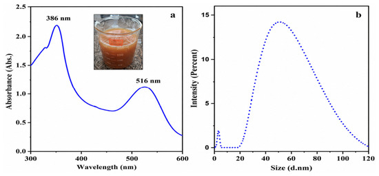
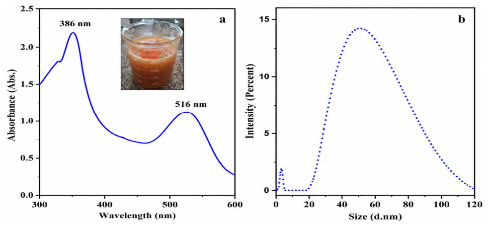
UV–visible absorption spectra of green synthesized copper nanoparticles are shown in Figure 5a. The two absorption bands due to the surface plasmon resonance (SPR) of CuO and Cu particles are observed at 386 nm and at around 516 nm, respectively (Figure 5a). The strong surface plasmon absorption band observed at 516 nm may be due to the formation of non-oxidized CuNPs. The broadness of the absorption band probably arises from the wide size distribution of copper nanoparticles. The considerably higher intensity of the short-wavelength band suggests that oxidized copper species constitute a substantial fraction of the synthesized material. These data are in good agreement with previously reported results [35,36,37]. Figure 5b shows the particle size distribution of copper nanoparticles (CuNPs). According to the obtained results, the average particle size of the CuNPs was approximately 44 nm (Figure 5b). Previous studies have demonstrated that CuNPs with particle sizes in the range of 10–50 nm exhibit enhanced antibacterial properties [38]. Therefore, the average particle size of the CuNPs obtained in this study falls within the optimal range associated with high antibacterial activity.
Figure 5.
UV-vis spectrum and of green synthesized CuNPs (a) and average particle size (b).
3.4. Synthesis of SCM-CNFs Biofilm and Mechanical Properties
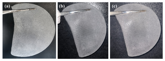
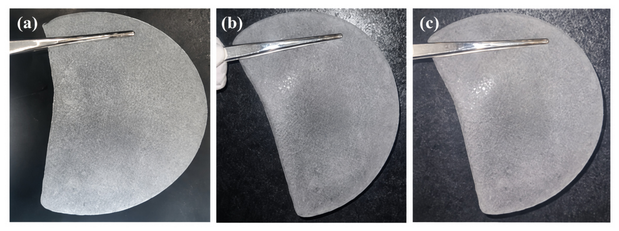
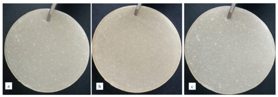
Figure 6a–c present macroscopic images of SCM–1CNF, SCM–3CNF, and SCM–5CNF biofilms containing 1%, 3%, and 5% CNFs, respectively. All obtained samples were observed to be semi-transparent, flexible, and possessed a uniform structure. This indicates that the CNFs were homogeneously dispersed and evenly distributed within the polymer matrix, which, in turn, positively influences the mechanical properties of the biofilms. The average thickness of the SCM–1CNF, SCM–3CNF, and SCM–5CNF biofilms was determined to be 38 ± 5 µm.
Figure 6.
The image of SCM-CNFs biofilm: (a)—SCM-1CNF; (b)—SCM-3CNF; (c)—SCM-5CNF.
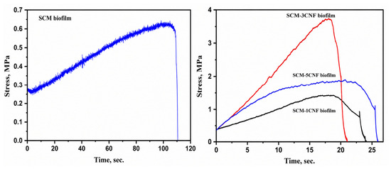
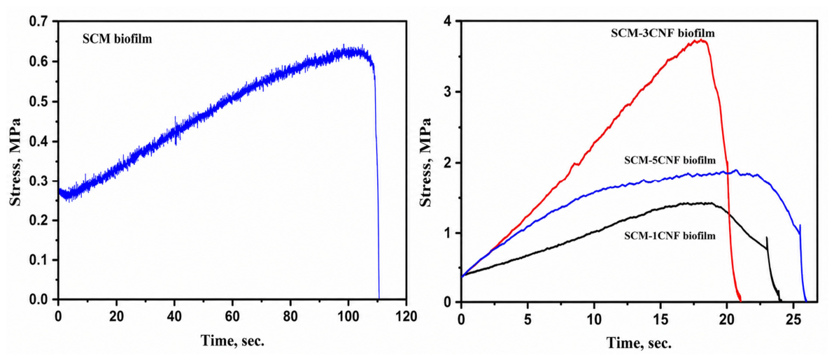
Figure 7 presents the comparative mechanical properties of the pristine SCM biofilm and the SCM–1CNF, SCM–3CNF, and SCM–5CNF biofilms. The mechanical strength of the pristine SCM biofilm without CNFs was 0.68 MPa. In contrast, the mechanical strength increased to 1.39 MPa for SCM–1CNF, reached a maximum of 3.87 MPa for SCM–3CNF, and then decreased to 1.85 MPa for SCM–5CNF. It was observed that increasing the CNF content to 5% led to structural deformation of the biofilm, resulting in premature rupture. In comparative terms, the mechanical strength of SCM–1CNF and SCM–5CNF films increased by 2 and 2.7 times, respectively, compared to the pristine SCM biofilm. Notably, the SCM–3CNF biofilm exhibited the highest performance among all concentrations, enhancing the mechanical strength of the initial SCM biofilm by approximately 5.7 times. Thus, it was determined that a CNF concentration of 3% is optimal for enhancing the mechanical properties of the biofilm. Ricardo et al. reported that CNF concentrations in the range of 5–7% significantly improve the mechanical performance of biofilms [39]. However, in the present study, a comparatively lower concentration—specifically 3% CNFs—resulted in a 5.7-fold increase in the mechanical strength of the pristine SCM biofilm. Therefore, the SCM–3CNF biofilm containing 3% CNFs was selected as the optimal composition.
Figure 7.
Mechanical properties of SCM-CNFs biofilms.
3.5. FTIR Spectrum of SCM-3CNFs Biofilm
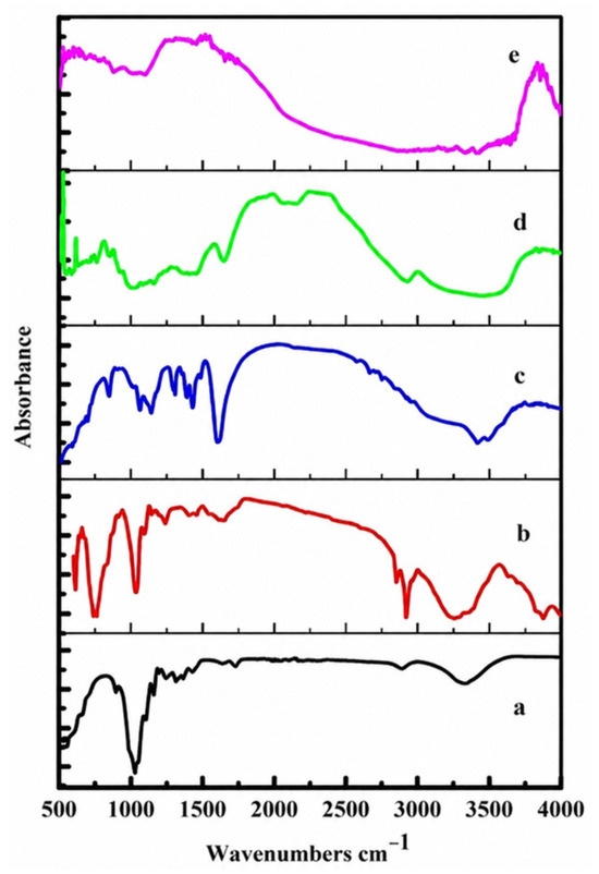
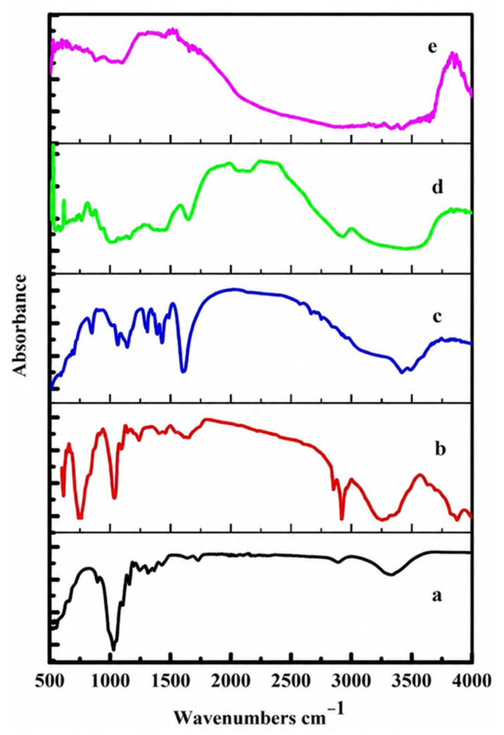
Figure 8 presents the comparative FTIR spectra of the SCM–3CNF biofilm and its initial components, including MCCCL, CNFs, CMC, and oxidized starch. As shown in Figure 8a,b, the spectra of MCC and CNFs exhibit characteristic absorption bands at 655.9 cm−1 corresponding to out-of-plane bending vibrations of C–OH bonds, at 891 cm−1 and 1153.6 cm−1 attributed to β-(1,4)-glycosidic C–O–C linkages (amorphous regions), and strong signals at 1014.7 cm−1 and 1153.6 cm−1 associated with C–O and C–C bonds in the aromatic ring. The band at 1053.3 cm−1 corresponds to stretching vibrations of the C–O–C bond in the pyranose ring, while peaks at 1253.9 cm−1, 1307.9 cm−1, and 1365.8 cm−1 are assigned to C–H vibrations. The absorption at 1412.1 cm−1 corresponds to CH2 groups, and the band at 1635.9 cm−1 is associated with O–H bending vibrations of water molecules adsorbed by cellulose. Additionally, bands observed at 2889.8 cm−1 and 3337.8 cm−1 correspond to C–H and O–H stretching vibrations, respectively. The obtained results are in good agreement with previously reported studies [40,41].
Figure 8.
FTIR spectrum of: (a)—MCCSFH; (b)—CNFs; (c)—CMC; (d)—Starch; (e)—SCM-3CNFs biofilm.
According to the FTIR spectrum of CMC (Figure 8c), characteristic absorption bands are observed at 3200–3600 cm−1 corresponding to the stretching vibrations of hydroxyl (–OH) groups, around 3000 cm−1 for C–H stretching vibrations, at 1680 cm−1 for carbonyl (C=O) stretching, at 1450 cm−1 for –CH2 bending vibrations, and in the range of 1000–1200 cm−1 for ester (–O–) stretching vibrations. These results are consistent with previously reported data [42].
Figure 8d presents the FTIR spectrum of oxidized starch. The broad absorption band in the region of 3500–3000 cm−1 indicates the presence of –OH groups, while the signal at 1053 cm−1 corresponds to C–O bond vibrations [43]. The absorption band at 2868 cm−1 is attributed to C–H stretching vibrations of CH2 groups. The peaks observed at 1147 cm−1, 1078 cm−1, and 990 cm−1 correspond to stretching vibrations of C–O, C–O–H, and C–O–C groups within the glucose ring, respectively. Absorption signals at 855–867 cm−1 indicate the presence of β-glycosidic linkages. Furthermore, the band at 1680 cm−1 is assigned to C=O stretching vibrations of carboxyl and carbonyl groups formed during the oxidation process [44]. These findings are consistent with the SEM analysis results presented in Figure 4b, confirming the structural modifications of the oxidized starch.
In the FTIR spectrum of the SCM–3CNFs biofilm (Figure 8e), a noticeable decrease in the intensity of the absorption band at 1680 cm−1, corresponding to the carboxyl and carbonyl groups of starch and CMC, is observed. This may be attributed to the interaction of the crosslinking agent, succinic anhydride, with the carboxyl and carbonyl groups of the polymer chains during synthesis, leading to a reduction in the intensity of these functional groups [45,46]. The FTIR results obtained in this study are consistent with those reported in previous work [47] for films based on oxidized starch and CMC.
3.6. The XRD Analysis of SCM-3%CNFs Biofilm
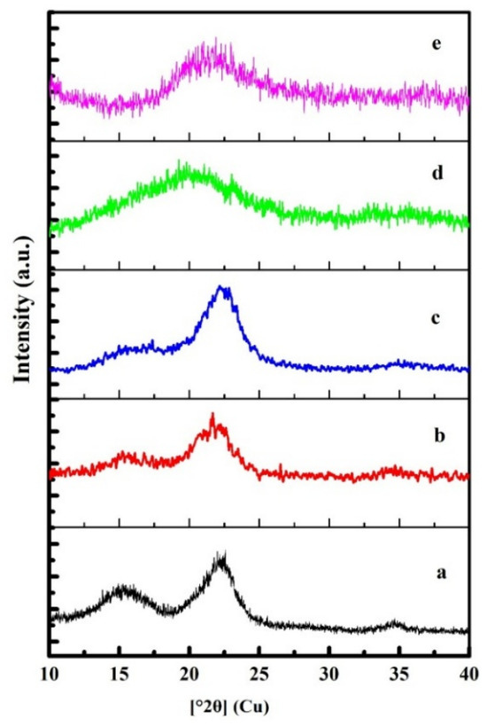
The crystalline structure of the obtained SCM–3CNFs biofilm was investigated using X-ray diffraction (XRD) in comparison with the initial MCCCH, CNFs, CMC, and oxidized starch. The corresponding diffractograms of the samples are presented in Figure 9. In the diffractograms of MCC (Figure 9a) and CNFs (Figure 9b), four characteristic diffraction peaks are observed at 2θ = 15.6° (1–10), 16.5° (110), 22.2° (200), and 34.5° (004), indicating that the crystal structure corresponds to a two-chain monoclinic unit cell [48]. These results are in good agreement with previously reported studies [49].
Figure 9.
The XRD diffractogram of (a)—MCCSFH; (b)—CNFs; (c)—CMC; (d)—Starch; (e)—SCM-3CNFs biofilm.
In the diffractogram of the synthesized CMC (Figure 9c), a characteristic peak corresponding to carbon is observed at 2θ = 21.3° (200). During the synthesis process, the weakening of inter- and intramolecular hydrogen bonds within the cellulose structure [50] leads to the disruption of its crystalline arrangement, which is evidenced by the decreased intensity of the diffraction peaks.
In the diffractogram of oxidized starch (Figure 9d), low-intensity peaks characteristic of the A-type polymorphic structure of amylose and amylopectin are observed at 2θ = 17.6° (110), 18.4° (110), and 23.0° (200). These findings complement the SEM (Figure 4b) and FTIR (Figure 8d) analysis results discussed above. The obtained results are in good agreement with those reported in previous studies [50].
In the diffractogram of the SCM–3CNFs biofilm, overlapping signals characteristic of the initial components, such as MCC, CNFs, CMC, and starch, are observed. This indicates that the original components underwent structural transformation, leading to modifications in their crystalline structure and a decrease in peak intensities. Consistently, the FTIR analysis (Figure 8) also revealed a reduction in the intensity of characteristic functional group signals of the initial components. Vedovello et al. reported similar XRD results for CMC/starch-based films crosslinked with citric acid [51].
3.7. Synthesis of SCM-3CNFs/CuNPs Biofilm and Antibacterial Properties
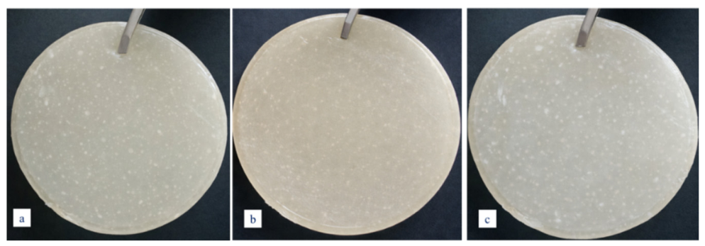
To impart antibacterial properties to the SCM–3CNF biofilm, CuNPs synthesized via a green method were immobilized onto the SCM–3CNFs biofilm at volumes of 0.5 mL, 0.75 mL, and 1 mL. Figure 10 presents the macroscopic images of the CuNPs-immobilized SCM–3CNFs-based biofilms. The obtained biofilms exhibited smooth surfaces with noticeable white inclusions, as well as a uniform and flexible structure. The average thickness of the biofilms was approximately 45 ± 2 µm.
Figure 10.
The SCM-3CNF/CuNPs biofilm: (a)—SCM-3CNF/0.5CuNPs; (b) SCM-3CNF/0.75CuNPs; (c)—SCM-3CNF/1CuNPs biofilms.
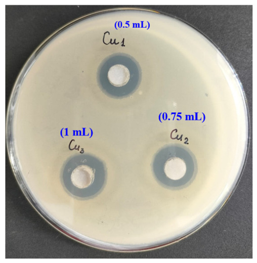
The antibacterial activity of SCM–3CNFs biofilms containing different concentrations of CuNPs was evaluated using the agar diffusion method. Antibacterial activity was assessed based on the diameter of the inhibition zones suppressing bacterial colony growth. The variation in inhibition zones clearly demonstrated a concentration-dependent trend (Figure 11). As shown in the figure, the inhibition zone of the film containing 0.5 mL of CuNPs was approximately 15 mm, while the film with 0.75 mL of CuNPs exhibited an inhibition zone of about 18 mm. In contrast, the SCM–3CNFs biofilm containing 1 mL of CuNPs demonstrated the largest inhibition zone, measuring approximately 22 mm. An increase in CuNPs concentration led to a corresponding enlargement of the inhibition zones, indicating that the antibacterial activity of CuNPs is directly dose-dependent. Accordingly, the SCM–3CNF/1CuNPs biofilm containing 1 mL of CuNPs was identified as the most effective formulation. Previous studies [52] have shown that the antibacterial mechanism of CuNPs involves their interaction with the bacterial cell membrane, facilitating ion penetration and inducing redox reactions. As a result, enzymatic activity is reduced, ultimately leading to disruption of the cell membrane integrity.
Figure 11.
The antibacterial properties of SCM-3CNF/0.5CuNPs; SCM-3CNF/0.75CuNPs; SCM-3CNF/1CuNPs biofilms.
3.8. The FTIR Spectrum of SCM-3CNFs/1CuNPs Biofilm
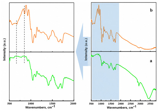
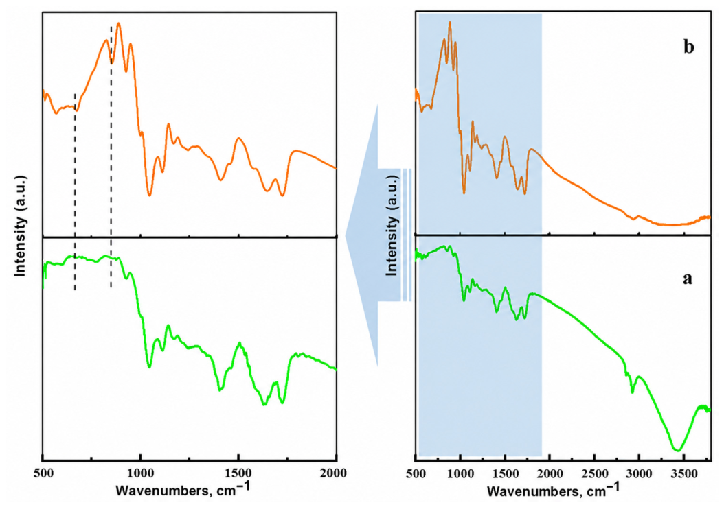
Figure 12 presents the comparative FTIR spectra of the SCM-3CNFs/1CuNPs biofilm and the parent SCM-3CNFs biofilm, illustrating their chemical structures. In the FTIR spectrum of the SCM-3CNFs biofilm (Figure 12a), the absorption bands observed in the 1000–1200 cm−1 region confirm the presence of characteristic C–O and C–O–C stretching vibrations associated with the original components, namely MCC, CMC, and oxidized starch [53,54]. These results are consistent with the FTIR analysis presented in Figure 8.
Figure 12.
The FTIR spectrum of: (a)—SCM-3CNFs biofilm; (b)—SCM-3CNFs/1CuNPs biofilm.
In the FTIR spectrum of the SCM–3CNFs/1CuNPs biofilm (Figure 12b), characteristic signals of the initial components are also observed. In addition, absorption bands appearing at 659 cm−1 and 880 cm−1 are attributed to Cu–O and Cu–OH bonds, respectively. This can be explained by the formation of interactions between CuNPs and oxygen-containing functional groups within the polymer matrix during immobilization [55]. These interactions may enhance the stability of CuNPs and positively influence their antibacterial activity [56]. The results of the antibacterial activity study of the SCM–3CNFs/1CuNPs biofilm (Figure 11) are consistent with this interpretation.
3.9. The XRD Analysis of SCM-3CNFs/1CuNPs Biofilm
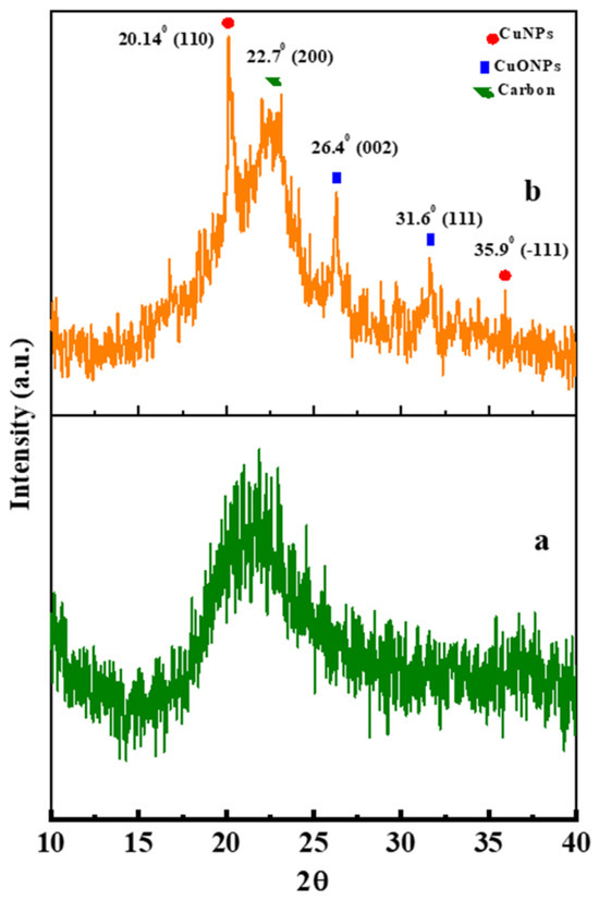
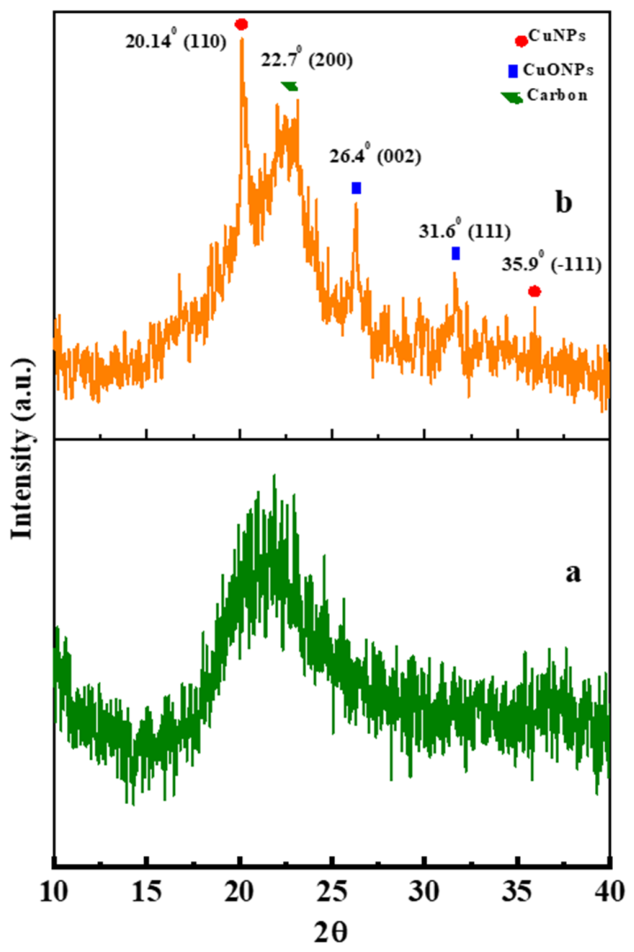
The comparative XRD diffractograms of the SCM–3CNFs/1CuNPs biofilm and the SCM–3CNFs biofilm are presented in Figure 13. In the diffractogram of the SCM–3CNFs biofilm (Figure 13a), a typical diffraction peak corresponding to carbon is observed at 2θ = 21.8° (200), along with low-intensity overlapping signals characteristic of the initial components, consistent with the results shown in Figure 9e.
Figure 13.
The XRD diffractogram of: (a)—SCM-3CNFs biofilm; (b)—SCM-3CNFs/1CuNPs biofilm.
In the diffractogram of the SCM–3%CNFs/CuNPs–1 biofilm (Figure 13b), characteristic diffraction peaks corresponding to face-centered cubic CuNPs (JCPDS 04-0836) and face-centered monoclinic CuO nanoparticles (JCPDS No. 00-048-1548) were identified at 2θ = 20.14° (110), 26.4° (002), 31.6° (111), and 35.9° (−111). The obtained results are in good agreement with previously reported studies [57,58]. The average percentage shifts in the diffraction peaks (2θ) for CuNPs and CuO nanoparticles relative to the JCPDS 04-0836 and JCPDS No. 00-048-1548 standards were insignificant, amounting to 0.063% and 0.124%, respectively. This indicates that the copper nanoparticles were uniformly and successfully immobilized within the SCM–3CNFs biofilm matrix.
3.10. The Mechanical Properties of SCM-3CNFs/1CuNPs Biofilm
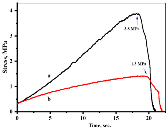
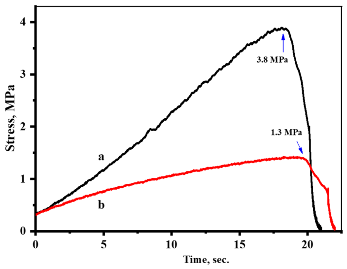
Figure 14 presents the comparative mechanical properties of the SCM–3CNFs/1CuNPs biofilm and the SCM–3CNFs biofilm. The mechanical strength of the SCM–3CNFs biofilm without CuNPs immobilization was 3.87 MPa (Figure 14a). With increasing stress, the biofilm undergoes gradual deformation and eventually exhibits abrupt fracture at higher stress levels. This indicates that the biofilm exhibits relatively high stiffness and strength, which can be attributed to the reinforcing effect of CNFs within the matrix. In contrast, as shown in Figure 14b, the SCM–3CNFs/1CuNPs biofilm undergoes deformation at lower stress and demonstrates gradual fracture during elongation. This behavior indicates increased elasticity of the film; however, its mechanical strength is lower compared to the SCM–3CNFs biofilm, with a value of 1.3 MPa. This value is approximately three times lower than that of the SCM–3CNFs biofilm. Thus, while the immobilization of CuNPs enhances the elasticity of the film, it adversely affects its mechanical strength. This phenomenon can be attributed to the interaction between CuNPs and the oxygen-containing functional groups in CNFs, which weakens the intermolecular hydrogen bonding between cellulose chains and, consequently, reduces the mechanical integrity of the film. The obtained results are in good agreement with previously reported studies [59,60]. The interaction between CuNPs and oxygen-containing functional groups is also confirmed by the FTIR analysis presented in Figure 12.
Figure 14.
The mechanical properties of: (a)—SCM-3CNFs, (b)—SCM-3CNFs/1CuNPs biofilms.
3.11. The Surface Morphology of SCM-3CNFs/1CuNPs Biofilm
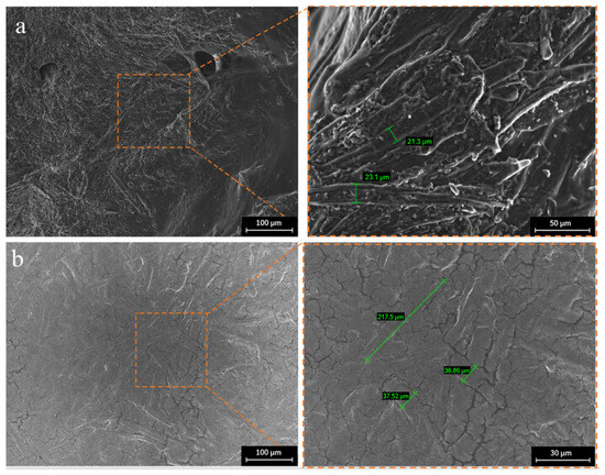
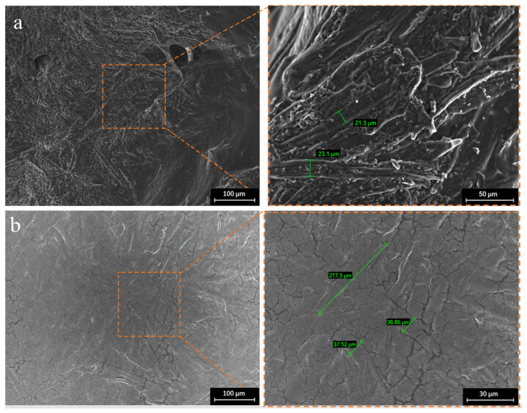
The surface morphology of the SCM–3CNFs biofilm and the SCM–3CNFs/1CuNPs biofilm was comparatively analyzed using SEM (Figure 15). The surface of the SCM–3CNFs biofilm appears slightly rough, with no visible cracks or distinct phase separation; however, it is relatively dense (Figure 15a). In addition, uniformly distributed cellulose micro- and nanofibers can be clearly observed. This contributes to the enhancement of the mechanical properties of the biofilm, confirming the results obtained from the mechanical analysis presented in Figure 14. Figure 15b shows the surface morphology of the SCM–3CNFs/1CuNPs biofilm. The cellulose fibers are clearly visible on the film surface. The incorporation of CuNPs increased the surface roughness and induced pronounced heterogeneous structural changes. These modifications enhance the direct contact area with bacteria and facilitate the release of CuNPs, thereby improving the antibacterial performance, as reported in previous studies [61]. In addition, the formation of microcracks on the film surface can be observed. These structural changes may contribute to the reduction in the mechanical properties of the film. The mechanical analysis results of the SCM–3CNFs/1CuNPs biofilm presented in Figure 15b confirm this interpretation. Similar findings have also been reported in earlier studies [62].
Figure 15.
The SEM image of (a)—SCM-3CNFs, (b)—SCM-3CNFs/1CuNPs biofilms.
3.12. The TGA Analysis of SCM-3%CNFs/CuNPs-1 Biofilm
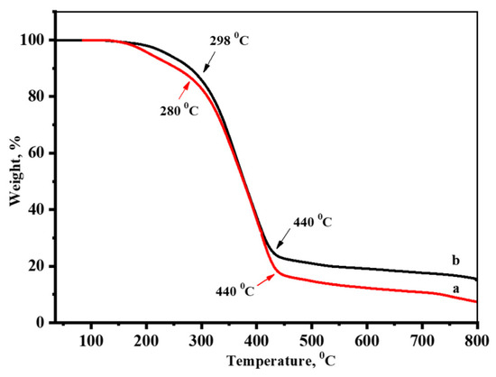
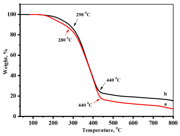
Figure 16 presents the comparative thermograms of SCM–3CNFs and SCM–3CNFs/1CuNPs samples. The thermograms of both samples reveal a multistage mass loss characteristic of polysaccharide-based materials. In both cases, a slight mass loss occurs up to 300 °C, during which the samples lose approximately 15% of their initial mass. This behavior is attributed to the evaporation of physically adsorbed water and inherent moisture within the films. Notably, the mass loss of the SCM–3CNFs/1CuNPs biofilm occurs more gradually during this stage (Figure 16b). This may be explained by the interaction of CuNPs with oxygen-containing groups in the polymer matrix and with bound water molecules, as reported by Lakshmanan et al. [63].
Figure 16.
The TGA curves of: (a)—SCM-3CNFs, (b)—SCM-3CNFs/1CuNPs biofilms.
The main thermal degradation process was found to occur in the temperature range of 300–440 °C, during which a sharp decrease in sample mass was observed. The SCM–3CNFs biofilm lost approximately 83% of its initial mass (Figure 16a), whereas the SCM–3CNFs/1CuNPs biofilm exhibited a mass loss of about 75%. Upon increasing the temperature to 800 °C, the total mass loss reached 93% for the SCM–3CNFs biofilm and 85% for the SCM–3CNFs/1CuNPs biofilm (Figure 16b). At this stage, carbonization processes occur.
Notably, the residual mass of the SCM–3CNFs/1CuNPs biofilm after thermal degradation was approximately two times higher than that of the SCM–3CNFs biofilm. Previous studies [64,65] have reported that polymer films embedded with metal nanoparticles tend to form inorganic (oxide) compounds during thermal decomposition, contributing to an increased residual mass. Furthermore, metal nanoparticles promote the formation of a stable carbonaceous phase. These findings are in full agreement with the results obtained in the present study.
3.13. The Biodegradation Properties of SCM-3%CNFs/CuNPs-1 Biofilm
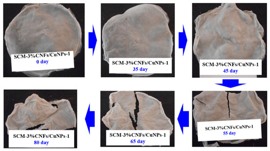
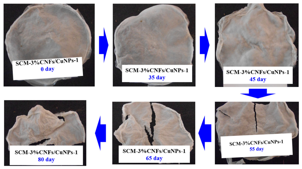
Figure 17 presents visual images illustrating the biodegradation behavior of the SCM–3CNFs/1CuNPs biofilm in soil. During the study, the temperature was maintained within the range of 25–30 °C. Based on the visual appearance of the film, no significant changes in its initial mass were observed during the first 35 days. Starting from day 45, the macroscopic structure of the film began to deform, accompanied by a gradual decrease in mass. The mass remained at approximately 96% on day 45, decreased to 80% by day 55, and further declined to 60% by day 65. During this stage, microorganisms actively begin to degrade the main components of the film, namely polysaccharides. This stage is referred to as the exponential phase. In this process, cellulose macromolecules present in the film are transformed into oligomers and monomers, with water and carbon dioxide released as oxidation products [66,67]. After 70 days, a noticeable decrease in the biodegradation rate was observed; however, the degradation process continued. By day 80, only 43% of the initial mass of the film remained, indicating that 57% had undergone biodegradation. This slowdown may be attributed to the persistence of more resistant components of the material, specifically the highly crystalline CNFs and modified polymer chains containing Cu nanoparticles, which are more difficult to degrade. Previous studies [68,69,70] have concluded that the biodegradation behavior of films largely depends on the nature of the polymer matrix and the incorporated antibacterial agents.
Figure 17.
The biodegradation process of SCM-3CNFs/1CuNPs biofilm.
3.14. Effect of SCM–3CNFs/1CuNPs Biofilm on the Agrochemical Properties of Soil
To evaluate the effect of the biodegradation of the pristine SCM-3CNFs and SCM-3CNFs/1CuNPs biofilms on the agrochemical properties of the soil, a comparative analysis was performed relative to the soil prior to biodegradation. Table 1 comparatively presents the values of the main agrochemical parameters of the soil before and after biodegradation.
Table 1.
Main agrochemical parameters of soil before and after biodegradation.
From the results presented in Table 1, it can be seen that the pristine film without copper nanoparticles does not exert a detrimental effect on the main agrochemical properties of the soil during biodegradation. The only notable change was an increase in soil moisture content. This indicates that the film has a good water-retention capacity.
The analysis results showed that during the biodegradation period, the soil pH value exhibited only a minimal change, increasing from 6.9 to 7.0. This indicates that the soil maintained its initial pH conditions. Typically, the degradation of organic materials may lead to a decrease in pH due to the release of organic acids; however, in this case, the mineralization of the polymers within the film and the presence of CuNPs did not significantly affect the soil pH. Such minimal variation suggests that the bionanocomposite film is environmentally safe for the soil ecosystem. Previous studies [71] have also demonstrated that biopolymer-based materials have minimal adverse effects on soil ecosystems during biodegradation, which supports the validity of the results obtained in this study.
During biodegradation, the decrease in soil macronutrients such as potassium and available phosphorus can be attributed to their uptake by plants as essential nutrients for growth and development. Meanwhile, the increase in soil moisture content from 17.8% to 20.2% can be explained by the moisture-retaining ability of the film, as well as the release of hydrophilic cellulose-based monomers during biodegradation and the formation of water as a final product of the degradation process. This effect may be particularly associated with the high water absorption capacity of cellulose derivatives such as CMC present in the film composition [72].
In addition, despite the fungicidal properties of CuNPs, the soil microflora effectively degraded the polymer matrix. This indicates that the concentration of CuNPs incorporated into the film is within an optimal range, exerting no inhibitory effect on the microbial activity responsible for biodegradation. Thus, it can be concluded that the presence of CuNPs does not adversely affect the microbiological activity of soil microorganisms involved in the degradation process. Previous studies [73] have similarly reported that biopolymers immobilized with CuNPs do not negatively impact soil microbial communities.
4. Conclusions
A biodegradable antibacterial biofilm based on oxidized corn starch and cellulose nanofibers derived from corn husk was successfully developed using succinic anhydride as a crosslinking agent and green-synthesized CuNPs as an antibacterial component. Among the investigated formulations, the biofilm containing 3% CNFs exhibited the highest mechanical strength (3.87 MPa), which was approximately 2.7 and 2.0 times higher than those of the films containing 1% and 5% CNFs, respectively. Incorporation of CuNPs imparted pronounced antibacterial activity, with the SCM–3CNFs/1CuNPs formulation showing the highest inhibition zone of approximately 22 mm. Although CuNP immobilization reduced the mechanical strength by approximately threefold compared with the pristine SCM–3CNFs film, it increased film flexibility and resulted in an approximately twofold higher residual mass after thermal degradation. Furthermore, the SCM–3CNFs/1CuNPs biofilm underwent 57% mass loss after 80 days of soil burial, while producing only minimal changes in the evaluated agrochemical properties of the soil. Overall, these results demonstrate that SCM–3CNFs/1CuNPs provides a favorable combination of antibacterial performance, biodegradability, and limited impact on soil properties, supporting its potential application as a functional biodegradable material for agricultural purposes. Future studies should optimize the CuNP loading to achieve an appropriate balance between antibacterial efficacy and mechanical properties and should quantify Cu release, phytotoxicity, and long-term environmental safety under field conditions.
Author Contributions
Conceptualization, K.A. and S.K.; methodology, K.A., A.B. and S.K.; validation, N.S., N.K. and A.D.; formal analysis, A.B., E.S. and A.K.; investigation, K.A., A.B., N.S., N.K., A.D., E.S. and A.K.; resources, M.B., S.K. and S.T.; data curation, N.S., N.K. and E.S.; writing—original draft preparation, K.A., A.B. and N.S.; writing—review and editing, S.K., M.B. and S.T.; visualization, A.D., E.S. and A.K.; supervision, S.K., M.B. and S.T.; project administration, K.A. and S.K.; funding acquisition, K.A. and M.B. All authors have read and agreed to the published version of the manuscript.
Funding
This research was funded by the Science Committee of the Ministry of Science and Higher Education of the Republic of Kazakhstan, Grant No. AP23490029.
Institutional Review Board Statement
Not applicable.
Informed Consent Statement
Not applicable.
Data Availability Statement
All data generated or analyzed during this study are included in this published article. Further inquiries can be directed to the corresponding author.
Conflicts of Interest
The authors declare no conflicts of interest.
References
- Pagano, M.; Lunetta, E.; Belli, F.; Mocarli, G.; Cocozza, C.; Cacciotti, I. Advancements in Agricultural Nanotechnology: An Updated Review. Plants 2025, 14, 2939. [Google Scholar] [CrossRef] [Scilit] [PubMed]
- Donley, N. The USA lags behind other agricultural nations in banning harmful pesticides. Environ. Health 2019, 18, 44. [Google Scholar] [CrossRef] [Scilit] [PubMed]
- Abd-Elsalam, K.A. Special Issue: Agricultural Nanotechnology. Plants 2024, 13, 489. [Google Scholar] [CrossRef] [Scilit] [PubMed]
- Food and Agriculture Organization of the United Nations. Assessment of Agricultural Plastics and Their Sustainability: A Call for Action; FAO: Rome, Italy, 2021. [Google Scholar]
- Escrig Rondán, C.; Sevilla Gil, C.; Sanz Fernández, P.; Ferrer Crespo, J.F.; Furió Sanz, C. Incorporation of natural biostimulants in biodegradable mulch films for agricultural applications: Ecotoxicological evaluation. Polymers 2025, 17, 3027. [Google Scholar] [CrossRef] [Scilit] [PubMed]
- Chen, C.; Li, Z.; Yin, K.; Li, L.; Zhang, Z.; Xu, X.; Liu, H.; Qing, Y.; Li, X.; Wu, Y. Biodegradable liquid slow-release mulch film based on bamboo residue for selenium-enriched crop cultivation. Research 2025, 8, 0685. [Google Scholar] [CrossRef] [Scilit] [PubMed]
- Akhmetov, K.; Sagyndykova, Z.; Khazimov, M. Power need of an implement for removing polymer residues from the soil surface in Kazakh horticulture. J. Agric. Eng. 2022, 53, 1382. [Google Scholar] [CrossRef] [Scilit]
- Kenenbayev, S.B.; Ospanbayev, O.Z.; Kydyrov, A.K.; Musagodzhaev, N.T.; Aristangulov, S.S. Effectiveness of sugar beet cultivation under drip irrigation in South-East Kazakhstan. Biosci. Biotechnol. Res. Asia 2016, 13, 571–578. [Google Scholar] [CrossRef] [Scilit][Green Version]
- Wan, Y.; Yang, Y.; Zhou, W. Agricultural plastic mulch: A brief review of development, composition and catalytic upcycling strategies. Catalysts 2025, 15, 310. [Google Scholar] [CrossRef] [Scilit]
- Helm, L.T.; Venier-Cambron, C.; Verburg, P.H. The potential land-use impacts of bio-based plastics and plastic alternatives. Nat. Sustain. 2025, 8, 190–200. [Google Scholar] [CrossRef] [Scilit]
- Fraceto, L.F.; Grillo, R.; de Medeiros, G.A.; Scognamiglio, V.; Rea, G.; Bartolucci, C. Nanotechnology in agriculture: Which innovation potential does it have? Front. Environ. Sci. 2016, 4, 20. [Google Scholar] [CrossRef] [Scilit]
- Mridha, D.; Lamsal, B.; Antonangelo, J.A. Nanotechnology in agriculture: Innovations for sustainability and greenhouse gas mitigation—A review. Sci. Total Environ. 2025, 995, 180065. [Google Scholar] [CrossRef] [Scilit] [PubMed]
- Sultan, H.; Li, Y.; Ahmed, W.; Mu, Y.; Shah, A.; Faizan, M.; Ahmad, A.; Abbas, H.M.M.; Nie, L.; Khan, M.N. Biochar and nano biochar: Enhancing salt resilience in plants and soil while mitigating greenhouse gas emissions: A comprehensive review. J. Environ. Manag. 2024, 355, 120448. [Google Scholar] [CrossRef] [Scilit] [PubMed]
- Chemiru, G.; Gonfa, G. Preparation and characterization of glycerol-plasticized yam starch-based films reinforced with titanium dioxide nanofiller. Carbohydr. Polym. Technol. Appl. 2023, 5, 100300. [Google Scholar] [CrossRef] [Scilit]
- Chen, L.; Dai, R.; Shan, Z.; Chen, H. Fabrication and characterization of one high-hygroscopicity liquid starch-based mulching material for facilitating plant growth. Carbohydr. Polym. 2020, 230, 115582. [Google Scholar] [CrossRef] [Scilit] [PubMed]
- Rajgadia, N.; Debnath, M. Biodegradable mulch utilizing bioplastic biopolymer polyhydroxyalkanoates. Mater. Today Proc. 2023, 79, 411–419. [Google Scholar] [CrossRef] [Scilit]
- Han, Q.; Wang, Y.; Wang, H.; Zhou, T.; Song, Z.; Yu, D.; Liu, X.; Liu, W.; Ge, S. Fabrication and evaluation of an alginate-based dual-network mulch film reinforced with multiscale cellulose fibers. ACS Sustain. Chem. Eng. 2024, 12, 2120–2129. [Google Scholar] [CrossRef] [Scilit]
- Parida, M.; Jena, T.; Mohanty, S.; Nayak, S.K. Advancing sustainable agriculture: Evaluation of poly(lactic acid) (PLA)-based mulch films and identification of biodegrading microorganisms among soil microbiota. Int. J. Biol. Macromol. 2024, 269, 132085. [Google Scholar] [CrossRef] [Scilit] [PubMed]
- Wang, M.; Li, W.; Zhao, F.; Deng, S.; Li, T.; Wu, X.; Mu, Y.; Yang, K.; Zhang, A.; Yang, X. A review on the application of starch in biodegradable mulch films. Int. J. Biol. Macromol. 2025, 320, 145915. [Google Scholar] [CrossRef] [Scilit] [PubMed]
- Kim, J.T.; Kim, T.H.; Kim, S.; Seo, K.H. Structural, thermal, and mechanical properties of PLA/PBAT/MEA blend. Polym. Korea 2016, 40, 371–379. [Google Scholar] [CrossRef] [Scilit]
- Na, K.; Lee, K.H.; Lee, D.H.; Bae, Y.H. Biodegradable thermo-sensitive nanoparticles from poly(L-lactic acid)/poly(ethylene glycol) alternating multi-block copolymer for potential anti-cancer drug carrier. Eur. J. Pharm. Sci. 2006, 27, 115–122. [Google Scholar] [CrossRef] [Scilit] [PubMed]
- Hassanisaadi, M.; Saberi Riseh, R.; Rabiei, A.; Varma, R.S.; Kennedy, J.F. Nano/micro-cellulose-based materials as adsorbents for the remediation of chemical pollutants from agricultural resources. Int. J. Biol. Macromol. 2023, 250, 126128. [Google Scholar] [CrossRef] [Scilit] [PubMed]
- Saberi Riseh, R. Advancing agriculture through bioresource technology: The role of cellulose-based biodegradable mulches. Int. J. Biol. Macromol. 2024, 255, 128006. [Google Scholar] [CrossRef] [Scilit] [PubMed]
- Xu, Y.; Wu, Z.; Li, A.; Chen, N.; Rao, J.; Zeng, Q. Nanocellulose composite films in food packaging materials: A review. Polymers 2024, 16, 423. [Google Scholar] [CrossRef] [Scilit] [PubMed]
- Basak, M.; Arafat, K.M.Y.; Bera, S.; Hubbe, M.A.; Lucia, L.; Pal, L. Energy-efficient cellulose acetate nanofilms from hemp hurds for circular packaging and agricultural mulch applications. Cellulose 2026, 33, 2081–2105. [Google Scholar] [CrossRef] [Scilit]
- Doan Trang, T.Y.; Dzung, H.T.; Lan, V.P.; Duc, H.T.; Hanh, D.T. Corn starch and durian husk nanocellulose composite films: A green approach for biodegradable packaging. E3S Web Conf. 2025, 623, 01001. [Google Scholar] [CrossRef] [Scilit]
- Malekzadeh, E.; Tatari, A.; Dehghani Firouzabadi, M. Effects of biodegradation of starch-nanocellulose films incorporated with black tea extract on soil quality. Sci. Rep. 2024, 14, 18817. [Google Scholar] [CrossRef] [Scilit] [PubMed]
- Sagiyeva, N.; Akatan, K.; Battalova, A.; Kampitova, G.; Shaimardan, E.; Kabdrakhmanova, A.; Kabdrakhmanova, S.; Beisebekov, M. Eco-friendly conversion of corn husk agro-waste into microcrystalline cellulose and cellulose nanofibers. Nano-Struct. Nano-Objects 2026, 45, 101610. [Google Scholar] [CrossRef] [Scilit]
- Aqatan, Q.; Qaiyrbekov, N.; Qabdrakhmanova, S.; Ibraeva, Z. Qarasora (Cannabis) sabağynan alınğan tsellülozadan üş hlorldı sırke qışqılı kömegimen karboksimetiltsellüloza alu mümkinşiligin zertteu. In Proceedings of the International Scientific-Practical Conference “Actual Problems of Natural Science and Natural Science Education”, Dedicated to the Memory of Scientist and Educator T.D. Kuanyshbaev, Kyzylorda, Kazakhstan, 7–8 November 2019; pp. 12–16. Available online: https://korkyt.edu.kz/files/222/Konferenciya/7.%20%D0%9A%D0%BE%D0%BD%D1%84.%20%D0%BE%D0%B1%D1%89%D0%B8%D0%B9%20%D0%BC%D0%B0%D0%BA%D0%B0%D0%BB%D0%B0.pdf (accessed on 31 July 2026).
- Akatan, K.; Battalova, A.; Sagiyeva, N.; Kabdrakhmanova, S.; Kaiyrbekov, N.; Tursyngazykyzy, A.; Shaymardan, E.; Beisebekov, M.; Kampitova, G. Oxidized starch/CMC-based biofilm: Synthesis and characterization. Phys. Sci. Technol. 2024, 11, 73–84. [Google Scholar] [CrossRef] [Scilit]
- Battalova, A.; Akatan, K.; Kabdrakhmanova, S.; Ibraeva, Z.; Beisebekov, M.; Shaimardan, E.; Nurgamit, K.; Dzhumanazarova, A.; Joshy, K.S.; Thomas, S. Isolation of cellulose nanofibers from sunflower seed husks via formic acid hydrolysis. J. Polym. Sci. 2024, 63, 5392–5401. [Google Scholar] [CrossRef] [Scilit]
- Anh, L.T.K.; Vuong, L.D.; Bao, V.V.Q.; Nga, N.T.P.; Luan, L.V. Biosynthesis of silver nanoparticles using nanocurcumin extracted from fresh turmeric of Vietnam. Int. J. Mater. Res. 2022, 113, 776–784. [Google Scholar] [CrossRef] [Scilit]
- Pricop, M.A.; Negrea, A.; Pascu, I.B.; Ciopec, M.; Negrea, P.; Cristea, I.M.; Tatu, C.A.; Ivan, A. A novel green synthesis method of copper nanoparticles and their biological effects on cancer and normal cells. Int. J. Mol. Sci. 2026, 27, 2559. [Google Scholar] [CrossRef] [Scilit] [PubMed]
- Tolvanen, P.; Mäki-Arvela, P.; Sorokin, A.B.; Salmi, T.; Murzin, D.Y. Kinetics of starch oxidation using hydrogen peroxide as an environmentally friendly oxidant and an iron complex as a catalyst. Chem. Eng. J. 2009, 154, 52–59. [Google Scholar] [CrossRef] [Scilit]
- Khanehzaei, H.; Ahmad, M.B.; Shameli, K.; Ajdar, Z. Synthesis and characterization of Cu@Cu2O core–shell nanoparticles prepared in seaweed Kappaphycus alvarezii. Int. J. Electrochem. Sci. 2014, 9, 8189–8198. [Google Scholar] [CrossRef] [Scilit]
- Yin, H.; Zhao, Y.; Xu, X.; Chen, J.; Wang, X.; Yu, J.; Wang, J.; Wu, W. Realization of tunable localized surface plasmon resonance of Cu@Cu2O core–shell nanoparticles by the pulse laser deposition method. ACS Omega 2019, 4, 14404–14410. [Google Scholar] [CrossRef] [Scilit] [PubMed]
- Mohamed, E.A. Green synthesis of copper & copper oxide nanoparticles using the extract of seedless dates. Heliyon 2020, 6, e03123. [Google Scholar] [CrossRef] [Scilit] [PubMed]
- Almasov, N.; Kurbanova, B.; Kuanyshbekov, T.; Akatan, K.; Kabdrakhmanova, S.; Aimaganbetov, K. Study of the structure and electrical properties of graphene oxide (GO) and graphene oxide–nanocellulose (GO–NC). Complex Use Miner. Resour. 2024, 329, 103–109. [Google Scholar] [CrossRef] [Scilit]
- Andrade-Pizarro, R.D.; Skurtys, O.; Osorio-Lira, F. Effect of cellulose nanofiber concentration on the mechanical, optical, and barrier properties of gelatin-based edible films. DYNA 2015, 82, 219–226. [Google Scholar] [CrossRef] [Scilit]
- Akatan, K.; Kabdrakhmanova, S.K.; Abilev, M.B.; Ibraeva, Z.E.; Nurgamit, K.; Bukunova, A.; Imasheva, A.A. Effect of mechanical treatment on structural features of cellulose obtained from Cannabis ruderalis Janisch. Oxid. Commun. 2021, 44, 231–239. [Google Scholar]
- Kurhade, R.R.; Shaikh, M.S.; Nagulwar, V.; Kale, M.A. Advancements in Carboxymethyl Cellulose (CMC) Modifications and Their Diverse Biomedical Applications: A Comprehensive Review. Int. J. Polym. Mater. Polym. Biomater. 2025, 74, 1043–1067. [Google Scholar] [CrossRef] [Scilit]
- Kuanyshbekov, T.; Sagdollin, Z.; Zhasasynov, E.; Akatan, K.; Kurbanova, B.; Guseinov, N.; Tolepov, Z.; Kantay, N.; Beisebekov, M. Composite membrane based on graphene oxide and carboxymethylcellulose from local Kazakh raw materials for possible applications in electronic devices. J. Compos. Sci. 2023, 7, 342. [Google Scholar] [CrossRef] [Scilit]
- Zavareze, E.R.; Pinto, V.Z.; Klein, B.; El Halal, S.L.M.; Elias, M.C.; Prentice-Hernández, C.; Dias, A.R.G. Development of oxidised and heat–moisture-treated potato starch film. Food Chem. 2012, 132, 344–350. [Google Scholar] [CrossRef] [Scilit] [PubMed]
- Wilpiszewska, K. Hydrophilic films based on starch and carboxymethyl starch. Pol. J. Chem. Technol. 2019, 21, 26–30. [Google Scholar] [CrossRef] [Scilit]
- Vu, H.P.N.; Lumdubwong, N. Starch behaviours and mechanical properties of starch blend films with different plasticizers. Carbohydr. Polym. 2016, 154, 112–120. [Google Scholar] [CrossRef] [Scilit] [PubMed]
- Ortega-Toro, R.; Jiménez, A.; Talens, P.; Chiralt, A. Properties of starch–hydroxypropyl methylcellulose-based films obtained by compression moulding. Carbohydr. Polym. 2014, 109, 155–165. [Google Scholar] [CrossRef] [Scilit] [PubMed]
- Beghetto, V.; Conca, S.; Santandrea, D. Carboxymethyl Cellulose-Based Films for Sustainable Food Packaging: Modification Strategies and Structure–Property Relationships. Polymers 2026, 18, 552. [Google Scholar] [CrossRef] [Scilit] [PubMed]
- Podgorbunskikh, E.M.; Bychkov, A.L.; Bulina, N.V.; Lomovskii, O.I. Disordering of the crystal structure of cellulose under mechanical activation. J. Struct. Chem. 2018, 59, 201–208. [Google Scholar] [CrossRef] [Scilit]
- Vedovello, P.; Paiva, R.S.; Bortoletto-Santos, R.; Ribeiro, C.; Putti, F.F. The effect of the conformation process on the physicochemical properties of carboxymethylcellulose–starch hydrogels. Gels 2025, 11, 183. [Google Scholar] [CrossRef] [Scilit] [PubMed]
- Lin, Z.; Xia, Y.; Yang, G.; Chen, J.; Ji, D. Improved film formability of oxidized starch-based blends through controlled modification with cellulose nanocrystals. Ind. Crops Prod. 2019, 140, 111665. [Google Scholar] [CrossRef] [Scilit]
- Moustafa, H.; Nasr, H.E.; Youssef, A.M. Development of Antibacterial Carboxymethyl Cellulose/Quaternized Starch Bionanocomposites Based on Cinnamon Essential Oil Nanoemulsion for Wound Healing Applications. Biomass Convers. Biorefin. 2024, 14, 27477–27489. [Google Scholar] [CrossRef] [Scilit]
- Harikumar, P.S. Antibacterial activity of copper nanoparticles and copper nanocomposites against Escherichia coli bacteria. Int. J. Sci. 2016, 2, 83–90. [Google Scholar] [CrossRef] [Scilit]
- Nauryzova, S.; Battalova, A.; Demeukhan, A.; Akatan, K.; Kabdrakhmanova, A.; Imasheva, A.; Shaimardan, E.; Kabdrakhmanova, S.; Yeskaliyev, A. Production of sustainable fibrous material from renewable resource. Int. J. Biol. Chem. 2026, 19, 156–166. [Google Scholar] [CrossRef] [Scilit]
- Battalova, A.K.; Akatan, K.; Demeukhan, A.; Shaimardan, E.; Kaiyrbekov, N.R.; Sagdollin, Z.R.; Kabdrakhmanova, A.K.; Kabdrakhmanova, S.K.; Deepa, B.; Thomas, S. Valorization of sunflower seed husks into cellulose nanofibrils and nanocrystals for sustainable hydrogel adsorbents in heavy metal removal. Polymers 2026, 18, 1903. [Google Scholar] [CrossRef] [Scilit] [PubMed]
- Amaliyah, S.; Pangesti, D.P.; Masruri, M.; Sabarudin, A.; Sumitro, S.B. Green synthesis and characterization of copper nanoparticles using Piper retrofractum Vahl extract as a bioreductor and capping agent. Heliyon 2020, 6, e04636. [Google Scholar] [CrossRef] [Scilit] [PubMed]
- Dodange, S.; Shekarchizadeh, H.; Kadivar, M. Preparation of active/intelligent packaging antibacterial film and hydrogen sulfide-sensitive indicator for trout fillet freshness based on copper nanoparticles. Food Biosci. 2024, 60, 104323. [Google Scholar] [CrossRef] [Scilit]
- Okpara, C.E.; Ogunjinmi, E.O.; Oyewo, O.A.; Fayemi, E.O.; Onwudiwe, C.D. Green synthesis of copper oxide nanoparticles using extracts of Solanum macrocarpon fruit and their redox responses on SPAu electrode. Heliyon 2021, 7, e08571. [Google Scholar] [CrossRef] [Scilit] [PubMed]
- Akatan, K.; Kabdrakhmanova, S.; Shaimardan, E.; Ospanova, Z.; Selenova, B.; Toktarbay, Z. Application of X-ray diffraction method for research of copper nanoparticles obtained using a chemical method. Oxid. Commun. 2019, 42, 462–467. [Google Scholar]
- Bian, J.; Yang, L. Mechanical behaviors of copper nanoparticle superlattices: Role of lattice structure. Crystals 2025, 15, 884. [Google Scholar] [CrossRef] [Scilit]
- Shah, M.; Ullah, A.; Azher, K.; Rehman, A.U.; Akturk, N.; Juan, W.; Tüfekçi, C.S.; Salamci, M.U. The influence of nanoparticle dispersions on mechanical and thermal properties of polymer nanocomposites using SLA 3D printing. Crystals 2023, 13, 285. [Google Scholar] [CrossRef] [Scilit]
- Youssef, A.M.; Abdel-Aziz, M.S.; El-Sayed, S.M. Chitosan nanocomposite films based on Ag-NP and Au-NP biosynthesis by Bacillus subtilis as packaging materials. Int. J. Biol. Macromol. 2014, 69, 185–191. [Google Scholar] [CrossRef] [Scilit] [PubMed]
- Schio, A.L.; de Lima, M.S.; Perotti, B.L.; Michels, A.F.; Fongaro, G.; Roesch-Ely, M.; Figueroa, C.A. Polymer matrix films containing copper particles with bactericidal activity under different illumination conditions. Mater. Today Commun. 2024, 41, 111031. [Google Scholar] [CrossRef] [Scilit]
- Muthulakshmi, L.; Rajalu, A.V.; Kaliaraj, G.S.; Siengchin, S.; Parameswaranpillai, J.; Saraswathi, R. Preparation of cellulose/copper nanoparticle bionanocomposite films using a bioflocculant polymer as a reducing agent for antibacterial and anticorrosion applications. Compos. Part B Eng. 2019, 175, 107177. [Google Scholar] [CrossRef] [Scilit]
- Băjan, M.; Cursaru, D.L.; Mihai, S. The impact of graphene oxide nanoparticles decorated with silver nanoparticles (GrO/AgNP) on the cellulose acetate (CA) membrane matrix used for hydrocarbon removal from water. Membranes 2025, 15, 158. [Google Scholar] [CrossRef] [Scilit] [PubMed]
- Makwana, D.; Castaño, J.; Somani, R.S.; Bajaj, H.C. Characterization of agar-CMC/Ag-MMT nanocomposite and evaluation of antibacterial and mechanical properties for packaging applications. Arab. J. Chem. 2020, 13, 3092–3099. [Google Scholar] [CrossRef] [Scilit]
- Li, J.; Zhou, M.; Cheng, G.; Cheng, F.; Lin, Y.; Zhu, P.X. Fabrication and characterization of starch-based nanocomposites reinforced with montmorillonite and cellulose nanofibers. Carbohydr. Polym. 2019, 210, 429–436. [Google Scholar] [CrossRef] [Scilit] [PubMed]
- Zanetti, M. Development of biodegradable polymeric films containing CuO-NPs and evaluation of their antifungal activity. Mater. Res. 2023, 26, e20220626. [Google Scholar] [CrossRef] [Scilit]
- Li, H.; Shi, H.; He, Y.; Fei, X.; Peng, L. Preparation and characterization of carboxymethyl cellulose-based composite films reinforced by cellulose nanocrystals derived from pea hull waste for food packaging applications. Int. J. Biol. Macromol. 2020, 164, 4104–4112. [Google Scholar] [CrossRef] [Scilit] [PubMed]
- Kumari, A.; Rana, V.; Yadav, S.K.; Kumar, V. Nanotechnology as a powerful tool in plant sciences: Recent developments, challenges and perspectives. Plant Nano Biol. 2023, 5, 100046. [Google Scholar] [CrossRef] [Scilit]
- Hernández-García, E.; Vargas, M.; González-Martínez, C.; Chiralt, A. Biodegradable antimicrobial films for food packaging: Effect of antimicrobials on degradation. Foods 2021, 10, 1256. [Google Scholar] [CrossRef] [Scilit] [PubMed]
- Smola-Dmochowska, A.; Lewicka, K.; Macyk, A.; Rychter, P.; Pamuła, E.; Dobrzyński, P. Biodegradable polymers and polymer composites with antibacterial properties. Int. J. Mol. Sci. 2023, 24, 7473. [Google Scholar] [CrossRef] [Scilit] [PubMed]
- Syafiq, R.; Sapuan, S.M.; Zuhri, M.R.M. Antimicrobial activity and physical, mechanical, and barrier properties of sugar palm-based nanocellulose/starch biocomposite films incorporated with cinnamon essential oil. J. Mater. Res. Technol. 2021, 11, 144–157. [Google Scholar] [CrossRef] [Scilit]
- Das, D.; Ingti, B.; Paul, P. Soil microbial dynamics in response to the impact of nanoparticles on agricultural implications. Discov. Soil 2025, 2, 58. [Google Scholar] [CrossRef] [Scilit]

Disclaimer/Publisher’s Note: The statements, opinions and data contained in all publications are solely those of the individual author(s) and contributor(s) and not of MDPI and/or the editor(s). MDPI and/or the editor(s) disclaim responsibility for any injury to people or property resulting from any ideas, methods, instructions or products referred to in the content. |
© 2026 by the authors. Licensee MDPI, Basel, Switzerland. This article is an open access article distributed under the terms and conditions of the Creative Commons Attribution (CC BY) license.