


Abstract
Background/Objectives: The superficial circumflex iliac artery perforator (SCIP) flap offers thin coverage for upper-extremity defects, but existing syntheses largely combine recipient regions. This review examined upper-extremity indications, techniques and outcomes. Methods: PubMed and Europe PMC were searched from inception to 14 September 2026, supplemented by reference-list checking. English-language clinical studies with at least five upper-extremity flaps and extractable outcomes were eligible. Two authors performed study selection and data extraction. The review was registered on the Open Science Framework. Methodological quality was assessed using the JBI case-series checklist and RoB 2, followed by narrative synthesis without meta-analysis. Results: Of 2631 unique records, 51 reports were sought, 49 were assessed and 33 studies were included, representing 438 distinct upper-extremity flaps. Total flap loss was recorded in 7/438 flaps (1.6%), partial necrosis in 7/363 (1.9%), re-exploration in 14/303 (4.6%), secondary debulking in 42/241 (17.4%) and donor-site complications in 17/283 (6.0%). These are descriptive counts with outcome-specific denominators, not pooled rates. No total loss was recorded among pure skin perforator, chimeric or multi-paddle flaps, and secondary debulking was most frequent after conventional (12/26) and non-thinned groin-type (21/92) harvest. Functional and sensory reporting was inconsistent. Evidence was predominantly retrospective; JBI scores ranged from one to nine, and the randomised trial raised some concerns. Conclusions: In the published experience, free SCIP flaps provided thin, adaptable upper-extremity coverage with infrequent reported total loss. The evidence is descriptive, largely retrospective and heterogeneous, so the aggregate proportions should not be read as estimates of individual patient risk or as comparisons between techniques. Prospective studies with standardised outcomes are needed.
1. Introduction
Soft-tissue defects of the hand, wrist and forearm that expose tendon, bone, joint or neurovascular structures cannot be closed with skin grafts and require vascularised coverage [1]. The reconstructive requirements of this region are unusually demanding. The dorsal hand and digits are covered by thin, mobile skin over gliding extensor tendons, the palm and volar digits carry thick glabrous skin subjected to shear and load, and the whole unit must move through a large range of joint motion soon after surgery if stiffness is to be avoided. A flap that is too thick impairs tendon excursion, interferes with pinch and grip, and obliges patients to undergo one or more secondary debulking procedures. Pedicled abdominal and groin flaps, and later the free groin flap, provided coverage at the cost of prolonged immobilisation, staged procedures or bulk, and the free groin flap was largely abandoned in favour of the radial forearm, lateral arm and anterolateral thigh flaps [2,3,4,5,6].
The superficial circumflex iliac artery perforator (SCIP) flap was introduced by Koshima and colleagues in 2004 as a perforator-based evolution of the groin flap in which only a short segment of the superficial circumflex iliac system and its cutaneous perforator are harvested, allowing primary thinning of the flap and sparing of the deep fascia and lymphatic tissue [7]. Later refinements lengthened the pedicle by tracing the superficial or deep branch to the femoral vessels [8], introduced elevation in the superficial fascial plane to obtain a uniformly thin flap [9,10], defined the chimeric and osteocutaneous variants based on the deep branch [11], and produced the pure skin perforator flap, approximately two millimetres thick, for hand and finger reconstruction [12,13]. The flap can be harvested supine with a two-team approach; its donor site lies within the underwear line and is closed primarily, and no major axial vessel is sacrificed.
These features make the SCIP flap an attractive alternative to established workhorse flaps for the upper limb, each of which carries a recognised cost. The radial forearm flap provides thin, pliable tissue with a long pedicle but sacrifices a major artery of the same limb and leaves a conspicuous, graft-dependent donor site prone to tendon exposure, delayed healing and reduced grip [14]. The anterolateral thigh flap is versatile and large but is often thick, particularly in Western and obese patients, and may require thinning or staged debulking to achieve an acceptable contour on the hand or digits. The lateral arm and parascapular flaps demand lateral positioning, limit a simultaneous two-team approach and leave visible scars [15]. Supermicrosurgical anastomosis of submillimetre vessels has meanwhile become routine in many centres, so the small calibre of the superficial circumflex iliac vessels no longer precludes transfer to the digital vessels [16].
Several questions nevertheless remain open. The reported thickness of SCIP flaps varies several-fold between Asian series, in which flaps of two to five millimetres are typical, and Western series, in which mean thicknesses of nine to twelve millimetres have been reported and the need for thinning or later debulking is higher [15,17]. The short pedicle and small arterial diameter still oblige the surgeon to select recipient vessels close to the defect or to use retrograde-flow, perforator or interposition-graft techniques, and it is unclear how often these strategies are needed in the upper limb and whether they compromise flap survival. Venous drainage may rely on the superficial circumflex iliac vein, the venae comitantes or both, and the relative contribution of venous congestion to flap loss in the hand is uncertain. The chimeric and osteocutaneous variants that exploit the deep branch of the artery to include iliac bone or fascia for composite metacarpal or thumb reconstruction have been reported by only a small number of centres, mainly in East Asia. Finally, published series differ markedly in the plane of elevation, ranging from classic subfascial groin-flap harvest to suprafascial, super-thin and subdermal dissection, and it is not clear whether these technical variants share the same reliability when applied to the hand.
The existing synthesised evidence does not resolve these questions. A systematic review and meta-analysis by Altiparmak and colleagues pooled 907 SCIP flaps from 36 articles published up to January 2019 across all anatomical regions; lower-limb defects accounted for 62.7 percent of cases, and the analysis reported an overall flap failure rate of 2.7 percent and a complication rate of 4.2 percent without stratification by recipient site [18]. Berner and colleagues combined a single-surgeon limb series with a literature review of 34 reports, again without separating hand and upper-limb outcomes [19]. A 2025 review confined to paediatric patients compared thinned SCIP flaps with conventional superficial circumflex iliac artery flaps across all body regions [20], and a narrative review has described technique and indications in the upper extremity without a systematic search or quantitative summary [21]. A recent meta-analysis of free versus pedicled flaps for hand reconstruction pooled heterogeneous free flaps and did not analyse the SCIP flap separately [3]. Since 2019, however, the SCIP literature has expanded rapidly, with numerous hand- and upper-limb-specific series reporting pure skin perforator, chimeric, osteocutaneous, multi-paddle and compound designs, a randomised comparison with the parascapular flap, and series from Africa, the Middle East, South Asia, Southeast Asia and North America.
The objective of this review was to identify, appraise and synthesise all published primary clinical studies of free SCIP flap reconstruction of the upper extremity and to summarise, for this recipient site specifically, the indications, flap designs and technical variants, flap dimensions and thickness, recipient-vessel strategies, flap survival and complications, the need for secondary thinning or revision, donor-site morbidity and reported functional, sensory and aesthetic outcomes. By restricting the analysis to upper-limb recipient sites and to studies with extractable upper-limb data, the review aims to provide hand surgeons with a site-specific descriptive summary that the existing all-region syntheses cannot offer.
2. Materials and Methods
2.1. Review Design, Protocol and Reporting Guidelines
This study was designed as a systematic review of primary clinical studies with a structured narrative synthesis, and it was conducted and reported in accordance with the Preferred Reporting Items for Systematic Reviews and Meta-Analyses (PRISMA) 2020 statement [22]. A written protocol specifying the review question, eligibility criteria, information sources, search strategy, selection process, data items, quality appraisal tool and synthesis approach was prepared before the searches were run and was followed without deviation except where explicitly noted in the Results. The review was registered on the Open Science Framework (OSF). Registration identifier: https://osf.io/y7nb8 (accessed on 22 September 2026). The search dates, strategies and screening decisions are described below. The review question was structured in population, intervention, comparator and outcome (PICO) terms. The population was patients of any age undergoing reconstruction of a defect located anywhere in the upper extremity, from the fingertips to the shoulder girdle. The intervention was a free flap based on the superficial circumflex iliac artery system harvested as a perforator flap or thin flap, including the pure skin perforator variant and chimeric, osteocutaneous, multi-paddle, flow-through, sensate and compound designs. Comparators were not required, but comparative studies were included and their comparator arms were recorded. Outcomes of interest were complete flap survival, total flap loss, partial flap necrosis, vascular compromise requiring re-exploration, other recipient-site complications, secondary thinning or revision procedures, donor-site complications, and any reported functional, sensory or aesthetic measures. Because a randomised comparison was available for only one comparator flap and the remaining evidence consisted of single-arm case series with heterogeneous designs, populations and outcome definitions, statistical pooling was considered inappropriate a priori, and the synthesis was planned as a structured narrative synthesis following the Synthesis Without Meta-analysis (SWiM) reporting guideline [23]. The synthesis was planned around three tables: study characteristics, intervention and technical characteristics, and outcomes.
2.2. Eligibility Criteria
Studies were eligible if they satisfied all of the following criteria. First, the study reported original clinical data on human patients; randomised trials, prospective or retrospective cohort studies, comparative studies and case series were all eligible. Second, the intervention was a free (microvascular) flap based on the superficial circumflex iliac artery or its superficial or deep branch, described by the authors as a superficial circumflex iliac artery perforator flap, superficial circumflex iliac artery flap, pure skin perforator flap based on this artery, or an ilioinguinal or groin free flap harvested on an identified superficial circumflex iliac pedicle with dissection to the source vessel; chimeric, osteocutaneous, multi-lobed, flow-through, sensate and compound (dual-pedicle) variants were included provided the superficial circumflex iliac system supplied the principal skin paddle. Third, the recipient site was the upper extremity, defined as the hand, digits, wrist, forearm, elbow, arm or shoulder region, including donor-site defects of the forearm created by radial forearm flap harvest. Fourth, the study reported at least five flaps transferred to the upper extremity, and at least one outcome (flap survival, complications, secondary procedures or functional or donor-site outcomes) was extractable specifically for the upper-extremity flaps; series that mixed upper- and lower-limb or other recipient sites were eligible only when the number of upper-extremity flaps was stated and at least the primary outcome could be attributed to that subgroup, and all such cases are flagged in the tables (Table 1 and Table 2). The threshold of five flaps was chosen to exclude single-case reports and small technique notes, which seldom report complications or follow-up systematically, while retaining the small series that make up most of the upper-extremity literature; its possible effect on the findings is considered in Section 4.6. Fifth, the article was published in English in a peer-reviewed journal with an accessible full text. Studies were excluded if they were case reports or series with fewer than five upper-extremity flaps; reviews, systematic reviews, letters, editorials, commentaries, conference abstracts or technique notes without a patient cohort; anatomical, cadaveric, radiological or animal studies without clinical outcomes; studies of pedicled groin or superficial circumflex iliac artery flaps; studies of flaps based solely on the superficial inferior epigastric artery or the deep circumflex iliac artery; studies of lymphatic or lymph-node transfers; studies in which upper-extremity outcomes could not be separated from those of other sites; non-English publications; and publications for which no full text could be obtained despite publisher, repository and author-library requests. No restriction was placed on publication date, patient age, aetiology of the defect or country of origin. Where multiple publications reported overlapping cohorts from the same institution and period, both were retained for descriptive purposes, the overlap was documented, and only non-duplicated patients were counted in descriptive aggregates.
For the purposes of this review, therefore, the term SCIP flap denotes any free flap whose principal skin paddle is supplied by the superficial circumflex iliac system and whose pedicle is dissected to that vessel, irrespective of the plane of elevation. Groin-type flaps harvested in the subfascial or supra-aponeurotic plane on an identified SCIA pedicle were included because they share the vascular basis, recipient-vessel strategy and donor site of the perforator flap, because several of the largest recent upper-extremity series describe such flaps [24,25,26,27,28,29,30], and because excluding them would have removed the comparison between thick and thin harvests that is of most interest to hand surgeons. Because these flaps differ from thinned SCIP and pure skin perforator flaps in dissection plane and thickness, the flap type of every study is recorded (Supplementary Table S2), and total flap loss and secondary debulking are reported separately by flap type (Table 3).
2.3. Literature Search Strategy
The electronic search was performed on 14 September 2026 in PubMed (MEDLINE) and Europe PMC, the latter chosen because it indexes MEDLINE and PubMed Central together with preprints, theses and additional life-science sources. No date limits were applied. In PubMed, the primary strategy combined a concept block for the intervention, consisting of the title/abstract terms “superficial circumflex iliac artery perforator”, “superficial circumflex iliac artery”, “superficial circumflex iliac”, SCIP, “SCIP flap”, SCIAP, “SCIA flap”, “SCIA perforator”, “pure skin perforator”, “PSP flap”, “groin perforator flap”, “inguinal perforator flap” and “iliac perforator”, with a concept block for the recipient site consisting of the title/abstract terms hand, hands, wrist, forearm, finger, fingers, fingertip, thumb, digit, digits, digital, palm, palmar, “upper extremity”, “upper extremities”, “upper limb”, “upper limbs”, extremity, extremities, limb and limbs, together with the MeSH headings Hand Injuries, Upper Extremity, Finger Injuries, Hand, Fingers, Wrist and Forearm. The two blocks were combined with AND, retrieving 256 records. Because upper-limb recipient sites are not always mentioned in abstracts of mixed series, a deliberately broad supplementary search using the intervention block alone was also run, retrieving 1071 records, of which 815 had not been retrieved by the primary strategy; all of these were title-screened. In Europe PMC, the intervention terms “superficial circumflex iliac”, SCIP, SCIAP, “pure skin perforator” and “groin perforator flap” were combined with the same recipient-site terms, retrieving 1995 records, of which 1560 were not already present in the PubMed sets. Preprint servers, theses and the other non-MEDLINE sources indexed by Europe PMC were thereby covered. Supplementary searches used the same intervention terms combined with “groin flap”, “inguinal flap”, “iliac flap”, “superthin”, “ultrathin” and “thin flap” to capture series in which the flap was named differently. The reference lists of all included full texts and of the previously published reviews of the SCIP flap were hand-searched for additional eligible studies. Records were exported with their PubMed identifiers, digital object identifiers and PubMed Central identifiers, and duplicates were removed by identifier matching before screening.
Embase, Scopus and Web of Science were not searched because they were not accessible to the review team at the time of the search. Because PubMed and Europe PMC overlap substantially in their MEDLINE content, two measures were taken to limit the risk of missing relevant studies: the deliberately broad intervention-only search, which retrieved every record indexed under the flap terms regardless of recipient site, and manual checking of the reference lists of all included studies and of the previous reviews of the flap [18,19,20,21]. The potential impact of this restriction is considered in Section 4.6.
2.4. Study Selection and Data Extraction
Two authors carried out the literature searches, retrieved the reports, screened the records and assessed the full texts for eligibility. Records were screened in two stages. In the first stage, titles were screened against the eligibility criteria and records that were clearly irrelevant were removed; these included studies of other anatomical regions (head and neck, breast, trunk, genitalia, lower limb and foot), lymphatic surgery, anatomical or imaging studies, animal or laboratory studies, and records in which the acronym SCIP referred to unrelated concepts. In the second stage, the abstracts of all remaining records were examined, and any record that could not be excluded with confidence on the basis of its abstract was advanced to full-text assessment. Full texts were obtained from open-access repositories (PubMed Central and Europe PMC), from publisher websites where the article was free to read, and otherwise through institutional library access; the full text of every included study was read in its entirety, and reasons for exclusion at the full-text stage were recorded for each excluded record and are reported in the PRISMA flow diagram. Data were extracted using a piloted extraction form directly from the full text and its tables, and were cross-checked against the abstract. The extracted items were: first author, year of publication, country and institution, study design, study period, number of patients and number of upper-extremity flaps, age and sex distribution, aetiology and location of the defect, defect dimensions, flap variant and design, plane of elevation and any thinning technique, preoperative imaging, flap dimensions and thickness, pedicle length and vessel diameter, recipient vessels and anastomotic configuration, method of donor-site closure, duration of follow-up, complete flap survival, total flap loss, partial necrosis, vascular compromise and re-exploration, salvage outcome, other recipient-site complications, secondary thinning or revision procedures, donor-site complications, functional, sensory and aesthetic outcomes and the instruments used to measure them, and the authors’ principal conclusions. When a study reported a mixed series, only the upper-extremity subgroup was extracted where possible, and any outcome that was available only for the whole cohort was recorded as such and labelled in the tables. Where a study stated whether secondary debulking was a planned stage of the reconstruction or an unplanned revision, this was recorded. Values that were not reported or could not be reliably retrieved were entered as NR; no value was estimated, imputed or inferred from figures or narrative descriptions, and no author was contacted to supply unpublished data. Two authors performed data extraction and verified the extracted information against the source documents before the tables were finalised. Screening and extraction decisions were reached by discussion between the two authors rather than by independent duplicate assessment; disagreements were resolved by consensus, and inter-reviewer agreement was therefore not calculated. Artificial intelligence was used only to assist manuscript formatting and English-language editing; it was not used for literature searching, report retrieval, study selection, data extraction or quality appraisal.
2.5. Quality Assessment and Evidence-Synthesis Methods
The methodological quality of the included studies was appraised using the Joanna Briggs Institute (JBI) critical appraisal checklist for case series, which examines ten items covering clear inclusion criteria, a standard and reliable method of measuring the condition, valid identification of the condition, consecutive and complete inclusion of participants, clear reporting of demographics and clinical information, reporting of outcomes and follow-up, reporting of the presenting site or clinic demographics, and appropriateness of the statistical analysis [31]. Each item was scored as yes, no, unclear or not applicable on the basis of the full text, and the number of items satisfied is reported for each study; studies were not excluded on the basis of their appraisal. The single randomised comparison was additionally appraised with the Cochrane RoB 2 tool for randomised trials, considering the randomisation process, deviations from intended interventions, missing outcome data, outcome measurement and selection of the reported result [32]. Because the appraisal was performed by a single reviewer, disagreement statistics could not be calculated, and this is acknowledged as a limitation. For evidence synthesis, the included studies were grouped into six flap types (non-thinned groin-type harvest on an identified SCIA pedicle, conventional suprafascial SCIP flap with or without peripheral thinning, thin or super-thin suprafascial flap, pure skin perforator flap, chimeric or osteocutaneous flap, and multi-paddle or compound flap; Table 3) and by recipient region (hand and digits, wrist and forearm, arm), and outcomes were tabulated with their numerators and denominators. For each outcome, a study contributed to the descriptive aggregate only when the outcome could be attributed to its upper-extremity flaps, either because it was reported for that subgroup or because no event occurred in the whole mixed cohort; whole-cohort values with events that could not be located, and outcomes that were not reported, were excluded from both numerator and denominator. Of the two overlapping Taiwanese reports, the smaller [25] contributed only its two non-overlapping flaps, and only to outcomes in which it recorded no event. The studies contributing to each aggregate are listed in Supplementary Table S1, and total flap loss and secondary debulking were additionally tabulated by flap type (Table 3). Where a study reported both patients and flaps, the flap was used as the unit of analysis for survival and complication outcomes, and the patient as the unit for functional and donor-site outcomes, as reported by the authors. Simple descriptive summaries (ranges of reported rates and the total number of flaps contributing to each outcome) were calculated across studies to characterise the body of evidence; these totals are presented as a descriptive aggregate and not as a pooled effect estimate, because the studies differed in design, patient selection, defect complexity, elevation plane, definition of partial necrosis, duration of follow-up and outcome instruments, and because most series were small, single-centre and retrospective. Formal meta-analysis, including random-effects pooling of proportions, assessment of statistical heterogeneity and evaluation of publication bias, was therefore not undertaken. The strength of the conclusions is interpreted in the light of the study designs and appraisal results, and reported findings are distinguished throughout from the reviewers’ interpretation. No formal sensitivity analysis or outcome-level certainty assessment was undertaken. Potential differences by flap variant, recipient region and patient characteristics were considered descriptively; no subgroup effect estimates or meta-regression were calculated.
3. Results
3.1. Study Selection
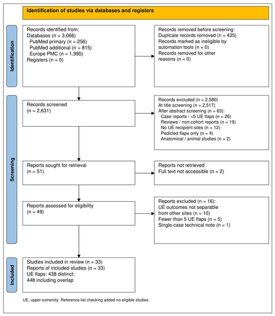
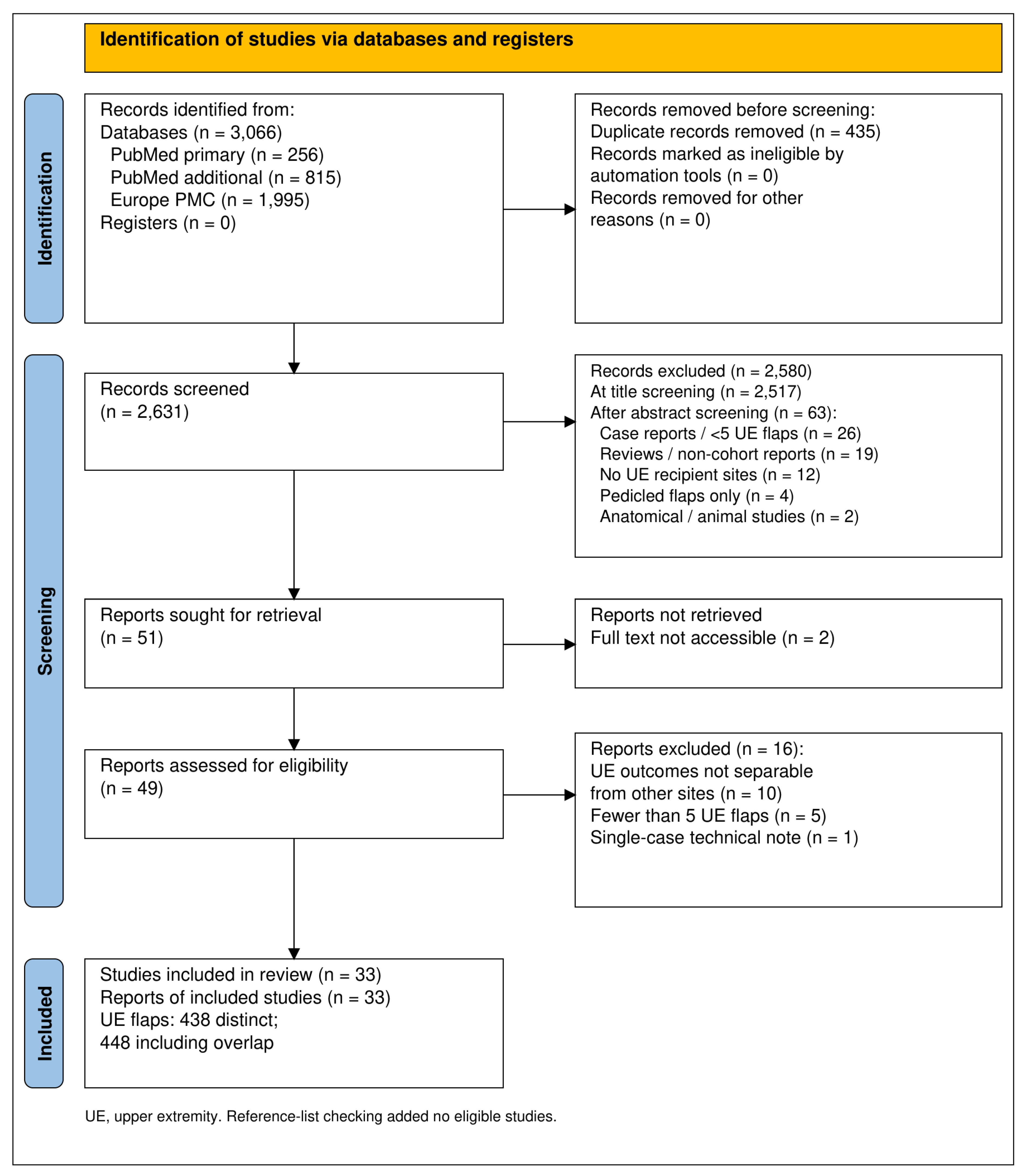
Figure 1 summarises study selection. The primary PubMed strategy retrieved 256 records, the supplementary intervention-only PubMed strategy added 815 records not retrieved by the primary strategy, and the Europe PMC search retrieved 1995 records, of which 1560 were not already present in the PubMed sets; after removal of 435 duplicates by identifier matching, 2631 unique records were screened by title. Title screening excluded 2517 records, most commonly because the flap was used at a recipient site other than the upper extremity (head and neck, breast, trunk, genitalia or lower limb), because the record concerned lymphatic or lymph-node surgery, anatomical or imaging work, or laboratory studies, or because the acronym SCIP denoted an unrelated concept. The abstracts of 114 records were examined, and 63 were excluded at this stage: 26 were case reports or series describing fewer than five upper-extremity flaps, 19 were reviews, letters, comments or technique notes without a patient cohort, 12 reported no upper-extremity recipient sites, four described pedicled flaps only, and two were anatomical or animal studies.
Figure 1.
PRISMA 2020 flow diagram of study identification, screening and inclusion. The database-only template was adapted from Page et al. [22]. UE, upper extremity. Of 114 abstracts examined after title screening, 63 were excluded. Known cohort overlap was removed from the aggregate flap count.
Full texts were sought for 51 records. Two could not be retrieved despite publisher, repository and library requests, namely a 2024 series of subdermally dissected pure skin perforator flaps in burn and trauma defects [33] and a 2017 series of chimeric iliac osteocutaneous flaps for metacarpal defects [34]; both are potentially eligible on the basis of their abstracts and are recorded in the flow diagram as full text not accessible. Forty-nine full texts were assessed. Sixteen were excluded: in ten, upper-extremity flaps were counted but no outcome could be attributed to that subgroup because all results were pooled across body regions [9,10,35,36,37,38,39,40,41,42]; in five, fewer than five upper-extremity flaps were reported [43,44,45,46,47]; and one was a single-case technical note [48]. Reference-list checking of the included full texts and of the previously published reviews identified no additional eligible study. Thirty-three studies were therefore included in the qualitative synthesis.
Two of these, from the same Taiwanese group and covering almost identical periods, were found on case-by-case comparison to share ten of twelve upper-extremity patients [24,25]; both are tabulated, but only the 22 upper-extremity flaps of the larger series and the two non-overlapping flaps of the smaller series were counted in descriptive aggregates.
3.2. Characteristics of the Included Studies
The 33 included studies were published between 2007 and 2026, and 24 of them appeared in or after 2020. They originated from ten countries: China contributed twelve studies [26,27,28,49,50,51,52,53,54,55,56,57], Japan eight [12,13,58,59,60,61,62,63], Taiwan five [8,24,25,29,64], the United States two [14,65], and Egypt [15], Vietnam [66], Turkey [67], Pakistan [30], the United Kingdom [19] and South Korea [68] one each. One study was a prospective randomised comparison of the SCIP flap with the parascapular flap [15], one was a retrospective three-arm comparison of the SCIP flap with two local perforator flaps for volar finger defects [67], one was a prospective descriptive series [30], and the remaining 30 were retrospective case series or cohorts, three of which also reported cadaveric anatomical studies [26,61,62]. Seventeen studies, or arms of studies, reported exclusively on upper-extremity recipients, whereas 16 were mixed-site series from which an upper-extremity subgroup could be identified from per-case tables, subgroup tables or explicit statements that no flap was lost. Table 1 presents study characteristics; study size ranged from five to 44 upper-extremity flaps, and most cohorts were retrospective.
Table 1.
Characteristics of included studies of free SCIP flap reconstruction of the upper extremity.
Taken together the 33 studies described 438 distinct free SCIP flaps transferred to the upper extremity, or 448 flaps when the overlapping Taiwanese cohort is counted twice. Individual contributions ranged from five flaps [12] to 44 flaps [66], and only five series reported more than 20 upper-extremity flaps [24,26,52,54,66]. The median study contributed ten flaps.
Patient age spanned childhood to old age: the youngest recipients were four years old in a paediatric bilobed-flap series [55] and in a series of multiple hand defects [54], the randomised trial enrolled children as young as nine years [15], and the oldest upper-extremity recipient was 89 years [13]. Men predominated in every study that reported sex, with male proportions from 50 percent [60] to 100 percent [65].
Trauma was the dominant indication overall, encompassing machine and crush injuries, degloving, avulsion, blast injuries from fireworks [65], hot-press and thermal injuries [51], and post-traumatic or post-burn contractures [13,15]. Oncological indications were confined to a small number of series, notably sarcoma resections of the posterior upper arm [61], tumour resections in the deep-branch series of Yoshimatsu and colleagues [62], and arteriovenous malformation resections in the original pure skin perforator series [12]. Two series addressed iatrogenic forearm defects created by radial forearm flap harvest [14] or reconstructed defects after compartment syndrome and open forearm fractures [58].
Recipient regions covered the entire limb, from fingertip pulps and volar digital defects [53,67] through the dorsal and palmar hand, first web space, wrist and forearm to the elbow and the posterior upper arm.
Study periods ranged from 15 months [66] to 16 years [19], and follow-up, where stated, ranged from six weeks to a mean of five years [19], although several series did not report follow-up at all [52,58] or reported it only for the whole mixed cohort.
3.3. Intervention and Technical Characteristics
The flap variant, source branch of the superficial circumflex iliac artery, preoperative imaging, flap dimensions, pedicle characteristics, anastomotic configuration and donor-site closure of each study are summarised in Supplementary Table S2. The included series spanned the entire technical evolution of the groin donor site. At one end were flaps harvested on an identified superficial circumflex iliac pedicle but elevated in the classic subfascial or supra-aponeurotic plane with the full thickness of subcutaneous fat, described by their authors as free iliac, ilioinguinal or superficial circumflex iliac artery flaps [26,27,28,30] or as axial “freestyle” groin flaps designed along two to four SCIA branches [24,25,29]. In the middle were conventional SCIP flaps elevated above the deep fascia or in the superficial fascial plane, with or without peripheral defatting, which constituted the majority of series [8,14,15,19,58,59,60,61,62,63,64,65,66,67,68]. At the other end were pure skin perforator and super-thin flaps in which the perforator was microdissected to the subdermal plexus or the flap was elevated in the subdermal plane [12,13,50], and thinned sensate flaps were trimmed under the microscope to four to eight millimetres [54].
Reported flap thickness reflected these planes of elevation. Pure skin perforator flaps measured approximately two millimetres in the original series [12], 1.0 to 2.5 millimetres in the subdermal-dissection series [13], 2 to 3 millimetres in the hand and wrist series of Zhu and colleagues [50] and a mean of 2.5 millimetres in the Vietnamese series, in which four flaps in overweight patients required additional microscopic defatting from 5.6 to 3.3 millimetres [66]. Suprafascial super-thin flaps averaged five millimetres [54,68], whereas the randomised Egyptian trial reported a mean of 11.75 millimetres [15], the Chinese retrograde-flow series 11 millimetres [27] and the earliest Taiwanese series 0.5 to 2 centimetres [8].
Pedicle length was generally short, with means of 2.8 centimetres for transfer to the posterior upper arm [61], 3.2 centimetres for the subfascial free iliac flap [28], 4.5 to 5.6 centimetres in most SCIP series [15,50,56,68] and up to 8 to 17 centimetres when the transverse branch of the deep branch was used as the pedicle [59]; the arterial diameter was between 0.5 and 1.3 millimetres in the majority of reports [12,15,50,58,60,68].
In the digits, the proper or common digital arteries were used with end-to-end anastomosis [28,54,60,67]; in the hand, the radial artery or its superficial palmar or dorsal carpal branches were used, frequently end-to-side or as retrograde-flow anastomoses in the anatomical snuffbox [14,26,50,54]; in the forearm the anterior interosseous artery [58] and the posterior interosseous artery [51] were used as size-matched recipients; and in the arm the deep brachial [61] and superior ulnar collateral arteries [63] were reported. Perforator-to-perforator supermicrosurgical anastomoses to metacarpal or interosseous artery perforators allowed transfer under local anaesthesia as day surgery [60], and two series performed the entire procedure under local or tumescent anaesthesia [24,29].
Venous drainage relied on the superficial circumflex iliac vein alone, the venae comitantes alone, or both, with most authors including a second vein whenever available.
Preoperative imaging ranged from none [60] or handheld Doppler alone [8,19,30,68] to colour Doppler ultrasonography [13,54,56], high-frequency ultrasound [50] and computed tomographic angiography of the groin and recipient limb [55,58,65,66].
Structural variants specifically for the hand included bilobed or multi-paddle designs for adjacent or multiple digital defects [24,52,54,55], compound dual-pedicle flaps extended with lumbar or intercostal perforators for long, narrow forearm-to-hand defects [51], chimeric flaps carrying iliac bone, fascia lata or deep fascia for metacarpal, thumb and extensor tendon reconstruction [49,53,56,57,62,63], and sensate flaps innervated by the lateral cutaneous branch of the subcostal or intercostal nerve or by the lateral femoral cutaneous nerve [12,54,56,65].
Donor sites were closed primarily in every series that reported closure.
3.4. Outcomes
Table 2 presents, for each study, the number of upper-extremity flaps and the counts and proportions of total flap loss, partial necrosis, vascular compromise or re-exploration, secondary debulking and donor-site complications, together with the functional, sensory or aesthetic measures reported and the quality appraisal score. Total loss was reported in 7/438 distinct flaps, whereas secondary debulking was attributable in only 241 flaps; the studies contributing to each denominator are listed in Supplementary Table S1.
Table 2.
Upper-extremity outcomes and methodological appraisal of the included studies.
Across the 438 distinct upper-extremity flaps, total flap loss was reported in seven (1.6 percent), and 28 of the 33 studies reported no total loss among their upper-extremity flaps. The losses comprised two of eight flaps with unsalvageable venous or arterial thrombosis in a Taiwanese series in which all four take-backs occurred in upper-limb flaps whose recipient vessels lay within the zone of trauma [64], two of 44 flaps in heavy smokers in the Vietnamese series, one of which had been anastomosed to a retrograde radial artery after a simultaneous toe transfer [66], one of 42 flaps after a vascular crisis in the ilioinguinal series [26], the first flap in the American blast-injury series, attributed to technical error [65], and one flap that was removed by the patient on the tenth postoperative day for non-vascular reasons [63]; the vascular loss rate was therefore six of 438 (1.4 percent).
Partial necrosis was reported in seven of the 363 flaps for which the outcome was attributable (1.9 percent), always as marginal or distal necrosis managed conservatively [14,15,26,30,50,54,64].
Vascular compromise requiring re-exploration or bedside intervention was reported in 14 of 303 flaps (4.6 percent), with study-level rates from zero in most series to 50 percent in the small post-traumatic series [64] and 27 percent in the volar-finger comparison, in which two episodes of venous congestion and one haematoma were all salvaged [67]; the flap was lost after three of the 14 events, all in two series [26,64], and survived after the remaining 11.
Secondary thinning or debulking was reported in 42 of 241 flaps with attributable data (17.4 percent), but ranged from zero in the pure skin perforator, super-thin and paediatric series [12,50,53,55,66,68] to 18.8 percent for osteocutaneous flaps [49], 25 to 26 percent for non-thinned ilioinguinal or SCIA flaps [26,30], 33 to 44 percent in Japanese and American conventional SCIP series [63,65], 75 percent (6/8) for the subfascial free iliac flap, in which staged debulking was an intended part of the syndactylised design [28], and five of eight flaps (63 percent; five of the six that survived) in the series of Tung and colleagues [64]. Because only one series stated whether debulking was planned [28], planned and unplanned procedures could not be separated. Table 3 presents total loss and debulking by flap type: no total loss was recorded among pure skin perforator, chimeric or multi-paddle flaps, and debulking was most frequent after conventional (12/26) and non-thinned groin-type (21/92) harvest and absent in the pure skin perforator and super-thin series that reported it (0/68); these are descriptive counts from non-comparative series and do not constitute a comparison between techniques.
Table 3.
Total flap loss and secondary debulking by flap type.
Donor-site complications occurred in 17 of 283 flaps (6.0 percent) and were minor, comprising wound dehiscence or delayed healing [26,50,66,67], infection or altered sensation [15] and scar widening or delayed healing [28,57]; transient numbness in the distribution of the lateral femoral cutaneous nerve or below the scar was reported in mixed cohorts that used the deep branch or included a cutaneous nerve [24,52,56,59].
Table 4 lists every functional, sensory and aesthetic measure by instrument. Seventeen studies reported at least one such measure, but validated patient-reported hand-function instruments were used in only four: the QuickDASH averaged 16.5 in patients without amputation after complex hand reconstruction, and was worse for multilayer than for skin-only defects (27.3 versus 12.7) [66], and a median of 27 after chimeric reconstruction of metacarpal hands [57]; the Michigan Hand Outcomes Questionnaire scored 80.5 for SCIP flaps in volar finger defects, between the dorsoulnar artery perforator flap (74.4) and the superficial palmar branch flap (85.8) [67], and was rated excellent in 38 of 41 patients after thinned sensate SCIP reconstruction [54].
Table 4.
Functional, sensory and aesthetic outcomes reported in the included studies, by instrument.
Sensory recovery ranged from static two-point discrimination of 5 millimetres in innervated flaps coapted to digital nerves [54] and 9.3 millimetres in children [55] to 15.4 millimetres or pressure sensation only in non-innervated subfascial flaps [28], with patchy, modality-dependent recovery in flaps innervated by the subcostal nerve [56].
Aesthetic outcomes were reported with a five-point Likert scale (mean overall score 4.5 of 5) [50], the modified Vancouver Scar Scale (4.8 for SCIP versus 3.7 for the superficial palmar branch flap and 9.4 for the dorsoulnar perforator flap [67]; 1 of 18 for recipient and donor sites in thinned flaps [54]) or patient-rated categories (excellent in 97.7 percent) [66]; most other series reported satisfaction narratively.
3.5. Quality Appraisal
Scores on the ten-item JBI case-series checklist ranged from one to nine, with a median of seven, and 20 of the 32 non-randomised studies satisfied at least seven items. The items most frequently unmet or unclear were consecutive and complete inclusion of participants, which was stated explicitly in only six studies [13,19,57,61,62,65], and clear reporting of outcomes and follow-up, which was compromised in studies that did not report follow-up duration [52,58] or that contained internal inconsistencies in patient numbers or complication counts [54,65]. The earliest series provided only ranges and no per-site outcomes and satisfied a single item [8], while the retrospective single-surgeon series without a case table satisfied four [19]. The randomised trial used computer-generated allocation but was unblinded, small, and assessed aesthetic outcome with a non-validated scale, and was judged to raise some concerns on the RoB 2 tool [15]. Statistical analysis was not applicable in most descriptive series and was appropriate in the comparative studies. No study was excluded on the basis of its appraisal.
4. Discussion
4.1. Principal Findings and Comparison with Previous Reviews
This systematic review identified 33 primary studies describing 438 distinct free SCIP flaps transferred to the upper extremity, and more than half of them were published since 2020, providing a synthesis confined to this recipient site. Three findings stand out. Total flap loss was uncommon and was recorded in only five of the 33 series. Reported secondary debulking varied widely between flap types, from none in the pure skin perforator and super-thin series to almost half of conventional flaps (Table 3), although these groups were not compared directly and differ in more than the plane of elevation (Section 4.3). Function was measured with a validated instrument in only four studies (Table 4), so the evidence describes flap survival far better than it describes what patients regain. The reported flap-loss rate in the upper extremity is of the same order as the 2.7 percent failure rate reported in the all-region meta-analysis of Altiparmak and colleagues [18] and considerably lower than the 18.7 percent loss reported in an early European extremity series that could not be included because its upper-limb outcomes were not separable [36]. Our aggregate is, however, descriptive, derived mainly from small single-centre series that may preferentially report favourable experience, and carries no confidence interval.
4.2. Interpreting Survival and Complication Estimates
The distinction between technical survival and successful hand reconstruction is central to interpreting these findings. A surviving flap may still require contour revision, tendon reconstruction, treatment of stiffness or prolonged rehabilitation. Conversely, a planned debulking procedure after staged reconstruction should not be classified automatically as treatment failure. Presenting survival, unplanned reoperation, planned contour revision and patient-reported function as separate outcomes would make the clinical meaning of success more consistent across reports.
Because the outcome-specific denominators differ (Supplementary Table S1), the aggregate proportions answer different questions, and a missing complication entry must remain distinct from a documented absence of complications. Event totals can describe the published experience, but they should not be interpreted as the probability of an outcome for an individual patient. Variations in follow-up are particularly relevant to later revision and sensory recovery, which may be missed in shorter series.
Excluding the duplicated flaps of the two overlapping Taiwanese reports [24,25] prevents known double counting but does not remove uncertainty about outcome-specific attribution; future publications should identify previous reports of the same cohort and report newly added cases separately.
4.3. Flap Design and Sources of Clinical Variation
“SCIP flap” is an umbrella term covering flaps of very different thickness and construction, and Table 3 shows how this heterogeneity maps onto outcomes. In the series that harvested the flap on an identified SCIA pedicle but in the classic subfascial or supra-aponeurotic plane, the flap behaved as a reliable groin flap: survival was uniformly high, but 25 to 75 percent of flaps underwent debulking [26,28,30], and sensory recovery in non-innervated flaps was poor [28]. Thin suprafascial SCIP flaps in Asian patients were about five millimetres thick [54,68], and no revision was reported in the one series that recorded this outcome [68], whereas comparable flaps in Egyptian and European populations measured nine to twelve millimetres [15,36] and conventional flaps in an obese American population were debulked in 44 percent of surviving cases [65]. Pure skin perforator and superficial-plane series reporting this outcome described no secondary thinning [12,50,66], while the subdermal-dissection series did not report it [13]. These techniques require microdissection, which took up to 140 min in one series [50], although subdermal dissection reduced elevation time to about 30 min in experienced hands [13]. The only randomised evidence, comparing SCIP and parascapular flaps, found similar survival and aesthetic outcomes but a shorter operative time and supine two-team positioning for the SCIP flap, offset by a pedicle diameter of 0.77 millimetres and a pedicle length of 5.6 centimetres [15]; the only comparison with local perforator flaps found that the SCIP flap was fastest to elevate but was outperformed cosmetically and functionally by the superficial palmar branch flap for volar finger defects with hair growth and colour mismatch reported as disadvantages [67]. Complication clusters were also informative: all four re-explorations and both losses in one series occurred in upper-limb flaps whose recipient vessels lay in the zone of injury [64], both losses in another series occurred in heavy smokers [66], and venous congestion of thin SCIP flaps was salvaged by venous revision in every series that described it [12,19,59,67], observations that echo the risk factors identified in larger mixed-site cohorts [37].
These between-type differences should be read with care. The groups differ in era, country, defect complexity, patient habitus and length of follow-up; debulking data were available for 241 flaps only, the smallest groups contain fewer than 25 flaps, and whether revision was planned was stated in a single series. These descriptive differences may inform counselling about the possibility of a second procedure after a non-thinned harvest, but they do not show that any flap design is safer or more effective than another; only prospective comparative studies can establish that.
4.4. Functional Recovery and Patient Priorities
Functional recovery depends on more than the tissue used for coverage. Tendon and nerve injury, bone loss, amputation, joint involvement and rehabilitation can influence the eventual outcome independently of flap survival. The poorer QuickDASH results reported for multilayer defects in the Vietnamese cohort illustrate the need to account for injury complexity when comparing reconstructions [66]. A flap-level survival analysis and a patient-level function analysis therefore address complementary outcomes and should retain their respective units of analysis.
The comparative evidence supports cautious, defect-specific interpretation. The small randomised SCIP–parascapular study informs technical trade-offs but cannot establish equivalence across uncommon complications or long-term function [15]. The volar-finger comparison also suggests that tissue match, hair growth and scar characteristics matter alongside the convenience of harvesting the flap [67]. Results from those defects cannot be extended automatically to extensive dorsal hand or composite forearm injuries.
Sensory restoration is particularly important where the reconstructed surface participates in pinch, grip or protective contact. The available series use different tests and follow-up intervals, so differences in two-point discrimination cannot isolate the effect of nerve coaptation from patient age, nerve injury or rehabilitation [28,54,55,56,57]. Future comparisons should specify the recipient nerve, coaptation method and sensory test protocol and report both protective sensation and discrimination at predefined follow-up visits.
4.5. Implications for Reconstructive Practice
The clinical implications of these findings are necessarily tentative, because they rest on descriptive, largely retrospective series without direct comparison between flap designs. First, the free SCIP flap may be considered one option for upper-extremity defects of small to moderate size when thin, pliable coverage is required and the recipient vessels are within a few centimetres of the defect, provided the surgical team is comfortable with anastomoses of one millimetre or less. Second, the plane of elevation should be chosen deliberately: pure skin perforator or superficial fascial-plane elevation may be appropriate for the dorsal hand and digits, where any bulk impairs tendon excursion, and the patient should be counselled that a non-thinned harvest may require secondary debulking, although this preference reflects reported revision counts rather than comparative evidence. Third, the chimeric capacity of the deep branch permits single-stage reconstruction of composite metacarpal, thumb and extensor tendon defects with iliac bone or fascia lata, with bone union reported in every series that included bone, within two to four months in three [49,56,57] and by nine to ten months in another [62], a capability reported by a small number of centres that has not yet been compared with alternative strategies. Fourth, several innovations broaden the indications: extension of the pedicle along the transverse branch to 8 to 17 centimetres [59], compound designs with lumbar or intercostal perforators for defects up to 35 centimetres long [51], multi-paddle designs for multiple adjacent digital defects [52,55], the use of the anterior interosseous, posterior interosseous, deep brachial and retrograde radial arteries as size-matched recipients [27,51,58,61], and performance of the transfer under local or tumescent anaesthesia as day surgery in selected patients [29,60]. Fifth, the donor site is a consistent strength: primary closure was achieved in every series that reported this outcome, and recorded donor complications were generally minor, although transient thigh or groin dysaesthesia should be discussed when the deep branch or a cutaneous nerve is harvested.
4.6. Limitations of the Evidence and Review Methods
The heterogeneity of the evidence base, however, sets clear limits on these conclusions. Studies differed in design, in the definition of the flap, in the plane of elevation, in whether upper-extremity outcomes were reported separately, in the definition and threshold of partial necrosis, in whether debulking was a planned second stage or an unplanned revision, in follow-up duration, and above all in the measurement of function, which was assessed with a validated instrument in only four series [54,57,66,67]. Sensory outcomes were reported with different instruments, follow-up intervals and thresholds, and the benefit of flap innervation cannot be judged from the available comparisons. Findings also conflicted on secondary revision, which was absent in Asian pure skin perforator series but frequent in Western conventional series, and it remains unclear how much of this difference reflects body habitus, technique or reporting. Ten otherwise relevant series, including the largest Western [35,36] and pure skin perforator cohorts [37], could not contribute because their upper-limb outcomes were pooled with other sites, and two potentially eligible reports could not be retrieved [33,34].
This review has limitations that arise from both the evidence and the methods. The included evidence consists almost entirely of small, single-centre, retrospective case series, many from a handful of high-volume East Asian units whose results may not generalise to other populations or to surgeons early in the learning curve; consecutive inclusion was rarely documented, follow-up was often short or unreported, several studies contained internal inconsistencies in patient numbers or complication counts, two overlapping cohorts were identified only by manual case-matching, and outcome definitions and instruments varied widely. The aggregates were derived from Table 2 using a single explicit attribution rule (Section 2.5), but they remain sensitive to how whole-cohort values and the overlapping cohort are handled. Restriction to studies with separable upper-limb data excluded several large mixed-site cohorts, and the requirement for at least five upper-extremity flaps excluded case reports that may describe important complications; two potentially eligible articles could not be retrieved, and non-English publications, which are numerous for this flap in the Chinese literature, were excluded, so that language and availability bias are possible. Only PubMed and Europe PMC were searched (Section 2.3); although the broad intervention-only search and reference-list checking identified no additional eligible study, series indexed only in Embase, Scopus or Web of Science may have been missed. Two authors undertook study selection and extraction, but decisions were reached by consensus rather than independently in duplicate, so inter-reviewer agreement could not be calculated. Quality appraisal was performed by a single reviewer, which may have introduced assessment error. Publication bias toward favourable single-surgeon experience remains possible and was not formally assessed. No formal sensitivity analyses or outcome-level certainty assessment were performed, which further limits the strength of the conclusions.
4.7. Priorities for Future Research
A practical next step would be a prospective multicentre registry using a common dataset for upper-extremity SCIP reconstruction. Core variables should include the recipient subregion, injury mechanism, exposed or missing structures, smoking status, body mass index, measured flap thickness, elevation plane, pedicle length, recipient-vessel and anastomotic configuration, and whether revision was planned. Consecutive enrolment and reporting of unsuccessful reconstructions would help reduce the selective publication of favourable experience.
Comparative studies, ideally prospective and multicentre, should enrol clinically similar defects stratified by body mass index and defect location, compare the SCIP flap with the anterolateral thigh, lateral arm, radial forearm and local perforator flaps, and specify the rationale for flap selection. Outcomes should include total and partial loss, unplanned re-exploration, time to definitive coverage, donor morbidity, planned and unplanned secondary procedures, validated hand-function scores at standardised intervals and return to usual activities. Reporting resource use, rehabilitation requirements and repeat operations would also help determine whether technical advantages translate into benefits meaningful to patients and reconstructive services.
5. Conclusions
In the published experience summarised here, free SCIP flaps provided thin, adaptable coverage for hand and upper-extremity defects, with infrequent reported total flap loss and generally minor recorded donor-site morbidity. Pure skin perforator, chimeric, osteocutaneous, multi-paddle, compound and sensate variants extend the reconstructive options. Reported secondary thinning varied with elevation plane, patient habitus and reconstructive intent, but these associations are descriptive and remain uncertain. Inconsistent functional and sensory reporting prevents firm comparison with alternative flaps. The proportions reported here are descriptive counts from small, heterogeneous and mostly retrospective series with outcome-specific denominators; they describe the published experience and should not be read as pooled complication rates, as estimates of individual patient risk or as evidence that the SCIP flap is superior to alternative flaps. Prospective comparative studies with standardised, site-specific outcomes are needed before its role can be defined.
Supplementary Materials
The following supporting information can be downloaded at https://www.mdpi.com/article/10.3390/surgeries7040133/s1. Completed PRISMA 2020 checklist and PRISMA 2020 flow diagram. Table S1: studies contributing to each descriptive outcome aggregate. Table S2: technical characteristics of the flaps in the included studies.
Author Contributions
Conceptualisation, D.I.O. and M.P.; methodology, D.I.O. and M.P.; software, D.I.O. and M.P.; validation, D.I.O. and M.P.; formal analysis, M.-A.B.; investigation, M.-A.B.; resources, M.-A.B.; data curation, M.-A.B.; writing—original draft preparation, D.I.O. and M.P.; writing—review and editing, V.A.B. and Z.P.C.; visualisation, V.A.B. and Z.P.C.; supervision, V.A.B. and Z.P.C.; project administration, V.A.B. and Z.P.C.; funding acquisition, V.A.B. and Z.P.C. All authors have read and agreed to the published version of the manuscript.
Funding
We would like to acknowledge the “Victor Babeș” University of Medicine and Pharmacy Timișoara (UMFVBT) for their support in covering the costs of publication for this research paper.
Institutional Review Board Statement
Not applicable. This study is a systematic review of previously published studies.
Informed Consent Statement
Not applicable.
Data Availability Statement
The extracted study-level information used for the narrative synthesis is presented in Table 1, Table 2, Table 3 and Table 4 and Supplementary Tables S1 and S2. The original reports are identified in the reference list. OSF registration and protocol URL: https://osf.io/y7nb8, (accessed on 22 September 2026).
Acknowledgments
Artificial intelligence was used only for manuscript formatting and English-language editing. Two authors performed the literature searches, report retrieval, study selection and data extraction. The authors reviewed and approved the edited text.
Conflicts of Interest
The authors declare no conflicts of interest.
References
- Rehim, S.A.; Chung, K.C. Local flaps of the hand. Hand Clin. 2014, 30, 137–151. [Google Scholar] [CrossRef] [Scilit] [PubMed]
- McGregor, I.A.; Jackson, I.T. The groin flap. Br. J. Plast. Surg. 1972, 25, 3–16. [Google Scholar] [CrossRef] [Scilit] [PubMed]
- Alzahrani, H.A.; Asiri, M.N.; Humedi, A.M.; Alammar, J.; Alrashed, H.; Hakami, F. Free versus pedicled flaps for hand reconstruction: A systematic review and meta-analysis. JPRAS Open 2025, 45, 278–288. [Google Scholar] [CrossRef] [Scilit] [PubMed]
- Daniel, R.K.; Taylor, G.I. Distant transfer of an island flap by microvascular anastomoses. A clinical technique. Plast. Reconstr. Surg. 1973, 52, 111–117. [Google Scholar] [CrossRef] [Scilit] [PubMed]
- Taylor, G.I.; Daniel, R.K. The anatomy of several free flap donor sites. Plast. Reconstr. Surg. 1975, 56, 243–253. [Google Scholar] [CrossRef] [Scilit] [PubMed]
- Harii, K.; Ohmori, K. Free groin flaps in children. Plast. Reconstr. Surg. 1975, 55, 588–592. [Google Scholar] [CrossRef] [Scilit] [PubMed]
- Koshima, I.; Nanba, Y.; Tsutsui, T.; Takahashi, Y.; Urushibara, K.; Inagawa, K.; Hamasaki, T.; Moriguchi, T. Superficial circumflex iliac artery perforator flap for reconstruction of limb defects. Plast. Reconstr. Surg. 2004, 113, 233–240. [Google Scholar] [CrossRef] [Scilit] [PubMed]
- Hsu, W.-M.; Chao, W.-N.; Yang, C.; Fang, C.-L.; Huang, K.-F.; Lin, Y.-S.; Lee, T.-H. Evolution of the free groin flap: The superficial circumflex iliac artery perforator flap. Plast. Reconstr. Surg. 2007, 119, 1491–1498. [Google Scholar] [CrossRef] [Scilit] [PubMed]
- Hong, J.P.; Choi, D.H.; Suh, H.; Mukarramah, D.A.; Tashti, T.; Lee, K.; Yoon, C. A new plane of elevation: The superficial fascial plane for perforator flap elevation. J. Reconstr. Microsurg. 2014, 30, 491–496. [Google Scholar] [CrossRef] [Scilit] [PubMed]
- Goh, T.L.H.; Park, S.W.; Cho, J.Y.; Choi, J.W.; Hong, J.P. The search for the ideal thin skin flap: Superficial circumflex iliac artery perforator flap--a review of 210 cases. Plast. Reconstr. Surg. 2015, 135, 592–601. [Google Scholar] [CrossRef] [Scilit] [PubMed]
- Iida, T. Superficial circumflex iliac perforator (SCIP) flap: Variations of the SCIP flap and their clinical applications. J. Reconstr. Microsurg. 2014, 30, 505–508. [Google Scholar] [CrossRef] [Scilit] [PubMed]
- Narushima, M.; Iida, T.; Kaji, N.; Yamamoto, T.; Yoshimatsu, H.; Hara, H.; Kikuchi, K.; Araki, J.; Yamashita, S.; Koshima, I. Superficial circumflex iliac artery pure skin perforator-based superthin flap for hand and finger reconstruction. J. Plast. Reconstr. Aesthet. Surg. 2016, 69, 827–834. [Google Scholar] [CrossRef] [Scilit] [PubMed]
- Yamamoto, T.; Yamamoto, N.; Fuse, Y.; Kageyama, T.; Sakai, H.; Tsukuura, R. Subdermal dissection for elevation of pure skin perforator flaps and superthin flaps: The dermis as a landmark for the most superficial dissection plane. Plast. Reconstr. Surg. 2021, 147, 470–478. [Google Scholar] [CrossRef] [Scilit] [PubMed]
- Moltaji, S.; Llewellyn, K.J.; Fuse, Y.; Felder, J.M., 3rd; Chaiyasate, K. Pilot study: Superficial circumflex iliac perforator flap for coverage of radial forearm free flap donor sites. Plast. Reconstr. Surg. Glob. Open 2026, 14, e7717. [Google Scholar] [CrossRef] [Scilit] [PubMed]
- Ayad, W.M.; Ouf, M.O.; Zayid, T.; Rashed, A.I. Free parascapular flap versus free superficial circumflex iliac artery perforator flaps in upper limb reconstruction. Plast. Reconstr. Surg. Glob. Open 2025, 13, e7202. [Google Scholar] [CrossRef] [Scilit] [PubMed]
- Escandón, J.M.; Ciudad, P.; Mayer, H.F.; Pencek, M.; Mantilla-Rivas, E.; Mohammad, A.; Langstein, H.N.; Manrique, O.J. Free flap transfer with supermicrosurgical technique for soft tissue reconstruction: A systematic review and meta-analysis. Microsurgery 2023, 43, 171–184. [Google Scholar] [CrossRef] [Scilit] [PubMed]
- Messa, C.A., 4th; Carney, M.J., 3rd; Tantillo, K.; Othman, S.; Moores, C.; Mirzabeigi, M.N.; Weissler, J.M.; Cook, T.; Kovach, S.J. Characteristics of the superficial circumflex iliac artery perforator flap in a Western population and a practice approach for free flap reconstruction. J. Reconstr. Microsurg. 2021, 37, 486–491. [Google Scholar] [CrossRef] [Scilit] [PubMed]
- Altiparmak, M.; Cha, H.G.; Hong, J.P.; Suh, H.P. Superficial circumflex iliac artery perforator flap as a workhorse flap: Systematic review and meta-analysis. J. Reconstr. Microsurg. 2020, 36, 600–605. [Google Scholar] [CrossRef] [Scilit] [PubMed]
- Berner, J.E.; Nikkhah, D.; Zhao, J.; Prousskaia, E.; Teo, T.C. The versatility of the superficial circumflex iliac artery perforator flap: A single surgeon’s 16-year experience for limb reconstruction and a systematic review. J. Reconstr. Microsurg. 2020, 36, 93–103. [Google Scholar] [CrossRef] [Scilit] [PubMed]
- Garbaccio, N.; Schonebaum, D.I.; Smith, J.E.; Cordero, J.J.; Foster, L.; Mehdizadeh, M.; Dowlatshahi, A.S.; Lin, S.J. Safety and utility of superficial circumflex iliac perforator versus superficial circumflex iliac artery flaps in pediatric reconstructive surgery. J. Reconstr. Microsurg. 2026, 42, 700–710. [Google Scholar] [CrossRef] [Scilit] [PubMed]
- Barnhill, C.W.; Greyson, M.A.; Iorio, M.L. Superficial circumflex iliac artery perforator flap reconstruction of the upper extremity. Hand Clin. 2024, 40, 179–187. [Google Scholar] [CrossRef] [Scilit] [PubMed]
- Page, M.J.; McKenzie, J.E.; Bossuyt, P.M.; Boutron, I.; Hoffmann, T.C.; Mulrow, C.D.; Shamseer, L.; Tetzlaff, J.M.; Akl, E.A.; Brennan, S.E.; et al. The PRISMA 2020 statement: An updated guideline for reporting systematic reviews. BMJ 2021, 372, n71. [Google Scholar] [CrossRef] [Scilit] [PubMed]
- Campbell, M.; McKenzie, J.E.; Sowden, A.; Katikireddi, S.V.; Brennan, S.E.; Ellis, S.; Hartmann-Boyce, J.; Ryan, R.; Shepperd, S.; Thomas, J.; et al. Synthesis without meta-analysis (SWiM) in systematic reviews: Reporting guideline. BMJ 2020, 368, l6890. [Google Scholar] [CrossRef] [Scilit] [PubMed]
- Chao, W.N.; Tsai, C.F.; Wang, P.H.; Chan, K.S.; Lee, Y.T.; Lin, D.B.; Chen, C.C.; Chen, S.C. Freestyle groin flaps: The real axial flap design and clinical application. Ann. Plast. Surg. 2015, 74, S75–S79. [Google Scholar] [CrossRef] [Scilit] [PubMed]
- Chao, W.N.; Wang, P.H.; Chen, B.R.; Chen, S.C. Chimeric groin free flaps: Design and clinical application. Microsurgery 2016, 36, 206–215. [Google Scholar] [CrossRef] [Scilit] [PubMed]
- Zhang, H.; Luo, Q.J.; Ding, M.C.; Qi, J.W. Anatomical foundations and clinical utility of personalized free ilioinguinal zone flap for hand skin and soft tissue defect reconstruction. Ther. Clin. Risk Manag. 2025, 21, 1645–1656. [Google Scholar] [CrossRef] [Scilit] [PubMed]
- Han, Z.; Zhang, K.; Liu, H.; Liu, Y.; Zhang, C. Clinical application of free inguinal flaps with retrograde blood supply anastomotic to repair soft tissue defects of extremities. Medicine 2022, 101, e31661. [Google Scholar] [CrossRef] [Scilit] [PubMed]
- Pan, Z.H.; Jiang, P.P.; Xue, S. Free iliac flap for treating multiple skin defects of the hand and digits. J. Hand Surg. Eur. Vol. 2013, 38, 952–958. [Google Scholar] [CrossRef] [Scilit] [PubMed]
- Chao, W.N.; Kuok, K.M.; Chen, C.J. Local anesthesia in superficial circumflex iliac artery free flap transfer. Ann. Plast. Surg. 2026, 96, S48–S52. [Google Scholar] [CrossRef] [Scilit] [PubMed]
- Hussain, T.; Khan, F.H.; Rahman, O.U.; Beg, M.S.A. Superficial circumflex iliac artery free flap for coverage of hand injuries. Cureus 2022, 14, e31520. [Google Scholar] [CrossRef] [Scilit] [PubMed]
- Munn, Z.; Barker, T.H.; Moola, S.; Tufanaru, C.; Stern, C.; McArthur, A.; Stephenson, M.; Aromataris, E. Methodological quality of case series studies: An introduction to the JBI critical appraisal tool. JBI Evid. Synth. 2020, 18, 2127–2133. [Google Scholar] [CrossRef] [Scilit] [PubMed]
- Sterne, J.A.C.; Savović, J.; Page, M.J.; Elbers, R.G.; Blencowe, N.S.; Boutron, I.; Cates, C.J.; Cheng, H.-Y.; Corbett, M.S.; Eldridge, S.M.; et al. RoB 2: A revised tool for assessing risk of bias in randomised trials. BMJ 2019, 366, l4898. [Google Scholar] [CrossRef] [Scilit] [PubMed]
- Lin Wu, Z.Q.; Bulla, A.; Aguilera Sáez, J.; Serracanta Domènech, J.; Barret, J.P.; Rivas Nicolls, D.A. Subdermal dissection technique for pure skin SCIA and ALT perforator flaps in burns and trauma defects: Clinical experience. Microsurgery 2024, 44, e31189. [Google Scholar] [CrossRef] [Scilit] [PubMed]
- Pan, Z.H.; Jiang, P.P.; Zhao, Y.X.; Wang, J.L. Treatment of complex metacarpal defects with free chimeric iliac osteocutaneous flaps. J. Plast. Surg. Hand Surg. 2017, 51, 143–148. [Google Scholar] [CrossRef] [Scilit] [PubMed]
- Pereira, N.; Venegas, J.; Oñate, V.; Camacho, J.P.; Roa, R. Extremity reconstruction with superficial circumflex iliac artery perforator free flap: Refinements and innovations after 101 cases. J. Plast. Reconstr. Aesthet. Surg. 2023, 85, 1–9. [Google Scholar] [CrossRef] [Scilit] [PubMed]
- Kueckelhaus, M.; Gebur, N.; Kampshoff, D.; Hiort, M.; Varnava, C.; Harati, K.; Hirsch, T.; Wiebringhaus, P. Initial experience with the superficial circumflex iliac artery perforator (SCIP) flap for extremity reconstruction in Caucasians. J. Plast. Reconstr. Aesthet. Surg. 2022, 75, 118–124. [Google Scholar] [CrossRef] [Scilit] [PubMed]
- Bae, J.; Lee, J.K.; Lee, K.T. Advancing pure skin perforator flap application: Microscope-free harvesting, versatile donor sites, and clinical outcomes. Plast. Reconstr. Surg. 2025, 156, 966–978. [Google Scholar] [CrossRef] [Scilit] [PubMed]
- Kim, H.B.; Min, J.C.; Pak, C.J.; Hong, J.P.J.; Suh, H.P. Maximizing the versatility of thin flap from the groin area as a workhorse flap: The selective use of superficial circumflex iliac artery perforator (SCIP) free flap and superficial inferior epigastric artery (SIEA) free flap with precise preoperative planning. J. Reconstr. Microsurg. 2023, 39, 148–155. [Google Scholar] [CrossRef] [Scilit] [PubMed]
- Kwon, J.G.; Pereira, N.; Tonaree, W.; Brown, E.; Hong, J.P.; Suh, H.P. Long pedicled superficial circumflex iliac artery flap based on a medial superficial branch. Plast. Reconstr. Surg. 2021, 148, 615e–619e. [Google Scholar] [CrossRef] [Scilit] [PubMed]
- Luo, H.; Bian, H.; Liu, Z.; Sun, C.; Li, H.; Ma, L.; Wang, X.; Huang, Z.; Mu, X.; Chen, S.; et al. Evaluation of patient benefits from the superficial circumflex iliac artery perforator flap in elderly patients. Bioengineering 2025, 12, 394. [Google Scholar] [CrossRef] [Scilit] [PubMed]
- Fuse, Y.; Yoshimatsu, H.; Karakawa, R.; Yano, T. Deep fat saving elevation of the superficial circumflex iliac artery perforator flap. Medicina 2022, 58, 670. [Google Scholar] [CrossRef] [Scilit] [PubMed]
- Pereira, N.; Parada, L.; Kufeke, M.; Troncoso, E.; Roa, R. A new planning method to easily harvest the superficial circumflex iliac artery perforator flap. J. Reconstr. Microsurg. 2020, 36, 165–170. [Google Scholar] [CrossRef] [Scilit] [PubMed]
- Abdelfattah, U.; Abdelwahab, N.; Emara, Y.O.; Elbanoby, T. The efficacy of chimeric superficial circumflex iliac artery perforator (SCIP) flap in treatment of posttraumatic chronic osteomyelitis. J. Reconstr. Microsurg. 2026, 42, 690–699. [Google Scholar] [CrossRef] [Scilit] [PubMed]
- Zubler, C.; Lese, I.; Pastor, T.; Attinger, M.; Constantinescu, M.A.; Olariu, R. The osteocutaneous SCIP flap: A detailed description of the surgical technique and retrospective cohort study of consecutive cases in a tertiary European centre. J. Plast. Reconstr. Aesthet. Surg. 2023, 77, 21–30. [Google Scholar] [CrossRef] [Scilit] [PubMed]
- Yoshimatsu, H.; Iida, T.; Yamamoto, T.; Hayashi, A. Superficial circumflex iliac artery-based iliac bone flap transfer for reconstruction of bony defects. J. Reconstr. Microsurg. 2018, 34, 719–728. [Google Scholar] [CrossRef] [Scilit] [PubMed]
- Yoshimatsu, H.; Hayashi, A.; Karakawa, R.; Yano, T. Combining the superficial circumflex iliac artery perforator flap with the superficial inferior epigastric artery flap or the deep inferior epigastric artery perforator flap for coverage of large soft tissue defects in the extremities and the trunk. Microsurgery 2020, 40, 649–655. [Google Scholar] [CrossRef] [Scilit] [PubMed]
- Franchi, A.; Perozzo, F.A.G.; Tiengo, C.; Walber, J.; Parisato, A.; Jandali, A.R.; Jung, F. SCIP/SIEA and PAP: The new workhorse flaps in soft tissue reconstruction for all body regions. J. Clin. Med. 2025, 14, 921. [Google Scholar] [CrossRef] [Scilit] [PubMed]
- Karakawa, R.; Yoshimatsu, H. The excised super-thin skin as a flap sizer for finger and hand free-mini-flap reconstruction. Plast. Reconstr. Surg. Glob. Open 2018, 6, e1767. [Google Scholar] [CrossRef] [Scilit] [PubMed]
- Pan, J.; Zhang, C.; Yu, G.; Wu, L.; Yin, S.; Wang, X. Reconstruction of combined bone and soft tissue defects of the hand and foot with free superficial circumflex iliac artery perforator osteocutaneous flaps: A retrospective analysis. Front. Surg. 2026, 13, 1748242. [Google Scholar] [CrossRef] [Scilit] [PubMed]
- Zhu, X.; Li, S.; Lv, Y.; Yang, J.; Zhou, F.; Chu, T.; Zhou, K.; Ding, J.; Chen, Y. The subdermal approach for harvesting superficial circumflex iliac artery-based pure skin perforator flaps in hand and wrist reconstruction. J. Hand Surg. Am. 2025, 50, 1056–1065. [Google Scholar] [CrossRef] [Scilit] [PubMed]
- Wang, H.; Shi, Z.; Zeng, D.; Wang, H.; Lv, P.; Li, P. Repair of a “long and narrow” skin defect of the upper extremity with a modified design of a compound SCIP flap: A series of 12 cases. Eur. J. Med. Res. 2024, 29, 275. [Google Scholar] [CrossRef] [Scilit] [PubMed]
- Chu, T.; Zhang, W.; Chen, T.; Shen, G.; Wang, L.; Huang, J.; Zheng, Z. Resurfacing of multiple adjacent defects with free multipaddle SCIAP flaps. J. Plast. Reconstr. Aesthet. Surg. 2024, 94, 119–127. [Google Scholar] [CrossRef] [Scilit] [PubMed]
- Chu, T.; Xiao, J.; Zhou, X.; Lin, K.; Tao, Z. Free chimeric superficial circumflex iliac artery perforator flap in reconstructing the distal complex extensor tendon injury. Plast. Reconstr. Surg. 2024, 153, 442e–447e. [Google Scholar] [CrossRef] [Scilit] [PubMed]
- Ou, C.L.; Li, J.; Zhou, X.; Luo, X.C.; Zou, Y.G. Repair of multiple hand defects with superficial circumflex iliac artery perforator flap. Injury 2023, 54, 940–946, Correction in Injury 2023, 54, 110998. https://doi.org/10.1016/j.injury.2023.110998. PMID: 37657412. [Google Scholar] [CrossRef] [Scilit] [PubMed]
- Li, Z.; Zheng, D.; Zheng, J.; Qi, W.; Qi, Q.; Liu, Y. Free superficial circumflex iliac artery perforator flap with a single-pedicle bilobed design for pediatric multi-digit defect reconstruction. J. Orthop. Surg. Res. 2020, 15, 216. [Google Scholar] [CrossRef] [Scilit] [PubMed]
- Pan, Z.; Jiang, P.; Xue, S.; Zhao, Y.; Li, H.; Gao, P.; Wang, J. Use of free sensate SCIA flap for reconstruction of distal limb defects of moderate size. J. Plast. Reconstr. Aesthet. Surg. 2020, 73, 434–442. [Google Scholar] [CrossRef] [Scilit] [PubMed]
- Pan, Z.H.; Jiang, P.P.; Xue, S.; Li, H.; Wang, J.L. Restoration of basic hand function following devastating hand injuries using a microsurgically fabricated chimeric iliac osteocutaneous flap. J. Plast. Reconstr. Aesthet. Surg. 2017, 70, 723–728. [Google Scholar] [CrossRef] [Scilit] [PubMed]
- Kodaira, S.; Fukumoto, K.; Koike, T.; Okada, Y. Anterior interosseous artery as a recipient vessel in superficial circumflex iliac artery perforator flap transplantation to the forearm. Microsurgery 2026, 46, e70221. [Google Scholar] [CrossRef] [Scilit] [PubMed]
- Yamamoto, T.; Fuse, Y.; Miyazaki, T.; Sakai, H.; Tsukuura, R.; Yoshimatsu, H.; Yamamoto, N. Transverse branch-based superficial circumflex iliac artery perforator flap: Expanding indications with a longer pedicle or multiple skin paddles. Plast. Reconstr. Surg. 2026, 158, 320e–327e. [Google Scholar] [CrossRef] [Scilit] [PubMed]
- Yamamoto, T. Day microsurgery: Further application of free flap transfer as an ambulatory surgery. J. Plast. Reconstr. Aesthet. Surg. 2023, 84, 567–573. [Google Scholar] [CrossRef] [Scilit] [PubMed]
- Yoshimatsu, H.; Karakawa, R.; Fuse, Y.; Yano, T.; Muro, S.; Akita, K. The use of the deep brachial artery as the recipient artery for free perforator flap transfer: An anatomic study and clinical applications. Medicina 2023, 59, 1087. [Google Scholar] [CrossRef] [Scilit] [PubMed]
- Yoshimatsu, H.; Yamamoto, T.; Hayashi, A.; Fuse, Y.; Karakawa, R.; Iida, T.; Narushima, M.; Tanakura, K.; Weninger, W.J.; Tzou, C.H.J. Use of the transverse branch of the superficial circumflex iliac artery as a landmark facilitating identification and dissection of the deep branch of the superficial circumflex iliac artery for free flap pedicle: Anatomical study and clinical applications. Microsurgery 2019, 39, 721–729. [Google Scholar] [CrossRef] [Scilit] [PubMed]
- Yoshimatsu, H.; Yamamoto, T.; Hayashi, A.; Iida, T. Proximal-to-distally elevated superficial circumflex iliac artery perforator flap enabling hybrid reconstruction. Plast. Reconstr. Surg. 2016, 138, 910–922. [Google Scholar] [CrossRef] [Scilit] [PubMed]
- Tung, Y.C.; Chen, Y.K.; Lin, Y.S. Reconstruction of posttraumatic distal limb defects with free superficial circumflex iliac artery perforator flap. Ann. Plast. Surg. 2025, 94, S13–S17. [Google Scholar] [CrossRef] [Scilit] [PubMed]
- Jain, N.S.; Bingham, E.; Goldberg, M. The superficial circumflex iliac artery perforator flap for devastating hand injuries in obese patients. Ann. Plast. Surg. 2023, 91, 441–445. [Google Scholar] [CrossRef] [Scilit] [PubMed]
- To, L.T.; Hoang, T.A.; Nguyen, H.H.; Tran, S.T.; Chuang, S.K.; Lee, K.C. Flexibility of the superficial circumflex iliac artery perforator free flap for hand reconstruction. Plast. Reconstr. Surg. Glob. Open 2025, 13, e6534. [Google Scholar] [CrossRef] [Scilit] [PubMed]
- Tatar, B.E.; Erçin, B.S. Comparison of dorsoulnar artery perforator, superficial palmar branch of the radial artery, and superficial circumflex iliac artery perforator flaps in volar finger defects. Ann. Plast. Surg. 2023, 91, 348–354. [Google Scholar] [CrossRef] [Scilit] [PubMed]
- Kim, J.H.; Kim, K.N.; Yoon, C.S. Reconstruction of moderate-sized distal limb defects using a superthin superficial circumflex iliac artery perforator flap. J. Reconstr. Microsurg. 2015, 31, 631–635. [Google Scholar] [CrossRef] [Scilit] [PubMed]

Disclaimer/Publisher’s Note: The statements, opinions and data contained in all publications are solely those of the individual author(s) and contributor(s) and not of MDPI and/or the editor(s). MDPI and/or the editor(s) disclaim responsibility for any injury to people or property resulting from any ideas, methods, instructions or products referred to in the content. |
© 2026 by the authors. Licensee MDPI, Basel, Switzerland. This article is an open access article distributed under the terms and conditions of the Creative Commons Attribution (CC BY) license.