


Abstract
Background: Management of high-grade carotid artery stenosis with concomitant ipsilateral parotid gland tumor presents a rare and challenging surgical condition. Due to the rarity of this dual pathology, standardized guidelines are lacking. Material and Methods: A retrospective study was conducted between January 2020 and May 2025. Six asymptomatic patients with significant carotid stenosis and coexisting ipsilateral Warthin’s tumor underwent simultaneous eversion CEA (carotid endarterectomy) and partial superficial parotidectomy/extracapsular enucleation via a single modified Blair’s incision. Results: Median follow-up was 43 months (range: 12–60 months). Postoperative and long-term survival at median follow-up was 100%, with a 5-year survival rate of 83.3% (one death at 50 months due to myocardial infarction). Disease-free survival at 5 years was 100% for Warthin’s tumor and 83.3% for carotid stenosis. No perioperative transient ischemic attacks (TIA), strokes, hematomas, salivary fistula or iatrogenic cranial nerve injuries occurred. Conclusions: In patients presenting with high-grade carotid stenosis and concomitant ipsilateral Warthin’s tumor, a simultaneous surgical approach is a feasible strategy. This approach eliminates the need for staged interventions, significantly reducing overall anesthetic risk, surgical burden, and healthcare resource utilization.
1. Introduction
Asymptomatic carotid artery stenosis (ACAS) and parotid gland tumors—most notably Warthin’s tumor (cystadenolymphoma)—are distinct clinical entities with different etiopathogenetic pathways. While ACAS is a manifestation of systemic atherosclerotic disease [1], Warthin’s tumor is a benign salivary gland neoplasm predominantly localized in the inferior pole of the parotid gland [2]. A direct biological or syndromic correlation between these two conditions does not exist, and large-scale epidemiological data quantifying the exact prevalence of their coexistence remains unavailable.
These two pathologies intersect in clinical practice due to shared demographic risk factors and close anatomical proximity. Both significant carotid stenosis and Warthin’s tumors exhibit a strong epidemiological predilection for elderly male patients with smoking habit. Indeed, tobacco consumption is a well-established driver of accelerated atherogenesis [1] and represents the most prominent risk factor for Warthin’s tumor, increasing its relative risk up to eight-fold compared to non-smokers [3,4].
The coexistence of these two conditions is typically an incidental finding during routine color Doppler ultrasound (DUS) screening of the supra-aortic trunks. Incidental findings are relatively common, with asymptomatic parotid masses detected in approximately 0.5 to 2.0% of cases [5,6]. Warthin’s tumor is among the most frequently diagnosed incidental parotid lesions during vascular or thyroid ultrasound assessments [6].
Due to the rarity of this association, no studies report its prevalence or incidence.
While carotid endarterectomy (CEA) is the gold standard for stroke prevention in patients with high-grade ACAS [7], the coexistence of Warthin’s tumor leads to changes in the surgical strategy.
This case series aims to evaluate the safety and efficacy of simultaneous CEA and parotidectomy, suggesting that a simultaneous combined procedure is a viable strategy.
2. Materials and Methods
2.1. Study Design and Population
A retrospective, multicenter case series was conducted at the Department of Surgery, Policlinico Umberto I (Rome, Italy), Department of Otolaryngology, Policlinico Umberto I (Rome, Italy), San Camillo de Lellis Hospital, Department of Vascular Surgery, (Rieti, Italy), San Camillo de Lellis Hospital, Department of Otolaryngology (Rieti, Italy), and San Carlo Hospital, Department of Vascular Surgery (Potenza, Italy), between January 2020 and May 2025.
Medical records of 6 patients who underwent simultaneous carotid endarterectomy and ipsilateral parotid gland tumor resection were retrospectively reviewed. Our cohort consisted of 4 males (66.7%) and 2 females (33.3%), with a mean age of 62 years (range, 59–65 years).
The study was conducted in accordance with the principles of the Declaration of Helsinki. Due to the retrospective nature of the study and the anonymization of patient data, the requirement for ethical approval was waived; written informed consent for both procedures and the use of data for research purposes was obtained from all patients.
2.2. Inclusion and Exclusion Criteria
Patients were included in the study if they met the following inclusion criteria:
- Confirmed diagnosis of ACAS ≥ 70% associated with an ipsilateral parotid gland tumor (Warthin’s tumor).
- Age ≥ 18 years.
- Availability of complete medical records and a minimum follow-up period of 12 months.
Exclusion criteria were:
- Patients with symptomatic carotid artery stenosis;
- Incomplete clinical data or loss to follow-up;
- Patients on dual antiplatelet therapy after coronary artery stent deployment [8,9,10].
Data were extracted from the institution’s electronic and paper medical records and entered into a secure, anonymized database. For each patient, the following variables were collected:
- Baseline demographic and clinical data: age, sex, body mass index (BMI), significant comorbidities.
- Procedural data and clinical and surgical outcomes.
Demography and comorbidities are summarized in Table 1.
Table 1.
Demography and comorbidities.
Signs and symptoms of the parotid neoplasm included swelling in the parotid region in all cases, pain and tension at the parotid site in 3 cases (50%) and recurrent acute sialadenitis in 2 patients (33.3%). Diagnosis was established with supra-aortic trunks DUS, parotid gland ultrasound, computed tomography angiography (CTA), magnetic resonance (MR) and fine needle aspiration cytology. Median tumor size was 3.75 cm (range, 3.0–5.0 cm). No patients showed clinical or radiological suspicion of malignancy. Postoperative histological examination confirmed the diagnosis of Warthin’s tumor in all patients. Enucleation was performed for small (<3 cm) Warthin tumors with a well-defined capsule (patients n = 3, 50%). Conversely, superficial parotidectomy was reserved for larger lesions (>3 cm) (patients n = 3, 50%).
All patients were operated under single antiplatelet therapy with acetylsalicylic acid (100 mg/day), which was withheld only on the day of surgery and resumed 24 h postoperatively. Patients taking clopidogrel (75 mg/day) discontinued the medication one week prior to surgery and switched to acetylsalicylic acid (100 mg/day). Postoperatively, in the absence of bleeding, clopidogrel (75 mg/day) was resumed 24 to 48 h after surgery.
Surgery was performed under general anesthesia.
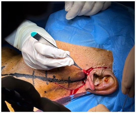
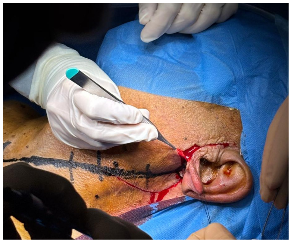
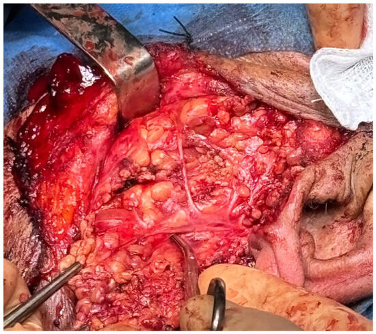
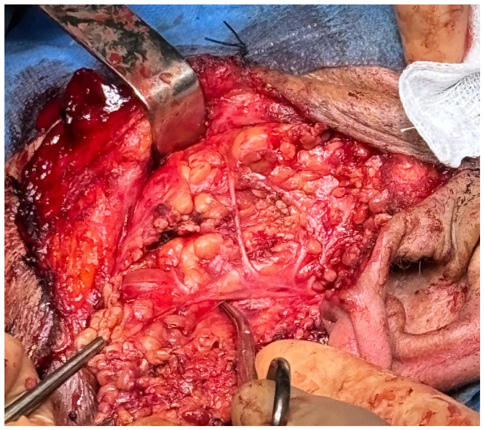
A single modified surgical approach was employed, consisting of a standard longitudinal incision along the anterior border of the sternocleidomastoid muscle that was extended superiorly into a preauricular hockey-stick configuration (modified Blair’s incision—Figure 1) to allow adequate exposure of the parotid gland (Figure 2).
Figure 1.
Modified Blair’s incision.
Figure 2.
Intraoperative view of Warthin’s tumor.
Eversion CEA was performed first. Before carotid clamping, systemic endovenous heparinization was performed with 5.000 IU, which was not reversed at the end of the procedure. Cerebral oximetry was monitored intraoperatively using a NIRS (near-infrared spectroscopy) system. Placement of an intraoperative shunt was required in one case (16.7%). Vascular clamps were applied, and eversion CEA was performed. Following plaque removal, the internal carotid artery (ICA) was re-anastomosed to the common carotid artery (CCA). The parotid loge was approached after de-clamping and hemostasis assessment.
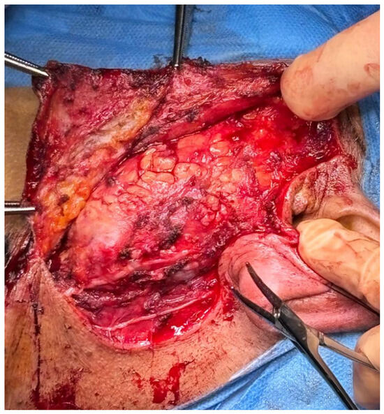
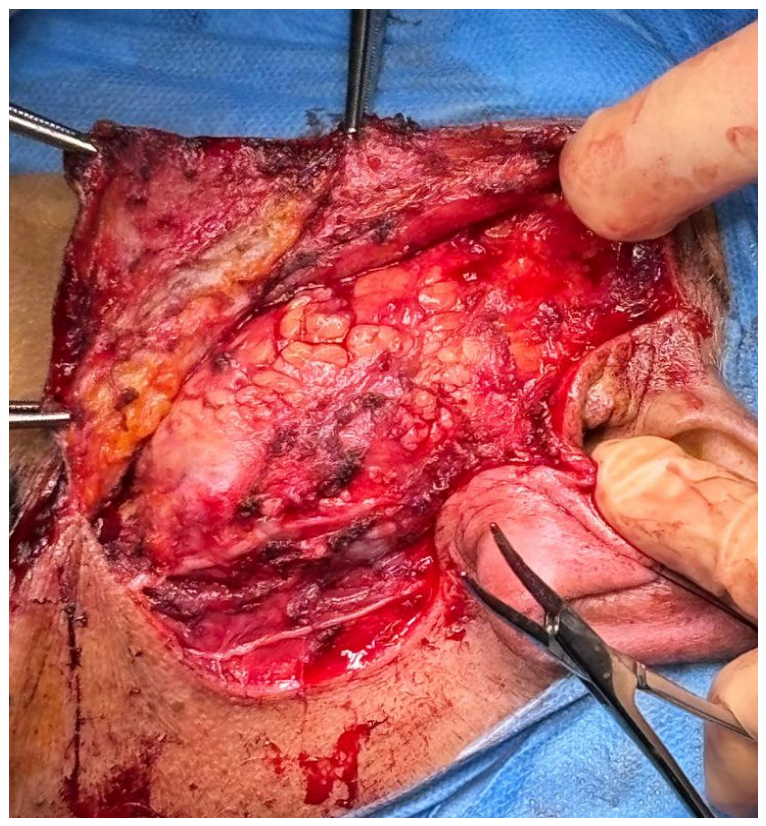
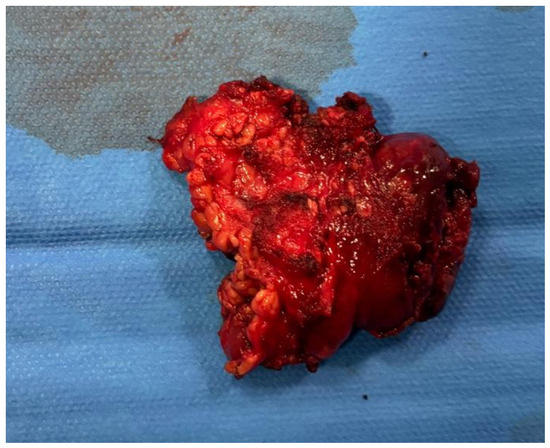
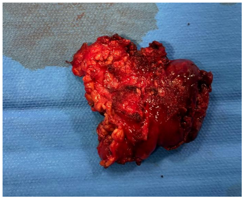
The main trunk of the facial nerve was identified at its outlet from the stylomastoid foramen, using the tragic pointer and the posterior belly of the digastric muscle as anatomical landmarks. Extracapsular dissection consisted of precise dissection along the tumor pseudocapsule without facial nerve neurolysis. Tissue division and microvascular sealing were achieved using the LigaSure™ Small Jaw device (Covidien LP/Medtronic plc, Boulder, CO, USA). Partial parotidectomy required identification of the main facial nerve trunk at the stylomastoid foramen (or a main peripheral branch) followed by neurolysis. LigaSure™ Small Jaw was employed for parenchymal transection and vessel sealing, ensuring complete tumor clearance with a collar of healthy tissue while safeguarding functional nerve branches (Figure 3 and Figure 4). A suction drain was placed to prevent hematoma formation.
Figure 3.
Intraoperative view after Warthin’s tumor resection with facial nerve preservation.
Figure 4.
Resected Warthin’s tumor.
Five patients (83.3%) returned to the surgical ward after the procedure. On the first postoperative day (POD), oral intake and autonomous mobilization were resumed, and the suction drain was removed. One patient (16.7%) with chronic obstructive pulmonary disease (COPD) was transferred to the Intensive Care Unit (ICU) for the first 24 postoperative hours to ensure monitoring of respiratory function. The patient returned to the surgical ward on POD 1.
2.3. Endpoints
Primary endpoints were perioperative and long-term survival and disease-free survival. Secondary endpoints were operative time, intraoperative and postoperative complications, and length of hospital stay. Median follow-up period was 43 months (range, 12–60 months).
2.4. Statistical Analysis
Statistical analyses were performed using SPSS version 26.0. Due to the small sample size (n = 6), a descriptive analysis was performed. Continuous variables were expressed as median and range (minimum–maximum). Categorical variables were expressed as absolute frequencies and percentages. Statistical inference tests and normality assessments were not applied due to the limited sample size.
Artificial intelligence (AI) tools (Gemini 2.0 Flash, chat GPT 4o) were used in the preparation of this manuscript to assist with grammar and stylistic improvements of the manuscript. All AI-generated content was thoroughly reviewed, edited, and verified for accuracy and originality by the authors, who take full responsibility for the final content.
3. Results
Postoperative and long-term survival rate at a median follow-up of 43 months was 100%, while the 5-year survival rate was 83.3%. One patient died after 50 months due to myocardial infarction. Disease-free survival at a median follow-up of 43 months and at 5 years was 100% for Warthin’s tumor. One patient (16.7%) developed a significant restenosis of the operated carotid artery at 48 months, which did not undergo reintervention due to the patient’s death from myocardial infarction. Restenosis was defined as a vessel lumen reduction ≥ 80% at Doppler ultrasound.
Secondary endpoints are summarized in Table 2.
Table 2.
Secondary endpoints.
4. Discussion
The simultaneous occurrence of significant ACAS and ipsilateral Warthin’s tumor is a rare clinical scenario that requires a personalized surgical strategy.
Warthin’s tumors often originate in the parotid tail, overlying the carotid bifurcation. A staged approach, performing either parotidectomy or CEA first, inevitably creates surgical scarring and fibrotic tissue which complicates a second intervention, increasing the risk of iatrogenic injury to the facial and hypoglossal nerve. Performing both procedures simultaneously ensures adequate visualization for both vascular and otolaryngologist teams [11,12,13].
The hallmark of this study protocol was a dual neuroprotection strategy:
- Continuous NIRS monitoring: Near-infrared spectroscopy provided real-time tracking of regional cerebral oxygen saturation. In the context of a combined procedure, which may involve more extensive neck manipulation than a standard CEA, the in vivo optical spectroscopy (INVOS) system acted as an early warning monitor for any hemodynamic shifts [14,15]
- Selective shunting: The use of an intraoperative shunt was guided and validated by INVOS data. This synergy ensured uninterrupted cerebral perfusion, effectively insulating the surgical procedure from the physiological stress associated with carotid cross-clamping [16].
Combined procedures require longer operative time and may increase the risk for blood loss. However, in this case series blood loss recorded remained within safety limits and no perioperative TIA, stroke or cranial nerves injury occurred.
Another concern is the development of a salivary fistula or sialocele directly over the reconstructed carotid artery increasing the risk of graft infection, arterial wall erosion, or pseudoaneurysm formation. In our series, patients underwent eversion CEA without patch closure; this avoids synthetic or autologous patch reconstruction reducing the risk of graft infection and material degradation in the event of salivary fistula. A single patient undergoing shunting required longitudinal arteriotomy and patch closure. Notably, this patient did not develop a postoperative salivary fistula. To mitigate this risk, meticulous intraoperative hemostasis, careful closure of the parotid fascia, and selective use of regional tissue flaps or sealants should be considered when the carotid bed is exposed.
Furthermore, perioperative antiplatelet and anticoagulation regimens require a tailored balance between thrombotic risk and hemorrhagic complications. While antiplatelet therapy or therapeutic anticoagulation is mandatory to maintain carotid patency, it inherently increases the risk of postoperative hematoma formation. In parotid surgery, hematomas can compromise wound integrity and cause mechanical compression on the facial nerve or on the airways [7,17,18]. In this series, antiplatelet timing (maintaining acetylsalicylic acid monotherapy perioperatively), systemic heparin reversal strategies, and intraoperative sequencing were tailored on a case-by-case basis.
The staged approach carries significant technical and clinical implications. Performing CEA first prioritizes neurological safety. By treating the high-grade stenosis, the patient is protected against potential embolic or hemodynamic strokes during the subsequent parotid surgery [19]. The primary drawback is post-surgical fibrosis, since CEA creates significant scarring in the carotid triangle. This distorts normal anatomical planes, significantly complicating the identification and preservation of both the hypoglossal nerve and the marginal mandibular branch of the facial nerve during the second stage [20]. Performing parotidectomy first allows the surgeon to operate on native tissue, ensuring the highest level of safety for nervous structures and providing an early histological diagnosis of the parotid mass [21]. However, the risk of intraoperative stroke is the main concern. The manipulation of the parotid gland, which lies directly over the carotid bifurcation, may lead to plaque destabilization or hemodynamic fluctuations in an unprotected brain.
Our findings support a simultaneous surgical approach. Combining both procedures into a single session eliminates the need to prioritize one pathology over the other. Furthermore, executing both interventions concurrently preserves optimal anatomical visualization before scar tissue develops. When paired with intraoperative shunting and NIRS monitoring, CEA enables a meticulous, unhurried parotid dissection without compromising cerebral perfusion.
Strengths and Limitations
This study addresses a rare clinical scenario for which standardized management guidelines are currently lacking. By documenting our diagnostic workup, treatment sequencing, and long-term outcomes, this series provides clinical insights and a practical framework for multidisciplinary teams facing this complex surgical case. However, several limitations must be acknowledged. The retrospective design inherently introduces selection bias. Furthermore, the small sample size precludes advanced statistical testing such as regression analysis or survival curves, limiting the generalizability of our findings. Despite these limitations, this cohort represents a contribution to literature on this dual pathology.
5. Conclusions
Simultaneous carotid endarterectomy and ipsilateral parotid tumor resection via a single surgical session is a feasible strategy. By eliminating the “priority dilemma” of staged approaches, this unified procedure reduces cumulative anesthetic risks and avoids post-surgical tissue fibrosis, thereby minimizing the risk of iatrogenic cranial nerve injury. Supported by intraoperative multimodal neuroprotection (NIRS and selective shunting), a combined single-stage approach optimizes both neurological safety and anatomical preservation, representing a highly viable clinical framework for this rare dual pathology.
Author Contributions
P.N.: Conceptualization, methodology, writing—original draft preparation, writing—review and editing; V.R.: methodology, formal analysis; G.D.: methodology, formal analysis; V.I.: methodology, formal analysis; S.L.: methodology, formal analysis; L.P.: methodology, formal analysis; M.V.: methodology, formal analysis; R.Z.: methodology, formal analysis; R.P.: visualization, supervision; G.P. (Giulia Pizzardi): visualization, supervision; M.R.: visualization, supervision; G.P. (Giampaolo Prezioso): visualization, supervision; V.D.: visualization, supervision; A.M.: visualization, supervision; G.N.: methodology, formal analysis; L.S.: methodology, formal analysis; M.D.V.: conceptualization, visualization, supervision; G.I.: conceptualization, visualization, supervision. All authors have read and agreed to the published version of the manuscript.
Funding
This research received no external funding.
Institutional Review Board Statement
This study was conducted in accordance with the principles of the Declaration of Helsinki. According to policies of the Ethics Committee of Azienda Ospedaliero-Universitaria Policlinico Umberto I (Comitato Etico Territoriale Lazio Area 1) and national regulations governing observational clinical research (AIFA Guidelines on Observational Studies and Italian Data Protection Authority Authorization No. 9/2016), formal ethical approval was not required as this represents a retrospective study using exclusively anonymized clinical data collected during routine care. The local institutional guidelines state that retrospective observational studies on anonymized cohorts do not alter patient management nor introduce periprocedural intervention risks.
Informed Consent Statement
Informed consent was obtained from all subjects involved in the study.
Data Availability Statement
The data presented in this study are available on request from the corresponding author due to privacy and ethical restrictions.
Acknowledgments
Artificial intelligence (AI) tools (Gemini 2.0 Flash, chat GPT 4o) were used in the preparation of this manuscript to assist with grammar and stylistic improvements of the manuscript. All AI-generated content was thoroughly reviewed, edited, and verified for accuracy and originality by the authors, who take full responsibility for the final content.
Conflicts of Interest
The authors declare no conflicts of interest.
Abbreviations
The following abbreviations are used in this manuscript:
| CEA | Carotid Endarterectomy |
| TIA | Transient Ischemic Attacks |
| ACAS | Asymptomatic Carotid Artery Stenosis |
| DUS | Doppler Ultrasound |
| BMI | Body Mass Index |
| CTA | Computed Tomography Angiography |
| MR | Magnetic Resonance |
| NIRS | Near-Infrared Spectroscopy |
| ICA | Internal Carotid Artery |
| CCA | Common Carotid Artery |
| DM | Diabetes Mellitus |
| COPD | Chronic Obstructive Pulmonary Disease |
| ICU | Intensive Care Unit |
| INVOS | In Vivo Optical Spectroscopy |
References
- de Weerd, M.; Greving, J.P.; Hedblad, B.; Lorenz, M.W.; Mathiesen, E.B.; O’Leary, D.H.; Rosvall, M.; Sitzer, M.; Buskens, E.; Bots, M.L. Prevalence of asymptomatic carotid artery stenosis in the general population: An individual participant data meta-analysis. Stroke 2010, 41, 1294–1297. [Google Scholar] [PubMed]
- Teymoortash, A.; Krasnewicz, Y.; Werner, J.A. Clinical features of cystadenolymphoma (Warthin’s tumor) of the parotid gland: A retrospective comparative study of 96 cases. Oral Oncol. 2006, 42, 569–573. [Google Scholar] [CrossRef] [Scilit] [PubMed]
- Saravakos, P.; Kourtidis, S.; Hartwein, J.; Preyer, S. Parotid Gland Tumors: A Multicenter Analysis of 1020 Cases. Increasing Incidence of Warthin’s Tumor. Indian J. Otolaryngol. Head Neck Surg. 2022, 74, 2033–2040. [Google Scholar] [CrossRef] [Scilit] [PubMed] [PubMed Central]
- Psychogios, G.; Vlastos, I.; Thölken, R.; Zenk, J. Warthin’s tumour seems to be the most common benign neoplasm of the parotid gland in Germany. Eur. Arch. Otorhinolaryngol. 2020, 277, 2081–2084. [Google Scholar] [CrossRef] [Scilit] [PubMed]
- Holgado, A.; León, X.; Llansana, A.; Valero, C.; Casasayas, M.; Fernandez-León, A.; Quer, M. Warthin’s Tumour as a Parotid Gland Incidentaloma Identified by PET-CT Scan in a Large Series of Cases. Indian J. Otolaryngol. Head Neck Surg. 2024, 76, 3046–3050. [Google Scholar] [CrossRef] [Scilit] [PubMed] [PubMed Central]
- Thompson, C.; Nolli, T.; Bannister, M. Parotid incidentalomas: A systematic review. J. Laryngol. Otol. 2021, 135, 765–769. [Google Scholar] [CrossRef] [Scilit] [PubMed]
- Naylor, R.; Rantner, B.; Ancetti, S.; de Borst, G.J.; De Carlo, M.; Halliday, A.; Kakkos, S.K.; Markus, H.S.; McCabe, D.J.H.; Sillesen, H.; et al. Editor’s Choice—European Society for Vascular Surgery (ESVS) 2023 Clinical Practice Guidelines on the Management of Atherosclerotic Carotid and Vertebral Artery Disease. Eur. J. Vasc. Endovasc. Surg. 2023, 65, 7–111. [Google Scholar] [CrossRef] [Scilit] [PubMed]
- Illuminati, G.; Ricco, J.B.; Caliò, F.; Pacilè, M.A.; Miraldi, F.; Frati, G.; Macrina, F.; Toscano, M. Short-term results of a randomized trial examining timing of carotid endarterectomy in patients with severe asymptomatic unilateral carotid stenosis undergoing coronary artery bypass grafting. J. Vasc. Surg. 2011, 54, 993–997; discussion 998–999. [Google Scholar] [CrossRef] [Scilit] [PubMed]
- Nardi, P.; Rinaldi, V.; Pasqua, R.; Prezioso, G.; D’ANdrea, V.; Palumbo, P.; Miraldi, F.; Tanzilli, G.; Recchioni, T.; Hostalrich, A.; et al. Systematic Preoperative Coronary Angiography in Patients with an Asymptomatic Coronary Artery Disease May be Recommended in Patients with Peripheral Artery Disease Undergoing Open Peripheral Revascularization: A Multicenter Retrospective Study. Ann. Vasc. Surg. 2025, 114, 330–339. [Google Scholar] [CrossRef] [Scilit] [PubMed]
- Nardi, P.; Rinaldi, V.; Costanzo, M.L.; Pasqua, R.; Loiacono, F.; Palumbo, P.; Miraldi, F.; Tanzilli, G.; D’Andrea, V.; Illuminati, G. Simultaneous Percutaneous Coronary Intervention (PCI) and Endovascular Aneurysm Repair (EVAR): A Preliminary Report. J. Clin. Med. 2024, 13, 5545. [Google Scholar] [CrossRef] [Scilit] [PubMed] [PubMed Central]
- Telischi, J.; Nisenbaum, E.; Nicolli, E. Carotid endarterectomy for the management of carotid stenosis occurring concurrently with head and neck cancer. Curr. Opin. Otolaryngol. Head Neck Surg. 2023, 31, 452–456. [Google Scholar] [CrossRef] [Scilit] [PubMed]
- Rechtweg, J.; Wax, M.K.; Shah, R.; Granke, K.; Jarmuz, T. Neck dissection with simultaneous carotid endarterectomy. Laryngoscope 1998, 108, 1150–1153. [Google Scholar] [CrossRef] [Scilit] [PubMed]
- Sanjuan-Sanjuan, A.; Ogledzki, M.J.; Haouilou, J.C.; Ramirez, C.A. Neck dissection for head and neck malignancies with concurrent carotid endarterectomy. Int. J. Oral Maxillofac. Surg. 2023, 52, 1120–1126. [Google Scholar] [CrossRef] [Scilit] [PubMed]
- Russo, M.F.; Gentile, P.; Fenga, M.; Izzo, S.; Denaro, F.; Luka, K.; Frattaroli, F.; Costanzo, A.; Castagneto-Gissey, L.; Salvati, B. Cerebral oximetry monitoring by means of INVOS-4100 as a predictor of ischemic events during carotid endarterectomy. Front. Surg. 2023, 10, 1170019. [Google Scholar] [CrossRef] [Scilit] [PubMed] [PubMed Central]
- Yürük, M.A.; Kılıç, A.; Sayar, U.; Arslan, A.K.; Hanedan, M.O. Cerebral pulse oximetry in carotid endarterectomy: An impact study. Rev. Assoc. Medica Bras. 2025, 71, e20250771. [Google Scholar] [CrossRef] [Scilit] [PubMed] [PubMed Central]
- Pennekamp, C.W.; Immink, R.V.; den Ruijter, H.M.; Kappelle, L.J.; Bots, M.L.; Buhre, W.F.; Moll, F.L.; de Borst, G.J. Near-infrared spectroscopy to indicate selective shunt use during carotid endarterectomy. Eur. J. Vasc. Endovasc. Surg. 2013, 46, 397–403. [Google Scholar] [CrossRef] [Scilit] [PubMed]
- Mazzolai, L.; Teixido-Tura, G.; Lanzi, S.; Boc, V.; Bossone, E.; Brodmann, M.; Bura-Rivière, A.; De Backer, J.; Deglise, S.; Della Corte, A.; et al. 2024 ESC Guidelines for the management of peripheral arterial and aortic diseases. Eur. Heart J. 2024, 45, 3538–3700. [Google Scholar] [CrossRef] [Scilit] [PubMed]
- Halvorsen, S.; Mehilli, J.; Cassese, S.; Hall, T.S.; Abdelhamid, M.; Barbato, E.; De Hert, S.; de Laval, I.; Geisler, T.; Hinterbuchner, L.; et al. 2022 ESC Guidelines on cardiovascular assessment and management of patients undergoing non-cardiac surgery. Eur. Heart J. 2022, 43, 3826–3924, Erratum in Eur. Heart J. 2023, 44, 4421. https://doi.org/10.1093/eurheartj/ehad577. [Google Scholar] [CrossRef] [Scilit] [PubMed]
- Kleindorfer, D.O.; Towfighi, A.; Chaturvedi, S.; Cockroft, K.M.; Gutierrez, J.; Lombardi-Hill, D.; Kamel, H.; Kernan, W.N.; Kittner, S.J.; Leira, E.C.; et al. 2021 Guideline for the Prevention of Stroke in Patients with Stroke and Transient Ischemic Attack: A Guideline from the American Heart Association/American Stroke Association. Stroke 2021, 52, e364–e467, Erratum in Stroke 2021, 52, e483–e484. https://doi.org/10.1161/STR.0000000000000383. [Google Scholar] [CrossRef] [Scilit] [PubMed]
- AbuRahma, A.F. Processes of care for carotid endarterectomy: Surgical and anesthesia considerations. J. Vasc. Surg. 2009, 50, 921–933. [Google Scholar] [CrossRef] [Scilit] [PubMed]
- İlhan, E.; Uyar, M.; Bektaş, S.; Arukan, Ç. Warthin’s tumour of the parotid gland: A 14-year retrospective review of surgical outcomes, diagnostic accuracy and patient-reported aesthetic satisfaction. Am. J. Otolaryngol. 2026, 47, 104775. [Google Scholar] [CrossRef] [Scilit] [PubMed]

Disclaimer/Publisher’s Note: The statements, opinions and data contained in all publications are solely those of the individual author(s) and contributor(s) and not of MDPI and/or the editor(s). MDPI and/or the editor(s) disclaim responsibility for any injury to people or property resulting from any ideas, methods, instructions or products referred to in the content. |
© 2026 by the authors. Licensee MDPI, Basel, Switzerland. This article is an open access article distributed under the terms and conditions of the Creative Commons Attribution (CC BY) license.