


Abstract
Background/Objectives: This paper synthesizes and evaluates the potential origins of ectopic salivary gland pathologies in the human nasopharynx. By combining historical reports with an original comparative analysis, this study aims to clarify the precise anatomical and functional status of the proposed tubarial salivary-type glands. Methods: A literature search through December 2025 across major electronic databases identified 84 relevant publications on nasopharyngeal glandular pathologies. The methodological quality in the histomorphological literature was evaluated using the AQUA tool. Concurrently, histological samples were harvested from the inner Eustachian tube opening/torus tubarius region of human, canine, and rodent cadavers and analyzed using histomorphological and immunohistochemical protocols. Results: Most of the literature addressing histologically indicated nasopharyngeal glandular pathology described these findings as controversial, largely overlooking salivary gland origins. The AQUA appraisal identified significant vulnerabilities, showing a high risk of bias in the Study Design domain and yielding moderate inter-rater agreement (κ = 0.435). The empirical morphological study revealed mixed glands with strong mucous predominance in the torus tubarius region of humans and dogs, whereas rodents displayed exclusive PAS positivity within a trabecular stroma. Human samples uniquely demonstrated distinct compartmentalization, duct- and capsule-like formations, and negative α-amylase immunoreactivity. Conclusions: Our pilot empirical evidence, combined with the literature data, suggests a possible pathogenetic link between specific nasopharyngeal neoplasms and the baseline glandular structures of the human torus tubarius. Exhibiting traits of both major and minor salivary systems, the tubarial glands may represent an intermediate structural entity. Furthermore, the animal models utilized do not provide a completely reliable translational framework, highlighting the need for standardized research using fresh human biopsies.
1. Introduction
Salivary gland tumors pose a persistent clinical challenge due to their pauci-symptomatic nature, complex locations within the maxillofacial region, and anatomical variability, including the presence of accessory glands or additional lobes [1]. While the integration of advanced clinical experience, morphological assessment, and modern imaging techniques generally ensures accurate diagnosis and effective management [1], significant diagnostic dilemmas arise when salivary tissue is unexpectedly identified in ectopic locations. In such scenarios, clinicians face a critical differential diagnosis between a metastatic focus and aberrant glandular tissue. Reflecting this, between 1989 and 2022, the scientific literature saw a progressive increase in case reports documenting nasopharyngeal neoplasms histologically identified as originating from salivary glandular tissue (Table 1, Figure 1).
Table 1.
Summary of reported cases of nasopharyngeal salivary-type neoplasms and their anatomical topography as suggested by the authors.
Figure 1.
Distribution of the reported cases by patient sex based on the data from Table 1. Males and females were equally represented across the documented cohort (χ2 = 0.263, df = 1, p = 0.608, φ = 0.022).
These accumulated data, along with the landmark discovery by Valstar et al. [36] of a pair of macroscopically distinct salivary glands in the human nasopharynx visualized via prostate-specific membrane antigen positron emission tomography/computed tomography (PSMA PET/CT), led to the hypothesis that glandular tissue surrounding the torus tubarius and the inner Eustachian tube opening could serve as the origin of the neoplasms documented in the literature. However, the exact anatomical status of these proposed ‘tubarial glands’ remains highly controversial and is currently under active debate within the scientific community. To address this question, the current pilot observational study combines a comprehensive literature review with original comparative anatomical research, including histomorphological evaluation and preliminary imaging studies in both humans and animals, aiming to provide a broader phylogenetic perspective on the glandular architecture of this region.
2. Materials and Methods
2.1. Literature Search Strategy and Quality Assessment
An electronic literature search was conducted up to 1 December 2025 across the PubMed/MEDLINE, Scopus, Google Scholar, and Cochrane Library databases, supplemented by a manual search of relevant journals. The search strategy utilized the following keywords and terms: ‘salivary glands’, ‘salivary gland tumors’, ‘new salivary glands’, ‘heterotopic salivary glands’, ‘tubarial salivary glands’, ‘nasopharynx’, and ‘nasopharyngeal neoplasms’ (Figure 2). Titles and abstracts of the retrieved articles were screened independently by two researchers to ensure objectivity. The inclusion criteria comprised systematic and narrative reviews presenting novel insights, randomized controlled trials, prospective and retrospective studies, original research articles, case reports, case series, letters to the editor, commentaries, and expert opinions. Publications reporting pathologies or findings outside the anatomical boundaries of the nasopharynx and torus tubarius were excluded. All selected publications were retrospectively and descriptively analyzed based on parameters of clinical relevance, including the specific type, anatomical location, and proposed tissue of origin of the findings.
The quality of the included histomorphological studies was independently evaluated using the Anatomical Quality Assessment (AQUA) tool to determine the risk of bias [37]. This assessment covered five core domains: (1) study design, (2) characteristics of the study population, (3) methodology of tissue preparation/evaluation, (4) descriptive anatomy, and (5) reporting of results. Inter-rater reliability between the two independent reviewers was quantified using Cohen’s Kappa coefficient (κ). The risk of bias within each domain was stratified into three levels: low, moderate, or high. If a study received a ‘NO’ response in even a single key domain, it was categorized as having a high risk of bias. These qualitative outcomes were visualized using a Traffic Light Plot (🟢—low risk/”YES”; 🟡—moderate risk/”UNCLEAR”, i.e., some concerns exist; 🔴—high risk/”NO”).
Statistical analyses were performed using the Statistical Package for the Social Sciences (SPSS, version 26.0; IBM Corp., Armonk, NY, USA). The distribution and trends across publication categories over the years were analyzed using Chi-Square (χ2) tests. At the same time, One-Way Analysis of Variance (ANOVA) (F) was utilized to assess between-group effects, supplemented by correlation analyses. The magnitude of the effect size (φ) was interpreted according to Cohen’s criteria: 0.10 indicated a small effect, 0.30 a medium effect, and 0.50 a large effect. For all statistical tests, a p-value < 0.05 was considered statistically significant, with a 95% confidence interval (CI).
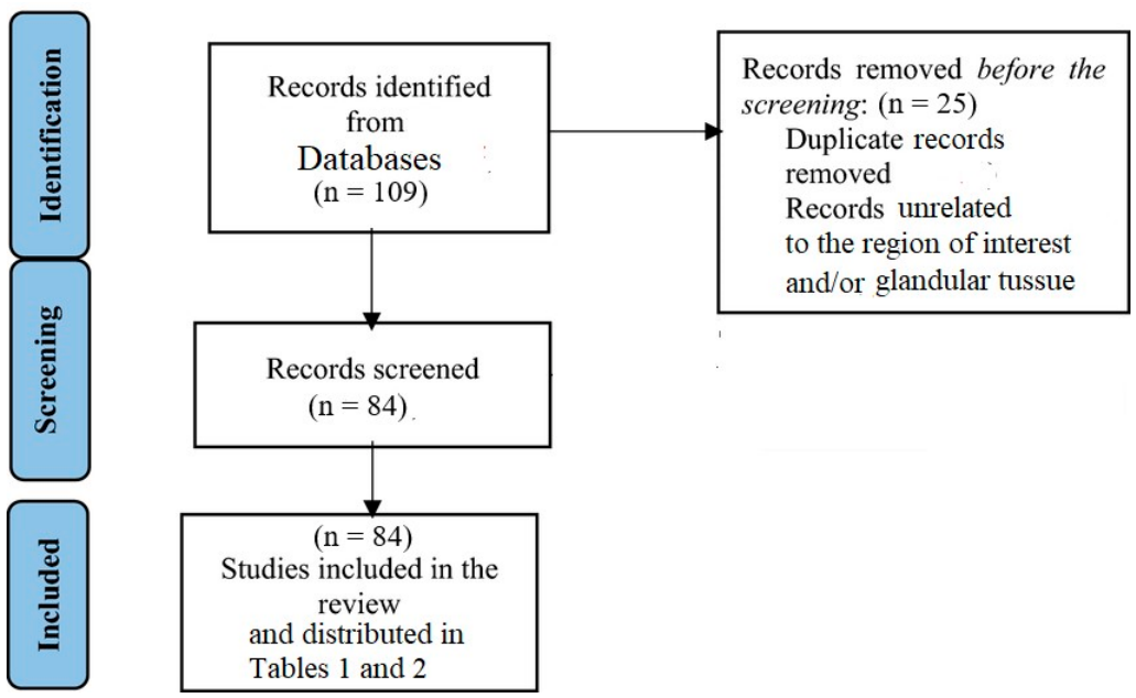
Table 2.
Summary of records discussing the tubarial salivary-type glands.
2.2. Comparative Anatomical and Histomorphological Evaluation
To evaluate the working hypothesis, a pilot imaging study was initially performed on a healthy human volunteer using magnetic resonance imaging (MRI) (Figure 3). Based on the anatomical dimensions obtained from the MRI and the existing literature, a pre-sized template was developed to standardize tissue sampling. Bilateral tissue blocks (3 × 3 × 3 cm) were harvested from the torus tubarius region of three human cadavers (two males and one female, aged 65–75 years; all of Caucasian ethnicity; Figure 4) fixed with a formaldehyde-containing soft embalming fluid. As a pilot morphological proof-of-concept study, the sample size was primarily dictated by the availability of cadaveric donations within the institutional anatomical program. None of the human subjects had a documented medical history of head and neck pathology. It must be explicitly acknowledged that the utilization of long-term embalmed cadaveric tissue, rather than fresh biopsies, carries an inherent risk of altering or masking certain histochemical and antigenic properties.
Figure 3.
High-resolution 3D MRI scan (field strength 1.5 T, slice thickness ≤ 2 mm) of a healthy human volunteer demonstrating the anatomical localization of the tubarial salivary-type glands (yellow circles). The panel displays (a) axial, (b) coronal, and (c) sagittal sections, alongside (d) an MRI-integrated 3D reconstruction and (e) corresponding T1/T2 relaxation analysis.
Figure 4.
Anatomical dissection and standardized tissue harvesting protocol from the human torus tubarius region (within the yellow circle). (a) Regional landmarks: 1—fossa of Rosenmüller; 2—torus tubarius; 3—auditory tube orifice; 4—salpingopharyngeal fold. (b) Application of a pre-sized template (5) to define sampling boundaries. (c) Post-harvest view showing the excision site (6) and the isolated tissue block (7) ready for histological processing.
To determine whether the presence of glandular tissue in this region represents an evolutionarily conserved feature in mammals—thereby providing a broader phylogenetic context rather than a direct translational framework for human physiology—an independent comparative morphological study was conducted in adult dogs and rats. Bilateral tissue blocks were obtained from the area surrounding the inner opening of the Eustachian tube from canine cadavers (two females and one male of medium-sized breeds; block size: ca. 3 × 3 × 3 cm) and rodent specimens (Wistar strain: n = 10, five males and five females; Long–Evans strain: n = 10, five males and five females; total rodent cohort, n = 20). For the rodent group, block sizes were approximately 0.5 × 0.5 cm and extended to the underlying bone depth. All animal specimens were donated for educational and research purposes, representing terrestrial mammalian species with comparable feeding habits.
All harvested tissue samples were fixed in 10% neutral buffered formalin, embedded in paraffin, and sectioned sagittally at a thickness of 4 µm. For the human specimens, the total pool of potential sections was estimated at approximately 7500 per block. To ensure statistically significant results with minimal error (coefficient of error < 0.10), a systematic random sampling protocol was implemented. A sampling interval (k = 500 sections) was set on the microtome counter, and the first section was randomly selected. Sections were collected at fifteen standardized depth intervals: #1 (2.00 mm), #2 (4.00 mm), #3 (6.00 mm), #4 (8.00 mm), #5 (10.00 mm), #6 (12.00 mm), #7 (14.00 mm), #8 (16.00 mm), #9 (18.00 mm), #10 (20.00 mm), #11 (22.00 mm), #12 (24.00 mm), #13 (26.00 mm), #14 (28.00 mm), and #15 (30.00 mm).
At each depth, four consecutive sections were processed using the following staining protocols: Hematoxylin and Eosin (H-E), Periodic Acid-Schiff (PAS) counterstained with Hematoxylin, Alcian blue, and immunohistochemical staining utilizing anti-alpha (α)-amylase polyclonal rabbit antibodies (all purchased from Thermo Fisher Scientific, Waltham, MA, USA). Histomorphological analysis was performed independently by two blinded morphologists. Finally, to optimize spatial characterization, the localized glandular areas were reconstructed into 3D graphical models (Figure 3).
Ethical Statement: All procedures involving human tissue and animal specimens were reviewed and approved by the Research Ethics Committee of the Department of Health Care and Social Work, NBU (Protocol No. 142/14 October 2025).
3. Results
3.1. Literature Review
Following the initial screening of titles and abstracts and the removal of duplicate records, a total of 84 publications met the inclusion criteria (summarized in Table 1 and Table 2). They were subjected to comprehensive qualitative and quantitative analysis. This finalized dataset primarily comprised research commentaries, letters to the editor, author replies, short communications, expert opinions, original research articles, systematic and narrative reviews, and case reports or series (Figure 5, Figure 6 and Figure 7). The evaluated literature focused predominantly on patients undergoing surgical intervention for various nasopharyngeal pathologies (Table 1, Figure 1), individuals subjected to advanced diagnostic imaging protocols targeting this region, and healthy volunteers participating in functional assessments of the proposed glandular structures (Table 2).
Figure 5.
Distribution of the records summarized in Table 2 by publication type. The different article types were unequally represented across the evaluated literature (χ2 = 14.080, df = 2, p < 0.001).
Figure 6.
Chronological distribution of the records compiled in Table 2 over the years, demonstrating an overall rising publication trend (indicated by the dashed linear trendline; r = 0.395, p = 0.332) in research dedicated to the tubarial salivary-type glands. The prominent spike in 2021 primarily reflects the immediate wave of academic letters, commentaries, and discussions triggered by the initial publication of the tubarial glands hypothesis in late 2020.
Figure 7.
Sex distribution of the analyzed subjects across the original research articles compiled in Table 2. The “Images” cohort encompasses all diagnostic scanning modalities detailed in the literature.
The comprehensive analysis of the publications summarized in Table 1 and Table 2 yielded several key conclusions:
- Over the documented period, there has been a progressive increase in reported clinical cases of patients presenting with diverse pathologies originating from glandular tissue in the nasopharynx, particularly within the torus tubarius region.
- Morphologically and in terms of biological behavior, these nasopharyngeal tumors closely resemble neoplasms typical of the major and minor salivary glands [51,68].
- The benign nature of several documented cases effectively rules out a metastatic process from a distant primary origin.
- The absence of an alternative anatomical explanation for these specific tumors likely provoked a second peak of scientific and clinical interest in the existence of the tubarial salivary-type glands following their initial formal description by Valstar et al. in 2020 [36] (Figure 6).
- This initial announcement triggered an increase in the volume of comments, letters to the editor, and related academic correspondence (F = 9.850, p = 0.048), which subsequently spurred deeper scientific investigation into the topic (F = 19.698, p = 0.003).
- Concurrently with the expansion of new findings regarding the tubarial anatomical debate, a nonsignificant growth in summary review articles was observed (r = 0.333, p = 0.385), alongside a nonsignificant reduction in the number of brief comments and letters (r = −0.045, p = 0.925) (Figure 6). Robust research outputs appear to stimulate more in-depth investigative designs (F = 11.832, p = 0.035) aimed at resolving emerging questions about the nature and clinical significance of the human tubarial glands.
- Within the imaging modalities cohort, despite a large pooled sample size (n = 1055), the sex distribution was significantly unequal (χ2 = 409.15, df = 1, p < 0.001), with a substantial effect size (φ = 0.62).
- Conversely, in the histomorphological cohort, while the sex distribution was statistically equitable (χ2 = 2.882, df = 1, p = 0.090), the effect size was moderate to high (φ = 0.412) due to the limited total number of evaluated subjects (Table 2).
- Despite the intense scientific debate, published histomorphological studies remain disproportionately scarce (n = 6, representing 27.27% of the original research) compared to imaging-based investigations (χ2 = 4.545, df = 1, p = 0.033), with a significant difference in effect size (φ = 0.45), underscoring the importance of further pilot anatomical studies.
The quality assessment of these morphological works via the AQUA tool is summarized in Table 3. The evaluation of five double-assessed criteria across six histomorphological studies indicated moderate inter-rater agreement (κ = 0.435), reflecting frequent variations in risk judgment between the low–moderate and moderate–high thresholds. Driven by the ‘weakest link’ rule, the overall risk assessment identified high-risk criteria in the studies by Valstar et al. [36], Pringle et al. [69], and Blagova et al. [84], whereas Kumari et al. [66], Ebrahim et al. [82], and Athavale et al. [86] were classified as presenting a moderate risk of bias. The highest risk of systematic error was localized within the Performance, i.e., Characterization of Methods, and Reporting, i.e., Results Reporting, domains, underscoring the need for stricter blinding protocols and more refined reporting criteria in future anatomical studies (Table 3).
Table 3.
Quality appraisal of the included histomorphological studies according to the AQUA tool.
Following additional analyses, Domain 2 was evaluated as the most problematic, with 33.33% of studies reporting a high risk of bias (🔴) (Figure 8). Domain 1 and Domain 3 accumulated the highest unclear risk scores (🟡), indicating that the publications themselves lacked detailed information regarding tissue procurement strategies or blinded morphometric protocols. Conversely, the most reliable baseline data were observed in Domain 4 and Domain 5, which demonstrated a low risk of bias (🟢) in 50.00% of the examined studies, serving as a favorable indicator of reporting quality with respect to descriptive anatomical completeness.
Figure 8.
Risk of bias summary illustrating the cumulative percentages of low (🟢), unclear (🟡), and high (🔴) risk of bias across the five analyzed AQUA domains for all included histomorphological studies.
Individual study profiles demonstrated that the work by Ebrahim et al. [82] exhibited the lowest overall risk of bias, accumulating the highest frequency of low-risk indicators (Figure 9). Furthermore, a chronological improvement was observed in more recent publications, specifically within the Detection Bias/Characterization of Methods (Domain 3) and Reporting Bias/Results Reporting (Domain 5) domains, as visually represented by the temporal progression of the plotted studies.
Figure 9.
Risk of bias traffic light plot illustrating the reviewers’ independent consensus judgments for each evaluated histomorphological study across the five analyzed AQUA domains (🟢—low risk; 🟡—unclear risk; 🔴—high risk) [36,66,69,82,84,86].
The analysis of the included histomorphological studies revealed highly variable methodological quality, which was critically dependent on the completeness of reporting within the literature. Overall, the predominance of moderate- and high-risk domains highlights an urgent need for standardized morphological investigations to facilitate the transition from purely descriptive morphology to evidence-based anatomy. To address this gap and provide robust data, our pilot study focused on characterizing the micromorphological features of the tubarial glands while exploring their long-term translational potential in research and clinical practice.
3.2. Pilot Imaging (MRI) Assessment
The high-resolution 3D MRI sequences of the healthy human volunteer confirmed the precise anatomical localization of the proposed tubarial salivary-type glands (Figure 3). On T1-weighted images, these bilateral glandular formations exhibited an isointense appearance relative to the adjacent parotid glands, whereas T2-weighted sequences revealed a slightly hyperintense signal compared to the surrounding muscles, bone, and cartilage. Morphometric analysis determined the dimensions of these structures to be 29.4 × 15.2 × 20.1 mm on the right side and 28.9 × 12.5 × 18.3 mm on the left side, situated between the skull base, the fossa of Rosenmüller, and the posterolateral nasopharyngeal wall, corresponding to the mapped T1 and T2 relaxation values.
3.3. Histomorphological and Comparative Anatomical Results
Histological evaluation of the tissue sections harvested from the torus tubarius region demonstrated the presence of mixed glands exhibiting distinct species-specific variations (Figure 10, Figure 11 and Figure 12). On Hematoxylin and Eosin (H-E) stained sections, the mucous acini presented a characteristic pale, “foamy” cytoplasm with basally flattened nuclei compressed against the basement membrane due to the dissolution of mucin during tissue processing. In contrast, serous acini displayed a deeply stained, basophilic cytoplasm with spherical, centrally to basally located nuclei. Crescent-shaped clusters of serous cells capping the terminal portions of mucous acini were identified, closely resembling classical Giannuzzi-Heidenhain demilunes observed following conventional tissue processing (Figure 10 and Figure 11).
Figure 10.
Histomorphological and immunohistochemical evaluation of human tissue samples from the torus tubarius region (magnifications ×10—second and third rows, ×20—first row, and ×40—j). Panels demonstrate: (a,g) Hematoxylin and Eosin (H-E) staining; (b,d,i) Periodic Acid-Schiff (PAS) staining; (c,e,f,h) Alcian blue staining; (j) anti-α-amylase immunohistochemistry showing negative immunoreactivity; and (k) 3D graphical reconstruction of the glandular spatial distribution. Structural landmarks include respiratory epithelium (RE), regional cartilage (C), salpingopharyngeus muscle (SM), interlobular duct-like structures (yellow circle), connective tissue septa (*), blood vessels (˄), serous demilunes (◊), and focal lymphocytic infiltrates (dashed circle).
Figure 11.
Histomorphological evaluation of canine tissue samples from the Eustachian tube region (magnification ×20). Panels demonstrate: (a) Hematoxylin and Eosin (H-E) staining; (b) Periodic Acid-Schiff (PAS) staining; and (c) Alcian blue staining. Secretory compartments consist of mixed seromucous glands with distinct serous demilunes (◊). Organized connective tissue septa, interlobular ducts, and an enveloping capsule-like formation are absent.
Figure 12.
Histomorphological evaluation of rat tissue samples from the Eustachian tube region (magnification ×20). Panels demonstrate: (a) Hematoxylin and Eosin (H-E) staining; (b) Periodic Acid-Schiff (PAS) staining; and (c) Alcian blue staining. Secretory compartments show prominent PAS-positive but Alcian blue-negative reactivity, with serous cells organized into demilunes (◊). Note the presence of a capsule-like formation with connective tissue septa (*), blood vessels (˄), and interlobular ducts (yellow circle).
Histochemical profiling confirmed that the human (Figure 10) and canine (Figure 11) glandular tissue consisted of mixed structures with a strong predominance of mucous acini. These structures exhibited strong Periodic Acid-Schiff (PAS) positivity, confirming the presence of neutral glycoproteins and serous secretory components, alongside robust Alcian blue affinity, which delineated the acidic mucins within the acinar cytoplasm. Notably, a comparative analysis of the PAS reaction across the three terrestrial mammalian species revealed the most intense staining reactivity in the human specimens. In contrast to humans and dogs, the glandular structures identified in the rat specimens (Figure 12) were exclusively PAS-positive and lacked Alcian blue reactivity.
Structurally, both human (Figure 10) and rodent (Figure 12) sections revealed prominent blood vessels and interlobular ductal-like structures coursing through the connective tissue septa arising from a capsule-like formation that enveloped the glandular conglomerates. Conversely, such organized stromal septa and distinct ductal networks could not be identified in the canine specimens (Figure 11), suggesting a morphology consistent with minor or small accessory glands. Additionally, the human samples uniquely displayed two distinct patterns of glandular conglomerates characterized by varying intensities of Alcian blue chromatic staining (Figure 10).
Finally, immunohistochemical evaluation for α-amylase, a functional marker used to visualize enzyme-producing serous acinar cells, demonstrated negative immunoreactivity within the serous components of the human tubarial glandular tissue (Figure 10). Rather than completely excluding a salivary-type lineage, this negative finding suggests a distinct, mucus-specialized functional profile for these structures, potentially further influenced by the antigen-masking effects of long-term formalin fixation.
4. Discussion
The evolutionary variability of human anatomy has established numerous precedents in clinical practice, spanning atavistic features such as supernumerary mammary glands, phylogenetic reductions including oligodontia, and vestigial structures such as the appendix vermiformis and Darwin’s tubercle. Within this evolutionary framework, it is crucial to determine whether the recently documented nasopharyngeal glandular tissue constitutes a constant anatomical feature in humans, whether it fulfills the established criteria to be classified as a distinct organ, and how its specific glandular lineage should be categorized. Such a determination carries profound implications for the interpretation of regional pathology, necessitating potential updates to current histological and clinical classifications. Accurately defining the structural topography of salivary and accessory glands, along with their associated diseases, remains vital for timely diagnosis, therapeutic decision-making, and prognostic evaluation. Accordingly, this paper aims to stimulate academic discussion and a rigorous reevaluation of the current understanding of human nasopharyngeal glands, specifically the tubarial glands, and their related clinical conditions, drawing upon novel comparative histomorphological findings and a comprehensive reassessment of historical reports. To fully contextualize these findings, the chronological evolution of this anatomical concept must be examined.
4.1. Chronological and Technical Evolution of the Tubarial Glands Concept
In 2020, a team of Dutch researchers led by Valstar announced the identification of what they described as a novel pair of macroscopic salivary glands situated above the torus tubarius, presenting their findings in three methodological phases [36,44]. The initial discovery stemmed from the incidental visualization of circumscribed, bilateral areas of high radiotracer uptake within the nasopharynx during prostate-specific membrane antigen positron emission tomography/computed tomography (PSMA PET/CT) in oncology patients [36]. Although primarily utilized for prostate cancer staging, PSMA PET/CT has been established as a highly sensitive modality for visualizing salivary glandular tissue due to its physiological expression of the target antigen [40,44,80]. Consequently, the authors characterized these structures as salivary-type glandular tissue overlying the torus tubarius [36,81].
A subsequent retrospective MRI analysis by Valstar et al. revealed a well-defined tissue structure characterized by low signal intensity on T1-weighted sequences and an isointense appearance relative to the normal parotid gland, localized on the medial aspect of the torus tubarius [36]. This structure appeared anatomically coherent with discrete T2-hyperintense focal areas, which the authors hypothesized to represent the macroscopic openings of excretory ducts subsequently observed during cadaveric dissections [36].
To validate these radiologic findings, macroscopic and microscopic evaluations of human cadaveric nasopharyngeal specimens were performed using standard Hematoxylin and Eosin (H-E) staining and immunohistochemical profiling [36]. Based on these morphological criteria, the structures were categorized as mixed, tubuloacinar, seromucous glands with an organized ductal framework opening into the nasopharyngeal cavity [36,44,52]. Furthermore, Valstar et al. investigated the clinical significance of these glands by evaluating the impact of xerostomia and dysphagia following radiotherapy targeting this region in patients with head and neck malignancies [36,44,62,79]. Finally, to demonstrate the functional contribution of these structures to regional lubrication, cyanine dye was injected around the auditory tube orifice in living subjects, confirming active mucinous secretion aimed at preventing nasopharyngeal desiccation [36,44,52].
Intriguingly, the initial reports emphasized that these tubarial glands were negative for α-amylase expression, producing predominantly mucous secretions [36,41], a feature shared with a minority of major and minor salivary structures, such as the unencapsulated posterior extension of the sublingual or palatine glands [44,66,69,70,81,82]. However, this categorical interpretation was later challenged by Kumari et al., whose independent histomorphological study detected focal α-amylase immunoreactivity within the nasopharyngeal glandular tissue, raising important questions regarding the functional classification and biochemical heterogeneity of these structures [66]. This notable discrepancy in the literature, which is also reflected in our own negative immunoreactivity findings, highlights a critical technical vulnerability in cadaveric studies. Long-term tissue procurement involving formaldehyde-based embalming fluids is well documented to induce extensive protein cross-linking, which can severely mask antigen epitopes or denature sensitive enzyme profiles like α-amylase. Therefore, unless antigen retrieval protocols are perfectly standardized or fresh clinical biopsies are utilized, a negative histochemical result cannot definitively rule out a salivary gland lineage but rather underscores a predominant mucus-specialized functional profile heavily influenced by tissue fixation artifacts.
4.2. Arguments “Pro” and “Contra” the Proposed Organ
4.2.1. Histomorphological Evidence
Following the initial description, the immunohistochemical characterization of the tubarial salivary-type glands was significantly expanded by independent research groups [62,64,69,82,84,86]. Multiple investigators validated the core findings of Valstar et al. [36,44] by confirming the presence of a distinct myoepithelial cell layer enveloping the glandular acini and ductal structures—a hallmark of true salivary lineage [52,64,66,69,82]. For instance, Pushpa et al. precisely dissected small bilateral samples of these glands from a 60-year-old male cadaver and evaluated their finer histomorphological details [52]. Using Periodic Acid-Schiff (PAS) staining, they identified active mucinous secretions characterized by intense magenta-purple chromogenic signaling within the submucosal glandular compartments [52]. Furthermore, they confirmed the presence of myoepithelial cells via strong immunoreactivity for CD10 and S100 markers [52,69].
The architecture of this contractility-supporting myoepithelial framework was corroborated by Kumari et al., who successfully utilized alpha-smooth muscle actin (α-SMA) to delineate these pericellular networks [66]. To further clarify the anatomical classification of these glands, Ebrahim et al. evaluated the structural benchmarks that traditionally differentiate major from minor salivary glands, focusing on the complexity of the branching ductal system, acinar spatial arrangement, and macroscopic visibility on standard radiological imaging [82]. Based on their histomorphological data and subsequent statistical analysis, they concluded that the tubarial structures align more closely with minor salivary glands or resemble the unencapsulated posterior extension of the sublingual gland, which naturally aggregates 8 to 30 mixed minor glandular units [36,41,81,82].
Consequently, the lack of a distinct macroscopic capsule and the technical difficulty of isolating these glands via standard surgical dissection led several authors to reject their classification as major salivary glands [64]. Conversely, conflicting macroscopic observations regarding their distinct boundaries and surgical isolation were reported by Athavale et al. in a substantial cohort comprising 118 anatomical dissections, further fueling the nosological debate [86].
4.2.2. Imaging Confirmation and Segmentational Delineation
Routine non-contrast and contrast-enhanced computed tomography (CT) scans historically failed to specifically identify the structures described as tubarial salivary-type glands [36]. Investigators pointed out that a primary reason for previously overlooking these entities was the inherent difficulty in delineating them on conventional cross-sectional imaging modalities, such as standard CT, due to their flat, elongated geometry and submucosal location within the nasopharynx [36]. Consequently, Valstar et al. concluded that radionuclide-based molecular imaging modalities were far more suitable for assessing the exact topography of the tubarial glands [36,39,77,80,81]. These nuclear medicine techniques provide the necessary high sensitivity and specificity required to detect salivary gland cells, yielding an exceptionally high contrast ratio relative to the surrounding prostate-specific membrane antigen (PSMA)-negative tissues [36,60].
Leveraging these unique uptake characteristics, Sample et al. developed an open-source computational program designed for the auto-segmentation of these glands [61]. By utilizing standard CT scans paired with contours derived from baseline PSMA PET images, this framework allowed for their accurate and reproducible anatomical delineation [61] (Figure 13). When integrated with the 3D histomorphological reconstructions and imaging datasets pioneered by Valstar et al., these computational approaches significantly improve the spatial illustration of the newly classified glandular tissue and its associated drainage duct system within the nasopharyngeal cavity [36,60] (Figure 13).
Figure 13.
Integrated spatial modeling of the tubarial structures: (a) interactive 3D reconstruction of the torus tubarius region and the overlying glandular tissue [36], and (b) the corresponding automated segmentation computational model [61]. Reprinted from Valstar et al. [36] and Sample et al. [61].
Based on advanced imaging modalities, the tubarial salivary-type glands were found to possess an average craniocaudal length of approximately 3.9 cm, spanning a range from 1.0 cm to 5.7 cm [36,55,61,86]. To standardize their anatomical delineation, the following precise topographic boundaries have been proposed: (1) anterior border—the skull base cranially and the dorsolateral pharyngeal wall caudally; (2) posterior border—the prevertebral longus musculature; (3) lateral border—the skull base at the cranial aspect and fibroadipose tissue at the caudal aspect; (4) superior border—the pharyngeal recess (fossa of Rosenmüller); and (5) inferior border—the posterolateral pharyngeal wall [36,55,61,86] (Figure 3, Figure 4, Figure 10 and Figure 13). This extensive macroscopic expansion and localized architecture of the glandular tissue were subsequently validated by Athavale et al. in a comprehensive anatomical study encompassing 118 hemipharyngeal dissections across 60 human cadavers [86].
4.2.3. Counterarguments and Methodological Critiques
The anatomical and functional status of the newly described tubarial structures remains a subject of intense debate within the scientific community [43,82]. Fundamental skepticism regarding the classification of the tubarial salivary-type glands originates primarily from their specific anatomical localization and biochemical profile. Critics have argued that because the glandular secretions are discharged directly into the nasopharyngeal cavity and do not enter the oral cavity proper, these structures do not actively participate in the systemic production of saliva. Furthermore, given the reported absence of α-amylase, a foundational enzymatic biomarker of canonical salivary secretions, several investigators maintain that designating the tubarial glands as true “salivary” entities is nosologically inappropriate [53].
From a micromorphological perspective, early independent evaluations failed to demonstrate well-defined lobular architecture or complex ductal differentiation [36], which are structural prerequisites necessary to substantiate the existence of a previously unrecognized pair of major salivary glands within the nasopharynx [64]. Additionally, selection biases and significant sex discrepancies documented across the index imaging cohorts have been cited as limitations undermining the generalizability of the initial findings [48] (Table 2, Figure 7).
Another major point of contention centers on the interpretation of the diagnostic imaging data. Based on cross-sectional evaluations, Guntinas-Lichius et al. emphasized that focal aggregation of prostate-specific membrane antigen (PSMA) ligands on PET/CT scans represents a functional tracer uptake pattern that, when standing alone, is methodologically insufficient to warrant the assignment of distinct major organ status [57].
4.2.4. Histochemical and Immunohistochemical Characterization of the Glandular Epithelia
Furthermore, several clinical anatomists have noted that the presence of glandular tissue within the pharyngeal orifice of the auditory tube is a well-established entity long documented in the classical anatomical literature [43,45,86,87]. Within the human nasopharynx, alongside the proposed tubarial clusters, traditional respiratory and minor seromucous intraepithelial or submucosal glands constitute a normal, constant finding [44,86,87]. The distinguishing histomorphological and histochemical features of these baseline glandular structures are comprehensively summarized in Table 4.
Table 4.
Histochemical and immunohistochemical comparison among the distinct glandular units within the human pharynx and salivary glands.
To map the biochemical microenvironment of true salivary and mucosal glands, a distinct panel of histochemical and immunohistochemical (IHC) staind is utilized:
- PAS (Periodic Acid-Schiff): Identifies neutral mucins and glycoproteins, staining them intense magenta.
- Alcian Blue (pH 2.5): Specifically targets acidic mucins (sialomucins and sulfomucins), rendering them deep blue.
- Combined Alcian Blue-PAS: Differentiates mixed glandular structures, yielding a mosaic of blue (acidic), magenta (neutral), or purple (mixed) secretions.
- Immunohistochemistry (IHC): Uses Pan-CK (AE1/AE3) for luminal epithelial differentiation, α-SMA, p63, and Calponin for myoepithelial lineage, and α-amylase for serous fluid secretion.
Within the upper airway, Periodic Acid-Schiff (PAS) and Alcian blue stains serve as reliable tools to delineate the specific composition of the protective mucus layer. In standard respiratory glands, Alcian blue effectively highlights the acidic mucins of the mucous acinar units and intraepithelial goblet cells lining the respiratory tract. Furthermore, while certain serous cells within the respiratory mucosa contain active secretory granules, the term “zymogen granule” is strictly reserved for storage organelles that contain specific antimicrobial defense proenzymes, such as lysozyme and lactoferrin. These defense-oriented structures are substantially less characteristic of general respiratory mucus-secreting unit than the enzyme-rich, salivary-type profiles observed in major salivary systems and the unique mucin-dominant profiles of the proposed tubarial glands (Table 4).
In conclusion, from an anatomical perspective, the presence of minor seromucous glands within the regional connective tissue is ubiquitous, extending consistently into the respiratory submucosa, as is characteristically observed in the vicinity of the tubarial glands [86]. Furthermore, heterotopic salivary gland pathologies, defined as the presence of salivary glandular tissue outside the canonical distribution of the major and minor salivary systems, have been well documented across diverse and atypical anatomical sites, including the middle ear, larynx, lower neck, chest wall, lungs, and central nervous system [88]. Within this conceptual framework, several authors have argued that the glandular clusters identified in the torus tubarius region may not represent a unique or isolated anatomical anomaly; however, they remain unable to elucidate with certainty the precise embryological or pathophysiological mechanisms driving such salivary gland heterotopia [88,89,90].
4.3. Clinical Significance
4.3.1. Neoplasms
As demonstrated in Table 1, the histogenesis of various neoplastic processes and anomalous formations within the human nasopharynx can be logically explained by the presence of this previously underrecognized glandular tissue. The literature suggests that this potential anatomical candidate is susceptible to an array of pathological conditions identical to those affecting the major and minor salivary systems [51,68]. Furthermore, the histopathological profiles and relative frequencies of salivary-type tumors arising within the nasopharynx mirror those observed in the canonical salivary glands [3,13,16,28,91]. For instance, Garcia et al. and Srinivas et al. documented benign salivary gland tumors within the nasopharynx, which were histologically characterized as pleomorphic adenomas [22,27].
The recent increase in clinical awareness regarding the tubarial glands is further substantiated by our retrospective analysis of historical cohorts; many of these previously described tumors were localized precisely at the predicted anatomical epicenter of these structures (Table 1). A similar perspective was posited by Jin et al., who reported a case of localized nasopharyngeal basal cell adenocarcinoma in a 57-year-old male [29]. In other oncological reports, Sell described a nasopharyngeal mucoepidermoid carcinoma [34], while Brandt et al. identified a rare hyalinizing clear cell carcinoma whose spatial distribution directly corresponded to the proposed tubarial gland topography [35].
Furthermore, benign lesions have also been localized to this site; Hasny et al. described an oncocytic papillary cystadenoma presenting specifically at the right torus tubarius [33]. Similarly, Hwang et al. published a case of extensive bilateral nasopharyngeal oncocytic cysts originating from salivary remnants that resulted in secondary Eustachian tube obstruction [32]. A rigorous reassessment of the available MRI [32] and CT [33] datasets from these index cases strongly supports the interpretation that these pathological processes likely arose from the tubarial glands, a finding entirely consistent with the broader patient data summarized in Table 1.
4.3.2. Functional Impairment and Clinical Implications
Valstar et al. [36], Aral et al. [79], and Yarlagadda et al. [85] highlighted the critical clinical significance of the tubarial salivary-type glands in the pathogenesis of dose-dependent xerostomia and secondary dysphagia following cancer treatment. Consequently, they suggested that specialized shielding protocols and careful contouring of this proposed novel “organ at risk” (OAR) should be routinely integrated into intensity-modulated radiation therapy planning [36,44,49,51,59,61,62,64,75,77,81,82,87]. To date, while these recommendations remain investigational, they are increasingly framed as biologically plausible and strictly patient-benefit-oriented.
Supporting these functional assertions, Mentis et al. hypothesized that the tubarial glands may actively participate in the systemic manifestation of Sjögren’s disease, potentially explaining why a subset of these autoimmune patients suffer from profound upper airway desiccation [54,76,80]. These investigators concluded that implementing targeted surgical access to the torus tubarius to obtain tissue biopsies could definitively clarify the histopathological involvement of these clusters in Sjögren’s syndrome [54,62]. Additionally, Athavale et al. postulated a novel role for the tubarial glands in general otorhinolaryngology, suggesting that their hypersecretion or chronic inflammation could be a key driving factor in the pathophysiological mechanism of postnasal drip [86].
5. Study Limitations and Methodological Considerations
A primary limitation of the current literature, as quantitatively demonstrated in Table 2 and Figure 5 and Figure 6, is the scarce number of original, empirical studies dedicated to the tubarial glands compared to the high volume of brief commentaries, letters to the editor, and author replies. To date, no alternative modalities of peer-reviewed evidence have been available for systematic evaluation. However, from a methodological standpoint, these rapid scientific communications should not be dismissed, as they represent peer-reviewed insights from experts published in high-impact journals. Rigorous analysis of these expert perspectives remains crucial for understanding the potential pathogenesis of regional diseases, designing optimized therapeutic modalities, and establishing foundational pillars for medical education. Consequently, every academic opinion was evaluated in this review as a valuable catalyst capable of inspiring robust, prospective research to resolve the ongoing anatomical and clinical debate.
Critical Synthesized Vulnerabilities of the Existing Literature
The primary methodological weaknesses cross-analyzed between the published literature and the present pilot study can be synthesized as follows:
- The morphological evaluations performed to date fail to provide definitive, evidence-based confirmation regarding the distinct major organ status of the tubarial salivary-type glands. Nevertheless, observed microarchitectural features, such as the lobule-like spatial organization of acini delineated by capsule-like and trabecular connective tissue structures, alongside interlobular vascularization and functional ductal-like networks, cannot be overlooked.
- The limited sample sizes and small cohort dimensions dictate that, with minor exceptions, the available literature must be classified predominantly as pilot or preliminary investigations (Table 2). Our own empirical section reflects this constraint, utilizing three cadavers as a qualitative proof-of-concept design dictated by institutional donation availability.
- Potential sexual dimorphism, as discussed by Ebrahim et al. and Valstar et al., regarding its histological presentation and the extent to which it may drive functional secretory variations between males and females has not been thoroughly evaluated [43,44,83].
- While imaging cohorts focused almost exclusively on adult populations, several histomorphological studies omitted the specific age profiles of the human cadavers, failing to discuss their pathophysiological significance regarding tissue involution or morphology [92,93] (Table 2). To address this gap, our pilot study explicitly included age-standardized specimens (65–75 years) to ensure baseline cohort clarity.
- There is a complete lack of adequate data regarding patient ethnicity, the geographical distribution of the clinical centers, or potential cross-ethnic anatomical variations. In an effort to improve reporting standards, our empirical cohort explicitly defined the donor ethnicity (Caucasian), though multi-ethnic validations remain necessary.
- The number of in-depth studies utilizing expanded immunohistochemical panels, molecular assays, and comprehensive immunophenotypic characterization remains highly insufficient.
- Inter-author variations in histological findings have not been discussed as potential reflections of normal baseline individual anatomical variations, which are common in well-established human organs.
- The current body of evidence is severely limited by its predominantly retrospective nature (Table 2).
In conclusion, the current literature offers highly fragmented data derived from limited preliminary investigations of the tubarial salivary-type glands, providing only baseline clues regarding their complex anatomy. Consequently, their definitive macroscopic boundaries, microscopic classification, and long-term clinical significance remain partially unresolved. The existing morphological descriptions suggest overlapping structural similarities with both minor and major salivary glands, positioning these novel pharyngeal structures as a potential “intermediate” entity or a structural “missing link” between the two classical glandular systems [82,84,86]. At present, the definitive question remains whether these conflicting, complementary, and often contradictory morphological profiles, derived from processed, embalmed cadaveric specimens, will be successfully validated by future standardized evaluations utilizing fresh tissue biopsies obtained from healthy living individuals.
6. Future Directions and Methodological Perspectives
The synthesized evidence from the scientific literature, paired with the present comparative histological data, underscores an ongoing debate regarding whether the tubarial clusters localized within the human nasopharynx—a critical anatomical junction between the respiratory and digestive tracts—should be classified as major or minor salivary glands, aggregates of minor glands, respiratory submucosal units, large nasal glands, a distinct organ, or components of a broader nasopharyngeal glandular system. Based on the reviewed case reports, it is posited that, regardless of their definitive taxonomical classification, these structures possess undeniable clinical significance. Their documented anatomical variability does not contradict but rather reflects the physiological variability inherent to all systems within the human body. Furthermore, the spectrum of documented pathologies within the torus tubarius region closely mirrors the oncological profiles of established salivary glands. This correlation logically explains the recent surge of interest among clinicians who view these glands as a potential key to resolving regional diagnostic ambiguities that have remained obscured until now.
From a historical perspective, the initial studies and conflicting reports surrounding the tubarial salivary-type glands resemble the early historical timeline of necrotizing sialometaplasia. This condition remained poorly recognized and frequently misdiagnosed until the landmark paper by Abrams et al. in 1973 [94] opened the door to subsequent rigorous clinical and histopathological evaluations [95]. Consequently, achieving scientific unanimity regarding the status of the tubarial glands is essential to enhance clinical awareness and prevent diagnostic confusion when presenting complex cases or discussing therapeutic modalities.
Currently, the literature still lacks definitive proof that these structures, when presented as isolated anatomical masses, possess an independent neurovascular supply or distinct lymphatic drainage—criteria necessary to characterize them as autonomous organs with specific functions. To address these fundamental gaps, future investigations must implement detailed stereological and volumetric analyses, combining advanced imaging mapping with subsequent morphological evaluations to establish a precise correspondence between diagnostic radiology and real microscopic architecture. Furthermore, the histomorphological diversity of these glands across different age groups, physiological states, and pathological conditions remains to be fully clarified.
Ultimately, resolving the true glandular nature of these structures requires explicit, multi-parametric research designs. These should encompass micro-CT angiography of cadaveric samples, advanced molecular and immunophenotypic characterization utilizing structural and functional biomarkers, mucin proteomics compared against major and minor salivary controls, comparative PSMA-PET versus non-PSMA radiotracer uptake analyses, and large-scale, sex-stratified autopsy series. Whether the outcomes of these prospective methodologies will fully substantiate the original authors’ hypothesis remains a matter of accumulating solid empirical argumentation. However, one thing is certain: having been opened, this scientific avenue will remain a focal point of interest for researchers and clinicians alike, ultimately reducing the risk of overlooking critical parameters essential to patient health.
7. Conclusions
The recent identification of the tubarial salivary-type glands within the human nasopharynx has fundamentally challenged classical anatomical paradigms. This comprehensive review and literature analysis synthesized the current literature alongside original empirical data to gain deeper insights into the morphology and clinical significance of this novel entity. Taken together, the systematic evaluation of the published literature and the findings of our comparative pilot studies strongly support the anatomical existence of distinct tubarial glandular aggregates.
Our microstructural findings demonstrated well-defined lobular configurations and clear ductal-like differentiation within potentially encapsulated mixed seromucous glands in both human and rodent specimens. In this regard, both species exhibited organizational traits comparable to major salivary glands, albeit with distinct variations in the histochemical profiles of their secreted mucopolysaccharides. Conversely, despite sharing secretory histochemical similarities with human tissue, the canine specimens displayed morphological features consistent with minor or accessory glands. Based on these data, it can be concluded that the evaluated animal models do not provide a completely reliable experimental framework for in-depth translational research aimed at direct human clinical application.
While initially conceived as a literature review, this paper was significantly enriched by our pilot research data, providing novel histomorphological arguments that strengthen the hypothesis surrounding the tubarial glands. Their constant bilateral topography further supports the concept of a highly determined, paired anatomical structure whose precise neurovascular, physiological, and biochemical characteristics warrant extensive future investigation. Paradoxically, the primary catalyst for identifying these structures has been their regional oncological pathology, which closely mirrors the disease spectrum of canonical salivary glands. By traversing the reverse pathway from documented pathology to the hidden norm, this study enhances clinical awareness, enriches the anatomical understanding of these historically obscured formations, and establishes clear methodological directions for future prospective research.
Author Contributions
Conceptualization, B.B. and G.T.; methodology, B.B.; validation, G.T.; formal analysis, B.B. and G.T.; investigation, B.B.; resources, B.B.; data curation, B.B. and G.T.; writing—original draft preparation, B.B.; writing—review and editing, G.T.; visualization, B.B.; supervision, G.T.; project administration, G.T.; funding acquisition, G.T. All authors have read and agreed to the published version of the manuscript.
Funding
The APC was funded by New Bulgarian University, Sofia, Bulgaria.
Institutional Review Board Statement
The study was reviewed and approved by the Research Ethics Committee of the Department of Health Care and Social Work, NBU (Protocol No. 142/14 October 2025). All participants were informed about the purpose and procedures of the study and provided their voluntary consent before participating. The study was conducted in compliance with the principles of confidentiality, anonymity, and data protection, following the ethical framework of the Declaration of Helsinki and the General Data Protection Regulation (GDPR).
Informed Consent Statement
Informed consent was obtained from all subjects involved in the study.
Data Availability Statement
The data presented in this study are available on request from the corresponding author due to ethical reasons.
Conflicts of Interest
The authors declare no conflicts of interest.
Abbreviations
The following abbreviations are used in this manuscript:
| AQUA | Anatomical Quality Assessment |
| CT | Computed Tomography |
| df | Degrees of Freedom |
| FLAIR | Fluid-Attenuated Inversion Recovery Image |
| H-E | Hematoxylin and Eosin |
| IHC | Immunohistochemistry |
| MRI | Magnetic Resonance Imaging |
| NA | Not available |
| NBU | New Bulgarian University |
| PAS | Periodic Acid-Schiff |
| PET/CT | Positron Emission Tomography/Computed Tomography |
| PRISMA | Preferred Reporting Items for Systematic Reviews and Meta-Analyses |
| PSMA | Prostate-Specific Membrane Antigen |
| SPECT | Single-photon emission computed tomography |
| Sp | pooled standard deviation |
References
- Lubin, D.; Song, S.; Baloch, Z.; LiVolsi, V.A. Pathology of benign and malignant neoplasms of salivary glands. Oper. Tech. Otolaryngol.-Head Neck Surg. 2018, 29, 101–115. [Google Scholar] [CrossRef] [Scilit]
- Wenig, B.M.; Harpaz, N.; DelBridge, C. Polymorphous low-grade adenocarcinoma of seromucous glands of the nasopharynx. A report of a case and a discussion of the morphologic and immunohistochemical features. Am. J. Clin. Pathol. 1989, 92, 104–109. [Google Scholar] [CrossRef] [Scilit] [PubMed]
- Li, T.S. [Minor salivary gland tumors of the nasopharynx]. Zhonghua Zhong Liu ZA Zhi 1990, 12, 127–129. (In Chinese) [Google Scholar] [PubMed]
- Nilles, R.; Lenarz, T.; Kaiserling, E. Myoepitheliales Karzinom des Nasopharynx. Fallbericht und Literaturübersicht [Myoepithelial carcinoma of the nasopharynx.] Case report and review of the literature. HNO 1993, 41, 396–400. (In German) [Google Scholar] [PubMed]
- Dehner, L.P.; Valbuena, L.; Perez-Atayde, A.; Reddick, R.L.; Askin, F.B.; Rosai, J. Salivary gland anlage tumor (“congenital pleomorphic adenoma”). A clinicopathologic, immunohistochemical, and ultrastructural study of nine cases. Am. J. Surg. Pathol. 1994, 18, 25–36. [Google Scholar] [CrossRef] [Scilit] [PubMed]
- Boccon-Gibod, L.A.; Grangeponte, M.C.; Boucheron, S.; Josset, P.P.; Roger, G.; Berthier-Falissard, M.L. Salivary gland anlage tumor of the nasopharynx: A clinicopathologic and immunohistochemical study of three cases. Pediatr. Pathol. Lab. Med. 1996, 16, 973–983. [Google Scholar] [CrossRef] [Scilit] [PubMed]
- Amilibia, E.; Nogués, J.; Sandoval, M.; Arias, G.; Dicenta, M. Tumor de glándulas salivares menores en rinofaringe [Minor salivary gland tumor in nasopharynx]. Acta Otorrinolaringol. Esp. 1997, 48, 671–673. (In Spanish) [Google Scholar] [PubMed]
- Nojeg, M.M.; Jalaludin, M.A.; Jayalakshmi, P. Papillary adenocarcinoma of the nasopharynx--case report and review of the literature. Med. J. Malays. 1998, 53, 104–106. [Google Scholar]
- Lengyel, E.; Somogyi, A.; Gõdény, M.; Szerdahelyi, A.; Németh, G. Polymorphous low-grade adenocarcinoma of the nasopharynx. Case report and review of the literature. Strahlenther. Onkol. 2000, 176, 40–42. [Google Scholar] [CrossRef] [Scilit] [PubMed]
- Schramm, V.L., Jr.; Imola, M.J. Management of nasopharyngeal salivary gland malignancy. Laryngoscope 2001, 111, 1533–1544. [Google Scholar] [CrossRef] [Scilit] [PubMed]
- Cohen, E.G.; Yoder, M.; Thomas, R.M.; Salerno, D.; Isaacson, G. Congenital salivary gland anlage tumor of the nasopharynx. Pediatrics 2003, 112, e66–e69. [Google Scholar] [CrossRef] [Scilit] [PubMed]
- Herrmann, B.W.; Dehner, L.P.; Lieu, J.E. Congenital salivary gland anlage tumor: A case series and review of the literature. Int. J. Pediatr. Otorhinolaryngol. 2005, 69, 149–156. [Google Scholar] [CrossRef] [Scilit] [PubMed]
- Pineda-Daboin, K.; Neto, A.; Ochoa-Perez, V.; Luna, M.A. Nasopharyngeal adenocarcinomas: A clinicopathologic study of 44 cases including immunohistochemical features of 18 papillary phenotypes. Ann. Diagn. Pathol. 2006, 10, 215–221. [Google Scholar] [CrossRef] [Scilit] [PubMed]
- Kusafuka, K.; Takizawa, Y.; Iida, Y.; Ebihara, M.; Onitsuka, T.; Kameya, T. Primary nasopharyngeal mucoepidermoid carcinoma in Japanese patients: Two case reports with histochemical and immunohistochemical analysis and a review of the literature. Virchows Arch. 2007, 450, 343–348. [Google Scholar] [CrossRef] [Scilit] [PubMed]
- Wei, Y.C.; Huang, C.C.; Chien, C.Y.; Hwang, J.C.; Chen, W.J. Polymorphous low-grade adenocarcinoma of the nasopharynx: A case report and brief review. J. Clin. Pathol. 2008, 61, 1124–1126. [Google Scholar] [CrossRef] [Scilit] [PubMed]
- Guo, Z.M.; Liu, W.W.; He, J.H. A retrospective cohort study of nasopharyngeal adenocarcinoma: A rare histological type of nasopharyngeal cancer. Clin. Otolaryngol. 2009, 34, 322–327. [Google Scholar] [CrossRef] [Scilit] [PubMed]
- Martínez-Capoccioni, G.; Martín-Martín, C.; Espinosa-Restrepo, F. Transnasal endoscopic resection of a nasopharyngeal pleomorphic adenoma: A rare case report. Eur. Arch. Otorhinolaryngol. 2012, 269, 2009–2013. [Google Scholar] [CrossRef] [Scilit] [PubMed]
- Tinsa, F.; Boussetta, K.; Bousnina, S.; Menif, K.; Nouira, F.; Haouet, S.; Sahtout, S. Congenital salivary gland anlage tumor of the nasopharynx. Fetal Pediatr. Pathol. 2010, 29, 323–329. [Google Scholar] [CrossRef] [Scilit] [PubMed]
- Marcelo, B.A.; Luv, R.J.; Evan, R.R.; Ken, K. Salivary anlage tumor of the nasopharynx: A case report and review of the literature. Int. J. Pediatr. Otorhinolaryngol. Extra 2011, 6, 69–71. [Google Scholar] [CrossRef] [Scilit]
- Dhawan, A.; Shenoy, A.; Sriprakash, D. Myoepithelial carcinoma of the nasopharynx: Case report of a rare entity. Natl. J. Maxillofac. Surg. 2011, 2, 207–209. [Google Scholar] [CrossRef] [Scilit] [PubMed]
- Pagella, F.; Chu, F.; Pusateri, A.; Matti, E. An unusual localization of a pleomorphic adenoma in the rhinopharynx. Case Rep. Med. 2012, 2012, 253186. [Google Scholar] [CrossRef] [Scilit] [PubMed]
- Garcia, G.J.A. Misdiagnosis of a nasopharyngeal pleomorphic adenoma as a nasopharyngeal carcinoma. J. Otolaryngol. ENT Res. 2014, 1, 23–25. [Google Scholar] [CrossRef] [Scilit]
- Swayampakula, A.K.; Ischander, M.; Zuppan, C.W.; Krishnan, M.; Tong, K.; Qureshi, S. Newborn with congenital salivary gland anlage tumor presenting with respiratory distress. Int. J. Pediatr. Otorhinolaryngol. Extra 2015, 10, 42–44. [Google Scholar] [CrossRef] [Scilit]
- Kim, S.H.; Park, S.E.; Bae, H.G.; Song, D.H.; Oh, H.H.; Cho, K.R.; Kim, H.J.; Sohn, B.S. Epithelial-myoepithelial carcinoma of the nasopharynx: A case report and review of the literature. Oncol. Lett. 2015, 10, 927–930. [Google Scholar] [CrossRef] [Scilit] [PubMed]
- Soon, G.; Petersson, F. Myoepithelial Carcinoma of the Nasopharynx: Report of a Rare Case and a Review of the Literature. Head Neck Pathol. 2015, 9, 474–480. [Google Scholar] [CrossRef] [Scilit] [PubMed]
- Rajeswari, B.; Sukumaran Nair, R.K.; Parukuttyamma, K.; Mathews, A. Low-grade papillary adenocarcinoma of nasopharynx with expression of thyroid transcription factor-1: Case report and review of literature. Indian J. Pathol. Microbiol. 2016, 59, 518–520. [Google Scholar] [CrossRef] [Scilit] [PubMed]
- Srinivasm, C.V.; Maheshm, A.; Nair, S. Nasopharyngeal pleomorphic adenoma: A difficult surgical proposition. J. Clin. Diagn. Res. 2017, 11, MD01–MD02. [Google Scholar] [CrossRef] [Scilit]
- Celik, S.; Kilic, O.; Zenginkinet, T.; Kalcioglu, M.T. Nasopharyngeal Pleomorphic Adenoma: A Rare Case Report and Review of the Literature. Case Rep. Otolaryngol. 2018, 2018, 2481370. [Google Scholar] [CrossRef] [Scilit] [PubMed]
- Jin, J.; He, X.Y. Basal cell adenocarcinoma of the nasopharyngeal minor salivary glands: A case report and review of the literature. BMC Cancer 2018, 18, 878. [Google Scholar] [CrossRef] [Scilit] [PubMed]
- Başak, K.; Günhan, Ö.; Akbulut, S.; Aydin, S. Salivary gland anlage tumour of the nasopharynx: A case report and review for histopathological characteristics. Malays. J. Pathol. 2019, 41, 345–350. [Google Scholar] [PubMed]
- Booth, J.R.; Unsal, A.A.; Tadros, S.; Byrd, J.K.; Kountakis, S.E. Salivary Gland Cancers of the Nasopharynx: A Population-Based Analysis of 383 Cases. Otolaryngol. Head Neck Surg. 2019, 161, 442–449. [Google Scholar] [CrossRef] [Scilit] [PubMed]
- Hwang, J.C.; Dedhia, R.D.; Bernard, J.E.; Steele, T.O. Oncocytic Cysts of the Nasopharynx: A Case Report. Allergy Rhinol. 2020, 11, 2152656720956594. [Google Scholar] [CrossRef] [Scilit] [PubMed]
- Hasny, N.S.; Amiruddin, F.M.; Hussain, F.A.; Abdullah, B. Unilateral Tubarial Oncocytic Papillary Cystadenoma Presenting with Epistaxis. Medeni. Med. J. 2021, 36, 343–347. [Google Scholar] [CrossRef] [Scilit] [PubMed]
- Sell, U. “Tubarial salivary glands”—Associated salivary gland carcinoma—A case report. Laryngorhinootologie 2021, 100, S302. [Google Scholar] [CrossRef] [Scilit]
- Brandt, H.H.; Baumhoer, D.; Tetter, N. Hyalinizing Clear Cell Salivary Gland Carcinoma of the Epipharynx: A Minor Salivary/Tubarial Gland Malignancy. Biomed. Hub. 2022, 7, 31–35. [Google Scholar] [CrossRef] [Scilit] [PubMed]
- Valstar, M.H.; de Bakker, B.S.; Steenbakkers, R.J.H.M.; de Jong, K.H.; Smit, L.A.; Klein Nulent, T.J.W.; van Es, R.J.J.; Hofland, I.; de Keizer, B.; Jasperse, B.; et al. The tubarial salivary glands: A potential new organ at risk for radiotherapy. Radiother. Oncol. 2021, 154, 292–298. [Google Scholar] [CrossRef] [Scilit] [PubMed]
- Henry, B.M.; Tomaszewski, K.A.; Ramakrishnan, P.K.; Roy, J.; Vikse, J.; Loukas, M.; Tubbs, R.S.; Walocha, J.A. Development of the anatomical quality assessment (AQUA) tool for the quality assessment of anatomical studies included in meta-analyses and systematic reviews. Clin. Anat. 2017, 30, 6–13. [Google Scholar] [CrossRef] [Scilit] [PubMed]
- Demirci, E.; Sahin, O.E.; Ocak, M.; Akovali, B.; Nematyazar, J.; Kabasakal, L. Normal distribution pattern and physiological variants of 68Ga-PSMA-11 PET/CT imaging. Nucl. Med. Commun. 2016, 37, 1169–1179. [Google Scholar] [CrossRef] [Scilit] [PubMed]
- Klein Nulent, T.J.W.; Valstar, M.H.; de Keizer, B.; Willems, S.M.; Smit, L.A.; Al-Mamgani, A.; Smeele, L.E.; van Es, R.J.J.; de Bree, R.; Vogel, W.V. Physiologic distribution of PSMA-ligand in salivary glands and seromucous glands of the head and neck on PET/CT. Oral Surg. Oral Med. Oral Pathol. Oral Radiol. 2018, 125, 478–486. [Google Scholar] [CrossRef] [Scilit] [PubMed]
- Dave, M. New salivary gland organs discovered. Br. Dent. J. 2020, 229, 573. [Google Scholar] [CrossRef] [Scilit] [PubMed]
- Bikker, F.J.; Vissink, A. Letter to the editor concerning Valstar et al., [Radiother Oncol 2020 Sep 23; S0167-8140(20)30809-4. doi: 10.1016/j.radonc.2020.09.034]. Radiother. Oncol. 2021, 154, 318. [Google Scholar] [CrossRef] [Scilit] [PubMed]
- Suvvari, T.K.; Arigapudi, N. The tubarial glands: Discovered but not defined—A narrative review. J. Radiat. Cancer Res. 2020, 11, 140–141. [Google Scholar] [CrossRef] [Scilit] [PubMed]
- Schumann, S. Salivary glands at the pharyngeal ostium of the Eustachian tube are already described in histological literature. Radiother. Oncol. 2021, 154, 326. [Google Scholar] [CrossRef] [Scilit] [PubMed]
- Valstar, M.H.; de Bakker, B.S.; Steenbakkers, R.J.H.M.; de Jong, K.H.; Smit, L.A.; Klein Nulent, T.J.W.; van Es, R.J.J.; Hofland, I.; de Keizer, B.; Jasperse, B.; et al. The tubarial glands paper: A starting point. A reply to comments. Radiother. Oncol. 2021, 154, 308–311. [Google Scholar] [CrossRef] [Scilit] [PubMed]
- Mudry, A.; Jackler, R.K. Are “tubarial salivary glands” a previously unknown structure? Radiother. Oncol. 2021, 154, 314–315. [Google Scholar] [CrossRef] [Scilit] [PubMed]
- Nascimento, J.J.C.; Ribeiro, E.C.O.; Silva-Neto, E.J. Letter to the Editor regarding “The tubarial salivary glands: A potential new organ at risk for radiotherapy”. Radiother. Oncol. 2021, 154, 323. [Google Scholar] [CrossRef] [Scilit] [PubMed]
- Goldemberg, D.C.; Novaes Pinheiro, T.; Santos-Silva, A.R.; de Melo, A.C.; Leão, J.C.; Fedele, S.; Porter, S. Comments on “The tubarial salivary glands: First description of a potential new organ at risk for head-neck radiotherapy”. Radiother. Oncol. 2021, 154, 316–317. [Google Scholar] [CrossRef] [Scilit] [PubMed]
- Ellsworth, S.G.; Winkfield, K.M.; Greenberger, J.S. RE: Valstar et al., “The tubarial salivary glands: A potential new organ at risk for radiotherapy”. Radiother. Oncol. 2021, 154, 312–313. [Google Scholar] [CrossRef] [Scilit] [PubMed]
- Singh, V.; Reddy, K.C. Discovery of new organs in the human throat. J. Anat. Soc. India 2021, 70, 1–2. [Google Scholar] [CrossRef] [Scilit] [PubMed]
- Matsusaka, Y.; Yamane, T.; Fukushima, K.; Seto, A.; Matsunari, I.; Kuji, I. Can the function of the tubarial glands be evaluated using [99mTc] pertechnetate SPECT/CT, [18F] FDG PET/CT, and [11C] methionine PET/CT? EJNMMI Res. 2021, 11, 34. [Google Scholar] [CrossRef] [Scilit] [PubMed]
- Thakar, A.; Kumar, R.; Thankaraj, A.S.; Rajeshwari, M.; Sakthivel, P. Clinical implications of tubarial salivary glands. Radiother. Oncol. 2021, 154, 319–320. [Google Scholar] [CrossRef] [Scilit] [PubMed]
- Pushpa, N.B.; Ravi, K.S.; Durgapal, P. Discovery of new salivary gland—A substantial histological analysis. Radiother. Oncol. 2021, 161, 92–94. [Google Scholar] [CrossRef] [Scilit] [PubMed]
- Narayan, R.K.; Kumari, C.; Panchal, P.; Ghosh, S.K.; Kumar, A. A macroscopic salivary gland and a potential organ, or simply tubarial sero-mucinous glands? Radiother. Oncol. 2021, 154, 324–325. [Google Scholar] [CrossRef] [Scilit] [PubMed]
- Mentis, A.A.; Chrousos, G.P. Tubarial Salivary Glands in Sjogren Syndrome: Are They Just a Potential Missing Link With No Broader Implications? Front. Immunol. 2021, 12, 684490. [Google Scholar] [CrossRef] [Scilit] [PubMed]
- Sainudeen, S.; Sabujan, A. Minor salivary glands and “Tubarial glands”—Anatomy, physiology, and pathology relevant to radiology. J. Radiol. Clin. Imaging 2021, 4, 1–4. [Google Scholar] [CrossRef] [Scilit]
- Iwanaga, J.; Ibaragi, S.; Nakano, K.; Takeshita, Y.; Tubbs, R.S. No convincing evidence for the presence of tubarial salivary glands: A letter to the editor regarding “The tubarial salivary glands: A potential new organ at risk for radiotherapy”. Radiother. Oncol. 2021, 154, 321–322. [Google Scholar] [CrossRef] [Scilit] [PubMed]
- Guntinas-Lichius, O.; Ihrler, S.; Freesmeyer, M.; Gühne, F.; Kluge, R.; Bräuer, L.; Iro, H.; Paulsen, F.; Dietz, A.; Bechmann, I. Gibt es eine neue Kopfspeicheldrüse?—Eher nicht! Is there a new salivary gland?—Rather not! Laryngorhinootologie 2021, 100, 12–14. (In German) [Google Scholar] [CrossRef] [Scilit] [PubMed]
- Sakthivel, P.; Thakar, A.; Arunraj, S.T.; Prashanth, A.; Kumar, R.; Sharma, S.C.; Kumar, R. Physiological PSMA Uptake in the Tubarial Salivary Glands and Its Implications in the PARIS Protocol-A First Study of Its Kind! Clin. Nucl. Med. 2021, 46, e398–e405. [Google Scholar] [CrossRef] [Scilit] [PubMed]
- Nagahata, K.; Kanda, M.; Kamekura, R.; Sugawara, M.; Yama, N.; Suzuki, C.; Takano, K.; Hatakenaka, M.; Takahashi, H. Abnormal [18F] fluorodeoxyglucose accumulation to tori tubarius in IgG4-related disease. Ann. Nucl. Med. 2022, 36, 200–207. [Google Scholar] [CrossRef] [Scilit] [PubMed]
- Silvera, E.; Ortega, R.; Eduardo Savio, E.; dos Santos, G.; Alonso, O.; Pablo Duarte, P.; Gambini, J. Identification of the tubal salivary glands by 68Ga PSMA and Al18F-PSMA. J. Nucl. Med. 2022, 63, 3110. [Google Scholar]
- Sample, C.; Jung, N.; Rahmim, A.; Uribe, C.; Clark, H. Development of a CT-Based Auto-Segmentation Model for Prostate-Specific Membrane Antigen (PSMA) Positron Emission Tomography-Delineated Tubarial Glands. Cureus 2022, 14, e31060. [Google Scholar] [CrossRef] [Scilit] [PubMed]
- Pringle, S.; Bikker, F.; Vogel, W.; Hofland, I.; van der Vegt, B.; Bootsma, H.; Kroese, F.; Vissink, A.; Valstar, M. The tubarial glands resemble the palatal salivary glands based on further histological characterization, and may represent an organ of interest in primary Sjögren’s Syndrome [abstract]. Arthritis Rheumatol. 2022, 74, 2039. Available online: https://acrabstracts.org/abstract/the-tubarial-glands-resemble-the-palatal-salivary-glands-based-on-further-histological-characterization-and-may-represent-an-organ-of-interest-in-primary-sjogrens-syndrome/ (accessed on 4 August 2023).
- Bhattacharya, K.; Bhattacharya, N.; Bhattacharya, A.S. Tubarial salivary gland—A new kid on the block. IP Indian J. Anat. Surg. Head Neck Brain 2022, 8, 80–81. [Google Scholar] [CrossRef] [Scilit]
- Li, W.; Wang, J.W.; Fang, X.; Gao, Y.; Zhang, W.G.; Yu, G.Y. Further comments on the tubarial glands. Radiother. Oncol. 2022, 172, 50–53. [Google Scholar] [CrossRef] [Scilit] [PubMed]
- Constantin, A.M.; Mihu, C.M.; Boşca, A.B.; Melincovici, C.S.; Mărginean, M.V.; Jianu, E.M.; Ştefan, R.A.; Alexandru, B.C.; Moldovan, I.M.; Şovrea, A.S.; et al. Short histological kaleidoscope—Recent findings in histology. Part I. Rom. J. Morphol. Embryol. 2022, 63, 7–29. [Google Scholar] [CrossRef] [Scilit] [PubMed]
- Kumari, C.; Gupta, R.; Sharma, M.; Jacob, J.; Narayan, R.K.; Sahni, D.; Kumar, A. Morpho-functional characterization of the submucosal glands at the nasopharyngeal end of the auditory tube in humans. J. Anat. 2023, 242, 771–780. [Google Scholar] [CrossRef] [Scilit] [PubMed]
- Wright, C.M.; Lee, D.Y.; Kim, M.; Barsky, A.R.; Teo, B.K.; Lukens, J.N.; Swisher-McClure, S.; Lin, A. Tubarial salivary gland sparing with proton therapy. Med. Dosim. 2022, 47, 222–226. [Google Scholar] [CrossRef] [Scilit] [PubMed]
- North, V.S.; Tran, A.Q.; Kazim, M. Orbital adenoid cystic carcinoma arising from nasopharyngeal salivary gland origin. Radiother. Oncol. 2022, 168, 104–105. [Google Scholar] [CrossRef] [Scilit] [PubMed]
- Pringle, S.; Bikker, F.J.; Vogel, W.; de Bakker, B.S.; Hofland, I.; van der Vegt, B.; Bootsma, H.; Kroese, F.; Vissink, A.; Valstar, M. Immunohistological profiling confirms salivary gland-like nature of the tubarial glands and suggests closest resemblance to the palatal salivary glands. Radiother. Oncol. 2023, 187, 109845. [Google Scholar] [CrossRef] [Scilit] [PubMed]
- Banik, S.; Gaikwad, M.R.; Bag, N.D. Tubarial Salivary Gland—The New Member of Nasopharynx. J. Oral Maxillofac. Surg. 2023, 81, 663–664. [Google Scholar] [CrossRef] [Scilit] [PubMed]
- Kumar, A.; Kant Narayan, R.; Kumari, C. Evidence is not sufficient to declare the tubal gland conglomerates as salivary. Radiother. Oncol. 2023, 190, 109960. [Google Scholar] [CrossRef] [Scilit] [PubMed]
- Pringle, S.; Bikker, F.J.; Vogel, W.; de Bakker, B.S.; Hofland, I.; van der Vegt, B.; Bootsma, H.; Kroese, F.G.; Vissink, A.; Valstar, M. Away from definition and back to the clinic—As response to Kumar et al., ‘Evidence is not sufficient to declare the tubal gland conglomerates as salivary’. Radiother. Oncol. 2023, 190, 109959. [Google Scholar] [CrossRef] [Scilit] [PubMed]
- Blagova, B.; Malinova, L. „Tubarial (salivary) Glands”—Do They Exist? Preliminary Results. Acta Morphol. Anthropol. 2023, 30, 177. [Google Scholar]
- Grootelaar, R.G.J.; van Ginkel, M.S.; Nienhuis, P.H.; Arends, S.; Pieterman, R.M.; Brouwer, E.; Bootsma, H.; Glaudemans, A.W.J.M.; Slart, R.H.J.A. Salivary gland 18F-FDG-PET/CT uptake patterns in Sjögren’s syndrome and giant cell arteritis patients. Clin. Exp. Rheumatol. 2023, 41, 2428–2436. [Google Scholar] [CrossRef] [Scilit] [PubMed]
- Zhao, D.W.; Teng, F.; Meng, L.L.; Fan, W.J.; Luo, Y.R.; Jiang, H.Y.; Chen, N.X.; Zhang, X.X.; Yu, W.; Cai, B.N.; et al. Development and validation of a nomogram for prediction of recovery from moderate-severe xerostomia post-radiotherapy in nasopharyngeal carcinoma patients. Radiother. Oncol. 2023, 184, 109683. [Google Scholar] [CrossRef] [Scilit] [PubMed]
- Guerreiro, F.; van Houdt, P.J.; Navest, R.J.M.; Hoekstra, N.; de Jong, M.; Heijnen, B.J.; Zijlema, S.E.; Verbist, B.; van der Heide, U.A.; Astreinidou, E. Validation of quantitative magnetic resonance imaging techniques in head and neck healthy structures involved in the salivary and swallowing function: Accuracy and repeatability. Phys. Imaging Radiat. Oncol. 2024, 31, 100608. [Google Scholar] [CrossRef] [Scilit] [PubMed]
- Alvarez-Lozada, L.A.; Salinas-Puente, E.J.; Torres-De León, R.A.; Arrambide-Garza, F.J.; Quiroga-Garza, A.; Elizondo-Riojas, G.; Elizondo-Omaña, R.E.; Guzmán-López, S.; Treviño-Gonzalez, J.L. Initial evaluation and external validation of 68Ga-PSMA-11 PET/CT in tubarial gland characterization. Radiother. Oncol. 2024, 190, 109975. [Google Scholar] [CrossRef] [Scilit] [PubMed]
- Aditya, D.W.N.; Santosa, W.N.; Marzuki, J.E. The Tubarial Glands: Key Anatomical Features and Clinical Significance. Folia Med. Indones. 2024, 60, 330–349. [Google Scholar] [CrossRef] [Scilit]
- Aral, İ.P.; Altinisik Inan, G.; Dadak, B.; Görtan, F.A.; Tezcan, Y. A Prospective evaluation of tubarial gland doses with acute dysphagia and treatment tolerance in head and neck cancer patients. Cureus 2024, 16, e56566. [Google Scholar] [CrossRef] [Scilit] [PubMed]
- Kumar, S.A.; Meena, A.; Sood, A.; Kumar, R.; Mittal, B.R. Tubarial salivary glands on PSMA ligand-based PET imaging and post 177Lu PSMA therapy scan: Reiterating its importance. Asia Ocean. J. Nucl. Med. Biol. 2024, 12, 43–45. [Google Scholar] [CrossRef] [Scilit] [PubMed]
- Ling, S.W.; van der Veldt, A.; Segbers, M.; Luiting, H.; Brabander, T.; Verburg, F. Tubarial salivary glands show a low relative contribution to functional salivary gland tissue mass. Ann. Nucl. Med. 2024, 38, 913–918. [Google Scholar] [CrossRef] [Scilit] [PubMed]
- Ebrahim, A.; Reich, C.; Wilde, K.; Salim, A.M.; Hyrcza, M.D.; Willetts, L. A comprehensive analysis of the tubarial glands. Anat. Rec. 2025, 308, 1425–1437. [Google Scholar] [CrossRef] [Scilit] [PubMed]
- Karabıyık, A.E.; Sumer, T.; Çam, K.; Uçak Semirgin, S.; Zengin, A.Z. Are dental professionals aware of the discovery of newly identified salivary glands? Is there a debate about this discovery? Clin. Oral Investig. 2025, 29, 161. [Google Scholar] [CrossRef] [Scilit] [PubMed]
- Blagova, B.; Hristakiev, L.; Malinova, L. Tubarial organ-like glandular tissue—A comparative histology study in humans and canines. J. IMAB—Annu. Proceeding Sci. Pap. 2025, 31, 6037–6046. [Google Scholar] [CrossRef] [Scilit]
- Yarlagadda, S.; Rohe, R.M.; McNeill, V.; Nemec, J.R.; Tohtz, C.P.; Fellows, Z.W.; McAllister, N.; Rzepczynski, A.E.; McConnell, K.A.; Kalman, N.S. Tubarial gland sparing for oropharyngeal cancer: Feasibility with intensity modulated proton therapy & intensity modulated radiation therapy. Radiother. Oncol. 2025, 207, 110882. [Google Scholar] [CrossRef] [Scilit] [PubMed]
- Athavale, S.A.; Kotgirwar, S.; Khan, M.M.; Rai, A.; Joshi, D.; Lalwani, R. Anatomical delineation of the tubarial gland amidst ongoing debate: Macroscopic study with microscopic validation and novel duct discovery. J. Anat. 2025, 249, 1–14. [Google Scholar] [CrossRef] [Scilit] [PubMed]
- Tos, M. Mucous glands in the developing human rhinopharynx. Laryngoscope 1977, 87, 987–995. [Google Scholar] [CrossRef] [Scilit] [PubMed]
- Valentino, W.; Paknezhad, H.; Sataloff, R.T. Heterotopic Salivary Gland Tissue in the Larynx. Ear Nose Throat J. 2020, 99, NP84–NP85. [Google Scholar] [CrossRef] [Scilit] [PubMed]
- Beule, A.G. Funktionen und Funktionsstörungen der respiratorischen Schleimhaut der Nase und der Nasennebenhöhlen [Physiology and pathophysiology of respiratory mucosa of the nose and the paranasal sinuses]. Laryngorhinootologie 2010, 89, S15–S34. (In German) [Google Scholar] [CrossRef] [Scilit] [PubMed]
- Berkovitz, B.O.B.; Evans, B.T.; Hopkins, C.; McHanwell, S. Upper aerodigestive tract. In Gray’s Anatomy: The Anatomical Basis of Clinical Practice, 41st ed.; Standring, S., Ed.; Elsevier: London, UK, 2015; pp. 507–604. [Google Scholar]
- Andreasen, S.; Bjørndal, K.; Agander, T.K.; Wessel, I.; Homøe, P. Tumors of the sublingual gland: A national clinicopathologic study of 29 cases. Eur. Arch. Otorhinolaryngol. 2016, 273, 3847–3856. [Google Scholar] [CrossRef] [Scilit] [PubMed]
- Li, N.; Ye, Y.; Wu, Y.; Li, L.; Hu, J.; Luo, D.; Li, Y.; Yang, J.; Gao, Y.; Hai, W.; et al. Alterations in histology of the aging salivary gland and correlation with the glandular inflammatory microenvironment. iScience 2023, 26, 106571. [Google Scholar] [CrossRef] [Scilit] [PubMed]
- Sivakumar, T.; Hand, A.R.; Mednieks, M. Secretory proteins in the saliva of children. J. Oral Sci. 2009, 51, 573–580. [Google Scholar] [CrossRef] [Scilit] [PubMed]
- Abrams, A.M.; Melrose, R.J.; Howell, F.V. Necrotizing sialometaplasia. A disease simulating malignancy. Cancer 1973, 32, 130–135. [Google Scholar] [CrossRef] [Scilit]
- Rushinek, H.; Keshet, N.; Maly, A.; Aframian, D.J. Necrotizing sialometaplasia related to vomiting and silastic ring vertical gastroplasty. Quintessence Int. 2016, 47, 147–150. [Google Scholar] [CrossRef] [Scilit] [PubMed]
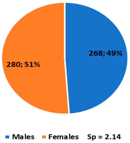
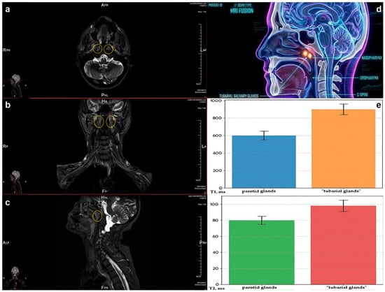
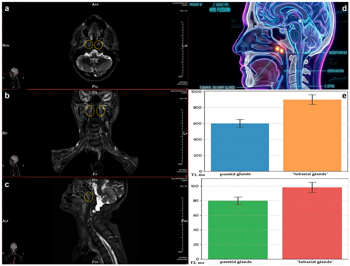
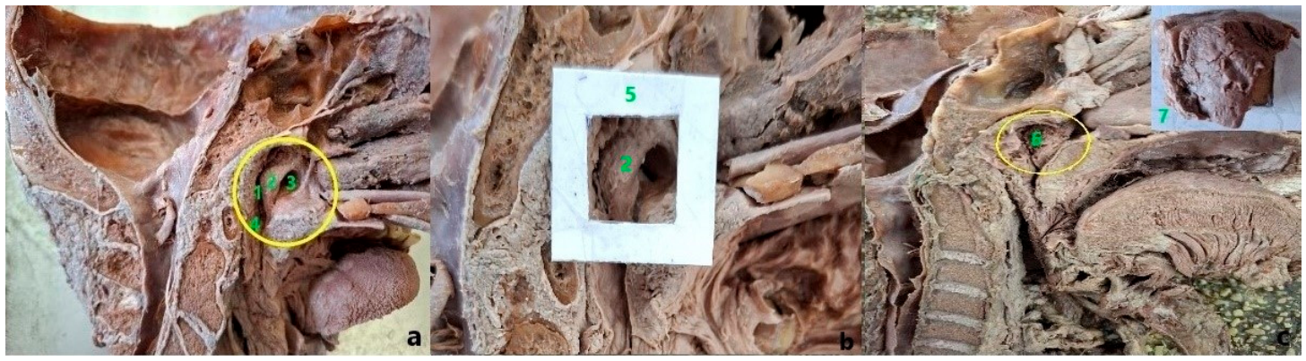
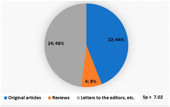
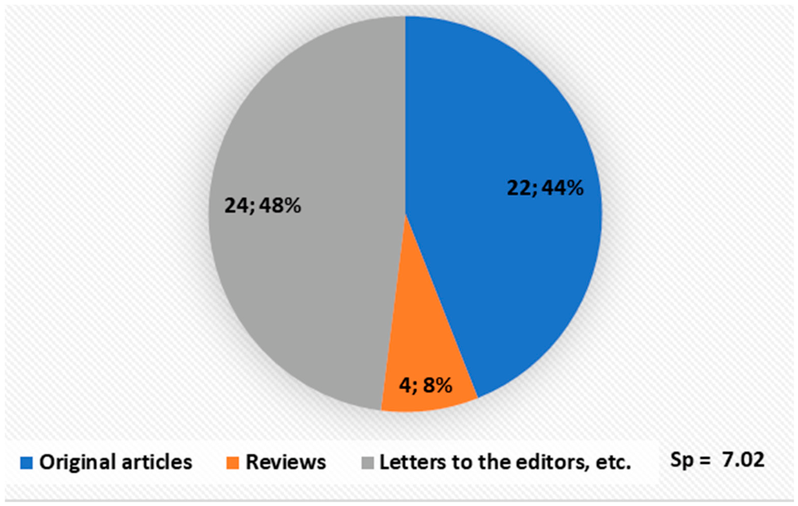
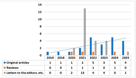
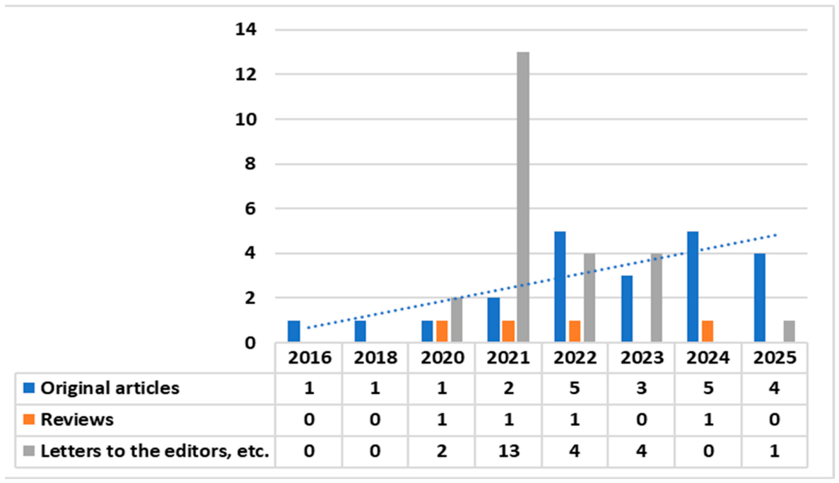
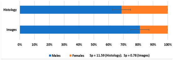
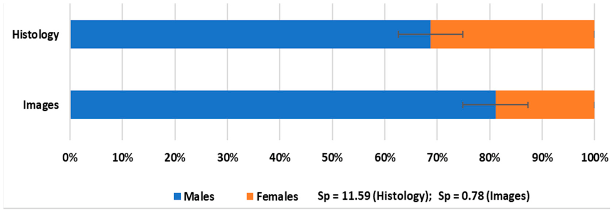
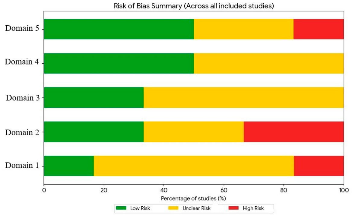
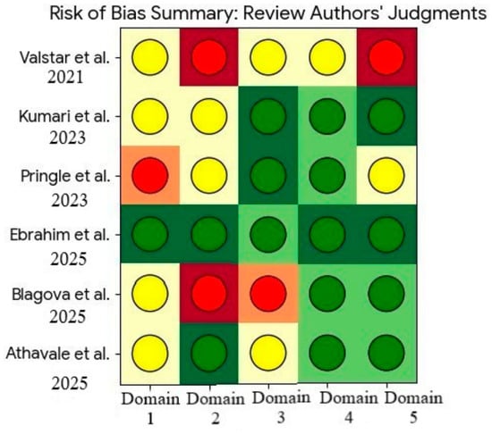
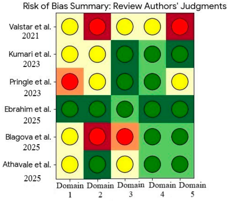
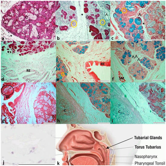
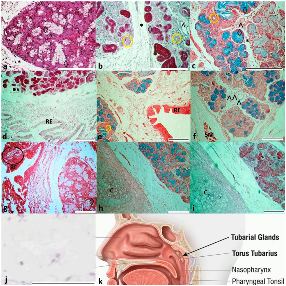
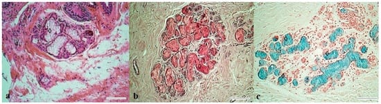
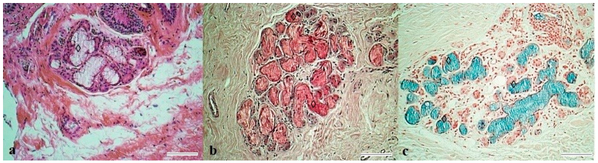
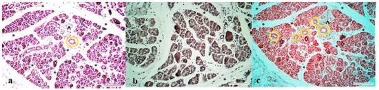
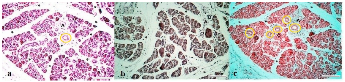
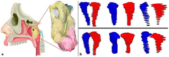
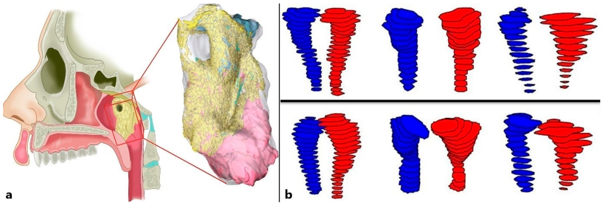

Disclaimer/Publisher’s Note: The statements, opinions and data contained in all publications are solely those of the individual author(s) and contributor(s) and not of MDPI and/or the editor(s). MDPI and/or the editor(s) disclaim responsibility for any injury to people or property resulting from any ideas, methods, instructions or products referred to in the content. |
© 2026 by the authors. Licensee MDPI, Basel, Switzerland. This article is an open access article distributed under the terms and conditions of the Creative Commons Attribution (CC BY) license.