


Abstract
Haemorrhagic stroke causes a disproportionate share of stroke deaths and disability, and its incidence rises steeply with age. Vagus nerve stimulation (VNS) targets modifiable secondary injury processes, including neuroinflammation, cerebral oedema, autonomic dysfunction and vasospasm, but its therapeutic role is uncertain. Animal studies and clinical randomised trials of VNS in intracerebral haemorrhage (ICH) or subarachnoid haemorrhage (SAH) were identified from Ovid MEDLINE (to 1 June 2026); appraised using SYRCLE, PEDro, and RoB 2; and synthesised narratively by level of evidence and pathology. Favourable functional outcome (modified Rankin Scale, 0–2) in SAH trials was pooled by random-effects meta-analysis. Nine reports were included: four preclinical (179 animals; three ICH models, one SAH model) and five clinical (four independent cohorts; 105 participants with haemorrhagic stroke: 97 SAH, 8 ICH). Animal studies reported reduced inflammatory signalling, haematoma volume and aneurysm rupture, as well as improved survival and motor recovery, with weaker effects after severe injury. In SAH trials, non-invasive VNS was feasible and well tolerated, with signals of reduced tumour necrosis factor-α and interleukin-6, less radiographic vasospasm and delayed cerebral ischaemia, and increased parasympathetic activity. The pooled functional outcome did not differ significantly (three trials: odds ratio [OR] 1.87, 95% confidence interval [CI] 0.65–5.37). Clinical ICH data were limited to 24 h safety in eight participants. VNS is biologically active and feasible but unproven; adequately powered, severity-stratified multicentre trials with standardised protocols and age-appropriate outcomes are needed.
1. Introduction
1.1. Haemorrhagic Stroke in an Ageing Population
Haemorrhagic stroke encompasses intracerebral haemorrhage (ICH), bleeding directly into the brain parenchyma, and subarachnoid haemorrhage (SAH), with bleeding into the subarachnoid space surrounding the brain. Together, they account for approximately 10–15% of all strokes, yet they carry a disproportionate burden of harm, causing around 40% of all stroke deaths and resulting in approximately half of the healthy life-years lost to stroke globally [1]. Despite advances in neurocritical care, including blood pressure management, haemostatic therapy and early aneurysm securing, outcomes after ICH and SAH remain poor [2]. Current management is largely supportive and directed at limiting further injury rather than actively promoting neurological recovery [3].
Both conditions are strongly age-related. The incidence of ICH rises steeply beyond the sixth decade, and rising rates of oral anticoagulant use, the accumulating prevalence of cerebral amyloid angiopathy and demographic ageing are expected to increase absolute case numbers over the coming decades. Older adults presenting with haemorrhagic stroke more frequently have pre-existing frailty, multimorbidity, cognitive impairment and polypharmacy, and consistently experience higher mortality, greater dependency and more limited access to rehabilitation than do younger patients [2]. Age-related autonomic changes, including reduced resting vagal tone and baroreflex sensitivity, together with the low-grade systemic inflammatory state often described as inflammaging, may plausibly alter both the pathophysiology of secondary injury and the response to interventions that act through autonomic and immune pathways. Interventions capable of attenuating secondary injury without surgical risk are therefore of particular relevance to the older populations who bear most of this disease burden.
1.2. Secondary Neurological Injury Mechanisms
Secondary injury processes are complex, interconnected and evolve over hours to days following the initial haemorrhage [4]. In ICH, haematoma expansion may occur during the acute phase, increasing the compression of surrounding tissue and worsening neurological injury [5]. The accumulation of blood and its breakdown products within the parenchyma initiates an inflammatory response involving activation of microglia and astrocytes and infiltration of peripheral immune cells. The subsequent release of pro-inflammatory cytokines contributes to blood–brain barrier (BBB) disruption, permitting further infiltration of inflammatory mediators and amplifying tissue damage [6]. BBB dysfunction also promotes cerebral oedema, which raises intracranial pressure, impairs cerebral perfusion and exacerbates perihaematomal injury [7].
Although many of these mechanisms also follow SAH, additional processes contribute to poor outcomes. Cerebral vasospasm, characterised by delayed narrowing of the cerebral arteries, commonly develops several days after aneurysmal rupture and may substantially reduce cerebral blood flow. This can precipitate delayed cerebral ischaemia (DCI), a major cause of neurological deterioration and long-term disability among SAH survivors, although DCI is now recognised to arise from a broader set of mechanisms than large-vessel vasospasm alone, including microcirculatory dysfunction, cortical spreading depolarisation and microthrombosis [8]. Together, these processes extend injury well beyond the initial bleed and represent the principal modifiable therapeutic target following haemorrhagic stroke. They are summarised in Figure 1A.
Figure 1.
(A) Principal secondary brain injury processes following intracerebral haemorrhage and aneurysmal subarachnoid haemorrhage. (B) Proposed mechanisms through which vagus nerve stimulation may attenuate secondary brain injury and the relationship between biological target engagement and functional recovery.
1.3. Vagus Nerve Stimulation
Vagus nerve stimulation (VNS) has emerged as a candidate therapeutic strategy capable of targeting several of the pathological mechanisms implicated in secondary brain injury. The vagus nerve is the tenth cranial nerve and serves as a major bidirectional communication pathway between the brain and peripheral organs, with important roles in autonomic regulation, inflammatory control and physiological homeostasis [9]. Traditionally, VNS has been delivered via surgically implanted electrodes wrapped around the cervical vagus nerve and connected to a pulse generator implanted in the chest wall. More recently, non-invasive techniques have been developed, including transcutaneous auricular VNS (taVNS) and non-invasive transcutaneous cervical VNS (tcVNS), which avoid the risks of surgical implantation while improving accessibility and ease of bedside delivery [10].
VNS is an established treatment for drug-resistant epilepsy [11] and is also approved for treatment-resistant depression and certain primary headache disorders [12]. Its established safety profile and the growing understanding of its biological mechanisms have driven increasing interest in its application across a range of neurological disorders, including traumatic brain injury, disorders of consciousness, ischaemic stroke and, more recently, haemorrhagic stroke [13].
Several mechanisms have been proposed to explain its therapeutic potential. Activation of vagal pathways can suppress systemic and central inflammation through the cholinergic anti-inflammatory pathway, modulate autonomic dysfunction, influence cerebral blood flow and promote neuroplasticity [14]. Because many of these pathways overlap with the mechanisms underpinning secondary injury after ICH and SAH, VNS represents an attractive candidate capable of targeting multiple pathological processes simultaneously rather than treating a single downstream complication. Across experimental models of ICH and SAH, VNS has reduced neuroinflammation, attenuated cerebral oedema and improved neurological recovery [15,16]. In clinical studies, VNS has shown promise in modulating autonomic dysfunction following SAH by restoring parasympathetic activity and improving cardiovascular regulation [17], and there is emerging evidence that it may reduce the incidence and severity of cerebral vasospasm after aneurysmal SAH (aSAH) [18].
1.4. Rationale, Hypotheses and Objectives
The application of VNS in ICH and SAH remains in its infancy, but the evidence base is expanding rapidly and has not previously been synthesised systematically. Existing reviews of VNS in stroke have focused predominantly on ischaemic stroke and upper limb rehabilitation, with haemorrhagic subtypes either excluded or represented by small subgroups. A dedicated synthesis is therefore warranted.
We hypothesised that VNS would demonstrate beneficial effects across preclinical and clinical studies through anti-inflammatory, autonomic and neurorestorative mechanisms; that these effects would be detectable as reductions in neuroinflammation, cerebral oedema and vasospasm; and that such biological effects would be accompanied by improved neurological outcomes. We further hypothesised, on the basis of injury-severity gradients described in experimental work, that any therapeutic effect may be attenuated once primary tissue destruction became extensive.
The objectives of this review were (i) to identify and systematically synthesise the current evidence investigating VNS in haemorrhagic stroke and related haemorrhagic brain injury; (ii) to evaluate the effects of VNS on key secondary injury mechanisms, including neuroinflammation, cerebral oedema, cerebral vasospasm and autonomic dysfunction; (iii) to assess the impact of VNS on neurological recovery, functional outcomes and disease-related biomarkers; (iv) to evaluate the safety and feasibility of VNS across preclinical and clinical studies; and (v) to compare preclinical and clinical findings and identify limitations and knowledge gaps to inform future research.
2. Materials and Methods
2.1. Design and Reporting
This systematic review was conducted and reported in accordance with the Preferred Reporting Items for Systematic Reviews and Meta-Analyses (PRISMA) 2020 statement [19] (Supplementary File S1). Given the small and emerging evidence base, both preclinical and clinical studies were included to provide a translational overview of the therapeutic potential of VNS in experimental models and human applications. Because substantial heterogeneity was anticipated in the study populations, stimulation modalities, and outcome measures, a narrative synthesis was adopted, with quantitative pooling undertaken only where sufficiently comparable data were available. Preclinical and clinical evidence was extracted, appraised and synthesised separately throughout, as well as within, each level of evidence, by pathology (ICH and SAH). Quantitative synthesis was restricted to clinical trials of the same pathology reporting the same outcome; in practice, only SAH trials met this criterion. This review was registered on OSF (10.17605/OSF.IO/H7WY4).
2.2. Eligibility Criteria
Studies were eligible if they investigated VNS in haemorrhagic stroke or related haemorrhagic brain injury and reported outcomes relating to neurological recovery, inflammation, autonomic regulation, cerebral oedema, vasospasm or functional performance. Preclinical studies using animal models of haemorrhagic injury were eligible. Clinical studies were eligible only if they employed a randomised controlled design. Reviews, conference abstracts, editorials, protocols and opinion papers were excluded, as were studies that did not investigate VNS as the primary intervention or were unrelated to the review question. Although review articles were excluded from formal analysis, they were used to provide contextual background, and their reference lists were screened for additional eligible reports.
2.3. Search Strategy
Literature searches were conducted in Ovid MEDLINE(R) ALL, using search strategies developed to capture both haemorrhagic stroke pathologies and VNS interventions. Population terms included intracerebral haemorrhage, intracranial haemorrhage, subarachnoid haemorrhage, haemorrhagic stroke, cerebral aneurysm and aneurysmal SAH; UK and US spellings were incorporated to maximise sensitivity. Intervention terms included vagus nerve stimulation, transcutaneous VNS, transcutaneous auricular VNS, non-invasive VNS, cervical VNS, invasive VNS and associated abbreviations. The population and intervention concepts were combined with the Boolean operator AND; no NOT operator and no language, publication-type or study-design limits were applied. The full search strategy is provided in Supplementary Table S1. Searches were conducted from database inception until 1 June 2026. A single bibliographic database was searched. MEDLINE was chosen because it indexes the core neurology, neurosurgery, neurocritical care and brain-stimulation journals in which this literature is published, and all eight published reports eventually included appeared in MEDLINE-indexed journals. To reduce the risk of missed studies, the search was supplemented by screening the reference lists of relevant reviews and included reports, as well as by the research team’s knowledge of recent work, which identified one additional report. The implications of this approach are considered in Section 4.9.2.
2.4. Study Selection
Search results were exported and duplicate records removed before screening. Titles and abstracts were assessed against the predefined eligibility criteria to remove clearly irrelevant records. Full texts were obtained and reviewed for potentially eligible reports, and eligibility was reassessed by two of the authors (M.T. and A.A.), using the same criteria independently. Any discrepancies or uncertainties were discussed and reviewed by a third author (A.M.). Reports were excluded if they did not investigate haemorrhagic pathology, did not use VNS as the primary intervention, did not report a relevant outcome, or represented an ineligible publication type. Reference lists of relevant reviews and included reports were also examined for eligible studies not identified through the database searches.
2.5. Data Extraction
Data extraction was performed using a standardised spreadsheet developed for this review, enabling consistent comparison across heterogeneous studies. Extracted information included author, publication year, country, study population, sample size, study design, VNS modality, stimulation site, stimulation frequency, intervention duration, comparator group and outcome measures. Principal findings and expanded summary results were also recorded. For preclinical studies, additional data included animal species, haemorrhage model, injury severity and experimental neurological, behavioural, inflammatory, histological and structural outcomes. For clinical studies, safety, tolerability and feasibility data were extracted, including the number and rate of adverse events and serious adverse events, their reported relationship to VNS, treatment withdrawals or discontinuations and reasons for withdrawal. Adherence data included the number of planned and completed stimulation sessions, treatment-completion rates, missed sessions and reasons for non-adherence, where available. Particular attention was given to stimulation parameters because of the considerable variation between studies.
2.6. Outcome Measures
The primary clinical outcome of the review was functional outcome, measured by the modified Rankin Scale (mRS); this was the only outcome considered for quantitative synthesis. Key secondary clinical outcomes were safety, tolerability and feasibility, and mortality. Inflammatory biomarkers, cerebral vasospasm, delayed cerebral ischaemia, cerebral infarction and autonomic measures were secondary; mechanistic (target-engagement) outcomes and were interpreted as exploratory. All preclinical outcomes were treated as exploratory and were not pooled.
Given the heterogeneity of the included studies, outcomes were grouped into seven domains to facilitate comparison: haemorrhage-related outcomes; inflammation; neurological and functional recovery; survival; structural or neuroimaging outcomes; safety, feasibility and adherence; and autonomic or cardiovascular function. Preclinical haemorrhage-related outcomes included haematoma volume (mL), aneurysm rupture (%), SAH severity and other measures of haemorrhage progression (e.g., haematoma expansion). Inflammatory outcomes included pro-inflammatory cytokines and inflammatory signalling proteins, together with measures of BBB dysfunction and cerebral oedema. Neurological and functional recovery were assessed using experimental neurological scores and behavioural measures, including the modified Neurological Severity Score (mNSS), rotarod performance, beam walking and task-specific motor assessments. Survival and structural or neuroimaging measures, including neuronal survival, myelin integrity, white-matter preservation and sensorimotor-network remodelling, were additionally extracted. Clinical outcomes included safety, tolerability and feasibility; circulating and cerebrospinal fluid (CSF) inflammatory biomarkers; cerebral vasospasm, DCI and cerebral infarction; heart rate, blood pressure, heart-rate variability (HRV) and related physiological measures; and the modified Rankin Scale (mRS), Barthel Index, ongoing neurological impairment, mortality and length of hospital stay.
2.7. Risk of Bias Assessment
Methodological quality and risk of bias were assessed separately for preclinical and clinical studies because of inherent differences in study design and experimental methodology. Preclinical studies were assessed using the Systematic Review Centre for Laboratory Animal Experimentation (SYRCLE) risk-of-bias tool [20], developed specifically for animal intervention studies, which evaluates selection, performance, detection, attrition and reporting bias, as well as and other potential sources of bias, and additionally accounts for animal-specific considerations such as random housing, random outcome assessment and blinding procedures. Clinical studies were evaluated using the Physiotherapy Evidence Database (PEDro) scale [21], an 11-item instrument assessing randomisation, allocation concealment, baseline comparability, blinding, completeness of outcome data, and statistical reporting; the eligibility-criteria item is not scored, giving a maximum of 10. One reviewer undertook the primary risk-of-bias assessments (MT), and a second reviewer independently reviewed the judgements (JB), with discrepancies resolved by discussion. Risk-of-bias findings were used to contextualise the strength of the evidence and inform interpretation. For robustness, any clinical trials included in quantitative syntheses were also assessed using the Cochrane RoB 2 tool (Supplementary File S3).
2.8. Statistical Analysis
Preclinical and clinical evidence was synthesised separately, with preclinical findings considered first. Because of substantial differences between animal models, intervention protocols and outcome measures, preclinical evidence was synthesised narratively and was not considered suitable for quantitative pooling.
A quantitative synthesis was undertaken for clinical reports providing sufficiently comparable binary functional-outcome data. Only three trials met this criterion, all of which enrolled patients with spontaneous, predominantly aneurysmal SAH; no ICH trial reported functional outcome, so no ICH estimate or ICH vs. SAH comparison was possible. These trials were pooled because they shared the same pathology, a sham-controlled randomised design and the same outcome construct. The purpose of pooling was to summarise the direction and imprecision of the available evidence, not to estimate efficacy, and trial-level estimates are presented alongside the pooled estimate. A favourable outcome was defined as mRS 0–2 and a poor outcome as mRS 3–6. The latest available functional assessment was selected from each report, i.e., first outpatient follow-up for Huguenard et al. [18], one-month follow-up for Myers et al. [22], and hospital discharge for Rebeiz et al. [23], for which no later mRS assessment was reported. All randomised participants were retained where group allocation was known; participants for whom functional-outcome data were unavailable were conservatively classified as having a poor outcome. This approach was applied consistently across reports to avoid excluding participants on the basis of post-randomisation outcome availability. Odds ratios (ORs) with 95% confidence intervals (CIs) were calculated for favourable functional outcome in the VNS group relative to the sham and visualised as forest plots. Log odds ratios were pooled using a random-effects inverse-variance model, with between-study variance estimated by restricted maximum likelihood (REML). Statistical heterogeneity was evaluated using Cochran’s Q, the I2 statistic, and τ2 [24]. With three trials, τ2 cannot be estimated reliably, and heterogeneity statistics have very low power; when τ2 is estimated as zero, the random-effects model reduces to a common-effect model, and the Wald-type confidence interval does not reflect between-trial heterogeneity. Pooled estimates were therefore interpreted as descriptive summaries. Tests were two-sided, with p < 0.05 considered statistically significant. Analyses were performed in IBM SPSS Statistics version 29.0.2.0 (IBM Corp., Armonk, NY, USA). Publication bias was not formally assessed because only three reports contributed to the meta-analysis; with so few reports, funnel-plot interpretation and tests of asymmetry would be underpowered and potentially misleading.
No subgroup analyses were prespecified. One post hoc exploratory sensitivity analysis was undertaken. Major haemorrhage was defined clinically as Hunt and Hess grade 5 (coma, decerebrate posturing) at presentation; no volumetric criterion was used because haemorrhage volume is not routinely quantified in SAH, and it was not reported by the included trials. Participants with major haemorrhage were removed from the Myers et al. dataset [22], and the meta-analysis was repeated using the same model. The analysis had two aims: to make the trial populations more comparable, since VANQUISH enrolled only participants who were alert and able to report headache intensity [23]; and to remove a chance baseline imbalance, since all four grade 5 participants in the Myers trial were randomised to taVNS. Because it altered one trial only, was not prespecified and did not test a treatment-by-severity interaction, it was treated as exploratory rather than as a subgroup analysis.
3. Results
3.1. Study Selection
The database search identified 409 records, and one additional record was supplied by the research team. After removal of 23 duplicates, 386 titles and abstracts were screened, and 356 records were excluded. Full texts manuscripts of 30 articles were obtained, and 21were excluded because they were reviews or protocols, investigated an ineligible population or intervention, or did not report relevant outcomes. The remaining nine full texts were assessed and included, comprising five clinical reports (168 unique participants, of whom 105 had haemorrhagic stroke: 97 with SAH and 8 with ICH) and four preclinical reports (179 animals). Three clinical reports provided binary functional-outcome data suitable for meta-analysis. The selection process is summarised in Figure 2.
Figure 2.
PRISMA 2020 flow diagram illustrating the identification, screening and inclusion of studies and reports.
3.2. Characteristics of Included Studies
Nine reports were included: five clinical and four preclinical. Three preclinical reports used rat models of ICH [16,25,26], and one used mild and severe mouse models of intracranial aneurysm rupture [27]. Preclinical sample sizes ranged from 26 to 53 animals. Interventions comprised auricular and cervical non-invasive VNS and implanted VNS paired with rehabilitation. Stimulation frequencies ranged from 25 to 30 Hz, and treatment ranged from single acute stimulation sessions to six weeks of rehabilitation-paired stimulation.
The clinical evidence comprised three published reports and one accepted manuscript involving patients with SAH or aSAH [17,18,22,23], together with one mixed acute-stroke trial that included eight participants with ICH [28]. Tan et al. [17] reported a cardiovascular analysis of 24 participants drawn from the NAVSaH trial population subsequently reported by Huguenard et al. [18]; these publications therefore do not represent independent cohorts, and the five clinical reports correspond to four independent clinical cohorts. Clinical sample sizes ranged from 24 to 71 randomised participants, although the haemorrhagic subgroup in the larger Arsava et al. study comprised only eight ICH participants, the remainder having ischaemic stroke. Study characteristics, stimulation protocols and principal findings are presented in Table 1 (preclinical) and Table 2 (clinical).
Table 1.
Characteristics, stimulation protocols and principal findings of included preclinical studies.
Table 2.
Characteristics, stimulation protocols and principal findings of included clinical studies.
3.3. Risk of Bias Assessment
3.3.1. Preclinical Studies
The SYRCLE assessment identified variable methodological reporting (Table 3). Randomisation was reported by Suzuki et al. [27] and Cáceres et al. [26], while three reports described comparable baseline characteristics and blinded outcome assessment. All four reported their prespecified outcomes. None met the criteria for allocation concealment, blinded intervention delivery or random selection for outcome assessment.
Table 3.
SYRCLE risk-of-bias assessment of included preclinical studies.
Attrition was a particular concern in the work of Hays et al. [25], in which 32 of 58 animals were excluded from the principal analysis because of death, insufficient or excessive post-lesion impairment, or stimulation-device failure. Although exclusion reasons were reported, and data for all animals were provided in the Supplementary Materials, the extent of exclusion introduces uncertainty regarding attrition bias. Random housing was poorly reported. A judgement of “no” indicates that a criterion was either unmet or insufficiently reported, and the “other bias” domain remained unclassified.
3.3.2. Clinical Studies
PEDro scores ranged from 8 to 9 out of 10, excluding the non-scored eligibility criterion (Table 4). All five clinical reports described random allocation, concealed allocation, adequate outcome data collection, between-group comparisons and measures of variability. Four reported baseline comparability, with this criterion not recorded for the UK VNS-SAH pilot. Blinding of intervention delivery was the most frequent limitation and was not achieved in the Huguenard, VNS-SAH, Arsava or Tan reports. Outcome-assessor blinding was not recorded for Rebeiz et al. [23], and intention-to-treat analysis was not explicitly reported by Tan et al. [17]. These uniformly high scores should be interpreted with caution for the reasons set out in Section 4.9.
Table 4.
PEDro assessment of the methodological quality of included clinical reports.
3.4. Preclinical Evidence
Three studies used rat models of ICH [16,25,26], and one used mouse models of intracranial aneurysm rupture and SAH [27]. Findings are presented by pathology and, within each, by outcome domain.
3.4.1. Intracerebral Haemorrhage Models
Cáceres et al. [26] delivered tcVNS or sham 30 min following induction of experimental ICH in Wistar rats and reported a smaller mean haematoma volume following tcVNS in the lower-dose collagenase model (5.51 vs. 8.97 mm3; p = 0.030), whereas no significant effect was observed in the higher-dose model (11.19 vs. 12.38 mm3; p = 0.38). Haemoglobin concentrations did not differ significantly at either dose.
Zhang et al. [16] paired taVNS or sham alongside rehabilitation exercises in Sprague Dawley rats induced with ICH (collagenase model) and reported less residual haematoma at week two with taVNS plus treadmill rehabilitation than with rehabilitation alone, although the gross anatomical comparison was based on selected samples. By week four, both groups showed near-complete resolution. taVNS was also associated with greater myelin basic protein expression, lower SMI-32 expression, greater fractional anisotropy, and enhanced corticospinal-tract reconstruction when compared to the results for sham stimulation.
Hays et al. [25] implanted cervical VNS in Sprague Dawley ICH model rats and found no significant differences in lesion volume between those receiving VNS-paired rehabilitation and rehabilitation alone (11.09 ± 1.58 vs. 13.55 ± 2.56 mm3; p = 0.38). The functional improvement associated with VNS therefore occurred without a measurable reduction in primary tissue loss.
Cáceres et al. [26] reported a borderline increase in AQP-4 following tcVNS (p = 0.053); no ICH study directly measured blood–brain barrier permeability or cerebral oedema.
Neurological recovery improved in all three ICH studies, although only in the lower-dose model in the work of Cáceres et al. [26]. Zhang et al. [16] reported lower mNSS scores with taVNS at weeks two (p < 0.05) and four (p < 0.01), along with increases in sensorimotor-network activity and centrality correlated with lower mNSS scores (r = −0.594, p = 0.042; r = −0.598, p = 0.040).
In the work of Hays et al. [25], the principal analysis included 14 rats receiving VNS-paired rehabilitation and 12 receiving rehabilitation alone. Recovery of forelimb performance was greater following VNS-paired rehabilitation (76.8 ± 11.3% vs. 29.4 ± 12.0%; p = 0.0062). Significant treatment effects were observed for hit rate (F [1,144] = 39.59; p = 3.54 × 10⁻⁹) and movement speed (F [1,144] = 57.67; p = 3.58 × 10⁻¹²). Training intensity did not differ between groups (p = 0.633), and the functional benefit persisted during week six after stimulation was withdrawn.
In Cáceres et al. [26], the decline in rotarod performance was smaller with tcVNS in the lower-dose model (−29.2 vs. −67.1 s; p = 0.0175), but this benefit was not reproduced in the higher-dose model. Beam-walking and Garcia scores did not differ significantly between treatment groups.
3.4.2. Intracranial Aneurysm and SAH Model
Suzuki et al. [27] induced cerebral aneurysms using stereotactic injections of elastase into the cerebrospinal fluid of mice and found that tcVNS reduced aneurysm rupture in the mild model (controlled blood pressure) from 80% with femoral-nerve stimulation to 29% (p = 0.036), and reduced SAH grade (p = 0.025). In the severe model (uncontrolled blood pressure), rupture rates and SAH grade did not differ between groups. Further, tcVNS reduced several inflammatory mediators within the circle of Willis, but only the 34% reduction in matrix metalloproteinase-9 (MMP-9) reached statistical significance; reductions in interleukin-1β (IL-1β), tumour necrosis factor alpha (TNF-α), C-C motif chemokine ligand 2 (CCL2), interleukin-6 (IL-6) and inducible nitric oxide synthase were non-significant. Blood–brain barrier permeability and cerebral oedema were not measured. In the severe aneurysm model, median survival increased from 6 to 13 days with tcVNS (p = 0.003), and deficit-free survival increased from 4 to 6 days (p = 0.029), despite similar rupture rates and SAH grades [27]. Resting arterial blood pressure did not differ between stimulation groups.
3.5. Clinical Evidence
The clinical evidence comprised one mixed acute stroke trial with an ICH subgroup [28] and three SAH trials, one of which also contributed a secondary cardiovascular report [17,18,22,23].
3.5.1. Intracerebral Haemorrhage
The only clinical ICH data came from the eight participants with ICH in the TR-VENUS trial of Arsava et al. [28], a randomised, sham-controlled, open-label safety and feasibility study of cervical nVNS in acute stroke. All prespecified stimulations were delivered, and among the eight ICH participants, no haematoma growth greater than 30% and no neurological deterioration occurred within 24 h. No functional, inflammatory or imaging efficacy outcome was reported for the ICH subgroup.
3.5.2. Subarachnoid Haemorrhage
VNS was generally deliverable without intervention-related serious adverse events. Huguenard et al. [18] reported no adverse events attributable to taVNS. In the UK VNS-SAH pilot, 221 of 259 possible sessions were delivered (85.3%), with no intervention-related serious adverse events; effects were mainly mild and transient, although one participant withdrew because of headache [22]. Rebeiz et al. [23] completed 950 of 1260 scheduled stimulations (75.4%); muscle twitching, lip pulling and nausea or vomiting were more frequent with active nVNS, but no serious device-related event or haemodynamic instability occurred.
Inflammation, Vasospasm and Delayed Cerebral Ischaemia: Huguenard et al. [18] found lower plasma TNF-α in the taVNS group on days 7 and 10 (p = 0.015, and p = 0.030) and lower plasma IL-6 on day 4 (p = 0.024). CSF TNF-α and IL-6 were also lower on day 13 (p = 0.031, and p = 0.025). Moderate-to-severe radiographic vasospasm affected 4 of 13 taVNS participants and 10 of 14 sham participants (30.8% vs. 71.4%; p = 0.018), and vessel-level analysis similarly favoured taVNS (17.7% vs. 27.7%; p = 0.015). Cerebral infarction was numerically less frequent with taVNS, although the difference was not significant (23.1% vs. 42.9%; p = 0.41).
In the VNS-SAH pilot, the change in serum TNF-α favoured taVNS (p = 0.028), with borderline between-group differences for IL-1β and interleukin-10 (both at p = 0.050) but not for IL-6 or C-reactive protein [22]. DCI occurred in 1 of 15 active participants and 5 of 14 sham participants (6.7% vs. 35.7%; p = 0.059). Rebeiz et al. [23] also reported fewer new cerebral infarctions with nVNS than sham (5.6% vs. 23.8%), although this difference was not statistically significant (p = 0.19).
Autonomic and Cardiovascular Outcomes: In the NAVSaH cardiovascular analysis, repetitive taVNS did not significantly alter heart rate, blood pressure or intracranial pressure and did not produce bradycardia or QT prolongation [17]. Relative to sham, taVNS increased the root mean square of successive differences between heartbeats (RMSSD; Bonferroni-corrected p = 0.025), overall HRV (p = 0.04) and the derived parasympathetic-activity factor (p = 0.04). Corrected QT prolongation was more marked in the sham group (p < 0.001).
Neurological and Functional Outcomes: Clinical functional outcomes were generally favourable in direction but statistically inconclusive. In the work of Huguenard et al. [18], favourable mRS outcomes occurred in 38.4% vs. 21.4% of patients at discharge and 76.9% vs. 57.1% at first follow-up for taVNS and sham, respectively; neither between-group comparison was statistically significant. Rebeiz et al. [23] reported favourable discharge mRS outcomes in 94.4% of active participants and 80.0% of sham participants (p = 0.34), with no significant difference in length of stay, alongside significantly reduced headache intensity and reduced opioid use. In the VNS-SAH pilot, neither the mRS nor Barthel Index differed significantly at discharge or one month; ongoing neurological impairment was numerically less frequent with taVNS (16.7% vs. 53.8%; p = 0.101), but inpatient mortality was 20.0% with taVNS and 7.1% with sham in the context of greater baseline haemorrhage severity in the active group [22].
3.6. Exploratory Meta-Analysis of SAH Trials
3.6.1. Included Reports and Outcome Definition
Three SAH trials contributed binary data for a favourable functional outcome, i.e., Huguenard et al. [18], Rebeiz et al. [23] and Myers et al. [22], comprising 96 participants. No ICH trial reported functional outcome, so no ICH estimate could be calculated. Two trials used auricular and one cervical stimulation, and functional outcome was assessed at hospital discharge [23], one month [22] and first outpatient follow-up [18]. Using RoB 2 (Supplementary File S3), the functional-outcome result was at high risk of bias in one trial [18] and raised some concerns in two [22,23]. A random-effects inverse-variance model with REML estimation was used to pool log odds ratios.
3.6.2. Primary Analysis
Trial-level estimates were imprecise and inconsistent in direction (Figure 3), with OR 2.50 (95% CI 0.47–13.27) for Huguenard et al., 5.31 (0.56–50.55) for Rebeiz et al. and 0.75 (0.14–4.17) for Myers et al. The pooled estimate favoured VNS but was not statistically significant (OR 1.87, 95% CI 0.65–5.37; p = 0.25). No statistical between-study heterogeneity was detected (I2 = 0%; τ2 = 0.00; Q = 2.03, df = 2, p = 0.36), although with three trials, this cannot be taken as evidence of homogeneity. These data do not permit a reliable estimate of efficacy: fewer than 100 participants contributed, or about one-sixth of the approximately 580 required to detect a 10-percentage-point absolute improvement in mRS 0–2 from the pooled sham rate of about 70% (80% power, two-sided α = 0.05). Individual and pooled estimates are shown in Figure 3.
Figure 3.
Forest plot of the association between vagus nerve stimulation and favourable functional outcome (modified Rankin Scale, 0–2) in the primary exploratory random-effects meta-analysis of three SAH trials: Myers et al. [22], Huguenard et al. [18], and Rebeiz et al. [23]. Odds ratios greater than 1 favour vagus nerve stimulation. OR, odds ratio.
3.6.3. Exploratory Sensitivity Analysis
In the post hoc exploratory sensitivity analysis defined in Section 2.8, excluding participants with Hunt and Hess grade 5 haemorrhage (n = 4) from the Myers dataset gave a pooled OR of 2.93 (95% CI 0.90–9.61; p = 0.08). No statistical heterogeneity was detected (I2 = 0%; τ2 = 0.00; Q = 0.39, df = 2, p = 0.82). Results are shown in Figure 4. Individual and pooled treatment effects are summarised in Table 5.
Figure 4.
Forest plot of the post hoc exploratory sensitivity analysis of three SAH trials, excluding participants classified as having major haemorrhage (Hunt and Hess grade 5, n = 4) from the Myers et al. [22] dataset. The Huguenard et al. [18] and Rebeiz et al. [23] data remained unchanged. Odds ratios greater than 1 favour vagus nerve stimulation. OR, odds ratio.
Table 5.
Summary of the exploratory meta-analyses of favourable functional outcome in SAH trials.
4. Discussion
This systematic review identified an emerging but limited evidence base indicating that VNS is biologically active following haemorrhagic brain injury. Across preclinical studies, VNS was associated with reduced haemorrhage-related injury and inflammatory signalling, alongside improvements in neurological recovery and neuroplastic remodelling, although these effects were less consistent in severe injury models. In early clinical studies, almost all in SAH, VNS was generally feasible and well tolerated in neurocritical care populations, and demonstrated preliminary effect signals on inflammatory biomarkers, cerebral vasospasm, DCI and autonomic regulation. These favourable biological findings did not, however, consistently translate into significant improvements in disability or independence. Overall, the preclinical findings support biological plausibility, but the clinical evidence does not demonstrate benefit; whether any benefit depends on pathology, injury severity, treatment timing or stimulation protocol remains a hypothesis.
4.1. Biological and Translational Effects of VNS by Level of Evidence and Pathology
4.1.1. Preclinical Evidence
Preclinical evidence suggests that VNS may attenuate acute brain injury, potentially via the cholinergic anti-inflammatory pathway, in which α7 nicotinic acetylcholine receptor signalling suppresses TNF-α, IL-1β and IL-6 production [29,30]. In a mouse aneurysm rupture model, Suzuki et al. [27] found that nVNS reduced rupture and SAH severity in mild but not severe disease, where only survival improved. In rat ICH models, Cáceres et al. [26] observed smaller haematomas and better motor performance after lower-dose, but not after severe injury. VNS may therefore modify secondary injury, with a ceiling once primary injury is extensive. Neither study showed BBB restoration or reduced oedema, and the borderline AQP-4 increase reported by Cáceres et al. is ambiguous, as AQP-4 contributes to both oedema formation and clearance.
When paired with rehabilitation, VNS may also enhance neuroplasticity by recruiting cholinergic and noradrenergic systems that strengthen task-engaged circuits [31,32]. In rat ICH, Hays et al. [25] reported substantially greater, durable forelimb recovery with VNS-paired rehabilitation, despite unchanged lesion volume, suggesting reorganisation of surviving networks rather than tissue preservation. Zhang et al. [16] associated taVNS with improved neurological scores, myelin preservation and corticospinal and sensorimotor-network remodelling, although correlation cannot establish causation. In ischaemic stroke, the 108-participant VNS-REHAB trial found greater upper limb improvement with VNS-paired than with sham-paired rehabilitation [33], though this cannot be directly extrapolated to ICH or SAH. VNS may therefore have two roles: reducing acute secondary injury and later enhancing rehabilitation-driven recovery.
4.1.2. Clinical Evidence
In SAH, Huguenard et al. [18] reported lower plasma and CSF TNF-α and IL-6, less moderate-to-severe radiographic vasospasm and numerically fewer infarctions with taVNS. Myers et al. [22] found a favourable change in serum TNF-α, but not in IL-6 or C-reactive protein, and a lower but inconclusive DCI rate. Rebeiz et al. [23] reported numerically fewer new infarctions with nVNS. These findings are compatible with vagal modulation of secondary injury. However, cytokine changes were not shown to cause the vascular effects, and reduced vasospasm does not demonstrate improved perfusion. Together, they indicate biological activity, not clinical efficacy.
Tan et al. [17] re-analysed the Huguenard (NAVSaH) cohort and found increased RMSSD, HRV and parasympathetic activity without concerning changes in heart rate, blood pressure or intracranial pressure. This suggests target engagement but is not independent confirmation, and HRV is confounded in sedated, ventilated patients. No intervention-related serious adverse events occurred in the SAH trials, and effects were mainly mild and transient. In ICH, the only data, obtained from Arsava et al. [28], showed no substantial haematoma growth or early deterioration within 24 h in eight participants. VNS thus appears feasible and well tolerated, but small samples cannot exclude uncommon adverse events or establish safety in ICH.
4.1.3. Pathology-Specific Mechanisms: ICH vs. SAH
ICH and SAH share inflammatory and BBB injury pathways. They differ in regards to which and when secondary processes dominate and hence, concerning which VNS effects matter most; trial-design implications are given in Table 6 (Section 4.7).
In ICH, haematoma expansion occurs mainly within hours of onset [5] and would require a pro-haemostatic effect, such as vagally mediated platelet priming [34,35] (Section 4.3). The smaller haematomas reported by Cáceres et al. [26] are consistent with this, although the mechanism was not tested. Perihaematomal oedema evolves over days through microglial activation, cytokine release and barrier disruption [6,7], and here, the cholinergic anti-inflammatory pathway is most relevant [29,30]. However, oedema was not measured directly, and the aquaporin-4 change is of uncertain direction [26]. The gains reported by Hays et al. [25] and Zhang et al. [16], without reduced lesion volume, suggest a third, later mechanism: rehabilitation-dependent plasticity. None has been examined in patients with ICH.
In SAH, rupture causes an intracranial pressure rise, global ischaemia and a catecholamine surge within 72 h [4], with cardiac and pulmonary complications [36]. The increased HRV and attenuated QTc prolongation reported by Tan et al. [17] are consistent with parasympathetic buffering, albeit as physiological endpoints. DCI develops over days 3–14 from vasospasm, microcirculatory constriction, microthrombosis, cortical spreading depolarisations and neuroinflammation [8]. VNS reduced susceptibility to cortical spreading depression in rats [37] and lowered relevant cytokines in two trials [18,22]. However, the reduced vasospasm in one trial [18] recalls earlier therapies that reduced angiographic narrowing without improving outcome [38]. A pro-haemostatic effect would also be undesirable, as microthrombosis contributes to DCI [8].
These mechanisms do not support a single strategy: their windows differ, and some effects conflict. Platelet priming could limit haematoma expansion but promote microthrombosis during DCI. Cytokine suppression could increase infection risk (Section 4.5), and parasympathetic augmentation could oppose haemodynamic augmentation for DCI, although heart rate and blood pressure were unchanged in the included trials [17,23]. VNS in ICH and VNS in SAH should therefore be regarded as distinct interventions, each with its own timing, dose and endpoints.
4.2. Functional Outcome and a Hypothesised Severity Threshold
Although the biological findings were encouraging, evidence that VNS improves patient-centred functional recovery was less conclusive. The meta-analysis of three SAH reports produced a pooled estimate favouring VNS for achieving a favourable mRS outcome, but the confidence interval crossed the null value. This should not be interpreted as evidence that VNS has no effect; rather, the wide interval remains compatible with clinically important benefit, with no effect, and with modest harm. No statistical heterogeneity was detected, with I2 and τ2 both equal to zero. This is very likely an artefact of low power rather than evidence of homogeneity: heterogeneity statistics have little power with three trials, and the trials differed in stimulation modality (auricular vs. cervical), haemorrhage severity, inclusion of perimesencephalic SAH, treatment duration and timing of outcome assessment. The pooled result should therefore be regarded as hypothesis-generating rather than confirmatory.
The gap between biological and functional findings may reflect low power or a severity-dependent treatment response. A post hoc sensitivity analysis excluded major haemorrhage, defined as Hunt and Hess grade 5 (Section 2.8), from the Myers dataset. Such patients were effectively ineligible for VANQUISH, which required alert participants able to report headache [23], and all four had been randomised to taVNS. The estimated benefit then increased, though not significantly. This is directionally consistent with the severe-model findings of Suzuki et al. [27] and Cáceres et al. [26]. VNS may act mainly on secondary processes and, by analogy with the ischaemic penumbra, may require sufficient salvageable tissue to improve function. We therefore propose a falsifiable but unproven hypothesis: that the effect of VNS on functional outcome declines as the volume of irreversibly injured tissue increases. It is testable through stratified randomisation and a prespecified treatment-by-severity interaction. It also implies that trials enrolling many high-grade haemorrhages may dilute a genuine effect. The sensitivity analysis cannot establish this: it altered only the Myers contribution, was not prespecified and did not test for interaction, and its result may reflect shifts in event distribution or chance.
4.3. Haemostatic Effects and the “Neural Tourniquet”: An Untested Hypothesis in Haemorrhagic Stroke
One mechanism of previously unexamined relevance to haemorrhagic stroke is vagal regulation of haemostasis. Electrical VNS primes platelets via splenic acetylcholine-secreting T lymphocytes, accelerating thrombin generation at injury sites without systemic thrombosis [35]. This pathway, the neural tourniquet, reduces blood loss in swine and mouse haemorrhage models and bypasses factor VIII deficiency in murine haemophilia A [34,35]. Two findings are consistent with it, i.e., reduced rupture and SAH grade in the mild model of Suzuki et al. [27] and smaller haematomas in the lower-dose model of Cáceres et al. [26], where nVNS began within the expansion window. This generates an untested clinical hypothesis: that acute VNS may reduce haematoma expansion after ICH, one of the few modifiable determinants of outcome. A local pro-haemostatic effect without systemic thrombosis could complement blood pressure lowering and haemostatic agents. No haematoma grew by more than 30% among the eight participants in the study by Arsava et al. [28]. This is compatible with the hypothesis, but the sample is far too small to test it. In SAH, any such effect must be balanced against DCI-related ischaemia and venous thromboembolism. The timing of any such effect is critical. Haematoma expansion occurs predominantly within the first few hours after onset [5], so a pro-haemostatic benefit could only be expected if stimulation begins within this hyperacute window, realistically within 6 h of onset and as early as possible. Applied later, once bleeding has stopped, platelet priming would offer no haemostatic benefit while carrying a potential pro-thrombotic cost, including ischaemia in perihaematomal tissue, where perfusion may already be reduced, and systemic arterial or venous thrombosis. Haemostatic drug trials in ICH illustrate both points: tranexamic acid given within 8 h reduced haematoma expansion, without improving functional outcome [39], and recombinant factor VIIa given within 4 h reduced haematoma growth, without functional benefit and with more arterial thromboembolic events [40]. The duration of vagally induced platelet priming is also unknown [35], which matters when stimulation is repeated over days. Trials testing the haemostatic hypothesis should therefore randomise within 6 h of onset, measure haematoma expansion on imaging at baseline and 24 h, and prespecify arterial and venous thrombotic events, whereas trials targeting oedema or recovery should be timed and designed separately (Table 6).
4.4. Biological Target Engagement vs. Functional Recovery
VNS produced favourable cytokine, vasospasm and autonomic signals, but the effects on mRS, Barthel Index and other disability measures were inconclusive. This is not necessarily contradictory. Biomarkers are intermediate steps, and modest biological changes may not overcome extensive primary injury, DCI, hydrocephalus, systemic complications or variable rehabilitation. The history of vasospasm-directed therapy in aSAH provides a cautionary precedent. Interventions that convincingly reduced angiographic vasospasm have repeatedly failed to improve functional outcome [34], reflecting the now well-established dissociation between large-vessel narrowing and DCI, which also arises from microcirculatory dysfunction, cortical spreading depolarisation, microthrombosis and early brain injury [8]. The reduction in radiographic vasospasm reported by Huguenard et al. [18] should therefore be regarded as a mechanistic signal rather than as a surrogate for benefit, and future trials of VNS in aSAH should be powered for patient-centred functional endpoints rather than for radiographic vasospasm.
Benefit may also have been underestimated. The mRS is insensitive to specific motor, cognitive or autonomic change, and dichotomisation discards information, so ordinal shift analysis would be more efficient. Follow-up may have been too short, and most studies were not powered for efficacy. The proposed relationships between VNS, biological target engagement and functional recovery are summarised in Figure 1B.
Autonomic indices are readily confounded in neurocritical care. Central α2-agonists such as dexmedetomidine can increase vagally mediated HRV, mimicking a VNS response. Beta-blockers, opioids, propofol, benzodiazepines, vasopressors and nimodipine also alter heart rate and its variability. Under controlled ventilation, ventilator settings largely determine respiratory sinus arrhythmia and high-frequency HRV [41], so they can mimic or mask a vagal effect. The changes reported by Tan et al. [17] should be interpreted accordingly. Trials should record these drug exposures and ventilator settings at each HRV epoch and should compare stimulation and rest epochs within patients under unchanged settings (Table 6).
4.5. Immunomodulation, Infection Risk and the Spleen
A consequence of the proposed mechanism that has received little attention in this literature is that the cholinergic anti-inflammatory pathway is, by design, immunosuppressive. Acute haemorrhagic stroke already induces a state of stroke-induced immunodepression [42], and infection, particularly pneumonia, is among the most common complications, as well as a strong determinant of poor outcome, especially in older and dysphagic patients [43]. It is therefore biologically plausible that effective vagal suppression of pro-inflammatory cytokine production could increase susceptibility to hospital-acquired infection and equally plausible that any such effect might offset neurological benefit. None of the included clinical reports were designed or powered to detect this, and infection outcomes were not systematically reported in the studies synthesised here. This is an important evidential gap: hospital-acquired infection should be a prespecified safety outcome in all future trials of VNS in acute haemorrhagic stroke, reported with the same rigour as device-related adverse events. The same reasoning implies that splenic integrity may modify treatment response, since the spleen is the principal effector organ of this pathway; splenectomy and functional hyposplenism are readily ascertainable and should be recorded.
4.6. Considerations Specific to Older Adults
Haemorrhagic stroke mainly affects older people, yet no included study reported age-stratified results, and all preclinical work used young adult animals. Ageing could modify effects in either direction. Declining vagal tone and baroreflex sensitivity [44] may reduce headroom for parasympathetic augmentation and complicate the use of HRV as a biomarker [45]. Conversely, chronic low-grade inflammation might increase responsiveness. Anticholinergic burden, high in older inpatients [46], could antagonise nicotinic signalling, and commonly prescribed beta-blockers alter HRV independently of VNS. In remote ischaemic conditioning, sulphonylureas abolish cardioprotection [47], illustrating how routine drugs can silently negate conditioning-type interventions.
Practical problems also arise. Auricular electrode contact may be impaired by ageing skin, oedema and supine positioning. Twice-daily adherence may be harder with delirium, itself a plausible inflammation-linked outcome not measured by any included study. The mRS also performs poorly with pre-existing disability, and pre-morbid mRS was inconsistently reported. Trials should report age-stratified results; record pre-morbid function, anticholinergic burden and frailty; and measure cognition, delirium and discharge destination (Table 6). Encouragingly, rehabilitation-paired VNS produced near-complete forelimb recovery in 18-month-old rats after ischaemic cortical lesions vs. about one-third with rehabilitation alone [48], so age need not preclude benefit.
4.7. Clinical Implications and Future Research
The evidence gives only provisional answers to the key questions framed in Table 6.
- Which patients? Signals come almost entirely from aneurysmal SAH after securing. Stratifying by grade would test the severity threshold (Section 4.2).
- When? Within the target window, i.e., 6 h for haematoma expansion, the first days for oedema, and from within 72 h to about day 14 for DCI. Rehabilitation pairing comes later.
- Which modality and dose? Non-invasive stimulation suits acute care, and implanted stimulation suits rehabilitation. Every trial used a fixed protocol, so dose-finding must precede efficacy testing.
- Which safety outcomes and biomarkers? Bradycardia, hypotension, arrhythmia, infection, thrombosis and, in ICH, haematoma expansion should be prespecified. HRV and cytokines remain unvalidated biomarkers.
VNS is most plausibly a stage-specific adjunct rather than a treatment for the primary haemorrhage. During neurocritical care, non-invasive VNS after aneurysm treatment or stabilisation could modulate inflammation and autonomic dysregulation, possibly reducing vasospasm and DCI. Bedside delivery without implantation suits frail patients, and Huguenard et al. [18], Rebeiz et al. [23] and Tan et al. [17] suggest that it is feasible and tolerable. It would not, however, reverse mass effect or established infarction. A separate, later role comprises rehabilitation-paired VNS. This is supported by Hays et al. [25], Zhang et al. [16], and ischaemic stroke data [33] but has not been tested after haemorrhagic stroke. These distinct applications require different protocols, outcomes and trials.
HRV may help personalise VNS. The increases in HRV and parasympathetic activity reported by Tan et al. [17] suggest that baseline HRV or an early response might identify patients with target engagement. Cooper et al. [49] found inverse associations between HRV and fibrinogen, C-reactive protein and IL-6. However, theirs was a cross-sectional community study, which cannot show that HRV predicts response to VNS or outcome after haemorrhagic stroke. Trials should test whether on-treatment HRV predicts cytokine, vascular or functional outcomes and whether stimulation can be titrated to it. Until then, HRV remains a candidate biomarker, not a tool for selection or dosing.
Adequately powered, multicentre, sham-controlled trials with registered analysis plans are needed. SAH and ICH should initially be studied separately, given their different injury processes and the asymmetry of the evidence: the preclinical signal is strongest in ICH, but almost all clinical data come from SAH. Acute trials should use a defined window and a patient-centred functional primary outcome at longer-term follow-up, with mechanistic markers as secondary outcomes. Rehabilitation trials require standardised task-specific pairing and domain-specific measures. VNS-REHAB shows that such a design is feasible, though its efficacy findings cannot be extrapolated to haemorrhagic stroke [33]. Dose-finding is an unaddressed priority, as no study compared intensities, durations or schedules. Stimulation parameters, sham procedures and adherence should be reported, as recommended by Farmer et al. [50]. Longer blinded follow-up, intention-to-treat analysis and complete adverse-event reporting are needed to establish efficacy.
Table 6.
Framework for the design and reporting of future clinical trials of VNS in haemorrhagic stroke.
4.8. Strengths
A key strength of this review is the integration of preclinical and clinical evidence, providing a translational overview while retaining separate synthesis and risk-of-bias assessment for each evidence type. Standardised extraction captured study design, stimulation parameters, biological and functional outcomes, safety and treatment adherence. Quantitative synthesis was restricted to three independent reports providing sufficiently comparable binary functional-outcome data, and the handling of missing outcomes and the post hoc sensitivity analysis are reported transparently. To our knowledge, this is the first systematic synthesis focused specifically on haemorrhagic stroke, a population that existing reviews of VNS in stroke have either excluded or represented only as small subgroups.
4.9. Limitations
4.9.1. Limitations of the Evidence Base
The evidence base was small, young and heterogeneous in VNS modality, pathology, experimental model, injury severity and study design. Integrating two pathologies and two levels of evidence in a single review risks conflating their distinct mechanisms, treatment windows and endpoint sensitivity; we therefore synthesised and interpreted them separately, and the preclinical findings should be read as hypothesis-generating rather than as evidence of clinical efficacy. The nine reports represented only four independent clinical cohorts and four preclinical studies, most of which were small pilot or feasibility studies not powered to establish functional efficacy or to detect uncommon adverse events.
The evidence is markedly imbalanced by haemorrhage subtype. Clinical evidence was dominated by SAH, whereas clinical evidence for ICH was limited to the eight-participant haemorrhagic subgroup of Arsava et al. [28], a study designed for safety and feasibility and reporting only 24 h outcomes. This is a striking translational mismatch, since three of the four preclinical studies used ICH models. The review therefore cannot support any inference about the efficacy of VNS in ICH, and the pooled estimate applies only to aSAH.
Preclinical studies differed substantially in regards to injury model, haemorrhage severity, VNS modality, stimulation protocol and outcome measurement, and, importantly, comparator: Suzuki et al. used active femoral-nerve stimulation, Cáceres et al. used an inactive device, and Hays et al. and Zhang et al. used rehabilitation alone. These are not equivalent controls, and the last of these cannot separate the effect of stimulation from that of the additional handling and attention received. Methodological reporting was incomplete in several animal studies, particularly for allocation concealment, random housing and blinding. All preclinical work used young adult animals of predominantly one sex, limiting relevance to the older, comorbid, mixed-sex populations who sustain most haemorrhagic strokes.
Several limitations attach specifically to the appraisal of clinical studies. PEDro scores were uniformly high (8–9), sitting uncomfortably alongside the small, exploratory nature of the trials, reflecting limitations of the instrument as much as the quality of the evidence. The subject-blinding item is also poorly calibrated for neurocritical care, in which many participants are sedated or have impaired consciousness and are therefore “blinded” trivially rather than by design; conversely, in awake participants, active cervical stimulation produces muscle twitching and lip pulling, as reported by Rebeiz et al. [23], which may unblind participants. Sham adequacy was not formally validated in any included study, and no study reported a blinding-integrity check. The autonomic evidence rests on a single secondary analysis [17], in which HRV was measured in sedated, partly ventilated patients receiving drugs that alter autonomic tone, so the changes cannot be attributed to VNS with confidence. Cochrane RoB 2 assessments of the three SAH trials contributing to the meta-analysis are provided in Supplementary File S3; the functional-outcome result was at high risk of bias in one trial and raised some concerns in two.
Finally, all included clinical studies reported directionally favourable results. In a field this young, with no published neutral or negative trials, the possibility of publication and outcome-reporting bias cannot be dismissed, even though it could not be assessed formally.
4.9.2. Limitations of the Review Process
The search was restricted to a single bibliographic database, Ovid MEDLINE; Embase, Web of Science, CENTRAL, CINAHL, Chinese-language databases, grey literature and clinical trial registries were not searched. Although all eight published included reports appeared in MEDLINE-indexed journals and reference lists were screened, single-database searches have lower recall, and eligible studies may have been missed, particularly non-English preclinical studies from China, where much of the taVNS literature originates. No language limit was applied, and one included report was available as a preprint when searched.
Several limitations attach to the quantitative synthesis. Favourable outcomes were assessed at different follow-up points across the three contributing reports (at discharge for Rebeiz, one month for Myers, and first outpatient follow-up for Huguenard), so the pooled estimate combines outcomes measured at different stages of recovery. An I2 of 0% should not be read as evidence of true homogeneity, since heterogeneity statistics have very low power with only three studies. The three trials also differed in stimulation modality, timing, treatment duration and population, so the pooled estimate should not be regarded as a reliable estimate of efficacy. The severity-based sensitivity analysis was post hoc, altered only the Myers dataset, and did not constitute a formal treatment-by-severity interaction test; it should be regarded as exploratory only.
5. Conclusions
VNS is a biologically plausible but clinically unproven adjunct for haemorrhagic stroke. Animal studies, mostly of ICH, linked it to reduced inflammation, smaller haematomas and better recovery, though less so after severe injury. In patients, almost all with SAH, non-invasive VNS was feasible and well tolerated, with preliminary signals of cytokine suppression, autonomic modulation and reduced vasospasm and DCI. Functional benefit was not demonstrated; a larger pooled effect excluding grade 5 haemorrhage emerged only post hoc. In ICH, the strongest preclinical signal contrasts with 24 h safety data from eight patients. Routine use is not supported, but separate, adequately powered multicentre trials in ICH and SAH are justified. These should distinguish acute from rehabilitation-paired treatment; stratify by severity; time stimulation to the targeted process; standardise protocols; prespecify haematoma expansion and infection; and report longer-term, age-stratified patient-centred outcomes (Table 6).
Supplementary Materials
The following supporting information can be downloaded at: https://www.mdpi.com/article/10.3390/neurosci7060124/s1, Supplementary File S1: PRISMA 2020 checklist; Table S1: Full Ovid MEDLINE search strategy. Supplementary File S3: Cochrane RoB 2 assessment of the three SAH trials included in the meta-analysis.
Author Contributions
Conceptualization, A.N.A.; methodology, A.N.A., J.B. and M.T.; formal analysis and data curation, A.N.A. and M.T.; writing—original draft preparation, A.N.A., M.T., M.A. and S.B.; writing—review and editing: A.N.A., M.T., S.B., M.A., L.S., M.M. and A.M.; visualization, A.N.A., M.T. and M.M.; supervision, A.N.A., L.S. and A.M. All authors have read and agreed to the published version of the manuscript.
Funding
This research was supported by the NIHR Sheffield Biomedical Research Centre (BRC). The views expressed are those of the authors and not necessarily those of the NHS, the NIHR or the Department of Health and Social Care (DHSC).
Institutional Review Board Statement
Not applicable. This systematic review analysed previously published data and did not involve new studies of humans or animals.
Informed Consent Statement
Not applicable.
Data Availability Statement
The original contributions presented in this study are included in the article and Supplementary Materials. The data extraction spreadsheet and analysis files are available from the corresponding author on reasonable request.
Acknowledgments
During the preparation of this manuscript/study, the authors used Claude AI (Opus 5.5) for the purposes of creating Figure 1. The authors have reviewed and edited the output and take full responsibility for the content of this publication.
Conflicts of Interest
Many of the authors of this review also conducted and authored the Myers et al. study and manuscript, respectively.
References
- GBD 2021 Stroke Risk Factor Collaborators. Global, Regional, and National Burden of Stroke and Its Risk Factors, 1990–2021: A Systematic Analysis for the Global Burden of Disease Study 2021. Lancet Neurol. 2024, 23, 973–1003. [Google Scholar] [CrossRef] [Scilit] [PubMed]
- Abulhasan, Y.B.; Teitelbaum, J.; Al-Ramadhani, K.; Morrison, K.T.; Angle, M.R. Functional Outcomes and Mortality in Patients with Intracerebral Hemorrhage after Intensive Medical and Surgical Support. Neurology 2023, 100, e1985–e1995. [Google Scholar] [CrossRef] [Scilit] [PubMed]
- Salinas, I.; Vela, L.; Santos, S.; Moncayo, A.; Moreno, K.; Guaillas, A.; Diaz, R.F.; Caicedo, A. MSC-Derived Exosomes for Hemorrhagic Stroke: Preclinical Evidence and Translational Challenges. Front. Neurol. 2026, 17, 1711050. [Google Scholar] [CrossRef] [Scilit] [PubMed]
- Lauzier, D.C.; Jayaraman, K.; Yuan, J.; Diwan, D.; Vellimana, A.K.; Osbun, J.W.; Chatterjee, A.; Athiraman, U.; Dhar, R.; Zipfel, G.J. Early Brain Injury after Subarachnoid Hemorrhage: Incidence and Mechanisms. Stroke 2023, 54, 1426–1440. [Google Scholar] [CrossRef] [Scilit] [PubMed]
- Li, Z.; You, M.; Long, C.; Bi, R.; Xu, H.; He, Q.; Hu, B. Hematoma Expansion in Intracerebral Hemorrhage: An Update on Prediction and Treatment. Front. Neurol. 2020, 11, 702. [Google Scholar] [CrossRef] [Scilit] [PubMed]
- Mayer, M.G.; Fischer, T. Microglia at the Blood–Brain Barrier in Health and Disease. Front. Cell. Neurosci. 2024, 18, 1360195. [Google Scholar] [CrossRef] [Scilit] [PubMed]
- Sorby-Adams, A.J.; Marcoionni, A.M.; Dempsey, E.R.; Woenig, J.A.; Turner, R.J. The Role of Neurogenic Inflammation in Blood–Brain Barrier Disruption and Development of Cerebral Oedema Following Acute Central Nervous System (CNS) Injury. Int. J. Mol. Sci. 2017, 18, 1788. [Google Scholar] [CrossRef] [Scilit] [PubMed]
- Rowland, M.J.; Hadjipavlou, G.; Kelly, M.; Westbrook, J.; Pattinson, K.T.S. Delayed Cerebral Ischaemia after Subarachnoid Haemorrhage: Looking beyond Vasospasm. Br. J. Anaesth. 2012, 109, 315–329. [Google Scholar] [CrossRef] [Scilit] [PubMed]
- Ma, L.; Wang, H.-B.; Hashimoto, K. The Vagus Nerve: An Old but New Player in Brain–Body Communication. Brain Behav. Immun. 2025, 124, 28–39. [Google Scholar] [CrossRef] [Scilit] [PubMed]
- Badran, B.W.; Austelle, C.W. The Future Is Noninvasive: A Brief Review of the Evolution and Clinical Utility of Vagus Nerve Stimulation. Focus 2022, 20, 3–7. [Google Scholar] [CrossRef] [Scilit] [PubMed]
- González, H.F.J.; Yengo-Kahn, A.; Englot, D.J. Vagus Nerve Stimulation for the Treatment of Epilepsy. Neurosurg. Clin. N. Am. 2019, 30, 219–230. [Google Scholar] [CrossRef] [Scilit] [PubMed]
- Fang, Y.-T.; Lin, Y.-T.; Tseng, W.-L.; Tseng, P.; Hua, G.-L.; Chao, Y.-J.; Wu, Y.-J. Neuroimmunomodulation of Vagus Nerve Stimulation and the Therapeutic Implications. Front. Aging Neurosci. 2023, 15, 1173987. [Google Scholar] [CrossRef] [Scilit] [PubMed]
- Hu, Y.; Xiong, R.; Pan, S.; Huang, K. A Narrative Review of Vagus Nerve Stimulation in Stroke. J. Cent. Nerv. Syst. Dis. 2024, 16, 11795735241303069. [Google Scholar] [CrossRef] [Scilit] [PubMed]
- Bu, Y.; Liang, A.; Hoffman, B.U.; Schiehser, D.M.; Case, O.; Simmons, A.; Klaming, R.; Gottfried-Blackmore, A.; Mittal, R.K.; Puleo, C.; et al. A Review of Vagus Nerve Stimulation for Disease: Comprehensive Theory and Evidence for Mechanisms of Action. Compr. Physiol. 2026, 16, e70109. [Google Scholar] [CrossRef] [Scilit] [PubMed]
- Fu, Y.; Li, X.; Chen, Y.; Xu, M.; Liu, S.; Xu, X.; Gao, J.; Cheng, C.; Jiang, T. Study on the Effects of Transcutaneous Auricular Acupuncture Stimulation of the Vagus Nerve on Post-Traumatic Cerebral Oedema. J. Integr. Neurosci. 2026, 25, 47066. [Google Scholar] [CrossRef] [Scilit] [PubMed]
- Zhang, J.; Zhang, Z.; Wei, L.; Ru, D.; Wang, M.; Fu, P.; Yang, W.; Wu, G.; Yang, L.; Xi, C.; et al. Transcutaneous Auricular Vagus Nerve Stimulation Enhances Structural and Functional Remodeling in Sensorimotor Networks Following Intracerebral Hemorrhage in Rats. J. Neuroeng. Rehabil. 2025, 22, 1700. [Google Scholar] [CrossRef] [Scilit] [PubMed]
- Tan, G.; Huguenard, A.L.; Donovan, K.M.; Demarest, P.; Liu, X.; Li, Z.; Adamek, M.; Lavine, K.; Vellimana, A.K.; Kummer, T.T.; et al. The Effect of Transcutaneous Auricular Vagus Nerve Stimulation on Cardiovascular Function in Subarachnoid Hemorrhage Patients: A Randomized Trial. eLife 2025, 13, RP100088. [Google Scholar] [CrossRef] [PubMed]
- Huguenard, A.L.; Tan, G.; Rivet, D.J.; Gao, F.; Johnson, G.W.; Adamek, M.; Coxon, A.T.; Kummer, T.T.; Osbun, J.W.; Vellimana, A.K.; et al. Auricular Vagus Nerve Stimulation for Mitigation of Inflammation and Vasospasm in Subarachnoid Hemorrhage: A Single-Institution Randomized Controlled Trial. J. Neurosurg. 2025, 142, 1720–1731. [Google Scholar] [CrossRef] [Scilit] [PubMed]
- Page, M.J.; McKenzie, J.E.; Bossuyt, P.M.; Boutron, I.; Hoffmann, T.C.; Mulrow, C.D.; Shamseer, L.; Tetzlaff, J.M.; Akl, E.A.; Brennan, S.E.; et al. The PRISMA 2020 Statement: An Updated Guideline for Reporting Systematic Reviews. BMJ 2021, 372, n71. [Google Scholar] [CrossRef] [Scilit] [PubMed]
- Hooijmans, C.R.; Rovers, M.M.; de Vries, R.B.M.; Leenaars, M.; Ritskes-Hoitinga, M.; Langendam, M.W. SYRCLE’s Risk of Bias Tool for Animal Studies. BMC Med. Res. Methodol. 2014, 14, 43. [Google Scholar] [CrossRef] [Scilit] [PubMed]
- Maher, C.G.; Sherrington, C.; Herbert, R.D.; Moseley, A.M.; Elkins, M. Reliability of the PEDro Scale for Rating Quality of Randomized Controlled Trials. Phys. Ther. 2003, 83, 713–721. [Google Scholar] [CrossRef] [Scilit]
- Myers, M.; Robson, F.; Baig, S.; Kular, S.; Aziz, M.; Burchi, E.; Bhattacharyya, D.; Su, L.; Majid, A.; Ali, A.N. Transauricular Vagus Nerve Stimulation for Aneurysmal Subarachnoid Haemorrhage (VNS-SAH): A Pilot Randomised Controlled Trial. medRxiv 2026. medRxiv:2026.08.25.26361366. [Google Scholar] [CrossRef] [Scilit]
- Rebeiz, T.; Sabirov, T.; White, T.G.; Ledoux, D.; Kim, J.-M.; Kerner, D.; Moclair, B.; Lin, A.; Khazanehdari, S.; Patel, A.; et al. Noninvasive Vagus Nerve Stimulation in Spontaneous Subarachnoid Hemorrhage (VANQUISH): A Randomized Safety and Feasibility Study. Brain Stimul. 2024, 17, 543–549. [Google Scholar] [CrossRef] [Scilit] [PubMed]
- Higgins, J.P.T.; Green, S. (Eds.) Cochrane Handbook for Systematic Reviews of Interventions; Version 5.1.0; The Cochrane Collaboration: London, UK, 2011. [Google Scholar]
- Hays, S.A.; Khodaparast, N.; Hulsey, D.R.; Ruiz, A.; Sloan, A.M.; Rennaker, R.L., 2nd; Kilgard, M.P. Vagus Nerve Stimulation during Rehabilitative Training Improves Functional Recovery after Intracerebral Hemorrhage. Stroke 2014, 45, 3097–3100. [Google Scholar] [CrossRef] [Scilit] [PubMed]
- Cáceres, E.; Salazar, P.; Shidoh, S.; Ortiz, M.J.; Bragin, D.E.; Kibria, F.; Divani, A.A. Noninvasive Vagus Nerve Stimulation Protects Neurons in the Perihematomal Region and Improves the Outcomes in a Rat Model of Intracerebral Hemorrhage. Neurocrit. Care 2025, 43, 277–289. [Google Scholar] [CrossRef] [Scilit] [PubMed]
- Suzuki, T.; Takizawa, T.; Kamio, Y.; Qin, T.; Hashimoto, T.; Fujii, Y.; Murayama, Y.; Patel, A.B.; Ayata, C. Noninvasive Vagus Nerve Stimulation Prevents Ruptures and Improves Outcomes in a Model of Intracranial Aneurysm in Mice. Stroke 2019, 50, 1216–1223. [Google Scholar] [CrossRef] [Scilit] [PubMed]
- Arsava, E.M.; Topcuoglu, M.A.; Ay, I.; Ozdemir, A.O.; Gungor, I.L.; Togay Isikay, C.; Nazliel, B.; Kozak, H.H.; Ozturk, S.; Yilmaz, İ.A.; et al. Assessment of Safety and Feasibility of Non-Invasive Vagus Nerve Stimulation for Treatment of Acute Stroke. Brain Stimul. 2022, 15, 1467–1474. [Google Scholar] [CrossRef] [Scilit] [PubMed]
- Borovikova, L.V.; Ivanova, S.; Zhang, M.; Yang, H.; Botchkina, G.I.; Watkins, L.R.; Wang, H.; Abumrad, N.; Eaton, J.W.; Tracey, K.J. Vagus Nerve Stimulation Attenuates the Systemic Inflammatory Response to Endotoxin. Nature 2000, 405, 458–462. [Google Scholar] [CrossRef] [Scilit] [PubMed]
- Wang, H.; Yu, M.; Ochani, M.; Amella, C.A.; Tanovic, M.; Susarla, S.; Li, J.H.; Wang, H.; Yang, H.; Ulloa, L.; et al. Nicotinic Acetylcholine Receptor Alpha7 Subunit Is an Essential Regulator of Inflammation. Nature 2003, 421, 384–388. [Google Scholar] [CrossRef] [Scilit] [PubMed]
- Hulsey, D.R.; Hays, S.A.; Khodaparast, N.; Ruiz, A.; Das, P.; Rennaker, R.L., 2nd; Kilgard, M.P. Reorganization of Motor Cortex by Vagus Nerve Stimulation Requires Cholinergic Innervation. Brain Stimul. 2016, 9, 174–181. [Google Scholar] [CrossRef] [Scilit] [PubMed]
- Hulsey, D.R.; Shedd, C.M.; Sarker, S.F.; Kilgard, M.P.; Hays, S.A. Norepinephrine and Serotonin Are Required for Vagus Nerve Stimulation Directed Cortical Plasticity. Exp. Neurol. 2019, 320, 112975. [Google Scholar] [CrossRef] [Scilit]
- Dawson, J.; Liu, C.Y.; Francisco, G.E.; Cramer, S.C.; Wolf, S.L.; Dixit, A.; Alexander, J.; Ali, R.; Brown, B.L.; Feng, W.; et al. Vagus Nerve Stimulation Paired with Rehabilitation for Upper Limb Motor Function after Ischaemic Stroke (VNS-REHAB): A Randomised, Blinded, Pivotal, Device Trial. Lancet 2021, 397, 1545–1553. [Google Scholar] [CrossRef] [Scilit] [PubMed]
- Czura, C.J.; Schultz, A.; Kaipel, M.; Khadem, A.; Huston, J.M.; Pavlov, V.A.; Redl, H.; Tracey, K.J. Vagus nerve stimulation regulates hemostasis in swine. Shock 2010, 33, 608–613. [Google Scholar] [CrossRef] [Scilit] [PubMed] [PubMed Central]
- Bravo-Iñiguez, C.E.; Fritz, J.R.; Shukla, S.; Sarangi, S.; Thompson, D.A.; Amin, S.G.; Tsaava, T.; Chaudhry, S.; Valentino, S.P.; Hoffman, H.B.; et al. Vagus nerve stimulation primes platelets and reduces bleeding in hemophilia A male mice. Nat. Commun. 2023, 14, 3122. [Google Scholar] [CrossRef] [Scilit] [PubMed] [PubMed Central]
- Lee, V.H.; Oh, J.K.; Mulvagh, S.L.; Wijdicks, E.F. Mechanisms in neurogenic stress cardiomyopathy after aneurysmal subarachnoid hemorrhage. Neurocrit Care 2006, 5, 243–249. [Google Scholar] [CrossRef] [Scilit] [PubMed]
- Chen, S.P.; Ay, I.; Lopes de Morais, A.; Qin, T.; Zheng, Y.; Sadeghian, H.; Oka, F.; Simon, B.; Eikermann-Haerter, K.; Ayata, C. Vagus nerve stimulation inhibits cortical spreading depression. Pain 2016, 157, 797–805. [Google Scholar] [CrossRef] [Scilit] [PubMed] [PubMed Central]
- Macdonald, R.L.; Higashida, R.T.; Keller, E.; Mayer, S.A.; Molyneux, A.; Raabe, A.; Vajkoczy, P.; Wanke, I.; Bach, D.; Frey, A.; et al. Clazosentan, an Endothelin Receptor Antagonist, in Patients with Aneurysmal Subarachnoid Haemorrhage Undergoing Surgical Clipping: A Randomised, Double-Blind, Placebo-Controlled Phase 3 Trial (CONSCIOUS-2). Lancet Neurol. 2011, 10, 618–625. [Google Scholar] [CrossRef] [Scilit] [PubMed]
- Sprigg, N.; Flaherty, K.; Appleton, J.P.; Al-Shahi Salman, R.; Bereczki, D.; Beridze, M.; Christensen, H.; Ciccone, A.; Collins, R.; Czlonkowska, A.; et al. TICH-2 Investigators Tranexamic acid for hyperacute primary IntraCerebral Haemorrhage (TICH-2): An international randomised placebo-controlled phase 3 superiority trial. Lancet 2018, 391, 2107–2115. [Google Scholar] [CrossRef] [Scilit] [PubMed] [PubMed Central]
- Mayer, S.A.; Brun, N.C.; Begtrup, K.; Broderick, J.; Davis, S.; Diringer, M.N.; Skolnick, B.E.; Steiner, T. FAST Trial Investigators. Efficacy and safety of recombinant activated factor VII for acute intracerebral hemorrhage. N. Engl. J. Med. 2008, 358, 2127–2137. [Google Scholar] [CrossRef] [Scilit] [PubMed]
- Heart rate variability: Standards of measurement, physiological interpretation and clinical use. Task Force of the European Society of Cardiology and the North American Society of Pacing and Electrophysiology. Circulation 1996, 93, 1043–1065. [PubMed]
- Meisel, C.; Schwab, J.M.; Prass, K.; Meisel, A.; Dirnagl, U. Central Nervous System Injury-Induced Immune Deficiency Syndrome. Nat. Rev. Neurosci. 2005, 6, 775–786. [Google Scholar] [CrossRef] [Scilit] [PubMed]
- Grossmann, I.; Rodriguez, K.; Soni, M.; Joshi, P.K.; Patel, S.C.; Shreya, D.; Zamora, D.I.; Patel, G.S.; Sange, I. Stroke and Pneumonia: Mechanisms, Risk Factors, Management, and Prevention. Cureus 2021, 13, e19912. [Google Scholar] [CrossRef] [Scilit] [PubMed]
- Monahan, K.D. Effect of Aging on Baroreflex Function in Humans. Am. J. Physiol. Regul. Integr. Comp. Physiol. 2007, 293, R3–R12. [Google Scholar] [CrossRef] [Scilit] [PubMed]
- Umetani, K.; Singer, D.H.; McCraty, R.; Atkinson, M. Twenty-Four Hour Time Domain Heart Rate Variability and Heart Rate: Relations to Age and Gender over Nine Decades. J. Am. Coll. Cardiol. 1998, 31, 593–601. [Google Scholar] [CrossRef] [Scilit] [PubMed]
- Fox, C.; Richardson, K.; Maidment, I.D.; Savva, G.M.; Matthews, F.E.; Smithard, D.; Coulton, S.; Katona, C.; Boustani, M.A.; Brayne, C. Anticholinergic Medication Use and Cognitive Impairment in the Older Population: The Medical Research Council Cognitive Function and Ageing Study. J. Am. Geriatr. Soc. 2011, 59, 1477–1483. [Google Scholar] [CrossRef] [Scilit] [PubMed]
- Kottenberg, E.; Thielmann, M.; Kleinbongard, P.; Frey, U.H.; Heine, T.; Jakob, H.; Heusch, G.; Peters, J. Myocardial Protection by Remote Ischaemic Pre-Conditioning Is Abolished in Sulphonylurea-Treated Diabetics Undergoing Coronary Revascularisation. Acta Anaesthesiol. Scand. 2014, 58, 453–462. [Google Scholar] [CrossRef] [Scilit] [PubMed]
- Hays, S.A.; Ruiz, A.; Bethea, T.; Khodaparast, N.; Carmel, J.B.; Rennaker, R.L., 2nd; Kilgard, M.P. Vagus Nerve Stimulation during Rehabilitative Training Enhances Recovery of Forelimb Function after Ischemic Stroke in Aged Rats. Neurobiol. Aging 2016, 43, 111–118. [Google Scholar] [CrossRef] [Scilit] [PubMed]
- Cooper, T.M.; McKinley, P.S.; Seeman, T.E.; Choo, T.-H.; Lee, S.; Sloan, R.P. Heart Rate Variability Predicts Levels of Inflammatory Markers: Evidence for the Vagal Anti-Inflammatory Pathway. Brain Behav. Immun. 2015, 49, 94–100. [Google Scholar] [CrossRef] [Scilit] [PubMed]
- Farmer, A.D.; Strzelczyk, A.; Finisguerra, A.; Gourine, A.V.; Gharabaghi, A.; Hasan, A.; Burger, A.M.; Jaramillo, A.M.; Mertens, A.; Majid, A.; et al. International Consensus Based Review and Recommendations for Minimum Reporting Standards in Research on Transcutaneous Vagus Nerve Stimulation (Version 2020). Front. Hum. Neurosci. 2021, 14, 568051. [Google Scholar] [CrossRef] [Scilit] [PubMed]
- Dowlatshahi, D.; Demchuk, A.M.; Flaherty, M.L.; Ali, M.; Lyden, P.L.; Smith, E.E. VISTA Collaboration. Defining hematoma expansion in intracerebral hemorrhage: Relationship with patient outcomes. Neurology 2011, 76, 1238–1244. [Google Scholar] [CrossRef] [Scilit] [PubMed] [PubMed Central]
- Hopewell, S.; Chan, A.W.; Collins, G.S.; Hróbjartsson, A.; Moher, D.; Schulz, K.F.; Tunn, R.; Aggarwal, R.; Berkwits, M.; Berlin, J.A.; et al. CONSORT 2025 statement: Updated guideline for reporting randomised trials. BMJ 2025, 389, e081123. [Google Scholar] [CrossRef] [Scilit] [PubMed] [PubMed Central]
- Hoffmann, T.C.; Glasziou, P.P.; Boutron, I.; Milne, R.; Perera, R.; Moher, D.; Altman, D.G.; Barbour, V.; Macdonald, H.; Johnston, M.; et al. Die TIDieR Checkliste und Anleitung—Ein Instrument für eine verbesserte Interventionsbeschreibung und Replikation [Better Reporting of Interventions: Template for Intervention Description and Replication (TIDieR) Checklist and Guide]. Gesundheitswesen 2016, 78, 175–188. (In Germany) [Google Scholar] [CrossRef] [PubMed]
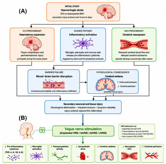
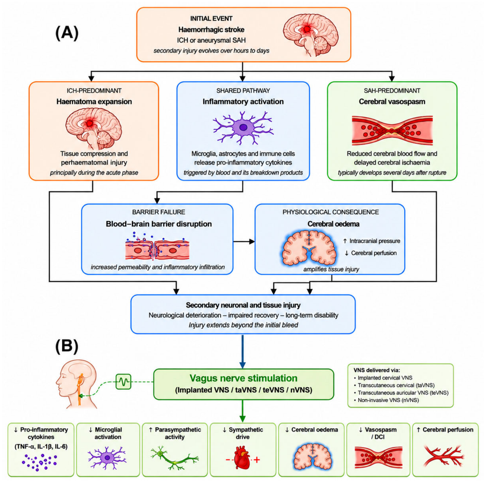
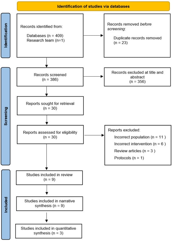
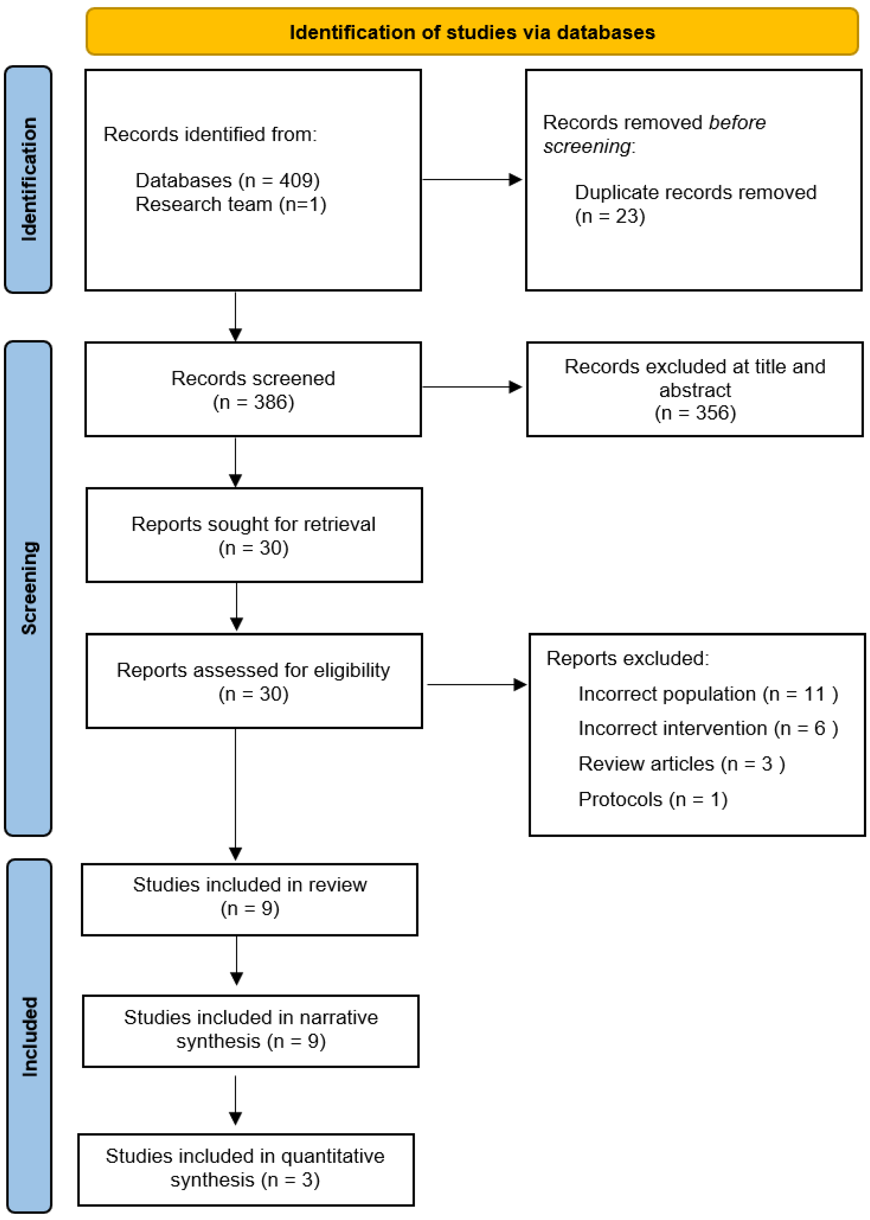
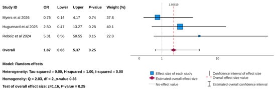
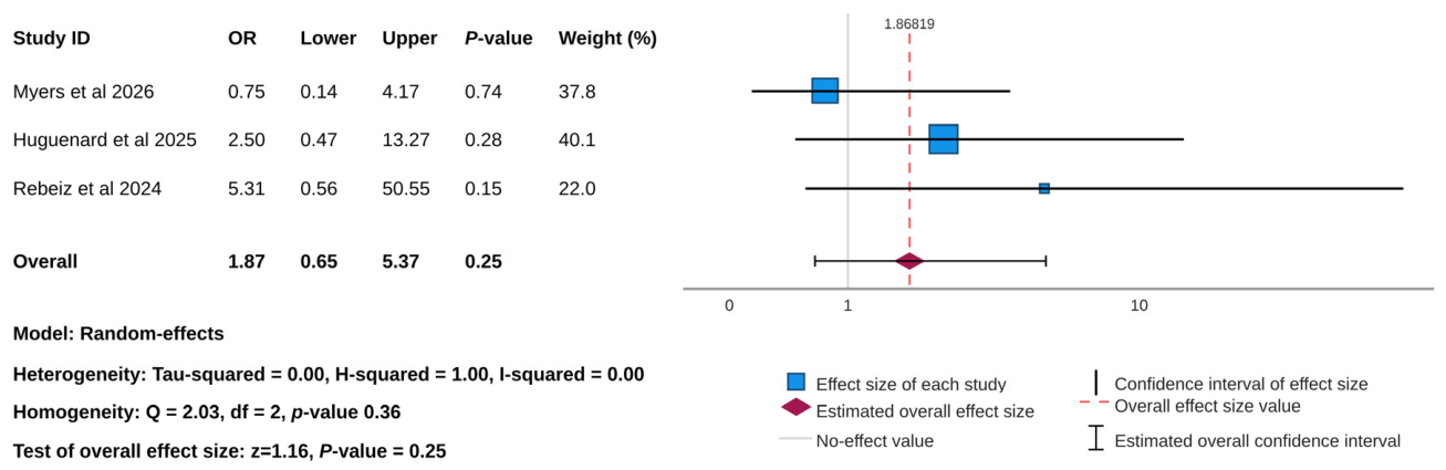
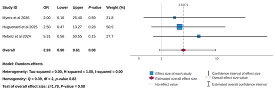
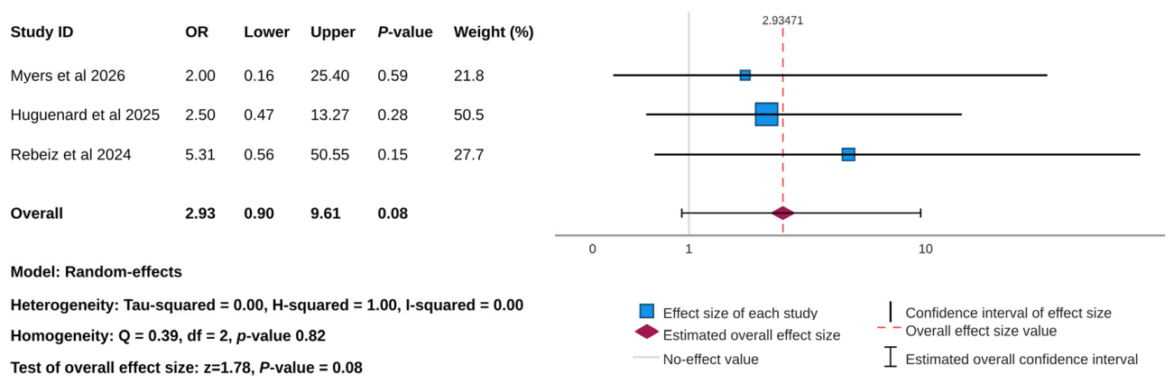

Disclaimer/Publisher’s Note: The statements, opinions and data contained in all publications are solely those of the individual author(s) and contributor(s) and not of MDPI and/or the editor(s). MDPI and/or the editor(s) disclaim responsibility for any injury to people or property resulting from any ideas, methods, instructions or products referred to in the content. |
© 2026 by the authors. Licensee MDPI, Basel, Switzerland. This article is an open access article distributed under the terms and conditions of the Creative Commons Attribution (CC BY) license.