
Harmonization for Parkinson’s Disease Multi-Dataset T1 MRI Morphometry Classification


Abstract
1. Introduction
2. Materials and Methods
2.1. Datasets
2.2. Terminology
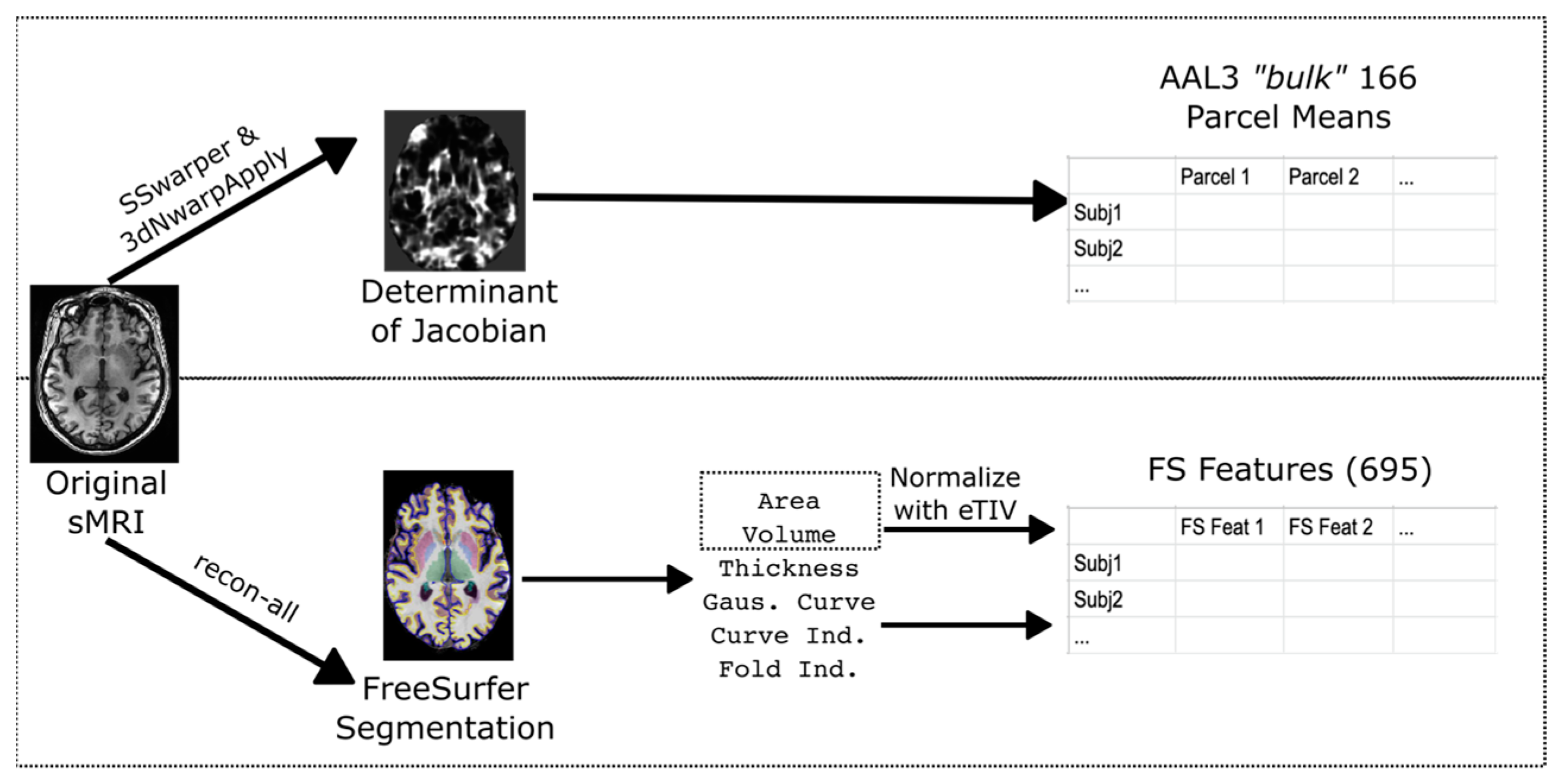
2.3. Data Processing
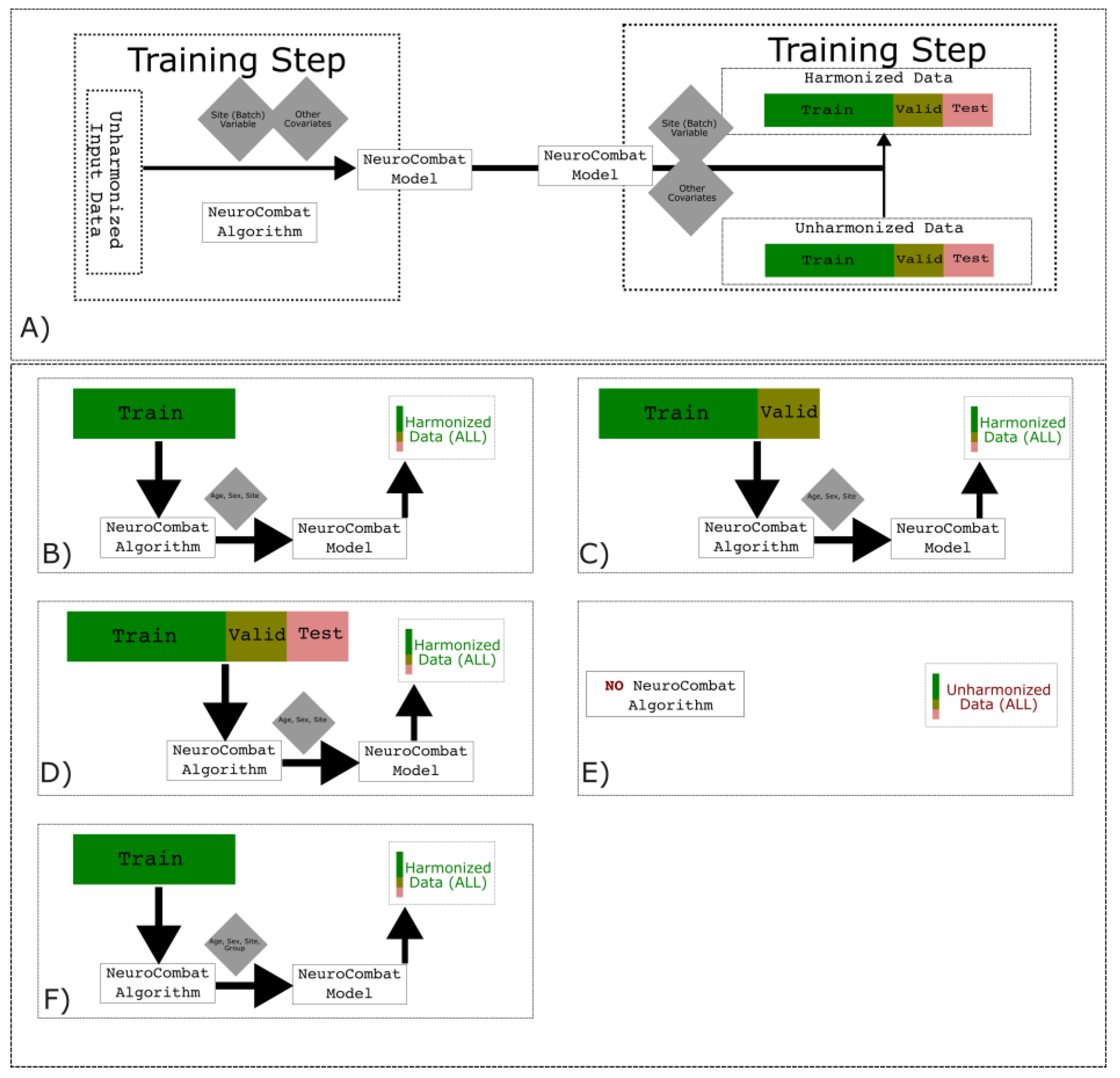
2.4. NeuroComBat Algorithm
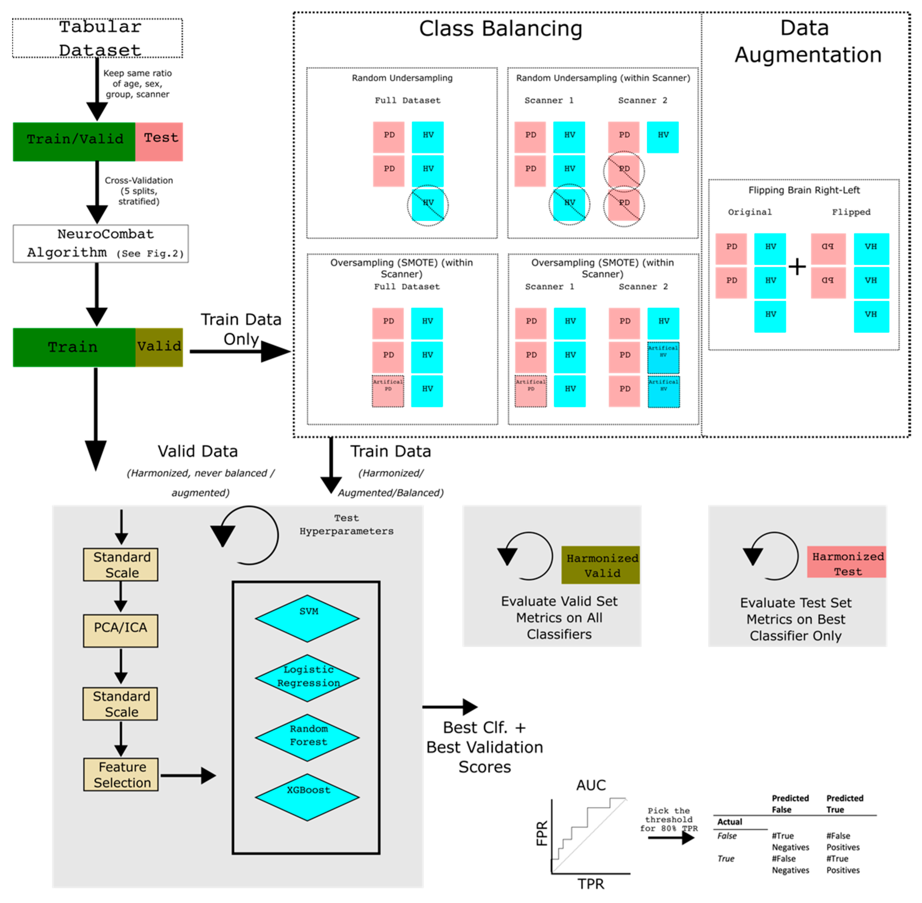
2.5. Data Augmentation/Class Balancing
2.6. Classifiers
2.7. Single Scanner Classifiers vs. Full Dataset
2.8. Evaluation
3. Results
3.1. Subject Demographics
3.2. Single Scanner Classifiers vs. Full Dataset Results
4. Discussion
5. Conclusions
Supplementary Materials
Author Contributions
Funding
Institutional Review Board Statement
Informed Consent Statement
Data Availability Statement
Acknowledgments
Conflicts of Interest
References
- Tysnes, O.-B.; Storstein, A. Epidemiology of Parkinson’s Disease. J. Neural Transm. 2017, 124, 901–905. [Google Scholar] [CrossRef] [Scilit] [PubMed]
- Miller, D.B.; O’Callaghan, J.P. Biomarkers of Parkinson’s Disease: Present and Future. Metabolism 2015, 64, S40–S46. [Google Scholar] [CrossRef] [Scilit] [PubMed]
- Sarasso, E.; Agosta, F.; Piramide, N.; Filippi, M. Progression of Grey and White Matter Brain Damage in Parkinson’s Disease: A Critical Review of Structural MRI Literature. J. Neurol. 2021, 268, 3144–3179. [Google Scholar] [CrossRef] [Scilit] [PubMed]
- Nemmi, F.; Sabatini, U.; Rascol, O.; Péran, P. Parkinson’s Disease and Local Atrophy in Subcortical Nuclei: Insight from Shape Analysis. Neurobiol. Aging 2015, 36, 424–433. [Google Scholar] [CrossRef] [Scilit] [PubMed]
- Laansma, M.A.; Bright, J.K.; Al-Bachari, S.; Anderson, T.J.; Ard, T.; Assogna, F.; Baquero, K.A.; Berendse, H.W.; Blair, J.; Cendes, F.; et al. International Multicenter Analysis of Brain Structure Across Clinical Stages of Parkinson’s Disease. Mov. Disord. 2021, 36, 2583–2594. [Google Scholar] [CrossRef] [Scilit] [PubMed]
- Button, K.S.; Ioannidis, J.P.A.; Mokrysz, C.; Nosek, B.A.; Flint, J.; Robinson, E.S.J.; Munafò, M.R. Power Failure: Why Small Sample Size Undermines the Reliability of Neuroscience. Nat. Rev. Neurosci. 2013, 14, 365–376. [Google Scholar] [CrossRef] [Scilit]
- Parkinson Progression Marker Initiative The Parkinson Progression Marker Initiative (PPMI). Prog. Neurobiol. 2011, 95, 629–635. [CrossRef] [Scilit]
- Peng, B.; Wang, S.; Zhou, Z.; Liu, Y.; Tong, B.; Zhang, T.; Dai, Y. A Multilevel-ROI-Features-Based Machine Learning Method for Detection of Morphometric Biomarkers in Parkinson’s Disease. Neurosci. Lett. 2017, 651, 88–94. [Google Scholar] [CrossRef] [Scilit]
- Long, D.; Wang, J.; Xuan, M.; Gu, Q.; Xu, X.; Kong, D.; Zhang, M. Automatic Classification of Early Parkinson’s Disease with Multi-Modal MR Imaging. PLoS ONE 2012, 7, e47714. [Google Scholar] [CrossRef] [Scilit]
- Salvatore, C.; Cerasa, A.; Castiglioni, I.; Gallivanone, F.; Augimeri, A.; Lopez, M.; Arabia, G.; Morelli, M.; Gilardi, M.C.; Quattrone, A. Machine Learning on Brain MRI Data for Differential Diagnosis of Parkinson’s Disease and Progressive Supranuclear Palsy. J. Neurosci. Methods 2014, 222, 230–237. [Google Scholar] [CrossRef] [Scilit]
- Huppertz, H.-J.; Möller, L.; Südmeyer, M.; Hilker, R.; Hattingen, E.; Egger, K.; Amtage, F.; Respondek, G.; Stamelou, M.; Schnitzler, A.; et al. Differentiation of Neurodegenerative Parkinsonian Syndromes by Volumetric Magnetic Resonance Imaging Analysis and Support Vector Machine Classification. Mov. Disord. 2016, 31, 1506–1517. [Google Scholar] [CrossRef] [Scilit] [PubMed]
- Badea, L.; Onu, M.; Wu, T.; Roceanu, A.; Bajenaru, O. Exploring the Reproducibility of Functional Connectivity Alterations in Parkinson’s Disease. PLoS ONE 2017, 12, e0188196. [Google Scholar] [CrossRef] [Scilit] [PubMed]
- Jovicich, J.; Czanner, S.; Greve, D.; Haley, E.; van der Kouwe, A.; Gollub, R.; Kennedy, D.; Schmitt, F.; Brown, G.; MacFall, J.; et al. Reliability in Multi-Site Structural MRI Studies: Effects of Gradient Non-Linearity Correction on Phantom and Human Data. NeuroImage 2006, 30, 436–443. [Google Scholar] [CrossRef] [Scilit] [PubMed]
- Han, X.; Jovicich, J.; Salat, D.; van der Kouwe, A.; Quinn, B.; Czanner, S.; Busa, E.; Pacheco, J.; Albert, M.; Killiany, R.; et al. Reliability of MRI-Derived Measurements of Human Cerebral Cortical Thickness: The Effects of Field Strength, Scanner Upgrade and Manufacturer. NeuroImage 2006, 32, 180–194. [Google Scholar] [CrossRef] [Scilit] [PubMed]
- Bayer, J.M.M.; Thompson, P.M.; Ching, C.R.K.; Liu, M.; Chen, A.; Panzenhagen, A.C.; Jahanshad, N.; Marquand, A.; Schmaal, L.; Sämann, P.G. Site Effects How-to and When: An Overview of Retrospective Techniques to Accommodate Site Effects in Multi-Site Neuroimaging Analyses. Front. Neurol. 2022, 13, 923988. [Google Scholar] [CrossRef] [Scilit] [PubMed]
- Fortin, J.-P.; Cullen, N.; Sheline, Y.I.; Taylor, W.D.; Aselcioglu, I.; Cook, P.A.; Adams, P.; Cooper, C.; Fava, M.; McGrath, P.J.; et al. Harmonization of Cortical Thickness Measurements across Scanners and Sites. NeuroImage 2018, 167, 104–120. [Google Scholar] [CrossRef] [Scilit]
- Chen, A.A.; Beer, J.C.; Tustison, N.J.; Cook, P.A.; Shinohara, R.T.; Shou, H.; Initiative, T.A.D.N. Mitigating Site Effects in Covariance for Machine Learning in Neuroimaging Data. Hum. Brain Mapp. 2022, 43, 1179–1195. [Google Scholar] [CrossRef] [Scilit]
- Reynolds, M.; Chaudhary, T.; Torbati, M.E.; Tudorascu, D.L.; Batmanghelich, K. Alzheimer’s Disease Neuroimaging Initiative. ComBat Harmonization: Empirical Bayes versus Fully Bayes Approaches. bioRxiv 2022. [Google Scholar] [CrossRef] [Scilit]
- Tafuri, B.; Lombardi, A.; Nigro, S.; Urso, D.; Monaco, A.; Pantaleo, E.; Diacono, D.; De Blasi, R.; Bellotti, R.; Tangaro, S.; et al. The Impact of Harmonization on Radiomic Features in Parkinson’s Disease and Healthy Controls: A Multicenter Study. Front. Neurosci. 2022, 16, 1012287. [Google Scholar] [CrossRef] [Scilit]
- Tinaz, S.; Lauro, P.; Hallett, M.; Horovitz, S.G. Deficits in Task-Set Maintenance and Execution Networks in Parkinson’s Disease. Brain Struct. Funct. 2016, 221, 1413–1425. [Google Scholar] [CrossRef] [Scilit]
- Tinaz, S.; Lauro, P.M.; Ghosh, P.; Lungu, C.; Horovitz, S.G. Changes in Functional Organization and White Matter Integrity in the Connectome in Parkinson’s Disease. Neuroimage Clin. 2017, 13, 395–404. [Google Scholar] [CrossRef] [Scilit]
- Hattori, T.; Reynolds, R.; Wiggs, E.; Horovitz, S.G.; Lungu, C.; Chen, G.; Yasuda, E.; Hallett, M. Neural Correlates of Working Memory and Compensation at Different Stages of Cognitive Impairment in Parkinson’s Disease. Neuroimage Clin. 2022, 35, 103100. [Google Scholar] [CrossRef] [Scilit] [PubMed]
- Hughes, A.J.; Ben-Shlomo, Y.; E Daniel, S.; Lees, A.J. What features improve the accuracy of clinical diagnosis in Parkinson’s disease: A clinicopathologic study. Neurology 1992, 57, S34–S38. [Google Scholar] [CrossRef] [Scilit] [PubMed]
- Ashburner, J.; Friston, K. Chapter 6. Morphometry. In Human Brain Function; Elsevier: Amsterdam, The Netherlands, 2004. [Google Scholar]
- Fischl, B. FreeSurfer. Neuroimage 2012, 62, 774–781. [Google Scholar] [CrossRef] [Scilit] [PubMed]
- Cox, R.W. AFNI: Software for Analysis and Visualization of Functional Magnetic Resonance Neuroimages. Comput. Biomed. Res. 1996, 29, 162–173. [Google Scholar] [CrossRef] [Scilit] [PubMed]
- Malone, I.B.; Leung, K.K.; Clegg, S.; Barnes, J.; Whitwell, J.L.; Ashburner, J.; Fox, N.C.; Ridgway, G.R. Accurate Automatic Estimation of Total Intracranial Volume: A Nuisance Variable with Less Nuisance. NeuroImage 2015, 104, 366–372. [Google Scholar] [CrossRef] [Scilit]
- Johnson, W.E.; Li, C.; Rabinovic, A. Adjusting Batch Effects in Microarray Expression Data Using Empirical Bayes Methods. Biostatistics 2007, 8, 118–127. [Google Scholar] [CrossRef] [Scilit]
- He, H.; Garcia, E.A. Learning from Imbalanced Data. IEEE Trans. Knowl. Data Eng. 2009, 21, 1263–1284. [Google Scholar] [CrossRef] [Scilit]
- Chawla, N.V.; Bowyer, K.W.; Hall, L.O.; Kegelmeyer, W.P. SMOTE: Synthetic Minority Over-Sampling Technique. J. Artif. Intell. Res. 2002, 16, 321–357. [Google Scholar] [CrossRef] [Scilit]
- Pedregosa, F.; Varoquaux, G.; Gramfort, A.; Michel, V.; Thirion, B.; Grisel, O.; Blondel, M.; Prettenhofer, P.; Weiss, R.; Dubourg, V.; et al. Scikit-Learn: Machine Learning in Python. J. Mach. Learn. Res. 2011, 12, 2825–2830. [Google Scholar]
- Lemaître, G.; Nogueira, F.; Aridas, C.K. Imbalanced-Learn: A Python Toolbox to Tackle the Curse of Imbalanced Datasets in Machine Learning. J. Mach. Learn. Res. 2017, 18, 1–5. [Google Scholar]
- Bergstra, J.; Bengio, Y. Random Search for Hyper-Parameter Optimization. J. Mach. Learn. Res. 2012, 13, 281–305. [Google Scholar]
- Guan, H.; Liu, M. Domain Adaptation for Medical Image Analysis: A Survey. arXiv 2021, arXiv:2102.09508. [Google Scholar] [CrossRef] [Scilit] [PubMed]
- Pomponio, R.; Erus, G.; Habes, M.; Doshi, J.; Srinivasan, D.; Mamourian, E.; Bashyam, V.; Nasrallah, I.M.; Satterthwaite, T.D.; Fan, Y.; et al. Harmonization of Large MRI Datasets for the Analysis of Brain Imaging Patterns throughout the Lifespan. NeuroImage 2020, 208, 116450. [Google Scholar] [CrossRef] [Scilit]
- Stein, C.K.; Qu, P.; Epstein, J.; Buros, A.; Rosenthal, A.; Crowley, J.; Morgan, G.; Barlogie, B. Removing Batch Effects from Purified Plasma Cell Gene Expression Microarrays with Modified ComBat. BMC Bioinform. 2015, 16, 63. [Google Scholar] [CrossRef] [Scilit] [PubMed]
- Noor, M.B.T.; Zenia, N.Z.; Kaiser, M.S.; Mamun, S.A.; Mahmud, M. Application of Deep Learning in Detecting Neurological Disorders from Magnetic Resonance Images: A Survey on the Detection of Alzheimer’s Disease, Parkinson’s Disease and Schizophrenia. Brain Inform. 2020, 7, 1–21. [Google Scholar] [CrossRef] [Scilit] [PubMed]
- Segato, A.; Marzullo, A.; Calimeri, F.; De Momi, E. Artificial Intelligence for Brain Diseases: A Systematic Review. APL Bioeng. 2020, 4, 041503. [Google Scholar] [CrossRef] [Scilit]
- Marzi, C.; Giannelli, M.; Barucci, A.; Tessa, C.; Mascalchi, M.; Diciotti, S. Efficacy of MRI Data Harmonization in the Age of Machine Learning: A Multicenter Study across 36 Datasets. Sci. Data 2024, 11, 115. [Google Scholar] [CrossRef] [Scilit] [PubMed]
- Bega, D.; Kuo, P.H.; Chalkidou, A.; Grzeda, M.T.; Macmillan, T.; Brand, C.; Sheikh, Z.H.; Antonini, A. Clinical Utility of DaTscan in Patients with Suspected Parkinsonian Syndrome: A Systematic Review and Meta-Analysis. NPJ Park. Dis. 2021, 7, 1–8. [Google Scholar] [CrossRef] [Scilit] [PubMed]
- Garcia-Dias, R.; Scarpazza, C.; Baecker, L.; Vieira, S.; Pinaya, W.H.L.; Corvin, A.; Redolfi, A.; Nelson, B.; Crespo-Facorro, B.; McDonald, C.; et al. Neuroharmony: A New Tool for Harmonizing Volumetric MRI Data from Unseen Scanners. Neuroimage 2020, 220, 117127. [Google Scholar] [CrossRef] [Scilit]
- Seoni, S.; Shahini, A.; Meiburger, K.M.; Marzola, F.; Rotunno, G.; Acharya, U.R.; Molinari, F.; Salvi, M. All You Need Is Data Preparation: A Systematic Review of Image Harmonization Techniques in Multi-Center/Device Studies for Medical Support Systems. Comput. Methods Programs Biomed. 2024, 250, 108200. [Google Scholar] [CrossRef] [Scilit] [PubMed]


| Group (PD/HV) | Age | Sex (M/F) | Disease Duration | H&Y Score | UPDRS3 | |||
|---|---|---|---|---|---|---|---|---|
| PD | HV | PD | HV | |||||
| NIH | ||||||||
| Scanner 1 | 24/12 | 62.8 ± 9.2 | 58.1 ± 7.4 | 16/8 | 7/5 | 9.4 ± 6.2 | 2.5 ± 0.4 | 17.6 ± 9.0 |
| Scanner 2 | 17/19 | 62.2 ± 7.3 | 63.2 ± 6.7 | 12/5 | 15/4 | 6.7 ± 3.1 | N.A. | 28.6 ± 8.6 |
| Scanner 3 | 23/20 | 60.4 ± 8.0 | 59.9 ± 7.4 | 12/11 | 9/11 | 7.8 ± 4.9 | 2.2 ± 0.5 | 20.6 ± 7.2 |
| PPMI | ||||||||
| Scanner 1 | 16/12 | 63.1 ± 8.3 | 61.9 ± 12.4 | 12/4 | 10/2 | 0.1 ± 0.1 | 1.0 ± 0.6 | 15.2 ± 8.7 |
| Scanner 2 | 22/12 | 62.0 ± 9.2 | 58.9 ± 6.9 | 13/9 | 9/3 | 0.1 ± 0.2 | 1.2 ± 0.8 | 15.5 ± 11.8 |
| Scanner 3 | 21/13 | 58.5 ± 10.9 | 62.6 ± 9.7 | 13/8 | 9/4 | 0.7 ± 1.1 | 1.0 ± 0.9 | 13.4 ± 11.8 |
| Scanner 4 | 14/10 | 61.7 ± 9.0 | 65.7 ± 3.1 | 6/8 | 7/3 | 0.3 ± 0.4 | 1.0 ± 0.8 | 15.6 ± 11.0 |
| Scanner 5 | 14/11 | 58.7 ± 8.2 | 60.5 ± 8.8 | 11/3 | 7/4 | 0.1 ± 0.03 | 0.8 ± 0.8 | 8.6 ± 9.2 |
| Scanner 6 | 19/10 | 63.5 ± 9.6 | 64.6 ± 6.8 | 12/7 | 4/6 | 0.3 ± 0.4 | 1.1 ± 0.8 | 12.0 ± 9.0 |
| Other | ||||||||
| Neurocon | 26/16 | 68.8 ± 10.7 | 67.6 ± 11.9 | 17/9 | 12/4 | N.A. | 1.9 ± 0.3 | 9.2 ± 5.3 |
| Tao Wu | 20/20 | 65.2 ± 4.4 | 64.8 ± 5.6 | 11/9 | 12/8 | 5.4 ± 3.9 | 1.9 ± 0.6 | N.A. |
| Classifier | Dataset Used | NC Fit Strategy | Validation AUC | Test AUC |
|---|---|---|---|---|
| XGBoost | FS Only | Fit on Train, w/Group Covariate | 0.863 ± 0.045 | 0.902 |
| RF | FS Only | Fit on Train, w/Group covariate | 0.841 ± 0.036 | 0.837 |
| RF | Bulk + FS | Fit on Train, w/Group covariate | 0.806 ± 0.056 | 0.866 |
| Classifier | Dataset Used | NC Fit Strategy | Validation AUC | Test AUC |
|---|---|---|---|---|
| LR | Bulk Only | Fit on all splits, no group covariate | 0.650 ± 0.052 | 0.550 |
| SVC | Bulk + FS | Fit on Train, no group | 0.646 ± 0.071 | 0.500 |
| XGBoost | Bulk + FS | No NeuroComBat | 0.644 ± 0.073 | 0.438 |
| Scanner | Validation AUC | |||
|---|---|---|---|---|
| Single-Scanner Classifier | Multi-Scanner Classifier (Best) | |||
| No NC | NC On Train Only (No Group) | NC On Train Only (Group) | ||
| NIH Dataset 1 | 0.768 | 0.822 | 0.736 | 0.966 |
| NIH Dataset 2 | 0.700 | 0.744 | 0.694 | 0.914 |
| NIH Dataset 3 | 0.618 | 0.583 | 0.555 | 0.732 |
| PPMI Scanner 1 | 0.486 | 0.614 | 0.643 | 1.0 |
| PPMI Scanner 2 | 0.560 | 0.682 | 0.503 | 0.871 |
| PPMI Scanner 3 | 0.593 | 0.653 | 0.653 | 0.785 |
| PPMI Scanner 4 | 0.519 | 0.444 | 0.467 | 0.488 |
| PPMI Scanner 5 | 0.573 | 0.518 | 0.527 | 0.873 |
| PPMI Scanner 6 | 0.617 | 0.509 | 0.621 | 0.857 |
| Neurocon | 1.000 | 0.564 | 0.553 | 0.878 |
| Tao Wu | 0.727 | 0.645 | 0.719 | 0.930 |
Disclaimer/Publisher’s Note: The statements, opinions and data contained in all publications are solely those of the individual author(s) and contributor(s) and not of MDPI and/or the editor(s). MDPI and/or the editor(s) disclaim responsibility for any injury to people or property resulting from any ideas, methods, instructions or products referred to in the content. |
© 2024 by the authors. Licensee MDPI, Basel, Switzerland. This article is an open access article distributed under the terms and conditions of the Creative Commons Attribution (CC BY) license (https://creativecommons.org/licenses/by/4.0/).
Share and Cite
Saqib, M.; Horovitz, S.G. Harmonization for Parkinson’s Disease Multi-Dataset T1 MRI Morphometry Classification. NeuroSci 2024, 5, 600-613. https://doi.org/10.3390/neurosci5040042
Saqib M, Horovitz SG. Harmonization for Parkinson’s Disease Multi-Dataset T1 MRI Morphometry Classification. NeuroSci. 2024; 5(4):600-613. https://doi.org/10.3390/neurosci5040042
Chicago/Turabian StyleSaqib, Mohammed, and Silvina G. Horovitz. 2024. "Harmonization for Parkinson’s Disease Multi-Dataset T1 MRI Morphometry Classification" NeuroSci 5, no. 4: 600-613. https://doi.org/10.3390/neurosci5040042
APA StyleSaqib, M., & Horovitz, S. G. (2024). Harmonization for Parkinson’s Disease Multi-Dataset T1 MRI Morphometry Classification. NeuroSci, 5(4), 600-613. https://doi.org/10.3390/neurosci5040042