


Abstract
Objective: High sperm DNA fragmentation (SDF) results in fewer euploid embryos. Although sperm retrieved by testicular sperm extraction (TESE) has low SDF compared to the ejaculated sperm, there is little data comparing the prevalence of chromosomal abnormalities in the resulting embryos. The objective of this study was to compare rates of chromosomal abnormalities in blastocysts from sperm obtained either by TESE or ejaculate with high SDF. Methods: The blastocysts were generated by ICSI in both groups. The preimplantation genetic testing for aneuploidy (PGT-A) was performed by next-generation sequencing (NGS). This study utilized 2242 blastocysts from 400 couples; 200 couples in each group. The TESE-retrieved sperm and ejaculated sperm with high SDF had 1037 and 1205 blastocysts, respectively. The sperm DNA fragmentation was determined by the Halosperm G2 assay. The rates of euploid, aneuploid and mosaic embryos were compared by multivariate logistic regression and mixed-effects models that accounted for female age and ovarian reserve. Results: The TESE-derived sperm group had a significantly higher percentage of euploid blastocysts (67.8%) as compared to those derived from ejaculated sperm with high SDF (48.2%, p = 0.003). The multivariate logistic regression indicated that the sperm origin (TESE/ejaculate) was an independent predictor of euploid blastocysts [OR = 1.85; 95% CI = 1.05–3.26; p = 0.034]. The probability of having euploid blastocysts decreased by 6% for every 1% increase in the SDF. The increased age of the female was a major negative predictor [OR = 0.92 per year; 95% CI = 0.88–96; p = 0.001]. Conclusions: The TESE-derived sperm group had significantly higher euploid blastocysts as compared to the ejaculated sperm with high SDF group.
1. Introduction
Male infertility is one of the serious worldwide health problems, as this type of infertility is observed in about 50 percent of all infertile couples, and about seven percent among all men [1,2]. Male infertility etiology is multifactorial and involves genetic issues, hormonal imbalances, anatomical abnormalities, lifestyle, and environmental factors [3]. Diagnosis should be made by history, physical examination, semen analysis, hormonal analysis, imaging and genetic testing [2]. Sperm DNA fragmentation (SDF) is one of the critical determinants of assisted reproductive technology (ART) outcomes. Higher levels of SDF are linked with lower fertilization, disrupted embryo growth, high levels of miscarriages, and poor live births, despite the regularity of conventional semen parameters [4,5]. The negative association with the number of chromosomally normal embryos highlights the clinical importance of SDF, especially when it is greater than 30% [5].
The sperm DNA reorganizes radically during spermiogenesis, with a density increase of about sixfold relative to somatic nuclei due to substitution of histones with protamines [6]. This restructuring results in very compact, toroidal DNA structures that are needed to accommodate the mammalian genome into the small sperm nucleus and allows it to be safeguarded against mechanical and chemical bombardments [7]. The DNA is bound by the protamine 1 (P1) and protamine 2 (P2) proteins via arginine-rich domains in the shape of doughnuts of about 50 kilobases each [7]. In addition to structural organization, sperm cells possess intricate epigenetic reminiscences such as DNA methylation signals, histone retention signals and non-coding RNA that jointly control the early embryonic gene expression [8]. Disturbances in these epigenetic characteristics related to high paternal age, environmental exposure, and oxidative stress were found to be associated with developmental changes in the embryo and poor offspring phenotype [8]. Sperm mitochondria are another important element of reproductive potential that generate adenosine triphosphate (ATP) molecules needed to provide motility, capacitance and acrosomal reaction [9]. Mitochondrial dysplasia is linked with low motility of sperm, excessive DNA damage and poor embryo development, which directly leads to infertility in males [9,10].
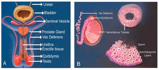
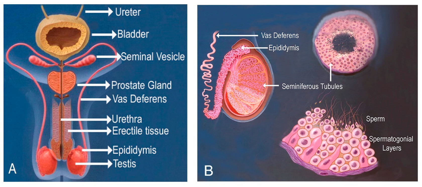
Sperm DNA fragmentation is caused by several mechanisms, such as oxidative stress, apoptosis, age-related alteration and environmental exposure [11]. The SDF is specific to ejaculated sperm since it is transferred through the epididymis and male reproductive tract and undergoes oxidative stress, apoptotic signals, and centrifugal forces that cluster DNA damage [12,13]. The effects of high SDF are not only limited to impairment of fertilization. There is a direct correlation between high SDF and more chromosomal abnormalities in resultant embryos, especially the aneuploidies of paternal origin [14]. This is because defective sperm DNA, due to improper repair through oocyte repair mechanisms, causes mitotic errors in the initial embryonic divisions and eventually leads to the production of aneuploid embryos with low implantation potential and high susceptibility to miscarriage [14,15]. The testicular sperm extraction (TESE) and its forms (percutaneous TESA, standard TESE, microsurgical micro-TESE) are surgical methods to extract sperm directly out of testicular tissue without involving the epididymis and ejaculatory ducts [16]. The method is especially effective in men with azoospermia and extreme SDF, because the testicular sperm normally undergoes much less DNA fragmentation than the ejaculated sperm of the same male [13,17]. Figure 1A is a diagram of the male reproductive system, and Figure 1B is a diagram of the testes, epididymis and seminiferous tubules at a microscopic level. The sperm are ejaculated from the urethra or retrieved surgically from the testes or epididymis.
Figure 1.
(A) Graphic presentation of the male reproductive system. The sperm can be obtained by ejaculation from the urethra, or retrieved surgically from the testis or epididymis. (B) is a microscopic presentation of testes, epididymis, vas deference, seminiferous tubules, and the spermatogonial layers that produce sperm.
The lesser fragmentation of TESE-derived sperm is because the testicular sperm are not subjected to the oxidative insults, apoptotic cues and mechanical damage that testicular sperm undergo during epididymal transit and ejaculation [12]. Direct comparative studies of paired samples showed that the amount of DNA damage in testicular sperm is significantly lower than that of ejaculated sperm, especially in men with a high SDF baseline [17]. The result of TESE-ICSI cycles demonstrates good outcomes with a fertilization rate of 62–68% and live birth rates per embryo transfer of 25–29%, which is comparable to results using other sources of sperm [18]. The correlation between lowered testicular sperm DNA fragmentation and the consequent embryonic chromosome integrity is, however, not wholly characterized in controlled comparative studies. Even though substantial literature has been written about SDF effects on fertility as well as TESE done to retrieve sperm, there is a large research gap in the direct comparative study of embryonic genetic abnormalities of TESE-derived and ejaculated sperm. Although TESE-derived sperm exhibits less DNA fragmentation by definition, direct empirical studies that have directly attributed this biological capability to a lower rate of embryonic aneuploidy are scarce [5,19]. The available literature on TESE-derived sperm is biased towards retrieval success rates, fertilization rates, and overall pregnancy/live birth outcomes in non-obstructive azoospermia cases [19,20], and has little embryo-level chromosomal profiling information. On the other hand, numerous studies have been done on naturally ejaculated sperm showing that high SDF, advanced age of a male, low semen quality, and high embryo aneuploidy rates are associated with high SDF [5,21], although not directly compared to TESE sperm in similar study populations [5]. This research aimed to fill this research gap by offering a systematic, controlled comparative analysis of chromosomal abnormalities of blastocysts between these two sperm origins: TESE-derived and ejaculated sperm with high DNA fragmentation.
2. Methods
This was a prospective cohort study at Thuriah Medical Center, Riyadh, Saudi Arabia. In total 2242 blastocysts from 400 couples (200 from TESE-derived and 200 from ejaculated sperm with high SDF) were compared. The ejaculated sperm group had 1205 blastocysts, and the TESE group had 1037 blastocysts. The blastocysts with no DNA detected were excluded. The patient inclusion criteria for the TESE-derived group were male with obstructive azoospermia; partner age between 18 and 45 years; normal ovarian reserve; and first or second ICSI cycle. The patient inclusion criteria for the ejaculated sperm group were SDF > 30%; partners age between 18 and 45 years; normal ovarian reserve; and first or second ICSI cycle. In both groups, patients with a history of recurrent miscarriages and female factor infertility were excluded. Only one cycle from each couple was included in both groups.
The TESE was performed on site under local anesthesia. The extracted tissues were subjected to mechanical and enzymatic treatments to isolate viable sperm. To obtain a clean sperm population, the tissues and suspension were processed by the density gradient technique. The ejaculated sperm cells were obtained by masturbation after 3 to 5 days of sexual abstinence. The density gradient technique was used to obtain motile sperm. The SDF was determined by the Halosperm G2 assay (Halotech@, 28049 Madrid, Spain). Each sample was evaluated in triplicate, and the average value was used for statistical analysis.
The ovarian stimulation, oocyte collection, denudation, ICSI, embryo culture, and trophectoderm biopsies were performed per standard protocols in both groups. The blastocysts were graded per the Gardner scoring system, and biopsies were performed on day-5 by taking about 5–7 blastomeres per biopsy [22]. The biopsied cells were lysed and put into polymerase chain reaction (PCR) tubes, which were then taken to the genetics laboratory for PGT-A by NGS. The results were classified as: euploid (normal chromosomal count); aneuploid (monosomy, trisomy, polysomy); and mosaic. The blastocysts in which no DNA was detected were excluded.
The demographic, clinical, and laboratory variables were entered into a standardized electronic database controlled by differing data of double-entry validation. The statistical analyses were performed using SPSS version 26.0 (IBM, Armonk, NY, USA). Comparisons of baseline traits between groups were performed using independent t-tests (continuous variables) and chi-square tests (categorical variables). Chi-square test was used to compare groups in terms of euploid rates. With the help of exact binomial techniques, ninety-five percent confidence intervals were obtained. The mixed-effects logistic regression was developed to assess the independent impact of sperm source on the probability of producing euploid embryos, conditions on female age, and measures of ovarian reserve. The Pearson correlation coefficients were estimated to assess the inter-relationships between important variables (SDF, aneuploidy rate, female age, oocyte parameters). The statistical significance was set at p < 0.05 (two-tailed).
This research study was approved by the Thuriah Medical Centre’s institutional review board (Organization number: IORG0008118, Approval Code: IRB number IRB00009734, Approval Date: 17 December 2024). Each of the participants gave written informed consent after extensive information on study goals, procedures, risks, and benefits. The information was de-identified and stored in safe, password-coded databases. The results of genetic testing were relayed to the participant using the laid-down counseling procedures.
3. Results
Table 1 shows the baseline demographic and clinical characteristics of the groups. The TESE-derived and the ejaculated sperm groups consisted of 400 couples (200 in each group). Non-significant differences were observed between groups regarding baseline demographic characteristics, indicating adequate group matching. Table 2 shows sperm DNA fragmentation percentages. The ejaculated sperm group had significantly higher DNA fragmentation (39.7 ± 8.3%) as compared to the TESE sperm group (18.4 ± 6.2%), p < 0.001.
Table 1.
Demographic and clinical characteristics of the study groups. Data from 400 couples (200 in each group) are presented. The studied parameters were similar in both groups.
Table 2.
Sperm DNA fragmentation (SDF)- comparison between TESE and ejaculated sperm. In the ejaculated sperm group, only men with >30% SDF were included.
Table 3 shows ICSI cycle outcomes and embryo development in both groups. Only one cycle from each couple was included, and all the blastocysts produced from that cycle were included in this study. The number of mature oocytes, fertilization rates, and number of mosaic blastocysts were similar in both groups. In total, 2242 blastocysts were analyzed, 1037 in the TESE group and 1205 in the ejaculated sperm group. The blastocysts with no DNA detected were excluded from both groups. There were seven and 14 blastocysts with no DNA detected in the TESE group and ejaculated sperm group, respectively. The day-3 cleavage rate, blastocyst rate, and number of euploid blastocysts were higher in the TESE group, whereas the number of aneuploid blastocysts was higher in the ejaculated sperm group. The most common abnormality in blastocysts of both groups was chromosome 16 aneuploidy. The second and third most common abnormalities were found for chromosomes 21 and 22, respectively (Table 4).
Table 3.
Cycle outcome and embryo development in both study groups. Only MII oocytes were included in the study. In the ejaculated sperm group, 1807, and in the TESE group, 2015 MII oocytes were injected.
Table 4.
Distribution of chromosomal abnormalities in aneuploid blastocysts in study groups. The data is based on aneuploid blastocysts shown in Table 3.
The origin of sperm was a predictive variable independent of the female age and ovarian reserve parameters in predicting the production of euploid blastocysts (Table 5). A unit change in SDF led to an odds decrease of six percent of getting euploid blastocysts. The maternal age was found to be a strong negative predictor, whereby one extra year was found to reduce the probability of euploidy by 8%. The blastocyst morphology quality and chromosomal normality were found to have strong relationships. Gardner scored blastocysts in category A exhibited 63.6% euploidy rates, and in category C, 42.9%.
Table 5.
Results of multivariate logistic regression analysis. The euploid blastocyst production was predicted against a mixed-effects logistic regression model to examine the independent predictors.
4. Discussion
The results of this study indicate that sperm DNA fragmentation plays a significant role in determining the blastocyst chromosomal integrity. The TESE-obtained sperm with low SDF had a higher number of euploid embryos 67.8% than 48.2% in the ejaculated sperm group with high sperm DNA fragmentation (p = 0.003). The sperm source was an independent predictor of production of euploid embryos after adjusting it in relation to the female age and ovarian reserve [OR = 1.85; 95% CI = 1.05–3.26; p = 0.034].
The observations of this study verify the earlier results of differences in the SDF by directly associating them with the chromosomally competent embryos. This is a clinically significant improvement in learning about mechanisms of male factor infertility and optimizing ICSI treatment. The higher chromosomal normality in TESE blastocysts is probably due to the lower level of genetic damage in testicular sperm. The testicular sperm avoids the epididymis and ejaculatory ducts, and therefore they are less affected by oxidative stress, apoptotic cues, and mechanical damage resulting in damage to DNA of ejaculated sperm [12,13,17]. Upon the introduction of fragmented paternal DNA into the oocyte, endogenous oocyte repairing mechanisms might fail to restore DNA damage completely, and instead the damage is carried into early embryonic divisions. This induces chromosomal instability and aneuploidy by a variety of possible pathways, including a defective recovery of meiosis division, centrosome dysfunction and an abnormal formation of the mitotic spindle [14,23]. The results of this study support the use of TESE sperm as a treatment option in instances of high DNA fragmentation in the ejaculated sperm. Men who have failed to correct high sperm DNA fragmentation following the improved lifestyle and medical optimization can consider using TESE sperm to achieve better embryonic chromosomal competence, and consequently better ICSI success. In this study, the multivariate analysis showed a decline of 6% in euploid probability in resulting blastocysts with a 1% increase in sperm DNA fragmentation. This justifies the need to identify and treat men with high SDF.
A recent comprehensive systematic review and meta-analysis identified a statistically significant but modest positive association between elevated sperm DNA fragmentation and an increased risk of embryo aneuploidy. The results of this review are consistent with multiple previously published studies [24]. The findings of this study also agree with the published literature that suggests the benefits of TESE in the context of high sperm DNA fragmentation [13,17]. Further, it offers a direct relationship of SDF with the chromosomal integrity of the resulting embryos. The difference between the rates of euploidy (67.8% vs. 48.2%) is a clinically significant difference that may be translated to significant improvement in cumulative live birth rate.
Further studies are needed that can compare embryo euploidy rate by allocating female partner’s oocytes between ejaculated sperm with high sperm DNA fragmentation and sperm retrieved by TESE in the same male. This was not possible in this study. Also, this study was performed on only 400 couples, 200 in each group. More studies on a larger number of couples with intra-patient comparisons are recommended.
5. Conclusions
This comparative study indicated that the TESE-derived sperm resulted in blastocysts of higher chromosomal integrity than the ejaculated sperm with high sperm DNA fragmentation. The 19.6% difference in the rate of euploid embryos (67.8% vs. 48.2%) is a clinically significant change. This finding helps in patient counseling in cases with high sperm DNA fragmentation in the ejaculated sperm.
Author Contributions
Conceptualization: N.K.A., S.Y.S. and M.J. Methodology: N.K.A., S.Y.S. and H.A. Formal analysis: N.K.A. and S.Y.S. Data curation: N.K.A., S.Y.S. and H.A. Project administration: S.Y.S., H.A. and M.J. Software: N.K.A. Investigation: N.K.A. and H.A. Writing—original draft: N.K.A., S.Y.S. and M.J. Writing—review and editing: N.K.A., S.Y.S. and M.J. Approval of final manuscript: N.K.A., S.Y.S., M.J. and H.A. All authors have read and agreed to the published version of the manuscript.
Funding
This study was a PhD research project for Noura Khalid Alfhead. This research received no external funding.
Institutional Review Board Statement
The study was approved by the Thuriah Medical Centre’s Institutional Review Board (Organization number: IORG0008118, Approval Code: IRB number IRB00009734, Approval Date: 17 December 2024).
Informed Consent Statement
Informed consent was obtained from all subjects involved in the study.
Data Availability Statement
Data are available by sending an email to <noura.kf3@gmail.com>.
Conflicts of Interest
The authors Noura Khalid Alfhead and Hamad Alsufyan were employed with Thuriah Medical Centre. Author Murid Javed was employed by the Embryogenex Inc. The remaining authors declare that the research was conducted in the absence of any commercial or financial relationships that could be construed as a potential conflict of interest.
References
- Leslie, S.W.; Soon-Sutton, T.L.; Khan, M.A.B. Male Infertility. In StatPearls; StatPearls Publishing: Treasure Island, FL, USA, 2024. [Google Scholar]
- Eisenberg, M.L.; Esteves, S.C.; Lamb, D.J.; Hotaling, J.M.; Giwercman, A.; Hwang, K.; Cheng, Y.S. Male infertility. Nat. Rev. Dis. Primers 2023, 9, 49. [Google Scholar] [CrossRef] [Scilit] [PubMed]
- Agarwal, A.; Baskaran, S.; Parekh, N.; Cho, C.L.; Henkel, R.; Vij, S.; Arafa, M.; Panner Selvam, M.K.; Shah, R. Male infertility. Lancet 2021, 397, 319–333. [Google Scholar] [CrossRef] [Scilit] [PubMed]
- Alahmar, A.T.; Singh, R.; Palani, A. Sperm DNA fragmentation in reproductive medicine: A review. J. Hum. Reprod. Sci. 2022, 15, 206–218. [Google Scholar] [CrossRef] [Scilit] [PubMed]
- Wan, B.; Fu, Y.; Ma, N.; Zhou, Z.; Lu, W. Effect of sperm DNA fragmentation on embryo euploidy rate in assisted reproductive technologies: A systematic review and meta-analysis. Eur. J. Med. Res. 2025, 30, 1138. [Google Scholar] [CrossRef] [Scilit] [PubMed]
- Miller, D.; Brinkworth, M.; Iles, D. Paternal DNA packaging in spermatozoa: More than the sum of its parts? DNA, histones, protamines and epigenetics. Reproduction 2010, 139, 287–301. [Google Scholar] [CrossRef] [Scilit] [PubMed]
- Moritz, L.; Hammoud, S.S. The art of packaging the sperm genome: Molecular and structural basis of the histone-to-protamine exchange. Front. Endocrinol. 2022, 13, 895502. [Google Scholar] [CrossRef] [Scilit] [PubMed]
- Balder, P.; Jones, C.; Coward, K.; Yeste, M. Sperm chromatin: Evaluation, epigenetic signatures and relevance for embryo development and assisted reproductive technology outcomes. Eur. J. Cell Biol. 2024, 103, 151429. [Google Scholar] [CrossRef] [Scilit] [PubMed]
- Mai, Z.; Yang, D.; Wang, D.; Zhang, J.; Zhou, Q.; Han, B.; Sun, Z. A narrative review of mitochondrial dysfunction and male infertility. Transl. Androl. Urol. 2024, 13, 2134–2145. [Google Scholar] [CrossRef] [Scilit] [PubMed]
- Vahedi Raad, M.; Firouzabadi, A.M.; Tofighi Niaki, M.; Henkel, R.; Fesahat, F. The impact of mitochondrial impairments on sperm function and male fertility: A systematic review. Reprod. Biol. Endocrinol. 2024, 22, 83. [Google Scholar] [CrossRef] [Scilit] [PubMed]
- Dos Santos Hamilton, T.R.; D’Ávila Assumpção, M.E.O. Sperm DNA fragmentation: Causes and identification. Zygote 2020, 28, 1–8. [Google Scholar] [CrossRef] [Scilit] [PubMed]
- Kugelman, N.; Hochberg, A.; Dahan, M.H. Impact of short abstinence versus testicular sperm on sperm DNA fragmentation: A systematic review and meta-analysis. Arch. Gynecol. Obstet. 2024, 310, 1831–1843. [Google Scholar] [CrossRef] [Scilit] [PubMed]
- Khoo, C.C.; Cayetano-Alcaraz, A.A.; Rashid, R.; Tharakan, T.; Yap, T.; Sofikitis, N.; Salonia, A.; Corona, G.; Giwercman, A.; Jayasena, C.N.; et al. Does testicular sperm improve intracytoplasmic sperm injection outcomes for nonazoospermic infertile men with elevated sperm DNA fragmentation? A systematic review and meta-analysis. Eur. Urol. Focus 2024, 10, 410–420. [Google Scholar] [CrossRef] [Scilit] [PubMed]
- Gao, J.; Yan, Z.; Yan, L.; Zhu, X.; Jiang, H.; Qiao, J. The effect of sperm DNA fragmentation on the incidence and origin of whole and segmental chromosomal aneuploidies in human embryos. Reproduction 2023, 166, 117–124. [Google Scholar] [CrossRef] [Scilit] [PubMed]
- Marinaro, J.A.; Schlegel, P.N. Sperm DNA damage and its relevance in fertility treatment: A review of recent literature and current practice guidelines. Int. J. Mol. Sci. 2023, 24, 1446. [Google Scholar] [CrossRef] [Scilit] [PubMed]
- Takeshima, T.; Karibe, J.; Saito, T.; Kuroda, S.; Komeya, M.; Uemura, H.; Yumura, Y. Clinical management of nonobstructive azoospermia: An update. Int. J. Urol. 2024, 31, 17–24. [Google Scholar] [CrossRef] [Scilit] [PubMed]
- Esteves, S.C.; Roque, M.; Bradley, C.K.; Garrido, N. Reproductive outcomes of testicular versus ejaculated sperm for intracytoplasmic sperm injection among men with high levels of DNA fragmentation in semen: Systematic review and meta-analysis. Fertil. Steril. 2017, 108, 456–467.e1. [Google Scholar] [CrossRef] [Scilit] [PubMed]
- Cano-Extremera, M.; Hervas, I.; Gisbert Iranzo, A.; Falquet Guillem, M.; Gil Juliá, M.; Navarro-Gomezlechon, A.; Pacheco-Rendón, R.; Garrido Puchalt, N. Superior live birth rates, reducing sperm DNA fragmentation, and lowering miscarriage rates by using testicular sperm versus ejaculates in ICSI cycles from couples with high SDF: A systematic review and meta-analysis. Biology 2025, 14, 130. [Google Scholar] [CrossRef] [Scilit] [PubMed]
- Hervas, I.; Gil Julia, M.; Rivera-Egea, R.; Navarro-Gomezlechon, A.; Mossetti, L.; Garrido, N. Switching to testicular sperm after a previous ICSI failure with ejaculated sperm significantly improves blastocyst quality without increasing aneuploidy risk. J. Assist. Reprod. Genet. 2022, 39, 2275–2285. [Google Scholar] [CrossRef] [Scilit] [PubMed]
- Tharakan, T.; Luo, R.; Jayasena, C.N.; Minhas, S. Non-obstructive azoospermia: Current and future perspectives. Fac. Rev. 2021, 10, 7. [Google Scholar] [CrossRef] [Scilit] [PubMed]
- García-Ferreyra, J.; Luna, D.; Villegas, L.; Romero, R.; Zavala, P.; Hilario, R.; Dueñas-Chacón, J. High aneuploidy rates observed in embryos derived from donated oocytes are related to male aging and high percentages of sperm DNA fragmentation. Clin. Med. Insights Reprod. Health 2015, 9, 21–27. [Google Scholar] [CrossRef] [Scilit] [PubMed]
- De Vos, A.; De Munck, N. Trophectoderm biopsy: Present state of the art. Genes 2025, 16, 134. [Google Scholar] [CrossRef] [Scilit] [PubMed]
- Setti, A.S.; Braga, D.P.A.F.; Provenza, R.R.; Iaconelli, A., Jr.; Borges, E., Jr. Oocyte ability to repair sperm DNA fragmentation: The impact of maternal age on intracytoplasmic sperm injection outcomes. Fertil. Steril. 2021, 116, 123–129. [Google Scholar] [CrossRef] [Scilit] [PubMed]
- Zou, M.; Raffi, N.A.B.; Mat Noor, M. Sperm DNA fragmentation and embryo aneuploidy: A comprehensive systematic review and meta-analysis. Reprod. Biomed. Online 2026, 53, 105639. [Google Scholar] [CrossRef] [Scilit] [PubMed]

Disclaimer/Publisher’s Note: The statements, opinions and data contained in all publications are solely those of the individual author(s) and contributor(s) and not of MDPI and/or the editor(s). MDPI and/or the editor(s) disclaim responsibility for any injury to people or property resulting from any ideas, methods, instructions or products referred to in the content. |
© 2026 by the authors. Licensee MDPI, Basel, Switzerland. This article is an open access article distributed under the terms and conditions of the Creative Commons Attribution (CC BY) license.