


Abstract
Advanced cases of unilateral condylar hyperplasia might need combined joint reconstruction and orthognathic surgery. This report illustrates the feasibility of integrating digital planning, patient-specific prosthesis design, and orthognathic correction within a single-stage surgical workflow. A 23-year-old female patient presented with skeletal Class III malocclusion, facial asymmetry, and mandibular midline deviation due to left condylar hyperplasia. After preoperative orthodontic alignment, virtual surgical planning was carried out using specialised software to simulate resection of the hyperplastic condyle, with concurrent total TMJ replacement, contralateral sagittal split ramus osteotomy, and Le Fort I osteotomy. Based on this plan, patient-specific prosthetic components, surgical guides, and fixation plates were designed and manufactured. Surgery was performed according to the digital plan using a combined intraoral and extraoral approach. At 3-month follow-up, clinical and radiological assessments showed stable prosthesis positioning, improved occlusal relationships, restoration of facial symmetry, and high patient-reported satisfaction. However, given the single-case design and short follow-up, these findings should be considered preliminary, and further studies are necessary to evaluate long-term functional outcomes and reproducibility.
1. Introduction
Unilateral condylar hyperplasia (UCH) is a rare developmental disorder characterised by disproportionate overgrowth of the condyle on one side, leading to progressive facial asymmetry and occlusal disturbances [1,2]. According to administrative health data, the prevalence of diagnostically and/or therapeutically relevant condylar hyperplasia or its sequelae has been estimated at 2.4 to 9.6 cases per 100,000 people [3]. Although its aetiology remains unclear, the condition is most often diagnosed in adolescence and early adulthood and appears to occur more frequently in females [4]. A three-category classification has been suggested, comprising hemimandibular elongation (HE), hemimandibular hyperplasia (HH), and a hybrid form that combines features of both entities [5,6,7].
Clinically, it presents with chin deviation, occlusal canting, midline discrepancies, and unilateral crossbite or open bite, often accompanied by functional impairments such as masticatory difficulty, speech alterations, and temporomandibular joint discomfort [3,4,5,6,7]. Diagnosis relies on clinical and imaging findings, with computed tomography (CT) used for structural assessment and single-photon emission computed tomography (SPECT) considered the most reliable method for identifying active condylar growth [8,9]. Management remains controversial and depends on growth activity, patient age, and severity of deformity [10,11]. Treatment options include high condylectomy, orthognathic surgery, or a combination of both. While condylectomy aims to arrest abnormal growth, orthognathic procedures address residual dentofacial deformities; however, conventional approaches may be associated with recurrence or limited long-term stability in advanced cases [12].
In complex or late-presenting cases with significant structural alteration of the condyle, total temporomandibular joint replacement (TMJ TJR) may be a viable alternative to conventional condylectomy. Recent advances in digital planning, additive manufacturing, and patient-specific implant design have enabled the development of patient-specific TMJ TJR prostheses, which may offer improved anatomical adaptation and surgical precision. However, despite these technological advances, robust comparative clinical evidence supporting the superiority of these methods over conventional approaches remains limited, particularly in complex cases requiring simultaneous orthognathic correction [13,14].
The present case report serves as an illustrative, exploratory example of a fully digital workflow that combines virtual surgical planning, TMJ prosthesis design, and patient-specific fixation systems in managing a complex clinical case of unilateral condylar hyperplasia.
2. Case Presentation
In May 2023, a 23-year-old woman visited our clinic reporting functional impairment, including difficulties with speech and mastication, along with noticeable facial asymmetry attributed to left-sided condylar hyperplasia. The patient was referred for orthodontic treatment to achieve teeth alignment and later returned in 2025 for surgical intervention.
Preoperative assessment confirmed no known allergies to prosthetic component materials, no evidence of uncontrolled masticatory muscle hyperfunction or parafunctional habits (such as clenching or bruxism), no active or suspected infection at the implantation site, no history of previous TMJ surgery, and no systemic conditions contraindicating the placement of an alloplastic joint prosthesis.
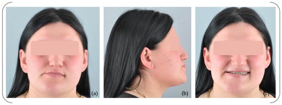
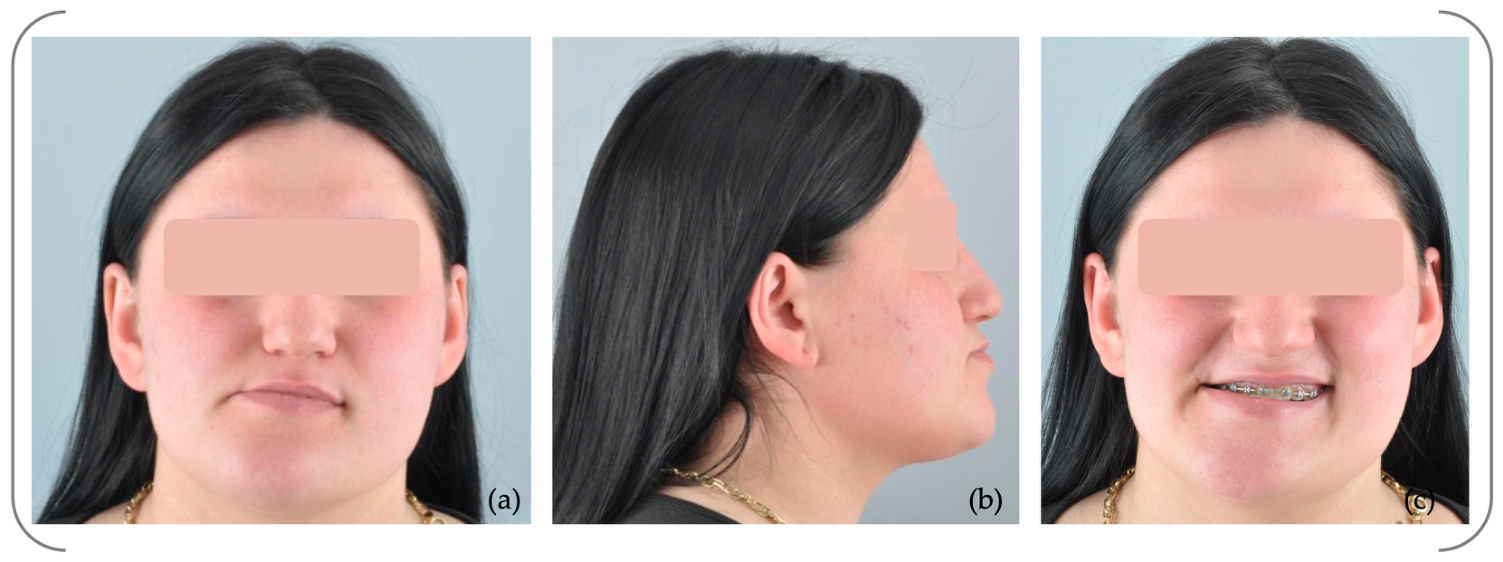
Facial examination revealed a skeletal Class III pattern with a concave profile and a pronounced right lateral deviation (Figure 1).
Figure 1.
Facial photographs during fixed orthodontic treatment: (a) frontal view; (b) lateral view; (c) frontal smiling (case belonging to Dr. Neculai Onică).
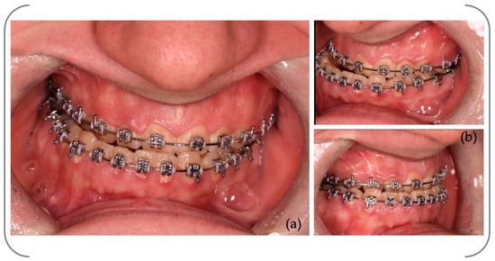
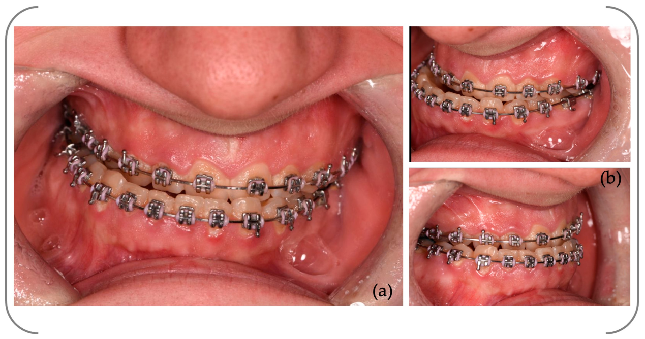
Intraoral photographs taken during fixed orthodontic treatment revealed a right-sided crossbite from tooth 3.4 to 4.7, a notable tilt of the occlusal plane towards the right, and an 8 mm deviation of the mandibular dental midline aligning to the right (Figure 2).
Figure 2.
Intraoral photographs at initial examination: (a) frontal view; (b) lateral view.
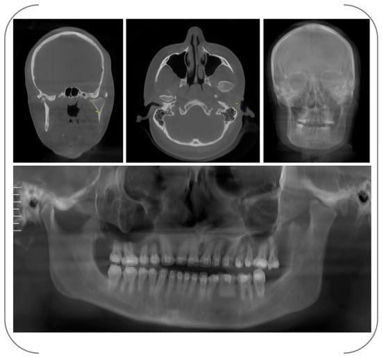
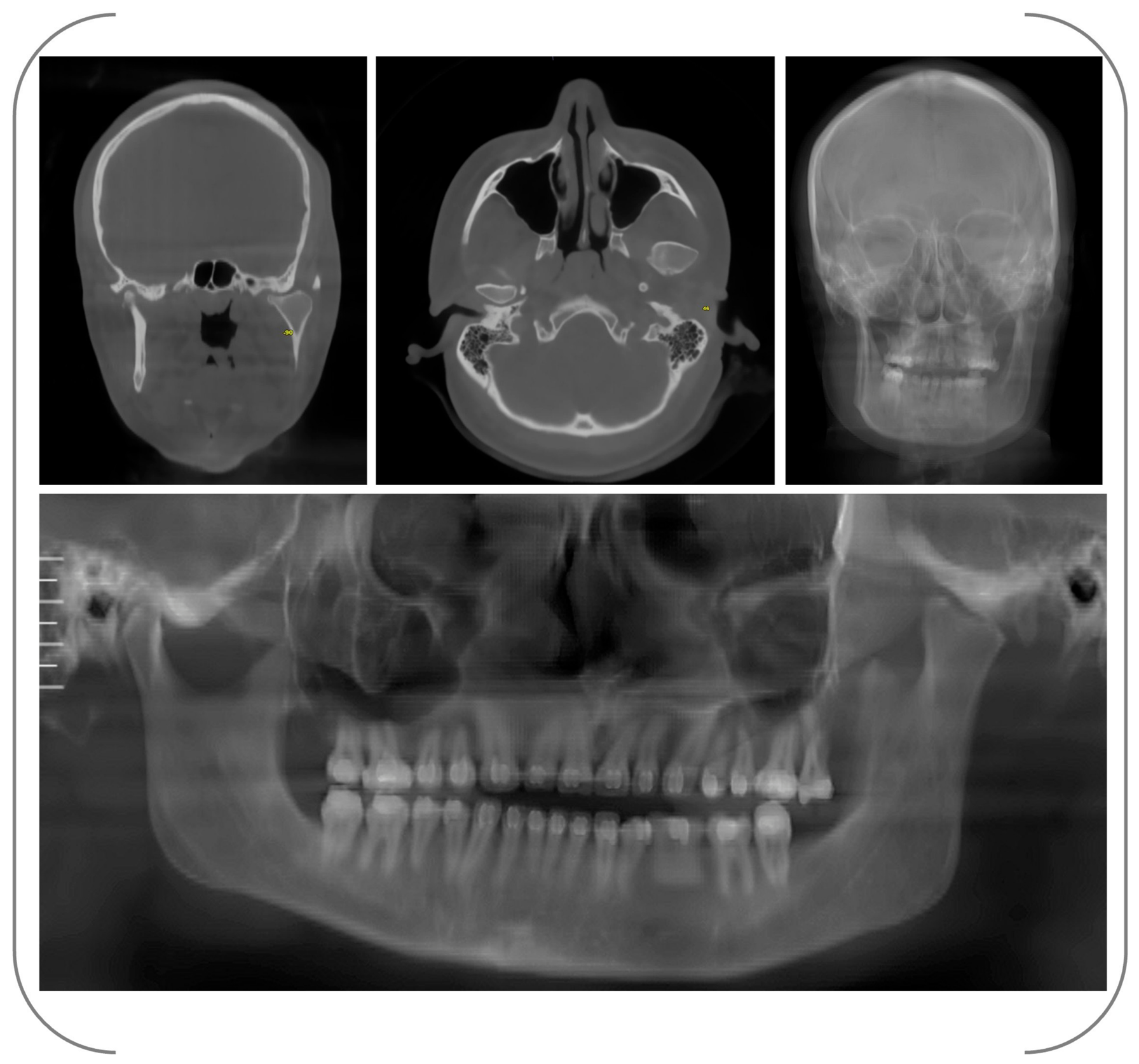
Cone-beam computed tomography (CBCT) scans were acquired using a DentriMax unit (DentriMax, HDX Will, Seoul, Republic of Korea) operating at 90 kV and 10 mA, with a field of view measuring 18 × 16.5 cm, a 0.5 mm focal spot, and a voxel size of 0.10 mm. The datasets were exported in the Digital Imaging and Communications in Medicine (DICOM) format. Tomographic assessment indicated an irregular enlargement of the left mandibular condylar head, consistent with hyperplastic changes, and anterior displacement relative to the glenoid fossa (Figure 3).
Figure 3.
Pre-surgical CBCT images.
Written informed consent was obtained after the patient was thoroughly informed of the prognosis with and without surgical treatment, the proposed therapeutic plan and procedural steps, and the potential benefits, specific risks, and possible adverse effects associated with the intervention.
2.1. Virtual Surgical Plan
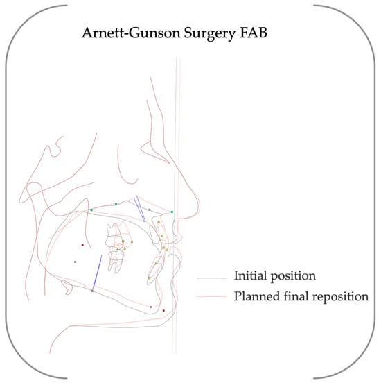
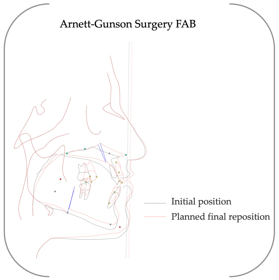
All data were first imported into NemoFab surgical planning software (version 2025, Nemotec, Madrid, Spain) prior to analysis (Figure 4).
Figure 4.
Arnett–Gunson cephalometric analyses (NemoFab, Nemotec, Madrid, Spain) illustrating the relationship between the initial skeletal configuration and the planned final repositioning. The superimposed tracings demonstrate the correction of maxillomandibular discrepancies and facial profile optimization achieved through virtual surgical planning.
The virtual surgical plan included both qualitative and quantitative objectives. Maxillary repositioning was planned to correct the occlusal cant through differential impaction, with an estimated vertical adjustment of approximately 3–4 mm between the right and left posterior segments. Mandibular repositioning aimed to correct a lateral midline deviation of about 7–8 mm towards the right side. The contralateral sagittal split ramus osteotomy was designed to achieve symmetrical mandibular alignment and a stable occlusion. No genioplasty was required, as adequate chin positioning was attained through mandibular repositioning alone.
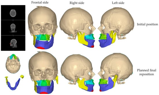
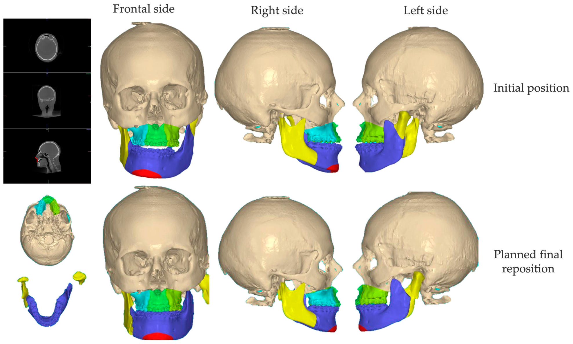
Initially, a Le Fort I osteotomy was simulated to correct the maxillary cant and level the occlusal plane, with slight overcorrection in the molar region to optimise postoperative symmetry. Next, resection of the hyperplastic left condylar segment was planned, ensuring an adequate safety margin between the residual mandibular ramus and the glenoid fossa to accommodate the prosthetic components. Finally, the placement of the total TMJ TJR prosthesis on the affected left side was combined with a contralateral sagittal split ramus osteotomy (SSRO) to restore mandibular symmetry and maintain the planned occlusal relationship (Figure 5).
Figure 5.
Three-dimensional virtual surgical planning performed by surgeon N.O. on a composite digital skull model using NemoFab software (version 2025, Nemotec, Madrid, Spain). Upper row: initial skeletal configuration showing facial asymmetry in frontal and lateral views. Lower row: planned final repositioning following maxillary and mandibular corrections. Color-coded segmentation highlights the osteotomized segments and their repositioning to achieve facial symmetry and occlusal alignment.
The indication for total temporomandibular joint replacement was based on the extent of condylar enlargement, morphological changes, and the expected instability linked to condylectomy alone. In this patient, the severity of the deformity and the requirement for precise three-dimensional reconstruction supported a reconstructive method using a patient-specific prosthesis. This enabled simultaneous removal of the pathological condyle and controlled restoration of joint anatomy within the planned digital workflow.
2.2. TMJ TJR Prosthesis Design
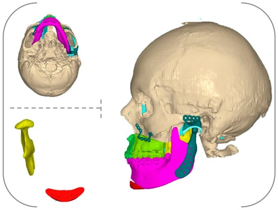
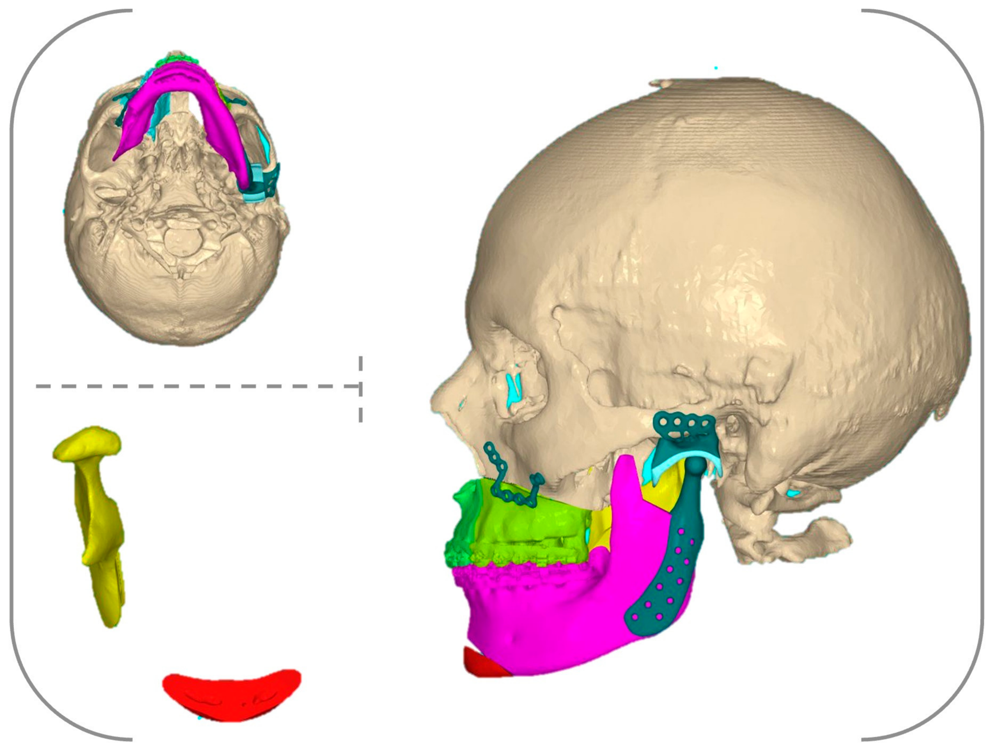
The prosthesis, comprising the glenoid fossa component, condylar head component, and mandibular (handle) component, was digitally designed at VEA MFG LTD using Geomagic Freeform (version 2024, Hexagon AB, Stockholm, Sweden) and SolidWorks (version 2025, Dassault Systèmes, Vélizy-Villacoublay, France) (Figure 6).
Figure 6.
Digital design of the TMJ prosthesis and patient-specific instrumentation. The figure illustrates the segmentation of anatomical structures, the design of the prosthetic components (glenoid fossa and mandibular component), and the development of patient-specific fixation plates.
The principal design considerations of the TMJ TJR prosthesis were as follows:
- −
- The osseous surface of the fossa component was personalised to match the anatomical morphology of the glenoid fossa, zygomatic arch, and residual articular eminence;
- −
- The condylar head component was designed with a cylindrical hollow structure, precisely fitted to a predefined cone frustum at the superior aspect of the mandibular handle component, based on a machine taper connection mechanism;
- −
- The internal surface of the mandibular handle component was personalised to conform to the external surface of the mandibular ramus.
2.3. Patient-Specific Cutting Guides Design
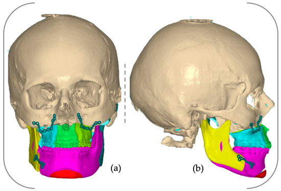
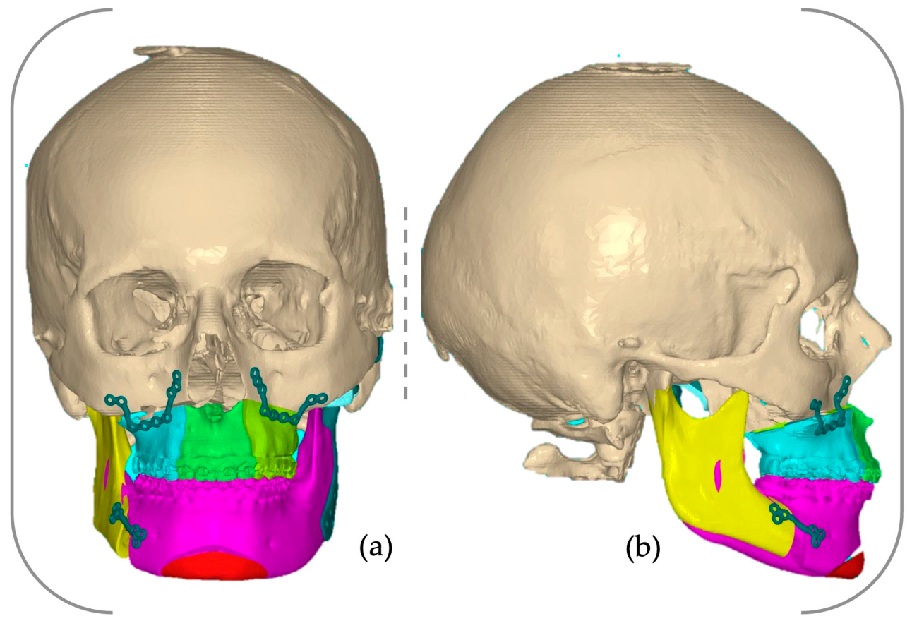
The cutting guides, along with the occlusal splints, were created within the affiliated digital design facility using the same software environment. The repositioned osteotomised bone segments were returned to their original anatomical position, allowing for precise transfer of the planned osteotomy lines and screw positions. The predetermined screw trajectories were converted into guide drill holes, specifying the exact intraoperative drilling sites. Cylindrical extensions were added as drill sleeves, ensuring controlled angulation and accurate placement of both the osteotomies and subsequent fixation hardware. Cutting slots were then designed along the predefined osteotomy planes, enabling exact execution of the planned bone sections. All guiding features, including drill holes and cutting slots, were integrated into a bone-supported guide to ensure stable intraoperative positioning (Figure 7).
Figure 7.
Virtual images of the designed patient-specific cutting guides: (a) frontal view; (b) right lateral view.
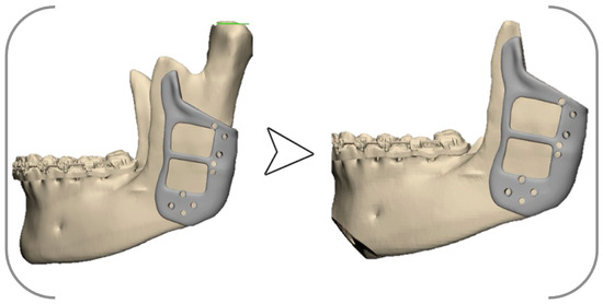
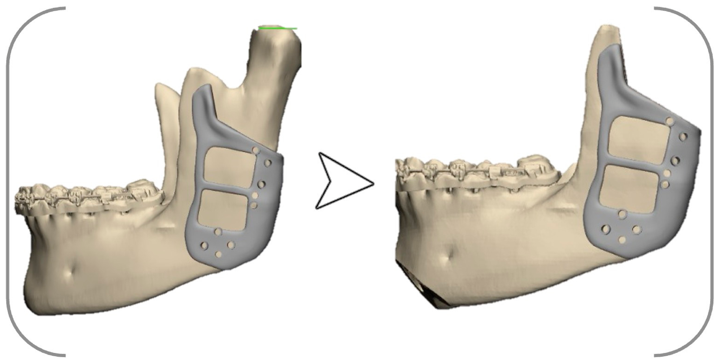
A mandible-first intermediate occlusal splint was digitally designed and produced to ensure precise intraoperative positioning. Additionally, patient-specific surgical guides were created to facilitate the accurate resection of the hyperplastic condylar segment and to ensure proper alignment and fixation of the prosthetic joint components (Figure 8).
Figure 8.
Virtual images of the designed left mandibular patient-specific cutting guide.
2.4. Patient-Specific Plate Design
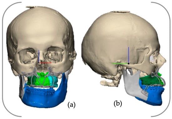
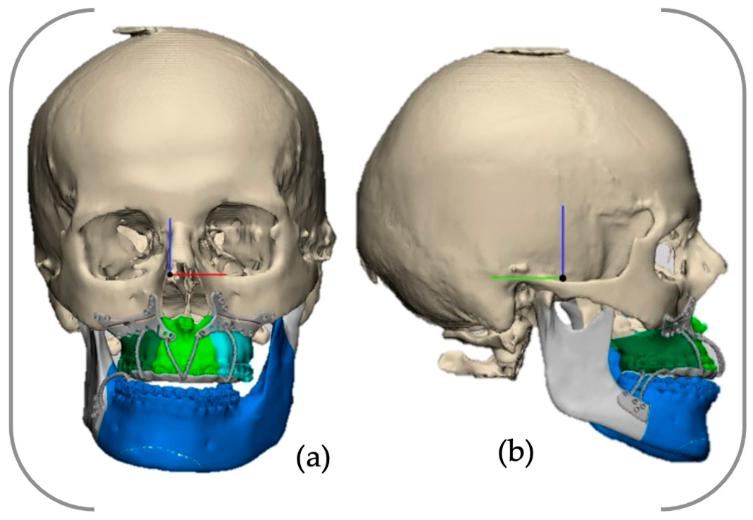
Subsequently, patient-specific fixation plates were digitally designed within the same computer-aided design workflow, based on the final repositioned skeletal configuration. Screw holes were initially positioned on the bone surface according to the planned fixation pattern. The plate body was then created by digitally linking these anchorage points while conforming to the anatomical contours. When the curvature between fixation points exceeded the tolerance of linear connections, intermediate design adjustments were made to ensure accurate surface fitting. In the maxilla, a single plate spanning both sides was preferred to improve the positional accuracy of the mobilised segment. Plate thickness was determined by balancing mechanical strength with minimal bulk; typically, a thickness of approximately 1.2–1.6 mm was chosen, with slight reinforcement in high-stress regions to reduce the risk of deformation during manufacturing and handling (Figure 9).
Figure 9.
Virtual images of the designed patient-specific plates: (a) frontal view; (b) right lateral view.
2.5. TMJ TJR Prothesis and Patient-Specific Implant Manufacturing
The fossa component was made from ultra-high-molecular-weight polyethylene (UHMWPE) by Orthoplastics (Orthoplastics Ltd., Bacup, Lancashire, UK) using subtractive manufacturing based on our personalised digital design.
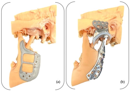
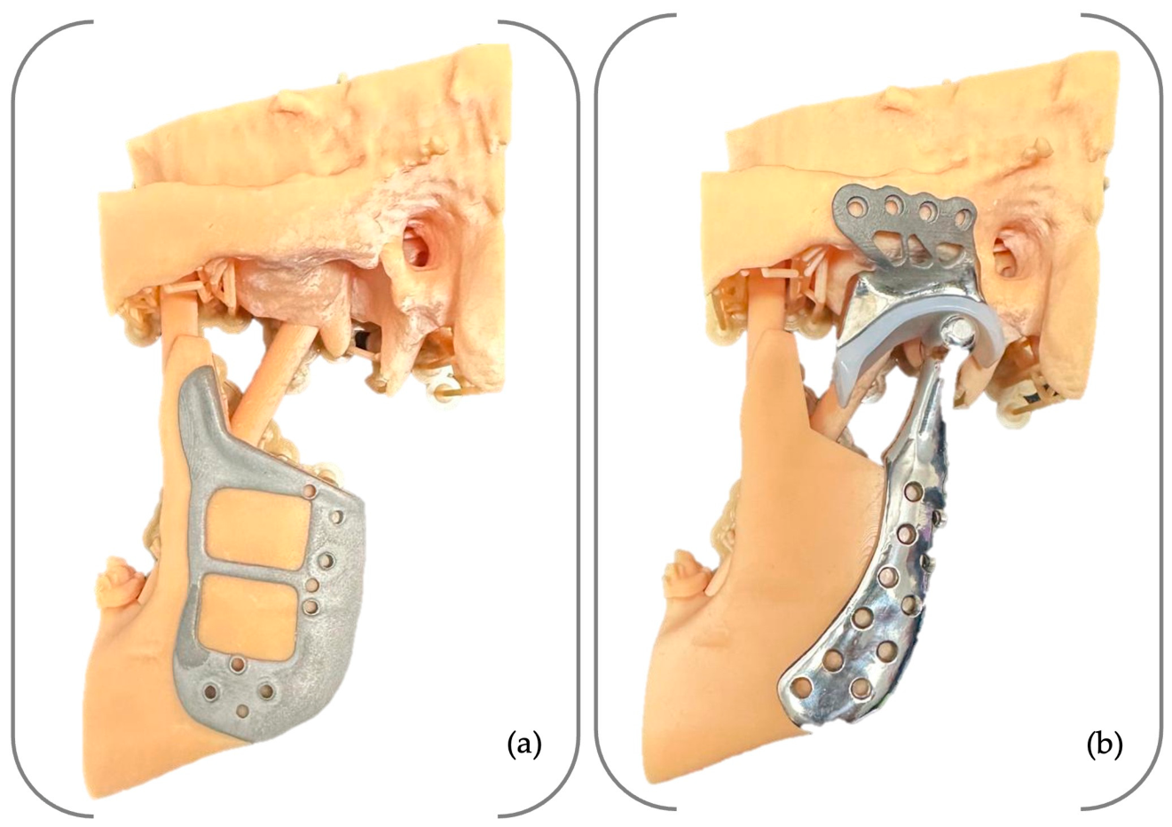
The condylar head, mandibular component, cutting guides, and patient-specific plates were exported in standard tessellation language (STL) format for 3D printing. They were fabricated from Ti6Al4V powder (PowderRange Ti64, Carpenter Technology Corporation, Philadelphia, PA, USA) using additive manufacturing with Selective Laser Melting (SLM) technology (mysint100, SISMA S.p.A., 36013 Piovene Rocchette, Italy) (Figure 10).
Figure 10.
The patient-specific (a) mandibular cutting guide and (b) TMJ TJR prosthesis.
All patient-specific components, including the TMJ TJR prosthesis, surgical cutting guides, and plates, were delivered in a clean (plasma-cleaned), non-sterile condition state.
Prior to surgical implantation, the prosthetic components and patient-specific guides were verified using three-dimensional printed anatomical models derived from the preoperative CBCT data. Trial fitting was conducted to assess the congruency between the prosthesis and the mandibular and cranial surfaces, as well as to confirm the accuracy of screw hole positioning.
The glenoid fossa component was sterilised with ethylene oxide gas, while the metallic elements were sterilised using steam autoclaving in accordance with established protocols. After sterilisation, each component was individually packaged and sealed to maintain sterility until the time of surgery.
The chronological sequence of the multidisciplinary treatment is summarised in Table 1.
Table 1.
Chronological timeline of the multidisciplinary treatment, including orthodontic preparation, digital planning, surgical intervention, and follow-up.
2.6. Surgical Intervention
In December 2025, surgeon N.O. performed a left mandibular condylectomy, combined with a contralateral SSRO and a Le Fort I osteotomy, under general anaesthesia with nasotracheal intubation, following the completion of preoperative orthodontic treatment.
Preoperative blood investigations were within normal limits. To minimise intraoperative bleeding and facilitate tissue dissection, local infiltration was performed with six cartridges (1.7 mL each) of articaine with epinephrine, supplemented with additional epinephrine and tranexamic acid. Furthermore, 100 mL of saline was used for local irrigation and haemostatic control [15].
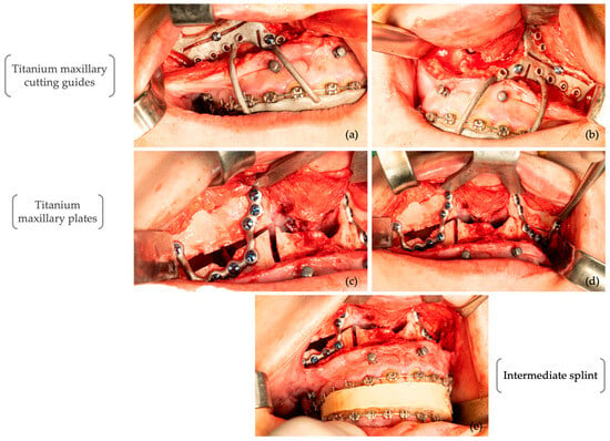
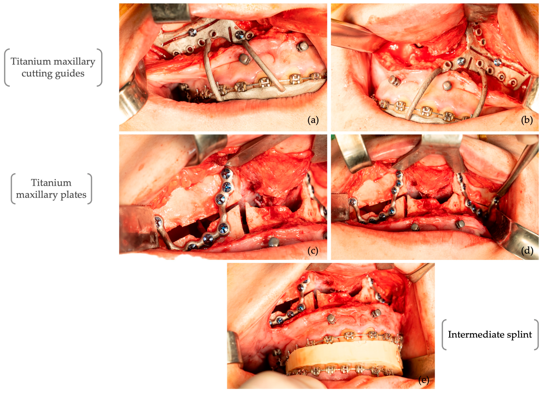
The procedure involved several wrappings to ensure strict separation between the intraoral and extraoral operative fields. The intervention started intraorally with a right SSRO, performed without splitting at the initial stage. A segmental Le Fort I osteotomy was then executed with down-fracture, mobilisation, and fixation using patient-specific maxillary implants, followed by layered suturing (Figure 11a–d). Osteotomy cuts were made using a piezoelectric surgical device (Woodpecker Ultrasurgery, Guilin Woodpecker Medical Instrument Co., LTD., Guilin, China) with dedicated tips (US25 and US1L Left Angle 90° Micro-Saw 0.5 mm, Guilin Woodpecker Medical Instrument Co., LTD., Guilin, China). After fixation, the position of the intermediate splint was verified to ensure precise reproduction of the planned occlusal relationship (Figure 11e).
Figure 11.
Intraoperative photographs of the multisegmental Le Fort I osteotomy performed using patient-specific cutting guides (a,b) and fixation plates (c,d). The intermediate splint position was verified to confirm the planned occlusion (e).
The operative field was then re-draped, and the patient was prepared for the extraoral phase of the procedure. The mandibular condylar resection site was preoperatively assessed using a three-dimensional (3D) jaw model.
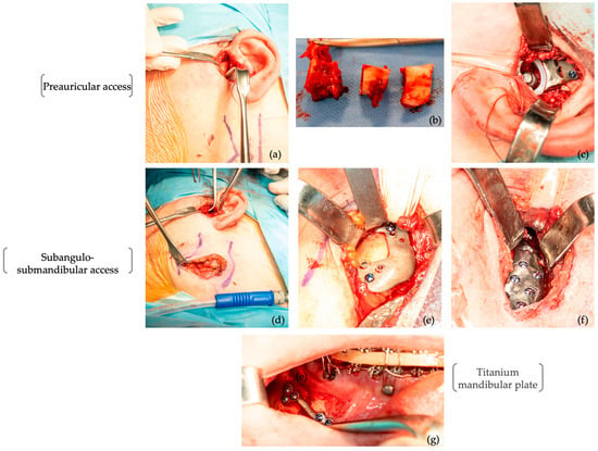
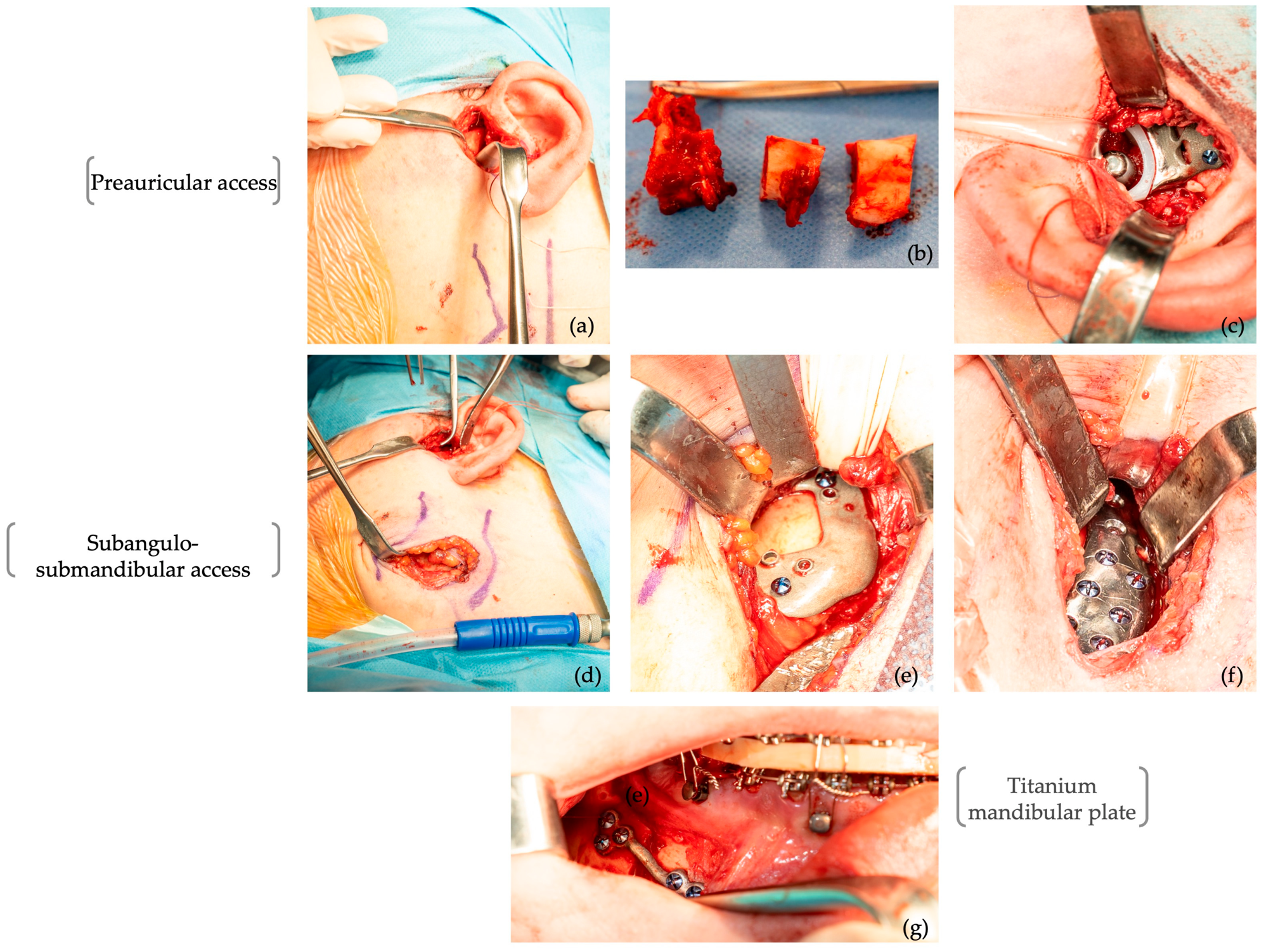
Surgical access was obtained via a dual approach, combining preauricular and subangulo-submandibular incisions. Due to its considerable size, the condyle was sectioned into three segments to facilitate extraction through the preauricular access (Figure 12b).
Figure 12.
Intraoperative photographs illustrating mandibular condylar resection (a,b,e); placement of the patient-specific total TMJ prosthesis through preauricular and subangulo-submandibular approaches (a–f); and right SSRO and fixation with patient-specific titanium plate (g).
Concomitantly with the condylar ostectomy, multiple fixation holes for the condylar prosthesis were prepared. The cutting and drilling guide was introduced via the previously performed subangulo-submandibular approach, which exposed and identified the inferior border of the mandible and the left mandibular angle, the posterior belly of the digastric muscle, the anterior margin of the sternocleidomastoid muscle, and the mastoid region, and protected the marginal mandibular nerve.
Following preparation of the TMJ replacement site and placement of gauze soaked in 10% vancomycin solution, the operative field was re-draped and the procedure continued intraorally. A right sagittal split osteotomy was completed, the final splint was positioned, and intermaxillary fixation (IMF) was established. The right SSRO was stabilised with a patient-specific fixation plate secured with six screws (Figure 12f).
After further adjustment of the draping, the extraoral field was re-entered. The gauze was removed, and the surgical site was thoroughly irrigated with a 10% vancomycin solution. The fossa component was inserted and manually positioned via the subangulo-submandibular approach to avoid contact between the UHMWPE surface and metallic instruments; it was then fixed with four 2.0 × 6 mm screws (Medicon eG, Tuttlingen, Germany). The condylar prosthetic component was subsequently introduced through the same approach, guided by a 2-0 non-resorbable suture passing from the preauricular access using haemostatic forceps. The component was seated in the fossa, properly centred, and secured with screws measuring 6–9 mm in length and 2 mm in diameter (Medicon eG, Tuttlingen, Germany).
The surgical site was irrigated again with vancomycin solution, covered with vancomycin powder, and closed in anatomical layers. Finally, intermaxillary fixation was released, the occlusion was verified, the intraoral incision was sutured, and two elastics were applied in a Class II configuration.
Postoperative management involved a 7-day course of intravenous antibiotic therapy, along with routine analgesic medication for pain relief. An ice pack was applied intermittently during the first 72 h after the procedure to reduce swelling and pain.
During the first three days following surgery, the surgical team managed the patient’s wound care. The patient was also provided with detailed instructions on maintaining oral hygiene, including the use of a soft toothbrush and chlorhexidine mouth rinses for 10 days. A liquid diet was advised for four weeks. Intermaxillary elastic therapy was continued for 40 days, after which postoperative orthodontic treatment was commenced. Physiotherapy was introduced from the second postoperative week onwards.
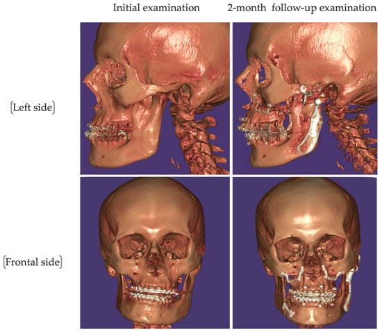
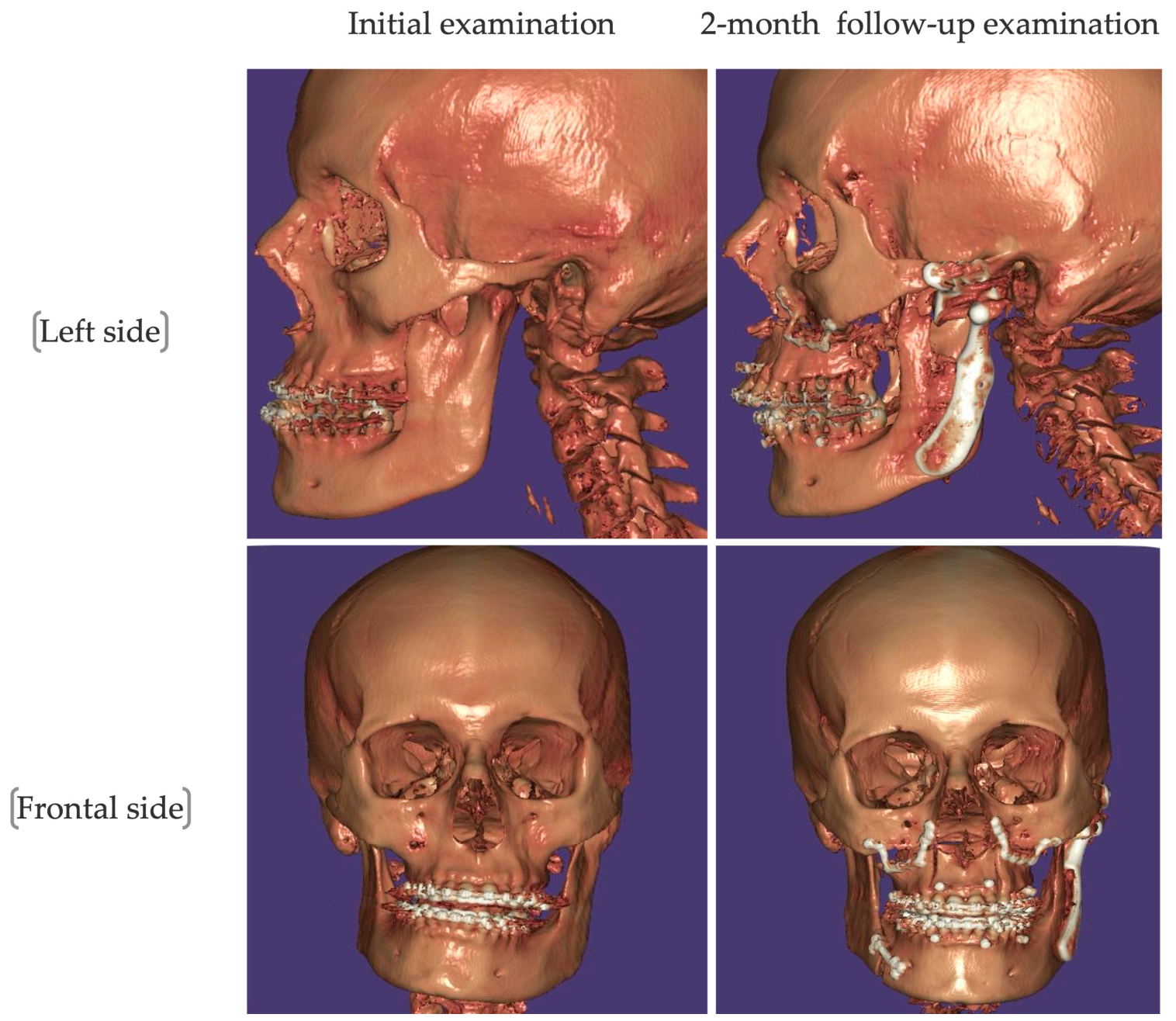
At the two-month postoperative review, clinical and radiological findings indicated successful early outcomes. Three-dimensional imaging confirmed the stable positioning of the prosthetic components with no signs of displacement or loosening (Figure 13).
Figure 13.
Three-dimensional CT reconstruction comparing initial examination and 2-month postoperative follow-up.
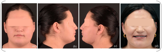
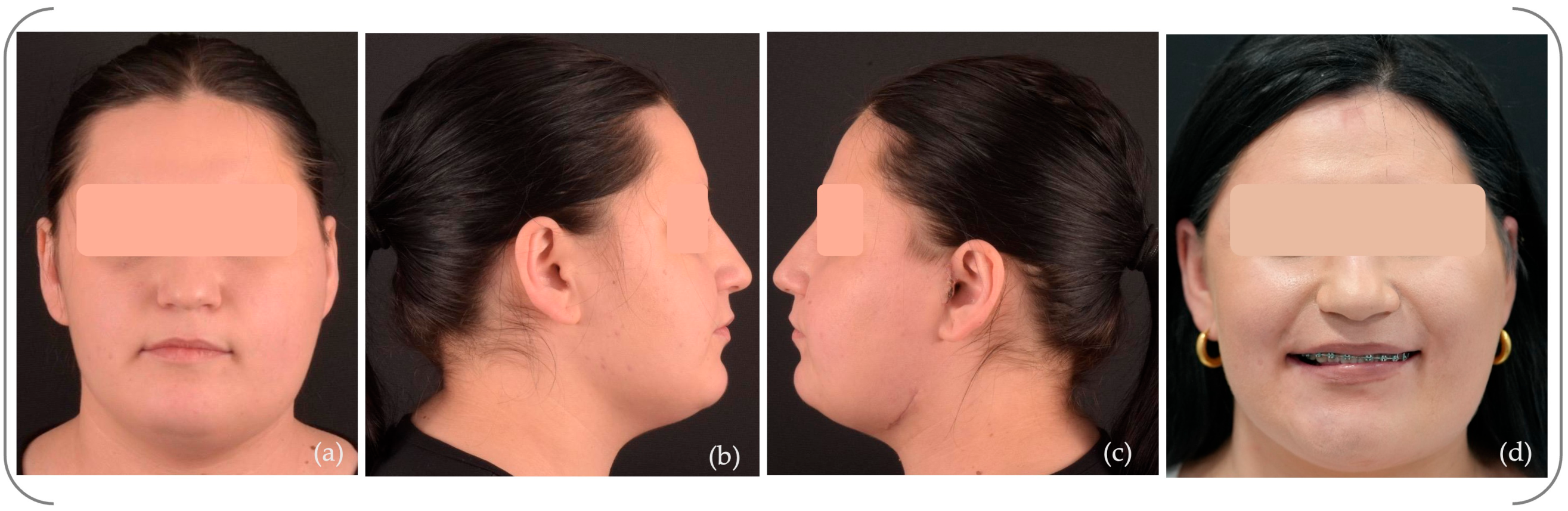
Clinically, a significant improvement in facial symmetry was observed (Figure 14a–d).
Figure 14.
Facial photographs at 2- month postoperative follow-up: (a) frontal view; (b) right lateral view; (c) left lateral view; and (d) frontal smiling.
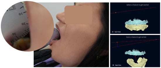
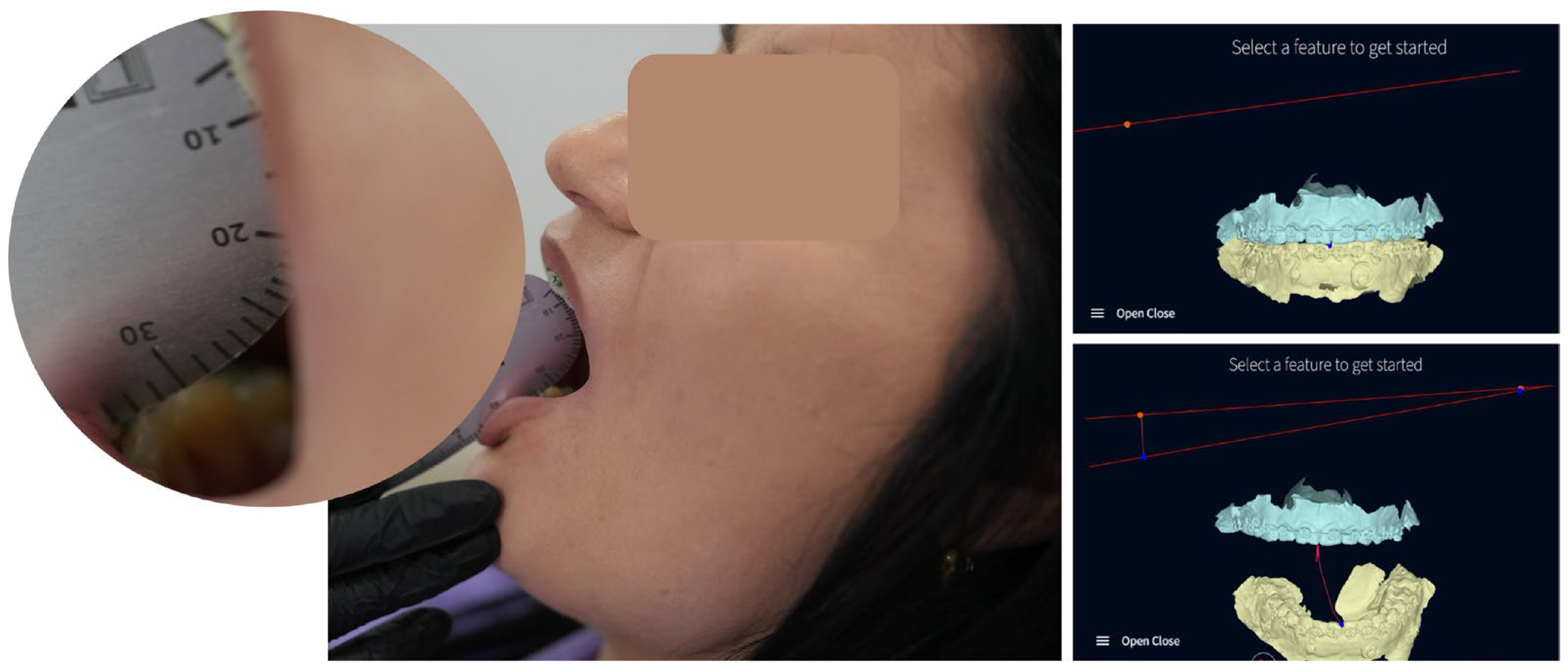
At the three-month follow-up, the patient showed successful functional recovery. Maximum interincisal mouth opening reached 32 mm, and mandibular movements recorded with the Modjaw system (Modjaw SAS, Lyon, France), incorporating the Twin in Motion™ (TWIM) software (version 3.6.3), revealed a deviation in the mandible towards the prosthetic side during maximum opening (Figure 15).
Figure 15.
Mandibular movement during maximum opening at 3-month postoperative follow-up.
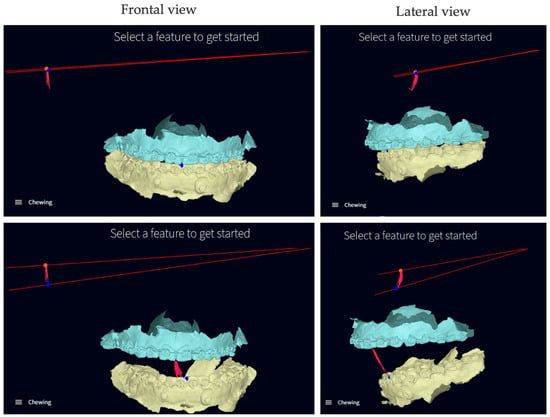
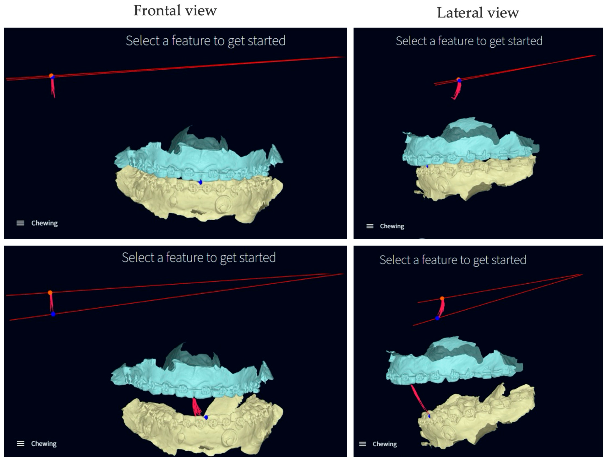
Mandibular kinematic analysis using the Modjaw system (Modjaw SAS, Lyon, France) revealed a predominantly rotational chewing pattern, with reduced translational components and limited lateral excursions (Figure 16).
Figure 16.
Modjaw-based dynamic analysis of mandibular movements during chewing, illustrating reduced lateral excursions and a predominantly rotational pattern.
The patient reported minimal postoperative discomfort, with a pain level of 2 on the visual analogue scale (VAS), where 0 indicates no pain, and 10 indicates maximum pain intensity.
Speech and mastication were subjectively improved, aligning with patient-reported outcome measures indicating a favourable postoperative state. Quality of life was evaluated using the orthognathic quality-of-life questionnaire (OQLQ) [16], which contains 22 items rated on a 5-point Likert scale from 0 (“does not bother me at all”) to 4 (“bothers me a lot”), with a total score from 0 to 88. The functional domain score was 9/20, suggesting a mild to moderate functional impact, mainly related to mastication and occlusal adaptation. The aesthetic (4/20) and awareness (4/16) domains showed minimal impact, while the social domain indicated very low impact (2/32), reflecting negligible psychosocial impairment. Additionally, assessment with the Oral Health Impact Profile (OHIP-14) resulted in a total score of 8/56, signifying minimal influence on oral health-related quality of life [17]. The patient occasionally reported minor difficulties related to speech.
Progressive dietary progress was made, with a return to a normal diet by the end of the follow-up period. Sensory function in the distribution of the inferior alveolar nerve was maintained. At this early postoperative stage, stability can be considered provisional.
3. Discussion
Unilateral condylar hyperplasia most commonly affects individuals aged between 11 and 30 years, although its exact cause remains only partly understood, with various factors suggested, including hormonal influences, trauma, infection, and genetic predisposition [2,12,18]. Hormonal involvement has been proposed, especially during periods of active skeletal growth, with oestrogen receptors found within the temporomandibular joint. However, previous studies show inconsistent results regarding sex distribution, and the link between UCH and hormonal factors remains speculative [19,20,21,22].
The optimal timing and surgical approach for managing unilateral condylar hyperplasia (UCH), especially when linked to dentofacial deformities, remain subjects of ongoing debate [21,22,23,24]. In this case, the pronounced mandibular midline deviation and severe facial asymmetry required a combined surgical strategy, including condylectomy, total temporomandibular joint replacement, and orthognathic correction.
Within this context, the present case illustrates the use of a fully digital workflow that combines virtual surgical planning and patient-specific solutions. Although the general principles of managing condylar hyperplasia and reconstructing the TMJ are well documented in the literature, this report highlights the practical application of these technologies in a complex clinical scenario. The digital workflow improved preoperative visualisation and planning of skeletal asymmetry, enabling accurate determination of maxillary levelling and mandibular repositioning. The utilisation of patient-specific cutting guides and fixation plates facilitated precise intraoperative transfer of the virtual plan, with no significant differences observed between the planned and actual outcomes. Additionally, the patient-specific prosthesis design, along with guided placement, allowed controlled positioning of the joint components, simplifying intraoperative procedures and reducing the need for adjustments.
Postoperative pain was minimal (VAS 2), aligning with the literature, which reports significant pain reduction following total TMJ replacement, with mean VAS scores falling from 6.6 preoperatively to around 1.7–1.9 at one year postoperatively [25,26]. Although follow-up is limited, these results suggest a positive early outcome, justifying further long-term assessment.
From a functional perspective, mandibular kinematic analysis revealed an adaptive pattern mainly characterised by rotational movements and limited lateral excursions. This changed chewing pattern aligns with the lack of functional reinsertion of the lateral pterygoid muscle onto the prosthetic condyle. Considering the crucial role of this muscle in translational and lateral mandibular movements, its detachment probably contributes to altered mandibular kinematics after total TMJ replacement. The debate in the literature continues regarding the functional implications of reattaching the lateral pterygoid [25,26,27].
Additionally, the observed maximum interincisal mouth opening reached 32 mm at the 3-month postoperative follow-up, which falls within the range reported in the literature for patients undergoing total TMJ replacement. Previous studies have shown that average mouth opening increases from approximately 27.5 mm preoperatively to 32.6 mm postoperatively [26]. The deviation of the mandible toward the prosthetic side during maximum mouth opening may be explained by the increased vertical dimension of the prosthetic fossa component, which effectively reduces the functional height of the reconstructed ascending ramus on the affected side, contributing to asymmetric mandibular motion.
Despite these biomechanical adaptations, functional recovery was satisfactory. Outcome assessment, based on clinical examination and patient-reported measures, including OQLQ and OHIP-14, indicated mild functional impact and minimal psychosocial impairment.
The long-term performance of TMJ prostheses remains affected by various factors, including infection, heterotopic ossification, material wear, and the possible need for revision surgery [14,28,29,30]. Therefore, caution is necessary when interpreting short-term results, and long-term follow-up is essential.
In the present case, no significant discrepancies were found between the virtual surgical plan and the manufactured components, and no intraoperative adjustments were necessary. Additionally, no early postoperative complications were observed during the available follow-up period. However, since this report concerns only a single patient with a relatively short follow-up period (3 months), further research involving larger cohorts and longer observation periods is needed to assess the reproducibility, long-term stability, and overall reliability of this treatment approach.
4. Conclusions
This case report provides insight into the feasibility of integrating virtual surgical planning, personalised instrumentation, and patient-specific TMJ prosthesis within a comprehensive surgical workflow. Positive short-term clinical and radiological outcomes were observed, including enhanced facial symmetry, stable occlusion, and satisfactory functional recovery; however, these findings should be interpreted with caution due to the single-case design and limited follow-up period.
Author Contributions
Conceptualization, E.-R.B. and N.O.; methodology, N.O. and C.A.O.; validation, G.L.G. and A.M.; investigation, C.I.L.; resources, N.O.; writing—original draft preparation, E.-R.B. and N.O.; writing—review and editing, C.A.O. and E.-R.B.; visualization, C.I.F.; supervision, G.L.G.; project administration, N.O. All authors have read and agreed to the published version of the manuscript.
Funding
This research was supported by the project “VEAINOV3D—Development of VEA MFG’s advanced innovation and production capacity in the customized medical prosthetics and automotive sector using 3D High-Tech” (SMIS code 338485), co-financed by the European Union through the North-East Regional Program 2021–2027 (Romania), part of the European Regional Development Fund (ERDF).
Institutional Review Board Statement
The study was carried out in accordance with the latest version of the Declaration of Helsinki and was approved by the Research Ethic Committee of the University of Medicine and Pharmacy, Iasi, Romania (Approval No. 566; Approval date: 18 March 2025).
Informed Consent Statement
Informed consent was obtained from the subject involved in the study. Written informed consent was obtained from the patient to publish this paper.
Data Availability Statement
The original contributions presented in this study are included in the article. Further inquiries can be directed to the corresponding author.
Acknowledgments
The authors acknowledge the support provided within the project “VEAINOV3D”, implemented by VEA MFG L.T.D. as lead partner, in partnership with Iasi University of Life Sciences (IULS). The project contributes to the development of advanced technologies and innovation capacity in customized medical prosthetics, in line with the Regional Smart Specialisation Strategy (RIS3 North-East 2021–2027).
Conflicts of Interest
Authors Elena-Raluca Baciu, Cezara Andreea Onică, and Neculai Onică were employed by the company VEA MFG L.T.D. The remaining authors declare that the research was conducted in the absence of any commercial or financial relationships that could be construed as a potential conflict of interest. The funders had no role in the design of the study; in the collection, analyses, or interpretation of data; in the writing of the manuscript; or in the decision to publish the results.
Abbreviations
The following abbreviations are used in this manuscript:
| CBCT | Cone-beam computed tomography |
| CT | Computed tomography |
| DICOM | Digital imaging and communications in medicine |
| OHIP | Oral health impact profile |
| OQLQ | Orthognathic quality-of-life questionnaire |
| SLM | Selective laser melting |
| SSRO | Sagittal split ramus osteotomy |
| STL | Standard tessellation language |
| TMJ | Temporomandibular joint |
| TMJ TJR | Total temporomandibular joint replacement |
| UCH | Unilateral condylar hyperplasia |
| UHMWPE | Ultra-high-molecular-weight polyethylene |
References
- Olate, S.; Netto, H.D.; Rodriguez-Chessa, J.; Alister, J.P.; de Albergaria-Barbosa, J.; de Moraes, M. Mandible condylar hyperplasia: A review of diagnosis and treatment protocol. Int. J. Clin. Exp. Med. 2013, 6, 727–737. [Google Scholar]
- Villanueva-Alcojol, L.; Monje, F.; González-García, R. Hyperplasia of the mandibular condyle: Clinical, histopathologic, and treatment considerations in a series of 36 patients. J. Oral Maxillofac. Surg. 2011, 69, 447–455. [Google Scholar] [CrossRef] [Scilit]
- Riechmann, M.; Gellrich, N.C.; Kress, B.; Schmidt, C.; Schröder, C.; Neff, A. Clinical Practice Guideline: Condylar Hyperplasia of the Mandible—Diagnosis and Treatment. Dtsch. Arztebl. Int. 2024, 121, 816–823. [Google Scholar] [PubMed]
- Raijmakers, P.G.; Karssemakers, L.H.; Tuinzing, D.B. Female predominance and effect of gender on unilateral condylar hyperplasia: A review and meta-analysis. J. Oral Maxillofac. Surg. 2012, 70, e72–e76. [Google Scholar] [CrossRef] [Scilit] [PubMed]
- Obwegeser, H.L.; Makek, M.S. Hemimandibular hyperplasia—Hemimandibular elongation. J. Maxillofac. Surg. 1986, 14, 183–208. [Google Scholar] [CrossRef] [Scilit] [PubMed]
- Verhoeven, T.J.; Nolte, J.W.; Maal, T.J.; Bergé, S.J.; Becking, A.G. Unilateral condylar hyperplasia: A 3-dimensional quantification of asymmetry. PLoS ONE 2013, 8, e59391. [Google Scholar] [CrossRef] [Scilit]
- Gateno, J.; Coppelson, K.B.; Kuang, T.; Poliak, C.D.; Xia, J.J. A better understanding of unilateral condylar hyperplasia of the mandible. J. Oral Maxillofac. Surg. 2021, 79, 1122–1132. [Google Scholar] [CrossRef] [Scilit]
- Pripatnanont, P.; Vittayakittipong, P.; Markmanee, U.; Thongmak, S.; Yipintsoi, T. The use of SPECT to evaluate growth cessation of the mandible in unilateral condylar hyperplasia. Int. J. Oral Maxillofac. Surg. 2005, 34, 364–368. [Google Scholar] [CrossRef] [Scilit]
- De Schepper, S.; Gnanasegaran, G.; De Vos, W.; Van de Casteele, E.; Dickson, J.C.; Van den Wyngaert, T. From SPECT/CT towards absolute quantification?—The case of unilateral condylar hyperplasia of the mandible. EJNMMI Phys. 2024, 11, 74. [Google Scholar] [CrossRef] [Scilit]
- Crank, S.; Gray, S.; Sidebottom, A.J. Condylar hyperplasia—Review of treatment outcomes and suggested pathway for management. Br. J. Oral Maxillofac. Surg. 2007, 45, e60–e61. [Google Scholar] [CrossRef] [Scilit]
- Mehrotra, D.; Dhasmana, S.; Kamboj, M.; Gambhir, G. Condylar hyperplasia and facial asymmetry: Report of five cases. J. Maxillofac. Oral Surg. 2011, 10, 50–56. [Google Scholar] [CrossRef] [Scilit] [PubMed]
- Suzuki, H.; Nogami, S.; Otake, Y.; Takeda, Y.; Sugawara, J.; Takahashi, T.; Yamauchi, K. Surgery-early approach combined with condylectomy for correction of severe facial asymmetry with mandibular condylar hyperplasia: A case report. J. Korean Assoc. Oral Maxillofac. Surg. 2024, 50, 227–234. [Google Scholar] [CrossRef] [Scilit]
- Zheng, J.; Chen, X.; Jiang, W.; Zhang, S.; Chen, M.; Yang, C. An innovative total temporomandibular joint prosthesis with customized design and 3D printing additive fabrication: A prospective clinical study. J. Transl. Med. 2019, 17, 4. [Google Scholar] [CrossRef] [Scilit]
- Huang, D.; Zou, L.; Lu, C.; Zhao, J.; He, D.; Yang, C. Simultaneous standard total joint prosthesis reconstruction with SSRO and Le Fort I osteotomy in the treatment of unilateral temporomandibular joint ankylosis with jaw deformity: A case cohort study. Clin. Oral Investig. 2024, 28, 163. [Google Scholar] [CrossRef] [Scilit]
- Onică, N.; Onică, C.A.; Tatarciuc, M.; Baciu, E.-R.; Vlasie, G.-L.; Ciofu, M.; Balan, M.; Gelețu, G.L. Managing predicted post-orthognathic surgical defects using combined digital software: A case report. Healthcare 2023, 11, 1219. [Google Scholar] [CrossRef] [Scilit] [PubMed]
- Cunningham, S.J.; Garratt, A.M.; Hunt, N.P. Development of a condition-specific quality of life measure for patients with dentofacial deformity: II. Validity and responsiveness testing. Community Dent. Oral Epidemiol. 2002, 30, 81–90. [Google Scholar] [CrossRef] [Scilit]
- Slade, G.D.; Spencer, A.J. Development and evaluation of the Oral Health Impact Profile. Community Dent. Health 1994, 11, 3–11. [Google Scholar]
- Toh, A.Q.J.; Becking, A.G.; Leung, Y.Y. Mirror-image unilateral condylar hyperplasia in monozygotic twins. Int. J. Oral Maxillofac. Surg. 2021, 50, 1177–1181. [Google Scholar] [CrossRef] [Scilit]
- Beltran, J.; Zaror, C.; Moya, M.P.; Netto, H.D.; Olate, S. Diagnosis and treatment in unilateral condylar hyperplasia. J. Clin. Med. 2023, 12, 1017. [Google Scholar] [CrossRef] [Scilit]
- Nelke, K.H.; Pawlak, W.; Morawska-Kochman, M.; Łuczak, K. Ten years of observations and demographics of hemimandibular hyperplasia and elongation. J. Craniomaxillofac. Surg. 2018, 46, 979–986. [Google Scholar] [CrossRef] [Scilit] [PubMed]
- Turvey, T.A.; Hannan, E.; Brader, T.; Bhatt, P.; Oliverson, T.; Magraw, C.B.; Pham, M. Active unilateral condylar hyperplasia treated with simultaneous condylectomy and orthognathic surgery: A clinical report. J. Oral Maxillofac. Surg. 2022, 80, 1593–1612. [Google Scholar] [CrossRef] [Scilit] [PubMed]
- Nolte, J.W.; Alders, M.; Karssemakers, L.H.E.; Becking, A.G.; Hennekam, R.C.M. Molecular basis of unilateral condylar hyperplasia? Int. J. Oral Maxillofac. Surg. 2020, 49, 1397–1401. [Google Scholar] [CrossRef] [Scilit]
- Wolford, L.M. Concomitant temporomandibular joint and orthognathic surgery. J. Oral Maxillofac. Surg. 2003, 61, 1198–1204. [Google Scholar] [CrossRef] [Scilit]
- Singh, V.; Verma, A.; Attresh, G.; Batra, J. Ortho-surgical management of condylar hyperplasia: Rare case reports. Natl. J. Maxillofac. Surg. 2014, 5, 54–59. [Google Scholar] [CrossRef] [Scilit]
- Yoda, T.; Ogi, N.; Yoshitake, H.; Kawakami, T.; Takagi, R.; Murakami, K.; Yuasa, H.; Kondoh, T.; Tei, K.; Kurita, K. Clinical guidelines for total temporomandibular joint replacement. Jpn. Dent. Sci. Rev. 2020, 56, 77–83. [Google Scholar] [CrossRef] [Scilit]
- Zou, L.; He, D.; Ellis, E. A comparison of clinical follow-up of different total temporomandibular joint replacement prostheses: A systematic review and meta-analysis. J. Oral Maxillofac. Surg. 2018, 76, 294–303. [Google Scholar] [CrossRef] [Scilit]
- Celebi, N.; Rohner, E.C.; Gateno, J.; Noble, P.C.; Ismaily, S.K.; Teichgraeber, J.F.; Xia, J.J. Development of a mandibular motion simulator for total joint replacement. J. Oral Maxillofac. Surg. 2011, 69, 66–79. [Google Scholar] [CrossRef] [Scilit]
- Lima, F.G.G.P.; Rios, L.G.C.; Paranhos, L.R.; Vieira, W.A.; Zanetta-Barbosa, D. Survival of temporomandibular total joint replacement: A systematic review and meta-analysis. J. Oral Rehabil. 2024, 51, 775–784. [Google Scholar] [CrossRef] [Scilit] [PubMed]
- Gakhal, M.K.; Gupta, B.; Sidebottom, A.J. Analysis of outcomes after revision replacement of failed total temporomandibular joint prostheses. Br. J. Oral Maxillofac. Surg. 2020, 58, 220–224. [Google Scholar] [CrossRef] [Scilit]
- Olate, S.D.; Ravelo, V.D.; Huentequeo, C.D.; Alister, J.P.D.; Parra, M.D. Indications for the use of TMJ prostheses in South America. J. Craniofac. Surg. 2025, 36, e434–e437. [Google Scholar] [CrossRef] [Scilit] [PubMed]

Disclaimer/Publisher’s Note: The statements, opinions and data contained in all publications are solely those of the individual author(s) and contributor(s) and not of MDPI and/or the editor(s). MDPI and/or the editor(s) disclaim responsibility for any injury to people or property resulting from any ideas, methods, instructions or products referred to in the content. |
© 2026 by the authors. Licensee MDPI, Basel, Switzerland. This article is an open access article distributed under the terms and conditions of the Creative Commons Attribution (CC BY) license.