Optimizing the Use of Ultrasound in Calcium Pyrophosphate Deposition (CPPD): A Review from the Ground Up



Abstract
1. Introduction
2. Crystal Formation and Deposition in Joints: Brief Review
3. Ultrasound Definitions for Crystal Identification in Tissues
4. Scanning Technique and Machine Setting Optimization for Crystal Visualization
4.1. General Considerations
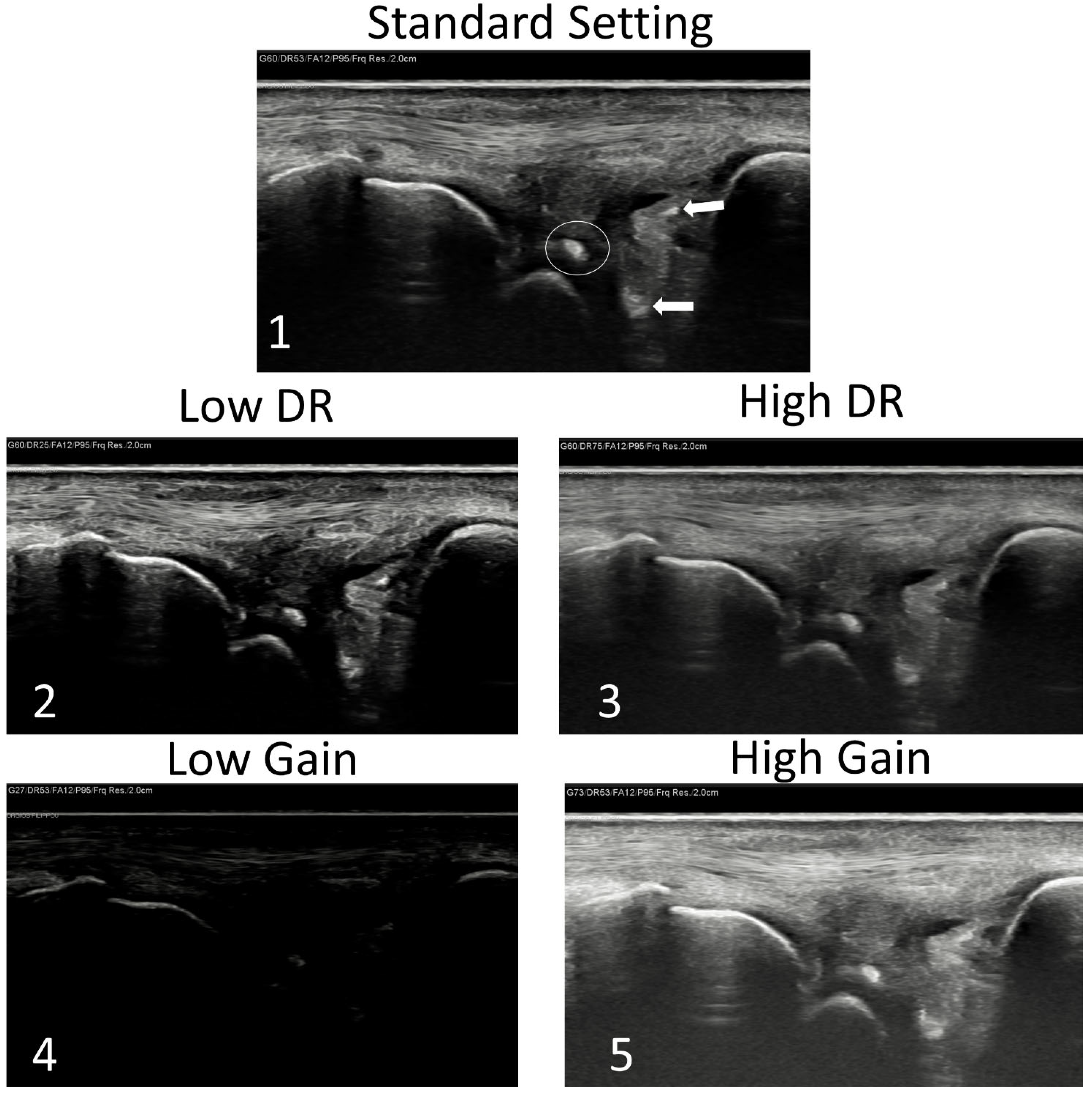
4.2. How to Set the Machine in Order to Enhance Crystal Appearance
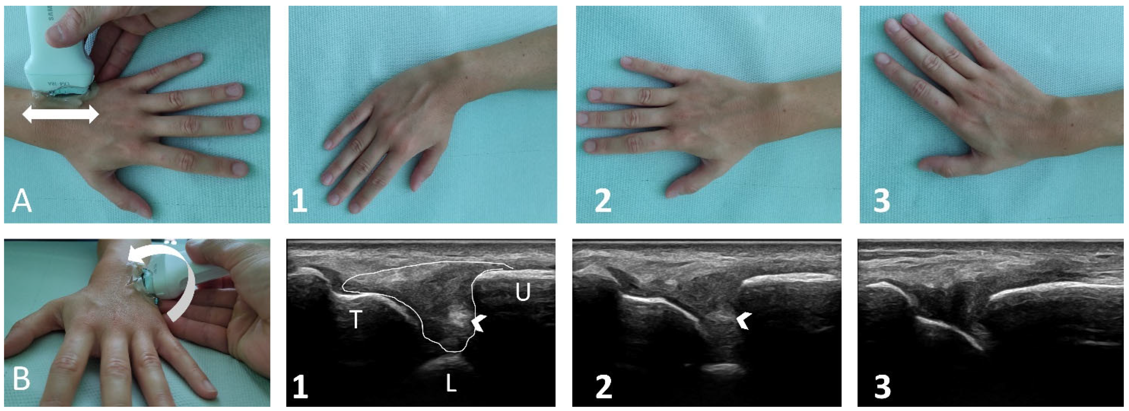
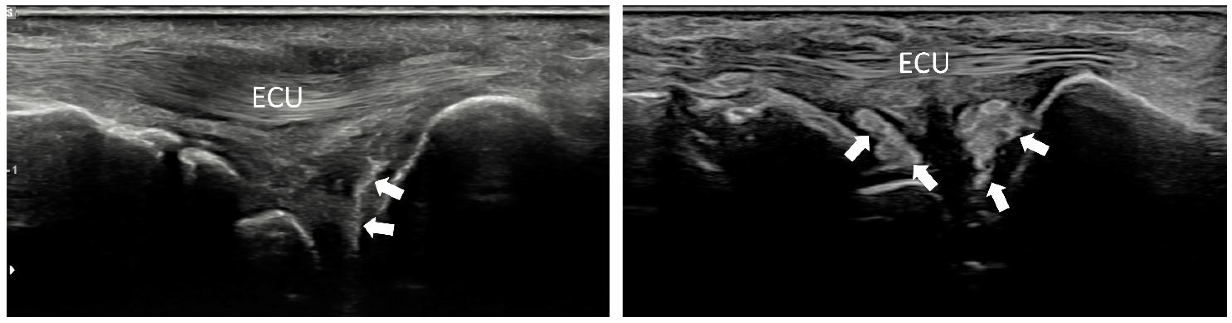
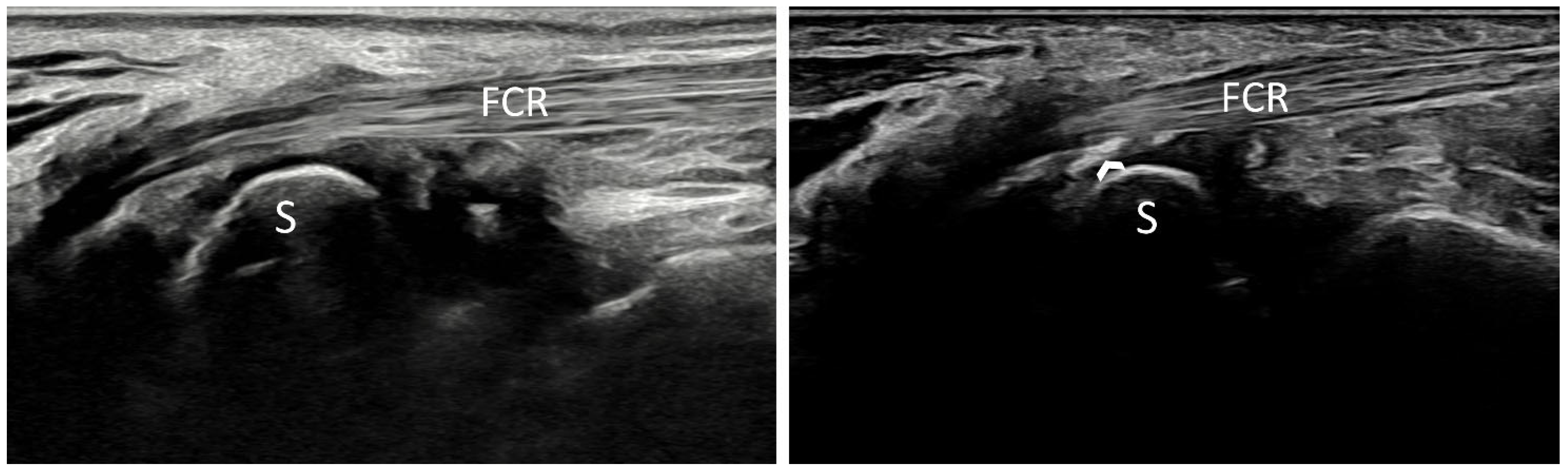
4.3. Site-Specific Scanning Techniques—The Wrist
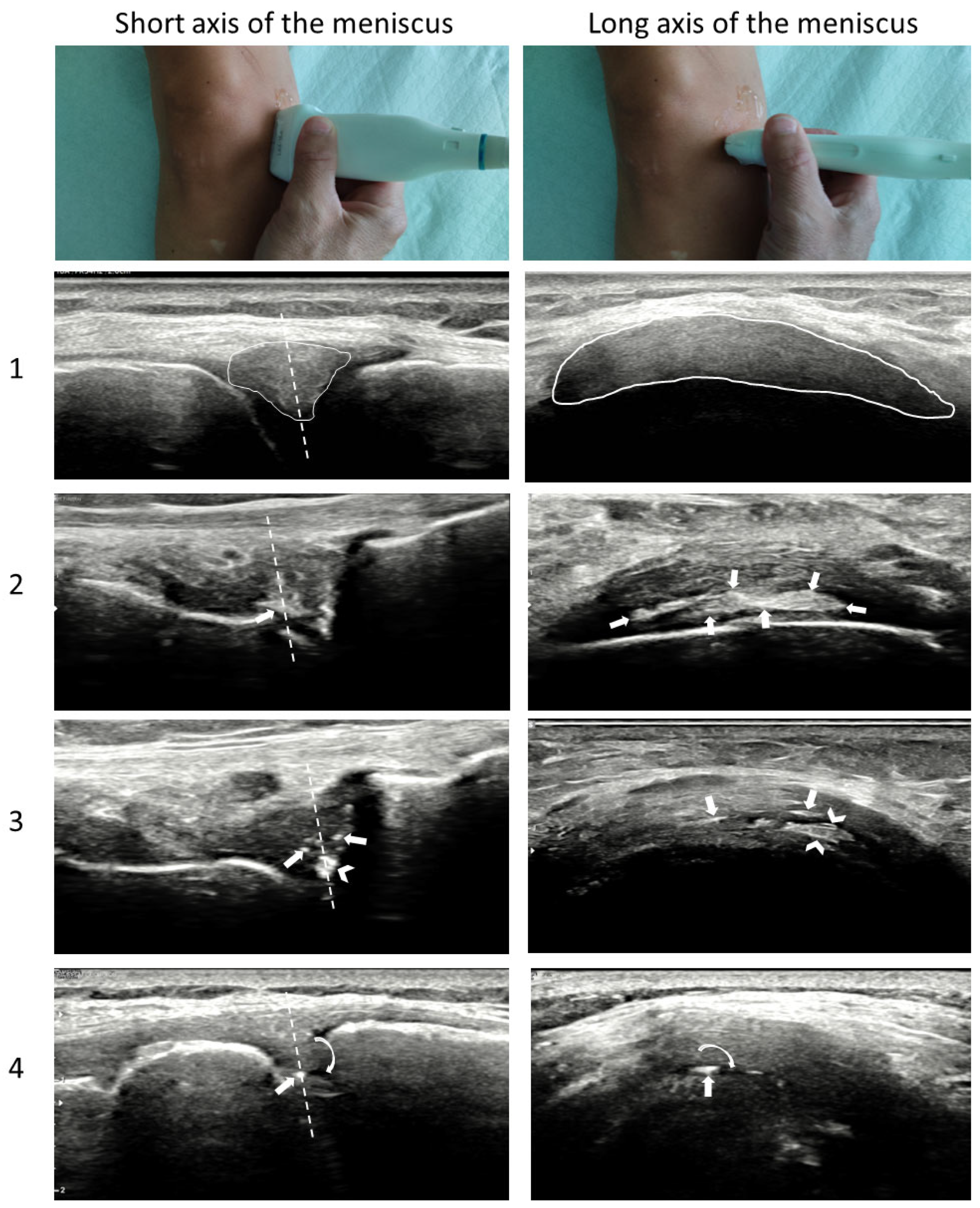
4.4. Site-Specific Scanning Techniques—The Knee
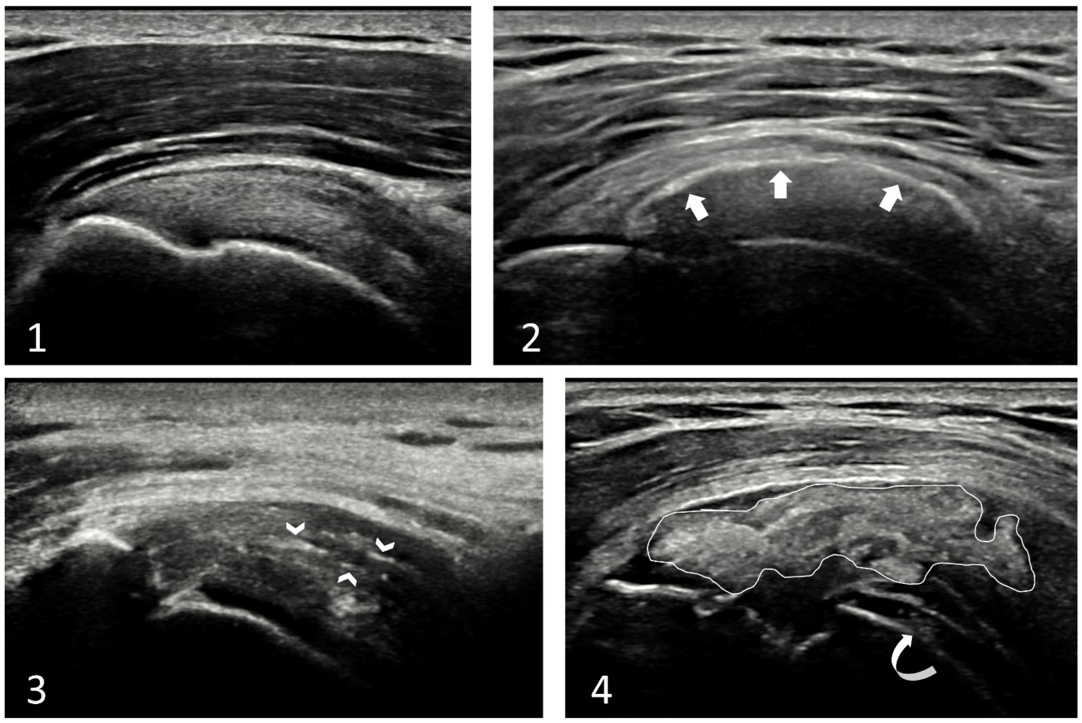
4.5. Site-Specific Scanning Techniques—Other Sites
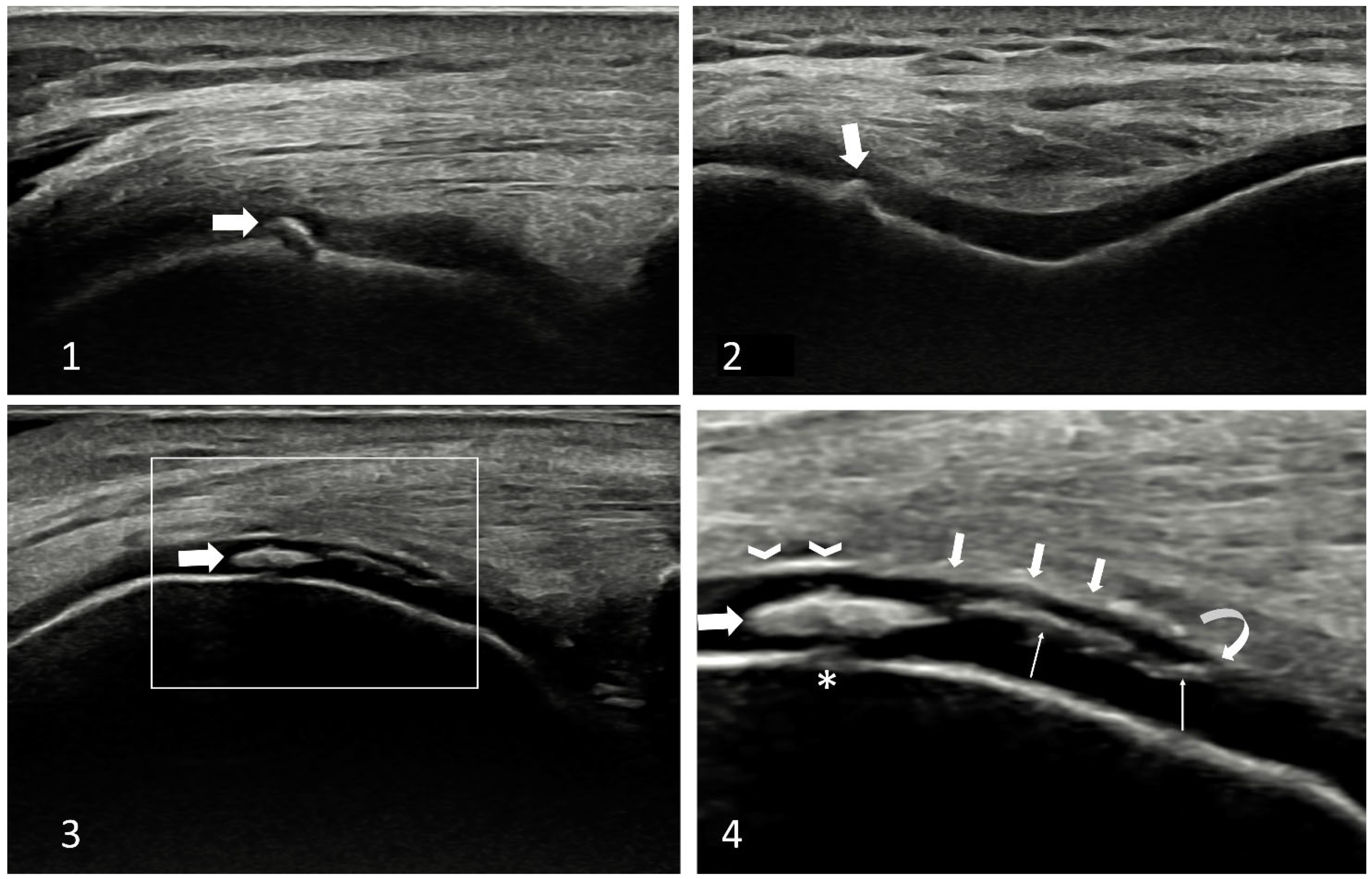
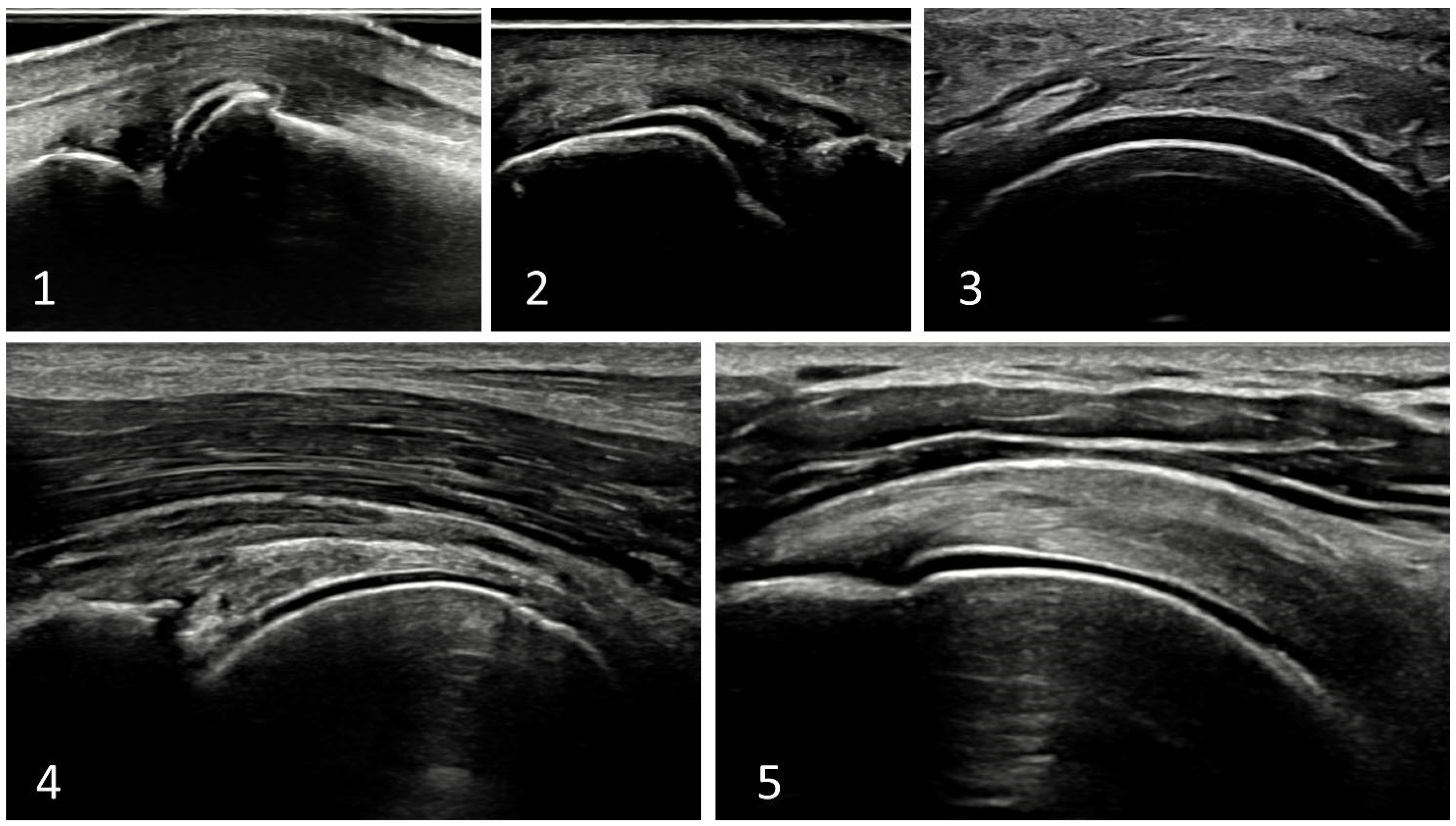
4.6. The Pseudo-Double Contour Sign—A New Hallmark of CPPD
5. Conclusions
Author Contributions
Funding
Conflicts of Interest
References
- Cipolletta, E.; Filippou, G.; Scirè, C.; Di Matteo, A.; Di Battista, J.; Salaffi, F.; Grassi, W.; Filippucci, E. The diagnostic value of conventional radiography and musculoskeletal ultrasonography in calcium pyrophosphate deposition disease: A systematic literature review and meta-analysis. Osteoarthr. Cartil. 2021, 29, 619–632. [Google Scholar] [CrossRef] [Scilit] [PubMed]
- Abhishek, A.; Tedeschi, S.K.; Pascart, T.; Latourte, A.; Dalbeth, N.; Neogi, T.; Fuller, A.; Rosenthal, A.; Becce, F.; Bardin, T.; et al. The 2023 ACR/EULAR classification criteria for calcium pyrophosphate deposition disease. Ann. Rheum. Dis. 2023, 82, 1248–1257. [Google Scholar] [CrossRef] [Scilit]
- Abhishek, A.; Neogi, T.; Choi, H.; Doherty, M.; Rosenthal, A.K.; Terkeltaub, R. Review: Unmet Needs and the Path Forward in Joint Disease Associated with Calcium Pyrophosphate Crystal Deposition. Arthritis Rheumatol. 2018, 70, 1182–1191. [Google Scholar] [CrossRef] [Scilit] [PubMed]
- Filippou, G.; Scirè, C.A.; Damjanov, N.; Adinolfi, A.; Carrara, G.; Picerno, V.; Toscano, C.; Bruyn, G.A.; D’agostino, M.A.; Sedie, A.D.; et al. Definition and Reliability Assessment of Elementary Ultrasonographic Findings in Calcium Pyrophosphate Deposition Disease: A Study by the OMERACT Calcium Pyrophosphate Deposition Disease Ultrasound Subtask Force. J. Rheumatol. 2017, 44, 1744–1749. [Google Scholar] [CrossRef] [Scilit] [PubMed]
- Filippou, G.; Scirè, C.A.; Adinolfi, A.; Damjanov, N.S.; Carrara, G.; Bruyn, G.A.W.; Cazenave, T.; D’agostino, M.A.; Sedie, A.D.; Di Sabatino, V.; et al. Identification of calcium pyrophosphate deposition disease (CPPD) by ultrasound: Reliability of the OMERACT definitions in an extended set of joints—An international multiobserver study by the OMERACT Calcium Pyrophosphate Deposition Disease Ultrasound Subtask Force. Ann. Rheum. Dis. 2018, 77, 1195–1200. [Google Scholar] [CrossRef] [Scilit]
- Filippou, G.; Scanu, A.; Adinolfi, A.; Toscano, C.; Gambera, D.; Largo, R.; Naredo, E.; Calvo, E.; Herrero-Beaumont, G.; Zufferey, P.; et al. Criterion validity of ultrasound in the identification of calcium pyrophosphate crystal deposits at the knee: An OMERACT ultrasound study. Ann. Rheum. Dis. 2020, 80, 261–267. [Google Scholar] [CrossRef] [Scilit]
- Sirotti, S.; Terslev, L.; Filippucci, E.; Iagnocco, A.; Moller, I.; Naredo, E.; Vreju, F.A.; Adinolfi, A.; Becce, F.; Hammer, H.B.; et al. Development and validation of an OMERACT ultrasound scoring system for the extent of calcium pyrophosphate crystal deposition at the joint level and patient level. Lancet Rheumatol. 2023, 5, e474–e482. [Google Scholar] [CrossRef] [Scilit]
- Filippou, G.; Sirotti, S. How can ultrasonography help in the management of CPPD? From diagnosis to clinical subset identification. Curr. Opin. Rheumatol. 2023, 35, 185–193. [Google Scholar] [CrossRef] [Scilit]
- Abhishek, A.; Doherty, S.; Maciewicz, R.; Muir, K.; Zhang, W.; Doherty, M. Evidence of a Systemic Predisposition to Chondrocalcinosis and Association Between Chondrocalcinosis and Osteoarthritis at Distant Joints: A Cross-Sectional Study. Arthritis Care Res. 2013, 65, 1052–1058. [Google Scholar] [CrossRef] [Scilit]
- Filippou, G.; Filippucci, E.; Tardella, M.; Bertoldi, I.; Di Carlo, M.; Adinolfi, A.; Grassi, W.; Frediani, B. Extent and distribution of CPP deposits in patients affected by calcium pyrophosphate dihydrate deposition disease: An ultrasonographic study. Ann. Rheum. Dis. 2013, 72, 1836–1839. [Google Scholar] [CrossRef] [Scilit]
- Williams, C.J.; Rosenthal, A.K. Pathogenesis of calcium pyrophosphate deposition disease. Best Pract. Res. Clin. Rheumatol. 2021, 35, 101718. [Google Scholar] [CrossRef] [Scilit]
- Nguyen, C.; Bazin, D.; Daudon, M.; Chatron-Colliet, A.; Hannouche, D.; Bianchi, A.; Côme, D.; So, A.; Busso, N.; Lioté, F.; et al. Revisiting spatial distribution and biochemical composition of calcium-containing crystals in human osteoarthritic articular cartilage. Arthritis Res. Ther. 2013, 15, R103. [Google Scholar] [CrossRef] [Scilit]
- Rosenthal, A.K.; Ryan, L.M. Calcium Pyrophosphate Deposition Disease. N. Engl. J. Med. 2016, 374, 2575–2584. [Google Scholar] [CrossRef] [Scilit]
- Bertrand, J.; Nitschke, Y.; Fuerst, M.; Hermann, S.; Schäfers, M.; Sherwood, J.; Nalesso, G.; Ruether, W.; Rutsch, F.; Dell‘Accio, F.; et al. Decreased levels of nucleotide pyrophosphatase phosphodiesterase 1 are associated with cartilage calcification in osteoarthritis and trigger osteoarthritic changes in mice. Ann. Rheum. Dis. 2012, 71, 1249–1253. [Google Scholar] [CrossRef] [Scilit] [PubMed]
- Abhishek, A.; Doherty, M. Pathophysiology of articular chondrocalcinosis—Role of ANKH. Nat. Rev. Rheumatol. 2010, 7, 96–104. [Google Scholar] [CrossRef] [Scilit] [PubMed]
- Williams, C.J.; Pendleton, A.; Bonavita, G.; Reginato, A.J.; Hughes, A.E.; Peariso, S.; Doherty, M.; McCarty, D.J.; Ryan, L.M. Mutations in the amino terminus of ANKH in two US families with calcium pyrophosphate dihydrate crystal deposition disease. Arthritis Rheum. 2003, 48, 2627–2631. [Google Scholar] [CrossRef] [Scilit] [PubMed]
- Abhishek, A.; Doherty, M. Update on calcium pyrophosphate deposition. Clin. Exp. Rheumatol. 2016, 34, 32–38. [Google Scholar]
- Pang, L.; Hayes, C.P.; Buac, K.; Yoo, D.-G.; Rada, B. Pseudogout-Associated Inflammatory Calcium Pyrophosphate Dihydrate Microcrystals Induce Formation of Neutrophil Extracellular Traps. J. Immunol. 2013, 190, 6488–6500. [Google Scholar] [CrossRef] [Scilit] [PubMed]
- Campillo-Gimenez, L.; Renaudin, F.; Jalabert, M.; Gras, P.; Gosset, M.; Rey, C.; Sarda, S.; Collet, C.; Cohen-Solal, M.; Combes, C.; et al. Inflammatory Potential of Four Different Phases of Calcium Pyrophosphate Relies on NF-κB Activation and MAPK Pathways. Front. Immunol. 2018, 9, 2248. [Google Scholar] [CrossRef] [Scilit] [PubMed]
- Liu, Y.; Jackson, A.; Cosgrove, S. Contribution of calcium-containing crystals to cartilage degradation and synovial inflammation in osteoarthritis. Osteoarthr. Cartil. 2009, 17, 1333–1340. [Google Scholar] [CrossRef] [Scilit]
- Beutler, A.; Rothfuss, S.; Clayburne, G.; Sieck, M.; Ralph, H.S. Calcium pyrophosphate dihydrate crystal deposition in synovium. relationship to collagen fibers and chondrometaplasia. Arthritis Rheum. 1993, 36, 704–715. [Google Scholar] [CrossRef] [Scilit] [PubMed]
- Fuerst, M.; Bertrand, J.; Lammers, L.; Dreier, R.; Echtermeyer, F.; Nitschke, Y.; Rutsch, F.; Schäfer, F.K.W.; Niggemeyer, O.; Steinhagen, J.; et al. Calcification of articular cartilage in human osteoarthritis. Arthritis Rheum. 2009, 60, 2694–2703. [Google Scholar] [CrossRef] [Scilit] [PubMed]
- Thouverey, C.; Bechkoff, G.; Pikula, S.; Buchet, R. Inorganic pyrophosphate as a regulator of hydroxyapatite or calcium pyrophosphate dihydrate mineral deposition by matrix vesicles. Osteoarthr. Cartil. 2009, 17, 64–72. [Google Scholar] [CrossRef] [Scilit] [PubMed]
- Neame, R.L.; Carr, A.J.; Muir, K.; Doherty, M. UK community prevalence of knee chondrocalcinosis: Evidence that correlation with osteoarthritis is through a shared association with osteophyte. Ann. Rheum. Dis. 2003, 62, 513–518. [Google Scholar] [CrossRef] [Scilit]
- Filippucci, E.; Gutierrez, M.; Georgescu, D.; Salaffi, F.; Grassi, W. Hyaline cartilage involvement in patients with gout and calcium pyrophosphate deposition disease. An ultrasound study. Osteoarthr. Cartil. 2009, 17, 178–181. [Google Scholar] [CrossRef] [Scilit] [PubMed]
- Bjelle, A.O.; Sundström, B.K.G. An ultrastructural study of the articular cartilage in calcium pyrophosphate dihydrate (CPPD) crystal deposition disease (Chondrocalcinosis Articularis). Calcif. Tissue Int. 1975, 19, 63–71. [Google Scholar] [CrossRef] [Scilit]
- Stücker, S.; Bollmann, M.; Garbers, C.; Bertrand, J. The role of calcium crystals and their effect on osteoarthritis pathogenesis. Best Pract. Res. Clin. Rheumatol. 2021, 35, 101722. [Google Scholar] [CrossRef] [Scilit]
- Filippou, G.; Miguel-Pérez, M.; Bong, D.; Coronel, L.; Sirotti, S.; Pacini, G.; Scanu, A.; Möller, I.; The “Anatomy for Image” EULAR Study Group. The ultrasonographic “pseudo-double contour” sign in calcium pyrophosphate deposition disease. Arthritis Rheumatol. 2022, 75, 639–640. [Google Scholar] [CrossRef] [Scilit]
- Ogdie, A.; Taylor, W.J.; Neogi, T.; Fransen, J.; Jansen, T.L.; Schumacher, H.R.; Louthrenoo, W.; Vazquez-Mellado, J.; Eliseev, M.; McCarthy, G.; et al. Performance of Ultrasound in the Diagnosis of Gout in a Multicenter Study: Comparison With Monosodium Urate Monohydrate Crystal Analysis as the Gold Standard. Arthritis Rheumatol. 2016, 69, 429–438. [Google Scholar] [CrossRef] [Scilit]
- Löffler, C.; Sattler, H.; Peters, L.; Löffler, U.; Uppenkamp, M.; Bergner, R. Distinguishing Gouty Arthritis from Calcium Pyrophosphate Disease and Other Arthritides. J. Rheumatol. 2014, 42, 513–520. [Google Scholar] [CrossRef] [Scilit]
- Adinolfi, A.; Picerno, V.; Di Sabatino, V.; Bertoldi, I.; Galeazzi, M.; Frediani, B.; Filippou, G. Inquiry Is Fatal to Certainty-Is the Ultrasonography Double Contour Sign Specific for Uric Acid-Induced Arthritis? Arthritis Rheum. 2013, 65, 1952. [Google Scholar] [CrossRef] [Scilit]
- Cipolletta, E.; Abhishek, A.; Di Matteo, A.; Grassi, W.; Filippucci, E. Dynamic assessment of the double contour sign by ultrasonography helps to distinguish between gout and calcium pyrophosphate deposition disease. RMD Open 2023, 9, e002940. [Google Scholar] [CrossRef] [Scilit] [PubMed]
- Filippou, G.; Pacini, G.; Sirotti, S.; Zadory, M.; Carboni, D.; Damiani, A.; Fiorentini, E.; Cipolletta, E.; Filippucci, E.; Froehlich, J.M.; et al. Comparison of ultrasound attenuation by calcium pyrophosphate, hydroxyapatite and monosodium urate crystals: A proof-of-concept study. Ann. Rheum. Dis. 2022, 81, 1199–1201. [Google Scholar] [CrossRef] [Scilit] [PubMed]
- Zander, D.; Hüske, S.; Hoffmann, B.; Cui, X.-W.; Dong, Y.; Lim, A.; Jenssen, C.; Löwe, A.; Koch, J.B.; Dietrich, C.F. Ultrasound Image Optimization (“Knobology”): B-Mode. Ultrasound Int. Open 2020, 6, E14–E24. [Google Scholar] [CrossRef] [Scilit] [PubMed]
- Adinolfi, A.; Sirotti, S.; Sakellariou, G.; Cipolletta, E.; Filippucci, E.; Porta, F.; Zanetti, A.; Ughi, N.; Sarzi-Puttini, P.; Scirè, C.A.; et al. Which are the most frequently involved peripheral joints in calcium pyrophosphate crystal deposition at imaging? A systematic literature review and meta-analysis by the OMERACT ultrasound—CPPD subgroup. Front. Med. 2023, 10, 1131362. [Google Scholar] [CrossRef] [Scilit] [PubMed]
- Wu, W.-T.; Chang, K.-V.; Mezian, K.; Naňka, O.; Yang, Y.-C.; Hsu, Y.-C.; Hsu, P.-C.; Özçakar, L. Ulnar Wrist Pain Revisited: Ultrasound Diagnosis and Guided Injection for Triangular Fibrocartilage Complex Injuries. J. Clin. Med. 2019, 8, 1540. [Google Scholar] [CrossRef] [Scilit] [PubMed]
- Gout, Hyperuricemia and Crystal-Associated Disease Network. Gout, Hyperuricemia and Crystal-Associated Disease Network (G-CAN) Conference 2022: Early-Career Investigators’ Abstracts. Gout Urate Cryst. Depos. Dis. 2023, 1, 167–191. [Google Scholar] [CrossRef] [Scilit]
- Bishop, A.T.; Gabel, G.; Carmichael, S.W. Flexor carpi radialis tendinitis. Part I. Minerva Anestesiol. 1994, 76, 1009–1014. [Google Scholar] [CrossRef] [Scilit]
- McGonagle, D.; Aydin, S.Z.; Tan, A.L. The Synovio-entheseal Complex and Its Role in Tendon and Capsular Associated Inflammation. J. Rheumatol. 2012, 89, 11–14. [Google Scholar] [CrossRef] [Scilit]
- Cipolletta, E.; Smerilli, G.; Mirza, R.M.; Di Matteo, A.; Carotti, M.; Salaffi, F.; Grassi, W.; Filippucci, E. Sonographic assessment of calcium pyrophosphate deposition disease at wrist. A focus on the dorsal scapho-lunate ligament. Jt. Bone Spine 2020, 87, 611–617. [Google Scholar] [CrossRef] [Scilit]
- Möller, I.; Janta, I.; Backhaus, M.; Ohrndorf, S.; Bong, D.A.; Martinoli, C.; Filippucci, E.; Sconfienza, L.M.; Terslev, L.; Damjanov, N.; et al. The 2017 EULAR standardised procedures for ultrasound imaging in rheumatology. Ann. Rheum. Dis. 2017, 76, 1974–1979. [Google Scholar] [CrossRef] [Scilit]
- Filippou, G.; Picerno, V.; Adinolfi, A.; Di Sabatino, V.; Bertoldi, I.; Galeazzi, M.; Frediani, B. Change perspective to increase diagnostic accuracy of ultrasonography in calcium pyrophosphate dihydrate deposition disease! A new approach: The axial scan of the meniscus. Reumatismo 2014, 66, 318–321. [Google Scholar] [CrossRef] [Scilit]
- Cipolletta, E.; Moscioni, E.; Sirotti, S.; Di Battista, J.; Abhishek, A.; Rozza, D.; Zanetti, A.; Carrara, G.; Scirè, C.A.; Grassi, W.; et al. Diagnosis of calcium pyrophosphate crystal deposition disease by ultrasonography: How many and which sites should be scanned? Rheumatology 2023, kead565. [Google Scholar] [CrossRef] [Scilit] [PubMed]
- Christiansen, S.N.; Filippou, G.; Scirè, C.A.; Balint, P.V.; Bruyn, G.A.; Dalbeth, N.; Dejaco, C.; Sedie, A.D.; Filippucci, E.; Hammer, H.B.; et al. Consensus-based semi-quantitative ultrasound scoring system for gout lesions: Results of an OMERACT Delphi process and web-reliability exercise. Semin. Arthritis Rheum. 2021, 51, 644–649. [Google Scholar] [CrossRef] [Scilit] [PubMed]
- Filippucci, E.; Scirè, C.A.; Sedie, A.D.; Iagnocco, A.; Riente, L.; Meenagh, G.; Gutierrez, M.; Bombardieri, S.; Valesini, G.; Montecucco, C.; et al. Ultrasound imaging for the rheumatologist. XXV. Sonographic assessment of the knee in patients with gout and calcium pyrophosphate deposition disease. Clin. Exp. Rheumatol. 2010, 28, 2–5. [Google Scholar] [PubMed]


Disclaimer/Publisher’s Note: The statements, opinions and data contained in all publications are solely those of the individual author(s) and contributor(s) and not of MDPI and/or the editor(s). MDPI and/or the editor(s) disclaim responsibility for any injury to people or property resulting from any ideas, methods, instructions or products referred to in the content. |
© 2024 by the authors. Licensee MDPI, Basel, Switzerland. This article is an open access article distributed under the terms and conditions of the Creative Commons Attribution (CC BY) license (https://creativecommons.org/licenses/by/4.0/).
Share and Cite
Filippou, G.; Sirotti, S.; Cipolletta, E.; Filippucci, E. Optimizing the Use of Ultrasound in Calcium Pyrophosphate Deposition (CPPD): A Review from the Ground Up. Gout Urate Cryst. Depos. Dis. 2024, 2, 17-33. https://doi.org/10.3390/gucdd2010002
Filippou G, Sirotti S, Cipolletta E, Filippucci E. Optimizing the Use of Ultrasound in Calcium Pyrophosphate Deposition (CPPD): A Review from the Ground Up. Gout, Urate, and Crystal Deposition Disease. 2024; 2(1):17-33. https://doi.org/10.3390/gucdd2010002
Chicago/Turabian StyleFilippou, Georgios, Silvia Sirotti, Edoardo Cipolletta, and Emilio Filippucci. 2024. "Optimizing the Use of Ultrasound in Calcium Pyrophosphate Deposition (CPPD): A Review from the Ground Up" Gout, Urate, and Crystal Deposition Disease 2, no. 1: 17-33. https://doi.org/10.3390/gucdd2010002
APA StyleFilippou, G., Sirotti, S., Cipolletta, E., & Filippucci, E. (2024). Optimizing the Use of Ultrasound in Calcium Pyrophosphate Deposition (CPPD): A Review from the Ground Up. Gout, Urate, and Crystal Deposition Disease, 2(1), 17-33. https://doi.org/10.3390/gucdd2010002