


Abstract
The development of less toxic nanoparticles capable of producing and selectively sensing organic pollutants remains a significant challenge in biological and environmental applications. In this study, ZnInS/ZnS quantum dots (QDs) were synthesized via an aqueous-phase doping approach to integrate manganese (Mn) and enhance the singlet oxygen quantum yield (SOQY). The as-synthesized QDs displayed an average particle size of 7.40 nm, prolonged photoluminescence lifetimes, and tunable emission characteristics. Introduction of Mn into the host QDs enhanced the absolute quantum yield by up to twofold at the optimal concentrations, while the increased concentrations resulted in reduced luminescence due to increased non-radiative recombination. The surface chemistry of stabilizing agents further improved the formation of singlet oxygen. Thioglycolic acid (TGA)- and gelatin-capped QDs generated higher SOQY than QDs capped with citrate and TGA. Furthermore, as-synthesized Mn-doped QDs detected different organic pollutants such as trinitrophenol (TNP), dichlorophenol (DCP), naphthalene (NP), phenanthrene (PH), and pyrene (PY). Among these pollutants, TNP showed quenching behavior, while the others showed enhancement behavior. The QDs detected 620 µM of TNP following a linear Stern–Volmer relationship. These results show the potential of water-soluble Mn–ZnInS/ZnS QDs as multifunctional nanoprobes for phototherapy and environmental sensing applications.
1. Introduction
Semiconductor quantum dots (QDs), also known as nanocrystals, have attracted great attention in different fields such as bioimaging, photodynamic therapy (PDT), and environmental monitoring, owing to their good optical properties [1]. Among the different ternary QDs, ZnInS-based QDs have been appealing due to their low toxicity and their broad emission wavelengths within the visible region [2]. Metal ions like zinc render them more biocompatible than traditional QDs, which contain cadmium and lead ions [3,4].
Metal-doping strategies, particularly with magnetically based metal ions, have been extensively used to enhance the photophysical properties of QDs [5]. Doping has also shown potential for improving reactive oxygen species (ROS) in PDT and for fluorescence detection of organic pollutants [6]. Furthermore, the surface chemistry of QDs plays an equally significant role in determining their detection properties and singlet oxygen generation. Stabilizing ligands influence both the structural integrity and interaction of QDs with their surrounding environment. Ligand selection can directly affect the efficiency and sensitivity of ROS generation, thereby lending itself to both biomedical and environmental sensing applications [7]. In this study, manganese (Mn) doping shows the potential to enhance emission intensity.
Organic pollutants, primarily persistent organic pollutants (POPs), pose a significant risk to the environment, as well as to terrestrial and human life worldwide. According to the USA Environmental Protection Agency (EPA), top priority pollutants of great concern are 2,4,6-trinitrophenol (TNP), dichlorophenol (DCP), naphthalene (NP), phenanthrene (PH), and pyrene (PY) [8,9,10]. Furthermore, these pollutants have been recognized as carcinogens [10,11]. TNP is a nitroaromatic compound with extensive use in the making of explosives. Owing to its widespread use in the military industry, large volumes of TNP are being released into the environment. Although there is no standard US EPA maximum drinking water concentration for TNP, given its low permissible level of 0.1 mg/m3 (0.436 nM) in air and provisional chronic oral dose of 0.002 mg/kg-day, it has become a great concern due to its toxicity and persistence [12].
Several conventional detection approaches have been employed in sensing persistent organic environmental pollutants. However, they are time-consuming and require sophisticated instrumentation, necessitating a rapid, sensitive, and cost-effective alternative approach [13]. Fluorescent nanoprobes, particularly doped-semiconductor nanocrystals, provide a promising solution owing to their ability to detect very small amounts of analytes, their size-dependent properties, and their ease of water-based synthesis. Zinc indium sulfide (ZnInS) QDs with a ZnS shell are known for their biocompatibility and tunable optical properties, making them suitable for different biological and photonic applications. However, their ability to generate 1O2 is limited due to different factors, including inefficient charge separation, inadequate energy transfer, surface defects, and trap states, etc. [14]. Mn-doping, as one of the ways to address these limitations, was employed in this work to enhance the yield of 1O2 by ZnInS/ZnS QDs as a potential photosensitizer (PS) and a chemical sensor.
Herein, we report the direct water-based synthesis of Mn-doped ZnInS/ZnS QDs stabilized with different ligands and investigate their photophysical behavior, singlet oxygen production, sensitivity and selectivity toward organic pollutants such as dichlorophenol (DCP), naphthalene (NP), pyrene (PY), phenanthrene (PH) and trinitrophenol (TNP).
2. Materials and Methods
2.1. Materials and Apparatus
Indium chloride (InCl3, 98%), zinc acetate dihydrate (Zn(OAc)2·2H2O, 99%), gelatin, thioglycolic acid (TGA), sodium sulphide (Na2S, 64%), sodium hydroxide (NaOH, 98%), thiourea (SC(NH2)2), methylene blue, dimethyl sulfoxide (DMSO), 1,3-diphenylisobenzofuran (DPBF) DCP, NP, PH, and PY were purchased from Sigma Aldrich, South Africa and used without further purification. Deionized (DI) water was employed in all the experiments.
2.2. Synthesis of ZnInS/ZnS Core/Shell QDs
The synthesis of the QDs was based on the method developed by Zikalala et al. [2,15]. In the current study, the cationic precursors, Zn(OAc)2 (0.0625 mmol) and InCl3 (0.25 mmol), were weighed and transferred into a 250 mL beaker that contained 80 mL of deionized water. Then, stabilizing agents, gelatin (0.297 g in 20 mL of DI water) and TGA (21.5 mmol), were added to the reaction solution, and the pH was adjusted to 7 via dropwise addition of NaOH (2 M). Sodium sulphide (1.25 mmol, 10 mL) was then added to the solution as a sulphur precursor. The reaction solution was then transferred into a three-necked flask and subsequently refluxed at 95 °C for 1.5 h to produce the ZnInS core. After the growth of ZnInS core QDs, the ZnS shell was grown by adding 0.0686 g of Zn(OAc)2 in 10 mL of DI water and 0.0119 g of thiourea dissolved in 10 mL of DI water to the core solution after reducing the temperature by 10 °C. The reaction mixture was allowed to reflux for 1 h to produce ZnInS/ZnS QDs. The synthesis was repeated by replacing gelatin with citrate as the capping agent.
2.3. Synthesis of Mn:ZnInS/ZnS QD Core/Shell
To synthesize Mn:ZnInS/ZnS, 80 mL of DI water was spiked with 5%, 10% and 15% of Mn2+, respectively, using MnCl2 as the Mn2+ precursor, and the other precursors were added following the same procedure as for undoped ZIS/ZnS QDs [15].
2.4. Evaluation of Singlet Oxygen
To measure the singlet oxygen, DPBF was used as a probe. A total of 0.8 mg of DPBF was dissolved in 100 mL of DMSO and then mixed with ZnInS/ZnS and 10% Mn:ZnInS/ZnS at an absorbance of about 0.1 in a 1:1 ratio. The irradiation of the samples was done at a wavelength of 532 nm using spectrofluorophotometry laser light for 16 min. The reduction in emission intensity of the mixture after every 2 min was measured at 471 nm. The singlet oxygen quantum yield (SOQY) was then calculated against MB as a reference (SOQYref = 0.52) using the following formula [16]:
where SOQY and SOQYref are the singlet oxygen quantum yields of the sample (ZIS/ZnS core/shell QDs and 10% Mn:ZIS/ZnS QDs), and the reference (MB, 0.52), mc and mref are the slopes of the sample and reference, and Aref and Ac are the absorbances of the reference and conjugate at the irradiation wavelength.
2.5. Fluorescence Sensing
The fluorescence sensing of organic pollutants using 10% Mn:ZnInS/ZnS QDs was carried out via the standard procedures [17,18]. A total of 500 μL of Mn:ZnInS/ZnS QD solution was transferred into a cuvette containing 2.5 mL of DI water. The emission spectrum of the mixture of DI water and Mn:ZnInS/ZnS QD solution was recorded as a blank with a spectrofluorometer at an excitation of 380 nm. For detection studies, 500 μL of the QD solution was added to a separate cuvette containing 2.5 mL of a solution of each pollutant (DCP, NP, PY, PH and TNP). To prepare the TNP working standards, different volumes of TNP were diluted with 50 mL of DI water.
2.6. Characterizations
The absorption and emission spectra of the as-synthesized ZnInS/ZnS QDs and Mn:ZnInS/ZnS QDs were obtained using Ultraviolet–visible spectrophotometry (UV–vis Lambda 25 spectrometer) (Perkin Elmer, Johannesburg, South Africa) and photoluminescence (PL) spectrofluorophotometry (RF-6000, Shimadzu, Tokyo, Japan). The absolute quantum yield and the decay lifetimes of the samples were obtained using a Spectrofluorometer FS5 (Horiba, EDINBURGH INSTRUMENTS, Northampton, UK). The morphology and particle size of the doped and undoped ZIS/ZnS QDs were obtained with a transmission electron microscope (TEM) (JEM 2100, JEOL, Tokyo, Japan) operated at an acceleration voltage of 200 kV.
3. Results and Discussion
3.1. Optical Analysis of the As-Synthesized ZnInS/ZnS QDs and Mn:ZnInS/ZnS QDs
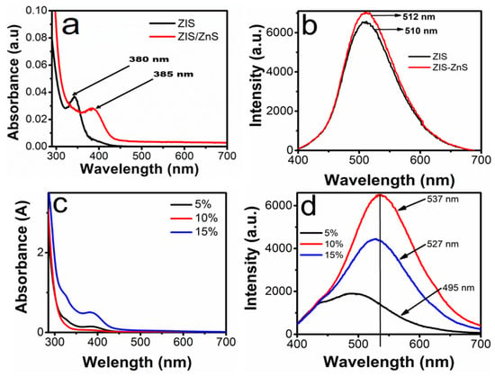
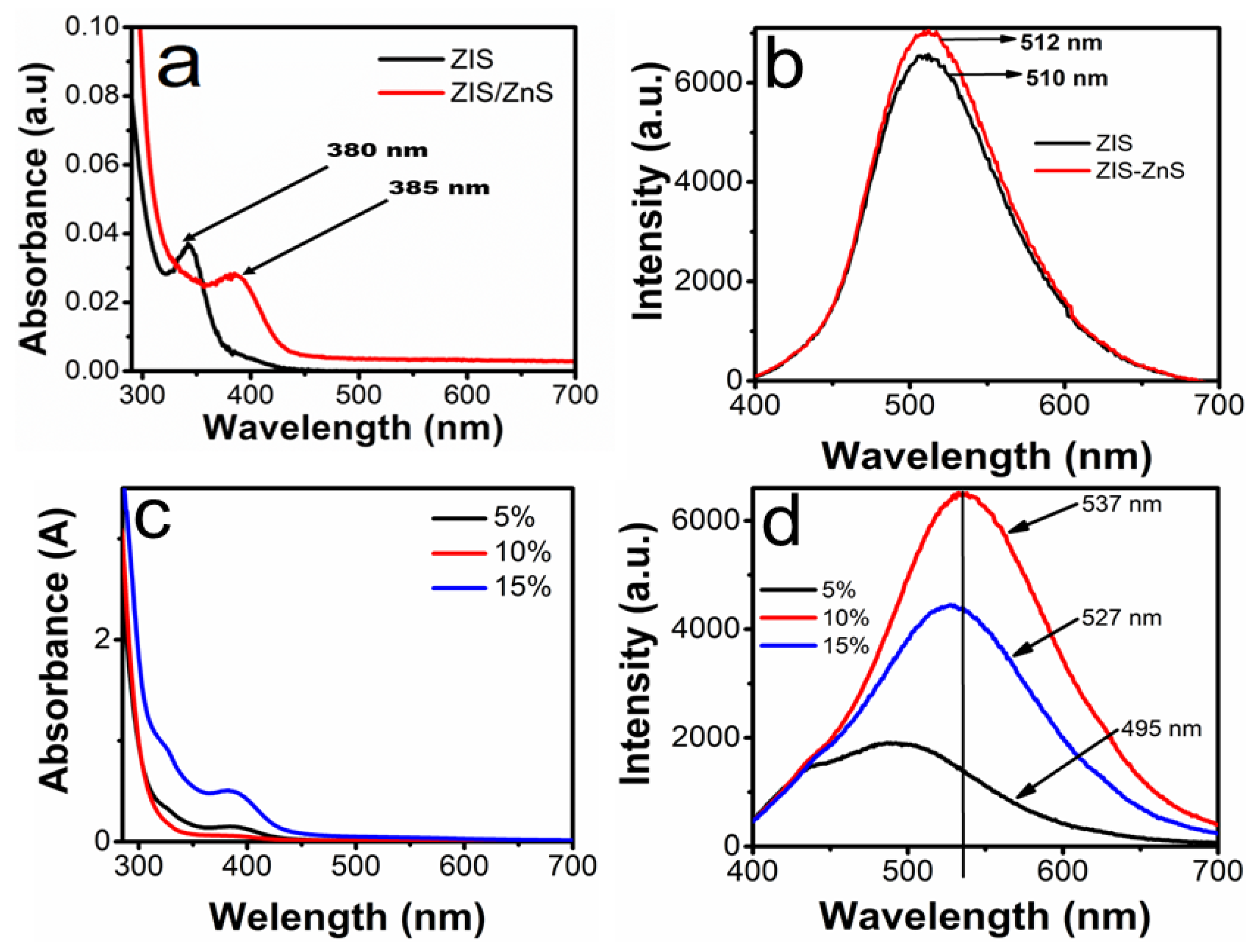
The doped QDs and undoped QDs were prepared via the reflux method using DI water as the solvent and dual stabilizing agents to prevent the high reactivity of cations such as Zn2+ and In3+. The absorption properties of the pristine ZIS core QDs and ZIS/ZnS core/shell QDs were evaluated with UV–Vis spectroscopy and photoluminescence spectroscopy, as displayed in Figure 1a. Both doped and undoped QDs showed broad absorption in the ultraviolet region, like the electronic transitions of typical ternary QDs [18]. The bare core displayed an absorption wavelength of 380 nm, while the core/shell QDs displayed an absorption wavelength of 385 nm, similar to a previous report [19]. A slight shift of 5 nm in absorption wavelength could be attributed to the quantum confinement effect influenced by the introduction of the ZnS shell.
Figure 1.
(a) The absorption spectra and (b) the PL spectra of undoped core QDs and core/shell QDs; (c) the absorption spectra and (d) the PL spectra of the (5%, 10%, 15% Mn-) doped QDs.
The PL emissions of both the bare core QDs and the core/shell QDs are shown in Figure 1b. The core QDs emitted at 510 nm, while the core/shell QDs emitted at 515 nm with enhanced intensity, which can be attributed to the successful passivation of ZIS by ZnS. This also suggests that non-radiative recombination was successfully reduced compared to the core QDs. The absorption spectra presented in Figure 1c display broad absorption peaks whose wavelengths range from 380 to 390 nm, suggesting that the incorporation of different levels of Mn2+ into the host lattice influences the energy transfer from the QD lattice to the dopant center.
The influence of different concentrations of Mn2+ on the emission properties of ZIS/Zn is presented in Figure 1d. A systematic red shift in the emissions of the QDs was observed as the Mn2+ dopant concentration was increased from 5% to 15%. At 5%, the emission wavelength was recorded at 495 nm, with the lowest intensity compared to the intensity of the QDs doped with 10% and 15% and bare QDs. This blue shift and low intensity might be due to cationic exchange between the Mn and Zn ions because of their relatively similar atomic radii, thus creating more defects [20]. Notably, a significant red shift from 515 nm in the ZIS/ZnS QDs and 495 nm in the 5% Mn:ZIS/ZnS QDs to 537 nm was observed when the level of Mn2+ was increased to 10% Figure 1d. This might be attributed to the fact that the dopant served as a passivating agent at the QDs’ surface against the In and Zn vacancies. This improved the optical properties by reducing the surface and structural defects and thus promoted radiative recombination [21]. This emission may also be due to the 4T1 → 6A1 ligand-field transition of Mn2+ ions, which is influenced by energy transfer from photogenerated excitons in the ZIS/ZnS centers [22]. However, a further increase in the concentration of Mn2+ drastically blue-shifted the emission wavelength, alongside a decrease in PL intensity compared to the intensity of the 10% doped core/shell QDs. This behavior is attributed to the excess of Mn2+ ions, which served as a new defect that increased both the surface and structural defects, which can also lead to lattice distortions, thereby promoting non-radiative recombination [23]. Furthermore, the Mn concentrations in the as-synthesized doped QDs obtained from ICP-OES, as shown in Table 1, also support the Mn concentration in the sample.
Table 1.
Mn concentrations obtained using ICP-OES.
3.2. Photoluminescence Quantum Yield and Lifetime Analysis
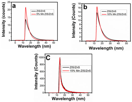
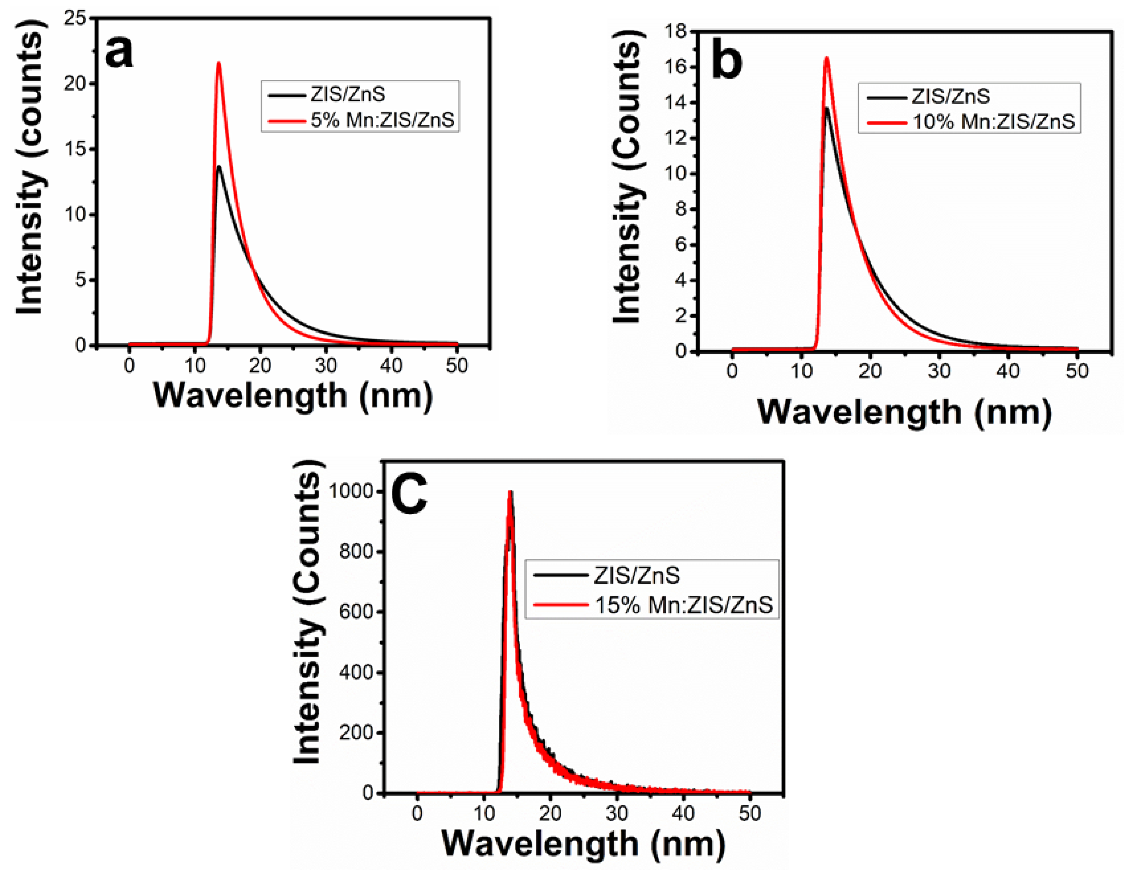
To quantitatively evaluate the influence of ZnS shell passivation and Mn doping on the optical properties of ZIS core QDs and ZIS/ZnS core/shell QDs, the photoluminescence quantum yield (PLQY) and decay lifetime (τ) (Figure 2) of all the as-synthesized materials were measured, and the data are presented in Table 2. The pristine ZIS core showed a very low PLQY of 0.04, which suggests the occurrence of surface defects due to the absence of a ZnS shell. On introduction of ZnS, the PLQY increases to 0.066%, accompanied by a slight increase in lifetime from 3.00 to 3.12 ns. This behavior could be attributed to the reduction in non-radiative decay channels, which influence photon emission and lifetime.
Figure 2.
Photoluminescence (PL) lifetime decay curves of (a) undoped ZnInS/ZnS and 5% Mn-doped ZnInS/ZnS QDs, (b) ZnInS/ZnS QDs and 10% Mn-doped ZnInS/ZnS QDs, and (c) ZnInS/ZnS QDs and 15% Mn-doped ZnInS/ZnS QDs.
Table 2.
Photoluminescence quantum yield (PLQY) and average PL decay lifetime of undoped and Mn-doped ZIS and ZIS/Zns QDs.
A steady order was observed in all Mn-doped QDs. The core/shell consistently produced a significant improvement in PLQY relative to the corresponding core, and this behavior did not show a dependence on dopant level. At 5% Mn2+, the PLQY increased from 0.03% (for the core only) to 0.59% when the shell was deposited on the core, while the lifetime increased from 3.71 ns to 4.74 ns. When the level of Mn2+ increased to 10%, a dramatic enhancement in the PLQY was observed, and 3.53% was recorded as the highest quantum yield. At a concentration of 15%, the PLQY rose from 0.04% to 1.20% after the shell deposition, while the lifetime reduced from 6.73 ns to 4.76 ns. This trend could be due to a reduction in non-radiative recombination [24,25].
Evaluation of the Mn-doped core revealed changes in the PLQY when the level of Mn2+ increased from 5% to 15%. The PLQY was recorded at 0.03% at 5% Mn2+, while a rise to 0.74% was observed when the concentration was 10%. However, a significant decrease in PLQY was observed when the concentration was further increased to 15%. This trend suggests that the Mn2+ ionic radius starts to compete with those of Zn2+ and In3+ at higher concentrations, as discussed in Section 3.1. This was further validated by the similar results exhibited by the bare core QDs. The core doped with 10% of Mn2+ displayed a very short lifetime of 1.21 ns, which was the lowest among the materials, suggesting that fast radiative recombination is dominant at this level. However, a significant increase to 6.73 ns in lifetime was observed when the concentration of Mn2+ in the core was raised to 15%. This could suggest that carriers are trapped in long-lived non-radiative states such as clusters caused by excessive levels of Mn2+. Among the core/shell QDs, lifetimes within the range of 3.12 ns (for undoped ZIS/ZnS) to 4.76 ns (for 15% Mn:ZnInS/ZnS) were observed. This could be attributed to the suppression of fast non-radiative decay on surface passivation [26].
3.3. Morphology and Particle Size of the As-Synthesized QDs
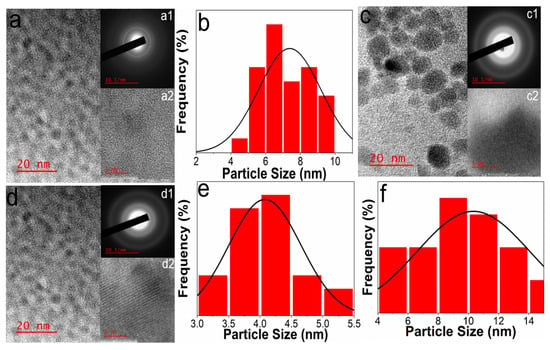
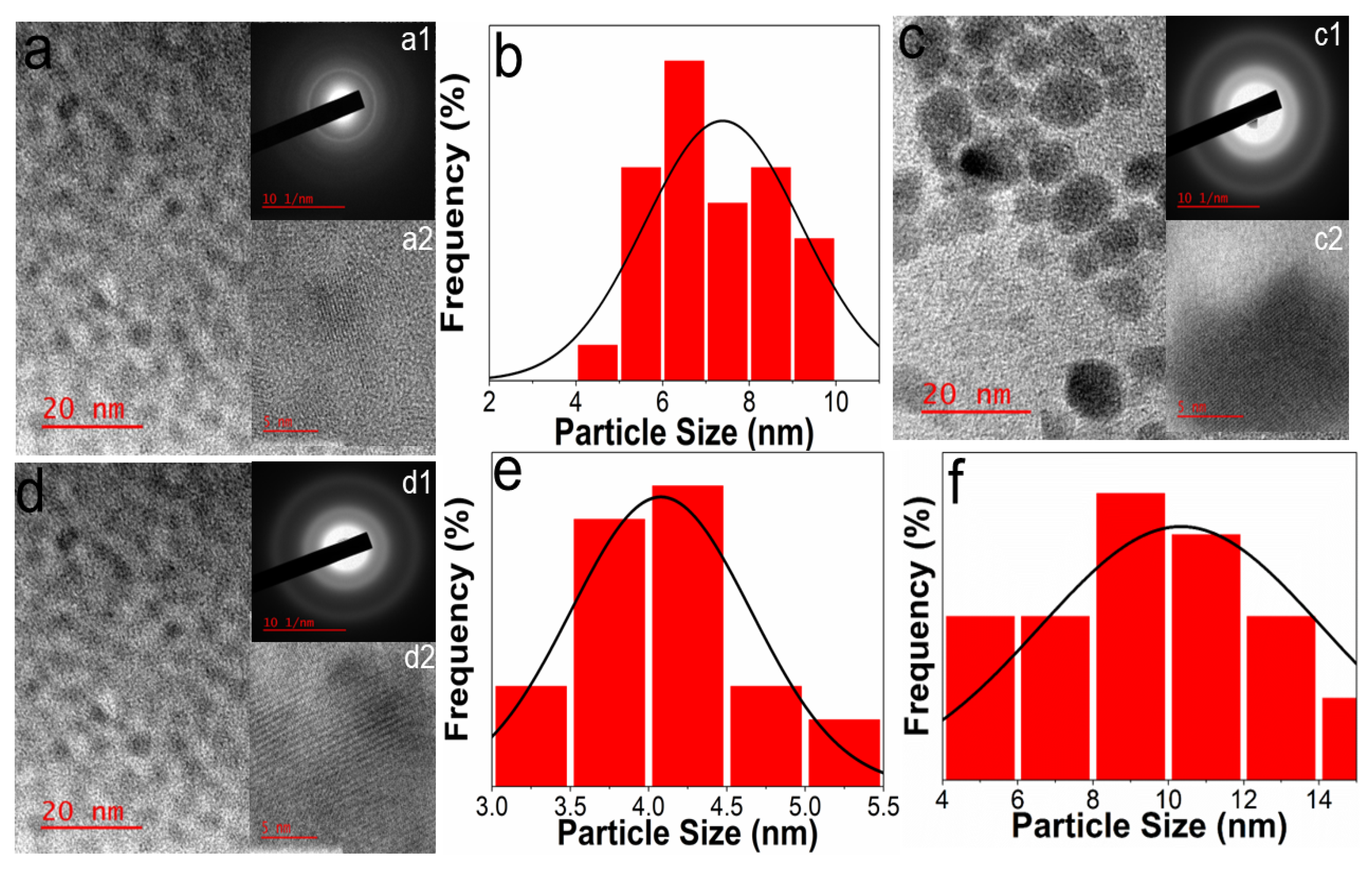
Figure 3 shows the micrographs of the undoped QDs, doped QDs and their corresponding average size. The size of the QDs revealed an intriguing trend related to manganese (Mn) doping concentration. Initially, the undoped ZnInS/ZnS QDs exhibited an average particle size of 7.4 nm. Upon introduction of a 5% Mn dopant, an unexpected reduction in particle size to 4.1 nm was observed. As the Mn concentration was further increased to 10%, the particle size increased once again from 4.1 nm to 10.5 nm. This complex size evolution can be attributed to several underlying mechanisms in the quantum dot synthesis process. At low Mn doping levels (5%), the dopant ions may interfere with the nucleation and growth processes, potentially creating more compact quantum dot structures and leading to the observed size reduction. This phenomenon could be related to changes in the crystal lattice, surface energy, and nucleation kinetics induced by the introduction of Mn ions [24,27,28]. The subsequent size increase at 10% Mn concentration suggests a transition in the quantum dot formation mechanism. At higher dopant concentrations, Mn ions may begin to play a more significant role in crystal growth, potentially altering the surface chemistry, modifying crystal nucleation rates, influencing Ostwald ripening processes, and creating additional sites for crystal expansion [29]. Furthermore, the size variation can be explained by the complex interactions between Mn dopant ions and the host ZnInS/ZnS lattice. At lower Mn concentration, the decrease in size (Figure 3d) might be due to cationic exchange between the Mn and Zn ions due to their relatively similar atomic radii, thus causing a decrease in emission intensity, as shown in Figure 1d [22]. However, at higher dopant concentrations, the dopant serves as a passivating agent at the QD surface against the In and Zn vacancies and thus increases the particle size (Figure 3c). This improves the optical properties (Figure 1d) by reducing surface and structural defects and thus promotes radiative recombination [30]. Also, both doped and undoped samples display high crystallinity and an amorphous phase, as indicated in the insets in Figure 3. The crystallinity and the phase are not different from those of undoped QDs that have been studied before. This could suggest that the introduction of Mn2+ does not affect the crystallinity or the phase of the host.
Figure 3.
(a) Micrograph of ZnInS/ZnS QDs, inset ((a1): SAED, (a2): lattice planes and (b) corresponding particle size distribution); (d) micrograph of 5% Mn:ZnInS/ZnS QDs, inset ((d1): SAED, (d2): lattice planes); (e) corresponding particle size distribution; (c) micrograph of 10% ZnInS/ZnS QDs, inset ((c1): SAED, (c2): lattice planes); (f) corresponding particle size distribution.
3.4. Singlet Oxygen Generation and Photophysical Analysis
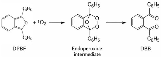
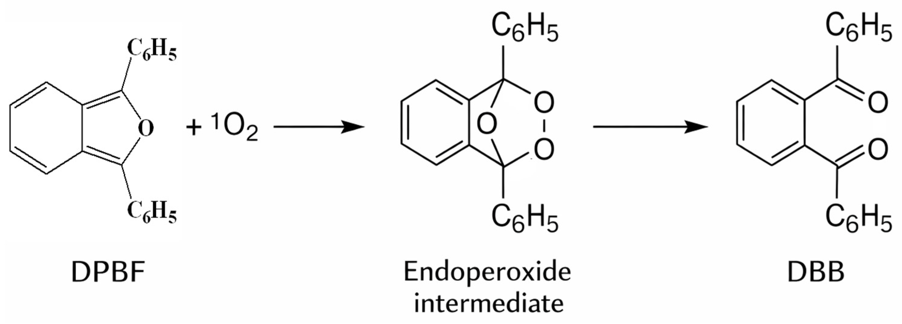
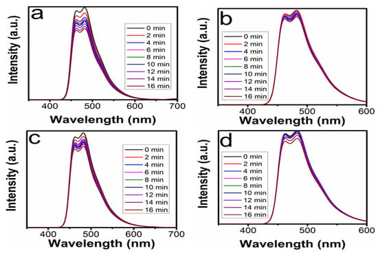
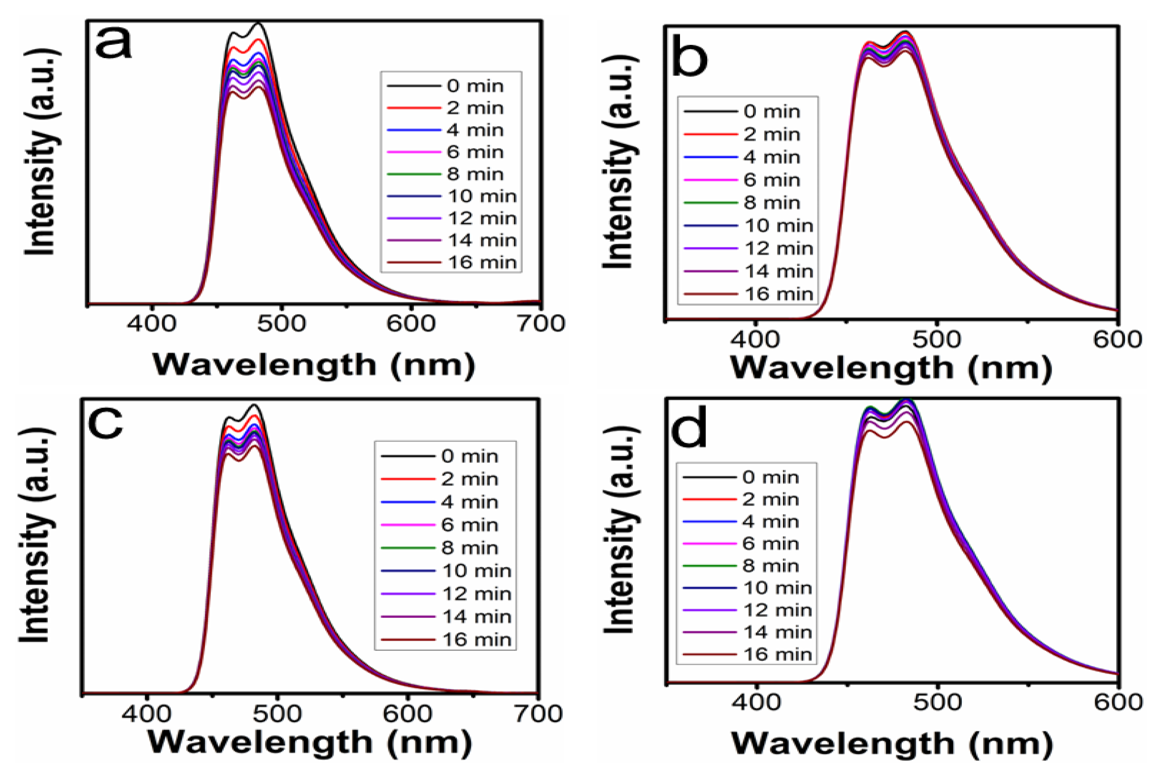
The 1O2 was investigated through a photochemical approach using DPBF as a scavenger and methylene blue as the reference. DPBF was chosen because of its ability to accept electrons from PSs. DPBF can react with 1O2 to produce o-dibenzoylbenzene (Figure 4), which opens up the π-system of isobenzofuran and prevents the product from absorbing or emitting visible light. As a result, any reduction in absorption or emission intensity of DPBF is reflected in the yield of 1O2 generated. Figure 5 shows a gradual decrease in the emission intensities of the mixture of DPBF and undoped ZIS/ZnS QDs. The decrease in emission intensity, also known as quenching, can be ascribed to the reaction of DPBF with the 1O2 generated from photoexcited QDs. This reaction occurred because of the transfer of electrons from the electron-rich group (PS, QDs) to the electron-deficient group (DPBF). The SOQY calculated from the spectra in Figure 5a,b are 0.16 and 0.32, respectively. Furthermore, the QDs that gave a SOQY of 0.16 showed little change in emission intensity during irradiation, while QDs that gave a SOQY of 0.32 displayed significant differences in the emission intensity of their spectra during irradiation. These differences could be due to the effect of the surface chemistry of thioglycolic acid and gelatin. This effect could be associated with Forster Resonance Energy Transfer (FRET), which can be ascribed to the spectral overlap between the absorption spectrum of Mn:ZIS/ZnS QDs and the emission spectrum of DPBF. In a typical reaction, DPBF reacts with 1O2 generated by the as-synthesized QDs upon light irradiation through Diels–Alder cycloaddition across the furan ring, producing a highly unstable endoperoxide intermediate. This intermediate subsequently undergoes ring opening and rearrangement to produce the stable, non-fluorescent product 1,2-dibenzoylbenzene (DBB) [31].
Figure 4.
Schematic representation of the reaction mechanism of DPBF with singlet oxygen (1O2) to form an endoperoxide intermediate.
Figure 5.
Variation in emission intensity of DPBF as a singlet oxygen (1O2) probe under an irradiation time of 0–16 min in the presence of: (a) Methylene blue, (b) ZnInS/ZnS QDs capped with sodium citrate and thioglycolic acid, (c) ZnInS/ZnS QDs capped with gelatin and thioglycolic acid, and (d) Mn-doped ZIS/ZnS core/shell QDs capped with gelatin and thioglycolic acid.
1O2 was generated when the QDs were excited at a wavelength of 450 nm. The production of 1O2 was observed as a function of the decrease in emission intensity of the QDs in the presence of DPBF. Radiation excites the electrons in the valence band of ZnInS/ZnS QDs, causing them to shift to the conduction band, thereby creating holes in the valence band and encouraging the development of energetically active electron–hole pairs. The energy from light is then transferred from the highly active electrons and holes to O2 and H2O in the tissues to form 1O2 [32]. The drop in the intensity of the DPBF peak occurred at a wavelength of 471 nm.
Figure 5c presents gradual changes in the emission intensity of the mixture of 10% Mn-doped ZIS/ZnS QDs under continuous irradiation at 450 nm for 16 min. The changes in intensity occur as a result of the interaction of 1O2 with DPBF, as shown in Figure 6. From the emission peaks, it can be observed that the intensity started to reduce after 2 min of irradiation. This observation shows that the 1O2 that is present in the PS takes a minimum time of approximately 120 s to start reacting with the probe to produce DBB. A similar observation has been reported in the literature [33]. The time it takes for oxygen to be released from PS could be dependent on different factors, including surface chemistry, the type of stabilizers, and concentration [34]. Therefore, we compared the effect of the stabilizers (TGA combined with gelatin and TGA combined with sodium citrate) on the SOQY. The effect of the stabilizers on 1O2 generation is also presented in Figure 5c. The intensity reduction was measured under continuous irradiation at 471 nm of DPBF as a function of time. The mixture of DPBF and TGA/sodium citrate-capped ZIS/ZnS QDs showed insignificant changes in fluorescence intensity when irradiated at 471 nm. These negligible changes can be ascribed to the absence of Förster Resonance Energy Transfer (FRET), which takes place when there is sufficient spectral overlap within 10 nm and/or beyond between the emission spectrum of the donor (PS) and the absorption spectrum of the electron acceptor (DPBF) [35,36]. This suggests that the surface of citrate anion mitigates the spectral overlap, as reflected in the SOQY. In contrast, the combination of TGA and gelatin significantly enhanced the quenching of the emission intensity of DPBF. This observation can be attributed to the interaction between the gelatin’s functional groups and DPBF molecules [37].
Figure 6.
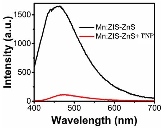
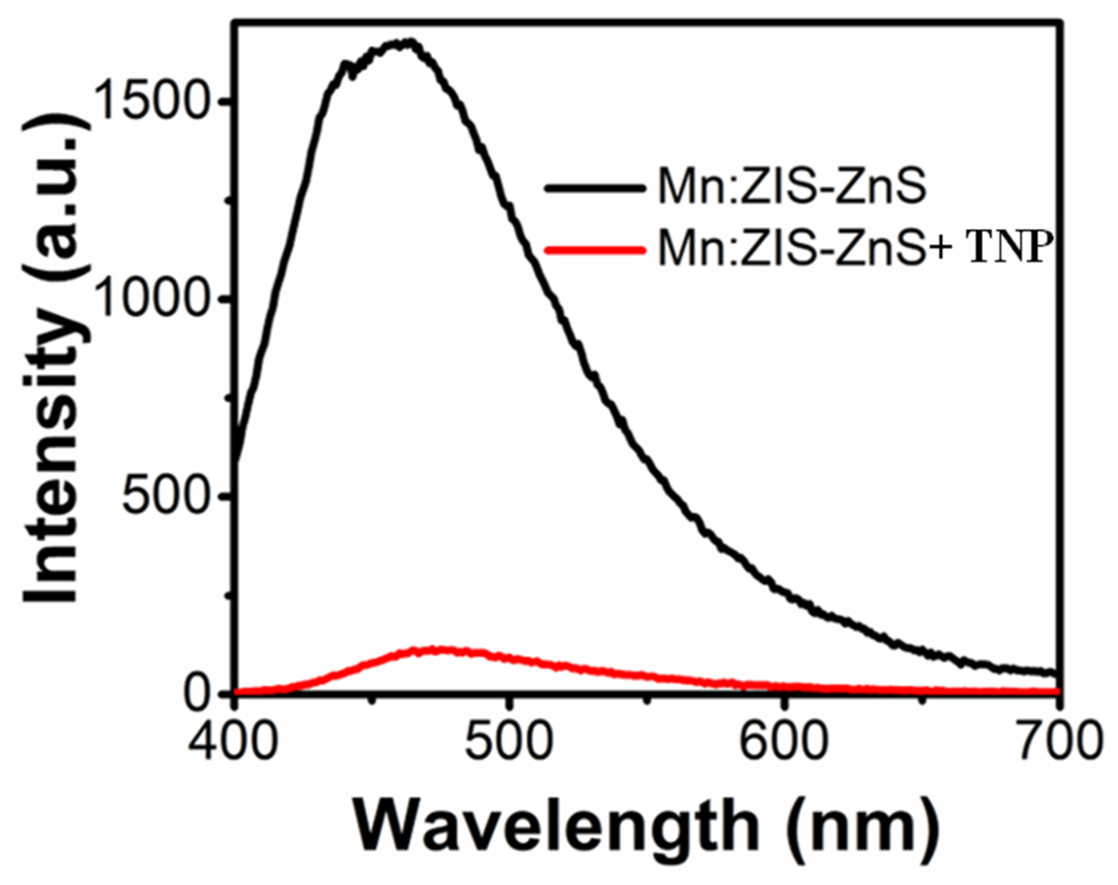
Photoluminescence spectra of the mixture of Mn:ZIS-ZnS QDs and TNP.
3.5. Chemical Sensing of Organic Pollutants
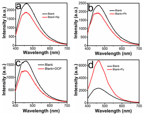
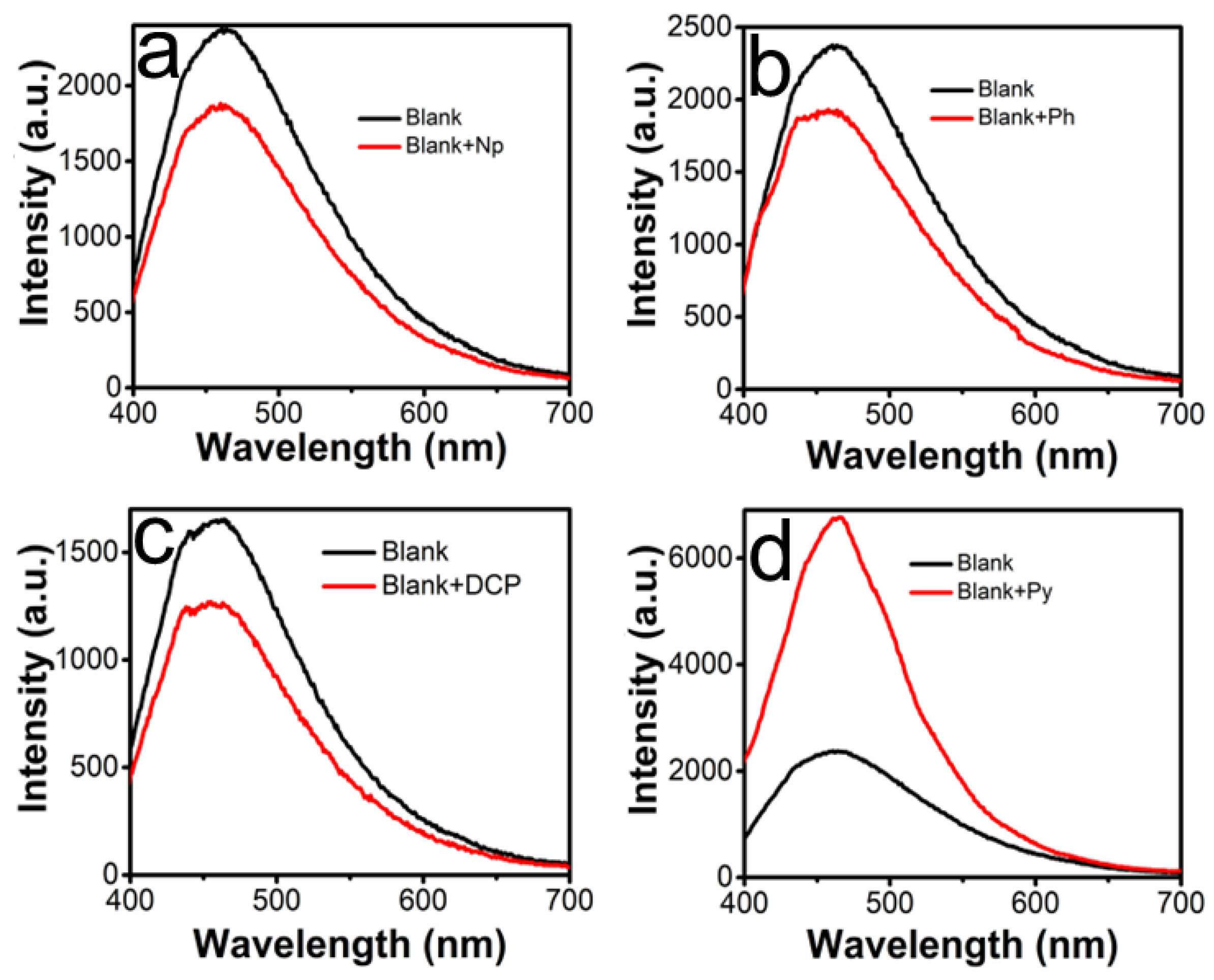
The Mn-doped ZnInS/ZnS QDs exhibited remarkable selectivity for 2,4,6-trinitrophenol (TNP) among the different organic pollutants (dichlorophenol (DCP), naphthalene (NP), phenanthrene (PH), and pyrene (PY)) that were tested. The significantly stronger quenching response, along with a distinct red shift in the emission wavelength of the quantum dots (QDs) upon their interaction with TNP, gives a very notable insight into their sensing mechanism. This observed red shift could be ascribed to the electrostatic reaction between some functional groups on the surface of QDs and the TNP surface, resulting in static quenching as a dominant mechanism. The electron-deficient nature of TNP could have facilitated its adsorption onto the QD surface, thereby transforming the local electronic structure and affecting the bandgap of the nanocrystal. This specific interaction explains why the doped QDs are significantly sensitive and selective toward TNP (Figure 6) over other phenols and polycyclic aromatic hydrocarbons (PAHs) (Figure 7 and Figure 8), which induced insignificant quenching. The fast, concentration-dependent quenching, which adhered to a linear relationship, also confirms the ability of the QDs to quantitatively identify TNP in an aqueous environment. However, the intensity of the QDs was enhanced when exposed to pyrene (PY). This enhancement could be due to different factors, including the fluorescence properties of PY, its ring structure and its electron system [38].
Figure 7.
Photoluminescence spectra of mixtures of Mn:ZIS-ZnS QDs with (a) NP, (b) PH, (c) DCP, and (d) PY.
Figure 8.
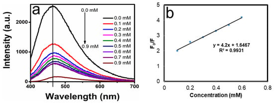
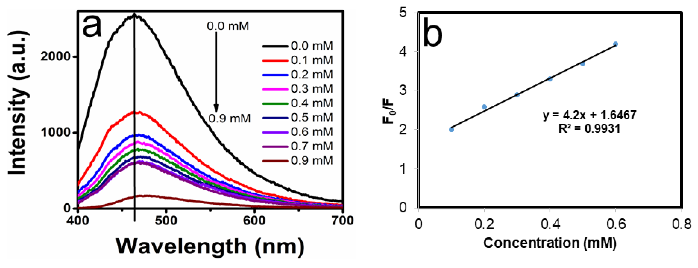
(a) Photoluminescence spectra of mixtures with different concentrations of TNP and Mn:ZIS-ZnS QDs, and (b) linear correlation between the intensity and the concentrations.
3.5.1. Fluorescence Sensing of Different Concentrations of TNP
The fluorescence quenching mechanism of Mn:ZIS/ZnS QDs when exposed to different concentrations of TNP was investigated to understand their degree of sensitivity as a potential chemical sensor for organic pollutants. In Figure 8, the PL intensity of Mn:ZIS/ZnS QDs gradually reduced with the increase in the concentration of TNP. The relationship between different concentrations of TNP as an electron acceptor and the consistent decrease in the intensity of the QDs was illustrated by a Stern–Volmer plot (Figure 8b). The QDs showed their maximum PL intensity at a wavelength position of 465 nm, before exposure to the minimum concentration of TNP. Upon the gradual addition of TNP at higher, a quenching of PL intensity consistent with the increased concentrations was observed, and this resulted in a sensitivity of 620 μM (Table 3). This observation indicates vacancy formation due to TNP and its dependence on TNP concentration. The quenching changes between the electron-deficient TNP molecules and the photoexcited QDs can be attributed to a combination of static and dynamic quenching [39]. Furthermore, this quenching effect could be attributed to electron transfer between the electron donor (QDs) and electron acceptor (nitro groups) of TNP [40]. The electron transfer happens from the conduction band of the excited QDs to the lowest unoccupied molecular orbital (LUMO) of TNP, resulting in reduced emission intensity and detection sensitivity of 620 µM [34].
Table 3.
Comparison of nanomaterial-based chemical sensors for TNP detection: LOD, sensing range, response time, mechanism, and reagent or precursor biocompatibility.
3.5.2. Fluorescence Sensing Mechanism of TNP
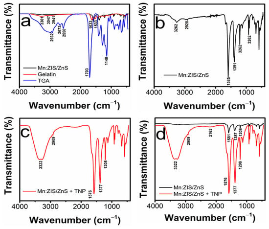
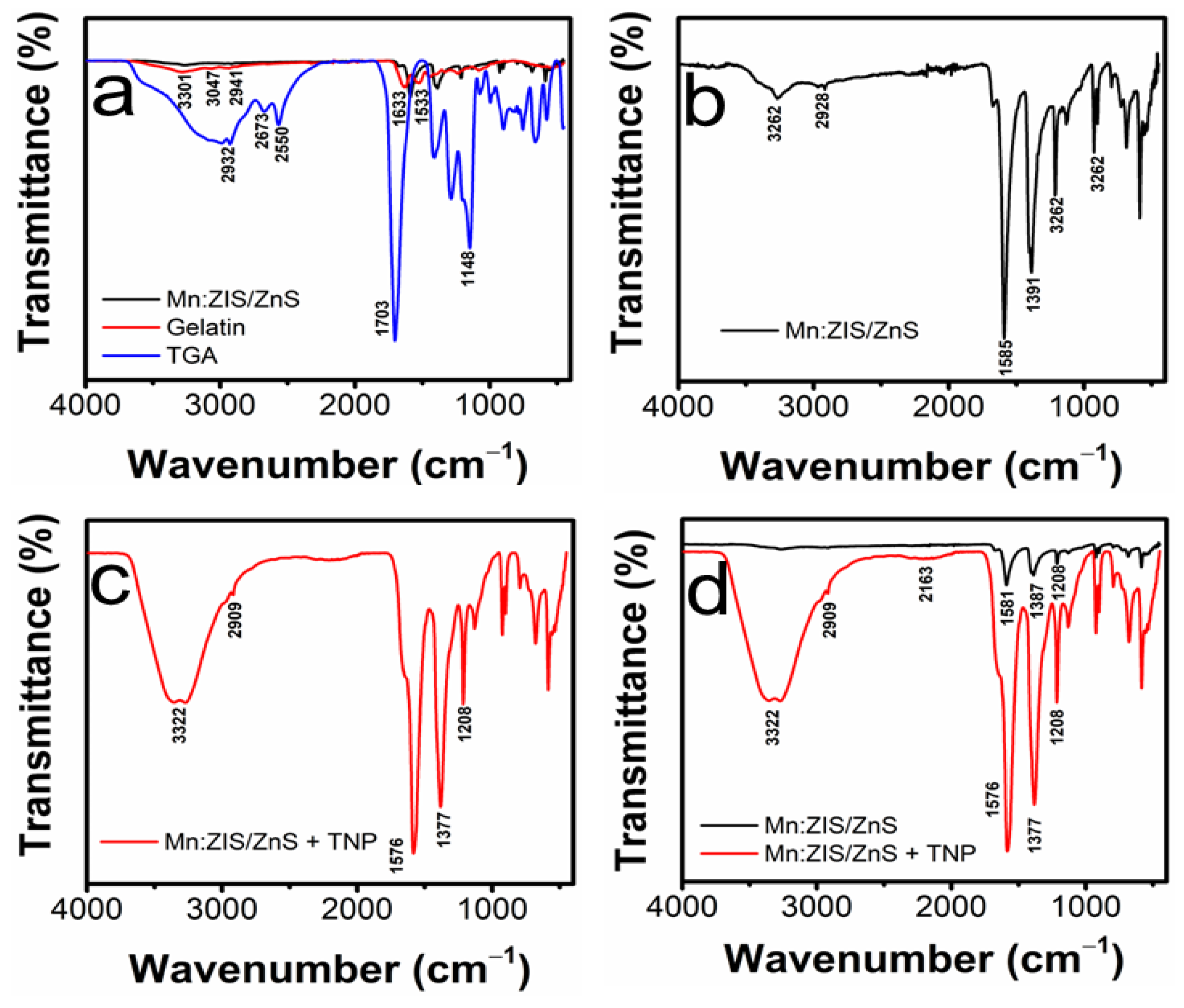
The interaction between TNP and the Mn-doped QDs was established by analyzing the functional groups. Shifts in the characteristic C=O stretching for amide and C-N-H deformation of amide were observed at 1576 cm−1 and 1377 cm−1, respectively, confirming the presence of gelatin and TGA. Also, OH stretching at 3322 cm−1 due to TNP and the formation of a weak band at 2163 cm−1 suggested two possible mechanisms, electrostatic and hydrogen bonding, between the functional groups of TNP and the QD surface, as presented in Figure 9.
Figure 9.
FTIR spectra illustrating the interaction mechanism between Mn:ZIS/ZnS QDs and TNP. (a) Comparison of FTIR spectra of Mn:ZIS/ZnS QDs, gelatin, and thioglycolic acid (TGA), (b) FTIR spectrum of pristine Mn:ZIS/ZnS QDs, (c) FTIR spectrum of Mn:ZIS/ZnS QDs after interaction with TNP. (d) Overlay comparison of Mn:ZIS/ZnS QDs before and after interaction with TNP.
4. Conclusions
In this study, ZnInS/ZnS QDs were successfully doped with Mn via an aqueous approach. The synthesis produced undoped particles with an average size of 7.40 nm and absorption within the visible region. The incorporation of 10% Mn significantly increased the size from 7.4 to 10.5 nm, with a red shift from 515 nm to 537 nm, due to its large atomic radius that causes lattice expansion in the host, ZnInS/ZnS. The results also demonstrate that the use of different stabilizing agents can contribute to the regulation of singlet oxygen, with the combination of TGA and gelatin giving an improved SOQY of 0.32 compared to the citrate-capped QDs, which gave 0.16. The Mn-doped ZIS-based QDs also demonstrated that they could be a potential chemical sensor for the detection of organic pollutants such as nitro aromatic compounds in different concentrations. The sensor showed the ability to detect TNP at a level of 620 µM. Thus, the as-synthesized doped QDs could be used as a potential material in phototherapy as a singlet oxygen generator and in the sensing of nitro aromatic pollutants.
Author Contributions
For O.S.O.; conceptualization, O.S.O.; project administration, O.S.O.; resources, O.S.O.; writing—review and editing, O.S.O.; funding, R.M.; writing—original draft, R.M.; methodology, R.M.; formal analysis. All authors have read and agreed to the published version of the manuscript.
Funding
This work was supported by the National Research Foundation (N.R.F) under the Competitive Programme for Rated Researchers (CPRR), Grants Nos. 129290 and 230509103956.
Data Availability Statement
We Dataset available on request from the authors.
Acknowledgments
The authors would like to thank the National Research Foundation (N.R.F) under the Competitive Programme for Rated Researchers (CPRR), Grants Nos. 129290 and 230509103956, the University of Johannesburg Research Committee (URC) and the Faculty of Science Research Committee (FRC) for financial support.
Conflicts of Interest
The authors declare no conflicts of interest.
Abbreviations
The following abbreviations are used in this manuscript:
| NP | Naphthalene |
| PH | Phenanthrene |
| DCP | Dichlorophenol |
| PY | Pyrene |
| DI | Dionized |
| MB | Methylene blue |
| ZIS | ZnInS |
| QDs | Quantum dots |
| TGA | Thioglycolic acid |
| DMSO | Dimethyl sulfoxide |
| PLQY | Photoluminescence quantum yield |
| SAED | Selected area electron diffraction |
| DPBF | 1,3-diphenylisobenzofuran |
| SOQY | Singlet oxygen quantum yield |
References
- Lv, Y.; Zhang, L.; Wu, R.; Li, L.S. Recent progress on eco-friendly quantum dots for bioimaging and diagnostics. Nano Res. 2024, 17, 10309–10331. [Google Scholar] [CrossRef] [Scilit]
- Cui, Z.; Qi, Z.; Diao, Z.; Qi, X.; Wang, S. GaN/HfSn2N4 heterojunction with superior polarization sensitivity and strong optical absorption. Micro Nanostruct. 2026, 215, 208688. [Google Scholar] [CrossRef] [Scilit]
- Zikalala, N.; Parani, S.; Tsolekile, N.; Oluwafemi, O.S. Facile green synthesis of ZnInS quantum dots: Temporal evolution of their optical properties and cell viability against normal and cancerous cells. J. Mater. Chem. C 2020, 8, 9329–9336. [Google Scholar] [CrossRef] [Scilit]
- Harris, S.; Kim, K. A Comparison of Common Quantum Dot Alternatives to Cadmium-Based Quantum Dots on the Basis of Liver Cytotoxicity. Nanomaterials 2024, 14, 1086. [Google Scholar] [CrossRef] [Scilit] [PubMed]
- Ncapayi, V.; Ninan, N.; Lebepe, T.C.; Parani, S.; Girija, A.R.; Bright, R.; Maluleke, R.; Aladesuyi, O.A.; Kodama, T.; Vasilev, K.; et al. Photostable fluorescent Near-infrared colloidal Mn-doped CuInSe/ZnS quantum dots for cancer-bacteria dual imaging. J. Lumin. 2023, 258, 119785. [Google Scholar] [CrossRef] [Scilit]
- Yuan, Y.; Han, Y.; Huang, B.; Zhang, L.; Yang, H.; Gu, B.; Cui, Y.; Zhang, J. Single-channel UV/vis dual-band detection with ZnCdS: Mn/ZnS core/shell quantum dots. Nanotechnology 2018, 30, 075501. [Google Scholar] [CrossRef] [Scilit] [PubMed]
- Dong, X.; Wang, K.; Bu, Y.; Wang, X. Ligand modification enhanced quantum dot LEDs: Principles and methods. J. Mater. Chem. C 2023, 11, 11755–11775. [Google Scholar] [CrossRef] [Scilit]
- Sanko, V.; Omeroglu, I.; Senocak, A.; Tumay, S.O. Highly selective pyrene-anchored halloysite nanotube for fluorometric determination of 2, 4, 6-trinitrophenol in environmental and food samples. ACS Omega 2025, 10, 7949–7963. [Google Scholar] [PubMed]
- Teixeira, J.; Delerue-Matos, C.; Morais, S.; Oliveira, M. Environmental contamination with polycyclic aromatic hydrocarbons and contribution from biomonitoring studies to the surveillance of global health. Environ. Sci. Pollut. Res. 2024, 31, 54339–54362. [Google Scholar] [CrossRef] [Scilit] [PubMed]
- Zango, Z.U.; Sambudi, N.S.; Jumbri, K.; Ramli, A.; Abu Bakar, N.H.H.; Saad, B.; Rozaini, M.N.H.; Isiyaka, H.A.; Osman, A.M.; Sulieman, A. An overview and evaluation of highly porous adsorbent materials for polycyclic aromatic hydrocarbons and phenols removal from wastewater. Water 2020, 12, 2921. [Google Scholar] [CrossRef] [Scilit]
- Feng, Y.; Li, Z.; Li, W. Polycyclic aromatic hydrocarbons (PAHs): Environmental persistence and human health risks. Nat. Prod. Commun. 2025, 20, 1934578X241311451. [Google Scholar] [CrossRef] [Scilit]
- Khan, I.; Shah, T.; Tariq, M.R.; Ahmad, M.; Zhang, B. Understanding the toxicity of trinitrophenol and promising decontamination strategies for its neutralization: Challenges and future perspectives. J. Environ. Chem. Eng. 2024, 12, 112720. [Google Scholar] [CrossRef] [Scilit]
- Kim, Y.; Jeon, Y.; Na, M.; Hwang, S.-J.; Yoon, Y. Recent trends in chemical sensors for detecting toxic materials. Sensors 2024, 24, 431. [Google Scholar] [CrossRef] [Scilit] [PubMed]
- Meng, J. Ultrafast Photophysics in Mn-Doped Semiconductor Quantum Dots for Optoelectronic Application. Ph.D. Thesis, Technical University of Denmark, Kongens Lyngby, Denmark, 2021. [Google Scholar]
- Zikalala, N.; Parani, S.; Oluwafemi, O.S. Synthesis of novel Zn-In-S/ZnS core/shell quantum dots: Temporal evolution and functionalization. Nano-Struct. Nano-Objects 2021, 25, 100673. [Google Scholar] [CrossRef] [Scilit]
- Ezquerra Riega, S.D.; Lantaño, B.; Rodríguez, H.B. Determination of relative singlet oxygen quantum yields via real-time fluorescence emission detection. Measurement 2026, 269, 120709. [Google Scholar] [CrossRef] [Scilit]
- Pan, Y.; Liu, T.; Li, C.; Shen, S.; Hou, X.; Wang, X. Synthetic scenarios, enzyme-mimetic activities, and colorimetric/fluorescent sensing applications based on carbon-based nanozymes for sensitively detecting environmental pollutants. Coord. Chem. Rev. 2026, 552, 217554. [Google Scholar] [CrossRef] [Scilit]
- Kaur, P.; Chopra, L.; Pal, K.; Gomes de Souza, F.; Kralj, S.; Oluwafemi, S.O.; Kyzas, G.Z. Guava leaf-derived carbon quantum dots as fluorescent probes for turn-on/off detection of Ba2+, Pb2+, Sn2+, and Hg2+ ions. Ceram. Int. 2025, 51, 47202–47217. [Google Scholar] [CrossRef] [Scilit]
- Jeong, S.; Ko, M.; Nam, S.; Oh, J.H.; Park, S.M.; Do, Y.R.; Song, J.K. Enhancement mechanism of quantum yield in core/shell/shell quantum dots of ZnS–AgIn5S8/ZnIn2S4/ZnS. Nanoscale Adv. 2024, 6, 925–933. [Google Scholar] [CrossRef] [Scilit] [PubMed]
- Oluwafemi, O.S.; Revaprasadu, N.; Adeyemi, O.O. A new synthesis of hexadecylamine-capped Mn-doped wurtzite CdSe nanoparticles. Mater. Lett. 2010, 64, 1513–1516. [Google Scholar] [CrossRef] [Scilit]
- Zhao, S.; Chen, Y.; Wang, L. Multiple underlying images tuned by Mn-doped Zn-Cu-In-S quantum dots. RSC Adv. 2023, 13, 34524–34533. [Google Scholar] [CrossRef] [Scilit] [PubMed]
- Mgedle, N.; Oluwafemi, O.S. Synthesis of Fe-doped ZnCuInS/ZnS QDs and their potential as fluorescence ‘‘switch OFF” for 2, 4, 6-trinitrophenol (TNP) detection. MRS Commun. 2026, 16, 481–486. [Google Scholar] [CrossRef] [Scilit]
- Chakraborty, S.; Mondal, P.; Viswanatha, R. Transition metal doping in CdS quantum dots: Diffusion, magnetism, and ultrafast charge carrier dynamics. Chem. Mater. 2023, 35, 2146–2154. [Google Scholar] [CrossRef] [Scilit]
- Cao, S.; Li, C.; Wang, J. Doping concentration-dependent photoluminescence properties of Mn-doped Zn-In-S quantum dots. J. Mater. Sci. 2018, 53, 1286–1296. [Google Scholar] [CrossRef] [Scilit]
- Cao, S.; Li, C.; Xu, L.; Wang, J. Long-lived and well-resolved Mn2+ ion emissions in CuInS-ZnS quantum dots. Sci. Rep. 2014, 4, 7510. [Google Scholar] [CrossRef] [Scilit] [PubMed]
- Cwilich, A.; Avidan-Shlomovich, S.; Lifshitz, E. Carrier dynamics and recombination pathways in Ag-In-Zn-S quantum dots. J. Phys. Chem. Lett. 2024, 15, 10479–10487. [Google Scholar] [CrossRef] [Scilit] [PubMed]
- Li, Z.-J.; Hofman, E.; Blaker, A.; Davis, A.H.; Dzikovski, B.; Ma, D.-K.; Zheng, W. Interface engineering of Mn-doped ZnSe-based core/shell nanowires for tunable host–dopant coupling. ACS Nano 2017, 11, 12591–12600. [Google Scholar] [CrossRef] [Scilit] [PubMed]
- Sreenan, B.; Lee, B.; Wan, L.; Zeng, R.; Zhao, J.; Zhu, X. Review of Mn-doped semiconductor nanocrystals for time-resolved luminescence biosensing/imaging. ACS Appl. Nano Mater. 2022, 5, 17413–17435. [Google Scholar] [CrossRef] [Scilit] [PubMed]
- Zhai, Y.; Shim, M. Benefitting from dopant loss and ostwald ripening in Mn doping of II-VI semiconductor nanocrystals. Nanoscale Res. Lett. 2015, 10, 423. [Google Scholar] [CrossRef] [Scilit] [PubMed]
- Talbi, A.; Khaaissa, Y.; Mansouri, F.; El Khouja, O.; Chaouiki, A.; Nouneh, K.; AlObaid, A.A. Advancing Solar Cell Efficiency: Experimental and Numerical Analysis of Mn-Doped ZnS as a Buffer Layer. ACS Omega 2025, 10, 28220–28232. [Google Scholar] [CrossRef] [Scilit] [PubMed]
- Stan, Ș.; Ramalho, J.P.P.; Holca, A.; Chiș, V. Photophysical Properties of 1, 3-Diphenylisobenzofuran as a Sensitizer and Its Reaction with O2. Molecules 2025, 30, 3021. [Google Scholar] [CrossRef] [Scilit] [PubMed]
- Wang, Z.; Zhu, K.; Hu, D.; Lu, X.; Zhou, W.; Liu, Z.; Li, P.; Li, Y.; Liu, X.; Hou, X. Colloidal quantum dots with enhanced reactive oxygen species generation for photodynamic therapy by hydrogen peroxide via deionization. J. Colloid Interface Sci. 2025, 700, 138432. [Google Scholar] [CrossRef] [Scilit] [PubMed]
- Tsolekile, N.; Parani, S.; de Macedo, E.F.; Lebepe, T.C.; Maluleke, R.; Ncapayi, V.; Matoetoe, M.C.; Songca, S.P.; Conceição, K.; Tada, D.B. Photodynamic Therapy and Antibacterial Activities of a Novel Synthesized Quaternary Zn-Cu-In-S/ZnS QDs-mTHPP Porphyrin Conjugate. Int. J. Nanomed. 2022, 17, 5315. [Google Scholar] [CrossRef] [Scilit] [PubMed]
- Maluleke, R.; Oluwafemi, O.S. Synthetic approaches, modification strategies and the application of quantum dots in the sensing of priority pollutants. Appl. Sci. 2021, 11, 11580. [Google Scholar] [CrossRef] [Scilit]
- Deng, R.; Wang, J.; Chen, R.; Huang, W.; Liu, X. Enabling Forster Resonance Energy Transfer from Large Nanocrystals through Energy Migration. J. Am. Chem. Soc. 2016, 138, 15972–15979. [Google Scholar] [CrossRef] [Scilit] [PubMed]
- Shi, J.; Tian, F.; Lyu, J.; Yang, M. Nnoparticle based fluorescence resonance energy transfer (FRET) for biosensing applications. J. Mater. Chem. B 2015, 3, 6989–7005. [Google Scholar] [CrossRef] [Scilit] [PubMed]
- Loskutova, A.; Seitkali, A.; Aliyev, D.; Bukasov, R. Quantum dot-based luminescent sensors: Review from analytical perspective. Int. J. Mol. Sci. 2025, 26, 6674. [Google Scholar] [CrossRef] [Scilit] [PubMed]
- Ayyavoo, K.; Velusamy, P. Pyrene based materials as fluorescent probes in chemical and biological fields. New J. Chem. 2021, 45, 10997–11017. [Google Scholar] [CrossRef] [Scilit]
- Zangi, R. Quenching Single-Fluorophore Systems and the Emergence of Nonlinear Stern–Volmer Plots. ACS Omega 2026, 11, 12713–12724. [Google Scholar] [CrossRef] [Scilit] [PubMed]
- Song, X.; Zhao, Q.; Hou, X.; Liu, S.; Luo, L.; Ren, Y. Three luminescent Zn/Cd-based MOFs for detecting antibiotics/nitroaromatic compounds and quenching mechanisms study. J. Mol. Struct. 2025, 1324, 140813. [Google Scholar] [CrossRef] [Scilit]
- Kumar, V.; Kumar, A.; Chini, M.K.; Satapathi, S. Fluorescent Fe2O3-CdSe nanocomposite probe for selective detection and removal of picric acid. Mater. Chem. Phys. 2021, 260, 124130. [Google Scholar] [CrossRef] [Scilit]
- Huang, J.; Wang, L.; Shi, C.; Dai, Y.; Gu, C.; Liu, J. Selective detection of picric acid using functionalized reduced graphene oxide sensor device. Sens. Actuators B Chem. 2014, 196, 567–573. [Google Scholar] [CrossRef] [Scilit]
- Mahyari, M. Electrochemical determination of picric acid based on platinum nanoparticles–reduced graphene oxide composite. Int. J. Environ. Anal. Chem. 2016, 96, 1455–1468. [Google Scholar] [CrossRef] [Scilit]
- Karthika, P.; Shanmuganathan, S.; Viswanathan, S. Electrochemical sensor for picric acid by using molecularly imprinted polymer and reduced graphene oxide modified pencil graphite electrode. Proc. Indian Natl. Sci. Acad. 2022, 88, 263–276. [Google Scholar] [CrossRef] [Scilit]
- Tan, X.; Zhang, T.; Zeng, W.; He, S.; Liu, X.; Tian, H.; Shi, J.; Cao, T. A fluorescence sensing determination of 2, 4, 6-trinitrophenol based on cationic water-soluble pillar[6]arene graphene nanocomposite. Sensors 2018, 19, 91. [Google Scholar] [PubMed]
- Bao, Z.; Zhang, K.; Jian, J.; Hu, Z.; Yuan, K.; Shao, H.; Peng, K.; Jiang, Z.; Zapien, J.A.; Yan, Y.; et al. Strongly fluorescent cysteamine-coated copper nanoclusters as a fluorescent probe for determination of picric acid. Microchim. Acta 2018, 185, 507. [Google Scholar] [CrossRef] [Scilit] [PubMed]
- Ibrahim, A.A.; Tiwari, P.; Al-Assiri, M.S.; Al-Salami, A.E.; Umar, A.; Kumar, R.; Kim, S.H.; Ansari, Z.A.; Baskoutas, S. A highly-sensitive picric acid chemical sensor based on ZnO nanopeanuts. Materials 2017, 10, 795. [Google Scholar] [CrossRef] [Scilit] [PubMed]

Disclaimer/Publisher’s Note: The statements, opinions and data contained in all publications are solely those of the individual author(s) and contributor(s) and not of MDPI and/or the editor(s). MDPI and/or the editor(s) disclaim responsibility for any injury to people or property resulting from any ideas, methods, instructions or products referred to in the content. |
© 2026 by the authors. Licensee MDPI, Basel, Switzerland. This article is an open access article distributed under the terms and conditions of the Creative Commons Attribution (CC BY) license.