


Abstract
Mercury contamination threatens aquatic ecosystems and human health because of its toxicity, persistence, and potential for bioaccumulation. This study investigated an electrochemically assisted self-assembly (EASA)-derived glutathione-modified thiol-functionalized mesoporous silica film on a fluorine-doped tin oxide (FTO) electrode as a proof-of-concept interface for Hg(II) detection. Films containing 5–15 mol% (3-mercaptopropyl)trimethoxysilane (MPTMS) were evaluated using charged redox probes, and the 10 mol% composition produced the highest electrochemical response. FTIR analysis supported silica-network formation and the retention of glutathione-associated functional groups. Under the selected conditions of pH 5.0, a deposition potential of −0.70 V, and a deposition time of 5 min, the MSF-GSH/FTO electrode exhibited a linear response from 5 to 60 µM Hg(II), with an estimated calibration-based detection limit of 4.27 µM. Resolved oxidation peaks were observed for Hg, Pb, and Cd in a mixed-metal solution. The interface combines direct EASA film formation, thiol incorporation, and post-deposition glutathione modification; however, further sensitivity improvement, replication, stability testing, and real-sample validation are required.
1. Introduction
Artisanal and small-scale gold mining (ASGM) remains an important source of gold production in the Philippines, particularly in gold-producing areas such as Davao del Norte, Benguet, Camarines Norte, and South Cotabato [1,2,3]. Despite the availability of improved gold-recovery technologies, mercury amalgamation continues to be practiced because it is inexpensive, rapid, simple, and accessible to small-scale miners [4,5]. In this process, elemental mercury is mixed with gold-bearing ore to form a gold–mercury amalgam, which is subsequently heated to recover the gold. The resulting release of mercury vapor and mercury-containing residues creates occupational and environmental exposure risks for miners, their families, and surrounding communities [5,6]. Consequently, the continued use of mercury in ASGM represents an important environmental and public-health concern, particularly in communities with limited access to analytical laboratories and exposure-monitoring services.
Mercury is highly toxic because of its strong affinity for sulfhydryl-containing proteins and enzymes, which can disrupt essential cellular and physiological processes [7,8,9]. Elemental mercury vapor can readily enter the bloodstream through inhalation and is subsequently oxidized to divalent mercury, Hg(II), which may accumulate in the kidneys, brain, liver, and other tissues [7,9]. Prolonged exposure has been associated with neurological, renal, developmental, immunological, and behavioral abnormalities, with children being particularly susceptible to impaired cognition, motor coordination, speech, and language development [10,11,12]. In Apokon, Tagum City, elevated mercury concentrations were previously detected in blood and hair samples collected from schoolchildren living near gold-processing activities [13]. These observations highlight the continuing need for accessible methods for monitoring mercury contamination in mining-affected areas.
Mercury is commonly quantified using techniques such as cold-vapor atomic absorption spectroscopy, cold-vapor atomic fluorescence spectrometry, and inductively coupled plasma mass spectrometry [14,15,16,17,18]. Although these methods provide sensitive and reliable measurements, they frequently require costly instrumentation, trained personnel, centralized facilities, and extensive sample preparation [19,20,21]. These requirements may limit their use for rapid or decentralized monitoring. Electrochemical methods offer an attractive alternative because of their relatively simple instrumentation, rapid analysis, low power consumption, portability, and potential for on-site measurement [21,22]. Nevertheless, the electrochemical determination of trace Hg(II) can be affected by low analyte concentrations, competing metal ions, nonspecific adsorption, and electrode-surface fouling. A selective electrode interface capable of capturing and preconcentrating Hg(II) near the electrode surface is therefore desirable.
The World Health Organization provides a guideline value of 0.006 mg L−1 (6 µg L−1) for inorganic mercury in drinking water, whereas the United States Environmental Protection Agency establishes a maximum contaminant level of 0.002 mg L−1 (2 µg L−1) [23,24]. These low concentrations emphasize the analytical challenge associated with regulatory mercury monitoring. The detection limit obtained in the present proof-of-concept study remains above these values; accordingly, the proposed interface is not presented as a validated method for direct drinking-water compliance measurements.
Mesoporous silica films are promising electrode-modification materials because of their ordered nanochannels, tunable surface chemistry, and charge-dependent transport properties [25,26,27]. Electrochemically assisted self-assembly (EASA) is an electrode-driven sol–gel method in which cathodically generated hydroxide ions locally promote silica condensation around surfactant assemblies, enabling rapid film formation directly on a conductive substrate [26,28]. Vertically oriented channels are characteristic of EASA films prepared under suitable conditions; however, channel orientation was not directly measured for the present films. Glutathione is a naturally occurring tripeptide containing thiol, amino, carboxyl, and amide groups that can participate in metal-ion coordination [29,30]. Previous studies have demonstrated GSH immobilization on silica-based materials and its use in adsorption and sensing [31,32,33]. The novelty of the present work does not arise from EASA or glutathione sensing alone. Rather, it lies in combining an EASA-derived thiol-containing mesoporous silica film with post-deposition GSH immobilization on FTO to create a confined, sulfur-rich interface for Hg(II) accumulation. Its current disadvantages include a comparatively high detection limit and the absence of replicate, stability, recovery, and real-sample validation. The study is therefore positioned as a material and sensing proof of concept rather than a fully validated trace-mercury method.
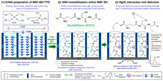
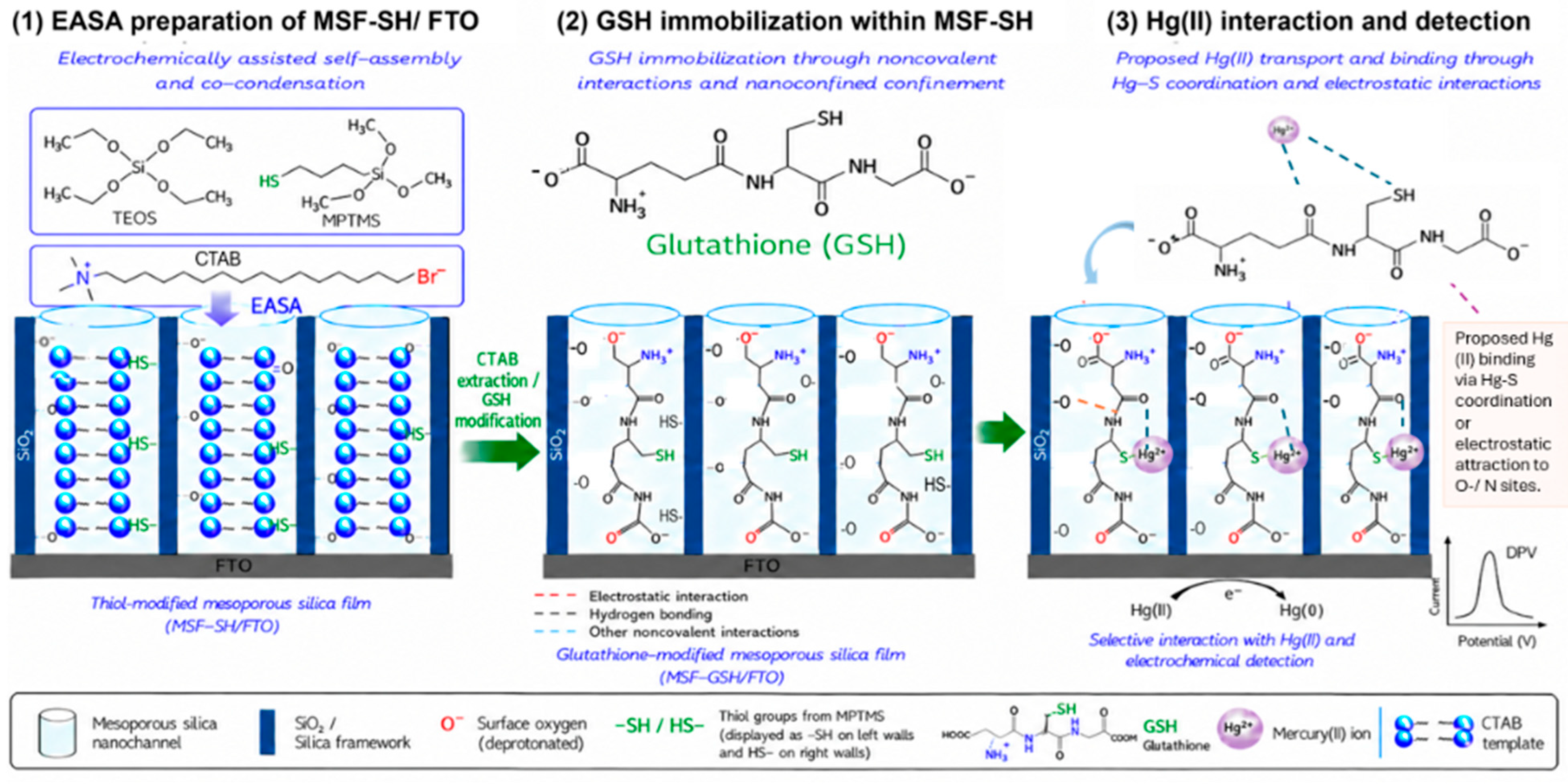
In the present study, a thiol-functionalized mesoporous silica film was deposited directly onto a fluorine-doped tin oxide electrode through the electrochemically assisted self-assembly and co-condensation of tetraethoxysilane and (3-mercaptopropyl)trimethoxysilane (Figure 1). Glutathione was subsequently immobilized within or on the thiol-functionalized film to produce the MSF-GSH/FTO electrode for Hg(II) detection. The mesoporous silica structure was designed to facilitate ion transport and provide a confined environment for analyte accumulation, while the immobilized glutathione introduced sulfur-, carboxyl-, and amino-containing functional groups capable of interacting with Hg(II). The influence of MPTMS content and selected experimental conditions, particularly solution pH, on the electrochemical response was evaluated. The analytical performance of the developed electrode was assessed in terms of sensitivity, linear concentration range, estimated limit of detection, and its ability to distinguish Hg(II) in the presence of potentially interfering Pb(II) and Cd(II). The resulting platform demonstrates the potential of an EASA-derived glutathione-modified mesoporous silica film for the electrochemical detection of Hg(II) in aqueous media.
Figure 1.
Schematic representation of the preparation of the glutathione-modified mesoporous silica film and its proposed interaction with Hg(II). MSF-SH was deposited on FTO by EASA using TEOS and MPTMS precursor in the presence of the template (CTAB), followed by template removal and GSH immobilization. The illustrated nanochannel arrangement and Hg(II)-binding interactions are conceptual and were not directly established by microscopy or surface spectroscopy in this study.
2. Materials and Methods
2.1. Materials
All chemicals were of analytical grade and were used as received without further purification. All aqueous solutions were prepared using distilled water. Tetraethoxysilane (TEOS, 98%, Sigma-Aldrich, St. Louis, MO, USA), (3-mercaptopropyl)trimethoxysilane (MPTMS, 95%, Sigma-Aldrich, St. Louis, MO, USA), and cetyltrimethylammonium bromide (CTAB, 98%, Sigma-Aldrich, St. Louis, MO, USA) were used for the electrochemically assisted self-assembly of thiol-functionalized mesoporous silica films on fluorine-doped tin oxide electrodes (FTO, Solaronix, Aubonne, Switzerland). Reduced L-glutathione (GSH, C10H17N3O6S, 98%, Sigma-Aldrich, St. Louis, MO, USA) was used for the post-functionalization of the thiol-modified films. Hydrochloric acid (HCl, Sigma-Aldrich, St. Louis, MO, USA), sodium nitrate (NaNO3, Scharlau, Sentmenat, Spain), sodium hydroxide (NaOH, Sigma-Aldrich, St. Louis, MO, USA), and ethanol (C2H5OH, Sigma-Aldrich, St. Louis, MO, USA) were used during film preparation, template extraction, and electrochemical measurements. Phosphate buffer solutions were prepared using dipotassium hydrogen phosphate (K2HPO4, 98%, Sigma-Aldrich, St. Louis, MO, USA), potassium dihydrogen phosphate (KH2PO4, Scharlau, Sentmenat, Spain), and phosphoric acid (H3PO4, ≥99%, Merck, Darmstadt, Germany). Certified standard solutions of Hg(II) (996 mg L−1 in 2% nitric acid, Buck Scientific, Ansonia, CT, USA), Cd(II) (997 mg L−1 in 2% nitric acid, Buck Scientific, Ansonia, CT, USA), Cd(II) solution (1000 mg/L in 2% nitric acid, Buck Scientific, Ansonia, CT, USA), and Pb(II) (1000 mg L−1 in 0.5 mol L−1 nitric acid, Buck Scientific, Ansonia, CT, USA)) were used to prepare the metal-ion working solutions. Hexaammineruthenium(III) chloride ([Ru(NH3)6]Cl3, 98%, Sigma-Aldrich, St. Louis, MO, USA) and potassium ferricyanide (K3[Fe(CN)6], 99.98%, Sigma-Aldrich, St. Louis, MO, USA) were used as redox probes for cyclic-voltammetry characterization.
2.2. EASA Approach for the Preparation of Thiol-Functionalized Mesoporous Silica Film (MSF-SH)
The thiol-functionalized mesoporous silica film-modified FTO electrode (MSF-SH/FTO) was prepared by electrochemically assisted self-assembly (EASA) using a µAutolab potentiostat (Metrohm Autolab, Utrecht, The Netherlands), following Ref. [28] with modifications. The precursor sol had a total silane concentration of 100 mM and contained TEOS and MPTMS. MPTMS constituted 5, 10, or 15 mol% of the total silane content. These formulations were selected as a limited low-to-moderate compositional series and do not represent exhaustive optimization. The sol also contained 32 mM CTAB and 0.10 M NaNO3 in water–ethanol (1:1, v/v) at pH 3. After hydrolysis under stirring for 2.5 h, the film was deposited at −1.5 V for 25 s using FTO as the working electrode, a silver-wire pseudo-reference electrode, and a stainless-steel plate counter electrode. The exposed FTO diameter was 0.8 cm. The electrode was rinsed, dried overnight at 130 °C, and extracted in 0.1 M HCl/ethanol for at least 15 min to remove CTAB.
2.3. Immobilization of Glutathione on MSF-SH/FTO
The template-extracted MSF-SH/FTO electrode was immersed in a freshly prepared 3.0 mM reduced glutathione solution in 0.10 M phosphate buffer at pH 7.4. The electrode was incubated for 1 h at ambient temperature under static conditions. After incubation, the electrode was rinsed with water to remove unbound or weakly retained glutathione and was subsequently dried or stored under ambient temperature. The resulting glutathione-modified electrode was denoted MSF-GSH. Because no separate coupling reagent or oxidizing agent was employed, this treatment is described as the immobilization or adsorption of GSH onto MSF-SH rather than as confirmed covalent grafting.
2.4. FTIR Characterization
Fourier-transform infrared spectroscopy was performed using a Spectrum Two™ FTIR spectrometer (PerkinElmer, Shelton, CT, USA) to identify the functional groups present in the silica films. Spectra were collected in transmission mode over the range of 400–4000 cm−1. Bare FTO, template-containing MSF-SH, template-extracted MSF-SH, and MSF-GSH were analyzed. The spectrum of reduced glutathione was also recorded as a reference. The spectra were compared to evaluate the formation of the siloxane network, the incorporation of mercaptopropyl groups, the removal of CTAB, and the presence of GSH-associated functional groups after immobilization.
2.5. Cyclic Voltammetry Characterization
Cyclic-voltammetry measurements were performed using an EMStat2 potentiostat controlled with PSTrace (PalmSens BV, Houten, The Netherlands). A conventional three-electrode cell comprised bare or film-modified FTO as the working electrode, a Ag/AgCl electrode in 3.0 M KCl as the reference electrode, and a stainless-steel rod as the counter electrode. The circular exposed FTO region had a diameter of 0.8 cm, corresponding to a geometric area of 0.503 cm2. The electrochemically active surface area was not independently determined; consequently, currents are reported as measured responses rather than active-area-normalized current densities. Electrolyte solutions were purged with nitrogen for 20 min before measurement. A 0.10 M NaNO3 solution was used as supporting electrolyte. Film accessibility and charge-selective behavior were evaluated using 5.0 mM [Ru(NH3)6]3+ and 5.0 mM [Fe(CN)6]3− as cationic and anionic redox probes, respectively.
2.6. Voltammetric Detection of Hg(II)
Voltammetric measurements for Hg(II) detection were performed using MSF-GSH/FTO as the working electrode, a Ag/AgCl electrode in 3.0 M KCl as the reference electrode, a stainless-steel rod as the counter electrode. Measurements were conducted in phosphate-containing electrolyte at the investigated pH values. Before each anodic stripping measurement, Hg(II) was deposited at −0.70 V for 5 min under static conditions, followed by an anodic differential-pulse scan with a pulse amplitude of 50 mV. Solution pH was the only parameter systematically varied; deposition potential, deposition time, and pulse amplitude were held constant and were not independently optimized. A newly prepared electrode was used for each measurement. The calibration measurements were single determinations and were not obtained from independent experimental replicates. Blank measurements were performed using supporting electrolyte without Hg(II).
3. Results and Discussion
3.1. Electrochemical Characterization of Thiol-Modified Mesoporous Silica Film
Electrochemically assisted self-assembly is a rapid electrode-driven sol–gel deposition method. Cathodic reactions generate hydroxide ions near the electrode surface, and the resulting local increase in pH promotes silica condensation around surfactant assemblies [24]. Vertically oriented nanochannels are characteristic of EASA films prepared under suitable conditions; however, nanochannel orientation, pore size, morphology, and film thickness were not directly determined for the present films. Accordingly, structural values cited from the literature are not treated as measurements of the electrodes examined here.
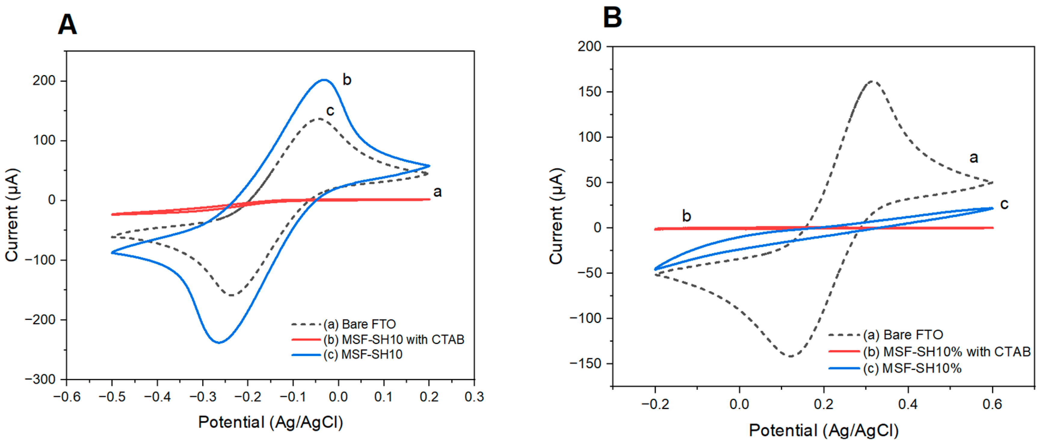
Thiol-functionalized mesoporous silica films were prepared through the co-condensation of TEOS and MPTMS using precursor sols containing 5, 10, or 15 mol% MPTMS relative to the total molar amount of TEOS and MPTMS [25,26]. The resulting films were denoted MSF-SH5%, MSF-SH10%, and MSF-SH15%, respectively. The accessibility and charge-selective properties of MSF-SH10%/FTO were evaluated by cyclic voltammetry using hexaammineruthenium(III), [Ru(NH3)6]3+, as a cationic redox probe and ferricyanide, [Fe(CN)6]3−, as an anionic redox probe. Figure 2 compares the voltammetric responses obtained at the bare FTO electrode and at MSF-SH10%/FTO before and after CTAB template extraction.
Figure 2.
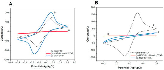
Cyclic voltammograms recorded at (a) bare FTO, (b) as-prepared CTAB-containing MSF-SH10%/FTO, and (c) template-extracted MSF-SH10%/FTO in 0.10 M NaNO3 containing (A) 5.0 mM hexaammineruthenium(III) chloride and (B) 5.0 mM potassium ferricyanide. Scan rate: 100 mV s−1.
Before template extraction, the electrochemical responses of both redox probes were strongly attenuated because the mesochannels remained occupied by CTAB, which restricted probe transport toward the underlying FTO surface. After template extraction, the response of the cationic [Ru(NH3)6]3+ probe increased markedly, indicating that the removal of CTAB generated accessible transport pathways through the mesoporous film. The preferential accessibility of [Ru(NH3)6]3+ can be attributed to its electrostatic attraction toward negatively charged silanol and siloxide groups within the silica nanochannels. In contrast, the response of the anionic [Fe(CN)6]3− probe remained considerably lower because of electrostatic repulsion from the negatively charged silica framework. The contrasting responses of the cationic and anionic probes therefore confirm that the template-extracted MSF-SH10% film retained the characteristic charge-selective behavior of mesoporous silica films.
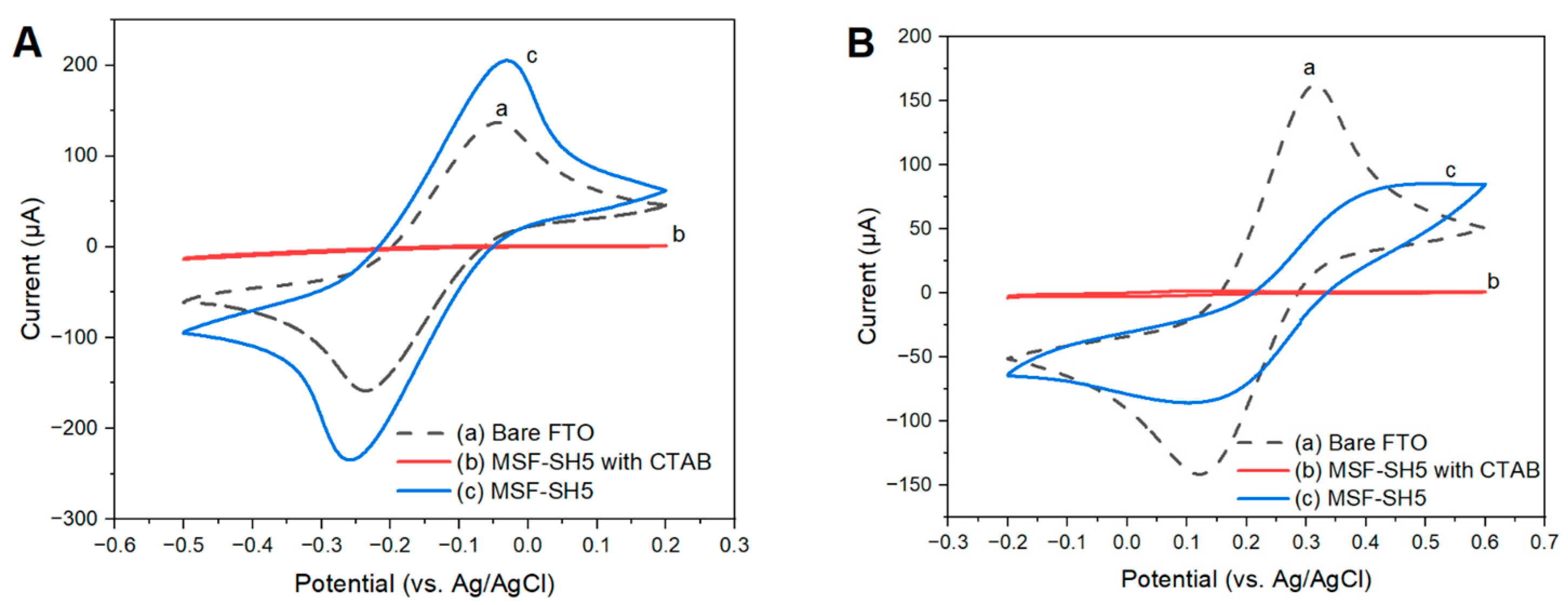
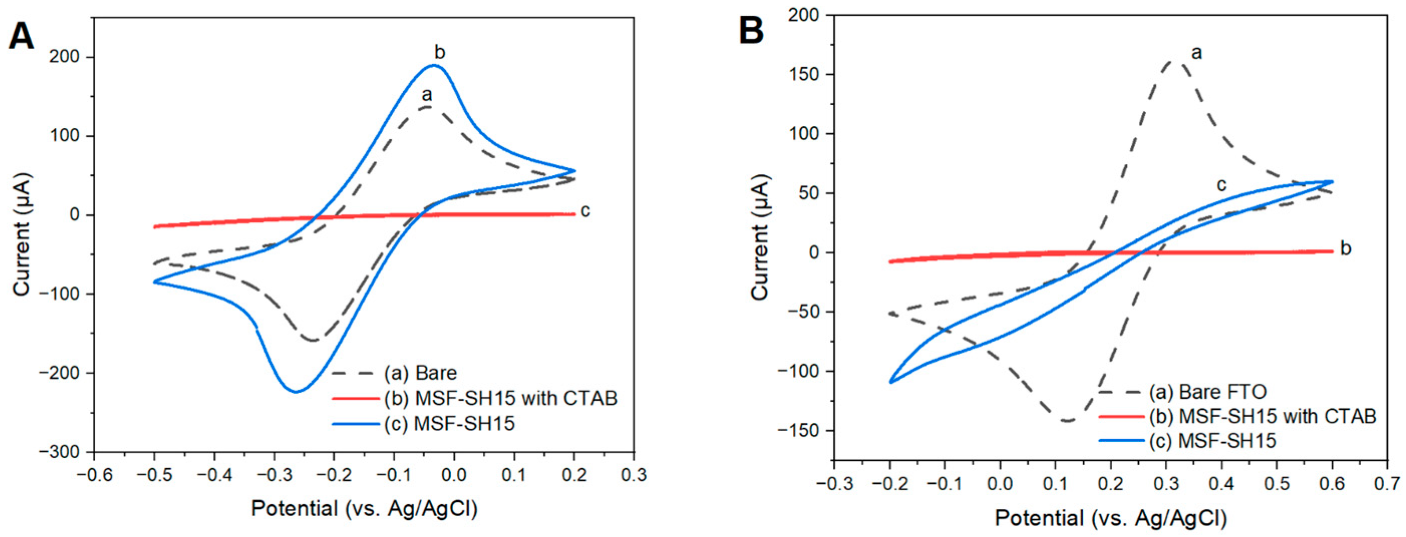
Although MSF-SH10%/FTO retained the characteristic charge-selective behavior of the unfunctionalized MSF/FTO electrode, it produced a higher voltammetric response, indicating improved electrochemical accessibility after the incorporation of MPTMS, as shown in Figure A1. The corresponding cyclic voltammetric characterization of MSF-SH5%/FTO and MSF-SH15%/FTO using hexaammineruthenium(III) and ferricyanide is presented in Figure A2 and Figure A3, respectively.
3.2. Effect of MPTMS Content on the Voltammetric Response of MSF-SH/FTO
The influence of MPTMS content in the initial sol–gel precursor on the electrochemical behavior of MSF-SH/FTO was investigated by cyclic voltammetry using 5.0 mM hexaammineruthenium(III) as positively and negatively charged redox probes. These probes were used to evaluate changes in film permeability and charge-selective transport resulting from the incorporation of mercaptopropyl groups into the silica framework.
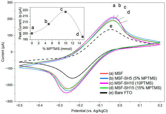
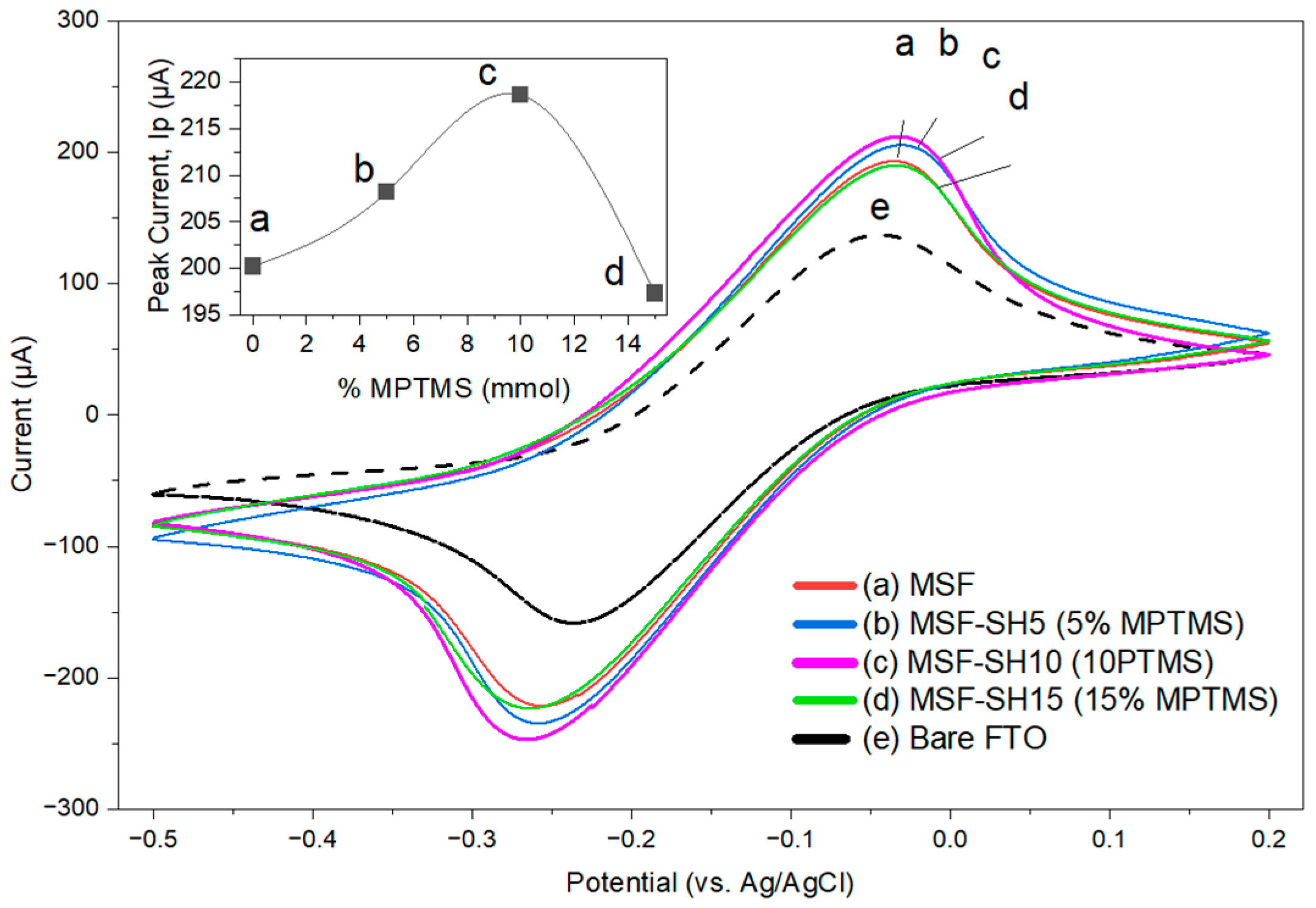
The increase in current from MSF-SH5% to MSF-SH10% indicates that the 10 mol% formulation provided greater electrochemical accessibility under the investigated conditions (Figure 3). The current response is influenced by multiple factors, potentially including film thickness, surface charge, pore accessibility, mesostructural organization, and defects. Because these properties were not independently measured, the voltammetric results do not establish a unique transport mechanism.
Figure 3.
Cyclic voltammograms recorded at the mesoporous silica film-modified FTO electrode (MSF/FTO, a), thiol-functionalized mesoporous silica film-modified FTO electrodes prepared with 5 mol% MPTMS (MSF-SH5%/FTO, b), 10 mol% MPTMS (MSF-SH10%/FTO, c), and 15 mol% MPTMS (MSF-SH15%/FTO, d), and the bare FTO electrode (e), using 5.0 mM hexaammineruthenium(III), [Ru(NH3)6]3+ as the redox probe. The inset shows the corresponding peak-current comparison for the MSF- and MSF-SH-modified electrodes (a–d).
Increasing the MPTMS content to 15 mol% resulted in a lower peak current (Figure 3). This observation may reflect changes in film permeability, local hydrophobicity, charge, or mesostructural organization; however, these explanations remain tentative because the morphology, thickness, and pore structure of the individual films were not directly measured. Among the three tested formulations, MSF-SH10%/FTO produced the highest redox-probe response and was therefore selected for subsequent experiments. The term ‘selected’ is used rather than ‘optimized’ because compositions outside the 5–15 mol% range were not investigated.
3.3. FTIR Characterization of MSF, MSF-SH, and MSF-GSH
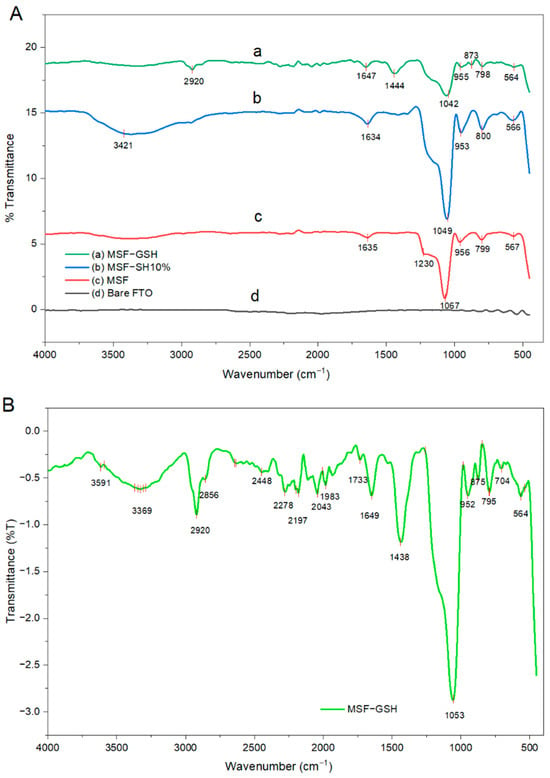
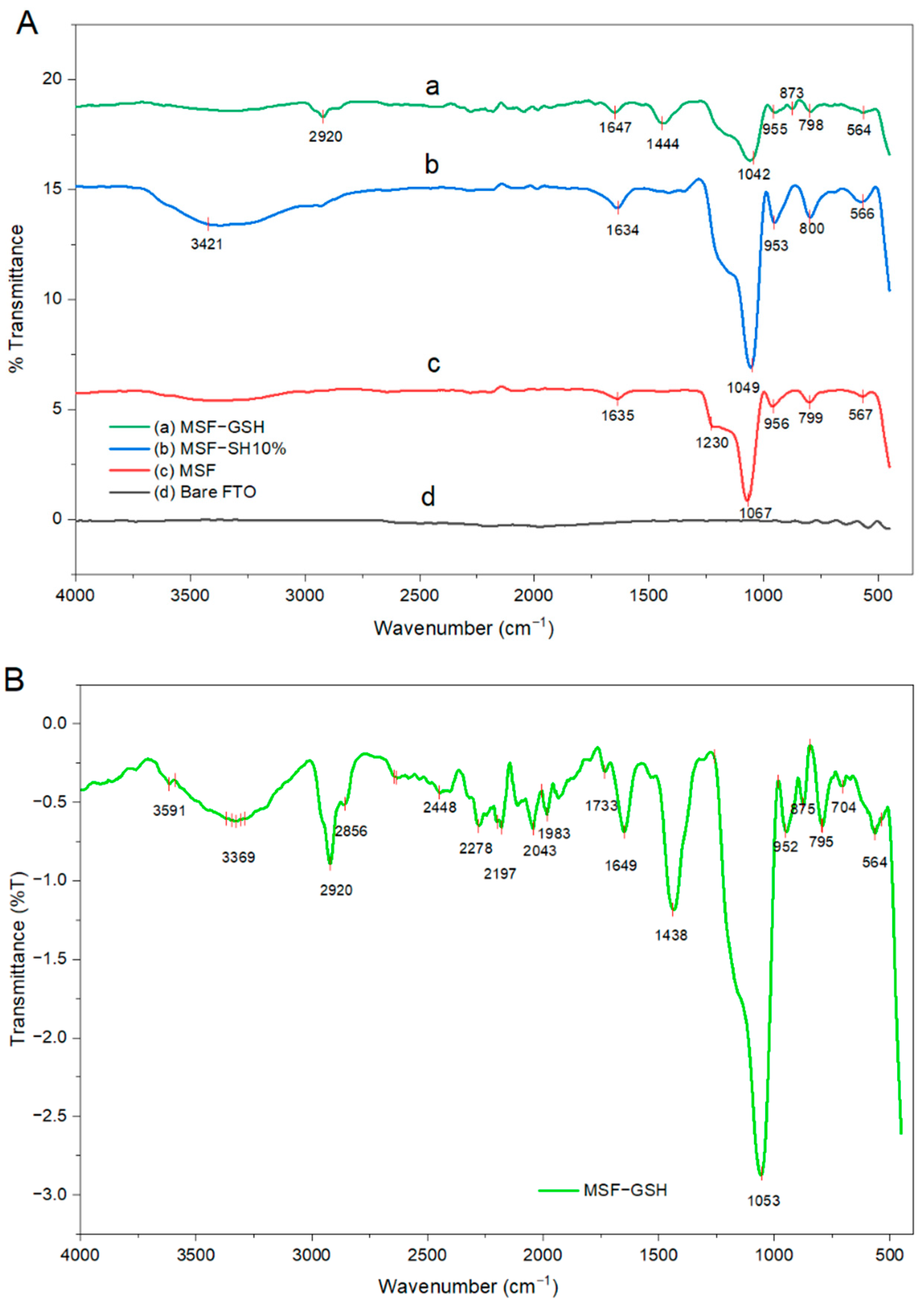
Figure 4 combines the spectra of bare FTO, MSF, MSF-SH, MSF-GSH, and free GSH to permit direct comparison. The prominent band at approximately 1042–1067 cm−1 is assigned to asymmetric Si–O–Si stretching and supports formation of the silica framework [34], while the feature near 955 cm−1 is associated with Si–OH. Relative to MSF and MSF-SH, MSF-GSH exhibited additional organic-associated features. The band near 2920 cm−1 is attributable to aliphatic C–H stretching; the feature near 1444 cm−1 may contain contributions from C–H bending and symmetric carboxylate stretching; and the bands near 1649 and 1733 cm−1 are consistent with amide/carboxylate and protonated carboxylic-acid carbonyl vibrations, respectively. These features support the presence of GSH-associated functional groups but do not, by themselves, establish their bonding configuration.
Figure 4.
FTIR spectra of the mesoporous silica films and modified electrodes. (A) Comparative spectra of MSF−GSH/FTO (a), MSF−SH/FTO (b), MSF/FTO (c), and bare FTO (d). (B) Enlarged FTIR spectrum of MSF-GSH/FTO showing the principal absorption bands associated with the presence of glutathione in the modified mesoporous silica film.
A weak band was observed at approximately 2448 cm−1 for MSF-GSH (Figure 4B), compared with the S–H stretching band of free glutathione at approximately 2519 cm−1 (Figure A4). This difference may reflect changes in the local chemical environment of the thiol groups following immobilization. However, because S–H absorption bands are generally weak, the observed shift alone cannot establish the formation of a specific chemical bond between glutathione and MSF-SH. The band at approximately 704 cm−1 may tentatively be assigned to C–S stretching vibrations, further indicating the presence of sulfur-containing organic groups in the modified film. Overall, the appearance of the C–H, amide, carboxyl, and sulfur-associated bands in the spectrum of MSF-GSH supports the retention of glutathione within or on the thiol-functionalized mesoporous silica film. Glutathione may be retained through noncovalent interactions, including hydrogen bonding with surface silanol groups and confinement within the mesoporous channels. Nevertheless, FTIR analysis alone does not establish the exact immobilization mechanism or confirm covalent attachment of glutathione to MSF-SH.
3.4. Effect of Solution pH on the Electrochemical Detection of Hg(II)
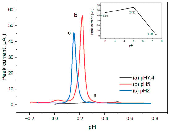
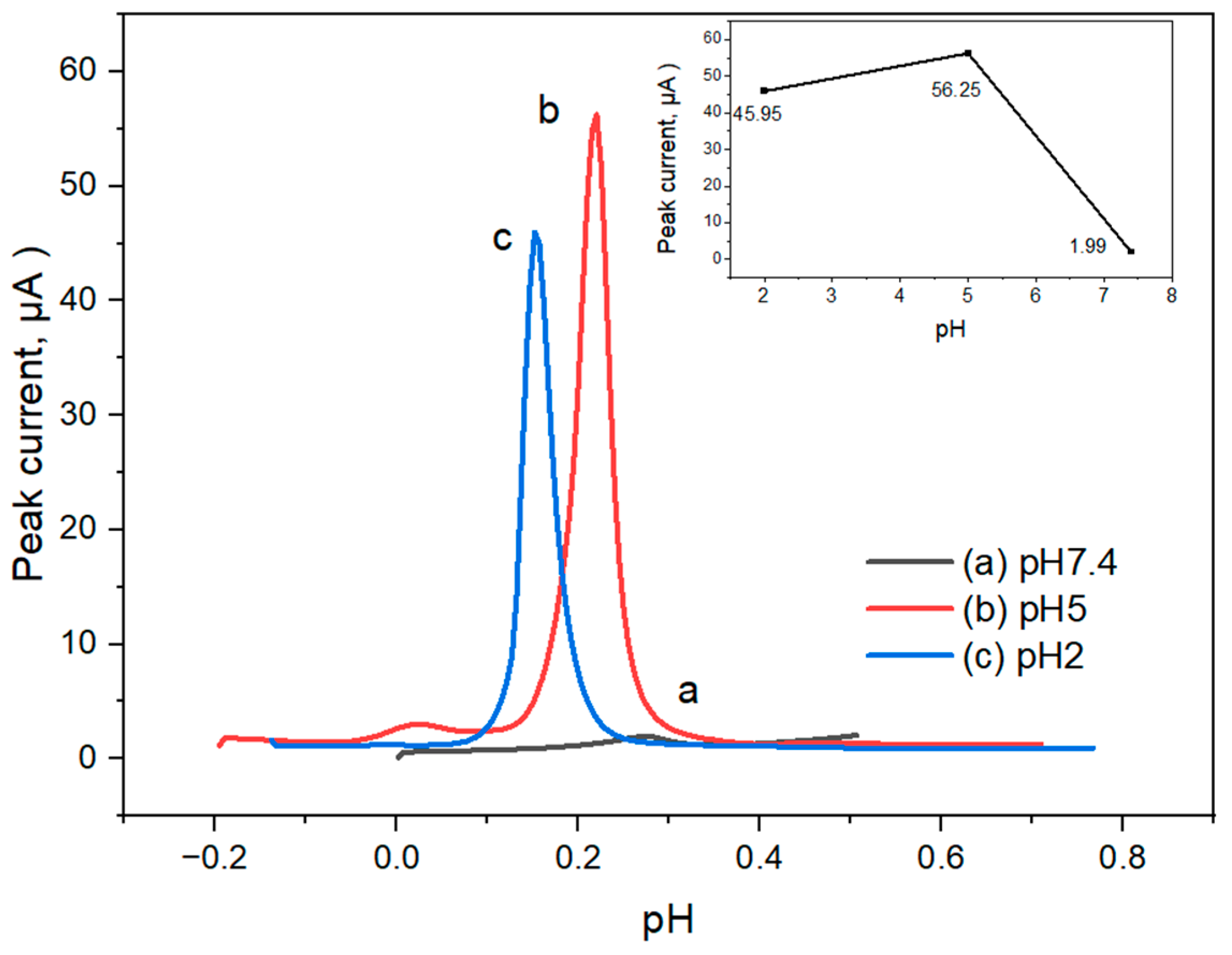
The influence of solution pH on the differential pulse voltammetric response of Hg(II) at the MSF-GSH/FTO electrode was investigated in phosphate-containing electrolyte solutions at pH 2.0, 5.0 and 7.4. The Hg(II) peak current increased significantly with increasing pH from 2.0 to 5.0 and then decreased at pH 7.4 (Figure 5). The results indicate that the pH of solution has an important effect on the accumulation and electrochemical detection of Hg(II) at the modified electrode. The highest DPV peak current was obtained at pH 5.0 among the studied conditions.
Figure 5.
Effect of solution pH on the differential pulse voltammetric response of 100 µM Hg(II) at MSF-GSH/FTO. Measurements were performed in phosphate-containing electrolyte at a deposition potential of −0.70 V and a constant deposition time of 5 min. Main panel: voltammograms at pH 2.0, 5.0, and 7.4. Inset: corresponding Hg(II) peak current as a function of pH.
The low response at pH 2.0 may be associated with protonation of film functional groups and competition from the high H+ concentration. Because the point of zero charge of silica is near pH 2, the silanol-bearing surface is expected to be nearly neutral or weakly charged in this strongly acidic region. The carboxyl groups of retained GSH are also predominantly protonated, which may reduce the availability of coordination sites. Hydrogen evolution at the applied deposition potential of −0.70 V may additionally affect the response. At pH 5.0, partial deprotonation of carboxyl and silanol groups increases the negative character of the interface and decreases proton competition. Hg(II)–sulfur coordination may also contribute to analyte retention. Thus, the maximum response at pH 5.0 is interpreted as an experimental compromise among functional-group ionization, Hg(II) availability, transport, and electrochemical deposition rather than as proof of a single mechanism.
The decrease in peak current at pH 7.4 may be related to the mercury speciation changes and electrochemical availability. At higher pH, Hg(II) can hydrolyze and form species like HgOH+ and Hg(OH)2. These species may carry different charges, mobilities and electrochemical behavior from free hydrated Hg2+ ions. Interactions of Hg(II) with phosphate species may also decrease the concentration of electrochemically available Hg(II). Therefore, although the MSF-GSH film should be more negatively charged at pH 7.4, the amount of Hg(II) available for accumulation and voltammetric detection can be reduced. Of the three pH values tested, pH 5.0 showed the highest DPV peak current, and the electrochemical determination of Hg(II) was subsequently carried out at this pH.
3.5. Comparison of the Voltammetric Responses of Hg(II) at Different Electrodes
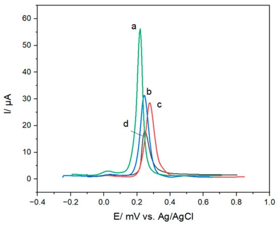
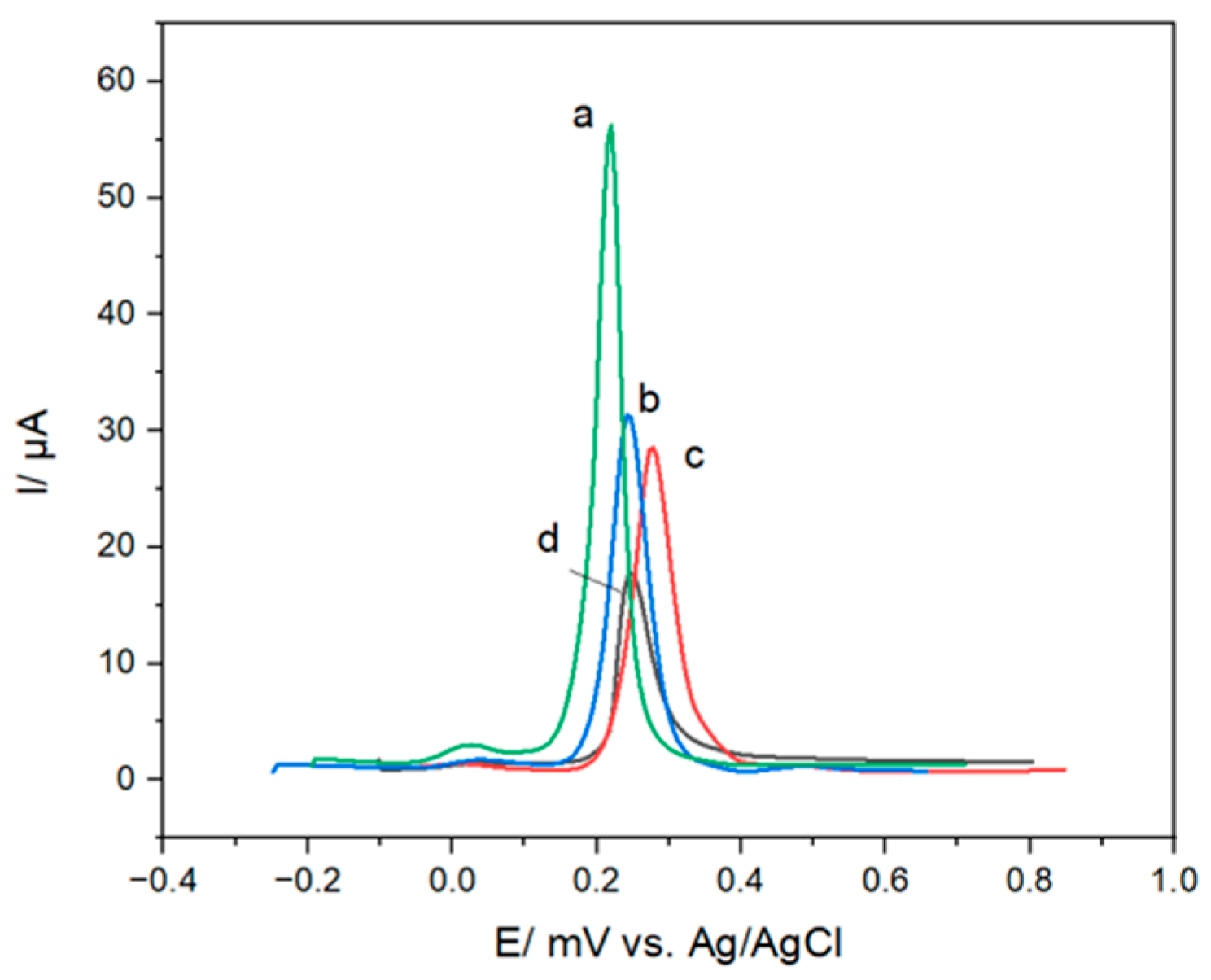
Figure 6 compares the differential pulse voltammetric responses of bare FTO, MSF/FTO, MSF-SH/FTO containing 10 mol% MPTMS, and MSF-GSH/FTO in pH 5.0 phosphate buffer containing 100 µM Hg(II), using a 5 min deposition time. Bare FTO, MSF/FTO, MSF-SH/FTO, and MSF-GSH/FTO produced peak currents of 15.96, 27.27, 30.21, and 53.91 µA, respectively. The increased response after film modification is consistent with enhanced Hg(II) accumulation or transport at the modified interface. The response of MSF-SH/FTO is consistent with a contribution from mercaptopropyl groups, while the larger response at MSF-GSH/FTO supports an additional effect of retained GSH-associated functionalities. These comparisons demonstrate enhanced voltammetric response but do not independently quantify binding affinity or establish the precise accumulation mechanism.
Figure 6.
Differential pulse voltammograms recorded at (a) MSF-GSH/FTO, (b) MSF-SH/FTO, (c) MSF/FTO, and (d) bare FTO in pH 5.0 phosphate buffer containing 100 µM Hg(II). Deposition time: 5 min; pulse amplitude: 50 mV.
This enhancement is primarily attributed to the strong affinity of Hg(II) for sulfur-containing groups in glutathione. The carboxyl and amino functionalities of GSH may also contribute to the local coordination environment and retention of Hg(II) within the modified film. The mesoporous structure facilitates analyte transport, whereas the GSH-containing functional groups promote Hg(II) accumulation near the electrode interface, resulting in a higher DPV peak current. These findings demonstrate that the combined effects of mesoporous transport and GSH-associated binding improve the voltammetric response of the MSF-GSH/FTO electrode toward Hg(II).
3.6. Electrochemical Detection of Hg(II) Using the MSF-GSH/FTO Electrode
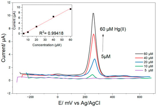
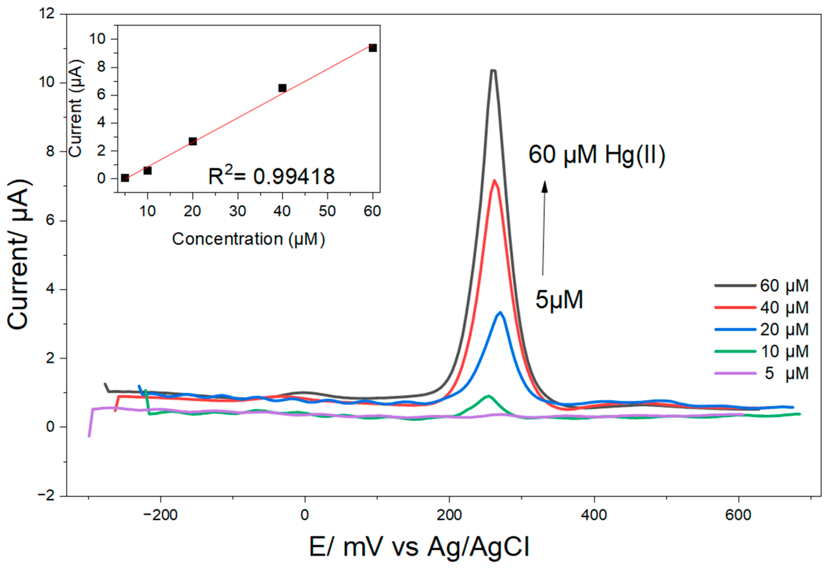
The electrochemical response of the MSF-GSH/FTO electrode toward Hg(II) was evaluated by differential pulse anodic stripping voltammetry. Hg(II) was deposited at −0.70 V for 5 min before the anodic scan. As shown in Figure 7, peak current increased with concentration from 5 to 60 µM. The calibration relationship was Ip (µA) = 0.17501 CHg(II) (µM)—0.86898, with R2 = 0.99418. The slope of 0.17501 µA µM−1 represents the sensitivity under the selected conditions. The estimated LOD was 4.27 µM, calculated as LOD = 3.3σ/m, where σ = 0.22641 µA was the standard error of the calibration intercept and m = 0.17501 µA µM−1 was the calibration slope. This value was derived from a single calibration regression rather than replicate blank measurements and should therefore be regarded as an estimated calibration-based LOD, not a validated method detection limit. Because independent replicate calibrations were not performed, error bars and between-electrode precision statistics cannot be reported.
Figure 7.
Differential pulse anodic stripping voltammograms at MSF-GSH/FTO for increasing Hg(II) concentrations in pH 5.0 phosphate buffer. Hg(II) was deposited at −0.70 V for 5 min before the anodic scan. Inset: calibration plot from 5 to 60 µM.
3.7. Interference Study for the Electrochemical Detection of Hg(II) Using MSF-GSH/FTO
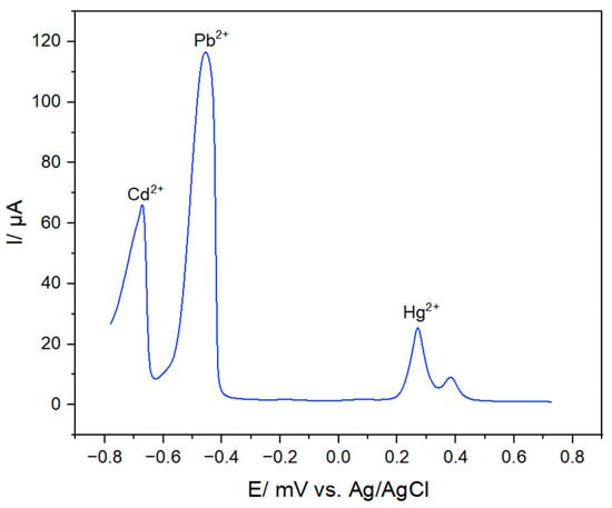
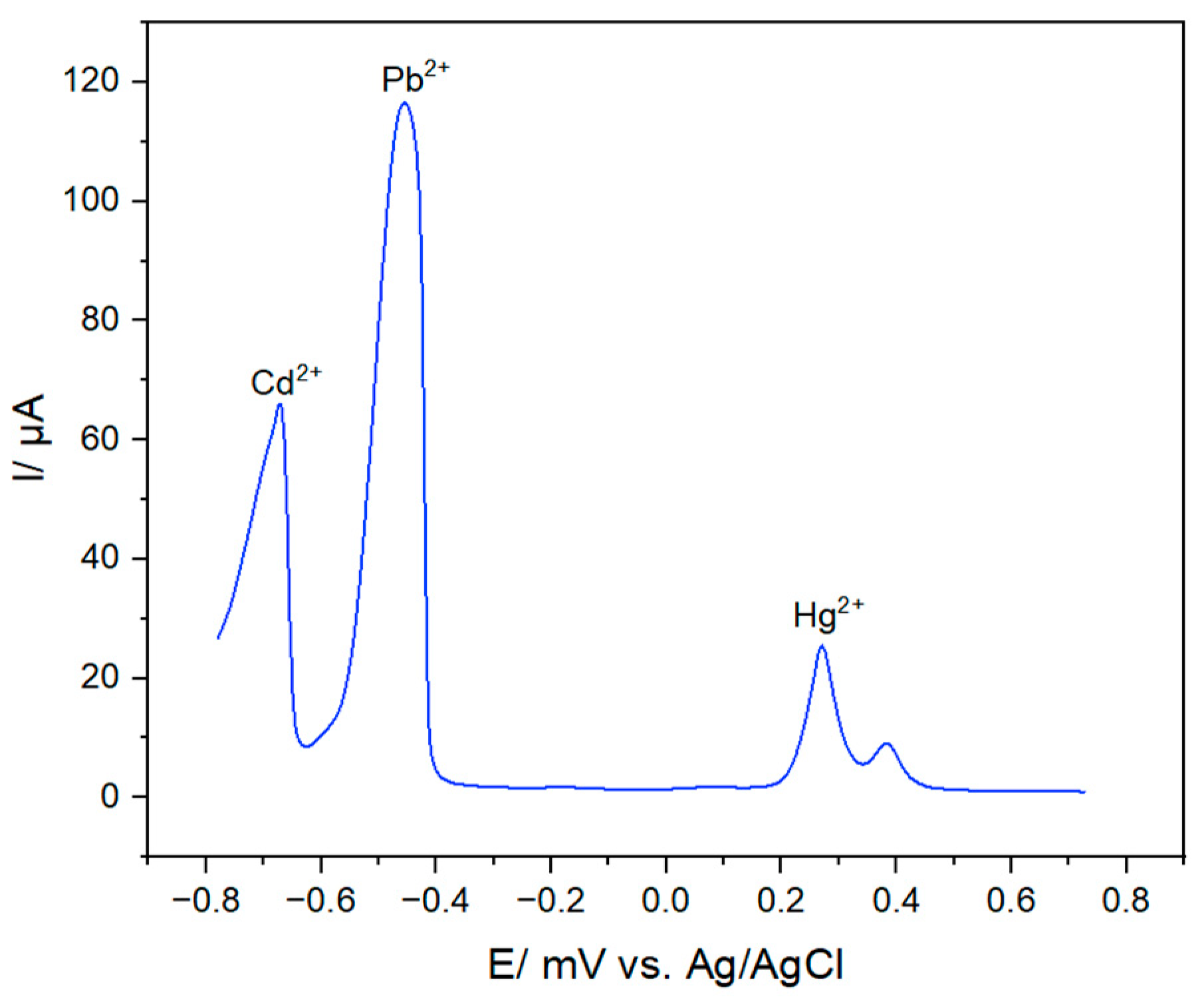
Pb(II) and Cd(II), which may coexist with Hg(II) in environmental samples, were selected as potential interfering metal ions to evaluate the selectivity of the MSF-GSH/FTO electrode. Their influence on the anodic stripping response of Hg(II) was examined under the selected experimental conditions using a mixed solution containing 100 µM each of Cd(II), Pb(II), and Hg(II). As shown in Figure 8, the differential pulse anodic stripping voltammogram exhibited three clearly resolved oxidation peaks at approximately −0.669, −0.454, and 0.271 V, which were assigned to Cd, Pb, and Hg, respectively. The distinct separation of the peak potentials allowed the three metal ions to be electrochemically distinguished in the mixed solution.
Figure 8.
Differential pulse anodic stripping voltammogram for a mixed solution containing 100 µM each of Cd(II), Pb(II), and Hg(II), recorded at MSF-GSH/FTO in pH 5.0 phosphate buffer after deposition at −0.70 V for 5 min.
The Hg peak remained identifiable in the presence of equimolar Cd(II) and Pb(II), demonstrating peak separation under this single mixed-metal condition. Because replicate Hg-only and mixed-metal measurements were not performed, the experiment does not establish the percentage signal change or comprehensive selectivity. Cu(II), Zn(II), Fe(III), Ni(II), other common ions, and complex sample components were not evaluated. Broader interference testing, individual calibration, and recovery studies are required before quantitative simultaneous determination can be claimed.
The present study has several limitations. Film morphology, thickness, nanochannel orientation, elemental distribution, surface chemical states, and interfacial impedance were not directly characterized. FTIR supports the retention of GSH-associated functional groups but does not establish the exact immobilization mechanism. Only solution pH was systematically varied, and the calibration was based on single measurements without independent electrode replicates. Storage stability, operational reuse, expanded interference, recovery, and real-water performance were not evaluated. Moreover, the estimated LOD remains higher than those of many advanced Hg(II) sensors and above drinking-water guideline concentrations. The results should therefore be interpreted as proof of concept for the MSF-GSH/FTO interface rather than validation of a field-ready analytical method.
4. Conclusions and Future Outlook
A glutathione-modified thiol-functionalized mesoporous silica film was prepared on FTO by EASA and evaluated as a proof-of-concept interface for Hg(II) detection. Among the three investigated compositions, the film containing 10 mol% MPTMS produced the highest redox-probe response and was selected for GSH immobilization. FTIR supported silica-network formation and the retention of GSH-associated functional groups, while voltammetry showed an enhanced Hg(II) response at MSF-GSH/FTO. Under the selected conditions, the response was linear from 5 to 60 µM, with an estimated calibration-based LOD of 4.27 µM. A single mixed-metal experiment produced resolved Cd, Pb, and Hg peaks, demonstrating peak separation but not comprehensive selectivity.
Future work should include SEM/TEM, AFM or profilometry, XPS, elemental mapping, and EIS; systematic optimization of deposition and pulse parameters; independently replicated calibration and electrode fabrication; validated LOD and LOQ determination; stability and regeneration studies; broader interference testing; recovery experiments; and analysis of authentic environmental samples.
Author Contributions
Conceptualization, D.A.B.-M.; methodology, D.A.B.-M. and M.C.C.; investigation, R.M.M.; resources, D.A.B.-M. and R.C.; data curation, R.M.M.; writing—original draft preparation, D.A.B.-M.; writing—review and editing, D.A.B.-M., R.M.M., M.C.C. and R.C.; visualization, D.A.B.-M.; supervision, M.C.C.; project administration, D.A.B.-M.; funding acquisition, D.A.B.-M. All authors have read and agreed to the published version of the manuscript.
Funding
This research was funded by DOST-PCIEERD (Philippine Council for Industry, Energy, and Emerging Technology Research and Development), PMIS 1211471.
Data Availability Statement
The data supporting the findings of this study are available within the article. Additional raw experimental data are available from the corresponding author upon reasonable request.
Acknowledgments
The authors gratefully acknowledge the Philippine Council for Industry, Energy and Emerging Technology Research and Development (DOST-PCIEERD) for the financial support provided for this study. The authors also thank the CSU Chemistry Department for its assistance and the MAE Group for their valuable administrative, technical, and laboratory support. During the preparation of this manuscript, the authors used ChatGPT (GPT-6.1 Sol, OpenAI) to assist in refining the language and readability of selected sections. The authors reviewed and edited all generated outputs and take full responsibility for the content of this publication.
Conflicts of Interest
The authors declare no conflicts of interest. The funders had no role in the design of the study; in the collection, analyses, or interpretation of data; in the writing of the manuscript; or in the decision to publish the results.
Abbreviations
The following abbreviations are used in this manuscript:
| Abbreviation | Definition |
| CTAB | Cetyltrimethylammonium bromide |
| CV | Cyclic voltammetry |
| DPV | Differential pulse voltammetry |
| EASA | Electrochemically assisted self-assembly |
| FTO | Fluorine-doped tin oxide |
| FTIR | Fourier-transform infrared spectroscopy |
| GSH | Glutathione |
| Hg(II) | Mercury(II) ion |
| MPTMS | (3-Mercaptopropyl)trimethoxysilane |
| MSF | Mesoporous silica film |
| MSF-GSH | Glutathione-functionalized mesoporous silica film |
| MSF-SH | Thiol-functionalized mesoporous silica film |
| TEOS | Tetraethyl orthosilicate |
Appendix A
Appendix A.1
Figure A1.
CV Characterization of Bare FTO as reference electrode (a), Mesoporous Silica Film (MSF) with template CTAB (b), and Mesoporous Silica Film (MSF) (c) using 5 mM/0.10 M NaNO3 hexaammineruthenium(III) (A) and ferricyanide (B).
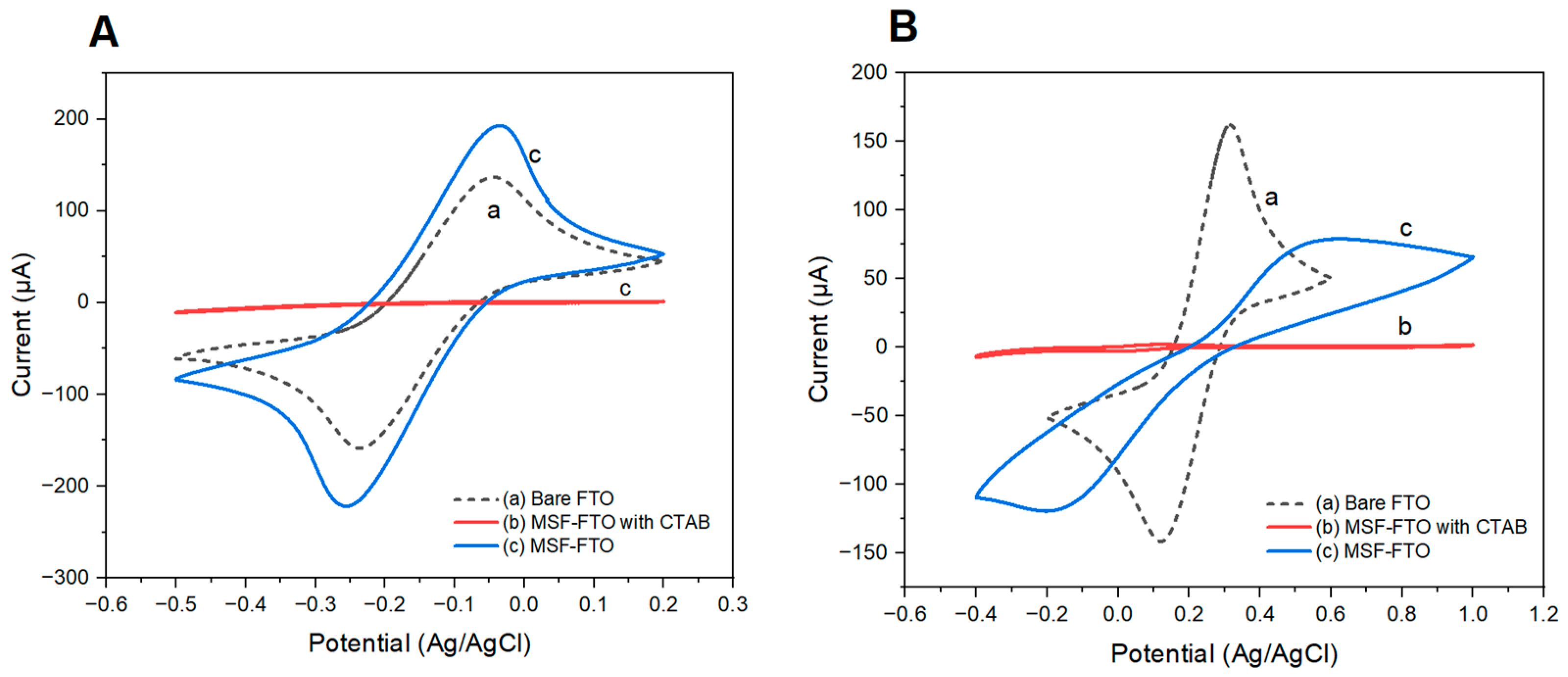
The prepared mesoporous silica film was characterized by cyclic voltammetry using the different redox probes ruthenium hexamine and methylene blue for the cationic probes and ferricyanide for the negatively charged redox probes. The plain MSF showed the expected charge-selective behavior of CTAB-templated mesoporous silica films. Before template extraction, the film strongly attenuated the response of charged probes, indicating effective blocking by the surfactant-filled mesopores. After template extraction, the cationic probe showed enhanced accessibility due to electrostatic attraction with negatively charged silanol/siloxide sites, whereas the anionic probe remained largely excluded because of electrostatic repulsion. The accessibility of the mesoporous electrode (MSF) is made as a reference for the thiol-functionalized mesoporous silica.
Appendix A.2
Figure A2.
CV Characterization of Bare FTO as reference electrode (a), Thiol-functionalized Mesoporous Silica Film (MSF-5) with template CTAB using 5% MPTMS precursor (b), and Thiol-functionalized Mesoporous Silica Film (MSF-SH5) using 5% MPTMS precursor (c) in 5 mM/0.10 M NaNO3 hexaammineruthenium(III) (A) and ferricyanide (B).
Appendix A.3
Figure A3.
CV Characterization of Bare FTO as reference electrode (a), Thiol-functionalized Mesoporous Silica Film (MSF-SH15% with CTAB) using 15% MPTMS precursor (b), and Thiol-functionalized Mesoporous Silica Film (MSF-SH15%) using 15% MPTMS precursor (c) in 5 mM/0.10 M NaNO3 hexaammineruthenium(III) (A) and ferricyanide (B).
Appendix A.4
Figure A4.
FTIR spectrum of Glutathione.
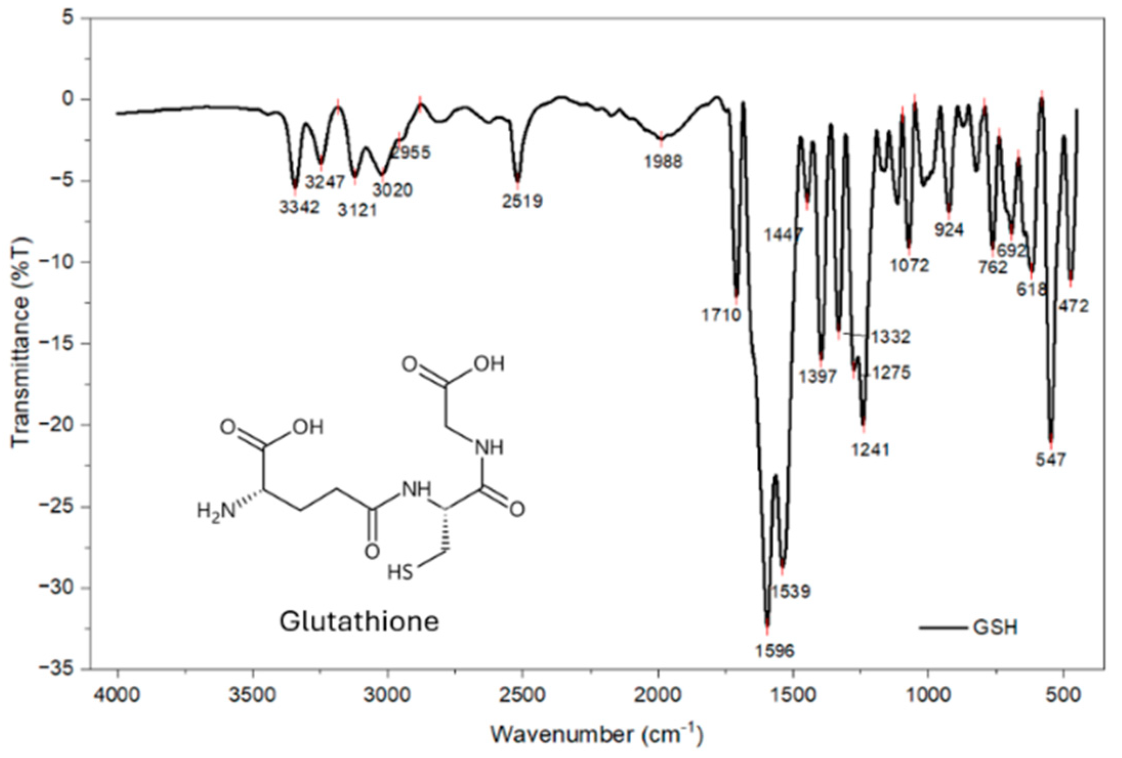
Glutathione contains one thiol group, one primary amine group, two carboxylic acid groups, and two amide groups. The FTIR spectrum of glutathione powder, obtained using the potassium bromide pellet method, exhibited prominent absorption bands at 3342, 3247, and 3121 cm−1, which were attributed to O–H and N–H stretching vibrations. The bands observed at 3020 and 2955 cm−1 were assigned to C–H stretching vibrations. A weak characteristic band at 2519 cm−1 corresponded to the S–H stretching vibration of the thiol group. The absorption band at 1710 cm−1 was attributed to the C=O stretching vibration of the carboxylic acid groups, whereas the bands at 1596 and 1539 cm−1 were associated with amide-related C=O stretching and N–H bending vibrations. The band at 1395 cm−1 was assigned to the symmetric stretching vibration of the carboxylate group, while the peak at 1241 cm−1 corresponded to C–N stretching vibrations [35,36].
References
- Espiritu, E.Q.; Claveria, R.J.R.; Bernadas, P.J.C. Assessment of surface water quality and mercury levels from Artisanal and small-scale gold mining (ASGM) along Acupan River, Benguet, Philippines. Environ. Geochem. Health 2022, 44, 3655–3676. [Google Scholar] [CrossRef] [Scilit] [PubMed]
- Logrosa, G. Integration of Health and Safety Risk Management into the Implementation of Gold Processing Technology in Artisanal and Small Scale Mining (ASM) Communities in the Philippines. Ph.D. Thesis, The University of Queensland, Brisbane, Australian, 2018. [Google Scholar]
- Murao, S.; Tomiyasu, T.; Ono, K.; Shibata, H.; Narisawa, N.; Takenaka, C. Mercury Distribution in Artisanal and Small-Scale Gold Mining Area: A Case Study of Hot Spots in Camarines Norte, Philippines. IJESD 2019, 10, 122–129. [Google Scholar] [CrossRef] [Scilit][Green Version]
- Appel, P.W.U.; Na-Oy, L. The Borax Method of Gold Extraction for Small-Scale Miners. J. Health Pollut. 2012, 2, 5–10. [Google Scholar] [CrossRef] [Scilit]
- Eisler, R. Health Risks of Gold Miners: A Synoptic Review. Environ. Geochem. Health 2003, 25, 325–345. [Google Scholar] [CrossRef] [Scilit] [PubMed]
- Soe, P.S.; Kyaw, W.T.; Arizono, K.; Ishibashi, Y.; Agusa, T. Mercury Pollution from Artisanal and Small-Scale Gold Mining in Myanmar and Other Southeast Asian Countries. Int. J. Environ. Res. Public Health 2022, 19, 6290. [Google Scholar] [CrossRef] [Scilit] [PubMed]
- Clarkson, T.W.; Magos, L. The Toxicology of Mercury and Its Chemical Compounds. Crit. Rev. Toxicol. 2006, 36, 609–662. [Google Scholar] [CrossRef] [Scilit] [PubMed]
- Martinez-Finley, E.J.; Chakraborty, S.; Fretham, S.J.B.; Aschner, M. Cellular transport and homeostasis of essential and nonessential metals. Metallomics 2012, 4, 593–605. [Google Scholar] [CrossRef] [Scilit] [PubMed]
- Tchounwou, P.B.; Ayensu, W.K.; Ninashvili, N.; Sutton, D. Review: Environmental exposure to mercury and its toxicopathologic implications for public health. Environ. Toxicol. 2003, 18, 149–175. [Google Scholar] [CrossRef] [Scilit] [PubMed]
- Azevedo, L.; Karpova, N.; Rocha, B.; Barbosa Junior, F.; Gobe, G.; Hornos Carneiro, M. Evidence on Neurotoxicity after Intrauterine and Childhood Exposure to Organomercurials. Int. J. Environ. Res. Public Health 2023, 20, 1070. [Google Scholar] [CrossRef] [Scilit] [PubMed]
- Castoldi, A.F.; Coccini, T.; Manzo, L. Neurotoxic and Molecular Effects of Methylmercury in Humans. Rev. Environ. Health 2003, 18, 19–32. [Google Scholar] [CrossRef] [Scilit] [PubMed]
- Kang, B.; Wang, J.; Guo, S.; Yang, L. Mercury-induced toxicity: Mechanisms, molecular pathways, and gene regulation. Sci. Total Environ. 2024, 943, 173577. [Google Scholar] [CrossRef] [Scilit] [PubMed]
- Akagi, H.; Castillo, E.S.; Cortes-Maramba, N.; Francisco-Rivera, A.T.; Timbang, T.D. Health assessment for mercury exposure among schoolchildren residing near a gold processing and refining plant in Apokon, Tagum, Davao del Norte, Philippines. Sci. Total Environ. 2000, 259, 31–43. [Google Scholar] [CrossRef] [Scilit] [PubMed]
- Abdelmonem, B.H.; Kamal, L.T.; Elbaz, R.M.; Khalifa, M.R.; Abdelnaser, A. From contamination to detection: The growing threat of heavy metals. Heliyon 2025, 11, e41713. [Google Scholar] [CrossRef] [Scilit] [PubMed]
- García-Mesa, J.C.; Montoro-Leal, P.; Maireles-Rivas, S.; López Guerrero, M.M.; Vereda Alonso, E. Sensitive determination of mercury by magnetic dispersive solid-phase extraction combined with flow-injection-cold vapour-graphite furnace atomic absorption spectrometry. J. Anal. At. Spectrom. 2021, 36, 892–899. [Google Scholar] [CrossRef] [Scilit]
- Kowa, E.; Telk, A.; Wieczorek, M. Flow techniques in the analysis of biological samples by inductively coupled plasma mass spectrometry—A review. J. Anal. At. Spectrom. 2024, 39, 1004–1023. [Google Scholar] [CrossRef] [Scilit]
- Narduzzi, F.; Covelli, S.; Floreani, F.; Pavoni, E.; Petranich, E.; Jantzi, S.C.; Pistone, M.; Černok, A.; Venier, M.; Crosera, M.; et al. Strengths and Weaknesses of the Analytical Techniques Used for Measuring Low Mercury Mass Fractions (<10 ng g−1) in Crystalline Rocks: Direct Mercury Analyser Versus Cold Vapour-Atomic Fluorescence Spectroscopy. Geostand. Geoanalytic Res. 2025, 49, 295–314. [Google Scholar] [CrossRef] [Scilit]
- Provete, C.S.; Dalfior, B.M.; Mantovaneli, R.; Carneiro, M.T.W.D.; Brandão, G.P. Comparison of the Performance of ICP-MS, CV-ICP-OES, and TDA AAS in Determining Mercury in Marine Sediment Samples. ACS Omega 2024, 9, 49229–49238. [Google Scholar] [CrossRef] [Scilit] [PubMed]
- Bulska, E.; Krata, A.; Kałabun, M.; Wojciechowski, M. On the use of certified reference materials for assuring the quality of results for the determination of mercury in environmental samples. Environ. Sci. Pollut. Res. 2017, 24, 7889–7897. [Google Scholar] [CrossRef] [Scilit] [PubMed]
- Díaz-González, M.; Gutiérrez-Capitán, M.; Niu, P.; Baldi, A.; Jiménez-Jorquera, C.; Fernández-Sánchez, C. Electrochemical devices for the detection of priority pollutants listed in the EU water framework directive. TrAC Trends Anal. Chem. 2016, 77, 186–202. [Google Scholar] [CrossRef] [Scilit]
- Sulthana, S.F.; Iqbal, U.M.; Suseela, S.B.; Anbazhagan, R.; Chinthaginjala, R.; Chitathuru, D.; Ahmad, I.; Kim, T. Electrochemical Sensors for Heavy Metal Ion Detection in Aqueous Medium: A Systematic Review. ACS Omega 2024, 9, 25493–25512. [Google Scholar] [CrossRef] [Scilit] [PubMed]
- Hara, T.O.; Singh, B. Electrochemical Biosensors for Detection of Pesticides and Heavy Metal Toxicants in Water: Recent Trends and Progress. ACS EST Water 2021, 1, 462–478. [Google Scholar] [CrossRef] [Scilit]
- World Health Organization (Ed.) Guidelines for Drinking-Water Quality: Fourth Edition Incorporating the First and Second Addenda, Fourth edition incorporating the first and Second addenda; World Health Organization: Geneva, Switzerland, 2022; pp. 425–426. [Google Scholar]
- United States Environmental Protection Agency. What EPA is Doing to Reduce Mercury Pollution, and Exposures to Mercury; US EPA, United States Environmental Protection Agency: Washington, DC, USA, 2026. Available online: https://www.epa.gov/mercury/what-epa-doing-reduce-mercury-pollution-and-exposures-mercury (accessed on 1 September 2026).
- Herzog, G.; Sibottier, E.; Etienne, M.; Walcarius, A. Electrochemically assisted self-assembly of ordered and functionalized mesoporous silica films: Impact of the electrode geometry and size on film formation and properties. Faraday Discuss. 2013, 164, 259–273. [Google Scholar] [CrossRef] [Scilit] [PubMed]
- Walcarius, A. Electroinduced Surfactant Self-Assembly Driven to Vertical Growth of Oriented Mesoporous Films. Acc. Chem. Res. 2021, 54, 3563–3575. [Google Scholar] [CrossRef] [Scilit] [PubMed]
- Yan, F.; Lin, X.; Su, B. Vertically ordered silica mesochannel films: Electrochemistry and analytical applications. Analyst 2016, 141, 3482–3495. [Google Scholar] [CrossRef] [Scilit] [PubMed]
- Basnig, D.; Vilá, N.; Herzog, G.; Walcarius, A. Voltammetric behaviour of cationic redox probes at mesoporous silica film electrodes. J. Electroanal. Chem. 2020, 872, 113993. [Google Scholar] [CrossRef] [Scilit]
- Wang, W.; Lu, Y.-C.; Huang, H.; Wang, A.-J.; Chen, J.-R.; Feng, J.-J. Solvent-free synthesis of sulfur- and nitrogen-co-doped fluorescent carbon nanoparticles from glutathione for highly selective and sensitive detection of mercury(II) ions. Sens. Actuators B Chem. 2014, 202, 741–747. [Google Scholar] [CrossRef] [Scilit]
- Yang, Z.; Lyu, J.; Qian, J.; Wang, Y.; Liu, Z.; Yao, Q.; Chen, T.; Cao, Y.; Xie, J. Glutathione: A naturally occurring tripeptide for functional metal nanomaterials. Chem. Sci. 2025, 16, 6542–6572. [Google Scholar] [CrossRef] [Scilit] [PubMed]
- Chaabane, L.; Nikolantonaki, M.; Weber, G.; Bezverkhyy, I.; Chassagnon, R.; Assifaoui, A.; Bouyer, F. Functionalization of SBA-15 mesoporous silica for highly efficient adsorption of glutathione: Characterization and modeling studies. J. Taiwan Inst. Chem. Eng. 2023, 152, 105169. [Google Scholar] [CrossRef] [Scilit]
- Kaabi, R.; Abderrabba, M.; Gómez-Ruiz, S.; Del Hierro, I. Bioinspired materials based on glutathione-functionalized SBA-15 for electrochemical Cd(II) detection. Microporous Mesoporous Mater. 2016, 234, 336–346. [Google Scholar] [CrossRef] [Scilit]
- Tian, Y.; Tang, R.; Liu, L.; Yu, Y.; Ma, S.; Gong, B.; Ou, J. Glutathione-modified ordered mesoporous silicas for enrichment of N-linked glycopeptides by hydrophilic interaction chromatography. Talanta 2020, 217, 121082. [Google Scholar] [CrossRef] [Scilit] [PubMed]
- Alahmadi, S.M.; Mohamad, S.; Maah, M.J. Synthesis and Characterization of Mesoporous Silica Functionalized with Calix[4]arene Derivatives. Int. J. Mol. Sci. 2012, 13, 13726–13736. [Google Scholar] [CrossRef] [Scilit] [PubMed]
- Basu, S.; Panigrahi, S.; Praharaj, S.; Kumar Ghosh, S.; Pande, S.; Jana, S.; Pal, T. Dipole–dipole plasmon interactions in self-assembly of gold organosol induced by glutathione. New J. Chem. 2006, 30, 1333–1339. [Google Scholar] [CrossRef] [Scilit]
- Qi, W.; Tian, Y.; Lu, D.; Chen, B. Detection of glutathione in dairy products based on surface-enhanced infrared absorption spectroscopy of silver nanoparticles. Front. Nutr. 2022, 9, 982228. [Google Scholar] [CrossRef] [Scilit] [PubMed]
Disclaimer/Publisher’s Note: The statements, opinions and data contained in all publications are solely those of the individual author(s) and contributor(s) and not of MDPI and/or the editor(s). MDPI and/or the editor(s) disclaim responsibility for any injury to people or property resulting from any ideas, methods, instructions or products referred to in the content. |
© 2026 by the authors. Licensee MDPI, Basel, Switzerland. This article is an open access article distributed under the terms and conditions of the Creative Commons Attribution (CC BY) license.