


Abstract
In many aerobic and anaerobic bacteria, degradation of chemically inert ketones such as acetone or acetophenone is initiated by a nucleotide triphosphate-dependent carboxylation reaction. Most previously studied acetone carboxylases were described as hexameric enzymes consisting of three different subunits. In contrast, in the homologous acetophenone carboxylase one of these subunits is duplicated. This results in a heterooctameric core complex, which additionally requires a detachable fifth subunit E for activity. A recent study identified an apparent acetophenone carboxylase gene cluster in the strictly aerobic actinobacterium Rhodococcus aetherivorans and showed that the encoded enzyme (termed Acr) unexpectedly catalyzes acetone rather than acetophenone carboxylation. Here, we purified this novel type of acetone carboxylase and characterized its biochemical properties. Mass photometry with the purified enzyme confirmed the formation of a heterooctameric, acetophenone carboxylase-like core complex. Although not visible in the mass photometry spectrum, mass spectrometric analysis revealed that a small amount of subunit E was co-purified with the core complex. The purified enzyme was active with acetone and additionally carboxylated an exceptionally broad range of other aliphatic ketones. Moreover, unexpectedly and in contrast to the previously studied acetone carboxylases, the enzyme was active with any of the five different nucleotide triphosphates tested. Also with regard to its other biochemical properties, Acr resembled acetophenone carboxylase rather than other acetone carboxylases. For instance, Acr formed nucleotide diphosphates during hydrolysis and contained Zn2+ as the sole catalytic metal. Putative applications of Acr include implementation in synthetic pathways and heterologous production in biotechnologically relevant strains of Rhodococcus spp., e.g., for bioremediation purposes.
1. Introduction
Microbial activation of chemically inert substrates such as ketones is commonly accomplished by oxygenation or carboxylation reactions. While oxygenations can only be employed under aerobic conditions, carboxylation is an important functionalization strategy in both aerobes and (facultative) anaerobes. Intriguingly, carboxylases evolved independently several times for the activation of multiple substrates [1,2], e.g., phenols [3], epoxides [4,5,6], 4-hydroxyacetophenone [7], acetophenone [8], and acetone [9,10,11,12,13,14]. These carboxylases belong to different protein families and exhibit distinct reaction mechanisms.
Acetone and acetophenone carboxylases are homologous enzymes affiliated with the hydantoinase/oxoprolinase family [2,8,9,10,11,12,13,14]. Apart from these carboxylases, this protein family also includes different hydrolases such as 5-oxoprolinases in mammals [15] or hydantoinases [16] and caprolactamases in bacteria [17]. The catalyzed reactions involve phosphorylation of the substrate’s ketone group and/or the cosubstrate bicarbonate in the first step and thus require nucleotide triphosphates, in most cases ATP. In the second step of the reaction, the phosphorylated intermediates are either hydrolyzed or carboxylated via condensation with carboxyphosphate [2,17]. A typical feature of the carboxylases from this enzyme class is the involvement of catalytic metal ions such as Zn2+, Mn2+, or Fe2+ instead of organic cofactors [2].
The known hydantoinases and caprolactamases form tetrameric (AB)2 complexes consisting of only two distinct, highly conserved subunits, and 5-oxoprolinases are homodimeric fusion proteins of these two subunits [2,17]. Acetone and acetophenone carboxylases, on the other hand, contain additional subunits. The well-characterized acetone carboxylases from Xanthobacter autotrophicus [9,10,18], Aromatoleum aromaticum [12], Rhodobacter capsulatus [10,11], and Cupriavidus metallidurans [13] form heterohexameric complexes out of two subunits homologous to those of the hydantoinases (AcxAB) and an additional smaller subunit (AcxC). In contrast, acetophenone carboxylase, which has been studied only in A. aromaticum so far, consists of five different subunits. Four of the subunits are homologous to those of Acx, exhibiting an apparent duplication of one of the large subunits. This yields a heterooctameric Apc(ABCD)2 core complex in a similar structural arrangement as the heterohexameric Acx complex, but with an additional superficially attached subunit. Furthermore, acetophenone carboxylase requires a fifth subunit (ApcE), which is lost during purification and does not have a homolog in Acx [8,19]. In a recent study, we proposed that the acetone carboxylase from Rhodococcus aetherivorans represents a novel subtype of acetone carboxylases (Acr), which resembles Apc rather than Acx in its subunit composition. Apparently, Acr also forms a heterooctameric Acr(ABCD)2 core complex with one of the hydantoinase-like subunits being duplicated [20]. Moreover, as described for Apc [8], an additional fifth subunit (AcrE) seems to be indispensable for acetone carboxylation activity, although AcrE does not share detectable sequence identity with ApcE. Phylogenetic analyses showed that Acr mainly occurs in Actinomycetota, including Rhodococcus strains, and in Pseudomonadota [20]. The previously studied acetone carboxylase from Rhodococcus rhodochrous B276 [14] probably is an Acr-type acetone carboxylase, too [20].
The acetone and acetophenone carboxylases described so far differ with regard to their substrate and nucleotide spectra, the nucleotide species formed upon hydrolysis (i.e., di- or monophosphates), uncoupled NTPase activities, and metal contents [2]. Intriguingly, Acr seems to differ from Acx not only in the proposed Apc-like subunit composition, but also in its substrate spectrum, which is not restricted to acetone and 2-butanone, but extends to ketones with longer chain lengths (2- and 3-pentanone, 2-hexanone [14,20]). Therefore, Acr should be regarded as an aliphatic ketone carboxylase rather than as canonical acetone carboxylase. However, many other features of Acr have not been studied in detail yet, neither in R. rhodochrous B276 [14] nor in R. aetherivorans [20]. In this study, we purified the Acr from R. aetherivorans and characterized its biochemical properties.
2. Materials and Methods
2.1. Materials
Chemicals were purchased from Applichem (Darmstadt, Germany), Cayman Chemical (Ann Arbor, MI, USA), Fluka (Neu-Ulm, Germany), Fluorochem (Glossop, UK), Merck (Darmstadt, Germany), Roth (Karlsruhe, Germany), TCI (Tokyo, Japan), Thermo Fisher Scientific (Waltham, MA, USA), and VWR Life Sciences (Darmstadt, Germany). Materials for protein purifications were from GE Healthcare (Chicago, IL, USA).
2.2. Strains and Growth Conditions
Rhodococcus aetherivorans 10BC-312 (DSM 44752) was obtained from the Deutsche Sammlung von Mikroorganismen und Zellkulturen (DSMZ, Braunschweig, Germany), conserved in 25% glycerol at −80 °C, and maintained on LB agar (15 g L−1 agar). All cultivations were performed aerobically at 28 °C. R. aetherivorans colony material from LB agar plates was used to inoculate pre-cultures in minimal medium. The minimal medium (pH 8.0) contained 0.5 g L−1 KH2PO4, 0.3 g L−1 NH4Cl, 0.5 g L−1 MgSO4·7H2O, 0.1 g L−1 CaCl2·2H2O, 0.3 g L−1 NaNO3, 1 mL L−1 trace element solution SL-10 (10 mL L−1 25% HCl, 1.5 g L−1 FeCl2·4H2O, 70 mg L−1 ZnCl2, 100 mg L−1 MnCl2·4H2O, 6 mg L−1 H3BO3, 190 mg L−1 CoCl2·6 H2O, 2 mg L−1 CuCl2·2H2O, 24 mg L−1 NiCl2·6H2O, and 36 mg L−1 Na2MoO4·2H2O), 40 mM NaHCO3, and 10 mM or 40 mM acetone (added step-wise). Trace element solution and NaHCO3 were prepared in separate stock solutions and added after autoclaving. For cultivation in sealed flasks, acetone was added prior to autoclaving; for cultivation in Erlenmeyer flasks, acetone was added after autoclaving from a 100% acetone stock. Pre-cultures were grown on a rotary shaker (130 rpm) in butyl rubber-sealed flasks filled to 30% of their volume until they reached the stationary growth phase. Main cultures were cultivated either the same way or, for large-scale cultivation, grown in 5 L Erlenmeyer flasks filled with 1 L of minimal medium on a magnetic stirrer (500 rpm). Bacterial growth was monitored by optical density measurements at 578 nm. The cultures were harvested by centrifugation during the exponential growth phase.
2.3. Harvesting of Cells
Cells were harvested by centrifugation (either 9126× g or 2370× g, 4 °C, 15 min), washed with ice-cold 0.9% saline, and pooled in a second centrifugation step (17,000× g, 4 °C, 15 min). Cell pellets were stored at −80 °C.
2.4. Preparation of Cell Extracts
Frozen cell pellets were resuspended in a 1:2 (w/v) ratio in 50 mM Tris-HCl (pH 7.5) containing 10% (v/v) glycerol, 1 mM DTE, 0.4 mg mL−1 lysozyme, and 0.5 mg mL−1 DNase I. For measurements in cell extracts, cells were washed with ice-cold 0.9% NaCl prior to resuspension in the lysis buffer to ensure removal of any residual acetone or other medium components. Resuspension in lysis buffer took 15–20 min and was performed at room temperature. Glass beads (1 mm diameter) were added to the cell suspensions to promote cell disruption. Then, cells were lysed on ice by sonication with a Bandelin SONOPULS device (BANDELIN, Berlin, Germany) (30% amplitude, 1 s pulse, 2 s break, 3–6 min). Cell extracts were clarified by low-speed centrifugation (17,000× g, 4 °C, 10 min) and either immediately used for cell extract measurements or subjected to ultracentrifugation prior to protein purifications (100,000× g, 4 °C, 1 h).
2.5. Protein Purification
The acetone carboxylase core complex was purified in two steps at 12 °C under oxic conditions. Extract from acetone-grown cells (5 g wet mass) was applied to a DEAE-Sepharose column (GE Healthcare; diameter, 26 mm; volume, 35 mL), which had been equilibrated with buffer A (50 mM Tris-HCl, pH 7.5, 10% [v/v] glycerol). The column was washed with one column volume of buffer A and proteins were eluted with a gradient from 0 to 500 mM KCl in buffer A at a constant flow rate of 3 mL min−1. SDS-PAGE analysis revealed that the Acr core complex eluted as a separate peak at approximately 350 mM KCl when most other proteins had already eluted. Fractions containing the Acr core protein were pooled and concentrated by ultrafiltration (Vivaspin 20, MWCO 100 kDa, Sartorius, Göttingen, Germany). The concentrated fraction was further purified by gel filtration on a Superdex-200 high-load column (GE Healthcare, HiLoad 16/60; diameter, 16 mm; volume, 120 mL), which had been equilibrated with buffer B (20 mM Tris-HCl, pH 7.5, 100 mM KCl, 1 mM DTE, 10% [v/v] glycerol). The fractions containing the acetone carboxylase complex were pooled and concentrated (Vivaspin 20, MWCO 100 kDa).
2.6. Enzyme Assays
Enzyme assays were conducted aerobically at 28 °C either in glass cuvettes (0.5 mL volume, spectrophotometric assays) or in Eppendorf vials (0.3 or 0.5 mL volume, LC-MS assays). Acetone carboxylase was measured either spectrophotometrically or by LC-MS-based detection of reaction products. Spectrophotometric assays used heterologously expressed 3-hydroxybutyrate dehydrogenase as an auxiliary enzyme to couple the acetone-dependent formation of acetoacetate to the oxidation of NADH, which was continuously monitored at 365 nm (NADH, ε365nm = 3.4 mM−1 cm−1). The formation of carboxylation products or GTP hydrolysis products was detected by LC-MS-based assays. The assay mixtures contained 50 mM MOPS-KOH (pH 7.0), 10 mM MgCl2, 20 mM NH4Cl, 20 mM KHCO3, and either cell extract or 0.1 mg of purified enzyme. For assays conducted with cell extract, the mixture was additionally supplemented with 1% Triton X-100 to inhibit NADH oxidase activity [21]. Nucleotide triphosphates (GTP, ITP, CTP, UTP, or ATP; stock solutions adjusted to neutral pH) were added to final concentrations of 5 mM in spectrophotometric assays or 2 mM in LC-MS-based assays. The assay mixture for the spectrophotometric measurements additionally contained 0.4 mM NADH and 0.02 mg of self-prepared 3-hydroxybutyrate dehydrogenase from Rhodobacter sphaeroides (HBDH; EC 1.1.1.30, [22]). Reactions were started with 1 mM organic substrate (acetone, 2-butanone, 2-pentanone, 3-pentanone, 2-hexanone, 2-octanone, or 2-decanone) or water as a control. For LC-MS-based assays, the reactions were stopped after 30 min by mixing 100 µL of the sample with 100 µL of acetonitrile. Samples were immediately placed on ice and then centrifuged to remove the precipitated protein (17,000× g, 4 °C, 10 min.). The supernatants were filtered through Minisart RC 4 filters (Sartorius, Göttingen, Germany) and frozen at −20 °C. LC-MS measurements were conducted on the same day for nucleotides and within two days for organic acids.
2.7. Mass Photometry
Mass photometry was performed on a Two MP mass photometer (Refeyn, UK) with freshly purified protein using a method established previously [23]. The protein stock solution in buffer B was diluted to a concentration of 4 µM and then supplemented with 5 mM GTP and 10 mM MgCl2, as well as with 5 mM phosphonoacetic acid as a putative substrate analogon. After preparation, the protein solution was kept at 4 °C for approximately 3 h. Prior to the measurement, the protein was diluted to 500 nM in phosphate-buffered saline (8 g L−1 NaCl, 0.2 g L−1 KCl, 2.14 g L−1 Na2HPO4, 0.24 g L−1 KH2PO4, pH 7.4). For the measurement, 2 µL of this solution was mixed with 18 µL of PBS directly on a silicon gasket fixed to a 25 × 50 mm glass slide (Marienfeld, Cologne, Germany). Measurements were acquired with a regular field of view in AcquireMP version 2025 R1.2. The data were analyzed in Discover MP version v2025 R1 (Refeyn, Oxford, UK). The instrument was calibrated using an in-house protein standard.
2.8. Determination of Metal Content in Purified Protein
The metal content of purified Acr was analyzed in triplicates with gel filtration buffer B as a blank control, using inductively coupled plasma mass spectrometry (ICP-MS) [24]. 89Y was measured as an internal standard. The detection limit for Zn was about 0.1 µg L−1 for the main isotope 64Zn, and the Zn content measured was at least 30 times higher in all of the different sample dilutions.
2.9. Metabolomics
LC-MS measurements were performed as described previously [20] using an Agilent 6546 Q-TOF mass spectrometer (Agilent Technologies, Santa Clara, CA, USA) in negative ionization mode with a dual electrospray ionization source for qualitative determination of acetoacetate and other organic acids or an Agilent 6495 ion funnel QQQ mass spectrometer (Agilent Technologies, Santa Clara, CA, USA) for semi-quantitative determination of guanosine di- and monophosphates. Chemically pure standards of GDP and GMP (1.25 nM, 2.5 nM, 5 nM, 12.5 nM, 25 nM, 50 nM, 125 nM, 250 nM, 500 nM, 1.25 µM, 2.5 µM, 5 µM, 12.5 µM, 25 µM, and 50 µM) or acetoacetate were measured in parallel to the respective assay samples.
2.10. Prediction and Comparison of Protein Structures
The three-dimensional structure of the Acr core complex from R. aetherivorans was predicted using AphaFold3 [25] with default settings on the AlphaFold Protein Structure Prediction Server (https://alphafoldserver.com/). The full-length amino acid sequences of the four Acr core subunits were submitted (NGP29006.1, NGP29005.1, NGP29004.1, NGP29003.1). The determined structure of the Apc core complex [19] was downloaded from PDB (5L9W) and aligned with that of the predicted Acr core complex using ChimeraX (version 1.12). Calculation of RMSD values and visualization was also performed with ChimeraX [26].
2.11. Other Methods
Protein concentrations were determined by Bradford assays with bovine serum albumin as a standard [27]. SDS-PAGE (10% or 7% polyacrylamide) was conducted as described previously [28]. Mass spectrometric identification of proteins in excised bands of a polyacrylamide gel was conducted as described previously [20].
2.12. Statistical Analysis
Mass photometry was conducted twice with two injections from the same sample of purified protein. All enzyme assays with purified protein were performed at least three times independently from each other, using the same protein preparation each. Enzyme assays with cell extracts were performed in biological duplicates with at least two technical replicates each to confirm protein dependency of the measured reaction. ICP-MS analysis was conducted with three independent protein preparations, which were measured three times each. Where applicable, mean values and standard deviations were calculated using Microsoft Excel. In those cases in which only representative data are shown, it has been ensured that the additional duplicate or triplicate measurements yielded similar results.
3. Results
3.1. Purification, Subunit Composition, and Enzyme Stability
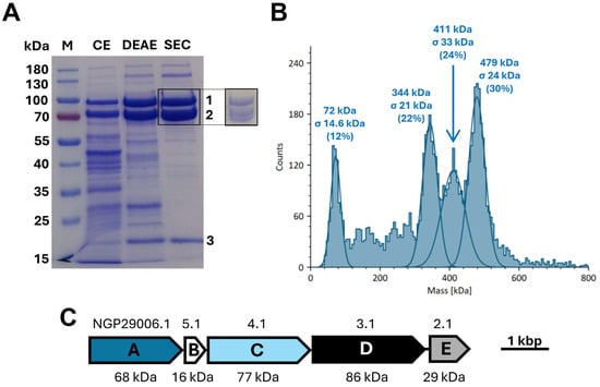
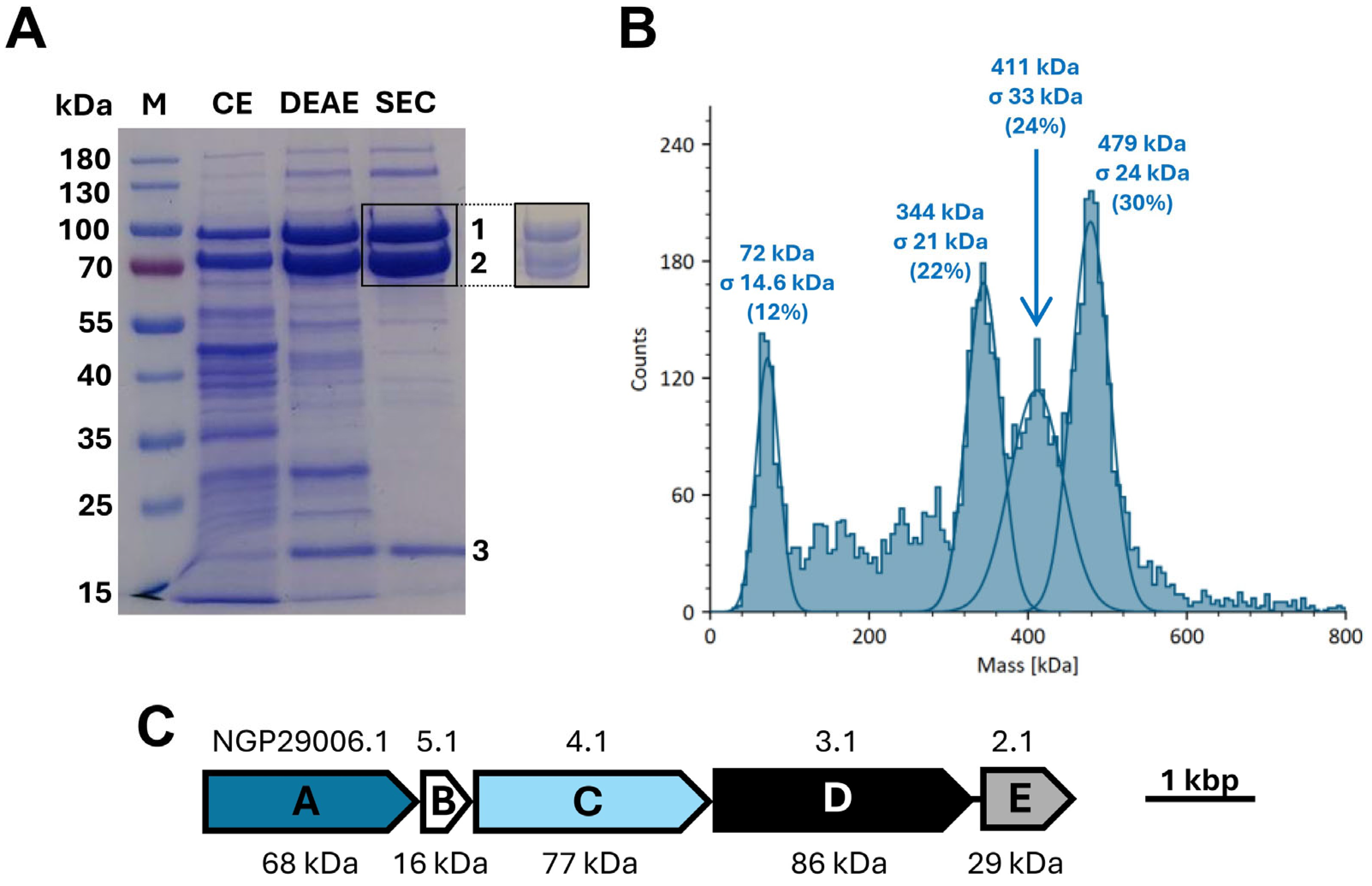
We have shown previously that all five genes of the acr operon are expressed during growth of R. aetherivorans on acetone. Moreover, growth experiments with an R. aetherivorans acrE deletion mutant indicated that the unique subunit E is required for acetone carboxylation, although complementation of this deletion strain with a cloned acrE gene did not succeed [20]. Here, we studied the molecular composition of Acr in more detail and therefore purified the protein by anion exchange and subsequent size exclusion chromatography (Figure 1A). Notably, purification turned out to be straightforward because Acr eluted as a separated peak in the anion exchange chromatography step at higher KCl concentrations than most other proteins of the extract (Supplementary Figure S1). The three large subunits (AcrACD) and the small subunit (AcrB) were detectable on SDS gels containing purified Acr protein (Figure 1A) and confirmed by mass spectrometry (Supplementary Table S1). In contrast, subunit AcrE was not visible on the gels (Figure 1A; Supplementary Table S1). However, when we analyzed purified Acr samples by mass spectrometry, we found that minor amounts of AcrE were still present after size exclusion chromatography (Supplementary Figure S2). The total yield of purified Acr was approximately 1.5–2 mg protein per g of R. aetherivorans wet cell mass.
Figure 1.
Purification and native size of the acetone carboxylase from R. aetherivorans (Acr). (A) Coomassie-stained SDS polyacrylamide gel (10%) of extract from acetone-grown cells (CE), Acr fraction after anion exchange chromatography (DEAE), and Acr fraction after size exclusion chromatography (SEC). A total of 20 µg protein was loaded per lane. The numbers indicate the positions of Acr subunits (two subunits comigrating in the second band). Protein identities were confirmed by mass spectrometry (Supplementary Table S1). In addition, 5 µg of the SEC-purified protein were separated on a 7% SDS polyacrylamide gel (excerpt). (B) Mass photometry analysis of freshly purified Acr complex. The major peaks are labeled with their calculated masses and the relative peak areas (percentages). The following numbers of counts were detected for the four peaks (from left to right): 838, 1603, 1681, and 2105. (C) Operon structure of acr with predicted masses of the Acr subunits. The corresponding NCBI protein accession numbers are indicated above the gene symbols.
Mass photometric analysis of freshly purified enzyme revealed the presence of a large protein complex of 479 ± 24 kDa (Figure 1B), which matches the theoretical size of a heterooctameric Acr(ABCD)2 core complex (494 kDa; Figure 1C) and suggests that most of the subunit E dissociated during purification. The additionally observed mass photometry peaks at 411 ± 33 kDa and 344 ± 21 kDa can be explained by dissociation of either one or both of the AcrA (68 kDa) subunits. Indeed, based on the structure of the Apc core [19], the AcrA subunits are expected to bind loosely to the periphery of the Acr(ABCD)2 complex. In turn, the peak at 72 ± 15 kDa probably corresponds to these dissociated AcrA subunits (Figure 1B). Unfortunately, this experiment did not provide clear evidence for the presence of the AcrE subunit, either separated or attached to the core complex.
As determined by spectrophotometric, 3-hydroxybutyrate dehydrogenase-coupled assays, the purified enzyme showed specific activities of only 2.7 mU mg−1 (i.e., ca. 2-fold enrichment compared to the specific activity measured in the corresponding cell extract). We first hypothesized that after lysing the cells, the enzyme may lose its activity in a time-dependent manner. However, at least in cell extract, Acr was quite stable and maintained about 70–80% of its activity within 6–12 h after cell disruption when kept on ice. Thereby, addition of DTE as a potential redox protectant did not affect enzyme stability (Supplementary Figure S3). We therefore presume that the relatively low enrichment of the specific activity of purified Acr may be attributed to the dissociation and loss of subunit AcrE (Figure 1A,B).
3.2. Molecular Properties, Substrate Spectrum, and Nucleotide Hydrolysis
Next, we studied the molecular properties, substrate spectrum, and range of nucleotide cofactors of the purified enzyme. ICP-MS analysis of the metal content revealed that Acr contains only Zn2+, but neither Mn2+ nor Fe2+ or any other catalytic metals (Table 1). Given the low amounts of AcrE present after purification (Figure 1A), we calculated the metal content per moles of heterooctameric Acr core complex and obtained values of approximately 2.9 mol Zn2+ per mol Acr(ABCD)2. Furthermore, we detected minor amounts of Mg2+, probably from the NTP coordination sites (Table 1). Assuming partial occupancy of the metal binding sites, which has been observed previously also for Apc [8,18,19], we conclude that Acr probably contains 2 Zn2+ per AcrABCD protomer.
Table 1.
Metal content of Acr. Samples were analyzed by ICP-MS, using Acr protein from three different purifications and the SEC buffer as a blank control. Data are displayed as mean ± standard deviation.
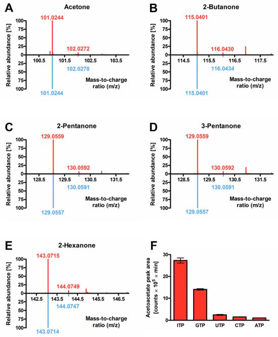
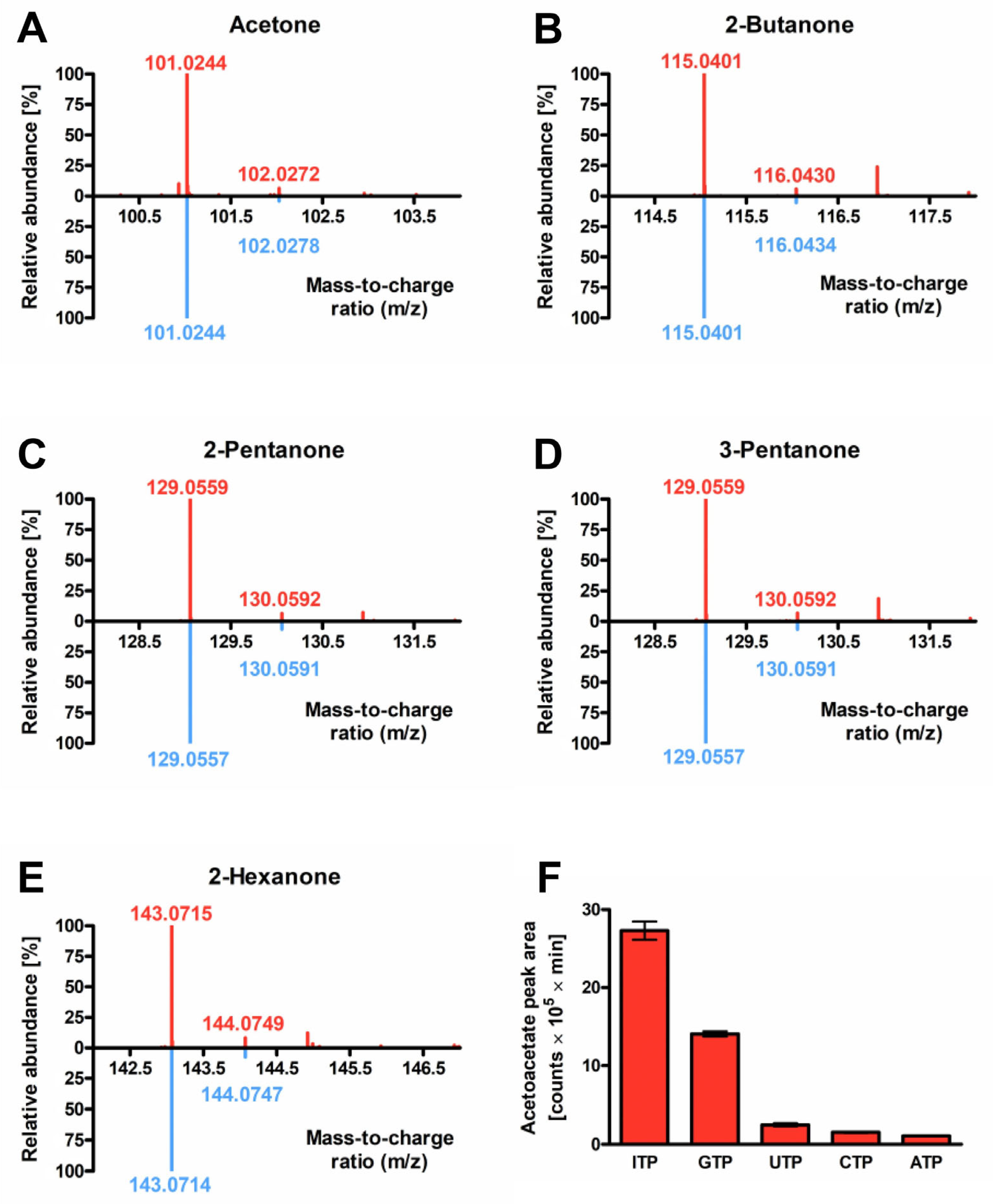
In previous studies, we had already detected carboxylation activity for acetone, 2-butanone, 2-pentanone, 3-pentanone, and 2-hexanone in R. aetherivorans cell extracts [20]. Here, we confirmed these results with purified enzyme, qualitatively detecting all of the five expected carboxylation products (acetoacetate, 3-oxopentanoate, 3-oxohexanoate, 2-methyl-3-oxopentanoate, 3-oxoheptanoate) by high-resolution mass spectrometry (Figure 2A–E). Carboxylation of the same ketones has been observed previously for the acetone carboxylase from R. rhodochrous [14]. In contrast, it had not been tested yet whether aliphatic ketones with even longer chain lengths might still be carboxylated by either of the two enzymes. In this study, we showed that Acr does not accept 2-octanone or 2-decanone as substrates, indicating an upper chain length limit at C6 or C7 (Figure 2A–E).
Figure 2.
Substrate and nucleotide spectrum of purified Acr. (A) Acetone, (B) 2-butanone, (C) 2-pentanone, (D) 3-pentanone, or (E) 2-hexanone were used as substrates and the corresponding carboxylation products were qualitatively detected by LC-MS. The diagrams show relative ion counts against mass-to-charge ratios (m/z). Spectra were extracted at the apex of the respective extracted ion chromatogram (EIC) peaks, which were generated for the theoretical m/z values of the respective carboxylation products, corresponding to retention times of (A) 2.782 min (acetoacetate), (B) 6.170 min (3-oxopentanoate), (C) 7.293–7.315 min (3-oxohexanoate), (D) 7.264–7.453 min (2-methyl-3-oxopentanoate), or (E) 7.536–7.981 min (3-oxoheptanoate). Experimental spectra (red) are displayed as mirror plots against the theoretical isotope patterns (blue) of the respective products, which were calculated based on natural isotope abundance. For all of the different substrates, three independent enzyme assays were performed each, using Acr protein from the same preparation, and the respective triplicate spectra were comparable to each other. No carboxylation products of the relevant masses were detected with 2-octanone or 2-decanone as substrates, or with the water control. All assays employed GTP as nucleotide cofactor. (F) LC-MS-based quantification of acetoacetate produced in assays with acetone as substrate and different nucleotide triphosphates added (ITP, GTP, UTP, CTP, or ATP). Data represent mean values and standard deviations of triplicate measurements (three independent enzyme assays each, with Acr protein from the same preparation).
The previously characterized, partially purified acetone carboxylase from R. rhodochrous hydrolyzed GTP, ITP, CTP, UTP, and XTP, but not ATP [14]. We have already hypothesized in our previous study that the Acr from R. aetherivorans shares this extended nucleotide spectrum and therefore used ITP in all enzyme activity measurements. However, these measurements were performed in cell extracts, which may still contain nucleotide kinases interconverting ITP with any residual nucleotide diphosphates, potentially yielding ATP or other NTPs [20]. Therefore, we determined the nucleotide specificity of Acr with purified enzyme in this study. We performed acetone carboxylase assays with purified Acr in the presence of different NTP species and measured acetoacetate formation by high-resolution mass spectrometry. Acetoacetate formation was highest with ITP and GTP, but we also observed acetone carboxylation at a 10- to 20-fold lower degree with CTP, UTP, or ATP (Figure 2F). Furthermore, to determine which phosphate species was produced upon Acr-catalyzed hydrolysis of nucleotide triphosphates, we measured the production of GDP or GMP from GTP in the assay samples and showed that the amount of GDP formed was at least 50-fold higher than that of GMP. Notably, we expect that much of this conversion occurred via uncoupled nucleotide hydrolysis, since we observed significant NTP hydrolysis rates in the absence of ketone substrates (Supplementary Table S2).
4. Discussion
4.1. Acr, a Novel Type of Acetone Carboxylase
In this study, we established a protocol for native purification of acetone carboxylase from R. aetherivorans (Acr) and characterized different biochemical properties of the purified enzyme (Table 2). Based on operon structure and proteomics, we proposed earlier that Acr forms an Apc-like heterooctameric core complex rather than an Acx-like heterohexamer [20]. Importantly, the mass photometry results unambiguously prove this hypothesis as the determined mass matches the theoretical size of an Acr octamer and shows more than 100 kDa mass difference to a putative hexameric protein (Figure 1B). In accordance with this, an AlphaFold model of the Acr core complex showed great similarity with the previously determined structure of the Apc core (RMSD = 2.437 Å across all 704 atom pairs; RMSD = 1.086 Å between 483 pruned atom pairs; Supplementary Figure S4; [19]). In conclusion, our data provide convincing evidence that Acr indeed represents a novel type of acetone carboxylase, which differs from the canonical enzyme variant in its subunit composition.
Table 2.
Biochemical properties of different acetone and acetophenone carboxylases described in the literature [2,8,9,10,11,12,18,19]. The data for Acr are from this study, except for the test for acetophenone carboxylation [20].
4.2. Biochemical Properties of Acr Compared to Canonical Acetone Carboxylases
Acr resembles Apc rather than Acx not only in its structural, but also in its biochemical properties. Intriguingly, Acr is the first acetone carboxylase to be discovered, which forms di- rather than monophosphates, shows uncoupled NTPase activity in the absence of ketone substrates, and does not contain other metals except Zn2+ ions (Table 2).
Previous studies detected 2 metal ions per protomer in the crystal structures of X. autotrophicus Acx (1 Mn and 1 Zn, [18]) and A. aromaticum Apc (2 Zn, replaced by 2 Hg in the analyzed crystal [19]). As inferred from the structures, the small subunits of these carboxylases contain a structural Zn ion, while the catalytic subunits contain either Mn, Zn, or Fe in the active site (Table 2). Hence, the measured number of Zn2+ ions per Acr(ABCD)2 complex is lower than expected (2.9 rather than 4; Table 1), suggesting that the metal binding sites were only partially occupied.
As described for the acetone carboxylase from R. rhodochrous [14], Acr exhibited an extended substrate and nucleotide range and showed the highest activities with ITP and GTP. However, to our surprise, Acr also accepted ATP as a nucleotide species (Figure 2F) and in this respect differs from the enzyme variant present in R. rhodochrous B276 [14]. Also, despite the apparent structural similarity, Apc is restricted to ATP [8]. Structural analyses will be required to investigate what determines nucleotide specificity in different Acr variants and in Acr compared to Apc.
4.3. Unclear Role of AcrE in Acetone Carboxylation
Previous studies with an R. aetherivorans deletion mutant suggested that the fifth subunit E encoded in the acr operon is required for enzyme activity [20]. As Apc, the structural homolog of Acr, also contains an indispensable subunit E of roughly the same size [8], this was in line with our expectations. Unfortunately, the data presented here do not allow any further conclusions on a putative role of AcrE in ketone carboxylation. The subunit is neither visible on SDS gels of purified protein (Figure 1A), nor in the mass photometry spectrum (Figure 1B). Therefore, if attached to the Acr heterooctamer during catalysis, this binding is only transient. A similar observation has been made for ApcE, which completely dissociates from the Apc core during purification [8]. On the other hand, we showed here that small amounts of AcrE were still present in the purified protein sample after size exclusion chromatography (Supplementary Figure S2). This indicates at least partial co-elution of the much smaller subunit E with the Acr core. Furthermore, we hypothesize that AcrE dissociation accounts for the low specific activities of purified Acr. Reconstitution experiments of purified Acr with additionally supplied AcrE would be required to test this hypothesis and to confirm or disprove the role of AcrE in catalysis. However, despite tremendous effort, we were not able to heterologously express AcrE in any of the tested host strains (i.e., E. coli, Rhodococcus erythropolis, and R. aetherivorans). Establishing a heterologous expression system for AcrE, subsequent reconstitution attempts, a thorough characterization of Acr kinetics, and elucidation of the (cryo-EM) structure of purified Acr will be the main focus of future work.
4.4. Biotechnological Applications of Acr
One putative application of novel enzymes is their implementation into synthetic metabolic pathways for the production of chemicals of interest [29] or for the degradation of toxins. For instance, the Acx from Xanthobacter autotrophicus has been successfully implemented in a synthetic system for degradation of the chemical warfare agent sarin in Escherichia coli [30]. Of note, Actinomycetota such as Rhodococcus spp. become increasingly important both for the production of chemicals and for bioremediation purposes [31,32,33]. The natural occurrence of (Acr-type) acetone carboxylases across Rhodococcus spp. is limited to only a few species [20] and heterologous expression of other acetone carboxylases [9,10,11,12,13] will not be possible in these strains due to codon incompatibilities. In contrast, the Acr characterized in this study may be easily introduced into biotechnologically relevant strains such as Rhodococcus jostii [32]. Finally, the exceptionally broad nucleotide spectrum of Acr may have biotechnological applications in the future. For instance, Acr may be a suitable platform to screen for putative NTP analogons used in orthogonal metabolism.
Supplementary Materials
The following supporting information can be downloaded at: https://www.mdpi.com/article/10.3390/reactions7040065/s1, Figure S1: Representative anion exchange chromatogram of Acr purification; Figure S2: AcrE fragments detected in the purified Acr protein after anion exchange and size exclusion chromatography; Figure S3: Stability of Acr protein in cell extract; Figure S4: Structural alignment of an AlphaFold model of the Acr complex from R. aetherivorans with the Apc core complex from A. aromaticum. Table S1: Mass spectrometric identification of proteins in SDS-PAGE bands from the gel shown in Figure 1A. Table S2: Acr-catalyzed nucleotide hydrolysis.
Author Contributions
Conceptualization, K.M. and J.H.; methodology, K.M., C.G., A.S., J.K., G.K.A.H., N.P. and J.H.; software, K.M., C.G., A.S., J.K. and N.P.; validation, K.M., C.G., A.S., J.K., N.P. and J.H.; formal analysis, K.M., C.G., A.S., J.K., N.P. and J.H.; investigation, K.M., C.G., A.S., M.F., J.K., G.K.A.H., N.P. and J.H.; resources, K.M., C.G., A.S., J.K., G.K.A.H., N.P. and J.H.; data curation, K.M., C.G., A.S., J.K., G.K.A.H., N.P. and J.H.; writing—original draft preparation, K.M.; writing—review and editing, K.M. and J.H.; visualization, K.M. and C.G.; supervision, K.M. and J.H.; project administration, J.H.; funding acquisition, J.H. All authors have read and agreed to the published version of the manuscript.
Funding
This work was funded by the University of Marburg and the Max Planck Institute for Terrestrial Microbiology. Publication costs were covered by the journal.
Data Availability Statement
The original contributions presented in this study are included in the article/Supplementary Materials. Further inquiries can be directed to the corresponding authors.
Acknowledgments
K.M. thanks I. Schall for purification of the heterologously expressed HBDH auxiliary enzyme.
Conflicts of Interest
The authors declare no conflicts of interest.
Abbreviations
The following abbreviations are used in this manuscript:
| Acr | Acetone carboxylase from Rhodococcus |
| Acx | Acetone carboxylase from Xanthobacter |
| Apc | Acetophenone carboxylase |
References
- Erb, T.J. Carboxylases in natural and synthetic microbial pathways. Appl. Environ. Microbiol. 2011, 77, 8466–8477. [Google Scholar] [CrossRef] [Scilit] [PubMed]
- Heider, J.; Schühle, K.; Frey, J.; Schink, B. Activation of acetone and other simple ketones in anaerobic bacteria. J. Mol. Microbiol. Biotechnol. 2016, 26, 152–164. [Google Scholar] [CrossRef] [Scilit] [PubMed]
- Schühle, K.; Fuchs, G. Phenylphosphate Carboxylase: A New C-C Lyase involved in Anaerobic Phenol Metabolism in Thauera aromatica. J. Bacteriol. 2004, 186, 4556–4567. [Google Scholar] [CrossRef] [Scilit] [PubMed]
- Allen, J.R.; Ensign, S.A. Carboxylation of epoxides to β-keto acids in cell extracts of Xanthobacter strain Py2. J. Bacteriol. 1996, 178, 1469–1472. [Google Scholar] [CrossRef] [Scilit] [PubMed]
- Allen, J.R.; Ensign, S.A. Purification to homogeneity and reconstitution of the individual components of the epoxide carboxylase multiprotein enzyme complex from Xanthobacter strain Py2. J. Biol. Chem. 1997, 272, 32121–32128. [Google Scholar] [CrossRef] [Scilit] [PubMed]
- Allen, J.R.; Ensign, S.A. Identification and characterization of epoxide carboxylase activity in cell extracts of Nocardia corallina B276. J. Bacteriol. 1998, 180, 2072–2078. [Google Scholar] [CrossRef] [Scilit] [PubMed]
- Wöhlbrand, L.; Wilkes, H.; Halder, T.; Rabus, R. Anaerobic degradation of p-ethylphenol by “Aromatoleum aromaticum” strain EbN1: Pathway, regulation, and involved proteins. J. Bacteriol. 2008, 190, 5699–5709. [Google Scholar] [CrossRef] [Scilit] [PubMed]
- Jobst, B.; Schühle, K.; Linne, U.; Heider, J. ATP-Dependent Carboxylation of Acetophenone by a Novel Type of Carboxylase. J. Bacteriol. 2010, 192, 1387–1394. [Google Scholar] [CrossRef] [Scilit] [PubMed]
- Sluis, M.K.; Ensign, S.A. Purification and characterization of acetone carboxylase from Xanthobacter strain Py2. Proc. Natl. Acad. Sci. USA 1997, 94, 8456–8461. [Google Scholar] [CrossRef] [Scilit] [PubMed]
- Sluis, M.K.; Larsen, R.A.; Krum, J.G.; Anderson, R.; Metcalf, W.W.; Ensign, S.A. Biochemical, Molecular, and Genetic Analyses of the Acetone Carboxylases from Xanthobacter autotrophicus Strain Py2 and Rhodobacter capsulatus Strain B10. J. Bacteriol. 2002, 184, 2969–2977. [Google Scholar] [CrossRef] [Scilit] [PubMed]
- Boyd, J.M.; Ellsworth, H.; Ensign, S.A. Bacterial acetone carboxylase is a manganese-dependent metalloenzyme. J. Biol. Chem. 2004, 279, 46644–46651. [Google Scholar] [CrossRef] [Scilit] [PubMed]
- Schühle, K.; Heider, J. Acetone and Butanone Metabolism of the Denitrifying Bacterium “Aromatoleum aromaticum” Demonstrates Novel Biochemical Properties of an ATP-Dependent Aliphatic Ketone Carboxylase. J. Bacteriol. 2012, 194, 131–141. [Google Scholar] [CrossRef] [Scilit] [PubMed]
- Rosier, C.; Leys, N.; Henoumont, C.; Mergeay, M.; Wattiez, R. Purification and characterization of the acetone carboxylase of Cupriavidus metallidurans strain CH34. Appl. Environ. Microbiol. 2012, 78, 4516–4518. [Google Scholar] [CrossRef] [Scilit] [PubMed]
- Clark, D.D.; Ensign, S.A. Evidence for an Inducible Nucleotide-Dependent Acetone Carboxylase in Rhodococcus rhodochrous B276. J. Bacteriol. 1999, 181, 2752–2758. [Google Scholar] [CrossRef] [Scilit] [PubMed]
- van der Werf, P.; Orlowski, M.; Meister, A. Enzymatic conversion of 5-oxo-L-proline (L-pyrrolidone carboxylate) to L-glutamate coupled with cleavage of adenosine triphosphate to adenosine diphosphate, a reaction in the -glutamyl cycle. Proc. Natl. Acad. Sci. USA 1971, 68, 2982–2985. [Google Scholar] [CrossRef] [Scilit] [PubMed]
- Kim, J.M.; Shimizu, S.; Yamada, H. Amidohydrolysis of N-methylhydantoin coupled with ATP hydrolysis. Biochem. Biophys. Res. Commun. 1987, 142, 1006–1012. [Google Scholar] [CrossRef] [Scilit] [PubMed]
- Marjanovic, A.; Rozeboom, H.J.; de Vries, M.S.; Mayer, C.; Otzen, M.; Wijma, H.J.; Janssen, D.B. Catalytic and structural properties of ATP-dependent caprolactamase from Pseudomonas jessenii. Proteins 2021, 89, 1079–1098. [Google Scholar] [CrossRef] [Scilit] [PubMed]
- Mus, F.; Eilers, B.J.; Alleman, A.B.; Kabasakal, B.V.; Wells, J.N.; Murray, J.W.; Nocek, B.P.; DuBois, J.L.; Peters, J.W. Structural Basis for the Mechanism of ATP-Dependent Acetone Carboxylation. Sci. Rep. 2017, 7, 7234. [Google Scholar] [CrossRef] [Scilit] [PubMed]
- Weidenweber, S.; Schühle, K.; Demmer, U.; Warkentin, E.; Ermler, U.; Heider, J. Structure of the acetophenone carboxylase core complex: Prototype of a new class of ATP-dependent carboxylases/hydrolases. Sci. Rep. 2017, 7, 39674. [Google Scholar] [CrossRef] [Scilit] [PubMed]
- Müller, K.; Kahnt, J.; Paczia, N.; Heider, J. An acetophenone carboxylase homolog from Rhodococcus aetherivorans initiates acetone rather than acetophenone catabolism. Front. Microbiol. 2026, 17, 1888828. [Google Scholar] [CrossRef] [Scilit] [PubMed]
- Boll, M. Studies with Triton X-100 treated electron transport particles from Rhodospirillum rubrum. Arch. Microbiol. 1970, 71, 1–8. [Google Scholar] [CrossRef] [Scilit] [PubMed]
- Bergmeyer, H.U.; Gawehn, K.; Klotzsch, H.; Krebs, H.A.; Williamson, D.H. Purification and properties of crystalline 3-hydroxybutyrate dehydrogenase from Rhodopseudomonas spheroides. Biochem. J. 1967, 102, 423–431. [Google Scholar] [CrossRef] [Scilit] [PubMed]
- Young, G.; Hundt, N.; Cole, D.; Fineberg, A.; Andrecka, J.; Tyler, A.; Olerinyova, A.; Ansari, A.; Marklund, E.G.; Collier, M.P.; et al. Quantitative mass imaging of single biological macromolecules. Science 2018, 360, 423–427. [Google Scholar] [CrossRef] [Scilit] [PubMed]
- Houk, R.; Fassel, V.; Flesch, G.D.; Svec, H.; Gray, A.; Taylor, C.E. Inductively coupled argon plasma as an ion source for mass spectrometric determination of trace elements. Anal. Chem. 1980, 52, 2283–2289. [Google Scholar] [CrossRef] [Scilit]
- Abramson, J.; Adler, J.; Dunger, J.; Evans, R.; Green, T.; Pritzel, A.; Ronneberger, O.; Willmore, L.; Ballard, A.J.; Bambrick, J.; et al. Accurate structure prediction of biomolecular interactions with AlphaFold 3. Nature 2024, 630, 493–500. [Google Scholar] [CrossRef] [Scilit] [PubMed]
- Pettersen, E.F.; Goddard, T.D.; Huang, C.C.; Meng, E.C.; Couch, G.S.; Croll, T.I.; Morris, J.H.; Ferrin, T.E. UCSF ChimeraX: Structure visualization for researchers, educators, and developers. Protein Sci. 2021, 30, 70–82. [Google Scholar] [CrossRef] [Scilit] [PubMed]
- Bradford, M.M. A rapid and sensitive method for the quantification of microgram quantities of protein utilizing the principle of protein-dye binding. Anal. Biochem. 1976, 72, 248–254. [Google Scholar] [CrossRef] [PubMed]
- Laemmli, U.K. Cleavage of structural proteins during the assembly of the head of bacteriophage T4. Nature 1970, 227, 680–685. [Google Scholar] [CrossRef] [Scilit] [PubMed]
- Erb, T.J. Back to the future: Why we need enzymology to build a synthetic metabolism of the future. Beilstein J. Org. Chem. 2019, 15, 551–557. [Google Scholar] [CrossRef] [Scilit] [PubMed]
- Brown, M.E.; Mukhopadhyay, A.; Keasling, J.D. Engineering bacteria to catabolize the carbonaceous component of sarin: Teaching E. coli to eat isopropanol. ACS Synth. Biol. 2016, 5, 1485–1496. [Google Scholar] [CrossRef] [Scilit] [PubMed]
- Ngo, A.C.R.; Haarmann, M.; Weindorf, N.; Guanzon, D.A.V.; Linke, V.; Smitka, J.; Tischler, D. Golden Gate Cloning in Actinobacteria: Opportunities and Challenges. Methods Mol. Biol. 2025, 2850, 377–386. [Google Scholar] [CrossRef] [Scilit] [PubMed]
- Yasin, R.; Rashid, G.M.M.; Ali, I.; Bugg, T.D.H. Engineering of Rhodococcus jostii RHA1 for utilisation of carboxymethylcellulose. Heliyon 2023, 9, e19511. [Google Scholar] [CrossRef] [Scilit] [PubMed]
- Nazari, M.T.; Simon, V.; Machado, B.S.; Crestani, L.; Marchezi, G.; Concolato, G.; Ferrari, V.; Colla, L.M.; Piccin, J.S. Rhodococcus: A promising genus of actinomycetes for the bioremediation of organic and inorganic contaminants. J. Environ. Manag. 2022, 232, 116220. [Google Scholar] [CrossRef] [Scilit] [PubMed]

Disclaimer/Publisher’s Note: The statements, opinions and data contained in all publications are solely those of the individual author(s) and contributor(s) and not of MDPI and/or the editor(s). MDPI and/or the editor(s) disclaim responsibility for any injury to people or property resulting from any ideas, methods, instructions or products referred to in the content. |
© 2026 by the authors. Licensee MDPI, Basel, Switzerland. This article is an open access article distributed under the terms and conditions of the Creative Commons Attribution (CC BY) license.