


Abstract
Paclitaxel-loaded alginate-chitosan complex microcapsules were successfully prepared by the high-voltage electrostatic technique. The effect of paclitaxel concentration and drug-loading methods on drug loading of alginate-chitosan microcapsules, and the effect of methanol concentration, chitosan concentration and chitosan molecular weight on encapsulation efficiency were investigated. Experimental results showed that calcium alginate beads had a good degree of sphericity and a uniform size of 299.8±3.6 µm. After encapsulation with chitosan membrane followed by freeze drying, microcapsules shrunk and collapsed with a mean particle size of about 60.0 µm. Drug loading results showed that as paclitaxel concentration increased from 2 mg/mL to 8 mg/mL, drug loading of microcapsules was increased from 1.95% to 11.16%. Microcapsules with dynamical paclitaxel-loading at 30 °C had the highest drug loading, followed by those with statical paclitaxel-loading at 4 °C and 37 °C. Encapsulation efficiency results showed that microcapsules washed with 0.5% (v/v) methanol had the highest encapsulation efficiency of 2.85%. The encapsulation efficiency was decreased with the increase of chitosan concentration, while it firstly decreased and then increased with the increase of molecular weight of chitosan. These results provide primary information about drug loading and encapsulation efficiency properties of paclitaxel-loaded alginate-chitosan microcapsules as a novel drug carrier.
1. Introduction
Paclitaxel (PTX) as an effective anticancer therapeutic agent has been widely applied in the treatment of a variety of cancers, including the breast, ovary, lung, head and neck, esophagus, and hematological malignancies [1,2]. However, the poor water solubility of PTX significantly hampers its clinical application [3,4]. To solve this problem, the current commercial product of PTX is dissolved in cremophor EL and ethanol mixture (1:1, v/v), which usually cause serious toxic side effects such as hypersensitivity, nephrotoxicity and neurotoxicity [5,6,7]. Therefore, the development of novel formulation converting PTX into water-soluble drug as well as reducing adverse reactions has attracted great interest recently [8,9,10,11]. The application of drug carriers such as nanoparticles [12], microspheres [13,14] and microcapsules [15] to entrap pharmaceutical agents into biodegradable polymers has been utilized as a preferred method for delivering PTX. These strategies can combine the conversion of PTX into a water-soluble drug with the release of drugs to specific tissues at a therapeutically optimal rate. The utilized polymers should possess good biodegradability, low toxicity and ideal bonding ability with bioactive molecules [16]. The most frequently studied polymers are chitosan [17], poly (lactic acid) (PLA) [18], poly (lacticco-glycolic acid) (PLGA) [19,20]. Chitosan as a natural biodegradable polymer, is abundant in source and inexpensive for price. It is a polycation polymer with excellent biocompatibility and widely used in drug delivery systems, accelerating drug absorption and inhibiting tumor cell [21,22,23,24]. Chitosan can be combined with the polyanion polymer such as alginate through ionic cross-link to form microcapsules with non-toxicity, excellent biocompatibility and pharmacological efficacy.
Several methods have been used for preparation of microcapsules, such as spray drying [25], emulsion solvent-extraction/evaporation [26,27,28], layer-by-layer assembly [29] and supercritical CO2 [30]. However, these methods have drawbacks of organic solvent removal in a subsequent procedure and tedious operation process. The high-voltage electrostatic technique, which can produce uniform size of calcium alginate beads in superior degree of sphericity without the usage of organic solvent, has aroused most interest [31,32,33]. Herein, in the present study, calcium alginate beads were produced by a high-voltage electrostatic droplet generator and then coated with chitosan as membrane materials through polyelectrolytical complex reaction between alginate and chitosan to prepare water-solvable drug carriers. The PTX loading and encapsulation efficiency of alginate-chitosan complex microcapsules were also investigated.
2. Materials and Methods
2.1. Materials
Sodium alginate (Alg), calcium chloride, sodium hydroxide and sodium bicarbonate were all purchased from National Pharmaceutical Group Chemical Reagent Co., Ltd. (China), and its aqueous solution was filtrated through 0.8 µm, 0.45 µm and 0.22 µm membranes in sequence. Chitosan (CS) was purchased from Zhejiang Golden-shell Biochemistry Co., Ltd. (China). Paclitaxel (PTX, purity ≥ 99.9%) was purchased from Beijing Yi-he Bioengineering Co., Ltd. (China). Other reagents were all of analytical reagent grade. All the chemicals were used as received without further purification.
2.2. Preparation of Calcium Alginate Beads
4 mL of sodium alginate solution (2.0%, w/v) was extruded into 100 mL calcium chloride solution (1.5%, w/v) by a syringe pump at a speed ratio of 50 mm/h under the high-voltage generator with a voltage of 6.3 kV. The distance between the flat pinhead (7#) and the surface of the calcium chloride solution was set at 20 mm. The prepared calcium alginate beads were collected and washed with distilled water twice to remove redundant calcium ion.
2.3. Preparation of PTX-loaded Alginate-chitosan Complex Microcapsules
PTX-loaded alginate-chitosan complex microcapsules were prepared by two-step method. The calcium alginate beads were incubated dynamically in a PTX methanol solution (4 mg/mL) at 30 °C for 24 h. Subsequently, PTX-loaded beads were separated with standard sieves and then transferred into the chitosan aqueous solution (0.2%, w/v) with a molecular weight of 100, 000 Da under shaking for 10 min to form the stable membrane of microcapsules. Finally, the PTXloaded alginate-chitosan complex microcapsules were washed with 3 mL methanol solution (0.5%, w/v) quickly, then separated from the reaction solutions, freeze-dried at 100 Pa and −80 °C for 24 h and then preserved in a dryer. Microcapsules with different PTX concentrations (2 and 8 mg/mL), drug-loading methods (37 °C statically and 4 °C statically), methanol concentrations (1% and 1.5%), chitosan concentrations (0.1% and 0.5%) and chitosan molecular weights (50 000 Da and 150 000 Da) were also prepared as the method mentioned above.
2.4. Characterization of Microcapsules
The shape and surface of calcium alginate beads and PTX-loaded alginate-chitosan complex microcapsules were characterized by optical microscopy (OLYMPUS PM20, Japan) and scanning electron microscopy (SEM, HITACHI S-4800, Japan). Particle sizes of calcium alginate beads were determined by a micrometer (Olympus, Japan), the number of calculated particles was 100.
2.5. Determination Method of PTX in Microcapsules
Freeze-dried microcapsules (10 mg) were swelled in 10 mL phosphate buffer solution (pH 7.4) for 1∼2 h, and then broken by ultrasonic cell crusher. 5 mL methylene chloride was added to extract PTX sample in the lower aqueous phase for 12 h. PTX concentration was determined by high performance liquid chromatography (HPLC). HPLC system includes: C18 column (150 mm × 4.6 mm); the flow rate of 1.0 ml/min; mobile phase of methanol and water (70:30, v/v); detection wavelength of 233 nm. The PTX standard solution was prepared by dissolving PTX in methylene chloride and diluted into a series of concentration to measure a standard curve.
2.6. Determination of Drug Loading and Encapsulation Efficiency
According to the “Pharmacopeia of People’s Republic of China” (2005), the drug loading and encapsulation efficiency were calculated by Equations (1) and (2), respectively.
where W1 is the weight of PTX in the microcapsules, W2 is the gross weight of microcapsules, W3 is the total weight of PTX, and W4 is the weight of PTX that remained in the liquid medium after encapsulation. Each experiment was performed in triplicate.
Drug loading (%) = 100 × W1/W2
Encapsulation efficiency (%) = 100 × (W3 − W4)/W3
2.7. Statistical Analysis
The final data are expressed as mean values ± standard deviation. Statistical analysis was performed using SPSS 15.0 software. Statistical significance was measured using one way analysis of variance (ANOVA) followed by Student-Newman-Keuls or Dunnett C post-hoc tests for all paired combinations, and the differences were considered significant when the p-value < 0.05.
3. Results and Discussion
3.1. Surface Morphology and Particle Size Distribution of Calcium Alginate Beads and PTX-loaded Alginate-chitosan Microcapsules
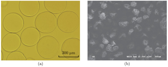
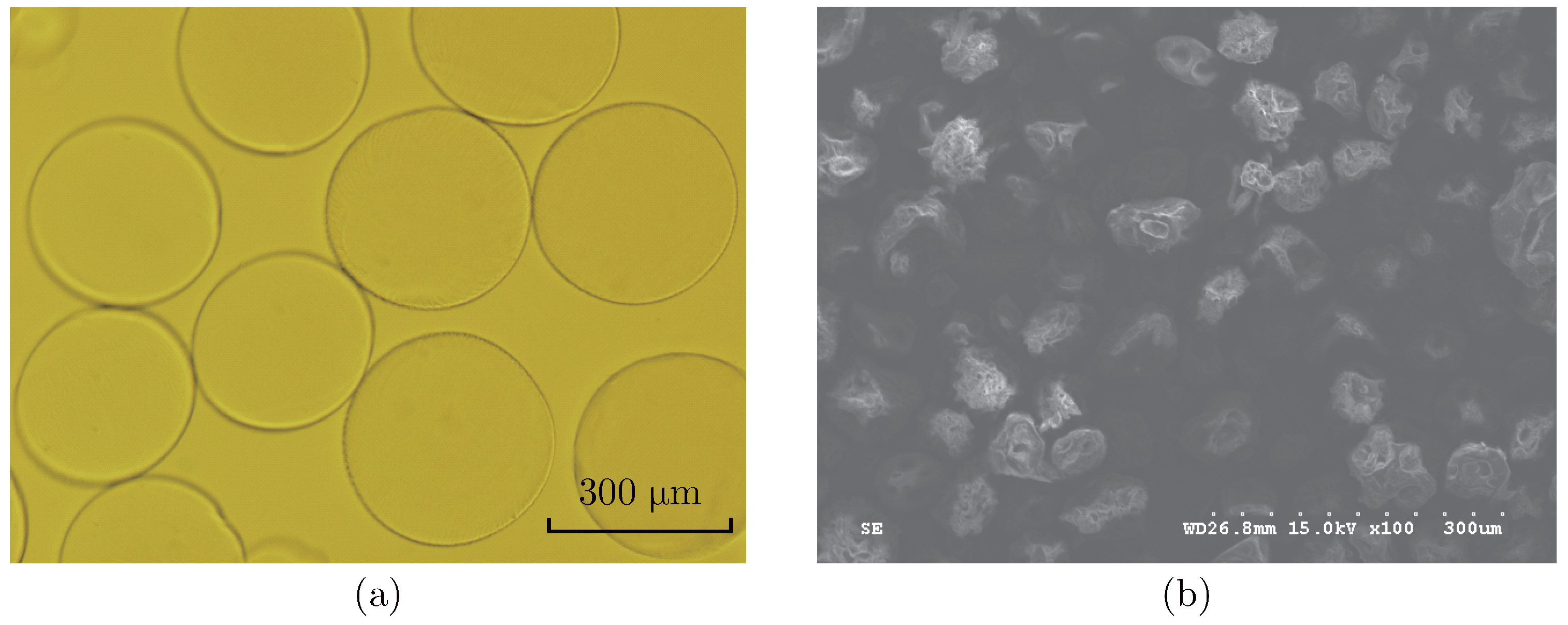
Figure 1 shows the surface morphology of the calcium alginate beads and PTX-loaded alginatechitosan microcapsules. It was found that the calcium alginate beads exhibited a smooth surface as well as good degree of sphericity and uniformity. The average particle size of calcium alginate beads and PTX-loaded alginate-chitosan microcapsules were 299.8±3.6 µm and 399.2±5.9 µm, respectively. After freeze drying, PTX-loaded alginate-chitosan microcapsules prepared by twostep method displayed apparent changes in sphericity and morphology. Surface folds and collapses were observed on the surface of microcapsules with a mean particle size of about 60.0 µm. It was ascribed to the rapid water sublimation during the process of freeze drying, which induced the shrinkage of pores in microcapsule walls and the collapse of crosslinking structure of microcapsules.
Figure 1.
Optical microscopy of calcium alginate beads (a) and SEM image of PTX-loaded alginate-chitosan microcapsules (b).
3.2. Maximum Absorption Wavelength and Standard Curve of PTX
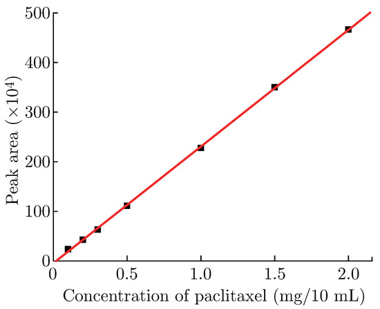
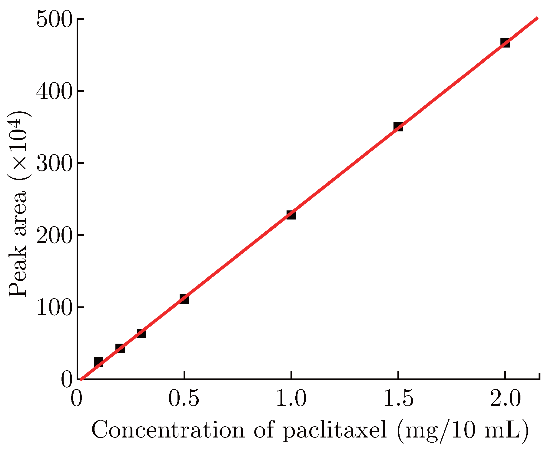
On the basis of wavelength scanning technology, the maximum absorption wavelength of PTX standard solution was 233 nm, therefore, we chose 233 nm as the detection wavelength of HPLC. As shown in Figure 2, the PTX concentration exhibited a good linear relationship with HPLC peak area. The equation of the standard curve was:
y = −4.63885 + 235.29045x, R2 = 0.9998 (n = 3)
Figure 2.
Calibration curve of PTX standard solution.
3.3. Drug Loading
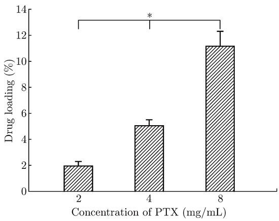
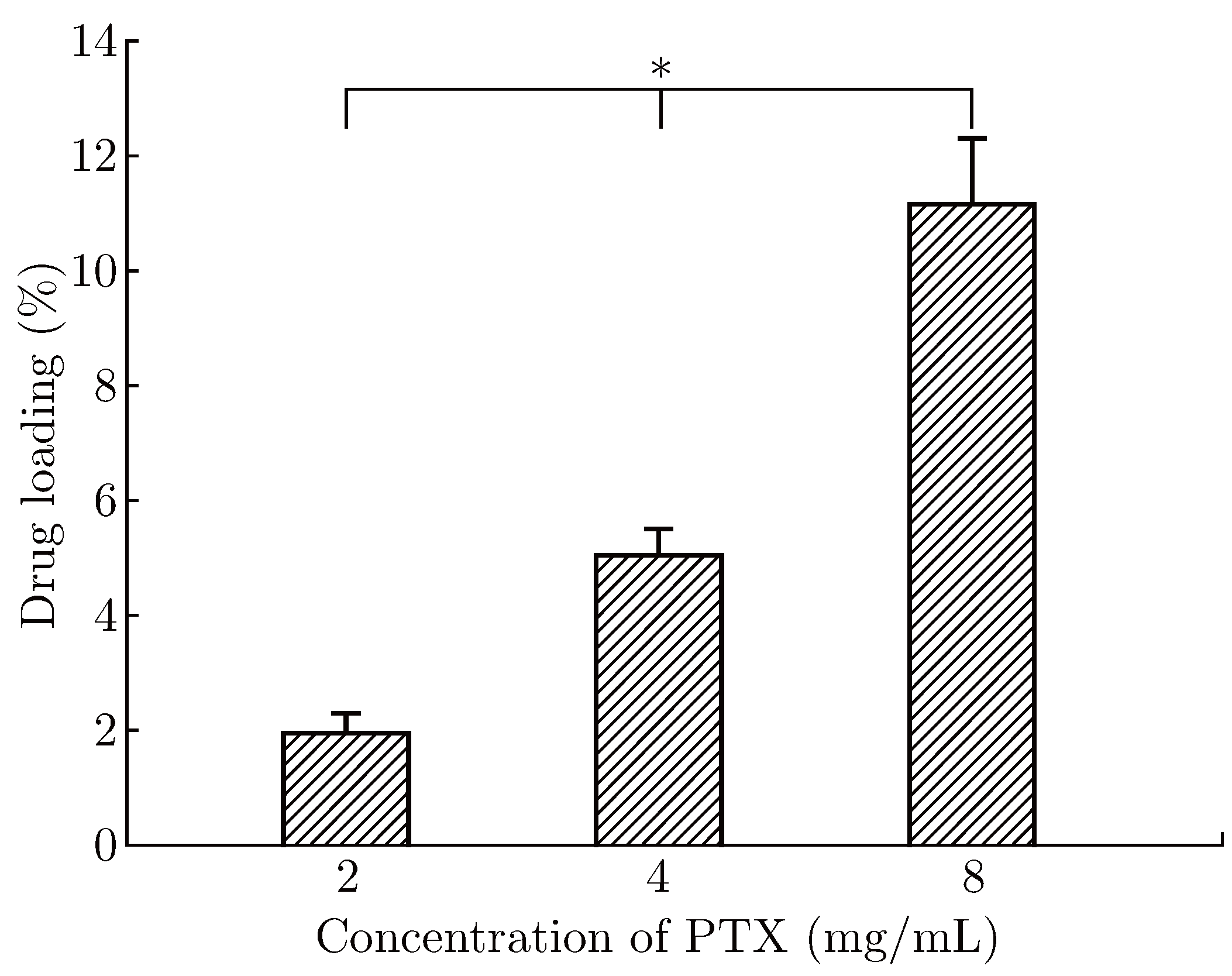
Drug loading is defined as the amount of drug per mass of drug carrier system. It is one of the most important parameters in the evaluation of microcapsules quality. Figure 3 shows the effect of PTX concentration on the drug loading of alginate-chitosan complex microcapsules. The drug loading of microcapsule was significantly (p < 0.05) increased from 1.95% to 11.16% as the PTX concentration increased from 2 mg/mL to 8 mg/mL. It was suggested that the electrostatic interaction between PTX as a cationic drug and sodium alginate as a polyanion polymer contributed to the increase of drug loading [34]. Furthermore, PTX as a small molecule drug could enter into microcapsules by diffusion [35,36]. The higher the PTX concentration increased, the more PTX molecules penetrated into the interior of calcium alginate beads. As the amount of drug loaded in calcium alginate beads per unit mass increased, the drug loading of alginate-chitosan microcapsules increased.
Figure 3.
Drug loading of alginate-chitosan microcapsules with different concentration of PTX. ∗p < 0.05.
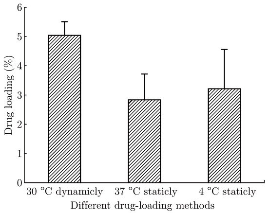
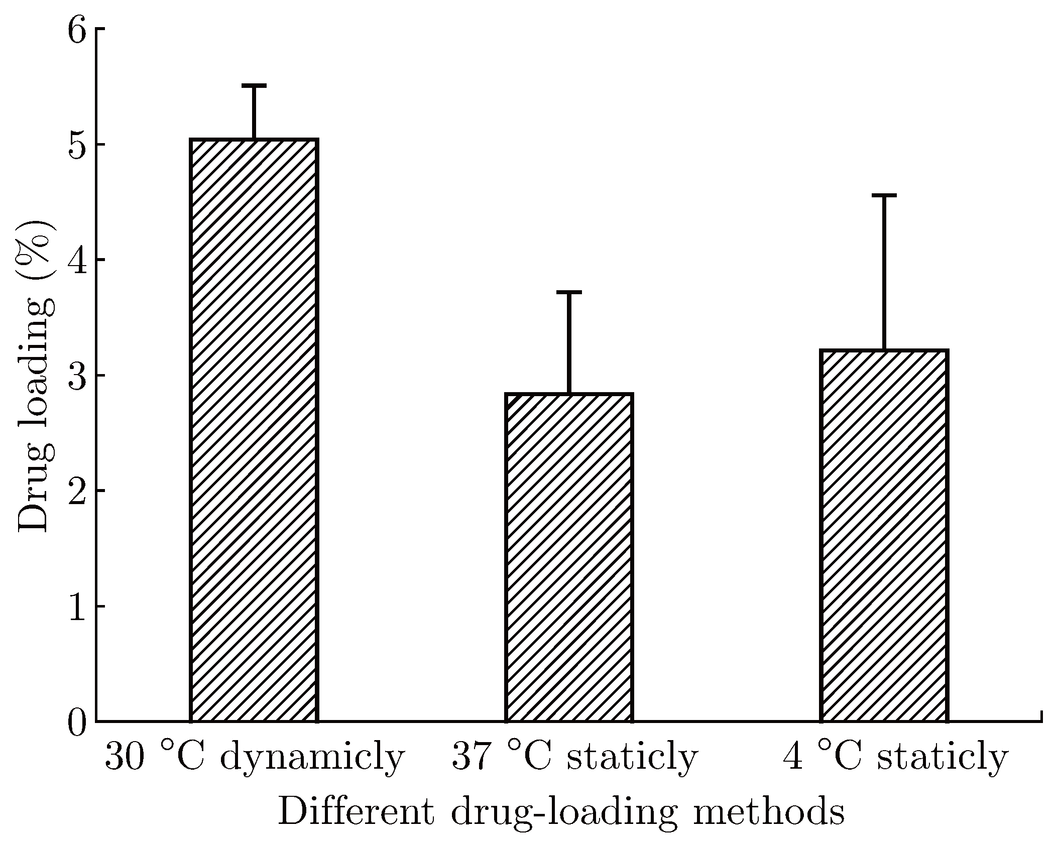
The effect of drug-loading methods on the drug loading of microcapsules was further investigated. As shown in Figure 4, microcapsules with dynamical PTX-loading at 30 °C has the highest drug loading, followed by those with statical PTX-loading at 4 °C and 37 °C. According to the principle of Brownian motion, dynamic drug-loading method and high temperature could accelerate molecular diffusion rate. Therefore, dynamic drug-loading method showed evident advantages with a drug loading of 5.04%. At static condition, the temperature played an important role in drug loading. 37 °C was supposed to have higher drug loading than 4 °C due to the accelerated molecular diffusion. However, as the temperature increased, PTX molecules not only diffused into the microcapsules, it also diffused out. As the amount of PTX diffused out of microcapsules was greater than that of PTX entering into microcapsules, the drug loading was decreased.
Figure 4.
Drug loading of alginate-chitosan microcapsules with different drug-loading methods.
3.4. Encapsulation Efficiency
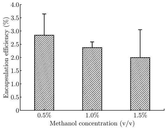
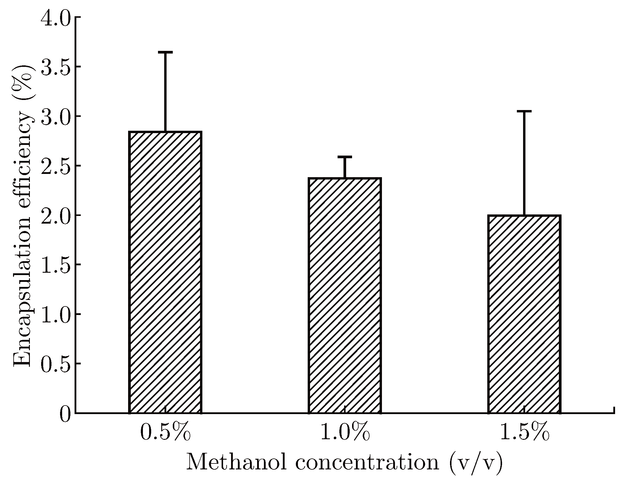
In the preparation of microcapsules, most of the drugs are encapsulated in the interior by the skeleton materials. The unwrapped drugs adsorbed on surfaces of microcapsules will firstly dissolve in tissue fluid in human body to achieve its therapeutic effect. However, the overdose of drug attached on surfaces may cause serious burst behavior in drug control release [37]. In order to avoid this phenomenon, a washing process of microcapsules using methanol was used and the effect of methanol on encapsulation efficiency was investigated. As shown in Figure 5, the encapsulation efficiency was decreased with methanol concentration. Among them, microcapsules washed with 0.5% (v/v) methanol had the highest encapsulation efficiency of 2.85%. This may be because more PTX was removed from microcapsule surface with the increase of methanol concentration. In addition, methanol could enter into the interior of microcapsules through polyelectrolyte holes, leading to drug leakage and decrease of encapsulation efficiency. Similar result was observed in preparation of PTX-loaded poly(lactic acid)–poly(ethyleneglycol)–poly(lactic acid) microspheres where acetone accelerated the release of PTX by increasing the number of mesh on microsphere surface [14].
Figure 5.
Encapsulation efficiency of alginate-chitosan microcapsules washed with different concentrations of methanol.
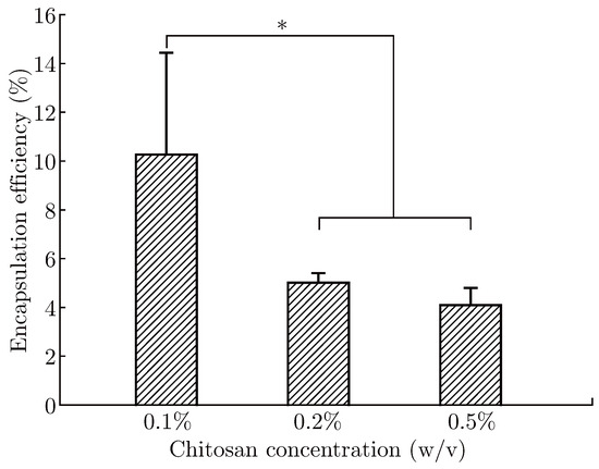
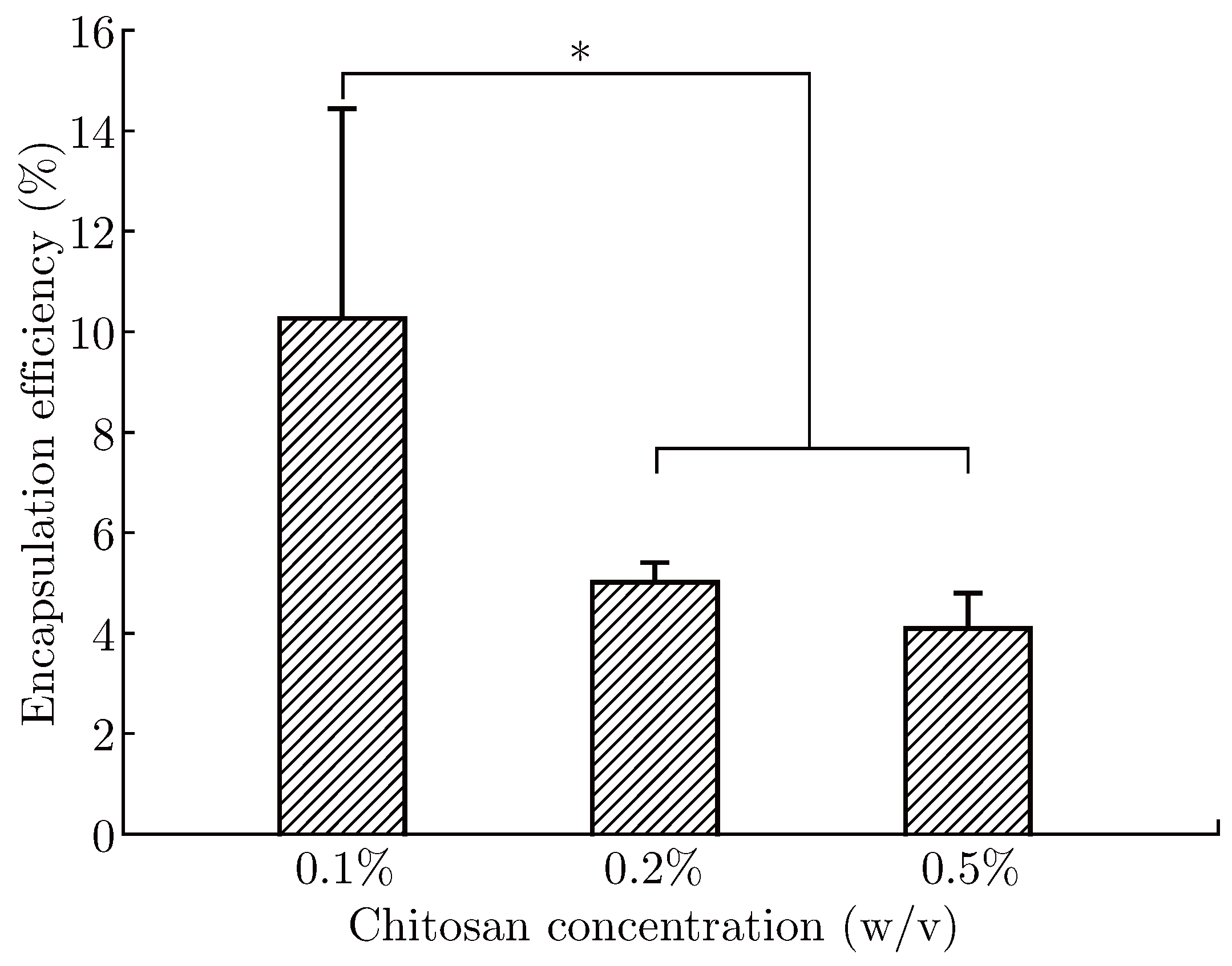
According to the above results, we chose 0.5% (v/v) methanol to wash alginate-chitosan microcapsules in the following experiments. The effect of chitosan concentration on encapsulation efficiency of microcapsules was further investigated. As shown in Figure 6, the encapsulation efficiency was significantly (p < 0.05) decreased with the increase of chitosan concentration. When chitosan concentration was 0.1%, 0.2% and 0.5%, encapsulation efficiency was 10.26%, 5.00% and 4.09% respectively. The increase of chitosan concentration could promote its infiltration into the calcium alginate beads and benefit the formation of microcapsule membrane, while the increase of bulk density of molecular chain could also thicken the membrane. The thickened membrane not only prevented PTX from diffusing out of microcapsules but also impeded the extracapsular PTX into microcapsules. In addition, the increased membrane thickness resulted in the increase of microcapsule weight [38]. When its contribution to reducing the loss of PTX was less than the increasing microcapsule weight, the encapsulation efficiency was decreased.
Figure 6.
Encapsulation efficiency of alginate-chitosan microcapsules with different concentrations of chitosan. ∗p < 0.05.
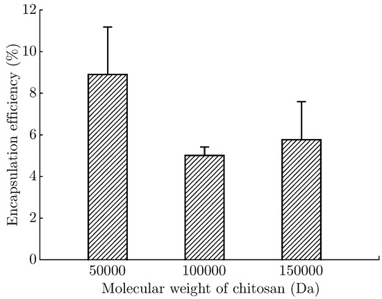
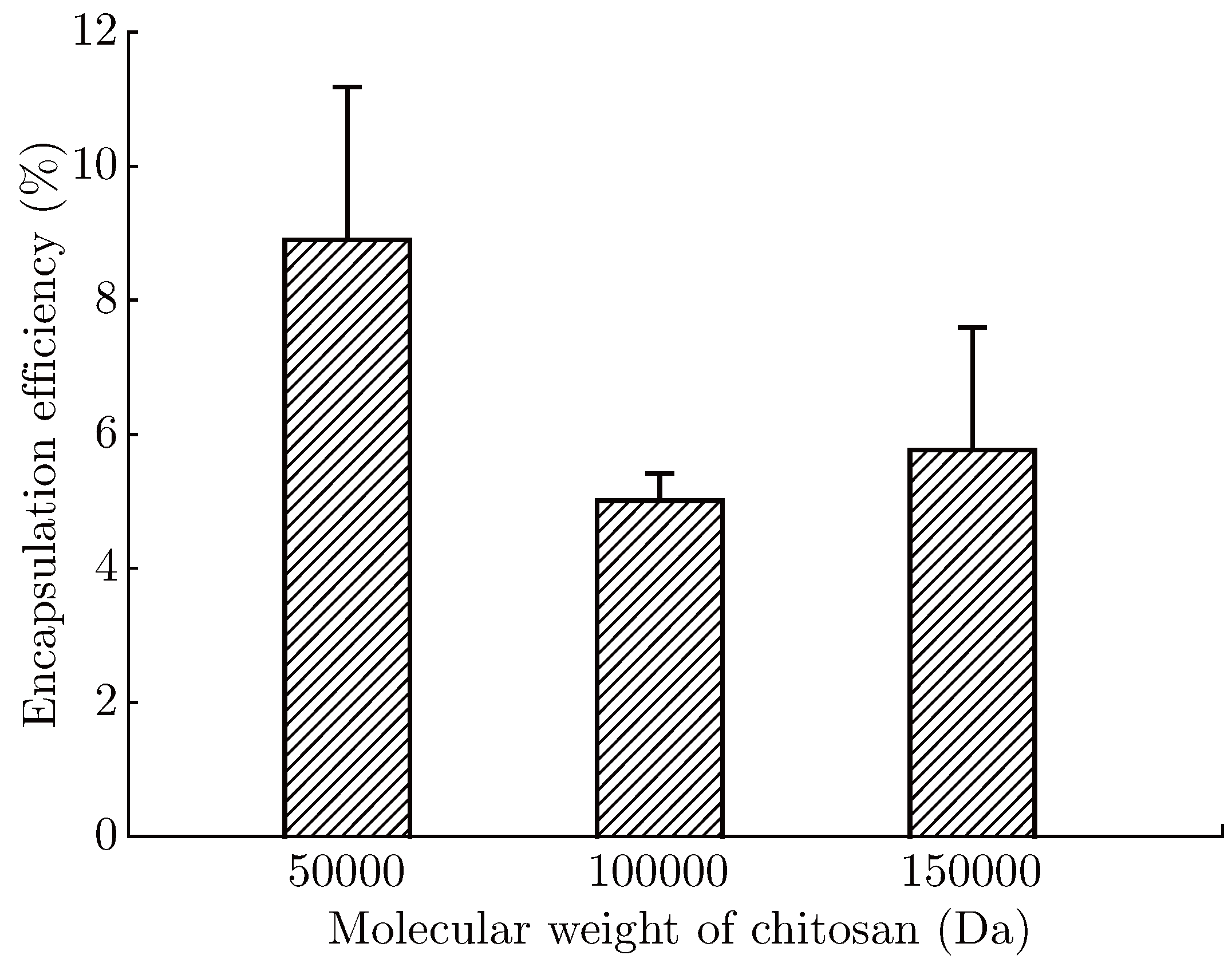
Figure 7 shows the effect of chitosan molecular weight on encapsulation efficiency of alginatechitosan microcapsules. When the molecular weight of chitosan was 50000 Da, 100000 Da and 150000 Da, the encapsulation efficiency of microcapsule was 8.89%, 5.01% and 5.76% respectively. The encapsulation efficiency of microcapsule was firstly decreased and then increased with the increase of molecular weight of chitosan. It was suggested that chitosan with high molecular weight had larger steric hindrance, which prevented it entering into calcium alginate beads and leaded to formation of thin composite film on the surface of calcium alginate beads [35]. However, chitosan with high molecular weight could provide more –NH+3 combined with –COO− of calcium alginate and form more dense composite film [32,39]. For microcapsules encapsulated with 50000 Da chitosan, they had the highest encapsulation efficiency due to the dominant role of membrane thickness. For microcapsules encapsulated with 150000 Da chitosan, they owned higher encapsulation efficiency than those wrapped with 100000 Da chitosan due to the dominant role of membrane strength. As a result of joint action between membrane strength and membrane thickness, microcapsules with 100000 Da chitosan showed the lowest encapsulation efficiency.
Figure 7.
Encapsulation efficiency of alginate-chitosan microcapsules with different molecular weights of chitosan.
4. Conclusion
PTX-loaded alginate-chitosan complex microcapsules were successfully prepared by the highvoltage electrostatic technique. The drug loading of microcapsules was increased from 1.95% to 11.16% as the PTX concentration increased from 2 mg/mL to 8 mg/mL. Microcapsules with dynamical PTX-loading at 30 °C had the highest drug loading, followed by those with statical PTX-loading at 4 °C and 37 °C. Microcapsules washed with 0.5% (v/v) methanol had the highest encapsulation efficiency of 2.85%. The encapsulation efficiency was decreased with the increase of chitosan concentration, while it firstly decreased and then increased with the increase of molecular weight of chitosan. All these results demonstrated that PTX-loaded alginate-chitosan microcapsules have potential application as a novel formulation in drug delivery.
Acknowledgments
Financial supports from the National Natural Science Foundation of China (NSFC 81301290), Quanzhou Science and Technology Plan Project (2013Z24) and Research Foundation for High-level Talents of Huaqiao University (12BS207) are gratefully acknowledged.
References
- Lopes, N.M.; Adams, E.G.; Pitts, T.W.; Bhuyan, B.K. Cell kill kinetics and cell cycle effects of taxol on human and hamster ovarian cell lines. Cancer Chemother. Pharmacol. 1993, 32, 235–242. [Google Scholar] [CrossRef] [Scilit]
- Rowinsky, E.K.; Cazenave, L.A.; Donehower, R.C. Taxol: A novel investigational antimicrotubule agent. J. Natl. Cancer 1990, 82, 1247–1259. [Google Scholar] [CrossRef] [Scilit]
- Videira, M.; Almeida, A.J. Preclinical evaluation of a pulmonary delivered paclitaxel-loaded lipid nanocarrier antitumor effect. Nanomedicine 2012, 8, 1208–1215. [Google Scholar] [CrossRef] [Scilit]
- Ndungu, J.M.; Lu, Y.J.; Zhu, S.J.; Yang, C.; Wang, X.; Chen, G.; Shin, D.M.; Snyder, J.P.; Shoji, M.; Sun, A.M. Targeted delivery of paclitaxel to tumor cells: synthesis and in vitro evaluation. J. Med. Chem. 2010, 53, 3127–3132. [Google Scholar] [CrossRef] [Scilit]
- Weiss, R.B.; Donehower, R.C.; Wiernik, P.H.; Ohnuma, T.; Gralla, R.J.; Trump, D.L.; Baker, J.R.J.; Van Echo, D.A.; Von-Hoff, D.D.; Leyland-Jones, B. Hypersensitivity reactions from taxol. J. Clin. Oncol. 1990, 8, 1263–1268. [Google Scholar] [CrossRef] [Scilit]
- Gelderblom, H.; Verweij, J.; Nooter, K.; Sparreboom, A. Cremophor EL: The drawbacks and advantages of vehicle selection for drug formulation. Eur. J. Cancer 2001, 37, 1590–1598. [Google Scholar]
- Ten Tije, A.J.; Verweij, J.; Loos, W.J.; Sparreboom, A. Pharmacological effects of formulation vehicles: implications for cancer chemotherapy. Clin. Pharmacokinet. 2003, 42, 665–685. [Google Scholar]
- Wong, J.; Brugger, A.; Khare, A.; Chaubal, M.; Papadopoulos, P.; Rabinow, B.; Kipp, J.; Ning, J. Suspensions for intravenous (IV) injection: A review of development, preclinical and clinical aspects. Adv. Drug Deliv. Rev. 2008, 60, 939–954. [Google Scholar] [CrossRef] [Scilit]
- Liu, Y.J.; Zhang, B.; Yan, B. Enabling anticancer therapeutics by nanoparticle carriers: The delivery of paclitaxel. Int. J. Mol. Sci. 2011, 12, 4395–4413. [Google Scholar] [CrossRef] [Scilit]
- Tekade, R.K.; Kumar, P.V.; Jain, N.K. Dendrimers in oncology: An expanding horizon. Chem. Rev. 2008, 109, 49–87. [Google Scholar] [CrossRef] [Scilit]
- Hawkins, M.J.; Soon-Shiong, P.; Desai, N. Protein nanoparticles as drug carriers in clinical medicine. Adv. Drug Deliv. Rev. 2008, 60, 876–885. [Google Scholar] [CrossRef] [Scilit]
- Xiao, K.; Luo, J.; Fowler, W.L.; Li, Y.; Lee, J.S.; Xing, L.; Cheng, R.H.; Wang, L.; Lam, K.S. A self-assembling nanoparticle for paclitaxel delivery in ovarian cancer. Biomaterials 2009, 30, 6006–6016. [Google Scholar] [CrossRef] [Scilit]
- Shiny, J.; Ramchander, T.; Goverdhan, P.; Habibuddin, M.; Aukunuru, J.V. Development and evaluation of a novel biodegradable sustained release microsphere formulation of paclitaxel intended to treat breast cancer. Int. J. Pharma Investig. 2013, 3, 119–125. [Google Scholar] [CrossRef] [Scilit]
- Ruan, G.; Feng, S.S. Preparation and characterization of poly(lactic acid)-poly(ethylene glycol)poly(lactic acid) (PLA-PEG-PLA) microspheres for controlled release of paclitaxel. Biomaterials 2003, 24, 5037–5044. [Google Scholar] [CrossRef] [Scilit]
- Boudou, T.; Kharkar, P.; Jing, J.; Guillot, R.; Pignot-Paintrand, I.; Auzely-Velty, R.; Picart, C. Polyelectrolyte multilayer nanoshells with hydrophobic nanodomains for delivery of Paclitaxel. J. Control Release 2012, 159, 403–412. [Google Scholar] [CrossRef] [Scilit]
- Lendlein, A.; Langer, R. Biodegradable, elastic shape-memory polymers for potential biomedical applications. Science 2002, 296, 1673–1676. [Google Scholar] [CrossRef] [Scilit] [PubMed]
- Yang, R.; Yang, S.G.; Shim, W.S.; Cui, F.; Cheng, G.; Kim, I.W.; Kim, D.D.; Chung, S.J.; Shim, C.K. Lungspecific delivery of paclitaxel by chitosan-modified PLGA nanoparticles via transient formation of microaggregates. J. Pharm. Sci. 2009, 98, 970–984. [Google Scholar] [CrossRef] [Scilit]
- Cui, F.; Li, Y.; Zhou, S.; Jia, M.; Yang, X.; Yu, F.; Ye, S.; Hou, Z.; Xie, L. A comparative in vitro evaluation of self-assembled PTX-PLA and PTX-MPEG-PLA nanoparticles. Nanoscale Res. Lett. 2013, 8, 301. [Google Scholar] [CrossRef] [Scilit]
- Danhier, F.; Lecouturier, N.; Vroman, B.; Jérōme, C.; Marchand-Brynaert, J.; Feron, O.; Préat, V. Paclitaxelloaded PEGylated PLGA-based nanoparticles: In vitro and in vivo evaluation. J. Control Release 2009, 133, 11–17. [Google Scholar] [CrossRef] [Scilit]
- Lim, H.J.; Nam, H.Y.; Lee, B.H.; Kim, D.J.; Ko, J.Y.; Park, J.S. A novel technique for loading of paclitaxelPLGA nanoparticles onto ePTFE vascular grafts. Biotechnol. Prog. 2007, 23, 693–697. [Google Scholar] [CrossRef] [Scilit]
- Rodrigues, S.; Dionísio, M.; Remuñán-López, C.; Grenha, A. Biocompatibility of chitosan carriers with application in drug delivery. J. Funct. Biomater. 2012, 3, 615–641. [Google Scholar] [CrossRef] [Scilit]
- Kumar, M.N.; Muzzarelli, R.A.; Muzzarelli, C.; Sashiwa, H.; Domb, A.J. Chitosan chemistry and pharmaceutical perspectives. Chem. Rev. 2004, 104, 6017–6084. [Google Scholar] [CrossRef] [Scilit]
- Alemdaroğlu, C.; Değim, Z.; Celebi, N.; Zor, F.; Oztürk, S.; Erdoğan, D. An investigation on burn wound healing in rats with chitosan gel formulation containing epidermal growth factor. Burns 2006, 32, 319–327. [Google Scholar] [CrossRef] [Scilit]
- Cao, J.; Zhou, N.J. Progress in antitumor studies of chitosan. Chin. J. Biochem Pharm. 2005, 26, 127–127. [Google Scholar]
- Beck-Broichsitter, M.; Schweiger, C.; Schmehl, T.; Gessler, T.; Seeger, W.; Kissel, T. Characterization of novel spray-dried polymeric particles for controlled pulmonary drug delivery. J. Control Release 2012, 158, 329–335. [Google Scholar] [CrossRef] [Scilit]
- Yuan, W.; Liu, Z. Controlled-release and preserved bioactivity of proteins from (self-assembled) core-shell double-walled microspheres. Int. J. Nanomed. 2012, 7, 257–270. [Google Scholar] [CrossRef] [Scilit]
- Jelvehgari, M.; Barar, J.; Valizadeh, H.; Shadrou, S.; Nokhodchi, A. Formulation, characterization and in vitro evaluation of theophylline-loaded Eudragit RS 100 microspheres prepared by an emulsionsolvent diffusion/evaporation technique. Pharm. Dev. Technol. 2011, 16, 637–644. [Google Scholar] [CrossRef] [Scilit]
- Liu, Y.G.; Zheng, Q.Y.; Wang, S.B.; Wu, K.J.; Chen, A.Z. Preparation and characterization of DHAD/HRP co-loaded multivesicular liposomes. J. Fiber Bioeng. Inf. 2013, 6, 95–102. [Google Scholar] [CrossRef] [Scilit]
- Tong, W.J.; Song, X.X.; Gao, C.Y. Layer-by-layer assembly of microcapsules and their biomedical applications. Chem. Soc. Rev. 2012, 41, 6103–6124. [Google Scholar] [CrossRef] [Scilit]
- Chen, A.Z.; Wang, G.Y.; Wang, S.B.; Feng, J.G.; Liu, Y.G.; Zheng, W. Preparation of ibuprofen-loaded poly-(methyl vinyl ether-co-maleic anhydride) nanoparticles by solution-enhanced dispersion by supercritical CO2. J. Fiber Bioeng. Inf. 2012, 5, 309–320. [Google Scholar] [CrossRef] [Scilit]
- Wang, S.B.; Liu, Y.G.; Weng, L.J.; Ma, X.J. Effect of molecular weights of poly-l-arginine on membrane strength and permeating property of poly-l-arginine group microcapsule. Macromol. Biosci. 2003, 3, 347–350. [Google Scholar] [CrossRef] [Scilit]
- Wang, S.B.; Chen, A.Z.; Weng, L.J.; Chen, M.Y.; Xie, X.L. Effect of drug-loading methods on drug load, encapsulation efficiency and release properties of alginate/poly-l-arginine/chitosan ternary complex microcapsules. Macromol. Biosci. 2004, 4, 27–30. [Google Scholar] [CrossRef] [Scilit]
- Wang, S.B.; Xu, F.H.; He, H.S.; Weng, L.J. Novel alginate-poly(l-histidine) microcapsules as drug carriers: in vitro protein release and short term stability. Macromol. Biosci. 2005, 5, 408–414. [Google Scholar] [CrossRef] [Scilit]
- Tu, J.; Bolla, S.; Barr, J.; Miedema, J.; Li, X.; Jasti, B. Alginate microparticles prepared by spraycoagulation method: preparation, drug loading and release characterization. Int. J. Pharm. 2005, 303, 171–181. [Google Scholar] [CrossRef] [Scilit]
- Gåserød, O.; Smidsrød, O.; Skjåk-Braek, G. Microcapsules of alginate-chitosan–I. A quantitative study of the interaction between alginate and chitosan. Biomaterials 1998, 19, 1815–1825. [Google Scholar]
- Honary, S.; Maleki, M.; Karami, M. The effect of chitosan molecular weight on the properties of alginate/chitosan microparticles containing prednisolone. Trop. J. Pharm. Res. 2009, 8, 53–61. [Google Scholar] [CrossRef] [Scilit]
- Lockman, P.R.; Mumper, R.J.; Khan, M.A.; Allen, D.D. Nanoparticle technology for drug delivery across the blood-brain barrier. Drug Dev. Ind. Pharm. 2002, 28, 1–13. [Google Scholar] [CrossRef] [Scilit] [PubMed]
- Chen, S.; Pieper, R.; Webster, D.C.; Singh, J. Triblock copolymers: synthesis, characterization, and delivery of a model protein. Int. J. Pharm. 2005, 2, 207–218. [Google Scholar] [CrossRef] [Scilit]
- Polk, A.; Amsden, B.; De Yao, K.; Peng, T.; Goosen, M.F. Controlled release of albumin from chitosanalginate microcapsules. J. Pharm. Sci. 1994, 83, 178–185. [Google Scholar] [CrossRef] [Scilit]
© 2014 Textile Bioengineering and Informatics Society. This article is an open access article distributed under the terms and conditions of the Creative Commons Attribution (CC BY) license (https://creativecommons.org/licenses/by/4.0/).