


Abstract
Electrospun nanofibers have increasingly attracted attention to be used as new generation tissue engineering scaffolds since they have the nanofibrous structure, which can biomimic the native Extracellular Matrix (ECM). This paper gives the review of our 10 years research on electrospun nanofibers for tissue engineering. Natural polymers like collagen and chitosan have been electrospun into complex nanofibers to biomimic the native ECM both in structure and components. Collagen-chitosan or silk fibroin (SF) was also blended with synthetic poly (L-lactide-co-ε-caprolactone) (P(LLA-CL)) and electrospun into collgen-chitosan-P (LLA-CL) nanofibers or SF-P(LLA-CL) nanofibers to achieve both good mechanical properties and biocompatibility. Coaxial electrospinning was used to encapsulate the biomolecules into nanofibers to display antithrombotic properties. The nanofiber scaffolds have been used for skin, nerve and blood vessel tissue engineering in vivo.
1. Introduction
1.1. Ideal Tissue Engineering Scaffolds Need Nanofibrous Structure
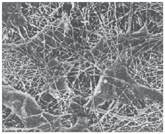
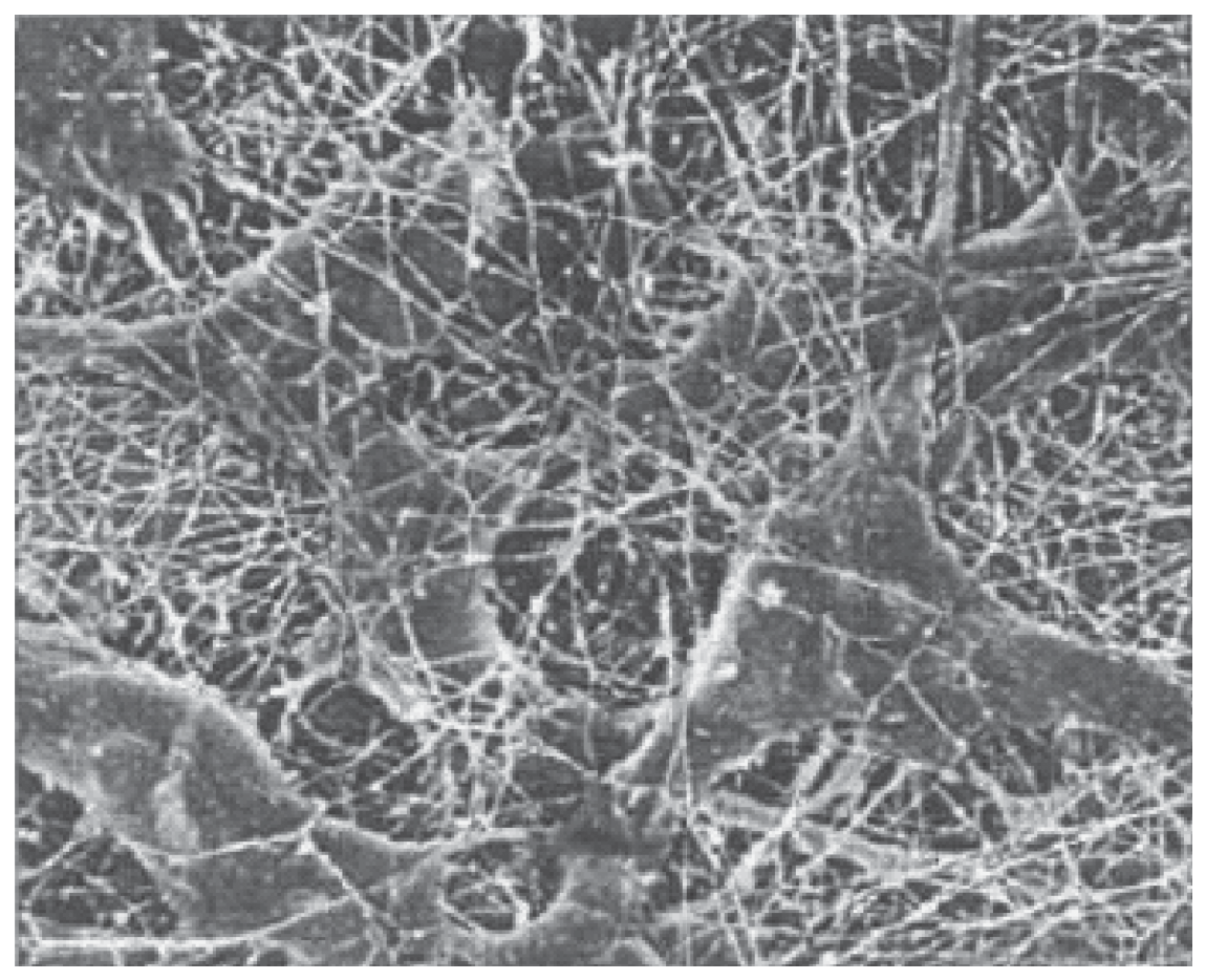
Tissue defect is a common phenomenon daily happening in clinic. Tissue engineering and translational medicine have introduced efficient methods for the repair and regeneration of defected tissue. The key issue for tissue regeneration is to provide a temporary scaffold for the defected tissue, which can induce the infiltration of cells and form new tissue with its gradually degradation. The existing procedures for constructing tissue engineering scaffolds are suitable to bulky tissue such as bone and cartilage. For some fine tissues and functional tissues, like vessel and nerve, there are no ideal regeneration scaffolds. Biomimetic functional electrospun nanofibers offer us new ideas and methods for those fine tissue engineering scaffolds and bring tissue engineering to a new stage, namely nano-biomimetic tissue Extracellular Matrix (ECM). Actually, the ECM in a body is essentially constructed by nanofibrous network containing protein and glycosaminoglycans, with a diameter ranging from 50 to 300 nm [1]. Cells are embedded in these nanofibrous network, thus form body tissue (Figure 1). Cells are specifically combined with the ligands on the ECM by the receptor in the cell membrane, which also respond to the external signal and affect the behaviors of cells. An ideal tissue engineering scaffold should mimic the native ECM. In addition, if the size of the scaffold’s skeleton is too large, during the formation of tissue, untimely degradation of the scaffold can always block the organized regeneration of the new tissue, which may lead to scar formation. This cannot achieve the aim of the regeneration of organized tissue, thus, new demands are required to the scaffold, which should be similar to the native ECM. That the phenomenon of scar formation is disappeared when the size of scaffold’s skeleton decreases from micron to nano fiber [2]. Recently, researchers found that the nano-fibrous structure has improved the application of tissue engineering scaffolds to bone, cartilage, cardiovascular, nervous and bladder regeneration with less scars [3].
Figure 1.
SEM photo of connective tissue (cells are embedded in nanofibrous network of collagen).
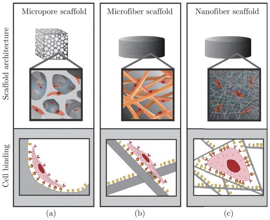
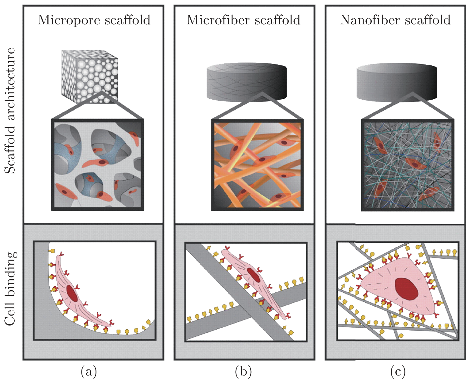
Researches showed that nano-materials can affect the behavior of cells notably. Pattison et al. [4] utilized nanoscale PLGA scaffold culture smooth muscle cell (SMC) to construct tissue engineering bladder. Compared with traditional microscale scaffold, cells demonstrated better adhesive and proliferation ability and secreted more collagen and elastin on nanoscale scaffold. Elias et al. [5] also proved that the proliferation ability of osteoblasts increased as the diameter of carbon nanofiber decreased. Osteoblasts secret more alkaline phosphatases and calcium on finer nanofiber scaffold. In 2005, Stevens et al. [6] published their article in Science, comparing the impact of scaffolds with different structure on the behavior of cells. They thought the cells adhere on the microscale scaffold in a tiled manner, which is similar to the stretched cells on a flat surface. On the other hand, with a high specific surface area, nanoscale scaffold could absorb more protein; provide more adhesion site for the receptors in cell membrane. The absorbed protein could expose more secret adhesion site by changing the conformation, facilitating the adhesion and proliferation of cells (Figure 2). Thus scaffolds made of nanofibers can mimic the physical structure of extracellular matrix and promote tissue regeneration.
Figure 2.
Schematic diagram of the effect of scaffold structure on cell adhesion and spreading. (a) and (b) indicate that cell adhesive on micropore and microfiber scaffold in a flat way, which is similar to that on flat surface. (c) Shows that nanofibrous scaffold can benefit the absorption of more protein, which also provide the receptors on cell membrane with more adhesion sites. The absorbed protein can expose more subtle site via changing the conformation of protein, thus contributing to cell adhesion [6].
1.2. Electrospun Nanofibers Biomimic the Structure and Function of ECM
Recently, electrospun nanofibers to biomimic native ECM for tissue engineering application has received more and more attention. Utilizing electrospinning technology, almost all the tissue engineering materials can be processed into nanofibers. These materials include synthetic polymers (PLA, PGA, PCL and their copolymers), natural polymers (collagen, gelatin, fibrous protein, silk, spider silk, hyaluronic acid, chitosan, cellulose) [7,8]. Different kind of structure could be obtained via different parameters, like randomly nanofibers, parallelly arranged nanofibers in varying degrees, randomly arranged layers or parallel layers or crossed layers. As cells proliferation can be oriented in direction along the nanofibers orientation [9], the nanofibrous materials can be designed for the regeneration of tissues with orientation texture. Apart from that the nanofibers can biomimic the structure of ECM, coaxial electrospinning also make nanofibers be possible for biomimicing the function of ECM. Via coaxial electrospinning, the active molecules and functional factors can be spun into the nanofibers and then continuously release from the fibers. Thus we can get tissue engineering scaffolds with special functions. For example, heparin was encapsulated in the nanofibrous vessel scaffold to form anticoagulant stent [10], growth factor was electrospun into nanofibrous nerve conduit which could promote nerve regeneration [11], bone morphogenetic protein was electrospun into the nanofibrous bone scaffold to induce Mesenchymal Stem Cells (MSC) differentiation into bone cells. Therefore, it is quite necessary for tissue regeneration to carry out the research on functional nanofiber scaffold fabrication to biomimic the native ECM, which is not only significant for the development of tissue engineering science but also meaningful for the health care of human beings.
2. Past 10 Years Research Results
Table 1 showed the electrospinning research results in the past 10 years in Biomaterials and Tissue Engineering Lab. of Donghua University. It can be seen that our research before 2009 gave only the nanofiber preparation and characterization for single materials or their complex. From 2009 we started to publish our coaxial electrospinning and emulsion electrospinning results to prepare serials of functional nanofibers. From 2010 our nanofiber for tissue engineering application had been gradually published, such as for skin tissue regeneration, nerve regeneration and blood vessel regeneration. In 2012 we published a dynamic fluid electrospinning method, with which a high porosity nanoyarn scaffold had been prepared, which allowed cell grow three-dimensionally for tendon and bone tissue engineering.
Table 1.
Historical development for electrospinning research in past 10 years.
2.1. Investigation on Protein-Polysaccharide Complex Nanofibers
Native ECM is constructed by the nanofibrous network containing collagen and glycosaminoglycan. In order to biomemic the structure and function of ECM, we have prepared serials of protein-polysaccharide composite nanofibers. Collagen and chitosan was electrospun into complex nanofibers by using a new developed cosolvents of HFIP and THF [12,13,14], silk fibroin and hyaluronic acid were electrospun into complex nanofibers [15] and silk fibroin and hydroxybutyl chitosan were also electrospun into complex nanofibers [16,17]. The mechanical properties of single nanofiber and nanofiber membrane consisting of collagen and chitosan were studied. It was found that the breaking strength and elongation at break of collagen and chitosan complex nanofiber were higher than that of single component nanofiber spun out from collagen or chitosan. When the content of chitosan in complex nanofiber was 20%, the fibers had an elongation at break of 45%, while that for collagen nanofiber was 4% and chitosan nanofiber was just 1%. When the content of chitosan in composite fiber was 40%, the fiber had a breaking strength of 63 MPa, while that for collagen nanofiber was 23 Mpa and 17 MPa for chitosan nanofiber. The collagen-chitosan complex nanofiber membrane showed elastic properties with the elongation at break of 73% and breaking strength of 2 MPa, when chitosan content was 20%. The good mechanical properties given by collagen-chitosan complex nanofibers could attribute to the intermolecular interaction existed between collagen and chitosan molecules [18]. The combination of collagen and chitosan can not only contribute to excellent mechanical properties of nanofibers, but can also improve its biological properties [19]. SMC was cultured on the nanofiber scaffold with different content of chitosan. It was found that the fastest proliferation rate occurred while the content of chitosan was 20%. Human tissue ECM was made up of collagen with a little mucooplysaccharide, which may be the reason why combination of collagen and chitosan would benefit the growth of cells.
2.2. Investigation on Collagen-Chitason-P(LLA-CL) Blend Nanofibers
Protein-polysaccharide composite nanofiber had a breaking strength of 2 MPa, which is quite suitable for the regeneration of skin but not enough for other tissue like small vessels, nerve and tendon etc. Synthetic materials have excellent mechanical properties, such as poly(L-lactide-co-caprolactone) (P(LLA-CL)), which is a elastic material when the ratio of lactic acid to caprolactone is 50:50. Blending natural materials with P(LLA-CL) may lead to new tissue engineering scaffolds with both good mechanical properties and biocompatibility.
We prepared collagen-chitosan-P(LLA-CL) blend nanofibers [20] and investigated the correlation of the mechanical properties and the blend ratios of these three components. Pure P(LLACL) Nanofibers had the breaking strength of 13 MPa and the elongation at break of 330%, showing the stress-strain behavior of elastic material. The collagen-chitosan-P(LLA-CL) nanofibers with the ratio of 20:5:75 reached the highest tensile strength with the value of 16.9 MPa, and it still kept the elasticity with elongation at break of 112% and elastic modulus of 10.3 MPa. The burst pressure strength of the collagen-chitosan-P(LLA-CL) nanofiber tube scaffold was as high as 3365 mmHg and the compliance value was 0.7%/100 mmHg. It has been reported that saphenous vein and mammary artery have burst pressure values of 1680-2273 and 2031-4225 mmHg, respectively [21,22]. Therefore, the nanofiber tube scaffold with the component ratio of 20:5:75 can resist burst pressure over 3365 mmHg, such strong and elastic scaffold has the potential for vascular graft applications. The contact angle of composite nanofibers was also related with the mixture ratio of three components. The contact angle decreased while the percentage of collagen and chitosan increased, illustrating a better biocompatibility. Endothelial cells proliferation on the nanofibers indicated that the proliferation activity of cells on collagen-chitosan-P(LLA-CL) nanofibers was better than that on pure P(LLA-CL) nanofibers or collagen-chitosan complex nanofibers. collagen-chitosan-P(LLA-CL) nanofibers with the blend ratio of 20:5:75 showed best mechanical properties and biocompatibility.
2.3. Investigation of SF-P(LLA-CL) Blend Nanofibers
Silk fibroin (SF) and P(LLA-CL) was blended together to be electrospun into nanofibers. The mechanical properties and biocompatibility of the obtained nanofibers were investigated [23]. As elastic material, P(LLA-CL) nanofibers showed the breaking strength of 6.96 MPa and elongation at break of 458%. When a small amount silk fibroin was added in (content of 25%), the SF-P(LLACL) nanofibers reached the highest tensile strength of 10.6 MPa, and it still keep the elasticity with the elongation at break of 279%. But with the further increasing of silk fibroin content the mechanical properties of SF-P(LLA-CL) nanofibers decreased. Pure silk fibroin nanofibers has the tensile strength of 2.72 MPa and the elongation at break of only 3.85%. The contact angle of the SF-P(LLA-CL) nanofibers decreased with the content of silk fibroin increasing, indicating the addition of silk fibroin improved the biocompatibility. Cell viability studies with pig iliac endothelial cells (PIEC) demonstrated that SF-P(LLA-CL) nanofibers significantly promoted cell proliferation in comparison with P(LLA-CL) nanofibers, especially when the weight ratio of SF to P(LLA-CL) was 25:75. SF-P(LLA-CL) nanofibers showed better mechanical properties and biocompatibility, which was consistent with that of the collagen-chitason-P(LLA-CL) nanofibers. So that, the best way to get tissue engineering scaffolds with both excellent mechanical properties and biocompatibility is to combine natural and synthetic material together to form the blend nanofibers via electrospinning.
The degradation performance of SF-P(LLA-CL) nanofibers in PBS (37 °C) was tested [24]. After degradation for 3 months, the fibrous morphology of pure P(LLA-CL) nanofibers was not observed and the weight loss was 20%. After degradation for 6 months, pure P(LLA-CL) nanofibers showed the weight loss of 50%, while SF-P(LLA-CL) blended nanofibers(SF:P(LLA-CL) is 25:75) also lost the nanofibrous morphology with the weight loss of 27%. SF nanofibers remained stable with the weight loss only 5%. P(LLA-CL) had a higher degradation rate in vitro, while the addition of silk fibroin slowed down this process. It was also found the degradation product of P(LLA-CL) contained acid which was reduced by the presence of silk fibroin.
2.4. Investigation in Core-shell Nanofibers
Electrospun nanofibers usually have a diameter of several hundred nanometers. Futher more, such fine fiber could also be constructed as core-shell structure. We successfully prepared core-shell structure nanofibers via coaxial electrospinning [25,26] and emulsion electrospinning [27,28].
Collagen was chosen as the shell layer and polyurethane (PU) as the core. Utilizing coaxial electrospinning process, collagen-PU core-shell structure nanofibers were manufactured. As the core provided excellent mechanical properties and the shell provided decent biocompatibility, scaffold made in this way could be applied into various tissue regeneration and repair [29].
Exploiting coaxial electrospinning and emulsion electrospinning, many active molecules could be spun into the core of the nanofiber and be released through the shell layer. For the first time, paclitaxel (PTX) was electrospun into the core of P(LLA-CL) nanofiber via coaxial electrospinning. PTX could be released from P(LLA-CL) nanofibers in a steady manner, effectively inhibited the proliferation of HeLa cells and showed the anticancer function [30]. This scaffold could be used as the tissue isolation membrane after tumor excision. Tetracycline hydrochloride (TCH) was encapsulated into the core of P(LLA-CL) electrospun nanofiber via coaxial electrospinning to get TCH loaded P(LLA-CL) nanofiber. TCH could be released slowly from TCH-P(LLA-CL) nanofibers in a long period and inhibited inhibit the activity of Gram-negative Escherichia [31]. This kind of nanofiber was suitable for antibacterial wound dressings. We successfully encapsulated nerve growth factor (NGF) in P(LLA-CL) nanofibers. It was found that the NGF could keep its activity in the high voltage electrostatic field. After released from the core-shell nanofibers, the NGF could induce PC-12 cell to grow and form the axons [32]. This nanofiber scaffold could be applied to prepare nerve conduits. Heparin was embedded in P(LLA-CL) nanofibers, which could prevent platelet adhesion [10,33]. This kind of tube scaffold was implanted into the femoral artery of dogs. The result indicated that the vascular patency was far better than those without heparin. Bone morphogenetic protein 2 (BMP2) and dexamethasone (DEX) have been coaxially electrospun into P(LLA-CL)/collagen nanofibers for controlled release during bone tissue engineering. The experimental results show that controlled release of BMP2 and DEX can induce hMSC to differentiate into osteogenic cells for bone tissue engineering [34].
2.5. Electrospun Nanofibers for Skin Tissue Regeneration
Electrospun nanofiber was developed into various tissue engineering scaffolds. Firstly we studied the application in skin tissue regeneration [35]. Collagen-chitosan complex nanofibers and silk fibroin-chitosan complex nanofibers were implanted in the full-thickness skin defect area on the back of SD rats. The results showed the biocompatibility of these two materials was excellent. Compared with gauze, those two complex nanofibers showed significant improvement function for the wound healing and the defects were healed in 3 weeks.
2.6. Electrospun Nanofiber for Nerve Tissue Regeneration
It was found that the SF-P(LLA-CL) blend nanofibers with the silk fibroin content of 25% had the most excellent mechanical properties in our previous studies. We have also found the parallelly aligned nanofibers can guide the cell orientation and induce the cell growth with high rate [36]. So we constructed 1.5 mm in diameter SF-P(LLA-CL) nanofiber nerve conduits with fiber alignment parallel to the axial direction, which was then implanted into the 1 cm nerve defect of sciatic nerve using rat model. After one month implantation, the nerve defect has been combined together by the new regenerated nerve. The regenerated nerve functions (nerve conduction velocity and distal compound motor action potential) were related to the materials components of the nerve conduit. SF-P(LLA-CL) nerve conduit showed better nerve function recovering than that of pure P(LLA-CL) conduit, the addition of silk fibroin accelerated the repairing of nerve [37].
To accelerate the nerve regeneration, NGF was spun in PLGA nanofibers by coaxial electrospinning, which was then made as nerve conduits and used for the repairing of the 1.5 cm sciatic nerve defect in rats [11]. It was found that the slow release of NGF significantly promote the regeneration of nerve. The recovery of the nerve function in conduit containing NGF was better than that in the conduit without NGF.
2.7. Electrospun Nanofiber for Blood Vessel Tissue Regeneration
Electrospun nanofibers are ideal scaffolds for small diameter blood vessel tissue regeneration. The purpose of our research was to prepare nanofiber tube scaffold and to investigate the blood vessel tissue regeneration in vivo. We produced two kinds of vascular grafts with the inner diameter of 3 mm, including heparin-P(LLA-CL) nanofiber graft by coaxial electrospinning and pure P(LLA-CL) nanofiber graft by conventional electrospinning, which were implanted to the dog’s femoral artery on left and right respectively. Angiography was used to observe the blood flow patency [38]. After three months implantation, the results showed that the patency of the scaffold without heparin was just 13% while that of the heparin-P(LLA-CL) tube was 87%, meaning the heparin played an important role in anticoagulation [39]. After 3 months implantation, endothelial cells proliferated in the lumun, but the P(LLA-CL) tube was not degraded and no new vascular tissue regenerated. Thus longer implantation time in animal test is needed in the future.
2.8. Dynamic Fluid Electrospinning for Nanoyarn Scaffold Fabrication
A new setup of dynamic fluid electrospinning was built in our laboratory, in this method nanofibers were first electrospun onto the water surface, which were then twisted into nanoyarns by water vortex. By collecting nanoyarns on a rotating drum we prepared nanoyarn membrane [40], and by collecting nanoyarns on a seven-pin plug-like roller we got 3-D nanoyarn scaffolds [41]. Compared with nanofiber membrane, nanoyarn membrane had higher porosity and bigger pore size. This porous structure would induce cells to penetrate into the deep side of the scaffold. Tendon cells were cultured on both nanoyarn membrane and nanofiber membrane, as a result, the cells proliferated faster on nanoyarns than on nanofibers and the gene expression with higher level on nanoyarns than on nanofibers [42]. The shortcoming of nanofibers is that the dense structure limited the cells growth only on the surface of nanofibers. New developed nanoyans has overcome that kind of shortage and shown great potential for tendon and cartilage tissue engineering.
3. Summary
In the past ten years we have developed a series nanofiber scaffolds based on natural and synthetic polymers by electrospinning and have investigated their great applications for skin, nerve and blood vessel tissue regeneration in vitro and in vivo. Collagen-chitosan complex nanofibers biomimetic the native ECM both in structure and components have shown higher elasticity and better cell proliferation ability. Those complex nanofibers have been succeeded used for skin regeneration in the back of SD rat. Collagen-chitosan-P(LLA-CL) nanofibers with the blend ratio of 20:5:75 showed the tensile strength of 16.9 MPa, elongation at break of 112% and burst pressure of 3365 mmHg, which was demonstrated to be suitable as blood vessel tissue engineering scaffold. SF-P(LLA-CL) nanofibers with the blend ratio of 25:75 showed the best mechanical properties with the tensile strength of 10.6 MPa and the elongation at break of 279%, as well as the best biological properties with highest cell proliferation rate among all SF-P(LLA-CL) nanofibers. Nerve conduits made from this SF-P(LLA-CL) nanofibers regenerated the 10 mm sciatic nerve defect of rat within four weeks and silk fibroin enhanced the nerve function recovery. Heparin-P(LLA-CL) nanofibers showed the antithrombotic properties by heparin release. Heparin-P(LLA-CL) tube scaffold was implanted in femoral artery of dog and it keep patency for three months. Nanoyarn scaffold has been developed through dynamic liquid electrospinning method, its high porosity allowed the cell penetrated three dimensionally in the scaffold and showed great potential for cartilage and tendon tissue regeneration.
Acknowledgments
The authors wish to acknowledge the Ministry of Science and Technology, Natural Science Foundation of China, Shanghai Science and Technology Commission and National Plan for Science and Technology for the funding support (2008AA03Z305, 30570503, 31070871, 31271035, 05DJ14006, 05PJ14013, 08520704600, 0852nm03400, 11nm0506200, 10-NAN1013-02), acknowledge to the Visiting Professor Program and special acknowledge to Prof. Cunyi Fan of Shanghai Jiaotong University Affiliated Sixth People’s Hospital, Prof. Sheng Wang of Tongji University Affiliated Tenth People’s Hospital and Prof. Wei Chen of Shanghai Eighth People’s Hospital for their cooperation on animal experiments of nerve, vessel and skin regeneration. The postgraduate students in Tissue Engineering Laboratory of Donghua University are greatly acknowledged for their contribution to our research.
References
- Piez, K.A. Molecular and aggregate structures of the collagens. In Extracellular Matrix Biochemistry; Reddi, A.H., Ed.; Elsevier: New York, 1984; pp. 1–35. [Google Scholar]
- Kim, B.S.; Mooney, D.J. Development of biocompatible synthetic extracellular matrices for tissue engineering. TIBTECH 1998, 16, 224–230. [Google Scholar] [CrossRef] [Scilit] [PubMed]
- Patch, K.; Body, S. E. Handles nanofiber better. Technol. Res. News 2003, 17, 24. [Google Scholar]
- Pattison, M.A.; Wurster, S.; Webster, T.J.; Haberstroh, K.M. Three-dimensional nano-structured PLGA scaffolds for bladder tissue replacement applications. Biomaterials 2005, 26(15), 2491–2500. [Google Scholar] [CrossRef] [Scilit] [PubMed]
- Elias, K.E.; Price, R.L.; Webster, T.J. Enhanced functions of osteoblasts on nanometer diameter carbon fibers. Biomaterials 2002, 23, 3279–3287. [Google Scholar] [CrossRef] [Scilit] [PubMed]
- Stevens, M.M.; George, J.H. Exploring and engineering the cell surface interface. Science 2005, 310, 1135–1138. [Google Scholar] [CrossRef] [Scilit] [PubMed]
- Yoshimoto, H.; Shin, Y.M.; Terai, H.; Vacanti, J.P. A biodegradable nanofiber scaffold by electrospinning and its potential for bone tissue engineering. Biomaterials 2003, 24, 2077–2082. [Google Scholar] [CrossRef] [Scilit] [PubMed]
- Min, B.M. Chitin and chitosan nanofibers: electrospinning of chitin and deacetylation of chitin nanofibers. Polymer 2004, 45, 7137–7142. [Google Scholar] [CrossRef] [Scilit]
- Xu, C.Y.; Inai, R.; Kotaki, M.; Ramakrishina, S. Aligned biodegradable nanofibrous structure: a potential scaffold for blood vessel engineering. Biomaterials 2004, 25, 877–886. [Google Scholar] [CrossRef] [Scilit] [PubMed]
- Su, Y.; Li, X.Q.; Liu, Y.N.; Su, Q.Q.; Lim, M.; Qiang, W.; Mo, X.M. Encapsulation and controlled release of heparin from electrospun Poly(L-Lactide-co--Caprolactone) nanofibers. J. Biomater. Sci. 2011, 22, 165–177. [Google Scholar] [CrossRef] [Scilit] [PubMed]
- Wang, C.Y.; Liu, J.J.; Fan, C.Y.; Mo, X.M.; Ruan, H.J.; Li, F.F. The Effect of aligned core-shell nanofibres delivering NGF on the promotion of sciatic nerve regeneration. J. Biomater. Sci. Polym. Ed. 2012, 23(1-4), 167–184. [Google Scholar] [CrossRef] [Scilit] [PubMed]
- Chen, Z.G.; Mo, X.M.; Qing, F.L. Electrospinning of collagen-chitosan complex. Mater. Lett. 2007, 61, 3490–3494. [Google Scholar] [CrossRef] [Scilit]
- Chen, Z.G.; Wei, B.; Mo, X.M.; Lim, C.T.; Ramakrishna, S.; Cui, F.Z. Mechanical properties of electrospun collagen-chitosan complex single fibers and membrane. Mater. Sci. Eng. C 2009, 29, 2428–2435. [Google Scholar] [CrossRef] [Scilit]
- Chen, Z.G.; Wei, B.; Mo, X.M.; Cui, F.Z. Diameter control of electrospun chitosan-collagen fibers. Polyp. Sci. Part B Polym. Phys. 2009, 47, 1949–1955. [Google Scholar] [CrossRef] [Scilit]
- Zhang, K.H.; Fan, L.P.; Yan, Z.Y.; Yu, Q.Z.; Mo, X.M. Electrospun biomimic nanofibrous scaffolds of silk fibroin/hyaluronic acid for tissue engineering. J. Biomaterilas Sci.-Polym. Ed. 2012, 23(9), 1185–1198. [Google Scholar]
- Zhang, K.H.; Yu, Q.Z.; Mo, X.M. Fabrication and intermolecular interactions of silk fibroin/hydroxybutyl chitosan blended nanofibers. Int. J. Mol. Sci. 2011, 12, 2187–99. [Google Scholar] [CrossRef] [Scilit] [PubMed]
- Zhang, K.H.; Qian, Y.F.; Wang, H.S.; Fan, L.P.; Huang, C.; Mo, X.M. Electrospun silk fibroin-hydroxybutyl chitosan nanofibrous scaffolds to biomimic extracellular matrix. J. Biomater. Sci. Ed. 2011, 22, 1069–82. [Google Scholar] [CrossRef] [Scilit] [PubMed]
- Chen, Z.G.; Mo, X.M.; He, C.L.; Wang, H.S. Intermolecular interactions in electrospun collagenchitosan complex nanofibers. Carbohydr. Polym. 2008, 72(3), 410–418. [Google Scholar] [CrossRef] [Scilit]
- Chen, Z.G.; Wang, P.W.; Wei, B.; Mo, X.M.; Cui, F.Z. Electrospun collagen-chitosan nanofiber: A biomimetic extracellular matrix for endothelial cell and smooth muscle cell. Acta Biomater. 2010, 6, 372–382. [Google Scholar] [CrossRef] [Scilit] [PubMed]
- Yin, A.L.; Zhang, K.H.; McClure, M. J.; Huang, C.; Fang, J.; Wu, J.L.; Bowlin, G. L.; Mo, X.M.; Al-Deyab, S.S.; El-Newehy, M. Electrospinning collagen/chitosan/poly(L-lactic acid-co-e-caprolactone) to form a vascular graft: mechanical and biological characterization. J. Biomed. Mater. Res. Part A 2013, 101A, 1292–1301. [Google Scholar] [CrossRef] [Scilit] [PubMed]
- Konig, G.; McAllister, T.N.; Dusserre, N.; Garrido, S.A.; Iyican, C.; Marini, A.; Fiorillo, A.; Avila, H.; Wystrychowski, W.; Zagalski, K.; Maruszewski, M.; Linthurst Jones, A.; Cierpka, L.; de la Fuente, L.M.; L’Heureux, N. Mechanical properties of completely autologous human tissue engineered blood vessels compared to human saphenous vein and mammary artery. Biomaterials 2009, 30, 1542–155. [Google Scholar] [CrossRef] [Scilit] [PubMed]
- L’Heureux, N.; Dusserre, N.; Konig, G.; Victor, B.; Keire, P.; Wight, T.N.; Chronos, N.A.F.; Kyles, A.E.; Gregory, C.R.; Hoyt, G.; Robbins, R.C.; McAllister, T.N. Human tissue engineered blood vessel for adult arterial revascularization. Nat. Med. 2006, 12, 361–365. [Google Scholar] [CrossRef] [Scilit] [PubMed]
- Zhang, K.H.; Wang, H.S.; Huang, C.; Su, Y.; Mo, X.M.; Ikada, Y. Fabrication of silk fibroin blended P(LLACL) nanofibrous scaffolds for tissue engineering. J. BioMed Mater. Res. A 2010, 93(3), 984–993. [Google Scholar] [CrossRef] [Scilit] [PubMed]
- Zhang, K.H.; Yin, A.L.; Huang, C.; Wang, C.Y.; Mo, X.M.; Al-Deyab, S.S.; El-Newehy, M. Degradation of electrospun SF/P(LLA-CL) blended nanofibrous scaffolds in vitro. Polym. Degrad. Stab. 2011, 96, 2266–75. [Google Scholar] [CrossRef] [Scilit]
- Li, X.Q.; Su, Y.; Chen, R.; He, C.L.; Wang, H.S.; Mo, X.M. Fabrication and properties of core-shell structure P(LLA-CL) nanofibers by coaxial Electrospinning. J. Appl. Polym. Sci. 2009, 111, 1564–1570. [Google Scholar] [CrossRef] [Scilit]
- Su, Y.; Li, X.Q.; Wang, H.S.; He, C.L.; Mo, X.M. Fabrication and characterizations of biodegradable nanofibrous mats by mix and coaxial electrospinning of Poly(L-lactid-co--caprolactone). J. Mater. Sci. Mater. Med. 2009, 20, 2285–2294. [Google Scholar] [CrossRef] [Scilit] [PubMed]
- Li, X.Q.; Su, Y.; Zhou, X.; Mo, X.M. Distribution of sorbitan monooleate in poly(l-lactide-co-caprolactone) nanofibers from emulsion electrospinning. Colloids Surf. B Biointerfaces 2009, 69, 221–224. [Google Scholar] [CrossRef] [Scilit] [PubMed]
- Su, Y.; Li, X.Q.; Liu, S.P.; Mo, X.M.; Ramakrishna, S. Controlled release of dual drugs from emulsion electrospun nanofibrous mats. Colloids Interfaces B Biointerfaces 2009, 73(2), 376–381. [Google Scholar] [CrossRef] [Scilit] [PubMed]
- Chen, R.; Huang, C.; Ke, Q.F.; He, C.L.; Wang, H.S.; Mo, X.M. Preparation and characterization of coaxial electrospun thermoplastic polyurethane/collagen compound nanofibers for tissue engineering applications. Colloids Surf. B Biointerfaces 2010, 79, 315–325. [Google Scholar] [CrossRef] [Scilit] [PubMed]
- Huang, H.H.; He, C.L.; Wang, H.S.; Mo, X.M. Preparation of core-shell biodegradable microfibers for long-term drug delivery. J. BioMed Mater. Res. A 2009, 90A(4), 1243–1251. [Google Scholar] [CrossRef] [Scilit] [PubMed]
- Su, Y.; Li, X.Q.; Tan, L.J.; Huang, C.; Mo, X.M. Electrospun Poly(L-lactide-co-ε-caprolactone) nanofibers for encapsulating and sustained releasing proteins. Polymer 2009, 17(50), 4212–4219. [Google Scholar] [CrossRef] [Scilit]
- Li, X.Q.; Su, Y.; Liu, S.P.; Tan, LianJ; Mo, X.M.; Ramakrishna, S. Encapsulation of proteins in poly(llactide-co-caprolactone) fibers by emulsion electrospinning. Colloids Surf. B Biointerfaces 2010, 75, 418–424. [Google Scholar] [CrossRef] [Scilit] [PubMed]
- Chen, F.; Huang, P.; Mo, X.M. Electrospinning of heparin encapsulated P(LLA-CL) core/shell nanofibers. Nano Biomed. Eng. 2010, 2, 84–90. [Google Scholar] [CrossRef] [Scilit][Green Version]
- Su, Y.; Su, Q.Q.; Liu, W.; Lim, M.; Venugopal, J.R.; Mo, X.M.; Ramakrishna, S.; Al-Deyab, S.S.; El-Newehy, M. Controlled release of bone morphogenetic protein 2 and dexamethasone loaded in core-shell PLLACL-collagen fibers for use in bone tissue engineering. Acta Biomater. 2012, 8, 763–771. [Google Scholar] [CrossRef] [Scilit] [PubMed]
- Yu, P.J.; Wang, L.P.; Guo, S.; Chen, W.; Mo, X.M. Collegen-chitosan complex nanofibers for skin tissue regeneration. J. Clin. Rehabil. Tissue Eng. Res. 2011, 15(51), 95619564. [Google Scholar]
- Zhang, K.H.; Wu, J.L.; Huang, C.; Mo, X.M. Fabrication of silk fibroin/P(LLA-CL) aligned nanofibrous scaffolds for nerve tissue engineering. Macromol. Mater. Eng. 2013, 298(5), 565–574. [Google Scholar] [CrossRef] [Scilit]
- Wang, C.Y.; Zhang, K.H.; Fan, C.Y.; Mo, X.M.; Ruan, H.J.; Li, F.F. Aligned natural-synthetic polyblend nanofibers for peripheral nerve regeneration. Acta Biomater. 2011, 7, 634–643. [Google Scholar] [CrossRef] [Scilit] [PubMed]
- Huang, C.; Wang, S.; Qiu, L.J.; Ke, Q.F.; Zhai, W.; Mo, X.M. Heparin loading and pre-endothelialization in enhancing the patency rate of electrospun small-diameter vascular grafts in a canine model. ACS Appl. Mater. Interfaces 2013, 5, 2220–2226. [Google Scholar] [CrossRef] [Scilit] [PubMed]
- Wang, S.; Mo, X.M.; Jiang, B.J.; Gao, C.J.; Wang, H.S.; Zhuang, Y.G.; Qiu, L.J. Fabrication of small-diameter vascular scaffolds by heparin-bonded P(LLA-CL) composite nanofibers to improve graft patency. Int. J. Nanomed. 2013, 8, 2131–2139. [Google Scholar] [CrossRef] [Scilit] [PubMed]
- Wu, J.L.; Huang, C.; Liu, W.; Yin, A.L.; Chen, W.M.; Liu, S.; Fan, C.Y.; Mo, X.M.; Bowlin, G.L. Electrospun nanoyarn scaffold and its application in tissue engineering. Mater. Lett. 2012, 89, 146–149. [Google Scholar] [CrossRef] [Scilit]
- Li, J.; Liu, W.; Yin, A.L.; Wu, J.L.; Al-Deyab, S.S.; El-Newehy, M.; Mo, X.M. Nano-yarns reinforced silk fibroin composites scaffold for bone tissue engineering. J. Fibre Bioeng. Inform. 2012, 5(1), 1–11. [Google Scholar] [CrossRef] [Scilit]
- Xu, Y.; Wu, J.L.; Wang, H.M.; Li, H.Q.; Di, N.; Song, L.; Li, S.T.; Li, D.W.; Xiang, Y.; Liu, W.; Mo, X.M.; Zhou, Q. Fabrication of Electrospun Poly(L-Lactide-co-e-Caprolactone)/collagen nanoyarn network as a novel, three-Dimensional, macroporous, aligned scaffold for tendon tissue engineering. Tissue Eng. Part C 2013. [Google Scholar] [CrossRef] [Scilit] [PubMed]

© 2013 Textile Bioengineering and Informatics Society. This article is an open access article distributed under the terms and conditions of the Creative Commons Attribution (CC BY) license (https://creativecommons.org/licenses/by/4.0/).