


Abstract
The Bombyx mori silk fibroin gels were prepared by adding various types of polyalcohol (Ethylene glycol, Polyethylene glycol 600) into the regenerated silk fibroin aqueous solution. The gelation time was recorded respectively. Gel structure was analyzed by X-ray diffraction, Fourier transform infrared spectroscopy, laser Raman spectroscopy and scanning electron microscopy. It was found that adding polyalcohol into the regenerated silk fibroin aqueous solution can accelerate the gelation process, with increase in the percentage of the polyalcohol except at 100%. Silk fibroin gels with Ethylene glycol mainly consist of silk II crystal structure. Ethylene glycol almost has no effect on the molecular conformation or crystal structure of silk fibroin gels. However, adding PEG600 to the solution can promote the silk fibroin molecules to form clusters rapidly, while it can also obstruct the structural transformation of silk fibroin molecules from random coil or α-helix to β-sheet. The crystallinity of silk gels was found to decline with an increase in the adding percentage of PEG600. While adding 100% polyalcohol of the mass of silk fibroin into the solution, whether it was Ethylene glycol or PEG600, the silk fibroin gels have more uniform morphology and pores distribution.
1. Introduction
Silk from Bombyx mori are natural fibrous polymers that have been increasingly studied as new biomaterials in recent years due to its biocompatibility, degradability and excellent mechanical properties. As the structural protein of Bombyx mori silk fiber, silk fibroin has also been prepared or designed into various formats such as nanospheres, films, porous materials, for controlled release-delivery of bioactive agents [1,2,3], cell culture and tissue engineering [4,5,6,7,8].
Among many Bombyx mori silk fibroin materials, silk fibroin gel with three a dimensional structure is an important form to be developed in tissue engineering materials. Besides the flexibility and plasticity, it also has permeability for some small molecular or polymer materials. Silk fibroin gel has been studied or applied in artificial skin, contact lenses, drug controlled-release carrier, enzyme immobilization carrier, cell culture matrix [9,10,11]. Therefore, it is necessary to explore the formation mechanism of silk fibroin gel and its influencing factors.
During the formation of silk fibroin gel, it mainly includes hydrophobic interactions, hydrogen bond formation and electrostatic interactions [12,13,14,15,16]. There are many influencing factors such as temperature, silk fibroin concentration, pH and ions, which significantly affect the gelation process. Gelation will occur in the regenerated silk fibroin solution under certain conditions, mainly due to a structural transition from random coil or α-helix to β-sheet [17]. Hirabayashi et al [18] reported the formation mechanism of silk fibroin gel and considered that the gel formation was due to hydrogen bond formation between silk fibroin molecular chains. Akira Matsumoto et al. [19] studied mechanisms of silk fibroin sol-gel transitions by monitoring the process under various physicochemical conditions with optical spectroscopy at 550 nm. This study indicated that there are two stages during the gelation process. In the early stage small precipitates (<10 μm) first appeared then sizes of these precipitates gradually increased. In the later stage, larger aggregates (10-100 μm) became immobile in the bulk or partially gelled state.
In vitro [20,21,22], the silk fibroin molecules in aqueous solution will turn into α-helix and β-sheet conformation after gelation. This sol - gel transition process was related to the concentration of silk fibroin solution, temperature, pH, and ion concentration. M. Fini et al [23] induced to prepare silk fibroin hydrogel for healing the critical size defects of rabbit distal femurs by adjusting the pH of the silk fibroin solution. Xiaoqin Wang et al [24] studied sonication-induced gelation of silk fibroin for cell encapsulation and found that ultrasonication could accelerate the formation of physical cross-links responsible for gel stabilization, because it initiated the formation of β-sheets by alteration in hydrophobic hydration, and then formed gels finally. Ung-Jin Kim et al [25] reported that strong hydrophilic substances such as PEO could facilitate the intermolecular or intramolecular interactions among the protein molecules and the subsequent formation of the β-sheet structure. Thus it promoted the sol-gel transitions, which could be induced by direct addition of PEO into the fibroin aqueous solutions or via separation from the aqueous solutions across a dialysis membrane (with PEG).
In the present study, we have studied in detail the strong hydrophilic substance such as polyalcohol (mainly Ethylene glycol or PEG 600) that influence silk fibroin sol-gel transitions, because it is one of the important factors which should not be overlooked. The Bombyx mori silk fibroin gels were prepared by adding various quality of polyalcohol (Ethylene glycol, PEG 600) into the regenerated silk fibroin aqueous solution. The gelation time of regenerated silk fibroin aqueous solutions with various qualities of polyalcohol was studied. The morphology and the structure of silk fibroin gels were also studied.
2. Materials and methods
2.1 Preparation of regenerated silk fibroin solution
According to the literature [26], the Bombyx mori silks were treated three times with 0.05 wt % Na2CO3 solution at 98~100°C for 30 min respectively to remove sericin. Then they were rinsed and air dried. The pure silk fibroin fibers were dissolved with triad solvent CaCl2·CH3CH2OH·H2O (mole ratio=1:2:8) at 78±2°C through stirring. The prepared solution was purified by dialyzed against water for three days, to obtain 2.7 wt % silk fibroin solution. The regenerated silk fibroin solution was prepared, and then it was stirred slowly at room temperature to make it to evaporate and concentrate up to 3.0 wt %.
2.2 Preparation of regenerated silk fibroin gels
The 25mL silk fibroin solutions at concentrations of 3.0 wt% were injected into two groups of culture dishes (diameter 8.5 cm, each group with six culture dishes). Added Ethylene glycol to the first group and PEG 600 to the second group, according to these percentages (0%, 100%, 300%, 500%, 700%, and 900%) of solute mass, respectively. Stirred while adding polyalcohol, and then were put aside at room temperature. Gelation time of each sample was recorded. After freeze-drying, silk fibroin gels were prepared.
2.3 X-ray diffraction
X-ray diffraction was performed by a Rigaku D/Max-3C diffractometer with Cu-Kα radiation (λ= 0.15418 nm). The X-ray source was operated at 40 kV and 40 mA. Diffraction intensity was measured in reflection mode at a scanning rate of 2°/min for 2θ=5-40°.
2.4 FTIR spectroscopy
Fourier transform infrared (FTIR) spectra were obtained by a Nicolet Avatar-IR360.
2.5 Laser Raman Spectroscopy
Raman spectra were recorded using a Dilor LabRam-1B spectrometer, operating at a resolution of 1 cm−1. The Spectra Physics Model 164 argon ion laser was operated at 632.8 nm with about 6mW power.
2.6 Scanning electron microscopy
The gel samples were cut horizontally at middle depth. The cross-sectional surface was examined with a Hitachi S-570 Scanning Electron Microscope (SEM).
3. Results and discussion
3.1 The effects of polyalcohol on the gelation time
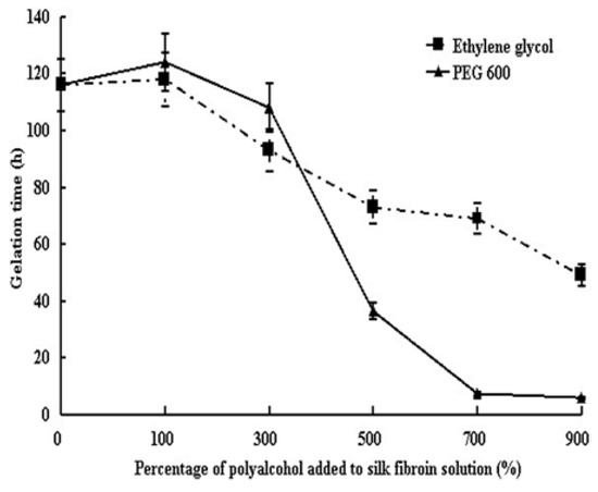
Adding ethylene glycol or PEG600 into the regenerated silk fibroin aqueous solutions can accelerate the gelation process with an increase in percentage except at 100%. The gelation time of silk fibroin solution without polyalcohol (the control sample) was 116h (Figure 1). This is because the water-bonding capacity of the strong hydrophilic substances, such as Ethylene glycol, PEG 600, broke hydration layer around the fibroin molecules in the solution.It then competed for water molecules with silk fibroin molecules, which reduced water molecules among the silk fibroin chains and facilitated to collide, leading to the acceleration of silk fibroin molecular aggregation and coagulation. In addition, while silk fibroin molecules collided, the hydrophobic interactions would occur with the folding of molecular chain. The breaking and rebuilding of hydrogen bonds, lead to silk fibroin molecules intertwining with each other and increase the opportunity to agglomerate together, thus the gelation process was promoted.
Figure 1.
Gelation time of Bombyx mori silk fibroin solutions by adding polyalcohol with various percentages.
Interestingly, when the percentage of polyalcohol added into silk fibroin is less than 300%, the ethylene glycol is more effective to promote the gelation process than PEG600. But when the percentage of polyalcohol added into silk fibroin is more than 300%, the PEG600 is more outstanding. It is speculated that this may be due to two factors. The number of hydrophilic groups in the solution and the molecular weight of polyalcohol. Initially, with the increase in the percentage of polyalcohol added into solution, ethylene glycol has more hydrophilic groups than that of the same percentage of PEG600. While the number of hydrophilic groups in the solutions formed by adding the ethylene glycol is absolute preponderant, under these circumstances, the molecular chain length and molecular weight of polyalcohol may have no effects on the gelation process, so the gelation time while adding ethylene glycol is even shorter. However, when the percentage of polyalcohol added into silk fibroin is greater than 300%, the difference of the number of hydrophilic groups in the solution does not seem so important, and PEG600 with the longer molecular chain make the silk fibroin molecular fold more easily after combining with the silk fibroin molecules, which facilitated silk fibroin molecules to tangle with each other and accelerated the aggregation of silk fibroin until gels were formed completely.
3.2 The effects of ethylene glycol on the structure of silk fibroin gels
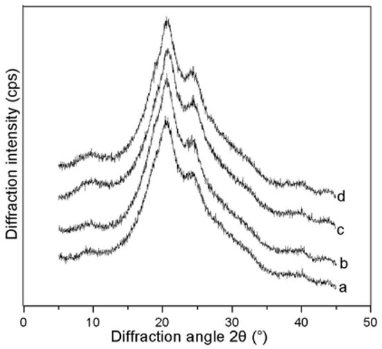
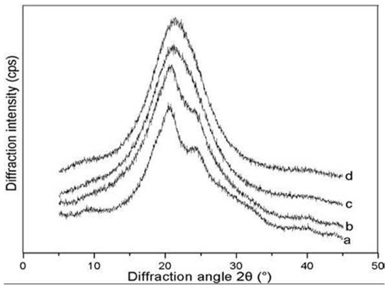
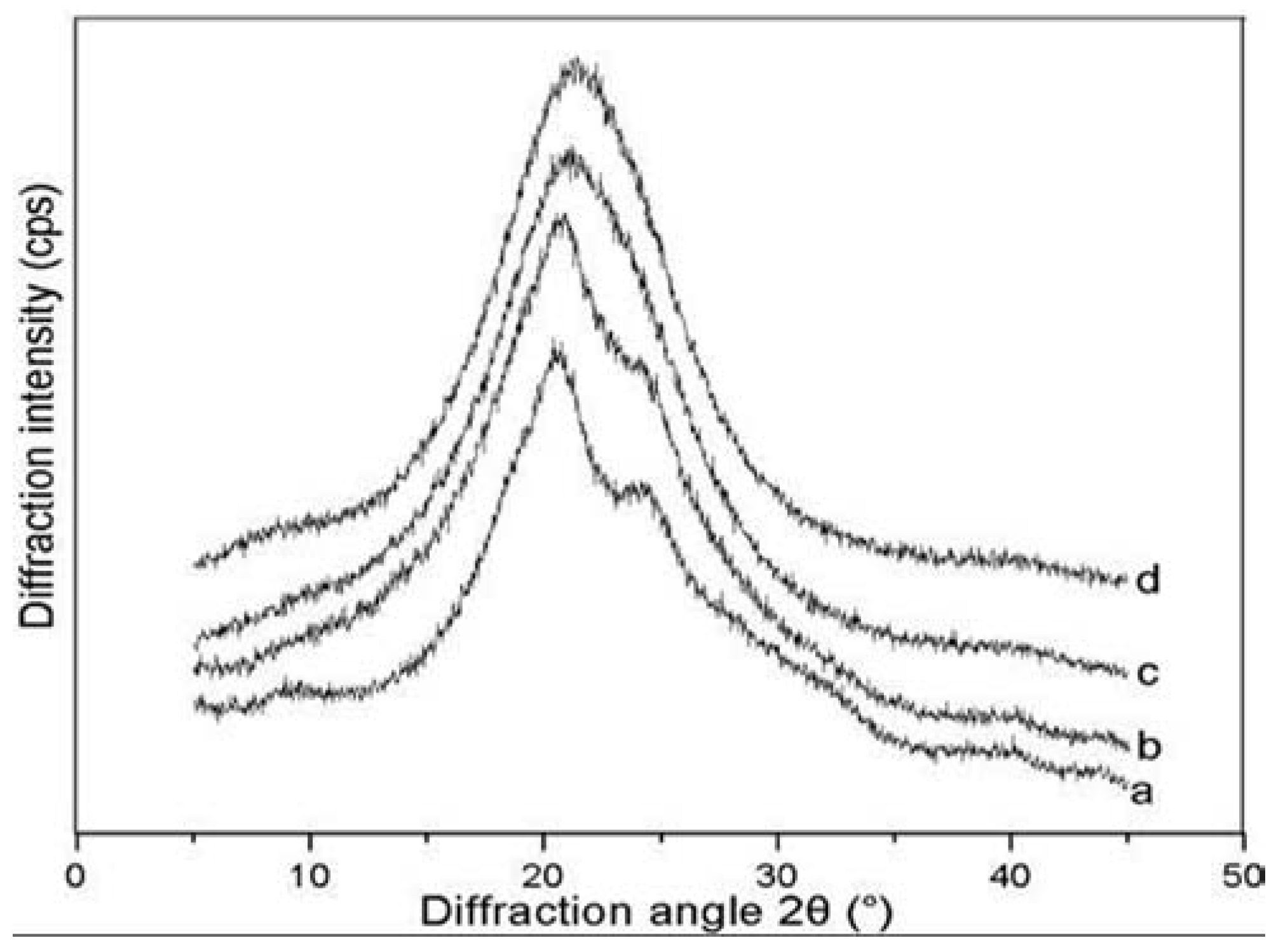
In Figure 2(A), there are some diffraction peaks around 9.3° (weak), 20.7° (strong), 24.7° (middle strong) in XRD curve of Bombyx mori silk fibroin gels with Ethylene glycol. According to the X-ray diffraction patterns of Bombyx mori silk fibroin materials determined in earlier studies [27,28,29,30], it means that these silk fibroin gels mainly consist of silk II crystal structure and Ethylene glycol has no effect on the molecular conformation or crystal structure of silk fibroin gels.
Figure 2(A).
X-ray diffraction curves of Bombyx mori silk fibroin gels with Ethylene glycol; the percentage of Ethylene glycol: (a) 0%, (b) 100%, (c) 500%, (d) 900%.
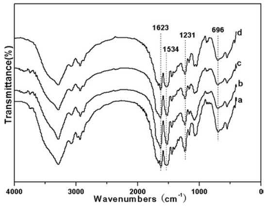
Figure 2(B) shows the FTIR spectra of Bombyx mori silk fibroin gels with Ethylene glycol. The control sample (curve a) shows strong bands at 1623 cm-1(amide I), 1231.8 cm-1(amide III), 696.5 cm-1 (amide V), which attributed to β-sheet structure based on these studies [31,32].
Figure 2(B).
FTIR spectra of Bombyx mori silk fibroin gels with Ethylene glycol; the percentage of Ethylene glycol: (a) 0%, (b) 100%, (c) 500%, (d) 900%.
When the added amount of Ethylene glycol was 100% of the mass of silk fibroin in aqueous solution, the bands at 1627.1 cm-1 (amide I), 1230.7 cm-1 (amide III), 696.6 cm-1 (amide V) became slightly weaker(curve b),but basically the same spectrum with that of control sample. The silk fibroin gels with 500% or 900% ethylene glycol almost had no changes in the spectrum compared with that of the controlled sample or silk fibroin gels with 100% ethylene glycol, indicating that the molecular conformation of silk fibroin in the gels with ethylene glycol almost had no changes, regardless of the proportion of ethylene glycol. These changes closely correspond to those of X-ray diffraction profile.
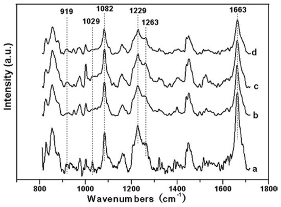
According to our experiment results, as shown in Figure 2(C), the Raman spectrum of silk fibroin gels exhibits bands near 1229 cm-1(amide III), 1082 cm-1, 1029 cm-1, respectively, which are obviously the characteristic bands of β-sheet conformation. It also exhibits bands at 1663 cm-1(amide I), 919 cm-1, which are attributed to random coil and α-helix conformation, respectively. With the increase in the percentage of ethylene glycol, the relative intensity of all of these bands has no significant changes, corresponding to the results of X-ray diffraction profile and that of the FTIR spectrum, indicating that Ethylene glycol almost has no effect on the molecular conformation of silk fibroin in the gels.
Figure 2(C).
Raman spectrum of Bombyx mori silk fibroin gels with Ethylene glycol; the percentage of Ethylene glycol: (a) 0%, (b) 100%, (c) 500%, (d) 900%.
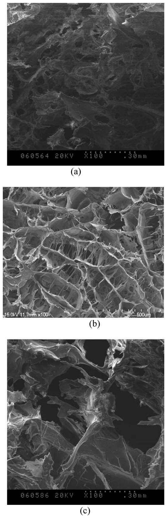
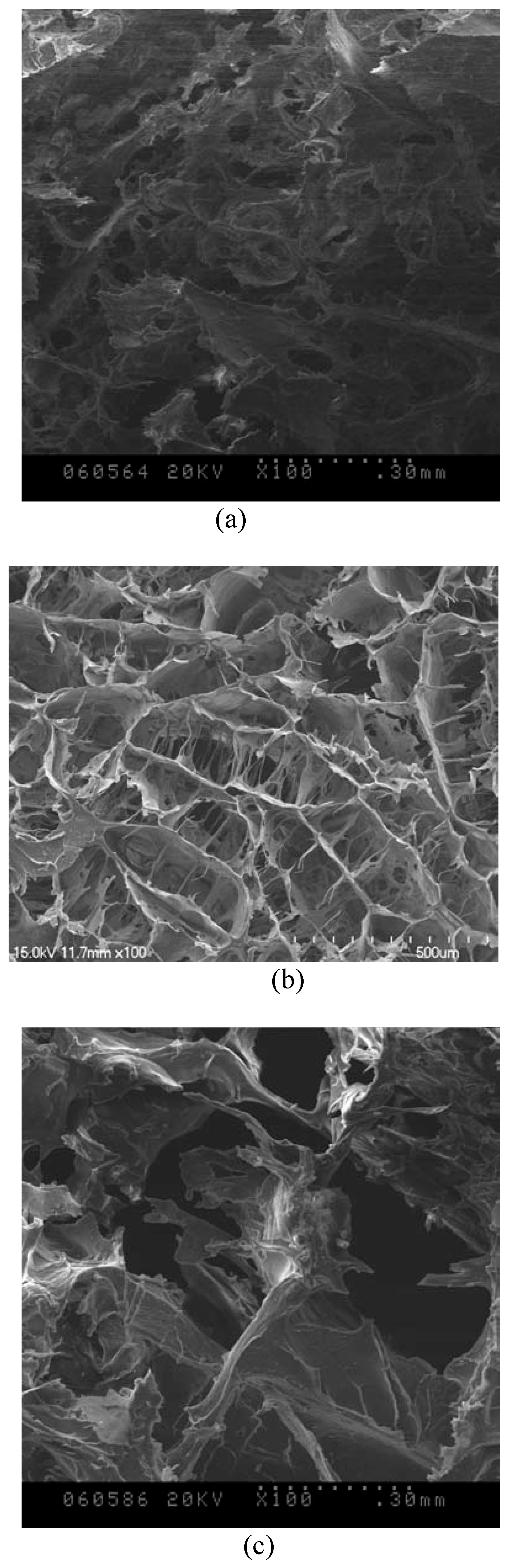
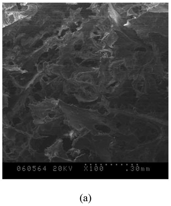
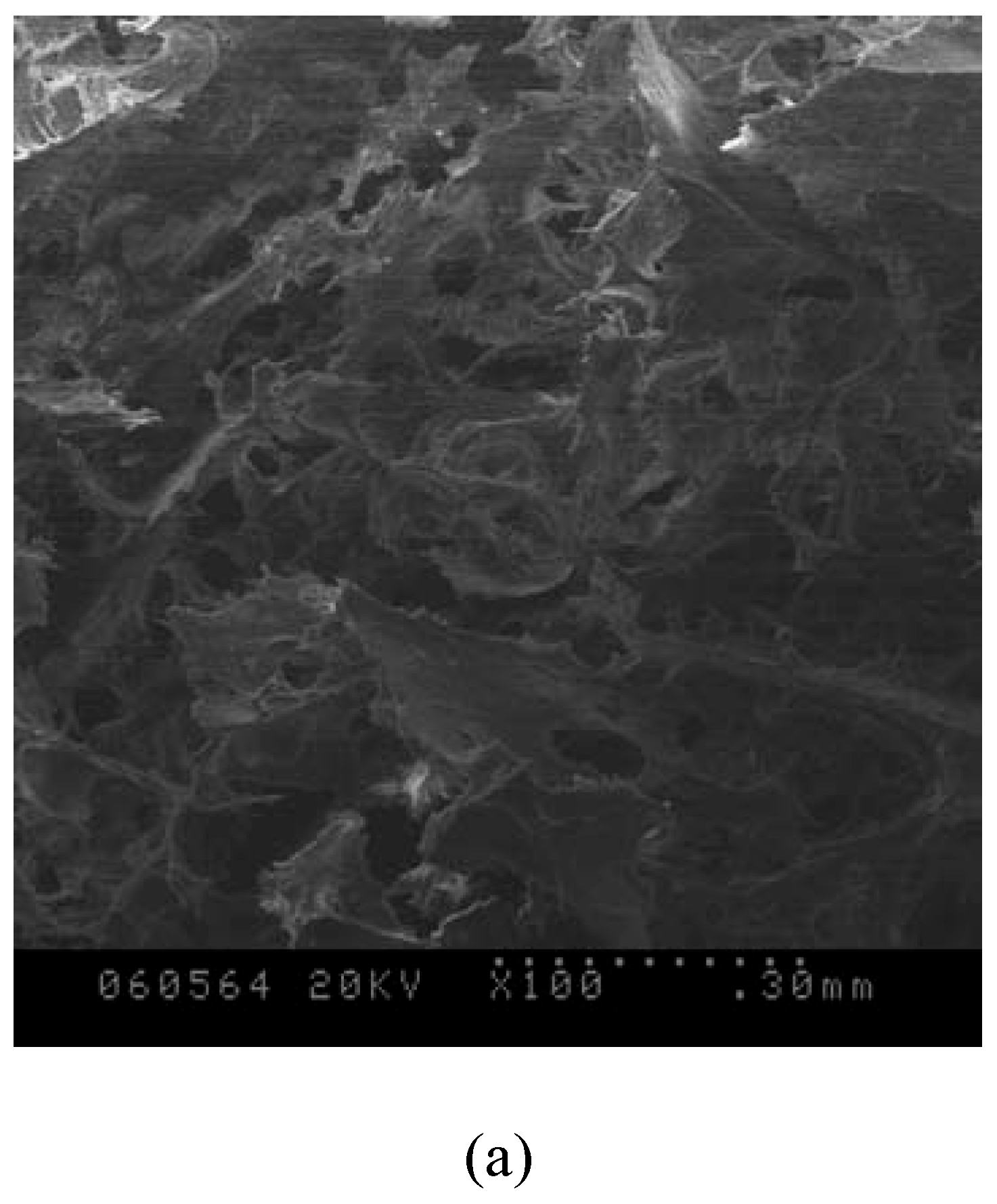
In Figure 3, the silk fibroin gels with 100% of ethylene glycol had more uniform morphology and pores distribution. This may be related to its gel time. The longer the gel time, the better was the uniformity for the aggregation of fibroin molecules.
Figure 3.
SEM of Bombyx mori silk fibroin gels with ethylene glycol; the percentage of ethylene glycol: (a) 0%, (b) 100%, (c) 500%; ×100.
3.3 The effects of PEG600 on the structure of silk fibroin gels
In Figure 4(A), diffraction peaks appears around 9.3° (weak), 20.7° (strong), 24.7° (middle strong) in the curve (a), which are attributed to the silk II crystal structure of Bombyx mori silk fibroin gels with PEG600. However, with an increase in the percentage of PEG600, the peaks around 9.3° and 24.7° disappear, the peak around 20.7° becomes broader, indicating that the molecular conformation of silk fibroin changes obviously and silk II crystal structure in those gels may decline. This is because when PEG 600 was added to the solution, it can promote the silk fibroin molecules to form clusters rapidly, while it also obstructs the structural transformation of silk fibroin molecules from random coil or α-helix to β-sheet and the crystallinity was declined with an increase in the adding percentage.
Figure 4(A).
X-ray diffraction curves of Bombyx mori silk fibroin gels with PEG600; the percentage of PEG600: (a) 0%, (b) 100%, (c) 500%, (d) 900%.
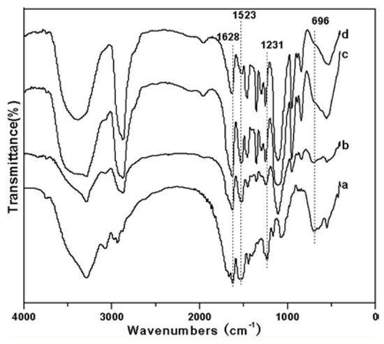
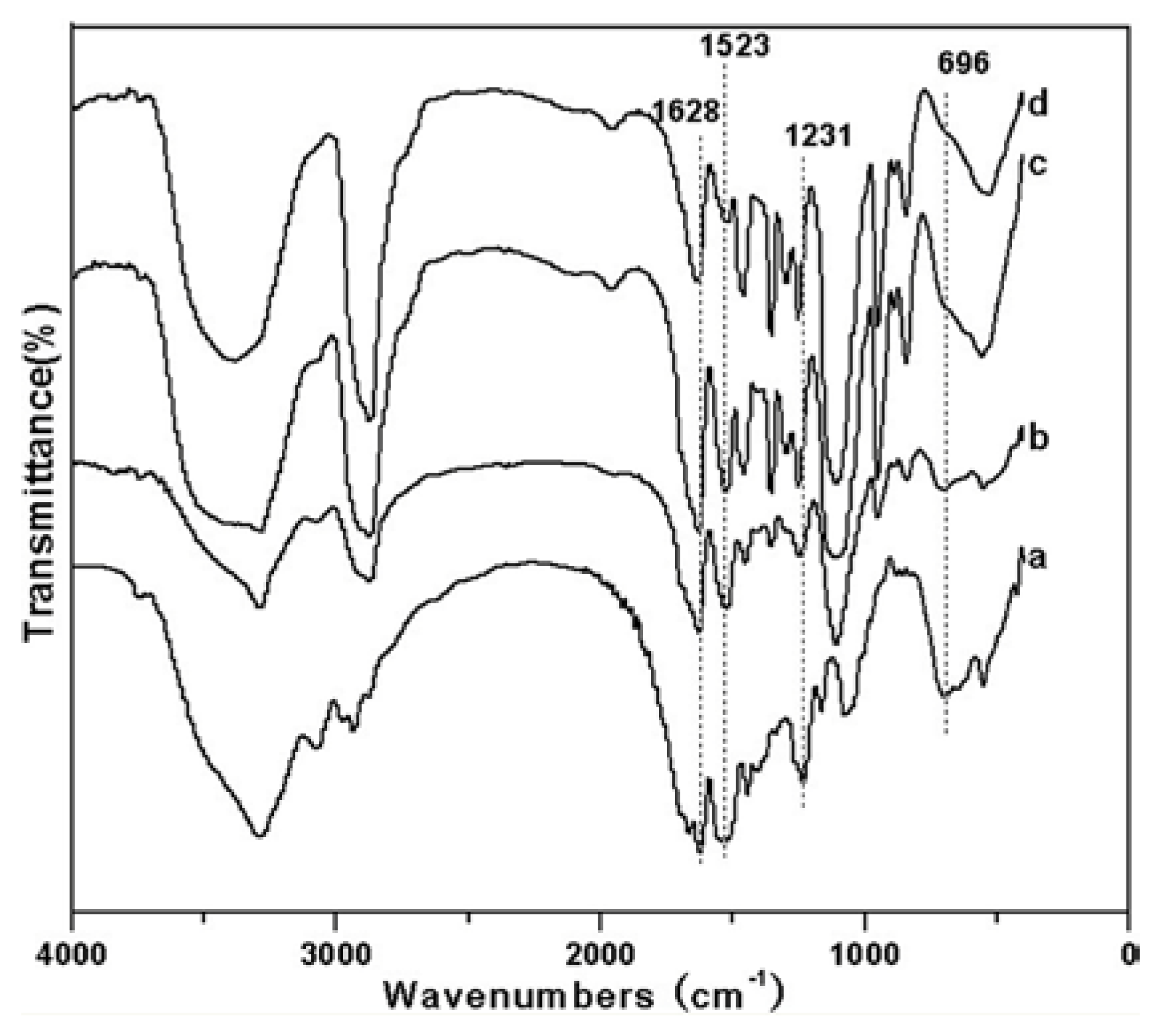
As shown obviously in Figure 4(B), the FTIR spectra of Bombyx mori silk fibroin gels with 100% PEG600 (curve b) almost has no changes compared with that of the control sample (curve a). When adding 500% PEG600 (curve c), some bands appear at 1628.8 cm-1(amide I), 1523.5 cm-1 (amide II), which are attributed to β-sheet structure. But when adding 900% PEG600 (curve d), only one weak band appears at 1631.3 cm-1 (amide I), which is attributed to β-sheet structure, indicating that with the increase in the percentage of PEG600, the β-sheet structure may be reduced to some extent, which is corresponding to the results of X-ray diffraction profile.
Figure 4(B).
FTIR spectra of Bombyx mori silk fibroin gels with PEG600; the percentage of PEG600: (a) 0%, (b) 100%, (c) 500%, (d) 900%.
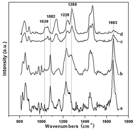
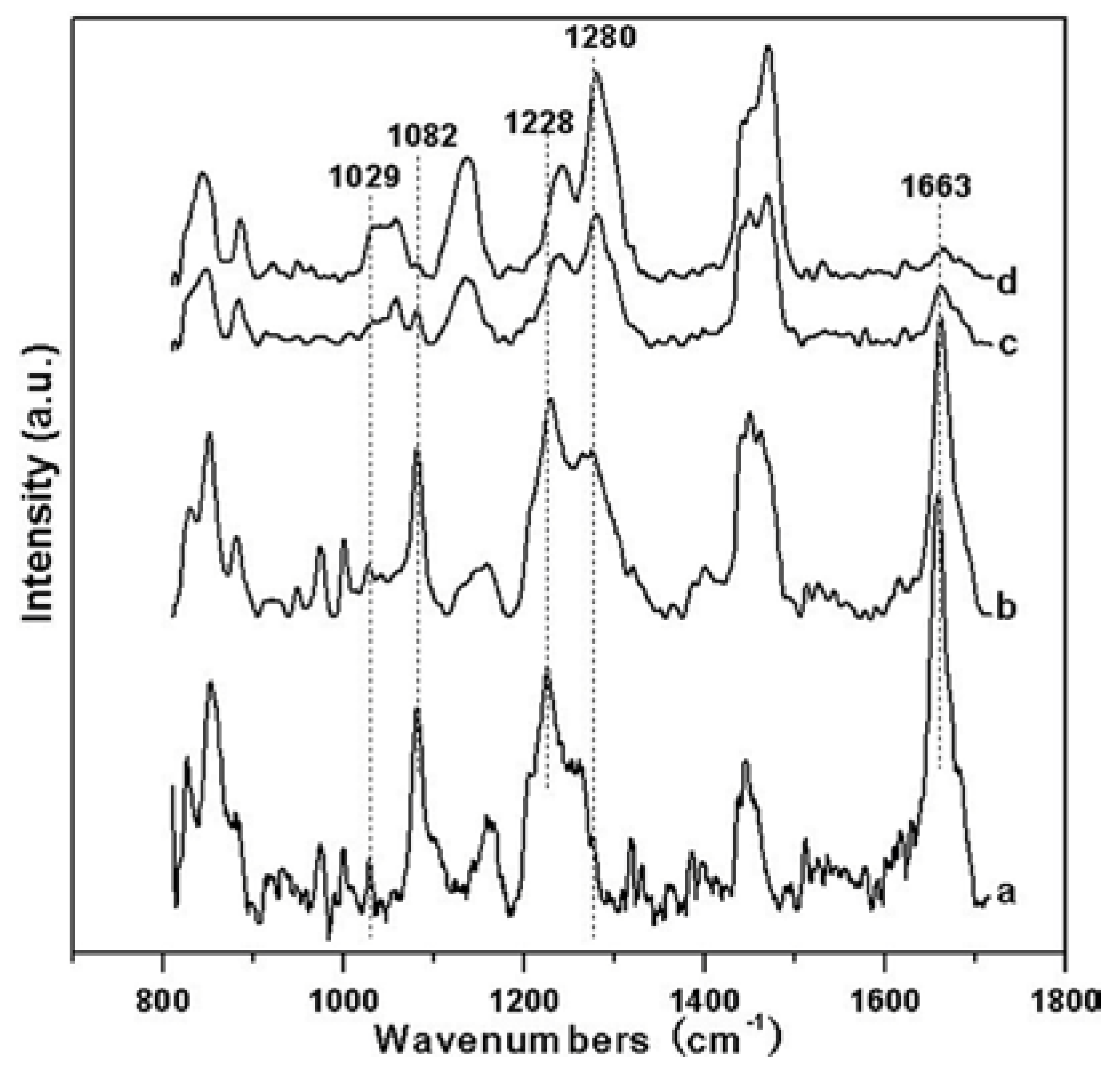
To analyze the fine structure further, the Raman spectrum of silk fibroin gels with PEG600 were determined and shown in Figure 4(C), when adding 100% PEG600, the bands at 1662.55cm-1 (amide I), 1230.05 cm-1 (amide III), 1081.72 cm-1 and 1026.91 cm-1, which are attributed to β-sheet, become stronger compared with the control sample. What is more, with the increase in the percentage of PEG600, the bands of α-helix become stronger and the bands of β-sheet at amide I go weaker and almost disappear with 900% PEG600. But medium strong bands appear at 1243.09 cm-1 (amide III), attributing to the random coil, in the Raman spectrum of silk fibroin gels with 900% PEG600 (curve d). This is because PEG600 breaks the hydrogen bonds between water molecules and silk fibroin molecules, while also facilitates hydrophobic interaction among silk fibroin molecules, and then promotes silk fibroin molecules to cluster rapidly, which accelerated the gelation process and makes a part of silk fibroin molecules to form β-sheet too late, until gels were formed. These results indicate that when adding less percentage of PEG600, such as 100%, it could promote silk fibroin molecules to transform from random coil or α-helix to β-sheet. But with the increase in the percentage of PEG600, it accelerated silk fibroin molecules to cluster rapidly and inhibit silk fibroin molecules transform from the random coil or α-structure to β-sheet, especially at 900%.
Figure 4(C).
Raman spectrum of Bombyx mori silk fibroin gels with PEG600; the percentage of PEG600: (a) 0%, (b) 100%, (c) 500%, (d) 900%.
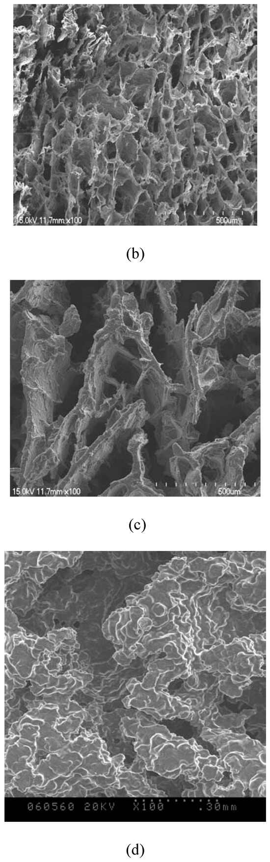
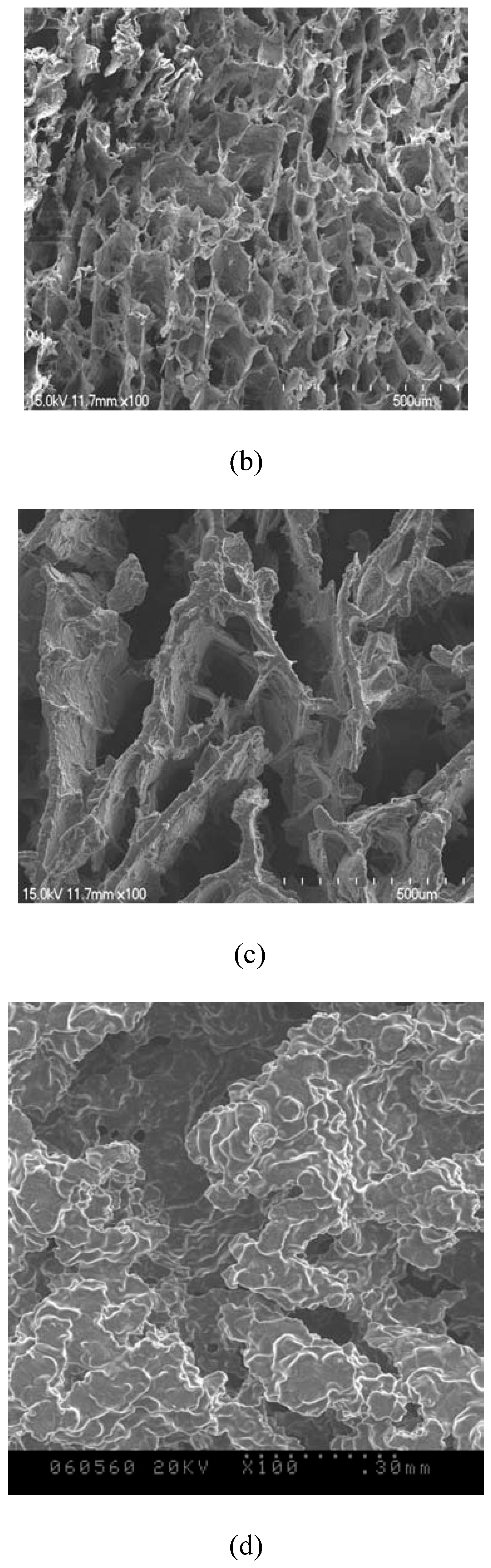
In Figure 5, the silk fibroin gels with 100% of PEG600 had more uniform morphology and the pores distribution was highly uniform, which is possibly concerned with the rate of formation of silk fibroin molecular clusters. These results are similar to that of gels with 100% of ethylene glycol.
Figure 5.
SEM of Bombyx mori silk fibroin gels with PEG600; the percentage of PEG600: (a) 0%, (b) 100%, (c) 500%, (d) 900%; ×100.
4. Conclusion
Silk fibroin was dissolved in triad solvent CaCl2 ·CH3CH2OH·H2O. By testing and analyzing the gelation time of regenerated silk fibroin aqueous solutions with various qualities of polyalcohol (Ethylene glycol, PEG 600), the morphology and the fine structure of silk fibroin gels were discussed and the following conclusions were obtained:
It can be concluded that adding polyalcohol into the regenerated silk fibroin aqueous solution can accelerate the gelation process with an increase in dosage. When adding more than 400% of polyalcohol, irrespective of Ethylene glycol or PEG 600, it accelerated the gelation process remarkably.
Silk fibroin gels with Ethylene glycol mainly consist of silk II crystal structure and Ethylene glycol almost has no effect on the molecular conformation or crystal structure of silk fibroin gels. But when adding PEG600 into silk fibroin aqueous solutions, with the increase in the percentage of PEG600, it accelerated silk fibroin molecules to cluster rapidly and inhibit silk fibroin molecules transforming from the random coil or α-helix to β-sheet, especially at 900%.
Silk fibroin gels with 100% of polyalcohol, both ethylene glycol as well as PEG 600, had more uniform morphology and pores distribution.
Acknowledgments
This work was supported by the Key Basic Research Program of China (973 program, 2005CB623902), National Natural Science Foundation of China (30970714), College Natural Science Research Project of Jiangsu Province (07KJA43010) and The Program of Science and Technology of Jiangsu Province (BA2007055).
References
- Haider, M.; Megeed, Z.; Ghandehari, H. Genetically engineered polymers: status and prospects for controlled release. J. Control Release 2004, 95, 1–26. [Google Scholar] [CrossRef] [Scilit] [PubMed]
- Megeed, Z.; Cappello, J.; Ghandehari, H. Controlled release of plasmid DNA from a genetically engineered silk-elastinlike hydrogel. Pharm. Res. 2002, 19, 954–959. [Google Scholar] [CrossRef] [Scilit] [PubMed]
- Hanawa, T.; Watanabe, A.; Tsuchiya, T.; Ikoma, R.; Hidaka, M.; Sugihara, M. New oral dosage form for elderly patients. II: Release behavior of benfotiamine from silk fibroin gel. Chem. Pharm. Bull. 1995, 43, 872–876. [Google Scholar] [CrossRef] [Scilit] [PubMed]
- Petrini, P.; Parolari, C.; Tanzi, M.C. Silk fibroin-polyurethane scaffolds for tissue engineering. J. Mater. Sci. Mater. Med. 2001, 12, 849–853. [Google Scholar] [CrossRef] [Scilit] [PubMed]
- Chiarini, A.; Petrini, P.; Bozzini, S.; Pra, I.D.; Armato, U. Silk fibroin/poly(carbonate)-urethane as a substrate for cell growth: in vitro interactions with human cells. Biomaterials 2003, 24, 789–799. [Google Scholar] [CrossRef] [Scilit] [PubMed]
- Dal Pra, I.; Petrini, P.; Charini, A.; Bozzini, S.; Fare, S.; Armato, U. Silk fibroin-coated three-dimensional polyurethane scaffolds for tissue engineering: interactions with normal human fibroblasts. Tissue Eng. 2003, 9, 1113–1121. [Google Scholar] [CrossRef] [Scilit] [PubMed]
- Cai, K.; Yao, K.; Lin, S.; Yang, Z.; Li, X.; Xie, H.; Qing, T.; Gao, L. Poly (D, L-lactic acid) surfaces modified by silk fibroin: effects on the culture of osteoblast in vitro. Biomaterials 2002, 23, 1153–1160. [Google Scholar] [CrossRef] [Scilit] [PubMed]
- Cai, K.; Yao, K.; Cui, Y.; et al. Influence of different surface modification treatments on poly(D,L-lactic acid) with silk fibroin and their effects on the culture of osteoblast in vitro. Biomaterials 2002, 23, 1603–1611. [Google Scholar] [CrossRef] [Scilit] [PubMed]
- Minour, N.; Tsukada, M.; Nagura, M. Physic-chemical properties of silk fibroin membrane as a biomaterial. Biomaterials 1990, 11, 430–434. [Google Scholar] [CrossRef] [Scilit] [PubMed]
- Opdahl, A.; Kim, S.H.; Koffas TSet, a.l. Surface mechanical properties of pHEMA contact lenses: Viscoelastic and adhesive property changes on exposure to controlled humidity. J. Biomed. Mater. Res. 2003, 67, 350–356. [Google Scholar] [CrossRef] [Scilit] [PubMed]
- Nowak, A.P.; Breedveld, V.; Pakstis, L.; et al. Rapidly recovering hydrogel scaffolds from self-assembling diblock copolypeptide amphiphiles. Nature 2002, 417, 424–428. [Google Scholar] [CrossRef] [Scilit] [PubMed]
- Hanawa, T.; Watanabe, A.; Tsuchiya, T.; Ikoma, R.; Hidaka, M.; Sugihara, M. New oral dosage form for elderly patients: preparation and characterization of silk fibroin gel. Chem. Pharm. Bull. 1995, 43, 284–8. [Google Scholar] [CrossRef] [Scilit] [PubMed]
- Kim, U.J.; Park, J.; Li, C.; Jin, H.J.; Valluzzi, R.; Kaplan, D.L. Structure and properties of silk hydrogels. Biomacromolecules 2004, 5, 786–92. [Google Scholar] [CrossRef] [Scilit] [PubMed]
- Motta, A.; Migliaresi, C.; Faccioni, F.; et al. Fibroin hydrogels for biomedical applications: preparation, characterization and in vitro cell culture studies. J. Biomater. Sci. Polym. Ed. 2004, 15, 851–64. [Google Scholar] [CrossRef] [Scilit] [PubMed]
- Ayub, Z.H.; Arai, M.; Hirabayashi, K. Quantitative structural analysis and physical-properties of silk fibroin hydrogels. Polymer 1994, 35, 2197–200. [Google Scholar] [CrossRef] [Scilit]
- Kang, G.D.; Nahm, J.H.; Park, J.S.; Moon, J.Y.; Cho, C.S.; Yeo, J.H. Effects of poloxamer on the gelation of silk fibroin. Macromol. Rapid Commun. 2000, 21, 788–91. [Google Scholar] [CrossRef]
- Ayub, Z.H.; Arai, M.; Hirabayashi, K. Mechanism of the gelation of gibroin dolution. Biosci. Biotech Bioch 1993, 57, 1910–1912. [Google Scholar]
- Hirabayashi, K.; Ayub, Z.H.; Kume, Y. Gelation of silk fibroin. SEN-I GAKKAISHI 1990, 46, 521–524. [Google Scholar] [CrossRef] [Scilit] [PubMed]
- Matsumoto, A.; Chen, J.; Collette, A.L.; Kim, U.J.; Altman, G.H.; Cebe, P.; Kaplan, D.L. Mechanisms of silk fibroin sol-gel transitions. J. Phys. Chem. B 2006, 110, 21630–21638. [Google Scholar] [CrossRef] [Scilit] [PubMed]
- Wang, H.; Zhang, Y.; Shao, H.; Hu, X. A study on the flow stability of regenerated silk fibroin aqueous solution. Int. J. Biol. Macromol. 2005, 36, 66–70. [Google Scholar] [CrossRef] [Scilit] [PubMed]
- Kim, U.J.; Park, J.; Li, C.; Jin, H.J.; Valluzzi, R.; Kaplan, D.L. Structure and properties of silk hydrogels. Biomacromolecules 2004, 768–792. [Google Scholar] [CrossRef] [Scilit] [PubMed]
- Matsumoto, A.; Chen, J.; Collette, A.L.; Kim, U.J.; Altman, G.H.; Cebe, P.; Kaplan, D.L. Mechanisms of silk fibroin sol-gel transitions. J. Phys. Chem. B 2006, 110, 21630–21638. [Google Scholar] [CrossRef] [Scilit] [PubMed]
- Fini, M.; Motta, A.; Torricelli, P.; Giavaresi, G.; Nicoli Aldini, N.; Tschon, M.; Giardino, R.; Migliaresi, C. The healing of confined critical size cancellous defects in the presence of silk fibroin hydrogel. Biomaterials 2005, 26, 3527–3536. [Google Scholar] [CrossRef] [Scilit] [PubMed]
- Wang, X.; Kluge, J.A.; Leisk, G.G.; Kaplan, D.L. Sonication-induced gelation of silk fibroin for cell encapsulation. Biomaterials 2008, 29, 1054–1064. [Google Scholar] [CrossRef] [Scilit] [PubMed]
- Kim, U.J.; Park, J.; Li, C.; Jin, H.J.; Valluzzi, R.; Kaplan, D.L. Structure and properties of silk hydrogels. Biomacromolecules 2004, 5, 768–792. [Google Scholar] [CrossRef] [Scilit] [PubMed]
- Li, M.; Lu, S.; Wu, Z.; Yan, H.; Mo, J.; Wang, L. Study on porous silk fibroin materials: 1. Fine structure of freeze-dried silk fibroin. Joumal Appl. Polym. Sci. 2001, 79, 2185–2191. [Google Scholar] [CrossRef] [Scilit]
- Shimizu, M. Sanshi Shikenjo Houkoku 1941, 10, 475.
- Hirabayashi, K.; Ishikava, H. Sen-i Gakkaishi 1967, 23, 538.
- Li, M.; Wu, Z.; Lu, S. Study on Silk Fibroin Materials. J. Dong Hua Univ. (Nat. Sci. Ed.) 2001, 27, 12–19. [Google Scholar]
- Li, M.; Lu, S.; Wu, Z.; Yan, H.; Mo, J.; Wang, L. Study on porous silk fibroin materials: 1. Fine structure of freeze-dried silk fibroin. Joumal Appl. Polym. Sci. 2001, 79, 2185–2191. [Google Scholar] [CrossRef] [Scilit]
- Tsukada, M.; Gotoh, Y.; Nagura, M.; et al. Structural changes of silk fibroin membranes induced by immersion in methanol aqueous solutions. J. Polym. Sci. Part A Polym. Chem. 1994, 32, 961–968. [Google Scholar] [CrossRef] [Scilit]
- Yoshimizu, H.; Asakura, T. The structure of Bombyx mori silk fibroin membrane swollen by water studied with ESR, 13C-NMR, and FT-IR spectroscopies. J. Appl. Polym. Sci. 1990, 40, 1745–1756. [Google Scholar] [CrossRef] [Scilit]
© 2011 Textile Bioengineering and Informatics Society. This article is an open access article distributed under the terms and conditions of the Creative Commons Attribution (CC BY) license (https://creativecommons.org/licenses/by/4.0/).