


Abstract
Cellulose ether, like hypromellose (HM), is an extremely versatile material that is widely used in pharmaceutical products as film coatings. To modify the surface properties of HM films, additives are routinely included during the film formulation process, which are typically hydrophobic lubricants or hydrophilic plasticizers. Plasticizers increase the flexibility and reduce the brittleness of the film. The first goal of this study is to demonstrate that plasticization of HM films by low-molecular-weight (400 g∙mol−1) polyethylene glycol (PEG) allows tuning adhesion and friction properties of HM films, both at nano- and macroscales. Surface morphology, surface energy, nano/macro adhesion, and nano/macro friction coefficient were studied by atomic force microscopy (AFM) in adhesion or friction modes at the nanoscale, wettability, and probe-tack adhesion, as well as pin-on-disk friction experiments at the macroscale. The results show that the addition of PEG decreases the Young’s modulus and the Tg of HM-plasticized films while increasing their strain at break and surface energy. The macroadhesion force increases from 9 to 90 mN by the addition of 40% w/w of PEG, whereas the macrofriction coefficient is reduced by 50%. The hypothesis of insertion of plasticizer molecules in HM chains’ nano-domains is evidenced and explains these results. The second goal of this study is to investigate nanoscale versus macroscale correlation of adhesion and friction properties and the role of adhesion in friction experiments. The results show, first, that the evolution of the adhesion energy at the macroscale as a function of adhesion energy at the nanoscale is linear. On the contrary, a high friction coefficient at the nanoscale corresponds to a low friction coefficient at the macroscale and vice versa, showing a first linear decrease for PEG contents ranging from 0 to 30% (w/w) and the second linear decrease, less pronounced, is observed for PEG contents ranging from 30 to 40% (w/w). The hypothesis of a difference in contact pressure applied on the probe at both scales, as well as HM-PEG surface phase separation at a high PEG content (>30% w/w), is proposed to explain this difference. The variations in friction coefficients are linear according to the PEG plasticizer content and suggest its lubricant role in HM-Plasticized films. Finally, the interplay between adhesion and friction, in friction experiments, is evidenced and appears dominant at the nanoscale.
1. Introduction
Hypromellose (HM) looks to be a dedicated biomaterial option for film-coating compositions. Indeed, HM is distinguished by its ease of use, availability, water solubility, and nontoxicity. Its primary application is as a controlled release matrix in pharmaceutical films [1]. The hydration of HM controls drug release from these systems, forming a gelatinous layer at the matrix’s surface through which the contained drug diffuses. Incorporating additives such as plasticizers [2] or surfactants [3,4] in film-coating formulations is the most effective way to improve specific features of HM film, including moisture content, barrier properties, mechanical, and surface characteristics. HM-based films must have strong mechanical qualities while avoiding ruptures and cracks in the film. As a result, plasticizers may be added to the formulation. Several studies have investigated the effect of plasticizer additives on the physicochemical characteristics of HM films. The most common plasticizers used for pharmaceutical film coatings are classified in three groups and include (i) polyols such as glycerol, propylene glycol, and polyethylene glycols (PEG), (ii) organic esters such as triacetin, phthalate esters (diethyl, dibutyl), and citrate esters, and (iii) oils and glycerides such as castor oil, acetylated monoglycerides, and fractionated coconut oil. The main requirements for plasticizers are efficiency, permanence, and nontoxicity. Permanence depends on the volatility of the plasticizer and on its solubility and interaction with the polymer matrix. The efficiency depends on the chemical structure, molecular weight, and concentration of plasticizer. For example, the plasticizing efficiency of polyethylene glycols decreases with increasing molecular weight from 200 to 6000 g∙mol−1. PEGs are good plasticizers for hypromellose (HM) and poor ones for ethylcellulose (EC). For HM polymer, the plasticizer efficiency increases in the order of propylene glycol < diethyl phthalate < triacetin < PEG. Moreover, good solubility and compatibility of PEG plasticizer with HM polymer are of major importance for effective plasticization. Plasticizers reduce interfacial tension between polymer chains by insertion and “swelling” mechanisms [5], thus reducing the glass transition temperature, Tg. Several studies on the plasticization of HM films have shown that polyethylene glycol (PEG) is highly effective. When the PEG plasticizer content of HM films was increased, they became softer and more viscoelastic. Plasticization effectiveness increases as the PEG molecular weight drops because there are more plasticizer molecules available to interact with the polymer. Increasing the concentration of PEGs gradually reduces the tensile strength, Young’s modulus, and glass transition temperature of cellulose ether films [6,7]. However, the molecular weight of PEG needs to be discussed. First, the physical state of PEG depends on its molecular weight. Indeed, for a molecular weight greater than 1000 g∙mol−1, PEG is a semi-crystalline solid with a melting temperature greater than 30 °C. As a consequence, plasticization efficiency is strongly reduced, and phase separation occurs, rendering film-forming difficult. Second, the targeted application of this research, namely, protective two-piece capsules or films for drug delivery applications, shows strong constraints in terms of formulation viscosity, drying kinetics, film water uptake, etc., that make PEG with a molecular weight of around 400 g∙mol−1 the best candidate for HM film plasticization. Nonetheless, only a few studies examined the surface characteristics of HM-formulated films [8]. Thus, this article will investigate, first, the influence of plasticization of HM films by low-molecular-weight (400 g∙mol−1) polyethylene glycol (PEG) on the thermomechanical and surface properties of HM-Plasticized films. To better understand how to tune the surface’s hydrophobicity/hydrophilicity, the impact of PEG additive concentration on surface energy, adhesion, and friction behaviors is studied. The surface energy of HM and HM-Plasticized films is assessed using contact angle analysis. Surface morphology was investigated using atomic force microscopy (AFM) in tapping mode (TM-AFM). Adhesion and friction measurements at the nanoscale were carried out using chemical force microscopy and lateral force microscopy. The second goal of this study is to investigate nanoscale versus macroscale correlation of adhesion and friction properties and the role of adhesion in friction experiments.
2. Materials and Methods
2.1. Materials
Dow Chemical Company (Midland, MI, USA) produces HyproMellose (HM), which was thereafter purchased by Colorcon, France. The HM grade used has a nominal viscosity of 3 mPa.s in a 2% aqueous solution at 20 °C and a glass transition temperature (Tg) equal to 138 °C in solid form. The degree of methoxy group substitution (DS) and molar substitution of hydroxypropoxy groups (MS) of HM are 1.87 and 0.25, respectively (by weight percent, the amount of OCH3 groups is 28.3% (w/w), and OCH2(CHOH)CH3 groups is 9.1%) [9]. This low-viscosity HM grade is ideal for film and coating applications as a film-forming matrix. Indeed, it allows for the preparation of an aqueous solution containing approximately 20% w/v solid polymer.
Polyethylene glycol (PEG) was purchased from Sigma-Aldrich (Saint-Quentin-Fallavier, France). A low-molecular-weight (Mn = 400 g∙mol−1) grade, hydrophilic, completely soluble in water, and biocompatible was used as a plasticizer. It is a clear, colorless, viscous liquid with a density of 1.128 g∙cm−3, a viscosity of 7.3 cSt, and a Tg equal to −72 °C.
2.2. Methods
2.2.1. Preparation of HM and HM-Plasticized Films
The “casting process” was used for film elaboration. It involves drying the film-forming solution following deposition on a nonstick substrate. The solvents used are limited to water and ethanol. The percentage w/v of a solution is computed using the following formula: % w/v = g of solute per 100 mL of solution. Pure HM films were elaborated by preparing the HM solution, casting it onto a glass plate, and allowing it to dry at room temperature. To avoid lumping, HM powder was moderately agitated in deionized water at 80–90 °C to produce the HM solution (6% w/v). Cooling to 25 ± 2 °C yields a translucent solution. The HM polymer was completely hydrated by maintaining the solution at 5 °C for an entire day. Solutions were then deposited onto a glass plate in a uniform layer with a thickness of 1.6 mm. Films were let dry for 48 h under natural convection. The thickness of each film is 100 ± 10 µm. The samples were conditioned at 25 ± 2 °C and 30 ± 5% RH after the dried films were removed from the glass plate.
HM-plasticized films were obtained by incorporating PEG plasticizer into the HM solution. The amount of PEG was calculated as a weight percentage of HM powder. PEG was added and homogenized with the HM solution. Four solutions were prepared: 10%, 20%, 30%, and 40% w/w HM. After 2 h of homogenization, all solutions were stored at 5 °C for 24 h. Film-forming solutions were deposited on a glass plate to form uniform films (1.6 mm thick) and then dried naturally. All HM and HM-Plasticized films were stored at 25 ± 2 °C and 30 ± 5% relative humidity before characterization.
2.2.2. Surface Energy Measurements
The most frequent approach for determining the surface energy of a solid surface is to deposit droplets of dedicated liquids on the surface being investigated. Liquid droplets (2–3 µL) of α-bromonaphtalene, diiodomethane, and water were deposited on the film surface and imaged using drop shape imaging equipment (Krüss G2 goniometer, Hamburg, Germany). The average contact angle (over 10 droplets per liquid) was measured at a relative humidity of 30 ± 5% and a room temperature of 22 ± 2 °C. The surface free energy of the HM and HM-plasticized films was deduced from the contact angle measurements and surface tensions of the probe liquids. Owens and Wendt extended Fowkes’ concepts to include both dispersion and polar forces. The Owens and Wendt approach [10] was linked with Young’s equation, resulting in:
where the contact angle of the probe liquid on the film solid surface is denoted by θ, while γlD, γlND, γsD, and γsND are the dispersive (D) and non-dispersive (ND) components of the surface tension of the probe liquid (l), and of the solid (s) film, and γl is the surface tension of the probe liquid. Measuring the contact angle with a dispersive liquid allows for the calculation of the dispersive component of the film’s solid surface energy (γsD), while a polar liquid allows calculation of the non-dispersive component (γsND). Surface free energy is defined as the sum of the surface energy’s dispersive and non-dispersive components.
γl × (1 + cos θ) = 2 × (γsD × γlD)1/2 + 2 × (γsND × γlND)1/2
2.2.3. Surface Morphology of HM and HM-Plasticized Films
Surface morphology and topography of HM and HM-Plasticized films are performed using atomic force microscopy (AFM) in the tapping mode. During scanning over the surface, the cantilever is sinusoidally vibrated by a piezo and the oscillating tip lightly taps the surface at the cantilever’s resonant frequency, with a constant oscillating amplitude introduced in the vertical direction via a feedback loop that keeps the average normal force constant. Topographical measurements are taken at a fixed scanning angle of 0° and a scan rate of 0.8 Hz, using a Nanoscope IIIa AFM (Digital Instruments, Santa Barbara, CA, USA). Silicon probes with a spring constant of 5 N∙m−1, a resonance frequency of 150 kHz, and a tip radius of less than 10 nm were used. Images are captured at a resolution of 512 × 512 pixels. Experiments were carried out at 25 °C.
2.2.4. Nanoadhesion of HM and HM-Plasticized Films
The adhesion force at the nanoscale between a pyramidal shape AFM tip and HM and HM-Plasticized films was measured using contact mode AFM through recording characteristic force–distance curves [11]. Characteristics of the silicon nitride cantilevers are a 0.3 N∙m−1 spring constant, a 115 µm length, and a 57 kHz resonance frequency. The AFM tip’s radius of curvature was 45 nm. While the tip is vertically moved forward and backward to the film surface thanks to a piezoelectric ceramic, the deflection of the cantilever is recorded. The adhesion force at the nanoscale is then calculated as Fadh = k∙Δz where k is the cantilever spring constant (N/m), and Δz is the cantilever deflection (nm). Experiments were carried out at 25 °C.
2.2.5. Macroadhesion of HM and HM-Plasticized Films
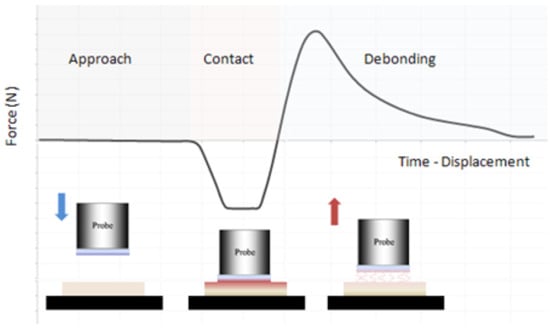
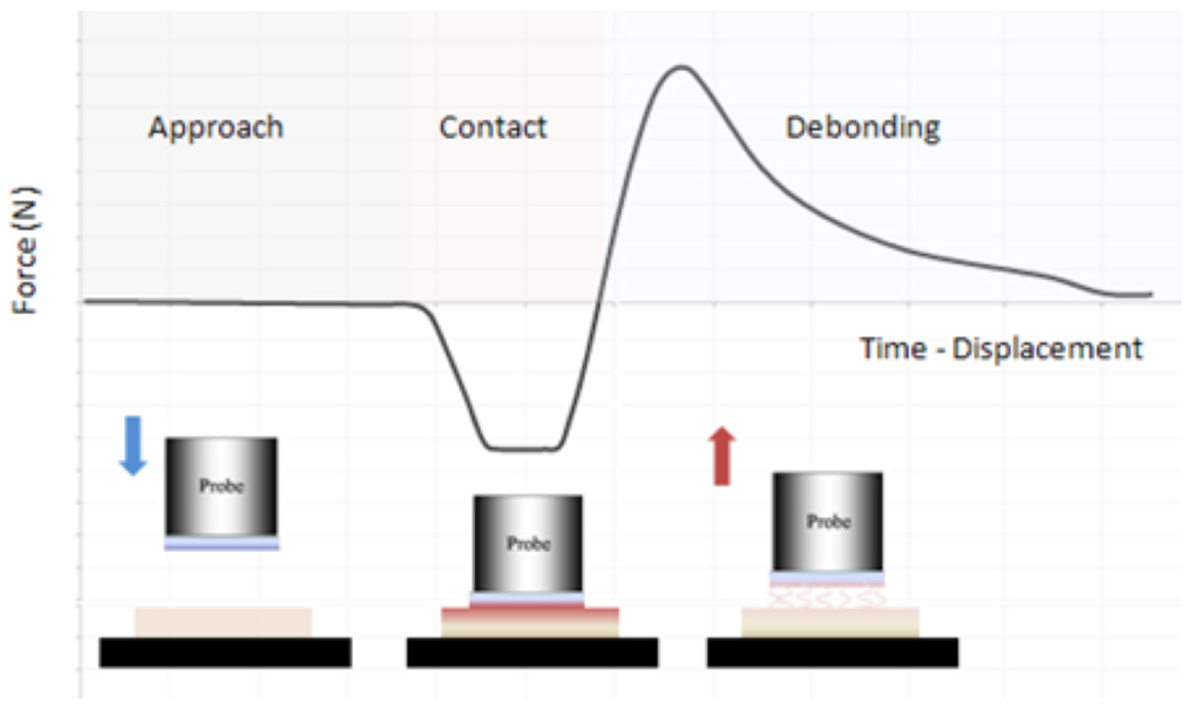
A conventional probe-tack test [12] was used to measure the adhesion behavior of HM and HM-Plasticized films at the macroscopic scale. Tack determines an adhesive ability to instantaneously form a bond with another surface under light contact pressure. Tack depends on the polymer’s viscoelastic characteristics. The HM film or HM-Plasticized film is deposited on an aluminum cylindrical flat punch. Then, the experiment consists of contacting the HM films with a clean glass substrate. The surface contact area measures 3.14 cm2. The two surfaces were brought into contact using a standard force of 10 N. After a contact time of 5 min, the two surfaces were separated at a constant velocity of 1 mm∙s−1. Separation force and energy were then measured. Figure 1 gives a schematic presentation of the probe-tack experiment.
Figure 1.
Schematic presentation of the probe-tack experimental stages: approach (blue arrow), contact, and debonding (red arrow) of the film.
2.2.6. Nanofriction of HM and HM-Plasticized Films
To access sliding and friction at the nanoscale, friction force microscopy (FFM) was used. The output of the horizontal two quadrants of the photodiode detectors is monitored while the sample is scanned back and forth (creating a friction loop), perpendicular to the long axis of the cantilever under a predetermined applied load. The cantilever twists as a result of the friction force between the film and the tip. The FFM signal represents the difference between the signals from the left and right photodiode detectors. Adhesion force at the tip–film interface can also impact the friction force, which is related to the degree of twisting of the cantilever. The difference between lateral forces scanning from left-to-right and right-to-left is known as TMR (Trace Minus Retrace, in volts (V)), which is used to quantify nanofriction force (FnF). The friction coefficient at the nanoscale (µnano) can be computed [13] by dividing the nanofriction force (FnF) by the sum of the nanoadhesion force (FnA) and the applied normal force (FnN).
µnano = FnF/(FnA + FN)
The same probe used to assess nanoadhesion forces was used to measure nanofriction forces. Values of nanofriction forces have been determined for a range of applied normal loads, from 0.1 to 3 V. Experiments were carried out at 25 °C.
2.2.7. Macrofriction of HM and HM-Plasticized Films
A pin-on-disk tribometer [14] was used to measure the macroscopic tribological characteristics of HM and HM-Plasticized films. Under a predetermined load, a glass ball (diameter = 6 mm) came into contact with HM and HM-plasticized films supported on a glass plate. The force opposing the ball’s motion (tangential force (FT)), equivalent to the friction force, was then measured while the glass plate was rotated at a specific speed. The macroscopic friction coefficient (µmacro) is determined by dividing the observed friction force (FT) by the applied normal force (FN). Each experiment was carried out at 25 °C with a normal load of 2 N, 5 N, and 10 N along a sliding distance of 100 mm and a sliding velocity of 5 mm∙s−1.
3. Results
3.1. Thermal and Mechanical Behaviors of HM and HM-Plasticized Films
The DSC thermograms of HM-Plasticized films show only one Tg that falls between the glass transitions of the two constituents, HM (−138 °C) and PEG (−72 °C). Furthermore, because both are water-soluble, they are miscible and do not phase separate. No peak corresponding to the melting of a crystalline phase is seen. However, the addition of PEG alters the thermal characteristics of HM-Plasticized films. Dynamic Mechanical Analysis (DMA) experiments [15] were also conducted to assess the Young’s modulus values and the temperatures at which the loss factor (tan δ) is maximum. The maximum of the tan δ value is related to the glass transition phenomenon and is considered a mechanical way to access Tg. Table 1 gathers Young’s modulus, the maximum of tan δ, and Tg values, obtained on the basis of DMA and DSC experiments. The values of Tg computed using the Fox equation [16] are also given. The Fox Equation (3) allows computation of Tg values for a binary mixture assuming perfect thermodynamic miscibility. Tg is the glass transition of the miscible blend, whereas Tg1 and Tg2 are the glass transition temperatures of the components 1 and 2 of the blend, and W1 and W2 are the weight fractions of component 1 and 2 of the blend.
1/Tg = W1/Tg1 + W2/Tg2
Table 1.
Maximum of tan δ and Tg values obtained on the basis of DMA, DSC, and Fox’s equation for HM and HM-Plasticized films.
The values of the maximum of tan δ acquired by DMA are consistent with those obtained by DSC and follow the same decreasing tendency when the PEG content increases. Nevertheless, tan δ values obtained by DMA differ from the Tg values found by DSC. This is due to the difference in sensibility and stress applied between the two techniques. The difference between the two methods is about 20 °C. Moreover, there is a good correlation between the experimental results and the computed Tg using Fox’s equation. HM-PEG miscibility indicates strong interactions between the two polymers [17], particularly through specific interactions between functional groups. One can deduce that PEG strongly influences the thermal characteristics of HM films. Indeed, the literature reports [18] that the hydroxyl groups on cellulose and methylcellulose create mostly hydrogen bonds with the ether oxygen of PEG. This opens up the way to strong interactions between HM’s hydroxyl groups and PEG, leading to miscible blends and the plasticizing effect of PEG.
The influence of PEG on the mechanical characteristics of HM-Plasticized films was also investigated using a uniaxial tensile test [19] (strain rate = 1 mm/min at 25 °C). Table 2 gathers the tensile strength and strain at break of HM and HM-Plasticized films as a function of PEG content.
Table 2.
Tensile strength and strain at break of HM and HM-Plasticized films.
Pure HM film has an estimated tensile strength and strain at break of 12 MPa and 0.8%, respectively. The addition of PEG to HM films drastically affects these values. Plasticized films become more flexible and less brittle. The addition of 40% PEG decreases the tensile strength by three times while increasing the strain at break by seven times. Indeed, PEG molecules may progressively diffuse into the polymer chains through interaction between hydrophobic ethylene segments of PEG molecules and the HM polymer chains’ hydrophobic methyl substituents, reducing the intermolecular attraction forces along the polymer chains, allowing the polymer chains to move more freely, and, therefore, lowering the maximum of tan δ temperature and the glass transition temperature. The insertion of PEG increases the free space around the HM polymer chains, allowing for boosting chain mobility. Mechanical properties are intimately related to HM/PEG intermolecular interaction, miscibility, and resulting glass transition temperature, which is reduced with the addition of plasticizer [20].
3.2. Surface Morphology of HM and HM-Plasticized Films
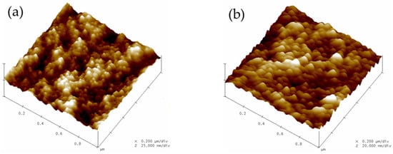
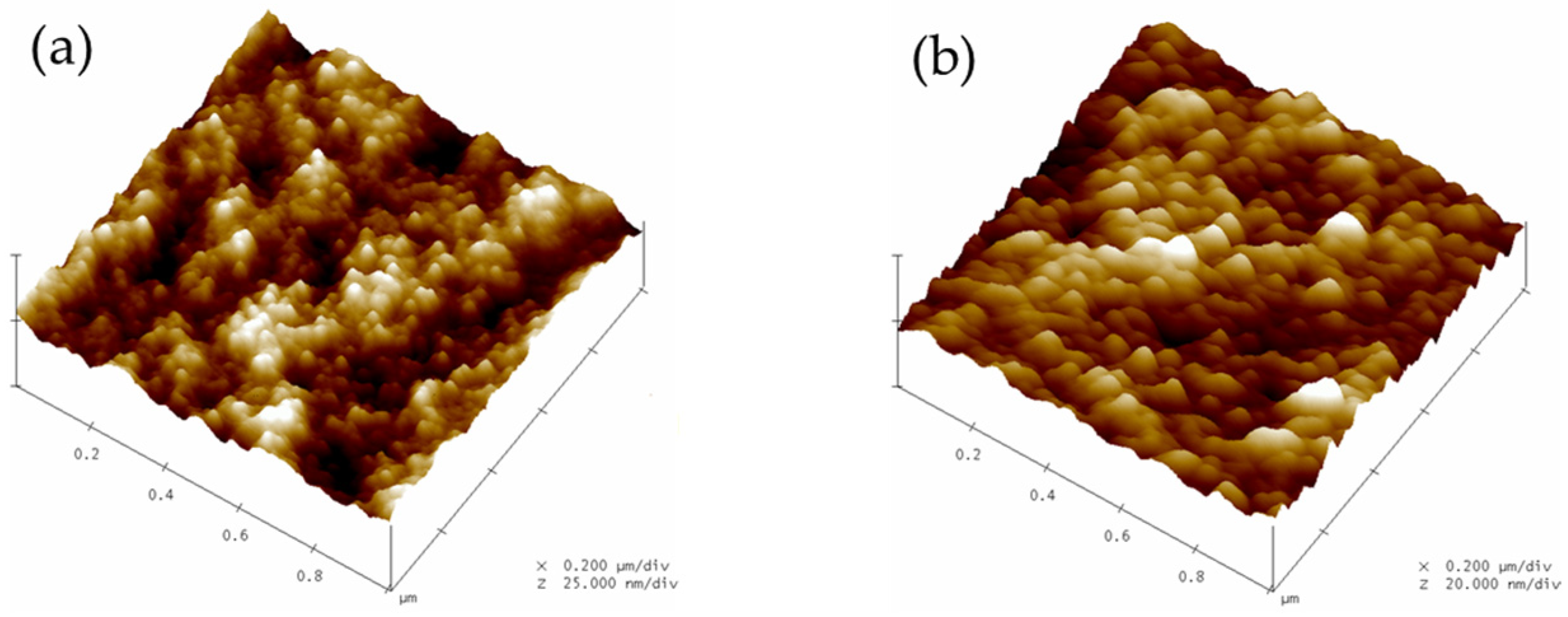
Tapping mode atomic force microscopy (TM-AFM) was used to measure the surface topography of HM and HM-Plasticized films. Figure 2 reports the surface aspect variation in TM-AFM topographic images.
Figure 2.
Topographic TM-AFM images of (a) HM film and (b) plasticized HM-Plasticized film containing 30% (w/w HM) of plasticizer.
Topographic images (1 µm × 1 µm) of the HM and HM-Plasticized films reveal spherical aggregates ranging in size from 20 to 30 nm. HM aggregates appear during the film-forming process [21]. Indeed, HM is a non-ionic cellulose derivative with methyl (hydrophobic) and hydroxypropyl (hydrophilic) groups added to the anhydroglucose backbone. These aggregates are formed as a result of interactions between HM polymer chains’ hydrophobic substituents. Aggregates are always visible in topographic images of all plasticized films. Table 3 summarizes the characteristics extracted from the section analysis profiles acquired on TM-AFM topographical images, namely the average aggregate radius and arithmetic average roughness (Ra) of the surface.
Table 3.
Average aggregate radius (AAR) and arithmetic average roughness (Ra) of HM and HM-Plasticized films.
The average aggregates radius varies with PEG content. Indeed, the addition of a larger proportion of PEG in HM films greatly increases the average aggregates radius, with an average value of 35 nm measured for 40% w/w PEG. Section analysis profiles show that the volume of aggregates increases, particularly when a large amount of PEG is introduced. No significant variation in average surface roughness was noticed. According to plasticizing theory, which entails an increasing intermolecular distance between polymer chains and thus increasing free volume [22], the fluctuation in aggregates’ characteristic size is thought to be caused by the insertion of PEG molecules within HM chain aggregates. This insertion mechanism is favored by first, interaction of the hydrophobic ethylene segments of the amphiphilic PEG molecules with the HM polymer chains’ hydrophobic substituents (aggregates), and second, forming subsequent hydrogen bonds between the hydroxyl end-groups of the PEG molecule and the hydroxypropyl groups substituted on the anhydroglucose backbone. As a result, the average size of HM aggregates rises.
3.3. Surface Free Energy of HM and HM-Plasticized Films
The determination of the contact angle of a water droplet deposited on HM and HM-Plasticized film surfaces reveals the hydrophilic character of the surface [23]. The water contact angle reduces continuously with the addition of PEG plasticizer, from 69° ± 2° for pure HM film to 21° ± 2° for HM-40% PEG film. This result indicates that the hydrophilicity of HM-plasticized film increases with PEG content. Surface properties are evaluated in terms of surface free energy. The surface free energies of HM and HM-Plasticized films were calculated by measuring contact angles with various liquid probes. Pure HM film shows a surface free energy of 43 mJ∙m−2. This surface energy is separated into components related to the hydrophilic interactions (Keesom, Debye) and hydrophobic interactions (London) exchanged by the liquid molecules with the film surface. These components are named non-dispersive (ND) and dispersive (D). Table 4 shows the surface free energy values for HM and HM-Plasticized films as a function of PEG content, as well as the contribution (%) of the dispersive (D) and of the non-dispersive (ND) components to the surface energy.
Table 4.
Surface free energy (γs) and contribution (%) of the dispersive (D) and non-dispersive (ND) components to the surface energy of HM and HM-Plasticized films.
The addition of hydrophilic PEG molecules raises the surface energy of the plasticized films. Hydrophilicity is measured by reduced contact angles and increased film surface energy. The addition of PEG affects both components of surface energy. The non-dispersive component is doubled by adding 30% PEG or more. The surface free energy is near 73 mJ∙m−2 (equivalent to the surface tension of water, γH2O = 72.8 mJ∙m−2). The addition of PEG polar molecules increases the adsorption of water molecules [24] on HM-Plasticized films. Furthermore, the inclusion of PEG molecules between HM chains enhances the free volume in the polymer film. This is an additional factor that helps to keep the water molecules trapped inside the film. Thus, the increased hydrophilic surface character of HM-Plasticized films might be explained by bound H2O molecules on their surface. To summarize, PEG increases the hydrophilic film character of HM and promotes the presence of water multilayers on its surface.
3.4. Nanoadhesion of HM and HM-Plasticized Films
Using AFM, we investigated how HM film/AFM tip adhesion at the nanoscale responds to the addition of plasticizer. On the basis of force–distance curves, the nanoadhesion force (Fadh) was estimated by multiplying the cantilever deflection (nm) by its stiffness (N∙m−1) during the jump out of contact of the tip from the film surface. Although the HM-Plasticized films became increasingly soft with the addition of PEG, the slope of the force curve remains constant. Moreover, as surface roughness is quite similar for all the films and experiments conducted at constant relative humidity, the nanoadhesion force results from van der Waals forces and capillary forces [25,26] due to water meniscus formation at the tip apex when contacting the hydrophilic HM and HM-Plasticized films. In an inert atmosphere, the nanoadhesion force is only due to van der Waals forces and is given by [25]:
FvdW = 4πRγS
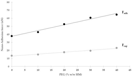
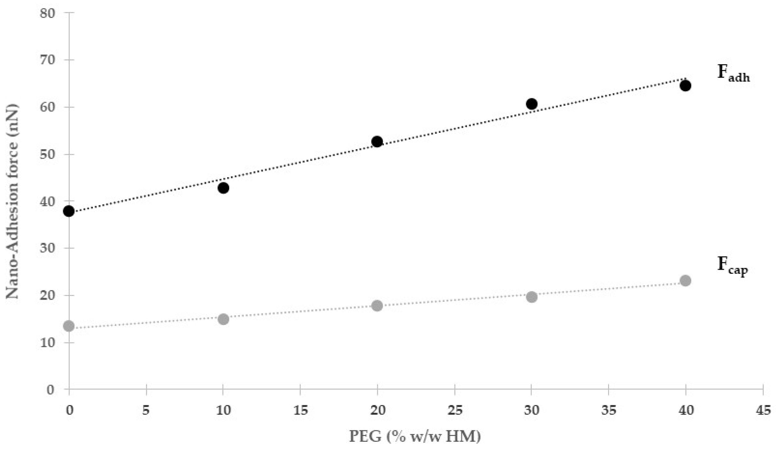
In this equation, R represents the tip radius and γS the surface free energy of the HM or HM-Plasticized films, as determined previously by wettability experiments. Thus, capillary force is computed by subtracting FvdW from Fadh. Figure 3 represents the evolution of the AFM adhesion force (Fadh) and capillary force (Fcap) with PEG content.
Figure 3.
Evolution of nanoadhesion force (Fadh) and capillary force (Fcap) with PEG content.
The simultaneous rise in van der Waals and capillary forces is the cause of the increase in adhesion force at the nanoscale with PEG concentration. AFM tip–film surface interactions are significantly impacted by an increase in surface free energy. Additionally, it is noted that there is no saturation stage with PEG addition and that capillary force increases with the hydrophilic film nature. The development of a meniscus bridge between the AFM tip and the film surface, which extends the adhesive force, can account for this outcome. One can conclude that the PEG amount is the sole source of force variation for HM films because the surface roughness and tip radius remain constant throughout the experiments. The contact zone between the AFM tip and the film surface is expanded by the capillary condensation of water on the film’s surface as PEG is added. The HM-PEG system’s thermodynamic miscibility, which modifies the glass transition temperature and thermomechanical characteristics, promotes a steady rise in the nanoadhesion force.
3.5. Macroadhesion of HM and HM-Plasticized Films
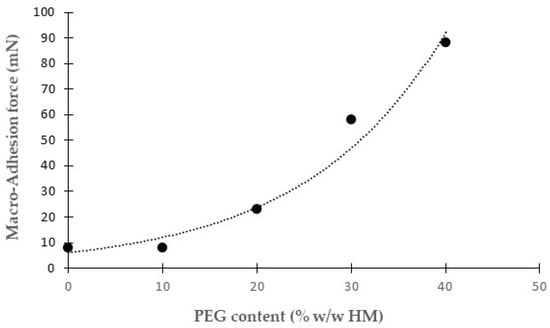
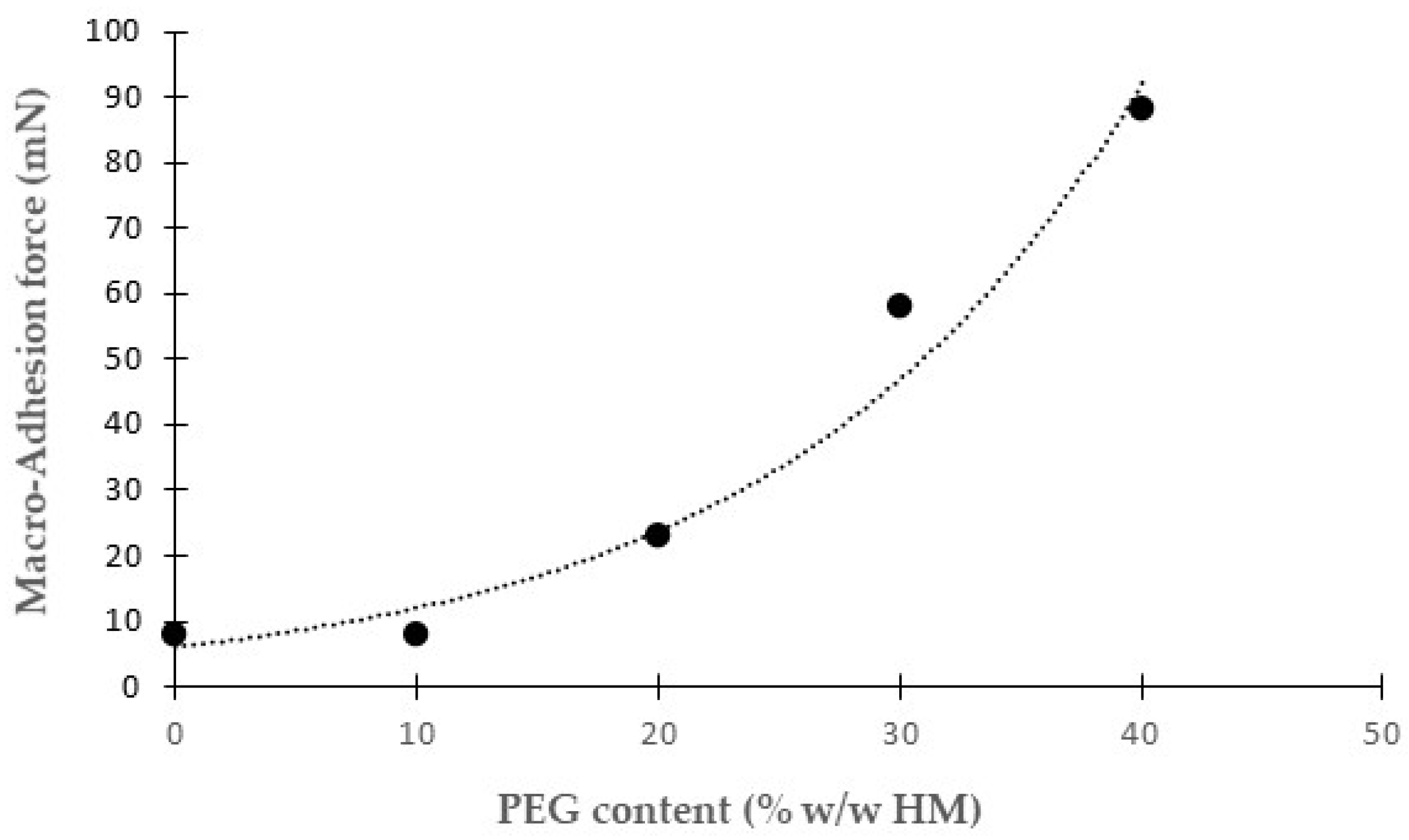
The conventional probe-tack test, presented in Figure 1, was used to determine the maximum value of the macroscopic adhesion force during the separation stage. The results show an increase in separation force between the glass substrate and the HM-Plasticized films. Figure 4 shows the influence of PEG content on the maximum value of the macroscopic adhesion force.
Figure 4.
Evolution of macroscopic adhesion force with PEG content.
The results reveal that the pure HM film has very poor adhesive behavior, but the forces are detectable and reproducible across samples. The PEG plasticizer significantly improves the macroscopic adhesion force required to separate the HM-Plasticized film from the glass substrate. This result can be explained by the fact that PEG lowers the glass transition temperature of HM chains through an insertion mechanism in HM aggregates, through the formation of hydrogen bonds between the hydroxyl end-groups of the PEG molecules and the hydroxypropyl groups substituted on the anhydroglucose backbone of the HM chains, boosting molecular mobility. In this situation, the presence of HM free chains on the film surface facilitates interfacial interactions with the glass plate, resulting in increased resistance to force during the separation stage.
3.6. Nanofriction of HM and HM-Plasticized Films
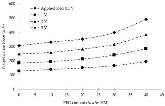
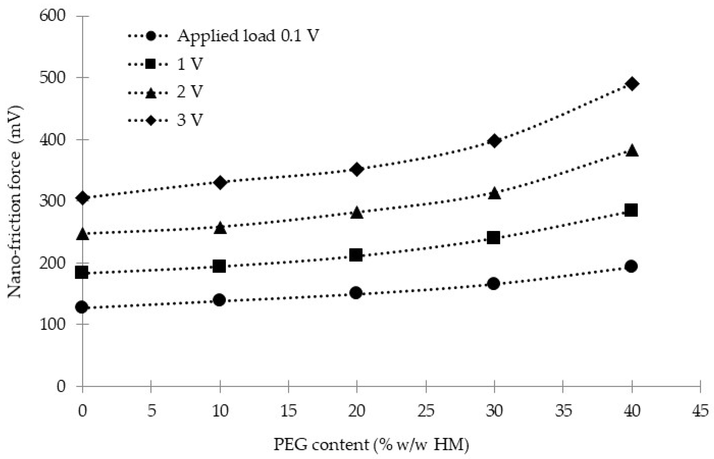
Friction force microscopy (FFM) was used to investigate how HM film/AFM tip friction at the nanoscale responds to the addition of plasticizer. The difference between lateral forces values while scanning from left-to-right and right-to-left, named TMR (Trace Minus Retrace, in volts (V)), was used to quantify the nanofriction force (FnF). Figure 5 shows the effect of plasticizer content on the nanofriction force of HM and HM-Plasticized films at different applied loads (V).
Figure 5.
Evolution of nanofriction force (TMR) with PEG content at various applied loads.
As shown in Figure 5, the evolution of nanofriction force (FnF) relies on PEG content and on the load applied (FN) on the AFM cantilever. Adding 20% PEG to HM films increases the nanofriction force by 15% for a set load, while adding 30% and 40% PEG to HM films increases the nanofriction force by 30% and 55%, respectively. Nanofriction force appears proportional to the applied load, and thus follows the classical Amonton’s law of friction, but it also depends on AFM tip/HM film adhesion, which is proportional to the contact area and adhesion shear strength. Indeed, for all HM and HM-Plasticized films, extrapolation of the nanofriction force at zero applied (FnF0) load is not equal to zero. Thus, FnF0 renders for the nanoadhesion force in a nanofriction experiment and should be taken into account to access the nanofriction coefficient, µnano [27], as suggested by Equation (2). Table 5 gathers the values of FnF0, FN, FnF, and µnano as a function of PEG content for two sets of applied loads, 100 and 3000 mV.
Table 5.
FnF0, FN, FnF, and µnano as a function of PEG content for a set of applied loads of 100 mV and 3000 mV.
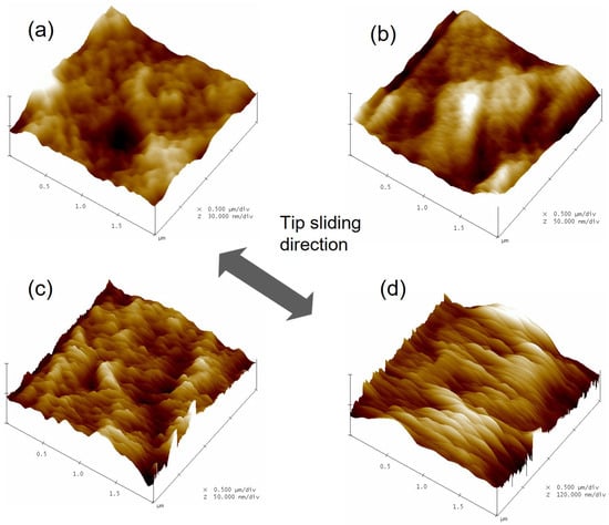
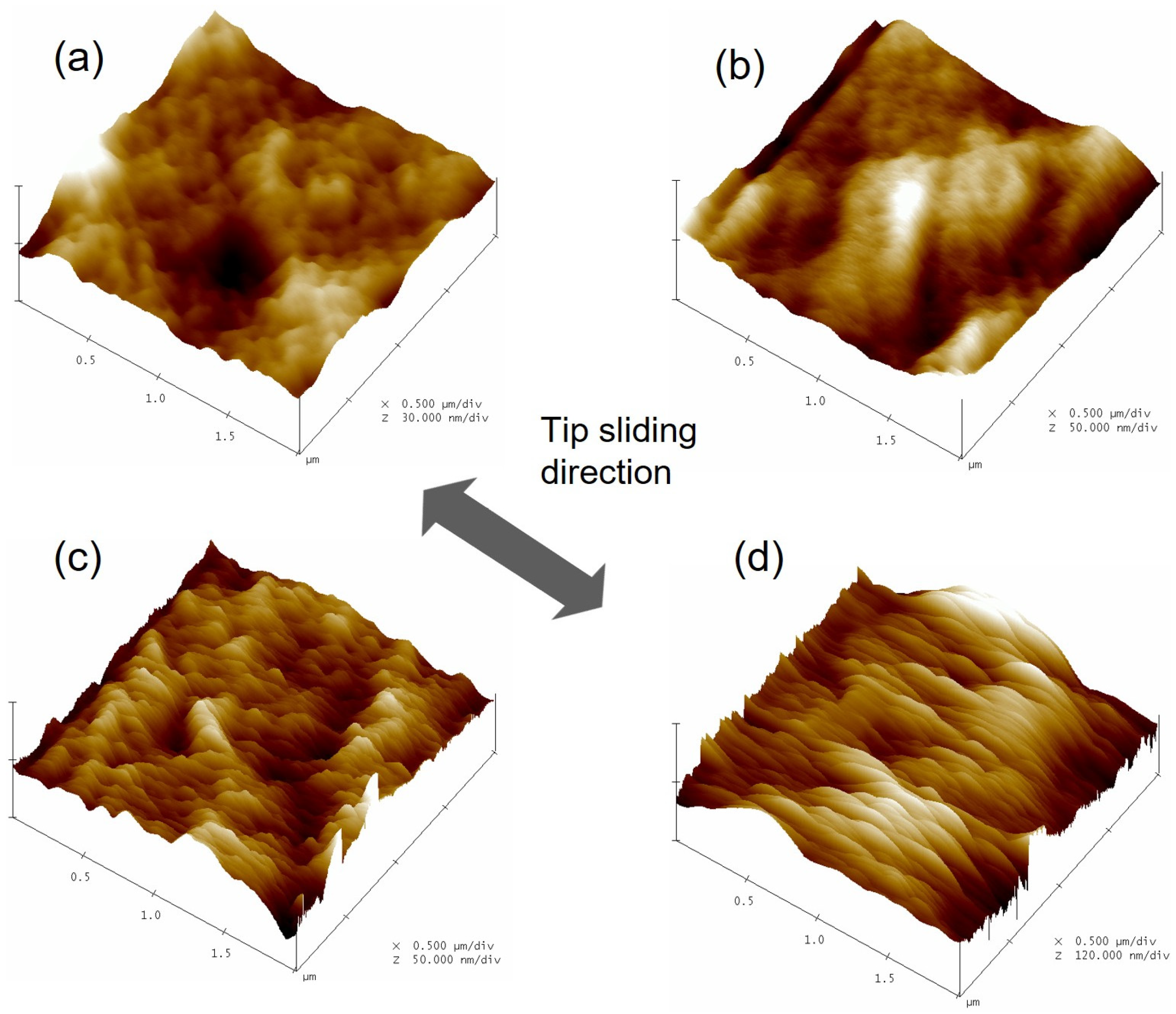
Table 5 shows that both µnano and FnF0 rise with PEG concentration, and nanofriction force increases as the films become more hydrophilic, i.e., as the PEG content increases. Thus, capillary condensation appears once more as a predominant factor that impacts [28] friction properties at the nanoscale. However, as the applied load increases, the friction coefficient at the nanoscale decreases. If the frictional behaviors of HM and HM-Plasticized films were governed by PEG content and applied load, other consequences like surface damage and wear phenomenon can occur on the surface of the films during nanofriction experiments. Figure 6 shows four topographic images (2 µm × 2 µm) obtained by FFM on HM and HM-Plasticized films at a set applied load of 0.1 V. The film surfaces were scanned in the orthogonal direction to the long axis of the cantilever.
Figure 6.
Topographic images (FFM) (2 µm × 2 µm) of (a) HM film and HM films containing (b) 20%, (c) 30%, and (d) 40% (w/w HM) of plasticizer. Applied load is set to 0.1 V.
Figure 6 clearly shows the wear scars induced by the FFM tip, while scanning, on the surface topography of HM and HM-Plasticized films during nanofriction measurements. The topographic images clearly illustrate surface changes that happened during friction measurements. The wear resistance of HM-Plasticized films reduces as their elastic modulus falls. The topographical changes are nicely visible at a high PEG content. To assess these changes, Table 6 displays the maximal scratch depth and arithmetic average roughness (Ra) following friction measurements for a set applied load of 0.1 V.
Table 6.
Maximal scratch depth and arithmetic average roughness (Ra) observed on HM and HM-Plasticized films.
These two parameters indicate a strong variation with the addition of plasticizer, and extreme changes are observed for HM-Plasticized films with 30% and 40% of PEG. Surface deformation is very different compared to that of pure films, even for a low value of the set applied load. The surface deformation and low wear resistance observed can be explained by the following points: First, the incorporation of PEG in HM aggregates, through the formation of hydrogen bonds between the hydroxyl end-groups of the PEG molecules and the hydroxypropyl groups substituted on the anhydroglucose backbone of the HM chains, increases the water affinity of HM-Plasticized films and favors the adsorption of water molecules on the film’s surface. Since HM and PEG are water-soluble polymers, the HM-PEG aggregates swell, expand in size, and thus the scratching phenomenon becomes easier under these conditions. Second, the increase of nanoadhesion and nanofriction forces for HM-Plasticized films is due to the development of adhesion within the contact zone between the AFM tip and the film’s surface. In this case, the contact pressure at the nanoscale can play an important role in the observed scratching phenomenon. Third, the important topographical modification observed for HM-Plasticized film containing 40% (w/w HM) of PEG is suspected to be due to excess [29] of plasticizer and thereby possible phase separation between the HM matrix and PEG.
3.7. Macrofriction of HM and HM-Plasticized Films
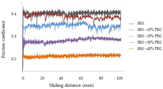
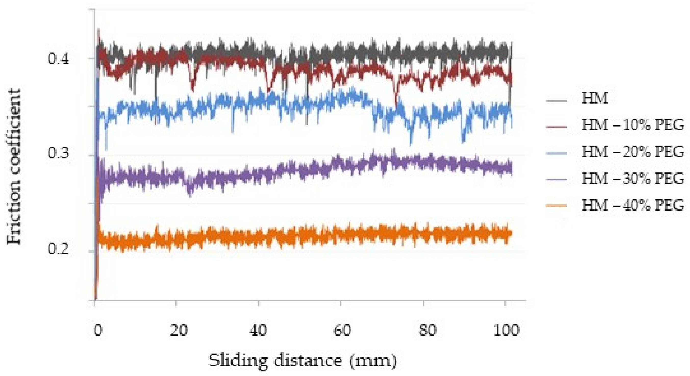
The macroscopic tribological behaviors of HM and HM-Plasticized films were analyzed using a conventional pin-on-disk tribometer that allows for recording the tangential force opposing the ball’s motion, equivalent to the friction force (FF), while the glass plate that supports the HM films was rotated at a specific speed. Various normal forces (FN), in the range of 2 to 10 N, were applied. To assess the friction properties of HM and HM-Plasticized films, the coefficient of friction was computed according to Amonton’s law, thus dividing the friction force FF by the normal force FN. Figure 7 illustrates the evolution of the friction coefficient as a function of sliding distance for HM and HM-formulated films. The applied normal force on the glass ball was 2 N, with a sliding velocity of 5 mm/s and a sliding distance of 100 mm.
Figure 7.
Evolution of the friction coefficient versus sliding distance for HM and HM-Plasticized films. The applied normal force is set to 2 N.
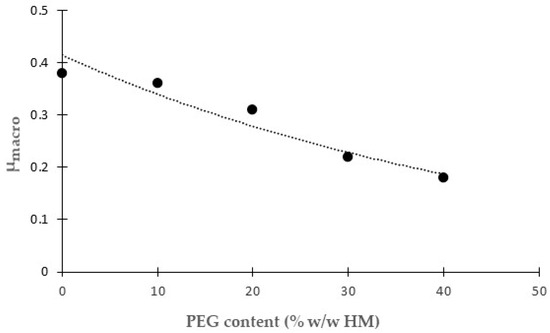
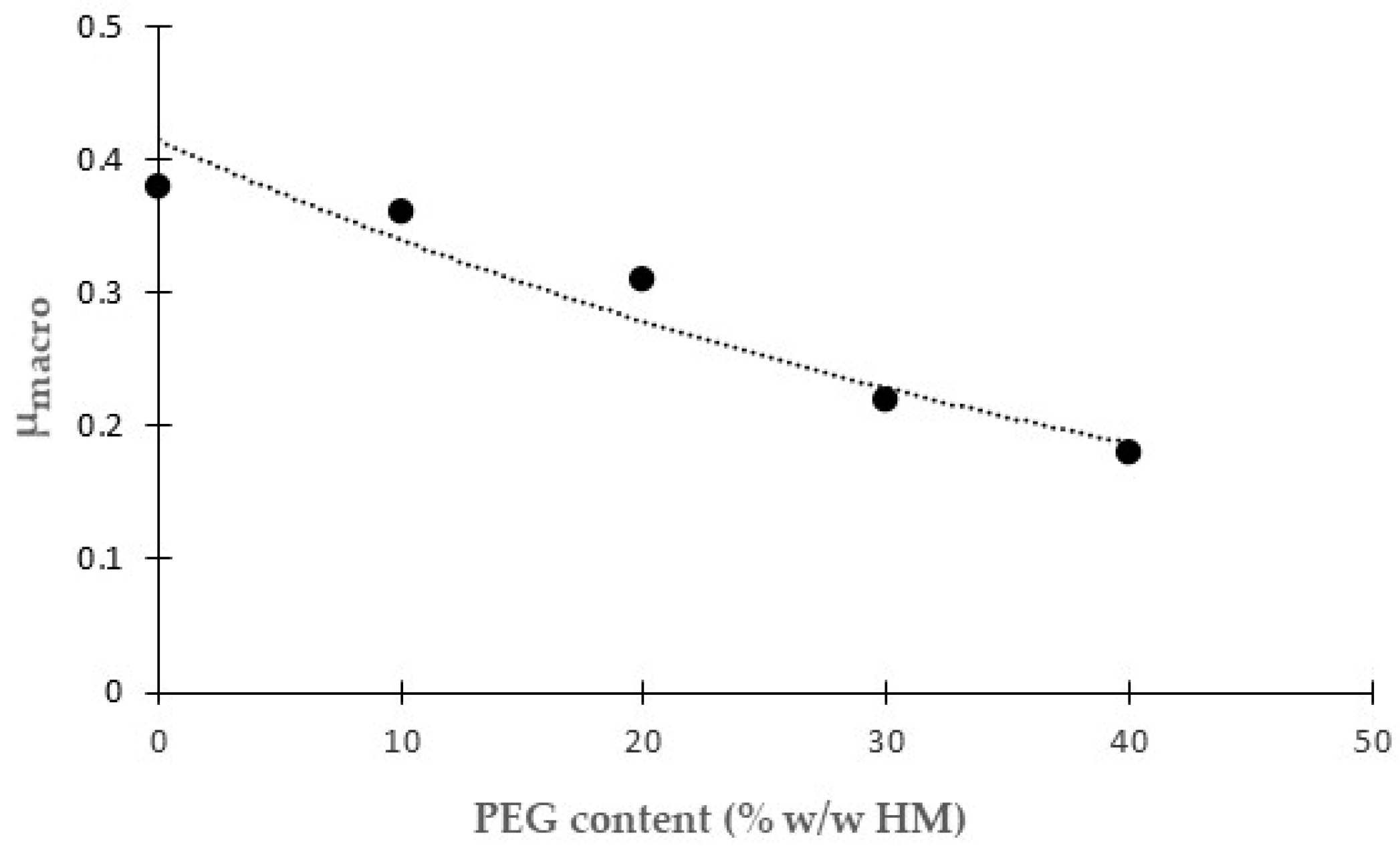
Tribological investigations at the macroscale reveal that the friction coefficient does not change with sliding distance for a fixed PEG concentration. Under 2 N normal force, one can clearly see a decrease in the friction coefficient with PEG content. PEG reduces the friction force between the glass ball and the HM-Plasticized film surface. Moreover, the results show (not represented) that the friction force values depend linearly on applied normal forces. In such a representation, the line slope represents the mean friction coefficient at the macroscopic scale. Figure 8 illustrates the evolution of the mean friction coefficient at the macroscale (µmacro) with the addition of plasticizer in HM films.
Figure 8.
Evolution of mean friction coefficient at macroscale (µmacro) versus PEG content for HM and HM-Plasticized films.
The mean friction coefficient decreases quite linearly from 0.38 for pure HM film to 0.18 with the addition of 40% PEG (w/w HM). The findings highlight PEG’s lubricating role on the surface of HM-Plasticized films, which is not observed at the nanoscale. This effect is due to the physical features within the contact sliding zone. The addition of PEG to HM aggregate softens the film and minimizes interactions between HM polymer chains’ hydrophobic substituents, replaced by the formation of hydrogen bonds between the hydroxyl end-groups of the PEG molecules and the hydroxypropyl groups substituted on the anhydroglucose backbone. The insertion of PEG between the HM chains promotes shear deformations during glass ball sliding. The decrease in friction forces is due to the simple deformation of HM chains swollen with PEG, which causes the glass ball to move easily over the film’s surface. The difference between nanoscale and macroscale sliding properties is due to the contact pressure within the contact zone and the involvement of adhesion force on both scales. At the macroscale, contact pressure is much lower than at the nanoscale. Nanoadhesion forces can interfere with nanofriction forces in ways that macrofriction cannot.
4. Discussion
Two important points have to be discussed following the results obtained. Indeed, the first point concerns the nanoscale versus macroscale correlation of adhesion and friction properties, whereas the second point concerns the role of adhesion in friction experiments.
4.1. Nanoscale vs. Macroscale Adhesion of HM and HM-Plasticized Films
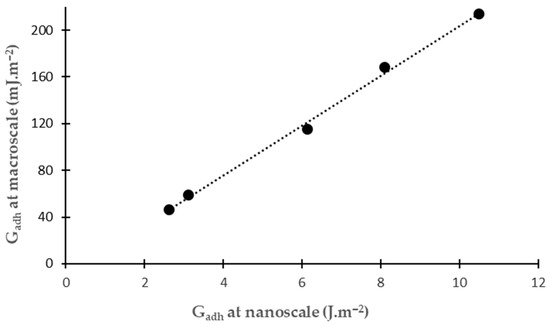
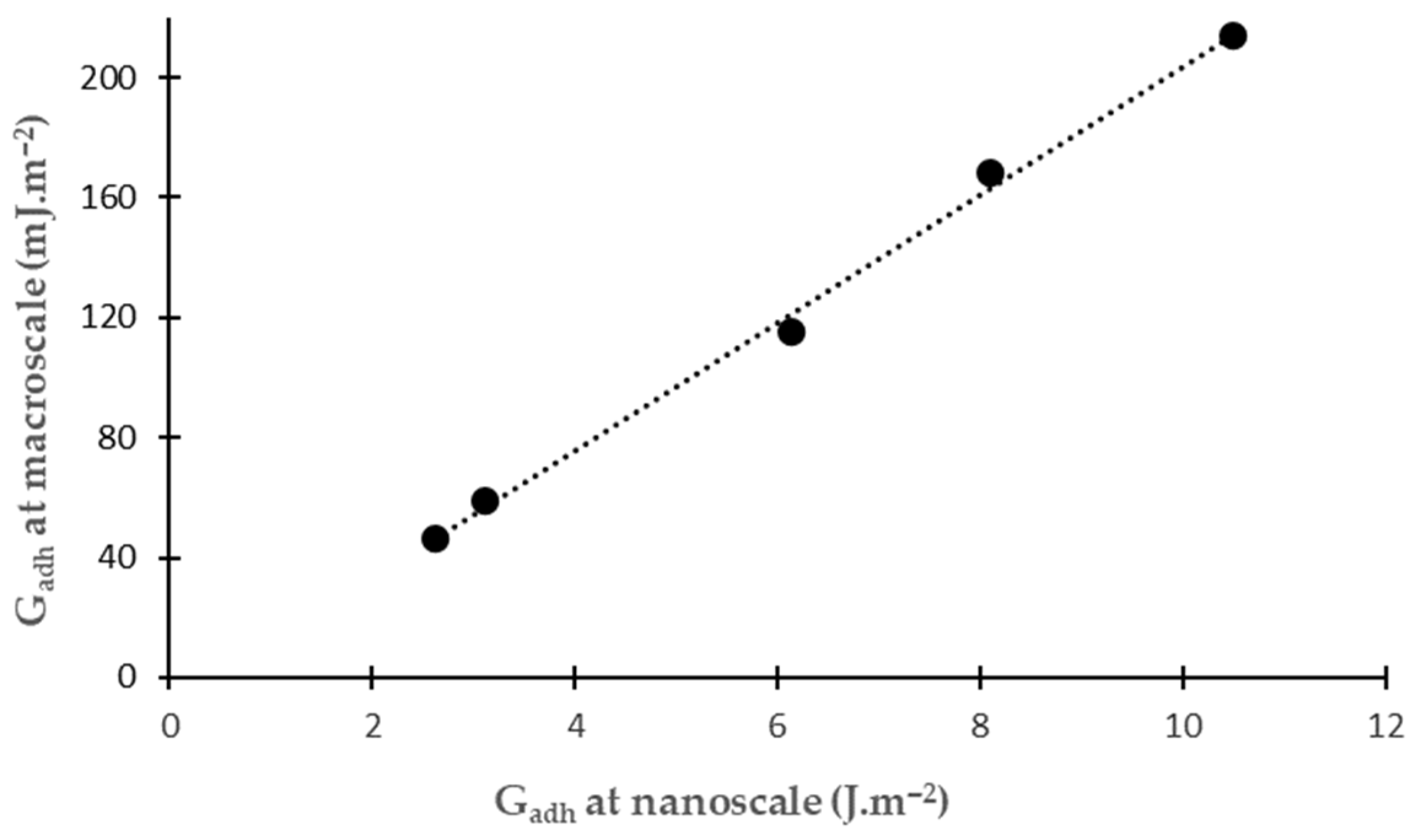
Figure 3 represents the evolution of the AFM adhesion force (Fadh) with PEG content for HM and HM-Plasticized films, whereas Figure 4 shows the influence of PEG content on the maximum value of the macroscopic adhesion force. At both scales, adhesion forces increase as the plasticizer content increases. Nevertheless, forces expressed in nN (nanoscale) or mN (macroscale) are not normalized values. For this reason, it is much more interesting to consider the adhesion energy, Gadh, expressed in J∙m−2. At the nanoscale, the adhesion energy required to separate the AFM tip from the HM or HM-Plasticized film surface is determined by computing the integral of the nanoadhesion force over the tip displacement associated with the separation step. At the macroscale, the adhesion energy required to separate the tack probe is determined by computing the integral of the adhesion force over the tack probe displacement associated with the separation step. Figure 9 represents the evolution of the adhesion energy at the macroscale as a function of adhesion energy at the nanoscale.
Figure 9.
Evolution of the adhesion energy at the macroscale as a function of adhesion energy at the nanoscale.
The results obtained at the macroscale are in agreement with those at the nanoscale, showing that the insertion of PEG molecules within the HM aggregates has the same effect at both scales. The adhesion behaviors of HM-Plasticized films exhibit good nano/macro correlation. HM-PEG compatibility, favored by, first, the interaction of the hydrophobic ethylene segments of the amphiphilic PEG molecules with the HM polymer chains’ hydrophobic substituents and, second, forming hydrogen bonds between the hydroxyl end-groups of the PEG molecules and the hydroxypropyl groups substituted on the anhydroglucose backbone, is a significant aspect, as it causes a change in adhesion behaviors at nano/macro scales to be linearly proportional to the amount of plasticizer in HM films. The increase in both components of nanoadhesion force (van der Waals and capillary forces) caused by plasticizer insertion in HM aggregates impacts adhesion energy at the nanoscale. Experiments at the macroscale show that adding PEG causes the films to soften and increase their molecular mobility, which increases their adhesion energy.
4.2. Nanoscale vs. Macroscale Friction of HM and HM-Plasticized Films
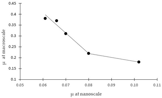
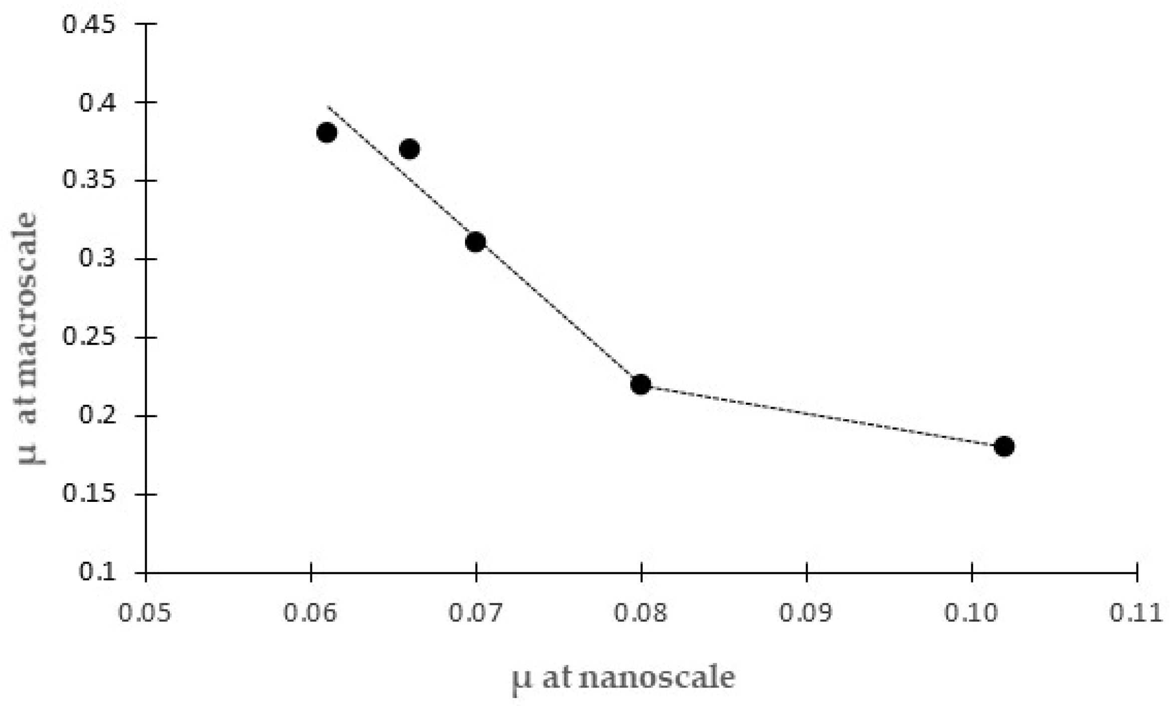
Previous results show that the adhesion energy of HM-Plasticized films exhibits the same tendency at the nano and macro scales. On the contrary, friction properties do not. For a clear comparison, Figure 10 represents the evolution of the friction coefficient at the macroscale as a function of the friction coefficient at the nanoscale.
Figure 10.
Evolution of the friction coefficient at the macroscale as a function of the friction coefficient at the nanoscale.
The addition of plasticizer in HM films induces an increase in friction coefficient at the nanoscale (µnano) (Table 5). However, a decrease in the friction coefficient is observed at the macroscale (µmacro) (Figure 8). Figure 10 indicates that a high friction coefficient at the nanoscale corresponds to a low friction coefficient at the macroscale, and vice versa. Two linear regimes are observed. The first linear trend line corresponds to a first linear decrease for PEG contents ranging from 0 to 30% (w/w), and the second linear decrease, less pronounced, is observed for PEG contents ranging from 30 to 40% (w/w). The hypothesis of HM-PEG phase separation at a high PEG content (>30% w/w) is proposed to explain the change in the linear trend line slopes. Moreover, the variations in friction coefficients are linear according to PEG content. The role of plasticizer in HM films, like a lubricant, was surprising at a larger scale (decreasing of friction coefficient), which was not found at the local scale (increasing of friction coefficient). The difference in contact pressure applied to the probe at both scales can explain this difference. Indeed, at the nanoscale, the applied load on the probe (AFM tip) ranges between 1 and 100 nN. The contact area is considered using the DMT model [30] and is equal to 2πR2, with R being the tip radius. Thus, the contact pressure at the nanoscale value is in the range 10–1000 N∙m−2. However, at the macroscale, the contact pressure on the glass ball is in the order of 100 N∙m−2. In addition, due to high contact pressure at the nanoscale, the adhesion strength might also be an additional parameter behind the increase in nanofriction behavior of HM-Plasticized films.
4.3. Interplay Between Adhesion and Friction of HM and HM-Plasticized Films
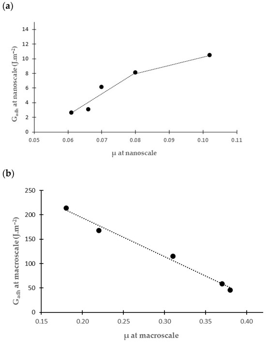
In order to unambiguously understand the interplay between adhesion and friction, Figure 11 reports the evolution of the adhesion energy (Gadh) as a function of the friction coefficient (µ), both at a nanoscale (a) and macroscale (b).
Figure 11.
Evolution of the adhesion energy (Gadh) as a function of the friction coefficient (µ), both at a nanoscale (a) and macroscale (b).
At the nanoscale, an increase in nanoadhesion, demonstrated to be due to capillary forces induced by the addition of plasticizer, contributes to increasing the friction coefficient in proportion. Two linear regimes are observed. The first linear trend line corresponds to a first linear increase for PEG contents ranging from 0 to 30% (w/w), and the second linear increase, less pronounced, is observed for PEG contents ranging from 30 to 40% (w/w). The hypothesis of HM-PEG phase separation at a high PEG content (>30% w/w) is proposed to explain the change in the linear trend line slopes. Moreover, this result (Figure 11a) validates the modification of the conventional Amonton’s law in nanofriction experiments. Indeed, the nanofriction force at zero applied (FnF0) renders the nanoadhesion force in the nanofriction experiment and has to be taken into account to access the nanofriction coefficient (Equation (2)). On the contrary, at the macroscale, an increase in adhesion energy, explained by the fact that plasticizer addition lowers the glass transition temperature of HM chains, thus boosting molecular mobility and interfacial interactions with the probe-tack plate, results in higher dissipation during the separation stage, and is associated with a decrease in the friction coefficient in proportion. Indeed, the mean friction coefficient decreases quite linearly with the addition of plasticizer (Figure 8), highlighting PEG’s lubricating role on the surface of HM-Plasticized films, which is not observed at the nanoscale.
5. Conclusions
The purpose of this work was to investigate the influence of low-molecular-weight PEG hydrophilic plasticizer on the bulk and surface characteristics of hypromellose films. Addition of plasticizer influences both thermal and mechanical properties. Glass transition temperature decreases from 138 °C to 41 °C, Young modulus decreases from 1500 MPa to 88 MPa, and tensile strength decreases from 12 MPa to 38 MPa when the plasticizer content increases from 0 to 40% w/w. The plasticizer also has an impact on surface properties. Indeed, the addition of plasticizer from 0 to 40% w/w raises the surface energy of the plasticized films from 43 mJ∙m−2 to 74 mJ∙m−2, thus demonstrating the hydrophilic film’s characteristics. The simultaneous rise in van der Waals and capillary forces causes the linear increase in adhesion force at the nanoscale with plasticizer concentration, whereas the lowering of the glass transition temperature of HM chains through an insertion mechanism in HM aggregates contributes to the increase of macroadhesion force from 9 to 90 mN when the plasticizer content increases from 0 to 40% w/w. Linear evolution of the nanofriction force is observed and relies on PEG content and on the applied load. Adding 20% PEG to HM film increases the nanofriction force by 15% for a set load, while adding 30% and 40% PEG to HM films increases the nanofriction force by 30% and 55%, respectively, as does the friction coefficient at the nanoscale. Consequently, the wear resistance of HM-Plasticized films is reduced as the plasticizer content increases. The tribological properties of plasticized HM films at the macroscale are reversed from those found at the nanoscale, and constitute an original result. Indeed, the mean friction coefficient decreases quite linearly from 0.38 for pure HM film to 0.18 with the addition of 40% PEG (w/w HM). The findings highlight PEG’s lubricating role on the surface of HM-Plasticized films, which is not observed at the nanoscale. In the last part, the nanoscale versus macroscale correlation of adhesion and friction properties and the role of adhesion in friction experiments are discussed. The adhesion behaviors of HM-Plasticized films exhibit good nano/macro correlation, indicating that the insertion of plasticizer molecules within the HM aggregates has the same effect at both scales. This constitutes an original result that has never been addressed previously. On the contrary, friction properties do not experience this. A high friction coefficient at the nanoscale corresponds to a low friction coefficient at the macroscale, and vice versa, showing, first, a linear decrease for PEG contents ranging from 0 to 30% (w/w), and, second, a linear decrease that is less pronounced, observed for PEG contents ranging from 30 to 40% (w/w). The hypothesis of a difference in contact pressure applied on the probe at both scales, as well as HM-PEG surface phase separation at a high PEG content (>30% w/w), is proposed to explain this difference. In addition, due to high contact pressure at the nanoscale, the adhesion within the contact zone constitutes an additional parameter behind the increase in nanofriction behavior of HM-Plasticized films. All these original results allow us to propose a modification of the conventional Amonton’s law in nanofriction experiments. Indeed, the nanofriction force at zero applied force renders the nanoadhesion force in a nanofriction experiment and has to be taken into account to access the nanofriction coefficient.
Author Contributions
Conceptualization, M.B. and S.B.; methodology, A.F.; validation, M.B., S.B., and A.F.; formal analysis, M.B.; investigation, A.F.; resources, M.B. and S.B.; data curation, M.B.; writing—original draft preparation, M.B. and A.F.; writing—review and editing, M.B. and S.B.; visualization, M.B. and S.B.; supervision, M.B. and S.B.; project administration, M.B.; funding acquisition, M.B. All authors have read and agreed to the published version of the manuscript.
Funding
This research was funded by the Region Alsace.
Institutional Review Board Statement
Not applicable.
Informed Consent Statement
Not applicable.
Data Availability Statement
The raw data supporting the conclusions of this article will be made available by the authors upon request.
Acknowledgments
The authors would like to thank the University de Haute Alsace for access to its scientific equipment.
Conflicts of Interest
The authors declare no conflicts of interest. The funders had no role in the design of the study; in the collection, analyses, or interpretation of data; in the writing of the manuscript; or in the decision to publish the results.
References
- Ford, J.L. Thermal analysis of hydroxypropylmethylcellulose and methylcellulose: Powders, gels and matrix tablets. Int. J. Pharm. 1999, 179, 209–228. [Google Scholar] [CrossRef] [Scilit]
- Navarro-Tarazaga, M.L.; Sothornvit, R.; Pérez-Gago, M.B. Effect of Plasticizer Type and Amount on Hydroxypropyl Methylcellulose−Beeswax Edible Film Properties and Postharvest Quality of Coated Plums (Cv. Angeleno). J. Agric. Food Chem. 2008, 56, 9502–9509. [Google Scholar] [CrossRef] [Scilit]
- Pérez, O.E.; Carrera Sánchez, C.; Rodríguez Patino, J.M.; Pilosof, A.M.R. Thermodynamic and Dynamic Characteristics of Hydroxypropylmethylcellulose Adsorbed Films at the Air−Water Interface. Biomacromolecules 2006, 7, 388–393. [Google Scholar] [CrossRef] [Scilit]
- Villalobos, R.; Hernández-Munõz, P.; Chiralt, A. Effect of surfactants on water sorption and barrier properties of hydroxypropyl methylcellulose films. Food Hydrocoll. 2006, 20, 502–509. [Google Scholar] [CrossRef] [Scilit]
- Suyatma, N.E.; Tighzert, L.; Copinet, A.; Coma, V. Effects of hydrophilic plasticizers on mechanical, thermal, and surface properties of chitosan films. J. Agric. Food Chem. 2005, 53, 3950–3957. [Google Scholar] [CrossRef] [Scilit] [PubMed]
- Thakhiew, W.; Devahastin, S.; Soponronnarit, S. Effects of drying methods and plasticizer concentration on some physical and mechanical properties of edible chitosan films. J. Food Eng. 2010, 99, 216–224. [Google Scholar] [CrossRef] [Scilit]
- Hansen, N.M.L.; Plackett, D. Sustainable films and coatings from hemicelluloses: A review. Biomacromolecules 2008, 9, 1493–1505. [Google Scholar] [CrossRef] [Scilit]
- Brogly, M.; Bistac, S.; Fahs, A. Surface migration of fatty acid to improve sliding properties of hypromellose-based coatings. Surfaces 2024, 7, 43. [Google Scholar] [CrossRef] [Scilit]
- Brogly, M.; Fahs, A.; Bistac, S. Determination of the chemical structure of cellulose based biopolymers. ARPN J. Eng. Appl. Sci. 2016, 11, 7188–7192. [Google Scholar]
- Owens, D.K.; Wendt, R.C. Estimation of the surface free energy of polymers. J. Appl. Polym. Sci. 1969, 13, 1741–1747. [Google Scholar] [CrossRef] [Scilit]
- Brogly, M.; Awada, H.; Noel, O. Contact atomic force microscopy: A powerful tool in adhesion science. In Applied Scanning Probe Methods XI: Scanning Probe Microscopy Techniques; Bushan, B., Fuchs, H., Eds.; Springer: Berlin/Heidelberg, Germany, 2009; pp. 73–95. [Google Scholar] [CrossRef] [Scilit]
- Lakrout, H.; Creton, C.; Ahn, D.; Shull, K.R. Influence of molecular features on the tackiness of acrylic polymer melts. Macromolecules 2001, 34, 7448–7458. [Google Scholar] [CrossRef] [Scilit]
- Bhushan, B. Nanotribology and Nanomechanics: An Introduction, 4th ed.; Springer: Cham, Switzerland, 2017. [Google Scholar]
- Galliano, A.; Bistac, S.; Schultz, J. The role of free chains in adhesion and friction of poly(dimethylsiloxane) (PDMS) networks. J. Adhes. 2003, 79, 973–991. [Google Scholar] [CrossRef] [Scilit]
- Nazhat, S.N. Thermal analysis of biomaterials. In Principles and Applications of Thermal Analysis; Gabbott, P., Ed.; Blackwell Publishing Ltd.: Oxford, UK, 2008; pp. 256–285. [Google Scholar] [CrossRef] [Scilit]
- Sakellariou, P.; Rowe, R.C. Phase separation and morphology in ethylcellulose/cellulose acetate phthalate blends. J. Appl. Polym. Sci. 1991, 43, 845–855. [Google Scholar] [CrossRef] [Scilit]
- Fuller, C.S.; MacRae, R.J.; Walther, M.; Cameron, R.E. Interactions in poly(ethylene oxide)–hydroxypropyl methylcellulose blends. Polymer 2001, 42, 9583–9592. [Google Scholar] [CrossRef] [Scilit]
- Kondo, T.; Sawatari, C.; Manley, R.S.J.; Gray, D.G. Characterization of hydrogen bonding in cellulose-synthetic polymer blend systems with regioselectively substituted methylcellulose. Macromolecules 1994, 27, 210–215. [Google Scholar] [CrossRef] [Scilit]
- Swarbrick, J. Encyclopedia of Pharmaceutical Technology, 3rd ed.; Informa Healthcare: New York, NY, USA, 2007. [Google Scholar]
- Peng, Y.; Wu, P.; Siesler, H.W. Two-dimensional/ATR infrared correlation spectroscopic study on water diffusion in a poly(ε-caprolactone) matrix. Biomacromolecules 2003, 4, 1041–1044. [Google Scholar] [CrossRef] [Scilit] [PubMed]
- Tanaka, M.; Ishizaki, S.; Suzuki, T.; Takai, R. Water vapor permeability of edible films prepared from fish water soluble proteins as affected by lipid type. J. Tokyo Univ. Fish. 2001, 87, 31–37. [Google Scholar]
- McHugh, T.; Krochta, J.M. Water vapor permeability properties of edible whey protein-lipid emulsion films. J. Am. Oil Chem. Soc. 1994, 71, 307–312. [Google Scholar] [CrossRef] [Scilit]
- Mittal, K.L. Contact Angle, Wettability and Adhesion, 1st ed.; CRC Press: London, UK, 2009; Volume 6. [Google Scholar] [CrossRef] [Scilit]
- Lepifre, S.; Froment, M.; Cazaux, F.; Houot, S.; Lourdin, D.; Coqueret, X.; Lapierre, C.; Baumberger, S. Lignin incorporation combined with electron-beam irradiation improves the surface water resistance of starch films. Biomacromolecules 2004, 5, 1678–1686. [Google Scholar] [CrossRef] [Scilit] [PubMed]
- Israelachvili, J.N.; Berman, A.D. Surface forces and microrheology of molecularly thin liquid films. In Handbook of Micro/Nano Tribology; Bushan, B., Ed.; CRC Press: Boca Raton, FL, USA, 2020; pp. 371–432. [Google Scholar]
- Stifter, T.; Marti, O.; Bhushan, B. Theoretical investigation of the distance dependence of capillary and van der Waals forces in scanning force microscopy. Phys. Rev. B 2000, 62, 13667–13673. [Google Scholar] [CrossRef] [Scilit]
- Song, S.; Ren, S.; Wang, J.; Yang, S.; Zhang, J. Preparation and tribological study of a peptide-containing alkylsiloxane monolayer on silicon. Langmuir 2006, 22, 6010–6015. [Google Scholar] [CrossRef] [Scilit] [PubMed]
- Garoff, N.; Zauscher, S. The influence of fatty acids and humidity on friction and adhesion of hydrophilic polymer surfaces. Langmuir 2002, 18, 6921–6927. [Google Scholar] [CrossRef] [Scilit]
- Hutchings, D.E.; Sakr, A. Influence of pH and plasticizers on drug release from ethylcellulose pseudolatex coated pellets. J. Pharm. Sci. 1994, 83, 1386–1390. [Google Scholar] [CrossRef] [Scilit]
- Derjaguin, B.V.; Muller, V.M.; Toporov, Y.P. Effect of contact deformations on the adhesion of particles. J. Coll. Interface Sci. 1975, 53, 314–326. [Google Scholar] [CrossRef] [Scilit]

Disclaimer/Publisher’s Note: The statements, opinions and data contained in all publications are solely those of the individual author(s) and contributor(s) and not of MDPI and/or the editor(s). MDPI and/or the editor(s) disclaim responsibility for any injury to people or property resulting from any ideas, methods, instructions or products referred to in the content. |
© 2026 by the authors. Licensee MDPI, Basel, Switzerland. This article is an open access article distributed under the terms and conditions of the Creative Commons Attribution (CC BY) license.