


Abstract
In cultural heritage conservation, the non-destructive characterisation of porous calcium-carbonate-based materials relies directly on assessing fluid transport, as water simultaneously governs primary deterioration processes and acts as the intrinsic probe for proton Nuclear Magnetic Resonance (1H-NMR). In this context, Nuclear Magnetic Resonance (NMR) has emerged as a powerful, non-invasive and non-destructive analytical tool. Recent advances in multimodal and multiparametric NMR approaches, including relaxometry, diffusion measurements, magnetic resonance imaging (MRI), and portable unilateral NMR, enable the comprehensive characterisation of porous systems across different spatial scales and under different experimental conditions. This review critically examines the contribution of proton NMR to the investigation of porous calcium-carbonate-based materials in cultural heritage. After relating water-induced deterioration to pore architecture, the review discusses the principles, measurable parameters, and complementary capabilities of NMR relaxometry, diffusion NMR, magnetic resonance imaging (MRI), and portable unilateral NMR. Particular attention is devoted to the effects of surface relaxation, restricted diffusion, and internal magnetic-field gradients, which are central to the interpretation of NMR data from mineral matrices. Representative applications to stones and marbles show that NMR can quantify accessible porosity, distinguish pore populations, map capillary water uptake, and monitor the penetration, effectiveness, and persistence of conservation products non-destructively, and, with portable instruments, without sampling. The evidence also highlights the strong dependence of treatment performance on lithotype and pore structure. By combining laboratory and portable NMR measurements with complementary analytical methods, future multi-parametric and multimodal workflows could provide more robust in situ diagnosis and support long-term, evidence-based conservation strategies.
1. Introduction
Natural porous stones, including marbles, granites, sandstones, conglomerates (such as historical puddingstone), and related lithotypes, have been employed since ancient times to create artefacts and architectural structures of inestimable cultural value. The need to preserve this vast heritage has driven increasing scientific interest in developing advanced strategies for monitoring and conservation. Given the historical and artistic significance of these materials, modern approaches prioritise analytical methodologies that minimise alteration of the original structure, favouring the use of non-invasive and non-destructive techniques. Among these, proton Nuclear Magnetic Resonance (1H-NMR) represents one of the most promising and innovative tools.
Exploiting the interaction between hydrogen nuclei (1H) and an external static magnetic field (B0) under radiofrequency (RF) excitation, magnetic resonance (MR) originated in physics and chemistry, but its greatest technological development was driven by biomedical imaging (MRI) [1,2]. These advances have proven equally powerful for the study of porous stone materials. Because the NMR signal originates directly from proton-bearing fluids, the water naturally residing or infiltrating the porous network serves as an exceptional intrinsic probe. Importantly, water represents simultaneously the primary agent of stone weathering and the vehicle that allows non-invasive NMR detection. Investigating water distribution, dynamics, and pore–surface interactions thus grants direct and quantitative insight into the microstructural degradation mechanisms of stone heritage [3,4,5,6].
NMR investigations of porous stones include relaxometry, diffusion measurements, internal gradient-related analyses, and magnetic resonance imaging (MRI), which can be performed using both high-field laboratory instruments and clinical scanners. In addition, since the introduction of the first portable unilateral NMR device (the NMR-MOUSE) in the mid-1990s, mobile NMR has expanded the in situ applications in cultural heritage [3,4], enabling the characterisation of fluid distribution and dynamics within porous systems, without constraints related to sample size or geometry.
In this context, portable NMR is particularly well suited for assessing the conservation state of stone materials: by directly probing water content, capillary uptake, and pore size distributions, it can non-invasively elucidate the fundamental water-driven deterioration mechanisms affecting porous inorganic substrates—such as superficial dissolution, runoff-mediated metallic staining, frost damage, and sub-florescence—as well as monitor the penetration and efficacy of modern conservation treatments.
This review critically examines the principles of NMR and presents the main instrumental approaches used for the investigation of porous materials. Key NMR and MRI methodologies, including acquisition sequences, models, and relevant measurable parameters for characterising porous structure, monitoring conservation treatments, and their potential contribution to preventive conservation, are illustrated.
In addition, because water is both the primary source of the detected NMR signal and the main driver of lithic decay, the essential water–pore interaction mechanisms and associated weathering processes are also outlined in the first part of the work.
Although NMR is a very versatile technique, suitable for the characterisation of various lithotypes, the representative applications discussed in this review mainly focus on carbonate stones (such as marbles, limestones, and calcarenites) to offer a more consistent framework for discussing the application and interpretation of NMR techniques on porous cultural heritage materials. Indeed, sandstones and puddingstones can exhibit highly heterogeneous, interconnected, and multi-scale porosity networks resulting from the arrangement of grains, pore bodies, pore throats, matrix, and/or cement. In such systems, interpreting the NMR parameter distribution in terms of pore size distribution is not straightforward. Specifically, because they are characterised by high interconnectivity. Moreover, carbonates not only represent a dominant substrate in monumental architecture, but their extreme vulnerability to water-mediated dissolution, weathering, and black crust formation makes them prime systems for advanced, non-destructive 1H-NMR relaxometry and imaging investigations. Representative case studies are subsequently discussed, followed by concluding remarks and future perspectives for the application of NMR techniques in cultural heritage conservation.
Previous reviews have addressed the instrumentation and methods of mobile single-sided NMR [3], the applications of the NMR-MOUSE to heritage objects [4], and the use of NMR for a wide range of cultural heritage materials [7]. The present review differs in three respects: (i) it focuses specifically on porous calcium-carbonate-based materials; (ii) it links water-related deterioration to the physical basis of NMR observables (surface relaxation, restricted and hindered diffusion, internal gradients, intermolecular double-quantum coherences, iDQC) and to their interpretative limits; and (iii) it compares laboratory relaxometry and diffusion, MRI and portable NMR in applications to cleaning, consolidation and water-repellent treatments, including studies published up to 2026. Here, “multimodal” refers to the combined use of complementary instrumental configurations (laboratory relaxometry, diffusion NMR, MRI, and unilateral NMR), and “multiparametric” to the different observables they provide (signal amplitude, T1, T2, and diffusion coefficient).
Within this scope, the practical aim of the review is to provide a physical and methodological basis for collaboration between NMR specialists and conservation scientists in diagnosing stone deterioration and monitoring interventions before, during and after treatment. By connecting conservation questions with measurable NMR observables and their interpretative limitations, this narrative review seeks to guide material-specific investigations and identify opportunities for further methodological development, laying the groundwork for systematic experimental studies and future validated protocols tailored to specific conservation tasks, such as diagnosing deterioration and monitoring treatment effects.
Literature Search Strategy
The scope of this narrative review is limited to porous calcium-carbonate-based materials (limestones, calcarenites, travertines, marbles and lime-based plasters and mortars), which constitute a large part of the built and sculptural heritage and are particularly vulnerable to water-related deterioration. The literature was retrieved from Scopus, Web of Science Core Collection and Google Scholar, complemented by the proceedings of the International Congresses on the Deterioration and Conservation of Stone. Search strings combined, with the Boolean operator AND, terms describing the NMR technique (e.g., relaxometry, MRI, unilateral NMR, diffusion NMR), the material (e.g., limestone, marble, calcarenite, lime mortar) and the conservation context (e.g., weathering, consolidation, water repellent, cleaning). For heritage applications, the search covered the period from January 1990 to October 2026, whereas methodological works on relaxation, diffusion, anomalous diffusion, iDQC and internal gradients in porous media were included without a time limit and based on the authors’ background. Background references on weathering and conventional porosimetry (Section 2) were not part of the structured search.
Studies were included if they applied 1H NMR relaxometry, diffusion NMR, MRI, micro-MRI or portable unilateral NMR to calcium-carbonate-based heritage materials and were published in English in peer-reviewed journals, books or full-text proceedings. Studies using NMR only for chemical characterisation (e.g., magic angle spinning, MAS NMR), dealing with non-carbonate or organic materials, or available only as abstracts were excluded. Studies on other substrates (e.g., sandstones, bricks, wood or phantoms) were retained only when they provide methods directly transferable to carbonate stones; in these cases, the substrate is indicated in the text and tables. Records were screened by title and abstract and then in full text by the authors. The selected studies are discussed in Section 2, Section 3 and Section 4 and summarised by technique and conservation treatment in Table 1, Table 2, Table 3 and Table 4. Studies in other languages and the non-indexed grey literature may be under-represented.
2. Weathering of Stones Due to Water Interactions
In all its physical states, water interacts with artificial and natural stones, activating most of the weathering mechanisms related to this material [6]. Material dissolution, black crust formation, and discolourations are well-known consequences of direct interaction between water and stones because water’s solvent properties are combined with the presence of various kinds of pollutants and particulate matter (PM) in the atmosphere [8]. Pollutants can be transported on material surfaces by rainwater (wet deposition) or by wind and other atmospheric agents (dry deposition). In the second case, weathering can be activated by water condensation, a phenomenon that occurs when the stone is exposed to a humid atmosphere and the temperature of the lytic surface drops to the dew point (i.e., the temperature at which saturation is reached) [9,10,11]. Liquid water can induce several decay processes (including acid attack, penetration of saline solutions, and biofilm formation), and some authors introduced the Time of Wetness (TOW) as a parameter that defines the time fraction during which liquid water could be present on a stone surface [8], as a “hazard” indicator. Dissolution of minerals and the stone’s binding medium, for example, is a phenomenon activated by water in different ways.
Among the most sensitive stones to these kinds of weathering mechanisms are calcareous stone materials (marbles, travertines, sandstones, etc.), because of the main presence of calcium carbonate in their mineralogical composition, which is highly susceptible to acid environments. However, other kinds of lithotypes are not excluded, such as granite [12] whose plagioclase mineral grain components are susceptible to acidic effects [13].
The dissolution mechanisms activated by water are mainly related to (1) the Karst Effect, produced by rain at pH ~ 5.6 in equilibrium with atmospheric CO2, (2) an acid rain effect, caused by rain with additional acidity due to the presence of sulfuric and nitric acid, and (3) the dry deposition of gaseous pollutants (SO2 and NOx), occurring between precipitation events [14,15].
2.1. Water-Driven Chemical and Physical Weathering (Dissolution, Crusts, Staining, and Salt Transport)
Degradation of stone materials of cultural interest has accelerated since air pollution became a major environmental issue. Calcareous stone materials are mainly composed of calcite, a polymorph of calcium carbonate, extremely sensitive to acid action [16]. The acid attack of a carbonate surface involves numerous problems of an economic and structural nature, since it leads to erosion of surfaces, loss of details, and total damage to the aesthetic appearance of the product.
The erosion of carbonate stone is the result of two processes, chemical erosion and physical erosion: the first involves the dissolution of the carbonate stone, and the second is the loss of the grains that make up the crystalline structure of the minerals that make up the rock, causing mechanical and structural damage [16].
The pH of rainwater is around 5.6, in equilibrium with atmospheric CO2. This type of rain alone is enough to damage a carbonate surface [14]; however, often in the rains, there are other compounds which contribute to further lowering the pH, such as sulfuric and nitric acid, and the presence of gaseous pollutants, such as SO2 and NOx. The resulting degradation process is greatly influenced by the direction of rain, wind, material, and surface geometry [9,15,16,17]. The presence of these pollutants dispersed in water, acting as a solvent, represents a major threat to outdoor cultural heritage, causing one of the most critical and widespread forms of stone degradation: the formation of black crusts. This phenomenon is particularly critical in sheltered areas [15]. Indeed, water mainly acts as a medium that allows chemical reactions to happen, rather than being the direct cause of black crusts. While direct rainfall washes over the stone too quickly and removes soluble materials, areas protected from rain allow moisture and air pollutants to deposit and accumulate over time. Because this contact lasts much longer, the slow chemical reaction between pollutants and the carbonate stone can take place, leading to the formation of gypsum and black crusts [9]. Black crust is a typical problem of carbonate stones (travertine, Carrara marble, Lecce stone, etc.) because of the prevalence of calcium carbonate in their chemical composition. This phenomenon results from the sulfation of the carbonate surface. Sulfur dioxide (SO2) deposited on the wet stone surface (dry deposition) is oxidised to sulfuric acid or sulfate, a reaction catalysed by metal oxides and carbonaceous particles, while additional sulfuric acid is supplied by acid rain (wet deposition) [9]. The acid reacts with calcite to form gypsum (calcium sulfate dihydrate) according to CaCO3 + H2SO4 + H2O → CaSO4·2H2O + CO2. Gypsum is about two orders of magnitude more soluble than calcite and has a molar volume about twice as large. On rain-exposed surfaces, it is therefore progressively washed away, causing surface recession, whereas in sheltered areas it accumulates and embeds atmospheric particulate matter, forming black crusts. Besides the aesthetic damage, the volume increase associated with gypsum crystallisation within the pores generates mechanical stresses that can lead to the detachment of the crust and of the underlying stone. Even some of the most important UNESCO masterpieces have not been spared, like the Trevi Fountain [13], the Cathedral of Florence [18] and the Parthenon [19].
Another relevant consequence of water interaction with built heritage materials is surface staining and differential discolouration, typically induced by runoff percolation. This is particularly evident in stone materials placed in direct contact with metal alloys, such as the marble bases of bronze statues (see Figure 1), and in stones with iron nails and structural bars, which are also caused by water percolation. Indeed, rainwater can transport soluble salts derived from the corrosion products of metals or alloys on the pedestals and reprecipitate as stains on the stone’s surfaces, causing the loss of the original aesthetic value of the artwork [14]. Depending on the nature of the metal or the alloy, the colour of the stains varies from green, bluish, or yellowish in the case of copper products; the red-rusty colour is typically associated with the presence of iron, while divalent lead oxides and minium give red stains, whereas lead dioxide contributes to the blackening of the stains [15,20] (Figure 1). In the literature, different study cases evidencing this kind of problem are reported, such as the equestrian statue of Michael the Brave (Cluj-Napoca, Romania) [21], the monument to Francis Garnier (Paris, France) [22], and the Fontana delle Tartarughe (Roma, Italia) [14].
Figure 1.
(a) The bronze copy of the Statue of the emperor Ottaviano Augusto (XX sec), exposed to the open environment (Via dei Fori Imperiali, Rome, Italy). The original “Augusto di Prima Porta” is a marble statue, preserved at the Vatican Museums. (b) Metallic patina percolated from the bronze statue into the marble basement. Photographs taken by C.G.
On the other hand, water is responsible for the weathering of porous inorganic materials not only by wetting the surface but also for its penetration of the inner structure of the porous matrix, mainly due to capillary rise (see Section 2.2). The presence of moisture inside natural or artificial stones, associated with some specific environmental conditions, can lead to freeze–thaw (F-T) and wetting-drying (W-D) cycles, for their part responsible for other mechanical, chemical, and biological weathering processes. During evaporation (W-D cycles), dissolved salts crystallise and accumulate either on the surface of the material, producing salt efflorescence, or within its porous structure, producing salt subflorescence [9]. It seems that the location and distribution of the salt crystals depend on the salt type: it was demonstrated that, under the same experimental conditions, halite might show efflorescence, while thenardite gives subflorescence, crystallising within the pore spaces [23]. Efflorescence mainly causes aesthetic deterioration that can be easily remedied by removing the salts, whereas subflorescence causes more serious damage, such as cracking and exfoliation, due to crystal expansion inside the capillaries. Salt crystallisation occurs in conditions of supersaturation or when the salt concentration grows due to temperature decreasing and water evaporation, depending on the salt type [24]. The damage related to the changes in the state of water and salt crystallisation is particularly relevant in the Mediterranean areas, where they are considered the main consequence of moisture presence [5].
In addition to salt crystallisation during wetting–drying cycles, freezing and thawing (F-T) of pore water can contribute to stone deterioration. The development of mechanical stresses during ice formation within the pore network can involve hydraulic pressure associated with the volumetric expansion of water upon freezing and crystallisation pressure exerted by growing ice crystals on the pore walls [25]. This process is influenced by both pore size and the presence of dissolved salts. Pore confinement lowers the equilibrium ice–water transition temperature as pore size decreases, consistent with the Gibbs–Thomson effect; consequently, water in smaller pores can remain unfrozen at temperatures at which ice is already present in larger pores [25,26]. Dissolved salts further lower the freezing point and, when retained within the pore liquid, become progressively concentrated as ice forms, influencing the amount of unfrozen water at a given temperature [26]. The resulting damage therefore depends on the interplay between intrinsic lithotype characteristics, including pore size distribution, pore connectivity, total porosity and mechanical resistance, and factors such as water saturation, solution composition and wetting properties [27].
Notably, many of these water-driven degradation phenomena, including capillary uptake dynamics, salt accumulation, and the formation of runoff-induced metallic patina, have recently been investigated using advanced and portable NMR techniques, which allow non-invasive tracking of fluid distribution and pore-network modifications, as well as the monitoring of related cleaning and consolidation treatments, which are detailed in Section 4.
2.2. The Influence of Porosity on Porous Stone Weathering and Current Methods for Pore Size Evaluation
The absorption of acid gases depends on the amount of moisture present in the porous matrix, which in turn depends on the absorption and permeability of liquid water, hygroscopic moisture content, evaporation, and permeability of water in the vapour phase.
All these properties can be related to the size and distribution of the pores and therefore to porosity, one of the main characteristics of a material when it comes to degradation [24]. The hygroscopic moisture content is very important in the dry deposition of acid gases and in the formation of sub-efflorescence and efflorescence phenomena, and is related to the relative humidity of the air, the nature of the material, pore diameter, and specific surface [10,24]. The presence of hygroscopic salts, such as nitrates, chlorides, and derivatives from previous SO2 deposits, increases the presence of atmospheric humidity, causing an accumulation of water located inside the pores, favouring the oxidation and absorption of atmospheric SO2, forming calcium sulphate [24].
Most inorganic building materials-bricks, stones, mortars, concretes-are intrinsically porous and hydrophilic, exhibiting open porosities that frequently exceed 10% [28].
In these systems, water penetration is predominantly driven by capillarity. In a hydrophilic pore (contact angle θ < 90°), the water–air interface forms a concave meniscus, and the resulting capillary pressure, Pc = 2σ cosθ/r, where σ is the surface tension of water and r is the pore radius, draws water into the pore network against gravity (Figure 2). In a vertical capillary, the equilibrium height is given by Jurin’s law, heq = 2σ cosθ/(ρw g r), where ρw is the density of water and g is the gravitational acceleration; heq therefore increases as the pore radius decreases. The rate of rise, instead, results from the balance between capillary pressure and viscous resistance. Neglecting gravity, the height reached at time t is described by the Lucas–Washburn law, h(t) = (σ r cosθ t/2η)1/2, where η is the viscosity of water. Consequently, at early times water rises faster in wider pores, whereas narrower pores fill more slowly but reach greater equilibrium heights (Figure 3) [9,10].
Figure 2.
Water meniscus inside capillaries. Arrows indicate the upward capillary forces and opposing gravity in the hydrophilic capillary, and downward capillary action in the hydrophobic capillary. A: convex meniscus; B: capillary depression.
Figure 3.
Capillary rise of water in ideal vertical cylindrical capillaries of radius r = 1, 10 and 50 µm (solid lines), calculated by numerically integrating the Lucas–Washburn equation including gravity, dh/dt = (r2/8ηh)(2σ cosθ/r − ρw g h), with σ = 0.072 N m−1, η = 1.0 mPa s, ρw = 1000 kg m−3 and θ = 0°. Dotted lines: Lucas–Washburn law without gravity; dashed lines: equilibrium heights given by Jurin’s law. Wider capillaries fill faster at early times, whereas narrower capillaries reach greater equilibrium heights. In real stones, tortuosity, the pore size distribution and evaporation reduce the heights actually reached.
In real stones, the capillary rise dynamics and the final height reached by the waterfront are governed by the pore size distribution and by the balance between capillary suction and surface evaporation [10]. In natural building stones, pore diameters span several orders of magnitude, from a few nanometres to hundreds of micrometres. Torraca [10] observed that the equivalent diameters of building stones are frequently concentrated in two ranges, large pores (>10 µm) and capillary pores (0.1–1 µm); this is an empirical observation rather than a complete classification, and pores of intermediate size (1–10 µm) and smaller than 0.1 µm also contribute to water transport and retention. In general, larger pores fill and drain rapidly but exert weak capillary suction, whereas smaller pores exert stronger suction, retain water for longer and, in the sub-micrometre range, are involved in adsorption and capillary condensation. The pore size distribution therefore largely determines moisture transport kinetics and the vulnerability of the lithotype to physical decay mechanisms, such as salt crystallisation and freeze–thaw cycles [10].
In this scenario, the determination of the pore size distribution of stones is crucial to predict the deterioration mechanisms and to evaluate stone durability over time. It is also fundamental for characterising the interaction between the pore network and the external environment. Throughout this review, the terms micropores, mesopores and macropores are used only in the IUPAC sense (<2 nm, 2–50 nm and >50 nm, respectively); since the pores relevant to water transport in building stones are mostly macropores in this sense, pore sizes are otherwise given explicitly in micrometres. Among the methods used for the characterisation of the porosity property of building material, it is possible to make a distinction between direct methods which allow direct documentation and measurement of pore space (i.e., thin section microscopy and scanning electron microscopy), and indirect methods which enable calculating the porosity data from the measurement results [29,30,31,32].
One of the most popular and effective methods enabling the determination of the total porosity (%), the median pore radius, the pore size distribution, and the pore volume (mm3/g) is Mercury Intrusion Porosimetry (MIP), which is widely adopted in the field of conservation of building materials [32].
Using this method, pore sizes ranging from about 0.002 µm to 300 µm can be investigated. The pressure P required to force mercury, a non-wetting liquid, into a cylindrical pore of radius rp is given by the Washburn equation, P = −2σHg cosθHg/rp, where σHg is the surface tension of mercury (≈0.485 N m−1), and θHg is the mercury–solid contact angle (typically 130–140°), so that P is positive. To probe pore radii of 0.01 µm and 0.005 µm, pressures of about 75 MPa and 150 MPa, respectively, are required, which may distort the pore structure [33]. The main limitation of MIP, however, is that it measures the size of the pore entrances (throats) through which mercury penetrates, rather than that of the pore bodies: large pores accessible only through narrower throats are assigned to the throat size (ink-bottle effect), so that the pore size distribution is shifted towards smaller sizes [32]. NMR relaxometry, instead, probes the surface-to-volume ratio (S/V) of the pore bodies (Section 3). This difference must be taken into account when MIP and NMR pore size distributions are compared, and it is the main source of the discrepancies between the two methods [34].
The Nitrogen-adsorption method provides the best results for nanopores detection when the measuring range is 0.001–0.1 μm. The adsorption isotherms can be analysed using different models, among which the BET (Brunauer–Emmett–Teller) theory allows the determination of the specific surface area of porous materials from the amount of N2 needed to form a monomolecular gas layer on the pore surface. The total pore volume (1):
can be derived from the amount of vapour adsorbed (Vads) at a relative pressure close to unity (assuming pores are filled with liquid adsorbate Vliq) and the average pore radius (rp) can be estimated from the pore volume assuming a cylindrical geometry [33].
Considering the limitations of the methods described above, researchers are increasingly investigating alternative techniques that can also be applied to the characterisation of the pore-size distribution of stone materials. In this context, methodologies based on nuclear magnetic resonance (NMR) are becoming increasingly important.
Zuena et al. [34] compared MIP and NMR relaxometry on limestones treated with a consolidating product, showing that the two methods provide complementary information and that NMR relaxometry is a valid non-destructive tool to follow the changes in pore size distribution induced by the treatment. Since NMR relaxometry can also be performed with portable instruments (Section 3.3), this approach can in principle be extended to in situ measurements on objects where sampling is not possible.
It should be noted that NMR relaxometry does not measure pore sizes directly. In the fast-diffusion regime, the relaxation rate of water in a pore is proportional to its surface-to-volume ratio (S/V) through the surface relaxivity ρ (Mean Pore Diameters Evaluation, in Section 3.4), so that a T2 (or T1) distribution corresponds to a distribution of S/V. Its conversion into pore sizes therefore requires the surface relaxivity and an assumption on pore geometry; since ρ depends on the mineralogy of the pore surface, and in particular on its content of paramagnetic impurities, it is usually calibrated against independent methods such as MIP [34].
In the following sections, the NMR methods applied over the past three decades to the study and conservation of stone heritage are described. Conventional methods, such as those described in this section, are complementary to NMR and are often used to calibrate NMR measurements and validate their interpretation.
3. Proton Nuclear Magnetic Resonance Techniques
To perform an NMR experiment, the sample must be inserted into a radiofrequency (RF) coil, surrounded by a static magnetic field B0. Proton Nuclear Magnetic Resonance (1H-NMR) is based on the stimulation of hydrogen spins nuclei by an electromagnetic field in the radiofrequency (RF) region, when they are exposed to a static magnetic field (B0), conventionally along the z-axis [1,2]. Hydrogen (1H) has only one proton in its nucleus, therefore 1H-NMR is also known as proton NMR.
Nuclei with a non-zero spin quantum number I possess an intrinsic angular momentum (spin) and an associated magnetic moment μ, which is an intrinsic property of the nucleus and is proportional to the spin angular momentum (2):
where γ is the gyromagnetic ratio, ħ = h/2π is the reduced Planck constant (h being Planck’s constant), and I is the spin angular momentum expressed in units of ħ. For the hydrogen nucleus (the proton), I = ½.
μ = γħI
When many hydrogen spins are present (spins ensemble), their magnetic moments will sum, generating a macroscopic magnetisation vector (M). In the presence of a static magnetic field B0, the macroscopic magnetisation vector M aligns with the direction of B0 (conventionally defined as the z-axis), resulting in an equilibrium magnetisation. At equilibrium, the longitudinal magnetisation Mz reaches its maximum value, M0. To obtain an NMR signal, the magnetisation must be carried in the plane transverse to the direction of the magnetic field. Toward this purpose, energy must be absorbed by the nuclear spins ensemble. With the application of a radiofrequency (RF) pulse of 90°, for instance, with a specific frequency, called resonance or Larmor frequency equal to Equation (3):
The nuclei are excited to absorb energy, causing the longitudinal magnetisation (Mz) to rotate into the transverse plane (Mxy). This is achieved through the application of a radiofrequency (RF) pulse, which generates a magnetic field (B1) in the transverse plane. As a result, the magnetisation begins to precess around B1.
After the RF pulse, the magnetisation returns to equilibrium through two relaxation processes. Longitudinal (spin–lattice) relaxation, characterised by the time constant T1, describes the recovery of Mz towards M0 and involves the exchange of energy between the spin system and its surroundings (the lattice). Transverse (spin–spin) relaxation, characterised by the time constant T2, describes the decay of Mxy due to the loss of phase coherence among the spins, caused by fluctuating local magnetic fields (mainly dipolar interactions); it does not require energy exchange with the lattice, and T2 ≤ T1. The decay of Mxy observed immediately after the RF pulse (free induction decay) is faster and is characterised by the time constant T2*, which also includes the dephasing due to static inhomogeneities of the magnetic field, both instrumental and induced by differences in magnetic susceptibility between the solid matrix and the pore fluid: 1/T2* = 1/T2 + 1/T2′, where 1/T2′ ≈ γΔB0 and ΔB0 is the field inhomogeneity within the sample. Since this static contribution is refocused by spin-echo sequences, T2 is measured with spin-echo or CPMG sequences (Section 3.4); in porous media; however, the diffusion of water through internal field gradients causes an additional attenuation that is not refocused, so that an apparent T2 is measured (Section 3.6 and Section 3.7). In porous stones, the relaxation of water is dominated by its interaction with the pore surfaces, where paramagnetic impurities strongly enhance relaxation (Mean Pore Diameters Evaluation, in Section 3.4).
The detected NMR signal is an electrical signal induced in the receiver coil by the time-varying magnetic flux generated by the precessing spins [1]. The intensity and characteristics of this signal depend on several measurable parameters, including T1, T2, T2*, the diffusion coefficient (D) and the chemical shift, which provide structural and physicochemical information about the system [2,5]. In porous stones, however, susceptibility-induced line broadening generally limits chemical-shift resolution, particularly with low-field and portable instruments, so that relaxation and diffusion parameters are the most informative.
Importantly, the NMR signal also depends on the number of hydrogen nuclei present and their binding environment. In systems where hydrogen nuclei are tightly bound, very short T2 relaxation times lead to rapid signal decay, often resulting in negligible detectable signal. In such cases, fast dephasing occurs during the instrumental dead time between the end of the RF pulse and the beginning of signal acquisition, typically ranging from 5 to 50 μs depending on the spectrometer hardware [35].
Conversely, in systems with more mobile or weakly bound hydrogen nuclei-such as soft matter, liquids, or waterlogged wood-longer T2 values result in stronger, detectable signals [36,37]. Intermediate binding conditions are commonly observed in porous materials, including bone, wood, and stone, where a distribution of relaxation times reflects the complexity of the microstructure [38,39]. These porous systems, however, differ from stones not only in pore size and pore size distribution, but also in pore interconnection and in the magnetic susceptibility difference between the pore fluid and the solid matrix, which governs internal gradients and apparent relaxation (Section 3.6 and Section 3.7); they are mentioned here only to help the reader place stones within the broader family of porous materials.
To accurately measure specific NMR parameters or to obtain images weighted by a selected contrast mechanism (see Section 3.2), appropriate NMR acquisition sequences must be carefully chosen [2,5].
3.1. Conventional NMR Instruments
Conventional NMR spectrometers are versatile, non-invasive analytical tools widely used in chemistry, biochemistry, and materials science for applications such as molecular structure determination, reaction monitoring, and the study of molecular dynamics. These instruments are typically based on a superconducting magnet capable of generating a strong and highly homogeneous static magnetic field, generally ranging from 1 T up to 21 T.
The magnet is equipped with a narrow vertical bore, within which the sample is placed and surrounded by radiofrequency (RF) coils for signal excitation and detection. The bore diameter typically ranges from 5 mm to 25 mm, limiting the analysis to relatively small samples. The same applies to laboratory MRI scanners for materials, whose radiofrequency coils typically accommodate samples a few centimetres in size or samples as large as a human body when clinical scanners are used. These measurements are non-destructive for the sample, but for heritage objects they imply sampling; only unilateral instruments (Section 3.3) allow in situ measurements without sampling, whereas clinical scanners do not. Conventional NMR spectrometers enable measurement of key parameters such as longitudinal and transverse relaxation times (T1 and T2), diffusion coefficients (D), and NMR spectra, providing detailed information on the chemical and physical properties of the sample.
Magnetic Resonance Imaging (MRI) systems, derived from the same physical principles, are primarily used for clinical applications but can also be employed for the investigation of materials and biological specimens. In contrast to conventional NMR spectrometers, MRI scanners feature a much larger bore (up to ~80 cm in diameter), allowing analysis of large objects, including the human body.
MRI relies on the application of radiofrequency (RF) pulses in the presence of controlled magnetic field gradients to generate spatially resolved images of the sample [40]. The sample (or subject) is placed within a strong external magnetic field, typically between 0.1 and 4.0 T for clinical applications, and up to 21 T for preclinical or materials-oriented systems [40].
3.2. Magnetic Resonance Imaging
Magnetic Resonance Imaging (MRI) is a non-invasive and non-destructive technique that enables visualisation of the internal structure of the investigated sample. As previously mentioned, the NMR signal depends on several measurable parameters, each providing specific information about the system. As a result, NMR images can be acquired with different contrast mechanisms. In addition to the structural (“anatomical”) information of the object, each voxel can provide quantitative information related to specific NMR parameters, such as T1, T2, effective transverse relaxation (T2*, which includes magnetic field inhomogeneities), and molecular diffusion (D) [1,5].
Conventional MRI acquisitions are typically based on T1, T2, or T2* contrast. Accordingly, T1-weighted, T2-weighted, and T2*-weighted images, widely used in clinical diagnostics [36], were among the first approaches applied in cultural heritage studies, together with relaxometry, to study water in porous stones [38,41,42]. Similar approaches have also been applied to other porous heritage materials, such as waterlogged wood [37].
The formation of NMR images relies on making the Larmor frequency dependent on the spatial position (r) of nuclear spins. This is achieved by applying magnetic field gradients (g) along the three spatial directions (x, y, z) [1,5,43]. Under a linear magnetic field gradient, spins located at different positions resonate at different frequencies. These gradients are generated by dedicated coils that produce controlled magnetic field variations along orthogonal directions.
MRI is inherently a three-dimensional imaging technique, typically implemented through the acquisition of a series of two-dimensional slices. Slice selection is achieved via frequency-selective RF excitation: only spins within a specific spatial region, defined by the gradient and RF bandwidth, are excited and contribute to the signal. Slice thickness is determined by the RF bandwidth and gradient strength and can be reduced by increasing the gradient strength or decreasing the RF bandwidth. Image formation is based on the proportionality between the resonance frequency and the total magnetic field (B0 + g·r) [44].
Following signal acquisition, Fourier transformation [1,45] converts frequency-encoded signals into spatially resolved images, where each voxel intensity reflects the local NMR signal. Differences in signal intensity arise from variations in relaxation parameters (T1, T2, T2*) and other properties, leading to image contrast, which is fundamental for interpretation [2].
Signal-to-noise ratio (SNR) and spatial resolution are key parameters in MRI. SNR is defined as the ratio between the signal intensity in a region of interest and the standard deviation of the background noise. It is proportional to the voxel volume and to the square root of the number of signal averages (NA), and therefore strongly dependent on acquisition time [36,40].
Spatial resolution is determined by voxel size: the in-plane resolution is defined by the voxel base area, while the slice thickness defines the third dimension [36,40]. Resolution depends on gradient strength, which in clinical systems typically ranges between 30 and 80 mT/m (up to 300 mT/m in dedicated research scanners) while dedicated systems for materials or small animals can reach values up to 2–3 T/m or higher.
SNR can be improved by increasing voxel size, reducing receiver bandwidth, minimising echo time (TE), and increasing the number of averages. However, increasing voxel size (e.g., by enlarging the field of view, reducing matrix size, or increasing slice thickness) leads to a reduction in spatial resolution. Therefore, MRI experiments require an optimal compromise between SNR and spatial resolution, depending on the specific application.
Magnetic Resonance Imaging in Porous Rocks and Marbles
As discussed above, the magnetic resonance signal depends on several measurable parameters-such as T2, T2*, internal magnetic field gradients (Gi), diffusion (D), and T1-which provide physicochemical and dynamic information about the investigated sample. MRI enables the spatial mapping of one or more of these parameters, allowing the acquisition of images that combine structural and functional information. In addition to the anatomical representation of the object, each voxel can provide quantitative insights into local microstructural and dynamical properties. A key advantage of MRI is its non-invasive and non-destructive nature, which preserves the integrity of the artefact while, under suitable conditions (e.g., sufficiently long T2 and high signal-to-noise ratio, SNR), producing high-quality images.
Image quality strongly depends on the amount of water present in the sample and on its interaction with the surrounding matrix. In porous systems, MRI contrast is influenced by water content, pore size and geometry [46], and magnetic susceptibility differences (Δχ) between the solid matrix and the pore-filling fluid (typically water) [47]. These susceptibility differences generate local magnetic field inhomogeneities, referred to as internal magnetic field gradients (Gi). Since Gi scales with Δχ·B0, its effect on NMR parameters becomes increasingly significant at higher magnetic field strengths [48].
In the analysis of porous materials, the role of MRI has evolved significantly over the past two decades. Early approaches followed the general trend of exploiting high magnetic field strengths, as in clinical MRI and high-resolution NMR. However, in heterogeneous porous systems, spatial resolution is fundamentally limited by susceptibility-induced internal gradients [49], making it impractical to resolve features at the scale of individual pores. As a result, MRI of porous stones was initially considered inefficient or unsuitable for such applications.
More recently, a paradigm shift has occurred, with increasing use of low-field MRI systems. These systems are not only more cost-effective and portable (see Section 3.2), but also reduce susceptibility-induced artefacts, enabling more reliable quantification of voxel intensities. In this regime, signal intensity can be more directly related to fluid content, allowing improved estimation of porosity and pore structure without the bias introduced by high-field susceptibility effects, which tend to underestimate pore size.
MRI studies of porous materials, including rock cores [49] and marble samples treated with water-repellent products [38], are typically performed using T1- or T2-weighted spin-echo sequences with short echo times (a few milliseconds). In such systems, the signal is governed by an apparent transverse relaxation time (T2,app), which reflects both intrinsic spin–spin relaxation and the coupling between internal gradients (Gi) and molecular diffusion (D). In contrast, T1 relaxation is largely independent of Gi and only weakly dependent on magnetic field strength [47,48,50].
Three-dimensional imaging of porous samples can be time-consuming. For example, acquiring a 128 × 64 × 64 voxel dataset using a spin-warp sequence, with a repetition time (TR) of 10 s and four signal averages, may require more than 45 h [49,51]. Conversely, two-dimensional imaging of marble samples at 7T using TR = 1 s, TE = 8 ms, 16 averages, a 128 × 128 matrix, and a 10.3 × 10.3 cm2 field of view can be completed in approximately 30 min [38]. The resulting images display voxel intensity variations that can be correlated with differences in porosity and local water content. The MRI studies of calcareous stones discussed in this review are summarised, with their acquisition parameters, in Table 1 (see also Section 4.1).
Fast imaging techniques such as echo-planar imaging (EPI) [52] have been developed for clinical applications to capture rapid processes. However, EPI relies on gradient-echo sequences and T2*-weighted contrast, making it highly sensitive to magnetic field inhomogeneities. In porous materials, this leads to significant distortions, artefacts, and signal loss, limiting its applicability.
More recently, ultrashort echo time (UTE) techniques [53] have been introduced to enable imaging at very short TE values (10–100 μs). These approaches allow the detection of signals from systems characterised by very short T2* relaxation times and enable rapid acquisition in samples with relatively long T1 values [49].
Table 1.
Applications of magnetic resonance imaging (MRI) to calcareous stones.
3.3. Unilateral Mobile NMR
A significant advancement in the application of NMR to inorganic materials in cultural heritage has been achieved through the development of portable instrumentation [7,61,62]. Mobile NMR systems are based on single-sided permanent magnets (open magnets), which generate an inhomogeneous magnetic field extending outside the sensor [3]. These instruments can be equipped with different radiofrequency (RF) coils, allowing variable penetration depths ranging from the surface to several millimetres, and up to approximately 2.5 cm [62,63,64,65].
Due to the intrinsic magnetic field inhomogeneity, the free induction decay (FID) signal in unilateral NMR decays very rapidly and is often difficult to detect [7]. However, the use of spin-echo-based acquisition sequences—such as single echoes or echo trains [1,2]—enables refocusing of the magnetisation and compensation of static field inhomogeneities, allowing reliable signal detection.
Unilateral NMR provides access to several key parameters, including proton density, signal amplitude (proportional to water content), longitudinal and transverse relaxation times (T1 and T2), their distributions, and the molecular diffusion coefficient (D) [3,7,62,64,66]. This enables in situ investigation of materials up to depths of approximately 25 mm from the surface, eliminating the need for sampling and preserving the integrity of the artefact [67].
The relatively low magnetic field strength (typically 0.2–0.5 T) results in reduced signal-to-noise ratio (SNR) and increased field inhomogeneity. Nevertheless, these limitations have been partially mitigated over time through improvements in magnet design, hardware, and acquisition strategies [63]. Key approaches include increasing the effective sensitive volume and optimising the magnetic field distribution to enhance local homogeneity.
These developments allow the definition of a region of relatively homogeneous magnetic field, commonly referred to as the “sweet spot” [62,64], typically shaped as a thin slice parallel to the sensor surface [64]. Depth profiling can then be performed by incrementally shifting this sensitive slice along the depth direction of the object.
A major advantage of unilateral NMR lies in its open geometry: only one side of the sample is required for measurement, and there are no constraints on sample size or shape. This makes the technique particularly well suited for cultural heritage applications, enabling the analysis of large and immovable objects while fully preserving their original appearance [4,7].
3.4. NMR Relaxometry on Inorganic Porous Materials
To acquire an NMR signal depending on the T2 relaxation time, the most commonly used acquisition sequence is the Carr-Purcell-Meiboom-Gill (CPMG) [1,2,68] A series of RF pulses is sent, generating a magnetic field B1 perpendicular to B0. The first pulse (90°) moves the magnetisation vector to the xy-plane, and then the first 180° pulse refocuses the magnetisation in the so-called echo signal. With the 180° pulse train, a series of coherent phase signals is induced, called an echo train. The time between an echo and its consecutive echo is called echo time (TE).
The echo train depends on the density of excited nuclei and on how they relax, according to the T2 relaxation time that describes the tendency of coherent spins to lose their phase coherence [1,2]. Once the echo envelope has been acquired, to extract the T2 relaxation times it is necessary to fit a theoretical function to the experimental data. The function is chosen based on hypotheses about the number of spin subsets characterised by a certain T2. In the fast diffusion limit, in a porous material with a distribution of N different pore sizes, the transient variation in the transverse magnetisation Mx,y(t) is a sum of exponentials (4):
where Mx,y,i(0) is proportional to the spin population characterised by the relaxation time T2,i.
When more than two T2 relaxation components are expected, the Laplace transform must be used to extract the values of the relaxation times and the related populations [50,69].
T2 and the echo envelope give useful information about the presence of fluids in rocks, such as water, and their surroundings, such as pores and their distribution [70], in particular:
- The porosity (Φ) of a porous medium is proportional to the strength of the echoes train at time 0;
- T2 distribution, obtainable using Laplace transform, provides T2 values and the associated spin populations.
- T2 value is determined by three effects: bulk relaxation (T2bulk), surface relaxation (T2s) and diffusion relaxation (T2D). T2bulk is strictly dependent on the interaction between the fluid spins themselves, with no matrix interaction. T2s depends on the interaction of the fluid spins with the pore walls; T2D depends on Brownian spin motions in the presence of an inhomogeneous magnetic field, i.e., it is the part of relaxation related to coupling of Gi with molecular diffusion quantified by D, which causes the relaxation of the echo envelope to depend on T2app (see Section Magnetic Resonance Imaging in Porous Rocks and Marbles, Section 3.6 and Section 3.7).
When there is only one type of fluid in the porous matrix of a rock, as in the case of samples with water, T2 expresses the interactions of the spins with the pore walls. In general, long T2 indicates the presence of large pores, while short T2 indicates the presence of small pores (Figure 4). Beyond this simple partitioning scheme, the T2 distribution can also be interpreted as a pore size distribution [70]. On the other hand, T1 of nuclei confined within a porous structure is largely determined by the strength of interactions between liquid and solid, since liquid molecules nearest to the solid surface relax much more rapidly. Due to the rapid exchange among bulk molecules and those close to the surface layers in contact with the solid matrix, a spin-lattice relaxation measurement gives averaged information: the larger the ratio between the pore volume and its surface area, the longer the T1 value [71]. Note that the relaxation times T1 and T2, which are two independent and measurable parameters (Figure 5), behave differently when the fluid spins are close to the pore surface. A rapid spin-spin dephasing occurs due to the difference in magnetic susceptibility between the solid matrix and the fluid, which strongly decreases the value of T2. Conversely, interactions between the fluid spins and the solid matrix increase the longitudinal relaxation rate (1/T1) of the fluid near the pore surface. In both cases, the molecular diffusion of the fluid averages the values of the times T2 and T1 near the surface (T2s and T1s) with those of the bulk fluid (T2bulk and T1bulk). Therefore, following the Brownstein and Tarr model [72], relaxation values and pore geometry detection also depend on the diffusion D of the fluid inside the pores [73] (Figure 4).
Figure 4.
Schematic illustration of the main mechanisms governing proton NMR relaxation in water-saturated porous rocks. The transverse relaxation time (T2) is primarily related to pore size through pore-wall interactions, whereas the longitudinal relaxation time (T1) depends on the surface-to-volume ratio. The figure also highlights the influence of internal magnetic-field gradients Gi due to the magnetic susceptibility difference between the solid porous matrix and fluid on the NMR signal and the different responses of T1 and T2 to pore structure and surface interactions.
Figure 5.
Schematic representation of the main basic NMR relaxation measurement sequences: inversion recovery (IR) and saturation recovery (SR) for T1 determination; spin-echo (SE) and Carr–Purcell–Meiboom–Gill (CPMG) for T2 characterisation. Pulse sequences and corresponding signal decay or recovery curves are shown, together with the mathematical expressions used for data fitting.
The most used acquisition sequences to quantify T1 relaxation time and observe the magnetisation recovery are the Inversion Recovery (IR), and the Saturation Recovery (SR), the latter most used with portable instrumentation [2,3]. The IR sequence involves the use of a first RF pulse at 180° and a second, after a time TI (inversion time), at 90°, and then the signal acquisition. The amplitude of the signal is collected as a function of the value of TI, until the magnetisation has finished relaxing and the signal reaches its maximum amplitude [1]. The SR sequence comprises a train of excitation pulses that force (saturate) the bulk magnetisation to zero. After saturation, a variable time interval τ is introduced during which the magnetisation recovers towards equilibrium. The recovered magnetisation is then detected by applying a single 90° pulse [61]. The following function was employed to fit experimental data in the case of N subsystems of spins characterised by different T1 Equation (5):
where a = 2 and t = TI for IR and a = 1 and t = τ for SR in the case of a perfect selected 180° pulse (Figure 5).
Mean Pore Diameters Evaluation
In general, mean pore diameters of inorganic porous materials were estimated from S/V quantification by combining T1 and T2 measurements, or T2 and D(t).
In the approximation of a homogeneous magnetic field, the longitudinal and transverse relaxation rates in saturated porous media may be written as follows Equations (6) and (7):
where T2bulk and T1bulk are the bulk relaxation times, ρS/V originates from the molecules in contact with the pore surface (i.e., is the 1/T2s component), ρ1 and ρ2 are the surface relaxivities (measured in μm/s), S is the surface area, and V is the volume of the pore. This means that S/V represents the specific surface area for pore volume in μm−1. Equations (6) and (7) hold in the fast-diffusion regime, i.e., when ρa/D0 ≪ 1, where a is the pore size and D0 the self-diffusion coefficient of water [72]. The bulk term can be neglected only when the surface term dominates, i.e., when 1/Tbulk ≪ ρ(S/V): the relative error introduced by neglecting it is approximately T/Tbulk. For pure water at room temperature (Tbulk ≈ 2–3 s), this condition is satisfied for relaxation times much shorter than Tbulk, i.e., in small pores and in materials with a sufficiently high surface relaxivity. It is not satisfied in large pores, in iron-poor carbonate stones with low surface relaxivity, or when the pore fluid contains paramagnetic ions or conservation products. In these cases, the bulk relaxation time should be measured on the pore fluid and subtracted [69]. The surface relaxivities depend on the mineralogy of the pore surface and, in particular, on the content of paramagnetic ions (mainly Fe3+ and Mn2+), which strongly enhance relaxation; carbonate stones, which are generally poor in iron, therefore tend to have lower surface relaxivities than iron-bearing sandstones [50]. Moreover, in a more real case in which an inhomogeneous magnetic field quantified by G is considered, due to magnetic susceptibility associated with pore geometry and solid matrix characteristics or from inhomogeneity in the applied magnetic field B0 (as in the case of unilateral NMR) transverse relaxation rate also depends on the G strength coupled with D Equation (8):
where TE = 2 is the echo time. The diffusion effect may be markedly reduced by selecting an echo time as short as possible [74].
Once the S/V value has been obtained, a characteristic pore size d can be estimated by assuming a pore geometry, since S/V = α/d, where α is a shape factor [50,69]. For quasi-cylindrical pores, with d being the diameter Equation (9):
S/V = 4/d
For quasi-ellipsoidal pores (prolate spheroids) with the long axis twice as long as the short axis, with d being the length of the short axis Equation (10):
where the coefficient follows from the surface area and volume of such a spheroid. Similarly, S/V = 6/d for spherical pores (d = diameter) and S/V = 2/d for slit-shaped pores (d = aperture). The pore size derived from relaxation data therefore depends on the assumed geometry, which should be stated explicitly when results from different studies are compared.
S/V = (2 × 2.56)/d ≈ 5.1/d
Another useful strategy to estimate the surface/volume ratio (V/S) for each sample without assumption of a specific shape of pores or cavities, is related to the estimation of diffusion a very short times t, as described in the next paragraph.
3.5. Diffusion NMR in Porous Systems
Molecular diffusion, called diffusion, is the random movement (Brownian motion) of molecules due to the thermal energy of particles at temperatures above absolute zero. The rate of this movement is directly related to the temperature and inversely to the viscosity of the fluid and the size (mass) of the particles [1]. Molecules move in a tridimensional space (n = 3) with random walk characterised by mean square displacement (MSD) equal to MSD = 6D, where D is the diffusion coefficient and is the diffusion time, i.e., the time during which the molecular motion is observed [1].
The diffusion-weighted (DW) NMR signal allows for extrapolating microstructural information from porous materials [75] and biological tissues [76]. Stejskal and Tanner introduced the molecular diffusion measurement by NMR [77], which captures a statistical average of microscopic dynamical features over a large ensemble of molecules, rather than their pathways. This inherent probabilistic description must necessarily rely on assumptions regarding the dynamics of molecular diffusion. In turn, these assumptions are implemented in mathematical models yielding the parametric functions used to fit the DW NMR signals [78]. The NMR acquisition sequences for obtaining a DW signal are based on applying pulse field gradient (PFG) [1] along different directions determined by the diffusion gradient g. The PFG spin-echo sequence for measuring diffusion is the most basic sequence for measuring diffusion. The diffusion during the period Δ (or diffusion time, the delay time between two consecutive diffusion gradients g) is characterised by a series of measurements with increasing gradient pulse strengths (g) of fixed duration δ.
Because of the stochastic nature of Brownian motion, molecular diffusion in the presence of a spatially dependent magnetic field (g) leads to an incoherent succession of phase shifts, which will not be refocused in a spin-echo [1,79]. The resultant echo attenuation may be used to quantify the molecular diffusion coefficient D. Under specific conditions for which only one kind of Gaussian dynamic is present in the investigated sample and in the narrow pulse gradient approximation, i.e., δ << Δ [80] the relationship between the echo attenuation and D is straightforward Equations (11) and (12):
with γ the nuclear gyromagnetic ratio, g the diffusion gradient strength, δ the pulse gradient field duration, ∆, the diffusion time; S(TE)0 is the echo amplitude acquired without gradient application. The functional form of the DW-NMR signal decay generally depends on the interplay between the type of molecular diffusion and the media where the diffusion occurs [78,79,81].
The value of the diffusion coefficient of a homogeneous and isotropic liquid (bulk liquid) not interacting with structures and microstructures remains constant as the diffusion time varies, D = D0. For example, the diffusion coefficient of pure free water at room temperature is D0 = 2.5 × 10⁻⁹ m2/s for any value of Δ (Figure 6, Figure 7 and Figure 8). Conversely, when the fluid diffuses in a porous matrix with diffusion length LD, where LD, equal to the square root of the product between D and Δ, is the mean free path of the molecule, or the intrinsic resolution of the diffusion investigation, the D value depends on ∆ [81,82].
Figure 6.
Schematic representation of water molecule diffusion in a porous system. The system is made of three interconnected pores. From the left side, the first and second pores are less interconnected than the second and third pores. For short Δ (a), D0 = 2.5 × 10⁻⁹ m2/s. For long Δ and high interconnections between pores (b), the water molecule has enough time to experience the walls of the pore, the one interconnection, and the other pore. In this case, the D value depends on ∆. For long Δ and poor interconnections between pores (c), the water molecule exhibit restricted diffusion in the pore, and the diffusion measurement provides an asymptotic constant value equal to Deff directly related to the mean pore dimension (see Figure 7).
Figure 7.
The behaviour of the square root of MSD, d, for water molecules diffusing between cylindrical structures (represented in blue) in the case of a diffusion gradient perpendicular to the cylinders’ main axis (yellow arrow, yellow data) and in the case of a diffusion gradient along the cylinders’ main axes (red arrow, red data). Diffusion along the axis perpendicular to the cylinders is restricted. For long diffusion times, d = √(MSD) reaches an asymptotic value from which the average value of the average diameter of the spaces between the cylinders can be extrapolated. Conversely, diffusion along the cylinders’ axis is always free or slightly hindered, with a diffusion coefficient equal to that of free water or slightly lower, given by the angular coefficient of the straight line passing through the red data.
Figure 8.
Schematic representation of the diffusion coefficient D in anisotropic and heterogeneous porous systems. Because the diffusion coefficient in heterogeneous and anisotropic systems depends on both the position and the diffusion time, it is called the apparent diffusion coefficient, ADC. The diffusion coefficient of free, pure water at room temperature is approximately 2.5 × 10⁻⁹ m2/s. Only for free water is the diffusion coefficient a scalar, D = D0. In anisotropic systems, D depends on the position vector r and takes different values along or perpendicular to anisotropic structures in the system. In heterogeneous systems, D depends on the diffusion time t = Δ. It expresses different values depending on the diffusion regime: hindered or restricted. In general, the regime is restricted when the pore is smaller than the mean free path of the diffusing particle, LD. The smaller the pore is relative to LD, the smaller the value of the diffusion coefficient will be.
In Figure 6, a schematic representation of water molecule diffusion in a porous system is displayed. The system is made of three interconnected pores. For short diffusion times ∆ (Figure 6a), the water molecule does not have the time necessary to experience the entire environment. It diffuses by moving in a random walk like bulk water. The measurement of its diffusion coefficient obtained with a PFG sequence will provide a value equal to D0 = 2.5 × 10⁻⁹ m2/s. For long ∆ and high interconnections between pores (Figure 6b), the water molecule has enough time to experience the pore walls, one interconnection, and the other pore. In this case, the D value depends on ∆. For long ∆ and poor interconnections between pores (Figure 6c), the water molecule has enough time to experience the pore walls several times. Its diffusion is restricted in the pore, and the diffusion measurement provides an asymptotic constant value equal to Deff directly related to the mean pore dimension (see Figure 7).
The diffusion coefficient decreases with increasing Δ, causing a deviation from the diffusion coefficient D0, associated with a bulk-free fluid in a porous medium; In this case, D depends on the total surface area, the S/V ratio of the pores, and the diffusion length LD. At short diffusion times, D decreases linearly with slope proportional to the S/V [67,68,83,84,85]. Equation (13):
where LD = . At long diffusion times [84], the mobility of the molecules also depends on the connection between the pores; therefore, they can go longer distances (Figure 6). The diffusion coefficient tends toward the Deff value asymptotically. In the limit of long Δ (i.e., Δ > > L2/D0, where L is the characteristic distance within the volume element of the porous space) and an impermeable wall, the water diffusion coefficient along one gradient direction varies according to Equation (14):
indicates that L, the maximum value of lD to be travelled in the pore confinement (i.e., the mean pore diameter), can be obtained from the slope of D vs. Δ−1. When, in a first approximation, the porous system is characterised by pores of about the same mean diameter with a low dispersion in size, the asymptotic value of the MSD at can directly furnish the mean pore diameter (see Figure 7) Equation (15):
The Deff/D0 ratio is equal to the inverse of the tortuosity factor τ of porous media and could give information about pore connections [85,86]. Equation (16):
The tortuosity τ is an intrinsic property of the porous system that describes the sinuosity of the pore space and the interconnection between them. A tortuous path of fluid in a porous system influences the diffusion of the fluid itself, providing valuable information about the system’s complexity [83,85,86].
By measuring diffusion along a different direction, identified by the diffusion gradient direction, it is also possible to evaluate the anisotropy degree of the distribution of the pores inside the porous medium (Figure 8). For example, Stagno et al. [83] assessed the anisotropic distribution of vessels’ cross-sections in wood using the diffusion coefficient ratio along two perpendicular directions. Fheed et al. [87] used diffusion along different gradient directions to identify fracture orientation in rocks.
The diffusion tensor imaging (DTI) technique has been developed for clinical applications to quantify the anisotropy of water diffusion in tissues through the fractional anisotropy parameter FA [76]. However, in the case of porous materials, it is not easy to apply this technique, since the FA value strongly depends on the SNR and the difference in magnetic susceptibility between the fluid and the porous matrix [88,89]. Furthermore, DTI requires diffusion-encoding periods, and therefore echo times, of the order of tens of milliseconds, so that the signal can be detected only if the apparent T2 of the pore water is not much shorter than the echo time. In porous stones, the diffusion of water through susceptibility-induced internal gradients shortens the apparent T2 [90], which further limits the applicability of DTI, particularly at high magnetic fields.
Some authors have identified carbonate rocks as possible porous systems to be analysed with the DTI technique [89]. Because the diffusion coefficient in heterogeneous and anisotropic systems depends on both the position and the diffusion time, the diffusion coefficient quantified by a PFG sequence by varying g strength at fixed Δ is called the apparent diffusion coefficient, ADC [76] (Figure 7).
3.6. More on the Magnetic Field Gradient G
In the previous Section 3.4, Mean Pore Diameters Evaluation (Equation (8)), we showed some consequences of the dependence of T2 on the gradient G that include B0 inhomogeneity, instrumental inhomogeneity, and internal inhomogeneity, named internal magnetic field gradient Gi, which are related to the geometrical and microstructural rearrangement of a sample immersed in a magnetic field.
Magnetic susceptibility difference (Δχ) at the boundary between the porous solid matrix and the fluid (water) generates local magnetic field gradients in porous systems. The inhomogeneities in the local magnetic field lead to a spatial magnetic field variation referred to as the “internal field gradient” Gi [91,92,93,94,95,96]. This results in a distribution of effective internal magnetic field gradients within pores [94,97,98,99]. As the strength of depends on the field variation ΔχB0 [91] (where B0 is the static magnetic field strength), the influence of on measured NMR parameters is more pronounced at high magnetic field strengths.
In general, using a Spin-Echo (SE) sequence, an apparent transverse relaxation time (T2,app) is measured in porous systems, rather than the “pure” spin–spin relaxation time (T2) that is governed by dipole interactions of spins in fluid and with those adsorbed on pore surface [68,94,95,96]. Indeed, T2,app is predominantly influenced by diffusion of the liquid-state protons in the local . This results in a more rapid coherence loss than expected from the usual spin–spin interaction and surface effects [96,100]. Equation (17):
where is the apparent diffusion coefficient measured in porous media. It is characterised by a feeble to moderate dependence on , eddy currents, and non-ideal diffusion gradients, rather than the ADC parameter quantifying the fluid dynamics only.
Assuming that a symmetric function may represent the spatial distribution of internal field gradients if the distance travelled by the diffusing molecules is small compared to the range of the Gi variations, the spatial distribution of internal field gradients may be represented by a Gaussian distribution. In this hypothesis, can be described by the following relation [100,101]. Equation (18):
where C is a constant that depends on the peculiar sequence used (i.e., PGSE or PGSTE).
In Equations (17) and (18), is , where is the variance of the internal effective gradient averaged over the ensemble of all inhomogeneities of the magnetic field, such as internal gradient, imaging gradients, and gradients due to inhomogeneities in the static magnetic field.
During the years, several approaches were developed to eliminate or reduce the contribution from T2 and ADC measurements. One of these approaches makes use of dipolar diffusion gradients in diffusion experiments [102,103,104] and CMPG experiments [105,106,107] at a very short echo time to quantify T2.
Other approaches quantify Gi to obtain information on the porous medium [94,108] or exploit the coupling between diffusion-encoding and internal gradients to generate a new contrast in MRI images of porous systems [109,110]. When the diffusion signal decay is described by a stretched-exponential (anomalous diffusion) model, this coupling produces pseudo-superdiffusion effects, and the exponent γ becomes strongly dependent on the internal gradients at interfaces between compartments with different magnetic susceptibility [110,111]. This approach has so far been validated only in controlled phantoms of packed polystyrene micro-beads in water with known susceptibility differences [110,111]. In these phantoms, γ-distinguished samples made of beads with diameters between 6 and 40 µm were changed when TiO2 particles (5 µm and 50 nm), which modify the local susceptibility difference, were added [110]; moreover, γ maps outlined interfaces between compartments with different susceptibility more clearly than T2*-weighted images [110]; however, no comparison with other contrast mechanisms is available for stones or other heritage materials, and its potential, for example, for detecting paramagnetic impurities or treatment residues in carbonate stones, remains to be demonstrated.
The application of this approach to heritage materials also faces practical limitations. First, the acquisition is time-consuming, since several diffusion-weighted images at different b values, and possibly at different diffusion times, are required [110]. Second, γ is obtained from a non-linear fit of the signal decay, and its estimate depends on the signal-to-noise ratio and on the range of b values: at high b values, the signal approaches the noise level, which biases the fitted parameters. Third, in stones, the short apparent T2 of pore water limits the echo time available for diffusion encoding and further reduces the signal-to-noise ratio. Finally, since internal gradients scale with ΔχB0, γ depends on field strength and acquisition parameters, so that values obtained under different conditions are not directly comparable; moreover, γ reflects both microstructural heterogeneity and susceptibility differences, which cannot be separated without additional measurements.
Since NMR experiments require the sample to be placed in a magnetic field, susceptibility differences are always present in heterogeneous samples. Consequently, every parameter used to characterise a heterogeneous system depends, more or less strongly, on internal gradients, according to the acquisition sequence and to the model used to describe the signal decay. Besides T2*, T2 and ADC, this also applies to the signals arising from intermolecular multiple-quantum coherences (iMQCs) [112,113]. iMQCs originate from the long-range dipolar field between spins of different molecules in the liquid. According to their coherence order n, they include intermolecular zero-, double- and higher-order quantum coherences, whereas n = 1 corresponds to the conventional single-quantum signal; they should not be confused with conventional double-quantum coherences, which arise from intramolecular or residual dipolar couplings. iMQCs are detected with the CRAZED sequence, which consists of two RF pulses (90°–t1–β) and a pair of correlation gradients: the area of the gradient after the second pulse is n times that of the gradient before it, so that only the coherence of order n is refocused. A 2:1 ratio selects intermolecular double-quantum coherences (iDQCs), whose signal is maximal for β = 120°, a 3:1 ratio selects triple-quantum coherences, and so on [112,113]. When the correlation gradient is applied continuously, iDQCs give rise to multiple spin echoes (MSE), the second of which arises from the iDQC signal [114]. The correlation gradient selects a correlation distance dc = π/(γGcτ), where Gc is its strength and τ its duration (equal to the inter-pulse delay in the MSE sequence, in which the gradient is applied continuously), so that the iDQC signal is sensitive to sample heterogeneity on the scale of dc [114,115,116].
Among heritage materials, iDQC methods have been applied only to water-saturated travertine [115], the first experimental application of this approach to a porous stone. The iDQC signal of water, measured as the ratio between the amplitudes of the second and first echoes, showed minima at characteristic values of the delay τ. By a suitable choice of τ and of the gradient strength, the sequence was made sensitive to specific length scales of the sample heterogeneity, which in travertine corresponded to length scales of the order of hundreds of micrometres [115], and iDQC images displayed a contrast different from that of conventional images [114,115]. Experiments on model porous systems have clarified the origin of these minima and indicate how iDQC methods could be applied to porous stones. The extension of this approach to other carbonate stones, and in particular to low-porosity marbles, has not yet been tested and remains a hypothesis. Moreover, iDQC signals are much weaker than conventional single-quantum signals and require long acquisition times, which currently limits their use for imaging, especially in low-porosity materials. The advanced approaches discussed in Section 3.5 and Section 3.6, and their applications to calcareous stones and model porous systems, are summarised in Table 2.
Table 2.
Advanced NMR approaches (diffusion, anomalous diffusion, relaxation exchange, intermolecular double-quantum coherences) applied to calcareous stones or validated on model porous systems.
3.7. More About the Interplay Between Diffusion and Internal Magnetic Field Gradient
The effective gradient Gi,eff introduced in Section 3.6 results from internal gradients, instrumental field inhomogeneities and applied gradients. Its effect on the measured signal depends on how far water molecules diffuse during the experiment compared with the pore size, as discussed below.
When we talk about fluids confined in porous structures with limited motion, diffusion follows the asymptotic short-time regime [47]. For a liquid diffusing in a porous matrix, three different length scales can be defined. The diffusion length provides a measure of the average distance that a spin diffuses during a given time lD = ; the structural length scale ls, which represents the mean pore size; the dephasing length l*, which may be thought of as the distance that a molecule has to cover for significant irreversible dephasing due to internal gradient Gi: l* = (D/γGi)1/3 [48,103].
The short-time (ST) regime essentially depends on the relative magnitude of the three length scales. The ST only applies in case lD ls l*; otherwise, we speak of fast-to-slow diffusion [47]. When dealing with small pores, the case of fast diffusion occurs, in particular, ls ˂˂ lD,l*; the spins explore the entire pore space several times before dephasing, so the local variation in the magnetic field is averaged by diffusion. For this reason, the Motional Averaging Regime (MAV) is used to describe this regime. When dealing with strong magnetic fields in large pores and slow diffusion occurs, l* ls, lD. In this slow-motion case, before the spins meet the walls of the pores, dephasing is greater than 2π radians. Significant magnetisations will be found only near the walls of the pores, so the magnetisation will not be uniform throughout the pore space [47].
According to the ST diffusion regime, transverse relaxation 1/T2eff will be given by Equation (19):
where D is the diffusion coefficient, γ is the gyromagnetic ratio; i are indices of pores of a particular size; a is the V/S ratio, independent of the shape of pores; ρ2 is the surface relaxivity; and geff is the effective gradient [93].
In the MAV regime [47], Equation (20):
These models in real case studies are not always applicable, due to a lack of data. However, diffusive decay can be dominant in large pores, while superficial relaxation can be dominant in smaller pores. Another limit to consider is Brownstein and Tarr’s slow-diffusion limit, for which it would be inconsistent to apply a model of surface relaxation dependent on uniform magnetisation if it were dominant in a case study [48,74,95].
In an arbitrary three-dimensional pore, the phenomena that can be verified are therefore [47]:
- The ST regime, in which the echo time defines the observed diffuse length scale. The decay signal of the magnetisation will be given by the sum of the diffusive decay and the surface relaxation and will have a dependence on TE3. This regime can be applied in any sample with a sufficiently short echo time.
- The MAV regime, in which the pore size affects the maximum diffusion distance. The decay of magnetisation will be affected by surface relaxation, pore size, and diffusion rate. This regime is typical of small pores with weak internal gradients.
- The localisation regime is when the magnetic field inside the pore affects the decay of magnetisation. Magnetisation will be observed only in areas of extreme gradient, where spin occurs in diffusion or near the pore walls.
Asymptotic localisation is very unlikely in a real case study; in any case, we will observe pre-asymptotic behaviour, in which the decay of magnetisation will have a TE dependence that is neither linear nor cubic. This case will occur in the presence of strong internal magnetic field gradients.
These regimes have direct practical consequences for measurements with unilateral instruments, whose static gradient G is of the order of 10–20 T/m [3] and, at their low field strength, generally dominates over internal gradients. For water at room temperature (D0 ≈ 2.3 × 10⁻⁹ m2 s−1), the dephasing length l* = (D0/γG)1/3 is then about 1 µm, i.e., comparable to the pore sizes of Lecce stone (Section 4.2) and of Carrara marble [118]. Since the diffusion length during an echo interval of 50–500 µs is also of the order of 1 µm, the three length scales are comparable, and the decay falls in an intermediate regime, in which the echo attenuation depends on TE neither linearly, as in the MAV regime (Equation (20)), nor cubically, as in the ST regime (Equation (19)) [47]. By contrast, with conventional relaxometers, whose field is homogeneous and in which internal gradients are weak at low field strength, water in small pores is generally in the MAV regime, and T2 depends only weakly on TE. In practice: (i) T2 distributions measured with unilateral instruments should be acquired with the shortest available echo time, and the echo time and the gradient strength should always be reported; (ii) the regime can be identified by repeating CPMG measurements at several echo times and examining how 1/T2,app scales with TE, and, where possible, by comparison with measurements performed with a homogeneous-field instrument or at a different field strength, or with T1, which is not affected by diffusion in field gradients; (iii) T2 cut-off values used to separate pore populations are not transferable between instruments or echo times: since the diffusion contribution increases with pore size up to l*, the peaks of larger pores shift towards shorter times and may overlap with those of smaller pores, so that cut-offs determined with a homogeneous-field instrument, or calibrated against MIP, must be re-determined for the echo time and gradient of the unilateral instrument used, or the separation should be based on T1. An example is discussed in Section 4.2.1.
4. Applications of Nuclear Magnetic Resonance for Conservation of Inorganic Porous Materials
In this section, some studies of inorganic porous materials, such as lithotypes, will be treated in depth, with a focus on porosimetry and structural characterisation, in situ and in the laboratory, by unilateral NMR and conventional NMR scanners. In addition, some monitoring studies on the application of restoration products, such as protective, consolidating, or cleaning gels, will be shown.
MRI has been applied to stone materials as a non-destructive method for visualising the spatial distribution and transport of proton-bearing fluids, particularly water, within porous networks. In Cultural Heritage research, this approach is especially valuable because water uptake, capillary rise, drying and the penetration or loss of effectiveness of conservation treatments are strongly controlled by the pore structure of the substrate. Unlike conventional bulk relaxometry, MRI adds spatial information and can therefore reveal heterogeneous wetting fronts, preferential pathways and treatment-induced changes within the stone. The literature is relatively limited and is concentrated mainly between the mid-1990s and late 2000s. A particularly relevant group of studies concerns carbonate stones and marbles, including Carrara and Candoglia marbles, travertine and Lecce stone [38,54,55,56,57,58,59]. It is very important to locate and understand how fluids move and diffuse within the pore matrix; the advantage of the MRI technique is to be able to simultaneously measure spin density and its diffusion within the geometry of the porous structure [119]. A limitation of this technique is its intrinsic low sensitivity, which is why it is crucial to have abundant and high γ nuclei, such as 1H [1,5]. Unilateral NMR instrumentation is an excellent resource for in situ analyses, thanks to the ease of handling spectrometers, resulting in effective and efficient sampling. This technique can superficially characterise [65] the porosity of a substrate and detect the presence of water in the structures, both from capillary rise and absorbed from the atmosphere, allowing the repetition of the analyses over time [120,121,122].
4.1. Calcareous Lithotypes: Magnetic Resonance Imaging
- As is well known, the application of many restoration products, used as protective and consolidating agents, over time tends to modify the porous structure of the treated substrates; these alterations can be studied and evaluated by NMR equipment, as done by Alesiani et al. [38,54,56,57] with a high-field MRI Bruker Biospec 70/15 operating at 7T, on two types of marble, the Candoglia marble and Carrara marble, through acquisition of relaxation times T1 and T2 and T2-weighted images. A birdcage radio-frequency coil with a diameter of 15 cm and a length of 60 cm, tuned to 300 MHz for the 1H resonance, was used to irradiate the samples. The sample container had a cylindrical volume with a diameter of 6 cm and a length of 6 cm. T2 is greatly influenced by variations in porosity induced by prolonged application of resins, which cause strong imbalances and consequent variations in T2.
- T2 are strongly affected by the intrinsic inhomogeneities of the marble samples.
The products, a fluorinated copolymer of vinylidene fluoride and hexafluoropropene (Fluorophase), an alkyl-alkoxysilane-based water repellent (Hydrophase) and an ethyl methacrylate–methyl acrylate copolymer (Paraloid B72), were applied by brush to one face of 5 × 5 × 2 cm3 samples with a porosity of 1.6–3.3%; SEM-EDX mapping showed that they formed a surface layer only a few micrometres thick [38,57]. The treated face was then placed in contact with wet filter paper, and the relaxation times were measured after capillary absorption times chosen from the capillary-rise curves so that the absorbed water reached the resin layer (25–480 min) [57], or after 600 s of absorption and at saturation (more than 12 h) [38]. T1 was measured by inversion recovery and T2 by CPMG (TE = 0.4 ms), and mean values were obtained from bi-exponential fits. In most cases, T1 of the treated samples differed only slightly from that of the untreated ones; appreciable changes were found only with Paraloid B72 (e.g., 590 ms vs. 682 ms in Carrara marble) and with Fluorophase on Candoglia marble, i.e., with the products that most reduced capillary uptake [57]. After 600 s of absorption, T1 was generally longer in the treated samples (e.g., 0.30–0.38 s vs. 0.24 s in Carrara marble), whereas at saturation the values levelled off to those of the untreated stone [38]. The authors attributed this behaviour to the occlusion of small pore throats by the resin, which leaves the smallest pores empty during the first stages of absorption [38]. T2 values were widely scattered because of the low signal-to-noise ratio, the non-equilibrium distribution of water at short absorption times and internal gradients at 7 T [38,57]. These relaxation data are therefore only indicative: they were obtained on partially and non-uniformly wetted samples, and a change in surface relaxivity due to the resin coating, or a different distribution of water in treated samples, could produce similar effects. Moreover, no measurements on dry treated samples were reported to exclude a contribution of polymer protons, although protons of solid resins, with very short T2, are not expected to contribute to a CPMG signal acquired with TE = 0.4 ms. The most robust results of these studies are provided by imaging: T2-weighted images acquired during capillary uptake showed the water-repellent effect of Fluorophase on Candoglia marble, the insufficient protection provided by Hydrophase on Carrara marble at long absorption times (Figure 9), and the retention of water by a Paraloid B72 layer on travertine, which prevented evaporation and made this treatment unsuitable for that stone [38,57].
Figure 9.
Section images of Carrara marble samples treated with Hydrophase (top) and untreated (bottom), acquired by Alesiani et al. [38,57] during water absorption by capillary rise. For long absorption times, the thin layer of water repellent does not prevent or appreciably reduce water ingress and diffusion within the porous structure. Acquisition parameters: 7.05 T, multi-slice multi-echo (spin-echo) sequence, TR = 1 s, TE = 8 ms, 16 averages, 128 × 128 matrix, field of view 10.3 × 10.3 cm2. Qualitative grey-level images; no intensity calibration is available. Reprinted from Magn. Reson. Imaging, 21, Alesiani, M.; Capuani, S.; Maraviglia, B., NMR applications to low porosity carbonate stones, 799–804, Copyright (2003), with permission from Elsevier [38].
From a conservation-science perspective, the main contribution of these early studies is therefore broader than the simple demonstration that water can be imaged inside stone. They introduced a genuinely spatial approach to the assessment of conservation treatments: relaxation measurements provide information related to the microscopic environment and water–surface interactions, whereas MRI reveals the macroscopic spatial distribution of the same probe fluid. The combination of the two makes it possible to distinguish between a treatment that merely reduces total water uptake and one that modifies the depth, distribution, and pathways of liquid penetration within the stone. This distinction is particularly important when comparing lithotypes with markedly different pore structures and provides an early conceptual basis for the subsequent use of spatially resolved NMR methods to monitor cleaning, consolidation and protective treatments in Cultural Heritage materials
Relaxation-weighted MRI can also detect biofilms in porous media. Hoskins et al. [123] showed, in a model parallel-plate bioreactor, that T1- and T2-sorted images, obtained by classifying pixel by pixel the short- and long-relaxing signal components, identify biofilm-associated water more selectively than conventional T1-weighted images. This study was not performed on stone. In calcareous stones, its application would be complicated by the short relaxation times of pore water, by paramagnetic impurities and internal gradients, which also shorten T2, and, in low-porosity stones, by the small amount of water; the relaxation contrast between biofilm-associated and pore water should therefore be verified on colonised stone samples before being used for diagnosis.
These images are compared with a conventional T1-weighted 2D Fourier-transform NMR image and with optical photographs of the biofilm distribution [123].
For more porous stones, MRI can follow water transport dynamically with a much higher signal-to-noise ratio. An early study on Tercé Sculpture limestone [54] monitored drying after capillary saturation and distinguished two drying regimes: an initial constant-rate stage dominated by evaporation at external surfaces and a subsequent falling-rate stage in which evaporation progressively moved into the interior.
Lecce stone, a highly porous Italian biocalcarenite, has been used as a model material in several quantitative MRI studies [55,56,58,59]. Borgia and co-workers [55,56] visualised water diffusion in Paraloid B72-treated stone and used the water distribution as an indirect probe of the spatial distribution and hydrophobic effectiveness of the polymer. The aim of Borgia et al. was to compare the evaluation of the efficacy of hydrophobic treatments on stone with traditional methods and by MRI. MRI analyses were performed by an ARTOSCAN™ 0.2T instrument (Esaote S.p.A., Genoa, Italy), equipped with a 0.2T magnetic field, having a maximum gradient strength of 15 mT/m. Each multi-slice spin-echo sequence produced four images, corresponding to four contiguous slices, each 5 mm thick. The pixel dimensions are 0.5 × 0.5 mm2. They analysed samples of Lecce Stone, already characterised by a helium pycnometer and a mercury pycnometer, treated with Paraloid B72 and Silirain 50. What has been done is the evaluation of water penetration in porous structures, after protective treatments, by means of MRI and classical methods, such as: capillary water absorption, according to NORMAL 11:85; residual permeability to water vapour, measured by the “bicchierino” method; kinetics of liquid water absorption by imbibition; saturation with water under vacuum. MRI has been used for the following two methodologies: imaging the kinetics of capillary water absorption and imaging fully water-saturated samples.
From the reported results, it was possible to understand that MRI techniques give useful information on the evaluation of the performance of the applied protectants. In particular, the water absorbed after saturation under vacuum was only slightly lower in treated samples (14.0–15.0 g) than in untreated ones (15.5–16.7 g), indicating that the treatments did not dramatically alter the water-accessible pore volume, whereas the protective efficacy ranged from 50% to 99% and the residual vapour permeability from 57% to 91%, depending on product, amount and application method [55]. MRI also showed the penetration depth of Paraloid B72, inferred from the water profile, and a more even distribution of Silirain 50 [55]. By means of MRI, it is therefore possible to detect the presence and distribution of free water within porous structures, in a non-destructive way, allowing the evaluation of the hydrophobic characteristics of a protective agent, visualising the distribution of liquid water inside a rock after treatment. The same approach was later extended by combining laboratory MRI with portable single-sided NMR during capillary water uptake in untreated and Paraloid PB72-treated Lecce stone: relaxation, imaging and gravimetric data were in good agreement, and the sorptivity of treated and untreated samples was quantified with the Washburn capillary-rise model [58]. Quantitative MRI of the height of the wetting front as a function of the square root of time was then used to validate a modified Washburn model for samples with a thin, low-permeability treated layer, showing that Paraloid PB72 mainly affects the larger pore channels and Silirain-50 the smaller ones [59] (Table 1).
4.2. Calcareous Lithotypes: Single-Sided Unilateral NMR
The pulse sequences used to measure T1 (inversion or saturation recovery) and T2 (CPMG) are similar for different carbonate stones, but the acquisition parameters (echo time, recycle delay, number of echoes) and the data processing (multi-exponential fitting or inverse Laplace transform) must be adapted to the relaxation times and signal-to-noise ratio of each material, since water in different pore populations relaxes with different time constants. In laboratory studies, samples are usually oven-dried and then saturated with water under vacuum and are wrapped in plastic film during measurements to prevent evaporation [34]. Full saturation provides a reference state in which the signal is proportional to the open porosity and relaxation-time distributions can be compared between samples. Real objects, however, are generally partially saturated and may contain soluble salts. At partial saturation, water fills the pores progressively during absorption [71], so that relaxation-time distributions reflect the distribution of water as well as the pore structure, while dissolved salts and paramagnetic ions modify bulk and surface relaxation. Measurements on objects should therefore be interpreted with reference to their moisture content; measurements at controlled relative humidity, as in [118,120,122], are closer to field conditions than measurements on saturated samples.
4.2.1. Unilateral NMR Validation
The highly porous biocalcarenite, Lecce Stone, is a typical South Italy lithotype. It is a calcite rock, homogeneous and fine-grained (medium grain size less than 100 μm), used in several architectural structures, and it could be taken as a model of a highly porous stone, with pores mainly in the range from a few tenths of a micrometre to a few micrometres. The possibility of conducting porosity analyses in situ on this substrate has been verified in several studies. Brai et al. conducted some experiments on samples of Lecce Stone, analysed both with a Single-sided spectrometer and in the laboratory, with a standard NMR apparatus. Single-sided measurements were performed with an mq-ProFiler (Bruker Biospin, Milano, Italy) that works at a Larmor frequency of 17.8 MHz [124]. The single-sided spectrometer has been tuned to get resonant conditions about 0.2 cm away from the probe, covering, in the surrounding porous medium, a sensitivity volume xyz of about 2.0 × 0.2 × 0.8 cm3, where the y-axis refers to the direction perpendicular to the magnet surface and z is the direction of the static magnetic field. The measurements were also performed with a standard NMR relaxometer characterised by a highly homogeneous field. It is based on a Jeol variable-field electromagnet (Larmor frequency, 20 MHz) equipped with an NMR data station, Spinmaster (Stelar, Mede, PV, Italy). Longitudinal relaxation curves were acquired by inversion–recovery sequences and transverse relaxation decays were acquired by the CPMG pulse sequence at different echo spacings of 2τ using both instruments (single-sided and standard NMR).
Brai et al. [124] compared the T1 distributions measured with the two instruments. For Lecce stone, the T1 distributions obtained with the two instruments are in good agreement, whereas for the brick sample, the maxima are shifted [124]. As illustrated by the simulation in Figure 10, T1 distributions measured in homogeneous and inhomogeneous fields are expected to coincide. T1 is not affected by diffusion in the static field gradient of the portable device, unlike T2; however, in unilateral instruments the inhomogeneity of the radiofrequency field B1 produces a distribution of flip angles, which affects the shape of the inversion-recovery curves, so that saturation-recovery sequences, less sensitive to flip-angle errors, are generally preferred [3]. The agreement with MIP reported in [124] should, however, be interpreted with caution: MIP provides the distribution of pore throats, whereas T1 reflects the S/V ratio of pore bodies, and the conversion of relaxation times into pore sizes requires surface relaxivity, which is often calibrated against MIP data. On the other hand, T2 strictly depends on the presence of magnetic field gradients Gi modulated by diffusion. As a consequence, the T2 distributions of the same Lecce stone sample differ when a single-sided instrument (characterised by a strong static gradient) or a conventional spectrometer is used, and, with the single-sided instrument, they depend strongly on the echo spacing [124] (see the simulation in Figure 11); the reasons and the practical consequences for the choice of T2 cut-off values are discussed in Section 3.7.
Figure 10.
Simulated T1 distributions for a model pore system representative of Lecce stone, as measured with a homogeneous-field relaxometer and with a single-sided instrument. (a) Model pore-radius distribution (volume-weighted, spherical pores, modes at 0.25 and 2.5 µm). (b) T1 distributions calculated in the fast-diffusion limit, 1/T1 = 1/T1,bulk + ρ1(S/V), with ρ1 = 12 µm s−1 and T1,bulk = 2.5 s. Since T1 is not affected by diffusion in magnetic field gradients, the two distributions coincide; B1 inhomogeneity effects of unilateral devices are not included. Figure prepared by the authors to illustrate the behaviour reported by Brai et al. [124].
Figure 11.
Simulated CPMG T2 distributions for the pore system of Figure 10a at different echo spacings: (a) homogeneous-field relaxometer, with internal gradients only (log-normal distribution, mean Gi ≈ 0.5 T/m); (b) single-sided instrument with a static gradient G = 15 T/m. Model: 1/T2 = 1/T2,bulk + ρ2(S/V) + 1/T2,D, with ρ2 = 20 µm s−1, T2,bulk = 2.0 s, and the diffusion term interpolated between free diffusion with short-time restriction, (γG·t_E)2D/12, and the motional-averaging limit (Equation (20)), with D0 = 2.3 × 10⁻⁹ m2 s−1. Dotted line: distribution without diffusion term. Figure prepared by the authors; it reproduces qualitatively the experimental behaviour reported by Brai et al. [124].
The experimental T2 distributions reported by Brai et al. [124] illustrate the diffusion regimes discussed in Section 3.7. With the homogeneous-field relaxometer, increasing the echo spacing from 30 µs to 2.4 ms only slightly modified the T2 distribution of Lecce stone, since, in the weak internal gradients, the diffusion contribution remains small compared with surface relaxation and, for pores smaller than the dephasing length, does not depend on the echo time (MAV regime, Equation (20)). With the single-sided instrument, increasing the echo spacing from 44 to 440 µs shifted the main peak from about 50 ms to 3–4 ms [124]: in the strong static gradient of the device, the dephasing length is comparable to the pore size and diffusion dominates the decay at long echo spacings. Figure 11 reproduces this behaviour qualitatively for the model pore system of Figure 10a: in the homogeneous field (Figure 11a) the distribution is only slightly shifted, whereas in the static gradient (Figure 11b) the peak of the larger pores moves towards shorter T2 values as the echo spacing increases and, at the longest echo spacing, merges with that of the smaller pores, whose relaxation is little affected by diffusion. Only the distribution measured at the shortest echo spacing is therefore close to that obtained in the homogeneous field, and T2 cut-off values used to separate pore populations cannot be transferred from one instrument to the other. T1 distributions, which are not affected by diffusion in field gradients, are instead in agreement [124] (Figure 10).
The portable unilateral NMR spectrometer has also been successfully applied to the study of cleaning systems based on hydrogels for stone substrates. In particular, Di Tullio et al. [66] investigated the release and diffusion of water from gellan gum formulations at the gel–stone interface, using Lecce Stone and Yellow Tuff of Via Tiberina as representative porous substrates with different porosimetric properties. The aim of the study was to investigate the influence of the porous structure of the stone on the performance of the cleaning system and, in particular, to monitor the kinetics of water release from the gel, its diffusion into the porous matrix, and its distribution and penetration depth within the stone [66].
The porosimetric properties of the two stones were first characterised by Mercury Intrusion Porosimetry (MIP) and subsequently investigated by unilateral NMR. The total open porosity was determined from the NMR signal obtained using a CPMG pulse sequence on water-saturated samples. The NMR-derived open total porosity, ΦNMR, was calculated according to Equation (21):
where M0s is the NMR signal intensity extrapolated to an echo time of (t = 0) for the water-saturated stone sample, whereas M0b is the corresponding intensity measured for bulk water, considered to represent 100% porosity [66]. The values obtained by NMR were 41% for Lecce Stone and 68% for Yellow Tuff. For Lecce Stone, the NMR value was consistent with the porosity determined by MIP, confirming the suitability of unilateral NMR for characterising the open pore volume of porous stone substrates [66]. This agreement concerns the total open porosity, i.e., the total accessible pore volume, which is measured by both techniques; the pore size distributions obtained by MIP and NMR are not expected to coincide, for the reasons discussed in Section 2.
The NMR depth profiles were then used to monitor the amount and distribution of water absorbed by the stone following application of the gellan gum gel. These measurements made it possible to evaluate the effective volume of water transferred from the gel to the porous substrate and to determine the degree of saturation of the stone as a function of depth and time. The results showed that the amount and distribution of absorbed water depend strongly on the porosimetric properties of the substrate. In particular, the different pore structures of Lecce Stone and Yellow Tuff affected the kinetics of water absorption and its penetration into the porous matrix [66].
The NMR measurements also allowed the behaviour of the gel after water release to be investigated. In particular, the shrinkage of the gellan gum gel was followed from NMR depth profiles of the gel itself: the decrease in gel thickness, measured at half the height of the profile, increased linearly with the square root of the contact time, and the resulting shrinkage coefficient was related to the rate of water absorption by the stone, i.e., to the amount of water released by the gel [66]. The study therefore highlighted that unilateral NMR can provide information not only on the porosity of the stone but also on the transfer and distribution of water at the gel–stone interface. These results highlight the importance of considering the porosimetric properties of the substrate when selecting and evaluating gel-based cleaning systems for cultural heritage [66].
Recently, the results obtained by portable single-sided NMR relaxometry were validated through comparison with independent analytical and theoretical approaches by Biribicchi et al. [118]. Measurements were performed using a BRUKER minispec mq-ProFiler (1H resonance frequency of 15.76 MHz) equipped with a single-sided permanent magnet, monitoring the transverse relaxation time (T2) of Carrara marble samples before and after Fatty Acid Methyl Esters (FAME) imbibition and during a 312 h monitoring period. Biexponential analysis of the NMR decay allowed the discrimination between two pore populations, with T2a and T2b associated with pores smaller and larger than 1 μm, respectively. The pore distribution estimated from the relative magnetisation of the two NMR components indicated that approximately 78% of the pore volume was associated with pores smaller than 1 μm. This value showed very good agreement with MIP, which independently indicated that more than 75% of the marble porosity consisted of pores below 1 μm, thus supporting the reliability of the NMR-based pore characterisation. The NMR measurements also provided consistent evidence of FAME penetration and retention within the porous structure. The increase in T2 immediately after imbibition, followed by a time-dependent decrease related to solvent evaporation, was consistent with the expected behaviour of fluids progressively filling and subsequently leaving the pores. The different retention trends observed for FAMEs with different chain lengths were further supported by contact angle measurements and by penetration rates calculated using the Lucas–Washburn equation. In particular, the faster penetration of the shorter-chain FAMEs and the greater persistence of the less volatile, longer-chain compounds were consistently reflected by the NMR relaxation trends and the complementary measurements. Overall, these results highlight that portable unilateral NMR can provide a reliable, non-invasive and non-destructive approach for validating and monitoring solvent penetration and retention in porous stone materials.
4.2.2. Cleaning Monitoring
Single-sided portable NMR has emerged as a valuable non-invasive approach for monitoring gel-based cleaning treatments of porous stone and marble, providing depth-sensitive information on both contaminant removal and modifications of the pore environment. In particular, NMR relaxation measurements, especially T1 and T2, can be used to distinguish changes associated with pore populations, water distribution, paramagnetic species, contaminants and possible gel residues. An application of one-sided NMR to evaluate the effectiveness of cleaning treatments aimed at removing biological patinas from stone surfaces is the one provided by Gabriele F. et al. [125], who studied an alginate-based hydrogel containing hypochlorite ions for the removal of biofilms from Lecce stone. The biofilms investigated in the study consisted mainly of filamentous cyanobacteria and green microalgae. The cleaning efficacy of the hydrogel was assessed using T2 relaxation measurements performed with a portable NMR instrument, together with optical microscopy and colorimetric measurements [125]. The study was initially performed on Lecce Stone specimens artificially colonised by microorganisms. The NMR measurements were used to investigate the relaxation behaviour of water in the porous material before and after application of the cleaning treatment. Because T2 relaxation depends on the mobility of water within the porous structure, changes in T2 can provide information on modifications to the surface and pore environment associated with the presence or removal of the biological layer [125]. In general, shorter T2 values are associated with water having lower molecular mobility and stronger interactions with pore surfaces, whereas longer T2 values indicate a greater contribution from more mobile water. At water saturation, the colonised samples retained more water than the uncolonised reference (signal amplitude higher by about 15% and 35% for low and high colonisation), and after cleaning, the highly colonised sample returned to the reference values of NMR signal and gravimetric water uptake within 5% [125]. These changes are consistent with the removal of the biological coating, which was confirmed by optical microscopy and colorimetry, but do not prove it by themselves: residues of the gel or of hypochlorite, a different degree of saturation and, with portable instruments, the dependence of T2 on the echo time could produce similar changes. Control measurements on clean stone treated with the gel or with hypochlorite alone, as performed in [125], are therefore essential. In the study by Gabriele et al., NMR relaxation measurements showed differences between the bio-deteriorated samples and the samples after treatment, supporting the effectiveness of the alginate–hypochlorite hydrogel in removing the microbial colonisation [125]. The NMR results were corroborated by optical microscopy and colorimetric measurements, which provided independent evidence of the removal of the biological patina. The treatment was subsequently tested in situ on a section of an altar rail in the rupestrian Church of San Pietro Barisano in Matera, confirming its potential for the removal of biological colonisation from calcareous stone artworks [125]. The study therefore highlights that portable unilateral NMR can be used as a non-invasive tool to monitor changes in the water dynamics of porous stone before and after cleaning and can provide complementary information for assessing the effectiveness of treatments for biodeterioration.
Early studies on PVA-PEO-borax hydrogels used to remove Paraloid B72 coatings from Lecce stone and travertine showed that T2 distributions obtained by CPMG measurements could monitor treatment-induced changes within the near-surface region and revealed a strong dependence of cleaning efficiency on the pore structure and accessibility of the substrate [122]. Similar investigations on travertine and Carrara marble contaminated with copper corrosion products showed that T1 was particularly sensitive to paramagnetic metal species, while the recovery of T1 and T2 towards the values of the untreated stone provided an indication of cleaning effectiveness. Importantly, incomplete recovery of the NMR parameters in travertine highlighted the possible persistence of contaminants and/or gel residues within its heterogeneous and macroporous structure with large pores and vugs, demonstrating the ability of portable NMR to detect changes below the surface that may not be evident from visual assessment [120]. Relaxation measurements alone, however, cannot distinguish residual contaminants from gel residues, since both shorten the relaxation times of the pore water. In [120], SEM-EDS and Raman analyses of the gel removed after cleaning showed that copper had been captured by the gel, and gel residues were visible to the naked eye in the pores of travertine, but the contaminants remaining in the stone were not quantified. Control measurements on clean stone treated with the gel alone, mapping of residual metals and the comparison between T1, more sensitive to paramagnetic ions, and T2 would help to separate them. This approach was subsequently extended to a more sustainable PVA-borax hydrogel incorporating lignin nanoparticles for the removal of metal corrosion products from Carrara and Candoglia marbles and travertine [126]. The combined use of single-sided NMR, portable ER-FTIR and colorimetry confirmed a lithotype-dependent cleaning response, with incomplete relaxation recovery in some areas indicating residual corrosion products in less accessible pores. These results further emphasised the influence of pore architecture, surface roughness and stone–gel interaction on cleaning efficiency [120,121,126]. More recently, highly viscous polymeric dispersions containing Origanum vulgare essential oil or carvacrol have been proposed for the removal of biofilms from stone [127]. In this case, high-resolution and diffusion 1H NMR were used to characterise the molecular interactions and dynamics of the formulations, in combination with rheology, ATR-FTIR and biological assays, while the cleaning tests were carried out on travertine and, in situ, on sandstone [127].
Overall, these studies highlight that portable NMR profiling can move the assessment of marble and stone cleaning from a purely surface-based evaluation towards a depth-sensitive characterisation of the stone–contaminant–cleaning system. The comparison of T1 and T2 before and after treatment provides a non-invasive means of assessing cleaning effectiveness and detecting residual materials, while simultaneously highlighting the key role of pore structure in controlling contaminant accessibility, gel penetration and residue retention [120,121,122,126]. The effect of cleaning materials and of their residues on the porosity of the stone has, however, been investigated by NMR only in a few cases and deserves systematic study. The applications of single-sided NMR to the monitoring of cleaning treatments are summarised in Table 3.
Table 3.
Applications of single-sided NMR to the monitoring of cleaning treatments on calcareous stones.
4.2.3. Consolidation Monitoring
Consolidation treatments aim to restore cohesion in decayed stone by introducing a liquid formulation that should penetrate through the weathered layer, reach the sound substrate and deposit a binding phase within the pore network. Their performance therefore depends on three aspects that conventional surface tests address only partially: how deeply and homogeneously the formulation penetrates, how the deposited phase modifies the pore space after curing, and whether the treated stone retains an adequate capacity to absorb, transport and release water. Proton NMR can address these aspects non-destructively, because the signal of a probe liquid, usually water, can be measured on the same specimen before and after treatment. Compared with cleaning and water-repellent treatments, however, NMR studies specifically devoted to the consolidation of calcareous materials are few and recent [128]. They are summarised in Table 4 together with the studies on water-repellent treatments discussed in Section 4.2.4.
The most direct application to calcareous lithotypes is the study by Zuena et al. [34], who compared NMR relaxometry and mercury intrusion porosimetry (MIP) on Lecce stone, Noto stone and Vicenza stone artificially aged by thermal cycles and treated by brushing with calcium ethoxide (Ca(OEt)2) nanosuspensions in different solvents (ethanol, 2-butanol and n-butyl acetate), using a commercial nanolime (Ca(OH)2 nanoparticles in ethanol) as a reference product. CPMG decays were acquired one month after application with a single-sided relaxometer (mq-ProFiler, about 15 MHz), and the T2 distributions were converted into pore-radius distributions through a power-law calibration against MIP. The NMR-derived open porosity showed that the Ca(OEt)2-based products brought Lecce and Noto stones back to porosity values close to those of the unaged material (e.g., about 34% for treated Lecce stone, compared with about 38% for the aged and 34% for the unaged stone), whereas in the less porous Vicenza stone they reduced the porosity below that of the aged material; the nanolime did not appreciably modify the porosity of Lecce stone, possibly because of deposition close to the surface. The agreement between NMR and MIP was good in most cases, particularly for T2 components longer than about 10 ms, corresponding to pores larger than about 1 µm, and no clear correlation was found between the amount of retained dry matter and the consolidating effect [34]. Two aspects of this work are relevant beyond the specific products. First, NMR allowed the same specimens to be measured before and after treatment, whereas MIP, being destructive, had to be performed on different specimens. Second, the conversion of T2 into pore size relies on a calibration that implicitly assumes constant surface relaxivity (Section 3.4). Since a consolidant coats the pore walls with a new phase (amorphous silica, newly formed calcite or a polymer), the surface relaxivity of treated stone may differ from that of the untreated stone, and a calibration obtained on untreated material should not be transferred to treated stone without independent verification.
A multimodal approach was adopted by Brizi et al. [60], who combined single-sided NMR, NMR relaxometry and magnetic resonance imaging (MRI) to compare two commercial consolidants, ethyl silicate and nanosilica, with a new formulation of nanosilica and a fluoroelastomer, applied to a highly porous limestone (Maastricht stone) with a wide range of pore sizes. All products penetrated uniformly and homogeneously into the porous structure, and the new formulation reduced the hydrophilicity of the stone more than the commercial products. The most original contribution of this study is the use of two-dimensional T2–T2 relaxation exchange experiments to verify that the treated stone preserved the communication between pore populations [60]. In these experiments, off-diagonal peaks arise from water molecules that migrate between pore environments with different relaxation times during the mixing time; their intensity is therefore governed by molecular diffusion and by the distance between connected pores (Section 3.4 and Section 3.5). Relaxation exchange thus provides an indirect, diffusion-mediated measure of pore connectivity, although it does not yield a self-diffusion coefficient. For consolidation, this information is crucial, because a product that occludes pore throats may reduce water uptake, and therefore appear effective in standard tests, while isolating pore populations and hindering moisture transport.
Diffusion NMR offers a complementary route to the same question. Di Tullio et al. [67] were the first to use NMR diffusion measurements explicitly to compare consolidating and protective treatments: for multifunctional formulations based on tetraethoxysilane (TEOS) combined with silica and polytetrafluoroethylene nanoparticles, unilateral NMR depth profiles and relaxation measurements provided the penetration depth, the water-repellent effect and the changes in open porosity, while the average pore radius and the degree of pore interconnection derived from diffusion measurements were introduced as parameters for treatment assessment. Although that study was carried out on a volcanic tuff, the approach is directly transferable to carbonate stones. The same group derived the average pore radius and the degree of pore interconnection of biocalcarenites from water diffusion measurements, and used micrometric-resolution depth profiles to evaluate how protective–consolidating treatments modified the total open porosity of plasters [117]. A similar diffusion-based characterisation, including the estimation of tortuosity, was applied to Lecce stone in contact with a hydrogel [66]. These parameters are directly relevant to consolidation, because a consolidant that preferentially fills or narrows pore throats is expected to increase tortuosity and reduce pore interconnection even when total porosity changes only slightly. They follow from the restricted-diffusion framework described in Section 3.5, in which the time dependence of the apparent diffusion coefficient reflects the surface-to-volume ratio and the tortuosity of the pore network [81,82,85] (see also Table 2). Since diffusion measurements in carbonate stones are affected by internal magnetic field gradients (Section 3.6 and Section 3.7) and, in unilateral devices, by the strong static gradient of the magnet, treated and untreated specimens can be meaningfully compared only when acquisition parameters and hydration state are kept constant. To the best of our knowledge, a systematic comparison of diffusion parameters before and after consolidation of calcareous stones has not yet been reported and represents a natural extension of these studies.
Unilateral NMR can also reveal when a consolidation treatment is inappropriate. Proietti et al. [129] reported depth profiles and depth-resolved T2 distributions of a water-saturated calcarenite treated with tetraethoxysilane (TEOS) in ethanol for increasing application times (5, 600 and 1800 s). The shortest application left the profile unchanged, whereas longer applications reduced the amount of absorbed water by about 80%. The T2 distributions acquired at different depths showed that the reduction mainly affected the large pores, which in the untreated stone hosted most of the water. Since TEOS is not a water repellent, this behaviour was ascribed to pore coating or the formation of a surface film, and the treatment was judged unsuitable because a consolidant should not markedly impair the water transport and vapour permeability of the stone [129]. This example shows that, for consolidants, a strong reduction in water uptake is not a measure of success, contrary to what is expected for water-repellent treatments (Section 4.2.4).
Overall, the available studies indicate that NMR can support the assessment of the consolidation of calcareous materials at three complementary levels: penetration and distribution of the treatment, from MRI and depth profiles [60,129]; modification of the pore space after curing, from relaxation-time distributions calibrated against independent porosimetry [34,129] and from diffusion-derived parameters such as average pore radius, pore interconnection and tortuosity [66,67,117]; and preservation of pore connectivity and water transport, from relaxation exchange experiments [60]. Unlike water-repellent treatments, for which a reduction in water uptake is the goal, a consolidant should improve cohesion while preserving porosity, pore connectivity and moisture transport as far as possible. A large decrease in water uptake, or a marked shortening of T1 and T2, may therefore indicate pore occlusion rather than effective consolidation. Conversely, changes in T1 and T2 alone cannot uniquely identify pore occlusion, since they also depend on surface relaxivity, wettability, residual solvent, degree of saturation and internal gradients (Section 3.4, Section 3.5, Section 3.6 and Section 3.7). Robust comparisons require untreated and, where relevant, solvent-only controls, measurements at matched hydration with identical acquisition parameters (field strength, echo time, sensitive depth), and the reporting of curing time and environmental conditions. Finally, NMR does not measure mechanical properties: a quantitative relationship between NMR parameters and cohesion or strength has not yet been established, and the consolidating effect must still be verified with independent methods such as drilling resistance, ultrasonic pulse velocity or peeling tests. Monitoring the formulation during application and curing, the systematic use of diffusion and exchange experiments before and after treatment, and in situ measurements on treated calcareous artefacts over long periods are the most promising directions for NMR in stone consolidation.
4.2.4. Hydrophobic Treatment Monitoring
Further application of unilateral NMR in the field of stone conservation concerns the evaluation of hydrophobic treatments. Hydrophobic products are commonly applied to stone artefacts to reduce water penetration and protect the material from water-related deterioration. Therefore, evaluating their penetration depth and effectiveness in reducing water absorption is essential. Di Tullio et al. [130] investigated these properties using NMR depth profiles on porous stone samples treated with a commercially available dimethylsiloxane solution in white spirit. The hydrophobic treatment was allowed to penetrate into the stone by capillary absorption. The penetration of the treatment and its effect on water absorption were subsequently investigated by acquiring NMR signal profiles as a function of depth. The NMR signal is directly related to the proton density within the investigated volume and, when water is used as the probing liquid, can therefore be used to monitor the amount of water present in the porous structure. Comparison of depth profiles acquired before and after treatment allowed the authors to determine the extent to which the hydrophobic product reduced water absorption at different depths [130]. The hydrophobic efficiency of the treatment was evaluated from the reduction in the NMR signal intensity observed in treated samples compared with untreated samples. In particular, the signal integrals measured at selected depths were used to quantify the reduction in water uptake produced by the treatment. Analysis of the depth profiles also made it possible to identify the penetration depth of the hydrophobic product and to detect possible inhomogeneities in its distribution within the porous material [130]. The authors therefore highlight that unilateral NMR can provide spatially resolved information on the effectiveness and distribution of hydrophobic treatments without requiring sampling or damaging the stone. The acquisition of T2 relaxation profiles provides complementary information on the mobility of water within the porous material. Since T2 relaxation is influenced by the interaction between water molecules and pore surfaces, changes in T2 can provide information on changes in the distribution and mobility of water caused by the presence of a protective treatment. Consequently, depth-resolved T2 measurements can contribute to the assessment of how a hydrophobic treatment modifies water absorption and water mobility within the porous structure [130].
Keine et al. [131] extended this approach to on-site quality assurance. Four sandstones of different porosity and one limestone were treated with two hydrophobic agents of different composition, and water ingress in the treated samples was measured with an NMR-MOUSE mounted on a lift, which provides depth profiles up to 25 mm. Three quality criteria were derived from the NMR data, namely the hydrophobic depth, the hydrophobic efficiency and the water absorption coefficient, and were compared with those of common quality-assurance methods: visual assessment of the hydrophobic depth and gravimetric water uptake on core samples, and the Karsten tube penetration test. The single-sided sensor provided a non-destructive alternative to core sampling, applicable on site [131]. Since only one of the five stones was a limestone, the transferability of these criteria to other calcareous stones remains to be verified. The studies on water-repellent treatments are summarised in Table 4.
Table 4.
Applications of single-sided and laboratory NMR to the monitoring of consolidation and water-repellent treatments on calcareous materials.
5. Conclusions
This review has highlighted the distinctive contribution of NMR to the study of inorganic porous materials in cultural heritage. Because the detected proton signal mainly arises from fluids within the pore network, NMR directly probes the component that drives or mediates many deterioration processes. Relaxation, diffusion, and spatially resolved measurements (by MRI) provide complementary information on accessible porosity, pore-size-related populations, water mobility, capillary transport, and fluid distribution. Unlike methods based solely on bulk uptake or surface observation, NMR can therefore reveal where water and treatment products are located and how their behaviour evolves with depth and time.
The applications reviewed here highlight that no single NMR parameter or instrumental configuration is sufficient for every conservation question. Laboratory relaxometry and diffusion measurements offer controlled multiparametric characterisation, MRI visualises heterogeneous transport and pore filling, and unilateral instruments enable genuinely non-invasive measurements on objects and architectural surfaces that cannot be sampled or placed inside a magnet. Their combined use is particularly valuable for evaluating consolidants, hydrophobic treatments, solvents, and gel-based cleaning systems, for which penetration depth, residual material, and lithotype-dependent response are as important as the apparent surface outcome.
At the same time, mineral porous media remain demanding NMR systems. Short relaxation times, low proton density, susceptibility differences, internal magnetic-field gradients, restricted diffusion, and broad pore-size distributions can affect signal intensity and the interpretation of relaxation decays. These effects should not be treated only as experimental limitations: when acquisition parameters and models are chosen appropriately, they can also provide information on pore geometry and fluid–surface interactions. Robust applications nevertheless require explicit reporting of magnetic-field strength, echo time, gradient conditions, investigated depth or sensitive volume, sample hydration, and data analysis procedures. Calibration against reference liquids and validation with independent porosimetric, imaging, spectroscopic, or surface-sensitive methods are equally important for avoiding over-interpretation.
Future progress will depend on integrated, longitudinal, and increasingly quantitative workflows. High-field microimaging remains comparatively underused for heritage stones and could offer improved spatial and multiparametric characterisation if susceptibility-induced gradients and rapid signal decay are properly managed. In parallel, portable NMR can move from isolated feasibility studies towards repeated in situ monitoring of treatments and environmentally driven moisture changes. The most effective strategy is likely to combine MRI, unilateral profiling, relaxometry, and diffusion measurements with complementary techniques selected for the specific material and conservation question. Such an approach would strengthen the link between laboratory characterisation and decisions made on real objects, supporting preventive conservation and the selection of treatments based on their spatial distribution, durability, compatibility, and reversibility. In this perspective, NMR can contribute to preventive conservation by detecting changes in moisture content and pore structure before visible damage occurs [33,64], and by assessing the durability of protective treatments over time, thus supporting the planning of maintenance interventions [56,131]. The expansion of multimodal and multiparametric NMR applications to porous calcium-carbonate stones of cultural heritage significance is highly desirable, with the ultimate aim of establishing validated protocols for diagnosing deterioration and monitoring conservation treatments.
Author Contributions
Conceptualization, G.F., A.C. and S.C.; methodology, V.S., C.G., E.V., L.G., A.M., L.M. and S.C.; data curation, V.S., C.G., E.V., L.G. and S.C.; writing—original draft preparation, V.S., C.G., E.V., L.G. and S.C.; writing—review and editing, all authors; visualisation, L.G., C.G. and S.C.; supervision, G.F. and S.C. All authors have read and agreed to the published version of the manuscript.
Funding
This research received no external funding.
Data Availability Statement
No new data were created or analysed in this study. Data sharing is not applicable to this article.
Conflicts of Interest
The authors declare no conflicts of interest.
References
- Callaghan, P.T. Principles of Nuclear Magnetic Resonance Microscopy; Clarendon Press: Oxford, UK, 1991. [Google Scholar]
- Farrar, T.C.; Becker, E.D. Pulse and Fourier Transform NMR: Introduction to Theory and Methods; Academic Press: New York, NY, USA, 1971. [Google Scholar]
- Blümich, B.; Perlo, J.; Casanova, F. Mobile single-sided NMR. Prog. Nucl. Magn. Reson. Spectrosc. 2008, 52, 197–269. [Google Scholar] [CrossRef] [Scilit]
- Blümich, B. Concepts and Applications of the NMR-MOUSE. In Advanced Characterization Techniques, Diagnostic Tools and Evaluation Methods in Heritage Science; Springer: Berlin/Heidelberg, Germany, 2018; pp. 61–75. [Google Scholar] [CrossRef] [Scilit]
- Mansfield, P.; Morris, P.G. NMR Imaging in Biomedicine; Academic Press: New York, NY, USA, 1982. [Google Scholar]
- Sansonetti, A.; Rosina, E.; Ludwig, N. Moisture Damage. Mater. Eval. 2011, 69, 41–46. [Google Scholar] [CrossRef] [Scilit]
- Capitani, D.; Di Tullio, V.; Proietti, N. Nuclear magnetic resonance to characterize and monitor cultural heritage. Prog. Nucl. Magn. Reson. Spectrosc. 2012, 64, 29–69. [Google Scholar] [CrossRef] [Scilit] [PubMed]
- Casati, M.; Rovelli, G.; D’Angelo, L.; Perrone, M.G.; Sangiorgi, G.; Bolzacchini, E.; Ferrero, L. Experimental Measurements of Particulate Matter Deliquescence and Crystallization Relative Humidity: Application in Heritage Climatology. Aerosol Air Qual. Res. 2015, 15, 399–409. [Google Scholar] [CrossRef] [Scilit]
- Torraca, G. Porous Building Materials: Materials Science for Architectural Conservation, 3rd ed.; ICCROM: Rome, Italy, 1988. [Google Scholar]
- Torraca, G. Lectures on Materials Science for Architectural Conservation; Getty Conservation Institute: Los Angeles, CA, USA, 2009. [Google Scholar]
- Camuffo, D. Physical weathering of stones. Sci. Total Environ. 1995, 167, 1–14. [Google Scholar] [CrossRef] [Scilit]
- Pozo-Antonio, J.S.; Cardell, C.; Comite, V.; Fermo, P. Characterization of black crusts developed on historic stones with diverse mineralogy under different air quality environments. Environ. Sci. Pollut. Res. 2022, 29, 29438–29454. [Google Scholar] [CrossRef] [Scilit] [PubMed]
- La Russa, M.F.; Fermo, P.; Comite, V.; Belfiore, C.M.; Barca, D.; Cerioni, A.; De Santis, M.; Barbagallo, L.F.; Ricca, M.; Ruffolo, S.A. The Oceanus statue of the Fontana di Trevi (Rome): The analysis of black crust as a tool to investigate the urban air pollution and its impact on the stone degradation. Sci. Total Environ. 2017, 593–594, 297–309. [Google Scholar] [CrossRef] [Scilit] [PubMed]
- Macchia, A.; Laurenzi Tabasso, M.; Salvi, A.M.; Sammartino, M.P.; Mangialardo, S.; Dore, P.; Postorino, P. Analytical characterization of corrosion products of copper and its alloys on stained stone surfaces. Surf. Interface Anal. 2013, 45, 1073–1080. [Google Scholar] [CrossRef] [Scilit]
- Bonazza, A.; Messina, P.; Sabbioni, C.; Grossi, C.M.; Brimblecombe, P. Mapping the impact of climate change on surface recession of carbonate buildings in Europe. Sci. Total Environ. 2009, 407, 2039–2050. [Google Scholar] [CrossRef] [Scilit] [PubMed]
- Baedecker, P.A.; Reddy, M.M. The Erosion of Carbonate Stone by Acid Rain: Laboratory and Field Investigations. J. Chem. Educ. 1993, 70, 104. [Google Scholar] [CrossRef] [Scilit]
- Franzoni, E.; Sassoni, E. Correlation between microstructural characteristics and weight loss of natural stones exposed to simulated acid rain. Sci. Total Environ. 2011, 412–413, 278–285. [Google Scholar] [CrossRef] [Scilit] [PubMed]
- Gioventù, E.; Lorenzi, P.F.; Villa, F.; Sorlini, C.; Rizzi, M.; Cagnini, A.; Griffo, A.; Cappitelli, F. Comparing the bioremoval of black crusts on colored artistic lithotypes of the Cathedral of Florence with chemical and laser treatment. Int. Biodeterior. Biodegrad. 2011, 65, 832–839. [Google Scholar] [CrossRef] [Scilit]
- Maravelaki-Kalaitzaki, P. Black crusts and patinas on Pentelic marble from the Parthenon and Erechtheum (Acropolis, Athens): Characterization and origin. Anal. Chim. Acta 2005, 532, 187–198. [Google Scholar] [CrossRef] [Scilit]
- Comite, V.; Miani, A.; Ricca, M.; La Russa, M.; Pulimeno, M.; Fermo, P. The impact of atmospheric pollution on outdoor cultural heritage: An analytic methodology for the characterization of the carbonaceous fraction in black crusts present on stone surfaces. Environ. Res. 2021, 201, 111565. [Google Scholar] [CrossRef] [Scilit] [PubMed]
- Chelaru, J.D.; Mureşan, L.M.; Barbu, L.; Kolozsi, T.; Pînzaru, S.C.; Tamas, T. Characterization of the corrosion products formed on Michael the Brave’s equestrian statue in urban atmosphere. Mater. Today Commun. 2022, 31, 103565. [Google Scholar] [CrossRef] [Scilit]
- Chiavari, C.; Rahmouni, K.; Takenouti, H.; Joiret, S.; Vermaut, P.; Robbiola, L. Composition and Electrochemical Properties of Natural Patinas of Outdoor Bronze Monuments. Electrochim. Acta 2007, 52, 7760–7769. [Google Scholar] [CrossRef] [Scilit]
- Ruedrich, J.; Siegesmund, S. Salt and ice crystallisation in porous sandstones. Environ. Geol. 2007, 52, 225–249. [Google Scholar] [CrossRef] [Scilit]
- Grossi, C.M.; Brimblecombe, P.; Menéndez, B.; Benavente, D.; Harris, I.; Déqué, M. Climatology of Salt Transitions and Implications for Stone Weathering. Sci. Total Environ. 2011, 409, 2577–2585. [Google Scholar] [CrossRef] [Scilit] [PubMed]
- Valenza, J.J., II; Scherer, G.W. Mechanism for Salt Scaling. J. Am. Ceram. Soc. 2006, 89, 1161–1179. [Google Scholar] [CrossRef] [Scilit]
- Zhou, T.; Mirzadeh, M.; Pellenq, R.J.-M.; Fraggedakis, D.; Bazant, M.Z. Theory of freezing point depression in charged porous media. Phys. Rev. E 2021, 104, 045102. [Google Scholar] [CrossRef] [Scilit] [PubMed]
- Le Dizès Castell, R.; Sinaasappel, R.; Fontaine, C.; Smith, S.H.; Kolpakov, P.; Bonn, D.; Shahidzadeh, N. Frost Damage in Unsaturated Porous Media. Phys. Rev. Appl. 2023, 20, 034025. [Google Scholar] [CrossRef] [Scilit]
- Benavente, D.; Sanchez-Moral, S.; Fernandez-Cortes, A.; Cañaveras, J.C.; Elez, J.; Saiz-Jimenez, C. Salt damage and microclimate in the Postumius Tomb, Roman Necropolis of Carmona, Spain. Environ. Earth Sci. 2011, 63, 1529–1543. [Google Scholar] [CrossRef] [Scilit]
- Luquot, L.; Gouze, P. Experimental determination of porosity and permeability changes induced by injection of CO2 into carbonate rocks. Chem. Geol. 2009, 265, 148–159. [Google Scholar] [CrossRef] [Scilit]
- Labus, M. Rock porosity determination in the historical monuments preservation. Civ. Environ. Eng. Rep. 2009, 3, 103–113. [Google Scholar]
- Kate, J.M.; Gokhale, C.S. A simple method to estimate complete pore size distribution of rocks. Eng. Geol. 2006, 84, 48–69. [Google Scholar] [CrossRef] [Scilit]
- Anovitz, L.M.; Cole, D.R. Characterization and Analysis of Porosity and Pore Structures. Rev. Mineral. Geochem. 2015, 80, 61–164. [Google Scholar] [CrossRef] [Scilit]
- Groh, M.; Orlowsky, J.; Schulte Holthausen, R. Analysing the Porosity Distribution in Stone Surfaces by Means of Unilateral NMR after Long-Term Outdoor Weathering. Materials 2022, 15, 4604. [Google Scholar] [CrossRef] [Scilit] [PubMed]
- Zuena, M.; Tomasin, P.; Francesca, M.; Longo, A.; Marrale, M.; Gallo, S.; Zendri, E. Comparison between Mercury Intrusion Porosimetry and Nuclear Magnetic Resonance Relaxometry to Study the Pore Size Distribution of Limestones Treated with a New Consolidation Product. Measurement 2019, 143, 234–245. [Google Scholar] [CrossRef] [Scilit]
- Nicot, B.; Fleury, M.; Leblond, J. Measurement of short NMR relaxation times: Effect of radio-frequency pulse length. Comptes Rendus Chim. 2008, 11, 506–514. [Google Scholar] [CrossRef] [Scilit]
- Serai, S.D. Basics of magnetic resonance imaging and quantitative parameters T1, T2, T2*, T1rho and diffusion-weighted imaging. Pediatr. Radiol. 2022, 52, 217–227. [Google Scholar] [CrossRef] [Scilit] [PubMed]
- Capuani, S.; Stagno, V.; Missori, M.; Sadori, L.; Longo, S. High-resolution multiparametric MRI of contemporary and waterlogged archaeological wood. Magn. Reson. Chem. 2020, 58, 860–869. [Google Scholar] [CrossRef] [Scilit] [PubMed]
- Alesiani, M.; Capuani, S.; Maraviglia, B. NMR applications to low porosity carbonate stones. Magn. Reson. Imaging 2003, 21, 799–804. [Google Scholar] [CrossRef] [Scilit] [PubMed]
- Capuani, S.; Gambarini, G.; Guarnieri, R.; Di Pietro, G.; Testarelli, L.; Di Nardo, D. Nuclear Magnetic Resonance Microimaging for the Qualitative Assessment of Root Canal Treatment: An Ex Vivo Preliminary Study. Diagnostics 2021, 11, 1012. [Google Scholar] [CrossRef] [Scilit] [PubMed]
- Geethanath, S.; Vaughan, J.T. Accessible magnetic resonance imaging: A review. J. Magn. Reson. Imaging 2019, 49, e65–e77. [Google Scholar] [CrossRef] [Scilit] [PubMed]
- Alesiani, M.; Capuani, S.; Curzi, F.; Mancini, L.; Maraviglia, B. Evaluation of stone pore size distribution by means of NMR. In Proceedings of the 9th International Congress on Deterioration and Conservation of Stone, Venice, Italy, 19–24 June 2000; pp. 579–585. [Google Scholar] [CrossRef] [Scilit]
- Sharma, S.; Casanova, F.; Wache, W.; Segre, A.; Blümich, B. Analysis of historical porous building materials by the NMR-MOUSE®. Magn. Reson. Imaging 2003, 21, 249–255. [Google Scholar] [CrossRef] [Scilit] [PubMed]
- Mlynárik, V. Introduction to nuclear magnetic resonance. Anal. Biochem. 2017, 529, 4–9. [Google Scholar] [CrossRef] [Scilit] [PubMed]
- Callaghan, P.T.; Eccles, C.D.; Xia, Y. NMR microscopy of dynamic displacements: K-space and q-space imaging. J. Phys. E Sci. Instrum. 1988, 21, 820–822. [Google Scholar] [CrossRef] [Scilit]
- Twieg, D.B. The k-trajectory formulation of the NMR imaging process with applications in analysis and synthesis of imaging methods. Med. Phys. 1983, 10, 610–621. [Google Scholar] [CrossRef] [Scilit] [PubMed]
- Van As, H.; Van Dusschoten, D. NMR methods for imaging of transport processes in micro-porous systems. Geoderma 1997, 80, 389–403. [Google Scholar] [CrossRef] [Scilit]
- Mitchell, J.; Chandrasekera, T.C.; Johns, M.L.; Gladden, L.F.; Fordham, E.J. Nuclear Magnetic Resonance Relaxation and Diffusion in the Presence of Internal Gradients: The Effect of Magnetic Field Strength. Phys. Rev. E 2010, 81, 026101. [Google Scholar] [CrossRef] [Scilit] [PubMed]
- Wilson, R.C.; Hürlimann, M.D. Relationship between susceptibility-induced field inhomogeneities, restricted diffusion, and relaxation in sedimentary rocks. J. Magn. Reson. 2006, 183, 1–12. [Google Scholar] [CrossRef] [Scilit] [PubMed]
- Mitchell, J.; Chandrasekera, T.C.; Holland, D.J.; Gladden, L.F.; Fordham, E.J. Magnetic resonance imaging in laboratory petrophysical core analysis. Phys. Rep. 2013, 526, 165–225. [Google Scholar] [CrossRef] [Scilit]
- Kleinberg, R.L. Pore size distributions, pore coupling, and transverse relaxation spectra of porous rocks. Magn. Reson. Imaging 1994, 12, 271–274. [Google Scholar] [CrossRef] [Scilit] [PubMed]
- Osment, P.A.; Packer, K.J.; Taylor, M.J.; Attard, J.J.; Carpenter, T.A.; Hall, L.D.; Herrod, N.J.; Doran, S.J. NMR imaging of fluids in porous solids. Philos. Trans. R. Soc. Lond. A 1990, 333, 441–452. [Google Scholar] [CrossRef] [Scilit]
- Mansfield, P. Multi-planar image formation using NMR spin echoes. J. Phys. C Solid State Phys. 1977, 10, L55–L58. [Google Scholar] [CrossRef] [Scilit]
- Holmes, J.E.; Bydder, G.M. MR imaging with ultrashort TE (UTE) pulse sequences: Basic principles. Radiography 2005, 11, 163–174. [Google Scholar] [CrossRef] [Scilit]
- de Barquin, F.; Dereppe, J.M. Drying of a white porous limestone monitored by NMR imaging. Magn. Reson. Imaging 1996, 14, 941–943. [Google Scholar] [CrossRef] [Scilit] [PubMed]
- Borgia, G.C.; Camaiti, M.; Cerri, F.; Fantazzini, P.; Piacenti, F. Study of water penetration in rock materials by nuclear magnetic resonance tomography: Hydrophobic treatment effects. J. Cult. Herit. 2000, 1, 127–132. [Google Scholar] [CrossRef] [Scilit]
- Borgia, G.C.; Bortolotti, V.; Camaiti, M.; Cerri, F.; Fantazzini, P.; Piacenti, F. Performance evolution of hydrophobic treatments for stone conservation investigated by MRI. Magn. Reson. Imaging 2001, 19, 513–516. [Google Scholar] [CrossRef] [Scilit] [PubMed]
- Alesiani, M.; Capuani, S.; Maraviglia, B.; Giorgi, R.; Baglioni, P. Effects induced in marbles by water-repellent compounds: The NMR contribution. Appl. Magn. Reson. 2002, 23, 63–73. [Google Scholar] [CrossRef] [Scilit]
- Bortolotti, V.; Camaiti, M.; Casieri, C.; De Luca, F.; Fantazzini, P.; Terenzi, C. Water absorption kinetics in different wettability conditions studied at pore and sample scale in porous media by portable single-sided and laboratory imaging devices. J. Magn. Reson. 2006, 181, 287–295. [Google Scholar] [CrossRef] [Scilit] [PubMed]
- Gombia, M.; Bortolotti, V.; Brown, R.J.S.; Camaiti, M.; Fantazzini, P. Models of water imbibition in untreated and treated porous media validated by quantitative magnetic resonance imaging. J. Appl. Phys. 2008, 103, 094913. [Google Scholar] [CrossRef] [Scilit]
- Brizi, L.; Camaiti, M.; Bortolotti, V.; Fantazzini, P.; Blümich, B.; Haber-Pohlmeier, S. One and two-dimensional NMR to evaluate the performance of consolidants in porous media with a wide range of pore sizes: Applications to cultural heritage. Microporous Mesoporous Mater. 2018, 269, 186–190. [Google Scholar] [CrossRef] [Scilit]
- Blümich, B. Essential NMR for Scientists and Engineers; Springer: Berlin/Heidelberg, Germany, 2005. [Google Scholar]
- Perlo, J.; Casanova, F.; Blümich, B. Profiles with microscopic resolution by single-sided NMR. J. Magn. Reson. 2005, 176, 64–70. [Google Scholar] [CrossRef] [Scilit] [PubMed]
- Sun, Z.; Xiao, L.; Zhang, Y.; Liao, G.; Xiang, W.; Tang, L.; Luo, S.; Liu, W.; Chen, W.; Tian, Z.; et al. A modular and multi-functional single-sided NMR sensor. Microporous Mesoporous Mater. 2018, 269, 175–179. [Google Scholar] [CrossRef] [Scilit]
- Blümich, B.; Haber, A.; Casanova, F.; Del Federico, E.; Boardman, V.; Wahl, G.; Stilliano, A.; Isolani, L. Noninvasive depth profiling of walls by portable nuclear magnetic resonance. Anal. Bioanal. Chem. 2010, 397, 3117–3125. [Google Scholar] [CrossRef] [Scilit] [PubMed]
- Eidmann, G.; Savelsberg, R.; Blümler, P.; Blümich, B. The NMR-MOUSE, a mobile universal surface explorer. J. Magn. Reson. A 1996, 122, 104–109. [Google Scholar] [CrossRef] [Scilit]
- Di Tullio, V.; Capitani, D.; Proietti, N. Unilateral NMR to study water diffusion and absorption in stone-hydrogel systems. Microporous Mesoporous Mater. 2018, 269, 180–185. [Google Scholar] [CrossRef] [Scilit]
- Di Tullio, V.; Cocca, M.; Avolio, R.; Gentile, G.; Proietti, N.; Ragni, P.; Errico, M.E.; Capitani, D.; Avella, M. Unilateral NMR investigation of multifunctional treatments on stones based on colloidal inorganic and organic nanoparticles. Magn. Reson. Chem. 2015, 53, 64–77. [Google Scholar] [CrossRef] [Scilit] [PubMed]
- Carr, H.Y.; Purcell, E.M. Effects of diffusion on free precession in nuclear magnetic resonance experiments. Phys. Rev. 1954, 94, 630–638. [Google Scholar] [CrossRef] [Scilit]
- Watson, A.T.; Chang, C.T.P. Characterizing porous media with NMR methods. Prog. Nucl. Magn. Reson. Spectrosc. 1997, 31, 343–386. [Google Scholar] [CrossRef] [Scilit]
- Westphal, H.; Surholt, I.; Kiesl, C.; Thern, H.F.; Kruspe, T. NMR Measurements in Carbonate Rocks: Problems and an Approach to a Solution. Pure Appl. Geophys. 2005, 162, 549–570. [Google Scholar] [CrossRef] [Scilit]
- Alesiani, M.; Capuani, S.; Maraviglia, B. NMR study on the early stages of hydration of a porous carbonate stone. Magn. Reson. Imaging 2003, 21, 333–335. [Google Scholar] [CrossRef] [Scilit] [PubMed]
- Brownstein, K.R.; Tarr, C.E. Importance of classical diffusion in NMR studies of water in biological cells. Phys. Rev. A 1979, 19, 2446–2453. [Google Scholar] [CrossRef] [Scilit]
- Davies, S.; Packer, K.J. Pore-Size Distributions from Nuclear Magnetic Resonance Spin-Lattice Relaxation Measurements of Fluid-Saturated Porous Solids. I. Theory and Simulation. J. Appl. Phys. 1990, 67, 3163–3170. [Google Scholar] [CrossRef] [Scilit]
- Mitchell, J.; Blümler, P.; McDonald, P.J. Spatially resolved nuclear magnetic resonance studies of planar samples. Prog. Nucl. Magn. Reson. Spectrosc. 2006, 48, 161–181. [Google Scholar] [CrossRef] [Scilit]
- Kärger, J.; Ruthven, D.M. Diffusion in Zeolites and Other Microporous Solids; John Wiley & Sons: New York, NY, USA, 1992. [Google Scholar]
- Jones, D.K. (Ed.) Diffusion MRI: Theory, Methods, and Applications; Oxford University Press: Oxford, UK, 2011. [Google Scholar]
- Stejskal, E.O.; Tanner, J.E. Spin diffusion measurements: Spin echoes in the presence of a time-dependent field gradient. J. Chem. Phys. 1965, 42, 288–292. [Google Scholar] [CrossRef] [Scilit]
- Costantini, G.; Capuani, S.; Farrelly, F.A.; Taloni, A. A new perspective of molecular diffusion by nuclear magnetic resonance. Sci. Rep. 2023, 13, 1703. [Google Scholar] [CrossRef] [Scilit] [PubMed]
- Price, W.S. NMR Studies of Translational Motion: Principles and Applications; Cambridge University Press: Cambridge, UK, 2009. [Google Scholar]
- Wang, L.Z.; Caprihan, A.; Fukushima, E. The narrow-pulse criterion for pulsed-gradient spin-echo diffusion measurements. J. Magn. Reson. Ser. A 1995, 117, 209–219. [Google Scholar] [CrossRef] [Scilit]
- Sen, P.N. Time-dependent diffusion coefficient as a probe of geometry. Concepts Magn. Reson. Part A 2004, 23A, 1–21. [Google Scholar] [CrossRef] [Scilit]
- Valiullin, R.; Skirda, V. Time dependent self-diffusion coefficient of molecules in porous media. J. Chem. Phys. 2001, 114, 452–458. [Google Scholar] [CrossRef] [Scilit]
- Stagno, V.; Egizi, F.; Corticelli, F.; Morandi, V.; Valle, F.; Costantini, G.; Longo, S.; Capuani, S. Microstructural features assessment of different waterlogged wood species by NMR diffusion validated with complementary techniques. Magn. Reson. Imaging 2021, 83, 139–151. [Google Scholar] [CrossRef] [Scilit] [PubMed]
- Seland, J.G.; Sørland, G.H.; Zick, K.; Hafskjold, B. Diffusion Measurements at Long Observation Times in the Presence of Spatially Variable Internal Magnetic Field Gradients. J. Magn. Reson. 2000, 146, 14–19. [Google Scholar] [CrossRef] [Scilit] [PubMed]
- Zecca, M.; Vogt, S.J.; Connolly, P.R.J.; May, E.F.; Johns, M.L. NMR Measurements of Tortuosity in Partially Saturated Porous Media. Transp. Porous Media 2018, 125, 271–288. [Google Scholar] [CrossRef] [Scilit]
- Ghanbarian, B.; Hunt, A.G.; Ewing, R.P.; Sahimi, M. Tortuosity in Porous Media: A Critical Review. Soil Sci. Soc. Am. J. 2013, 77, 1461–1477. [Google Scholar] [CrossRef] [Scilit]
- Fheed, A.; Kłodowski, K.; Krzyżak, A. Fracture orientation and fluid flow direction recognition in carbonates using diffusion-weighted nuclear magnetic resonance imaging: An example from Permian. J. Appl. Geophys. 2020, 174, 103964. [Google Scholar] [CrossRef] [Scilit]
- Capuani, S.; Rossi, C.; Alesiani, M.; Maraviglia, B. Diffusion tensor imaging to study anisotropy in a particular porous system: The trabecular bone network. Solid State Nucl. Magn. Reson. 2005, 28, 266–272. [Google Scholar] [CrossRef] [Scilit] [PubMed]
- Krzyżak, A.T.; Mazur, W.; Fheed, A.; Węglarz, W.P. Prospects and Challenges for the Spatial Quantification of the Diffusion of Fluids Containing 1H in the Pore System of Rock Cores. J. Geophys. Res. Solid Earth 2022, 127, e2021JB023299. [Google Scholar] [CrossRef] [Scilit]
- Hardy, P.; Henkelman, R.M. On the Transverse relaxation rate enhancement induced by diffusion of spins through inhomogeneous fields. Magn. Reson. Med. 1991, 17, 348–356. [Google Scholar] [CrossRef] [Scilit] [PubMed]
- Washburn, K.E.; Eccles, C.D.; Callaghan, P.T. The dependence on magnetic field strength of correlated internal gradient relaxation time distributions in heterogeneous materials. J. Magn. Reson. 2008, 194, 33–40. [Google Scholar] [CrossRef] [Scilit] [PubMed]
- Petković, J.; Huinink, H.P.; Pel, L.; Kopinga, K. Diffusion in porous building materials with high internal magnetic field gradients. J. Magn. Reson. 2004, 167, 97–106. [Google Scholar] [CrossRef] [Scilit] [PubMed]
- Xiao, L.; Liu, H.; Deng, F.; Zhang, Z.; An, T.; Zong, F.; Anferov, V.; Anferova, S. Probing internal gradients dependence in sandstones with multi-dimensional NMR. Microporous Mesoporous Mater. 2013, 178, 90–93. [Google Scholar] [CrossRef] [Scilit]
- De Santis, S.; Rebuzzi, M.; Di Pietro, G.; Fasano, F.; Maraviglia, B.; Capuani, S. In Vitro and in Vivo MR Evaluation of Internal Gradient to Assess Trabecular Bone Density. Phys. Med. Biol. 2010, 55, 5767–5785. [Google Scholar] [CrossRef] [Scilit] [PubMed]
- Hahn, E.L. Spin echoes. Phys. Rev. 1950, 80, 580–594. [Google Scholar] [CrossRef] [Scilit]
- Majumdar, S.; Gore, J.C. Studies of diffusion in random fields produced by variations in susceptibility. J. Magn. Reson. 1988, 78, 41–55. [Google Scholar] [CrossRef] [Scilit]
- Zhao, P.; Wanga, L.; Xuc, C.; Fud, J.; Shid, Y.; Maoa, Z.; Xiao, D. Nuclear magnetic resonance surface relaxivity and its advanced application in calculating pore size distributions. Mar. Pet. Geol. 2020, 111, 66–74. [Google Scholar] [CrossRef] [Scilit]
- Palermo, F.; Trocchi, M.; Fratini, M.; Bukreeva, I.; Massimi, L.; Coppa, A.; Cedola, A.; Capuani, S. Multi-parametric µMRI and Synchrotron radiation-based XPCT for studying human bone tissue in archaeological contexts. J. Archaeol. Sci. Rep. 2025, 66, 105315. [Google Scholar] [CrossRef] [Scilit]
- Hürlimann, M.D. Effective Gradients in Porous Media Due to Susceptibility Differences. J. Magn. Reson. 1998, 131, 232–240. [Google Scholar] [CrossRef] [Scilit] [PubMed]
- Zhong, J.; Kennan, R.P.; Gore, J.C. Effects of susceptibility variations on NMR measurements of diffusion. J. Magn. Reson. 1991, 95, 267–280. [Google Scholar] [CrossRef] [Scilit]
- Di Pietro, G.; Palombo, M.; Capuani, S. Internal Magnetic Field Gradients in Heterogeneous Porous Systems: Comparison Between Spin-Echo and Diffusion Decay Internal Field (DDIF) Method. Appl. Magn. Reson. 2014, 45, 771–784. [Google Scholar] [CrossRef] [Scilit]
- Wu, D.; Chen, A.D.; Johnson, C.S., Jr. An improved diffusion-ordered spectroscopy experiment incorporating bipolar-gradient pulses. J. Magn. Reson. A 1995, 115, 260–264. [Google Scholar] [CrossRef] [Scilit]
- Zheng, C.; Price, W.S. Suppression of background gradients in B0-gradient-based NMR diffusion experiments. Concepts Magn. Reson. Part A 2007, 30A, 261–277. [Google Scholar] [CrossRef] [Scilit]
- Zheng, C.; Price, W.S. MAG-PGSTE: A new STE-based PGSE NMR sequence for the determination of diffusion in magnetically inhomogeneous samples. J. Magn. Reson. 2008, 195, 40–44. [Google Scholar] [CrossRef] [Scilit] [PubMed]
- Meiboom, S.; Gill, D. Modified spin-echo method for measuring nuclear relaxation times. Rev. Sci. Instrum. 1958, 29, 688–691. [Google Scholar] [CrossRef] [Scilit]
- Ziegler, A.; Decorps, M.; Remy, C. T2 measurements with the Carr–Purcell–Meiboom–Gill sequence in the inhomogeneous RF field of surface coils. J. Magn. Reson. 1992, 96, 589–595. [Google Scholar] [CrossRef] [Scilit]
- Majumdar, S.; Orphanoudakis, S.C.; Gmitro, A.; O’Donnell, M.; Gore, J.C. Errors in the measurements of T2 using multiple-echo MRI techniques. II. Effects of static field inhomogeneity. Magn. Reson. Med. 1986, 3, 562–574. [Google Scholar] [CrossRef] [Scilit] [PubMed]
- Kuntz, J.-F.; Palmas, P.; Level, V.; Canet, D. Restricted diffusion and exchange of water in porous media: Average structure determination and size distribution resolved from the effect of local field gradients on the proton NMR spectrum. J. Magn. Reson. 2008, 191, 239–247. [Google Scholar] [CrossRef] [Scilit] [PubMed]
- Cho, H.; Ryu, S.; Ackerman, J.L.; Song, Y.-Q. Visualization of inhomogeneous local field gradient due to susceptibility contrast. J. Magn. Reson. 2009, 198, 88–93. [Google Scholar] [CrossRef] [Scilit] [PubMed]
- Palombo, M.; Gabrielli, A.; De Santis, S.; Capuani, S. The γ parameter of the stretched-exponential model is influenced by internal gradients: Validation in phantoms. J. Magn. Reson. 2012, 216, 28–36. [Google Scholar] [CrossRef] [Scilit] [PubMed]
- Palombo, M.; Gabrielli, A.; De Santis, S.; Cametti, C.; Ruocco, G.; Capuani, S. Spatio-temporal anomalous diffusion in heterogeneous media by nuclear magnetic resonance. J. Chem. Phys. 2011, 135, 034504. [Google Scholar] [CrossRef] [Scilit] [PubMed]
- Richter, W.; Warren, W.S. Intermolecular multiple quantum coherences in liquids. Concepts Magn. Reson. 2000, 12, 396–409. [Google Scholar] [CrossRef]
- Branca, R.T.; Capuani, S.; Maraviglia, B. About the CRAZED sequence. Concepts Magn. Reson. Part A 2004, 21A, 22–36. [Google Scholar] [CrossRef] [Scilit]
- Capuani, S.; Alesiani, M.; Branca, R.T.; Maraviglia, B. New openings for porous systems research from intermolecular double-quantum NMR. Solid State Nucl. Magn. Reson. 2004, 25, 153–159. [Google Scholar] [CrossRef] [Scilit] [PubMed]
- Capuani, S.; Alesiani, M.; Alessandri, F.M.; Maraviglia, B. Characterization of porous media structure by nonlinear NMR methods. Magn. Reson. Imaging 2001, 19, 319–323. [Google Scholar] [CrossRef] [Scilit] [PubMed]
- Alessandri, F.M.; Capuani, S.; Maraviglia, B. Multiple spin echoes in heterogeneous systems: Physical origins of the observed dips. J. Magn. Reson. 2002, 156, 72–78. [Google Scholar] [CrossRef] [Scilit] [PubMed]
- Di Tullio, V.; Capitani, D.; Trojsi, G.; Vicini, S.; Proietti, N. Nuclear magnetic resonance to investigate inorganic po-rous materials of interest in the cultural heritage field. Eur. J. Mineral. 2015, 27, 297–310. [Google Scholar] [CrossRef] [Scilit]
- Biribicchi, C.; Capuani, S.; Santonicola, M.G.; Ciarleglio, G.; Stagno, V.; Piancastelli, A.; Favero, G. Retention and capillary flow dynamics assessment of fatty acid methyl esters (FAMEs) in Carrara marble. Mater. Chem. Phys. 2025, 333, 130400. [Google Scholar] [CrossRef] [Scilit]
- Chen, Q.; Kinzelbach, W.; Oswald, S. Nuclear Magnetic Resonance Imaging for Studies of Flow and Transport in Porous Media. J. Environ. Qual. 2002, 31, 477–486. [Google Scholar] [CrossRef] [Scilit]
- Stagno, V.; Ciccola, A.; Curini, R.; Postorino, P.; Favero, G.; Capuani, S. Non-Invasive Assessment of PVA-Borax Hydrogel Effectiveness in Removing Metal Corrosion Products on Stones by Portable NMR. Gels 2021, 7, 265. [Google Scholar] [CrossRef] [Scilit] [PubMed]
- Giuliani, L.; Genova, C.; Stagno, V.; Paoletti, L.; Matulac, A.L.; Ciccola, A.; Di Fazio, M.; Capuani, S.; Favero, G. Multi-Technique Assessment of Chelators-Loaded PVA-Borax Gel-like Systems Performance in Cleaning of Stone Contaminated with Copper Corrosion Products. Gels 2024, 10, 455. [Google Scholar] [CrossRef] [Scilit] [PubMed]
- Stagno, V.; Genova, C.; Zoratto, N.; Favero, G.; Capuani, S. Single-Sided Portable NMR Investigation to Assess and Monitor Cleaning Action of PVA-Borax Hydrogel in Travertine and Lecce Stone. Molecules 2021, 26, 3697. [Google Scholar] [CrossRef] [Scilit] [PubMed]
- Hoskins, B.C.; Fevang, L.; Majors, P.D.; Sharma, M.M.; Georgiou, G. Selective imaging of biofilms in porous media by NMR relaxation. J. Magn. Reson. 1999, 139, 67–73. [Google Scholar] [CrossRef] [Scilit] [PubMed]
- Brai, M.; Camaiti, M.; Casieri, C.; De Luca, F.; Fantazzini, P. Nuclear magnetic resonance for cultural heritage. Magn. Reson. Imaging 2007, 25, 461–465. [Google Scholar] [CrossRef] [Scilit] [PubMed]
- Gabriele, F.; Tortora, M.; Bruno, L.; Casieri, C.; Chiarini, M.; Germani, R.; Spreti, N. Alginate-biocide hydrogel for the removal of biofilms from calcareous stone artworks. J. Cult. Herit. 2021, 49, 106–114. [Google Scholar] [CrossRef] [Scilit]
- Stagno, V.; Di Fazio, M.; Giuliani, L.; Zumpano, R.; Ciccola, A.; Villani, E.; Genova, C.; Medeghini, L.; Capuani, S. Preliminary investigation of a lignin-based hydrogel for the removal of metal corrosion products from stones. Waste Biomass Valorization 2026, 17, 8205–8229. [Google Scholar] [CrossRef] [Scilit]
- Giuliani, L.; Genova, C.; Villani, E.; Stagno, V.; Paoletti, L.; Oknazova, E.; Ciccola, A.; Magris, D.; Cruickshank, C.; Favero, G.; et al. Assessment and characterization of Origanum vulgare essential oil in highly viscous polymeric dispersions for cleaning cultural heritage stone materials. J. Cult. Herit. 2025, 77, 341–350. [Google Scholar] [CrossRef] [Scilit]
- Camaiti, M.; Bortolotti, V.; Fantazzini, P. Stone porosity, wettability changes and other features detected by MRI and NMR relaxometry: A more than 15-year study. Magn. Reson. Chem. 2015, 53, 34–47. [Google Scholar] [CrossRef] [Scilit] [PubMed]
- Proietti, N.; Capitani, D.; Di Tullio, V. Nuclear magnetic resonance, a powerful tool in cultural heritage. Magnetochemistry 2018, 4, 11. [Google Scholar] [CrossRef] [Scilit]
- Di Tullio, V.; Proietti, N.; Capitani, D.; Nicolini, I.; Mecchi, A.M. NMR depth profiles as a non-invasive analytical tool to probe the penetration depth of hydrophobic treatments and inhomogeneities in treated porous stones. Anal. Bioanal. Chem. 2011, 400, 3151–3164. [Google Scholar] [CrossRef] [Scilit] [PubMed]
- Keine, S.; Schulte Holthausen, R.; Raupach, M. Single-sided NMR as a non-destructive method for quality evaluation of hydrophobic treatments on natural stones. J. Cult. Herit. 2019, 36, 128. [Google Scholar] [CrossRef] [Scilit]
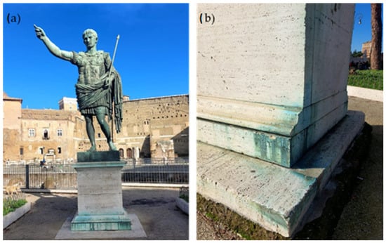
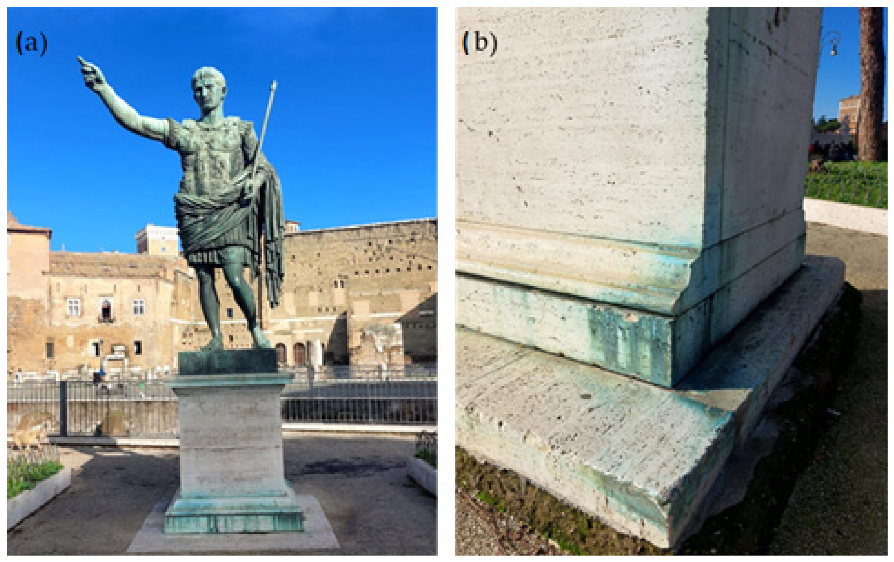
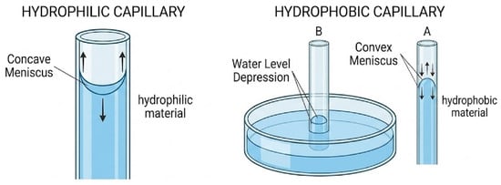
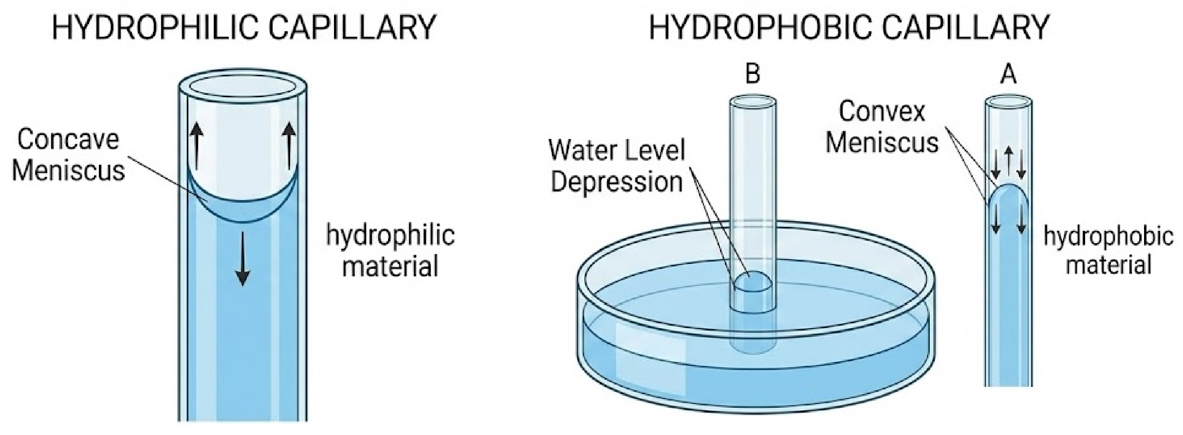
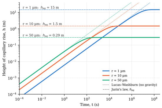
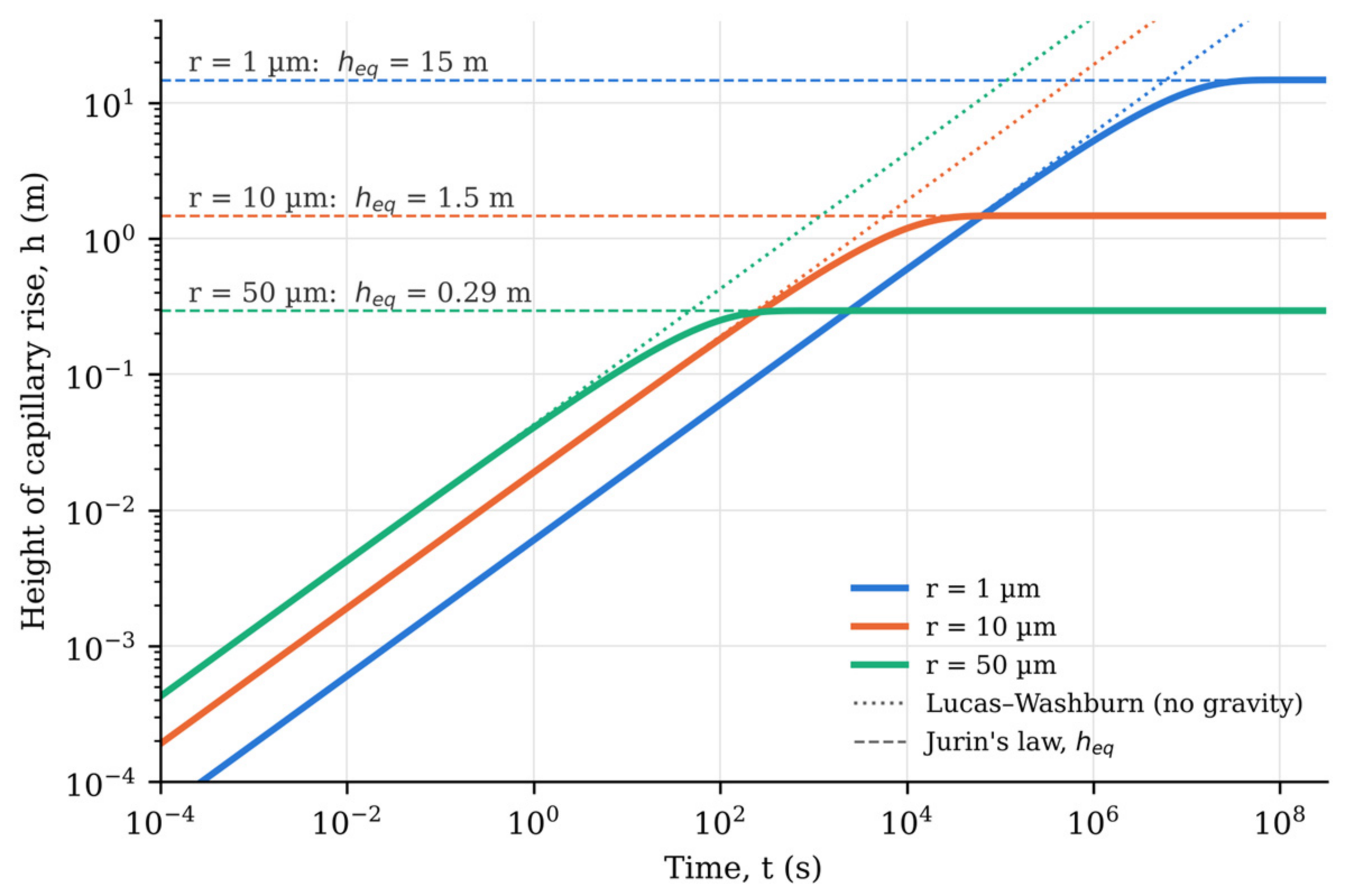
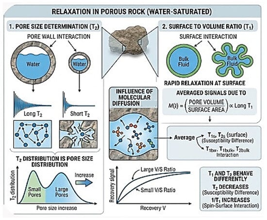
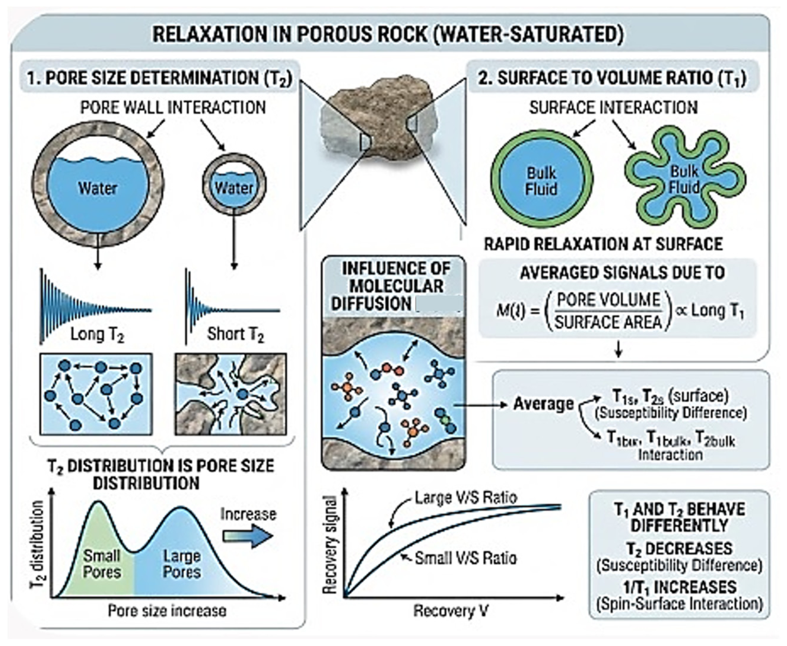
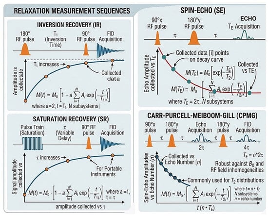
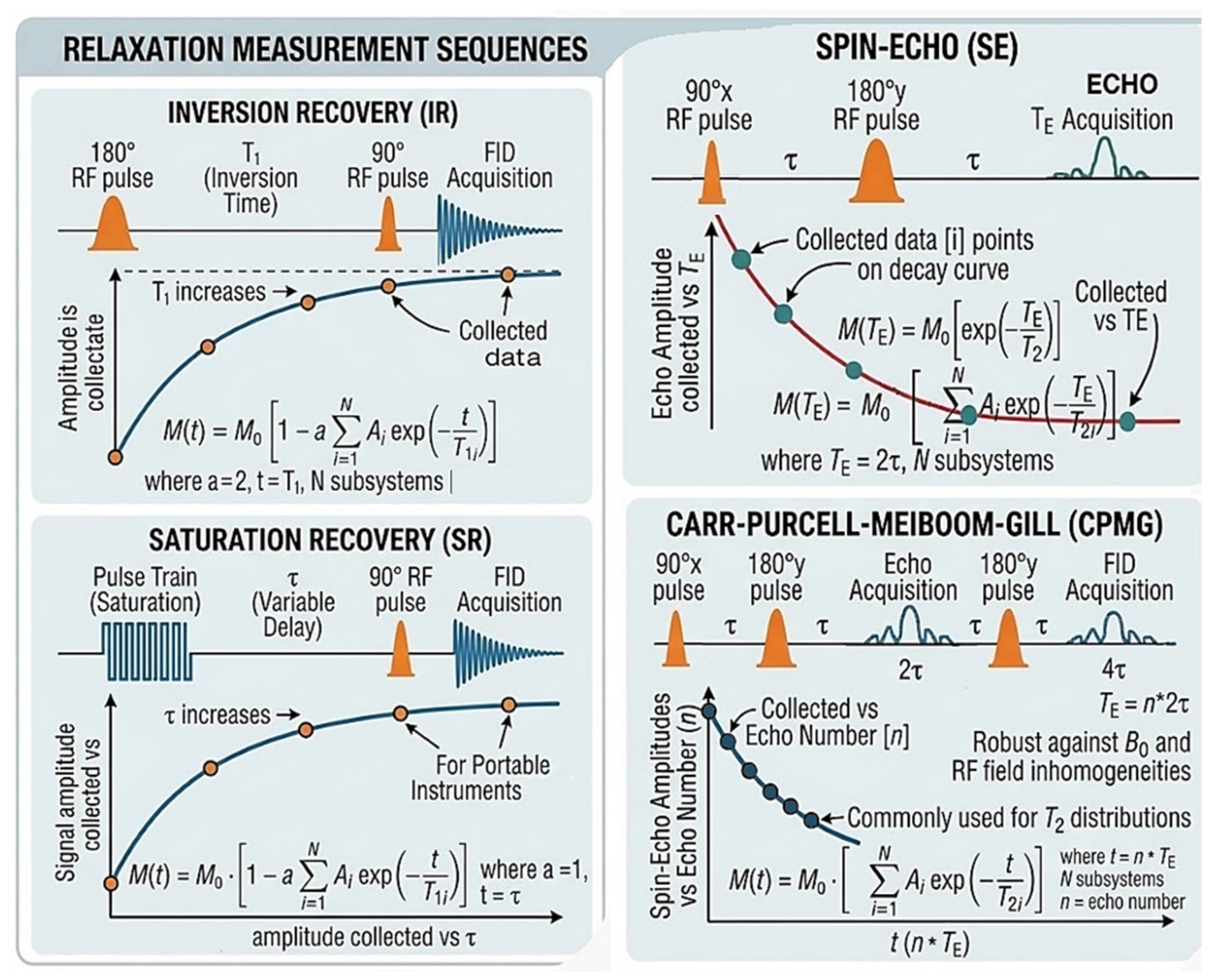
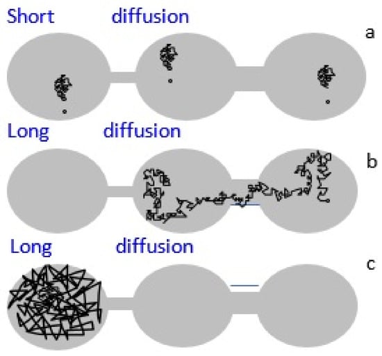
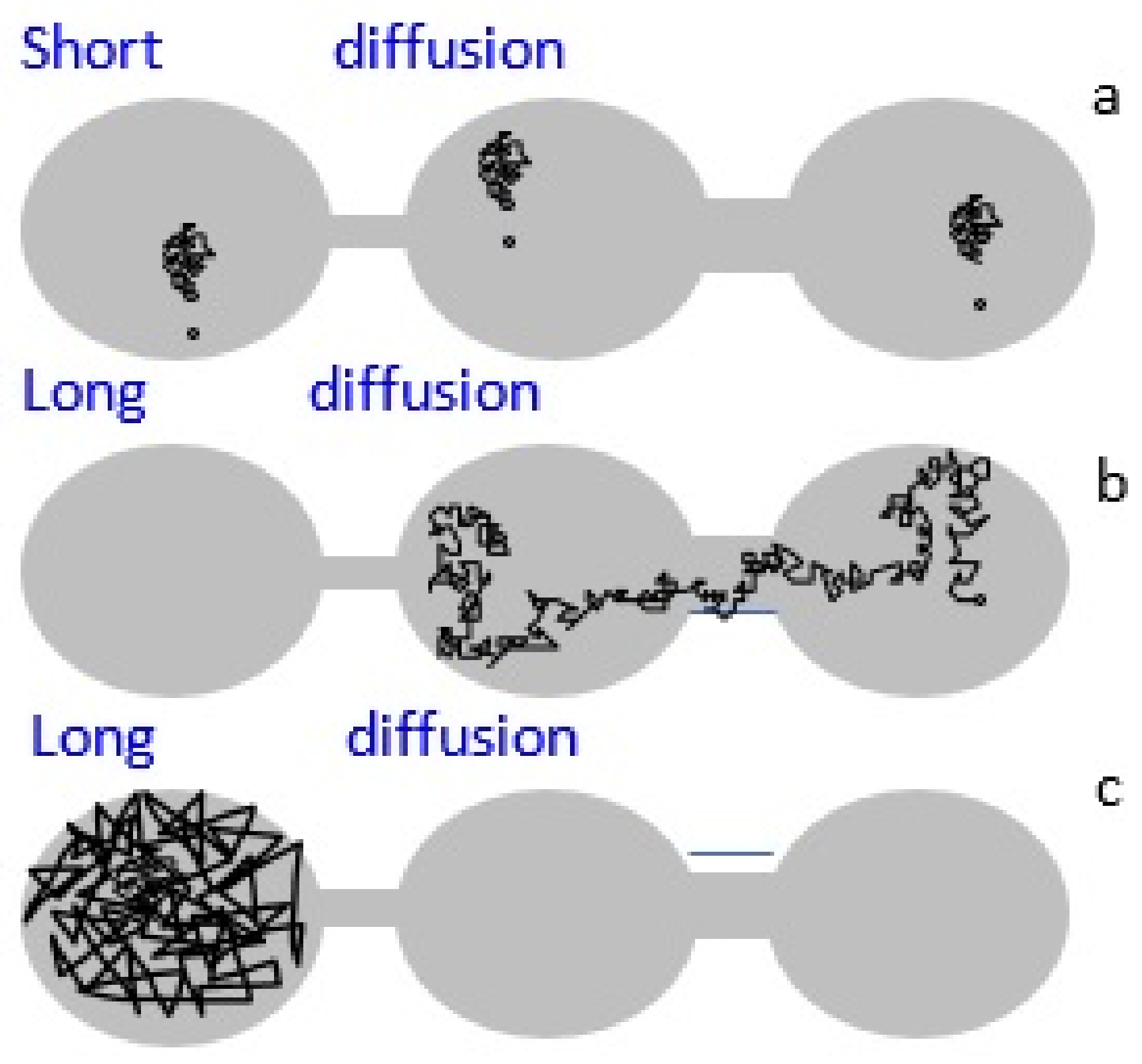
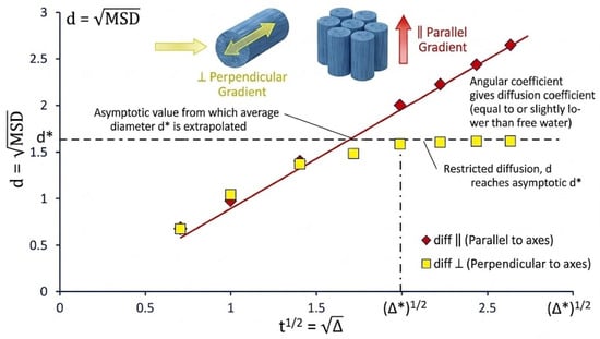
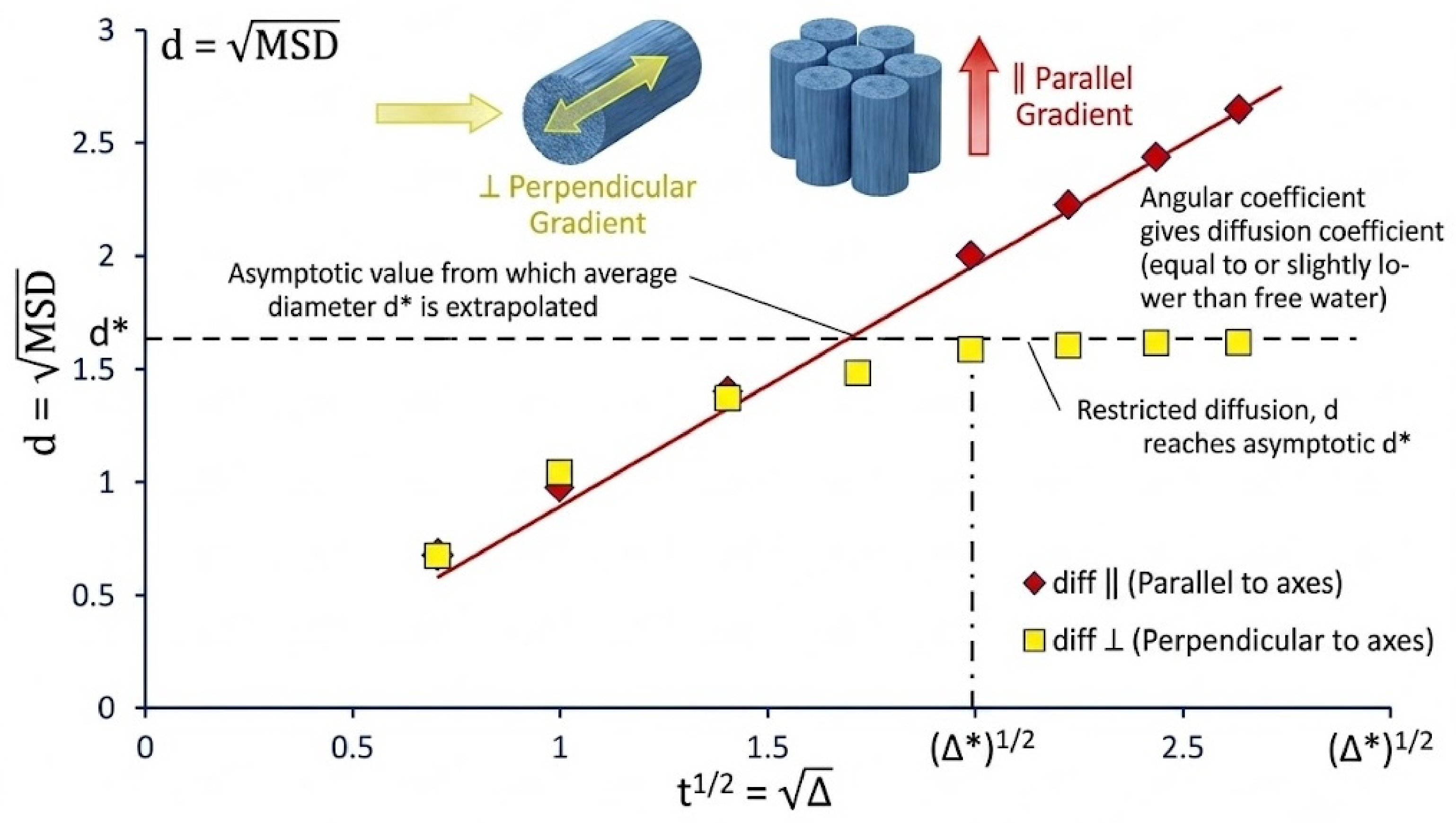
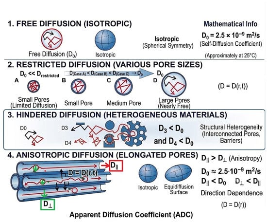
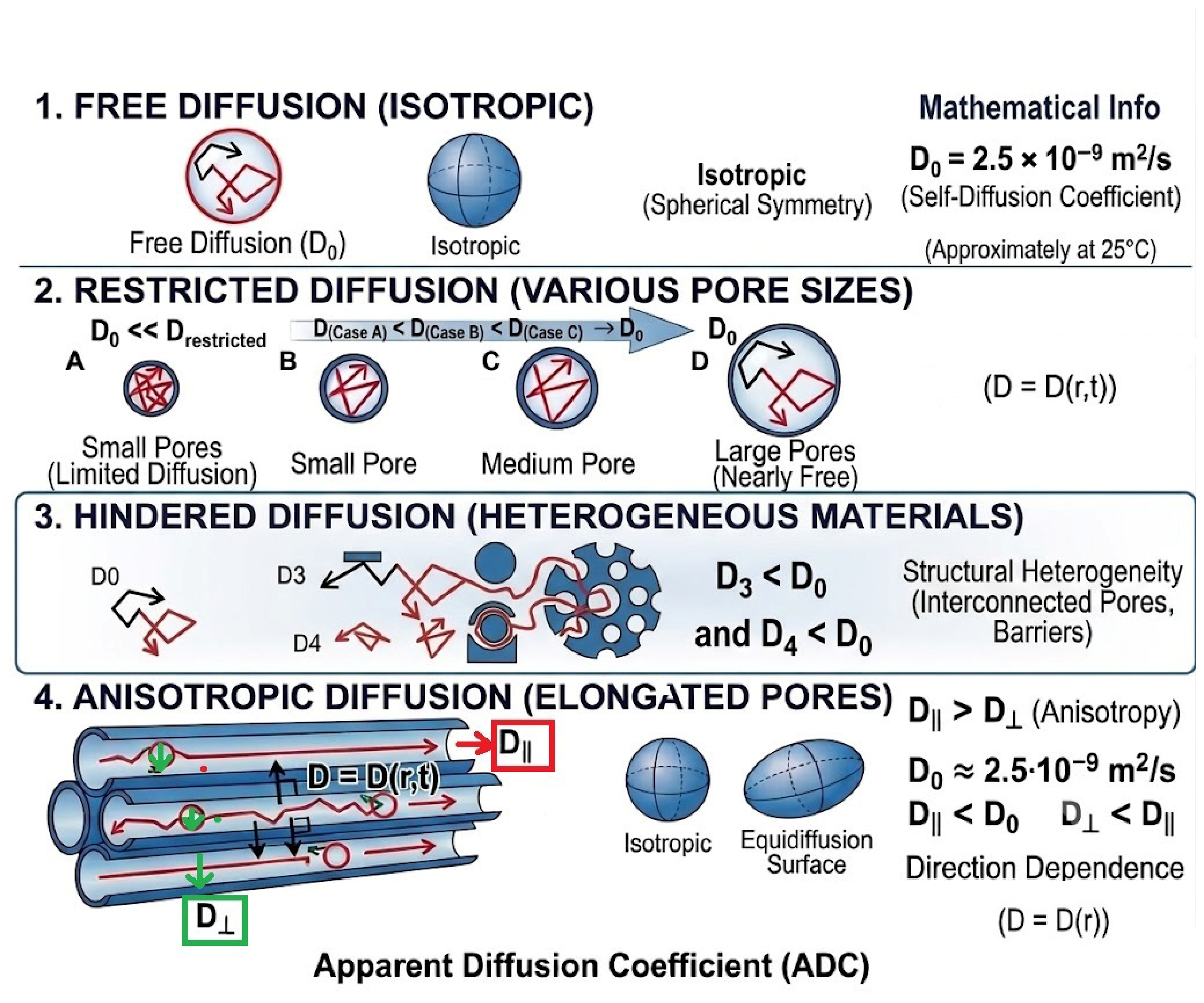
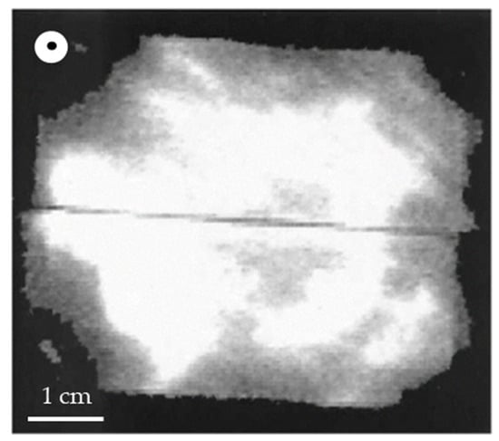
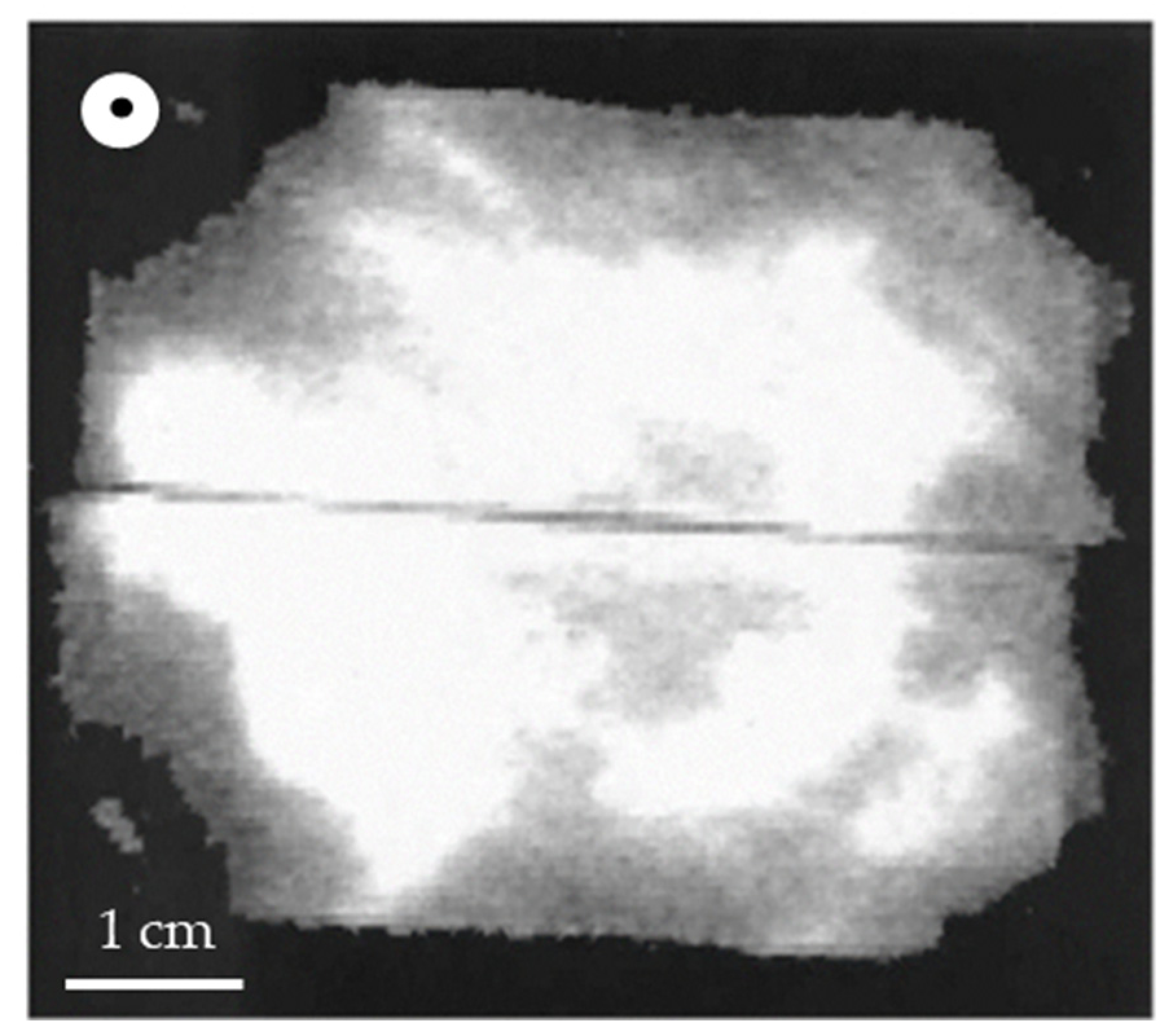
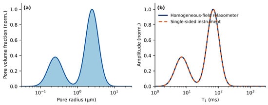
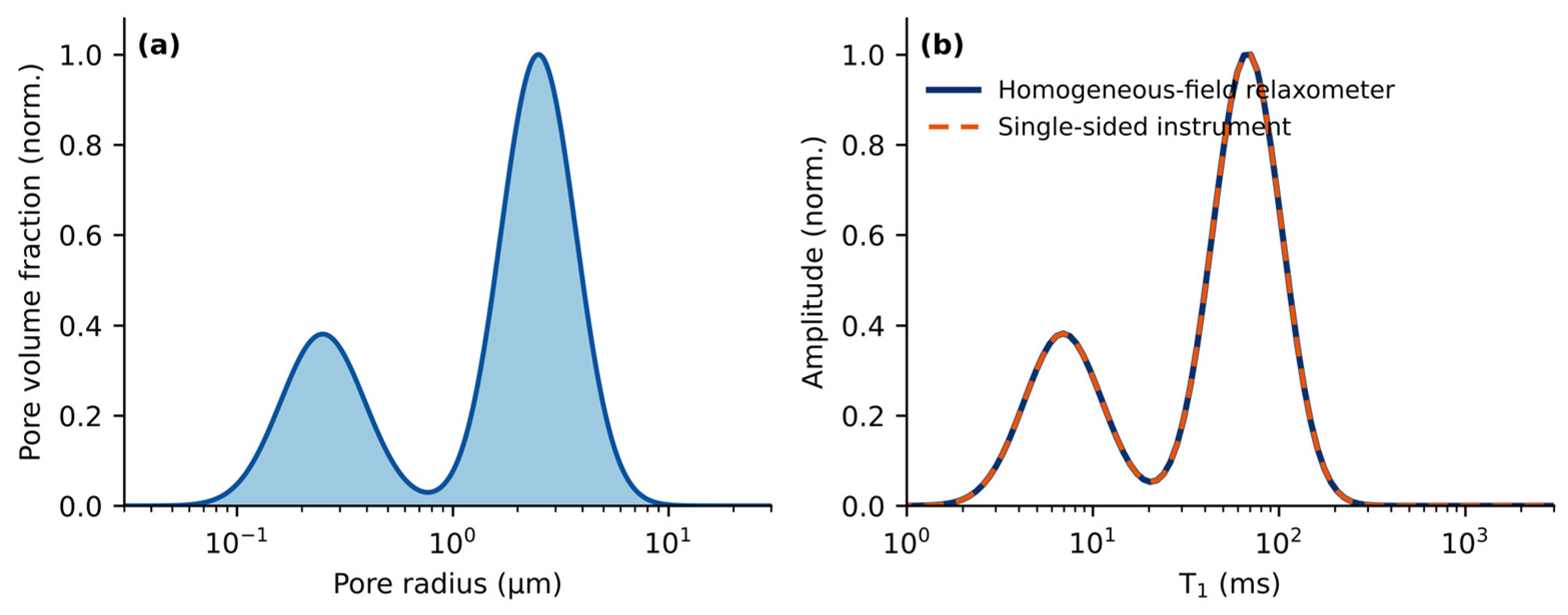
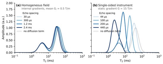
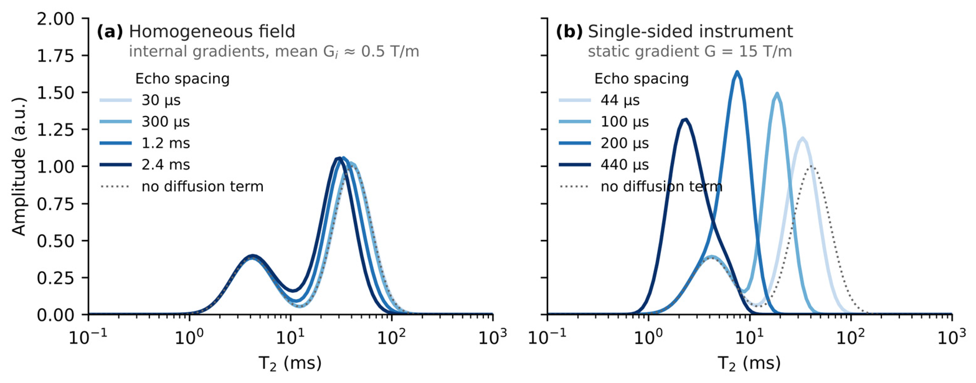
Disclaimer/Publisher’s Note: The statements, opinions and data contained in all publications are solely those of the individual author(s) and contributor(s) and not of MDPI and/or the editor(s). MDPI and/or the editor(s) disclaim responsibility for any injury to people or property resulting from any ideas, methods, instructions or products referred to in the content. |
© 2026 by the authors. Licensee MDPI, Basel, Switzerland. This article is an open access article distributed under the terms and conditions of the Creative Commons Attribution (CC BY) license.