


Abstract
The present work reports the results of a multianalytical study on a renowned Etruscan large bronze masterpiece from Florence’s National Museum of Archaeology known as the Arringatore of Pila. Thorough microstratigraphic characterization, metallotechnical examination, and measurement of the alloy compositions using the LIBS depth profile approach were carried out. This allowed us to collect new information on the execution processes of the present bronze sculpture and, more in general, on the materials and methods of the art foundry during the late Etruscan period, which is still little known. Furthermore, we provide the first compositional and microstructural demonstration of an intentional blackish patina based on thermally induced surface oxidation to form a cuprite–tenorite bilayer on the present bronze surface. The discrimination between ancient or modern production of such a bilayer is discussed in some detail along with microstructural clues supporting the intentionality of the tenorite-based blackish appearance. To achieve this goal, μRaman spectroscopy compositional mapping was successfully associated with the results of optical and SEM microscopy and XRD to achieve an exhaustive material characterization. With respect to the general objective of delving into issues of the art foundry of the past, seeking to uncover its unknown aspects, the results reported here offer various points for discussion and make a concrete contribution to advancing knowledge on the technological aspects of ancient large bronze statuary.
Keywords:
LIBS; Raman; Etruscan bronze; archaeometallurgy; Arringatore; art foundry; laser spectroscopy 1. Introduction
The alloy compositions, casting processes, and original patinations of ancient bronzes have attracted the interest of the scientific community since the birth of the modern sciences [1], and they have been extensively investigated over the last fifty years, under the stimulus of important archeological discoveries and restoration works, with the availability of increasingly advanced spectroscopic tools.
As well known, the study and restoration of the Bronzes of Riace in the laboratories of the Archaeological Superintendence of Tuscany (AST) during the second half of the 1970s played a key role in enhancing such interest. The thorough analysis of the alloys using AAS carried out on 36 metal powder samples per statue [2] collected by means of micromotor burs, and the pioneering metallotechnical examination [3] represent a milestone within the relevant research community. Such extensive sampling was aimed at analyzing independent pieces and overcoming problems due to intrinsic compositional variations associated with segregation phenomena of the metal components and structural defects of the alloys produced by casting dynamics and corrosion processes [4,5,6], which usually largely exceed the errors of AAS.
In the footsteps of this pioneering case study, several large bronzes have been investigated over the last decades, which have enriched the knowledge of ancient foundry materials and methods. At the same time, efforts have been dedicated to minimizing the invasiveness of the alloy characterization by avoiding extensive sampling, coring, and relatively large material removal. These have become increasingly unacceptable over time, due not only to possible visible alterations or undesirable fruition effects, but also to the gradual establishment of the general principle of non-invasiveness within the field of cultural heritage. This fostered the development and application perspective of portable analytical tools and Large-Scale Facilities (LSF) able to provide compositional and microstructural information without material sampling.
Up until around the first half of the 2000s, the only practicable alternative to the chemical analysis of the alloys based on metal sampling (i.e., the mentioned AAS or the less invasive and more sensitive ICP-OES [7]) was represented by portable X-Ray Fluorescence (XRF) [8,9], which is still extensively applied. However, deep mineralization, elemental surface enrichments or depletions, exogenous materials, and surface unevenness usually encountered in archeological artefacts can often seriously affect the reliability of quantitative measurements carried out using XRF and then limit its range of application.
Starting from the second half of the 2000s, in situ application of Laser-Induced Breakdown Spectroscopy (LIBS) in archaeometallurgical studies significantly grew thanks to important methodological advances [10,11,12,13,14,15], also involving the combination with XRF [16] and other techniques, and the development of dedicated portable instruments [17,18,19]. In parallel, non-invasive techniques based on LSF, such as neutron diffraction, were introduced [20] and similarly combined [21], although they can play a role only when dealing with small objects or fragments of large bronzes.
LIBS offers the most convenient compromise between the mentioned principle of non-invasiveness and the need to probe the metal sufficiently in depth in several areas suitably distributed all over the work of art, before formulating reasonable hypotheses on the alloys used by the ancient bronzesmiths. Reliable LIBS quantitative elemental analyzes can be achieved by limiting laser ablation around ~0.1 mg (i.e., a pair of order of magnitude lower than the typical sampling for ICP) and spot size < 100 μm (i.e., invisible to the naked eye). In other words, a suitable setting makes LIBS practically countable among the non-invasive elemental techniques, although this cannot be generalized to the technique tout court. Most of the handheld tools marketed over the last years [22,23] can be invasive and their quantification performances of Cu alloys are often unsatisfactory, thus making dedicated prototypes still the best choice to analyze archeological bronzes.
After Donatello’s David [24] and some other Renaissance bronze statue under restoration between 2007 and 2008, the Arringatore of Pila was the first archeological large bronze whose alloy compositions were entirely quantified through LIBS depth profiles measured in many sites over the sculpture, somehow in a similar way to that done thirty years before in the mentioned pioneering study but in a non-invasive way. The present masterpiece was investigated and restored in 2008–2009 within a collaboration between AST and our Institute, but only some aspects concerning the laser-assisted removal of modern coats have been reported to date [25], whereas the main questions concerning the interpretation of the execution processes, alloy compositions, and possible presence of traces of original intentional patina were postponed until after additional analyses.
Much belatedly, but with the benefit of hindsight, here, we present and discuss for the first time the results of the archaeometallurgical study of the Arringatore, which were achieved through the elaboration of the data collected during the mentioned restoration intervention and further recent analytical insights. These included thorough metallotechnical examination of the surfaces, in situ copper alloy LIBS analyses, optical and SEM characterizations, and XRD (X-ray diffraction) of several samples.
Stratified mineral phases that were formerly characterized using XRD have been recently re-examined by means of μRaman chemical imaging to further investigate the possible presence of an original intentional patina argued during laser-assisted removal of modern patination layers [25]. Raman spectroscopy pairs effectively with LIBS as the most congruent complementary technique, which underwent a similar technological evolution towards portable instruments [26] as the latter one. It allows identifying most of the minerals associated with the corrosion of copper alloys and then possible patination films, as systematically demonstrated since the beginning of the 2000s [27,28,29,30,31], although extensive applications to solve archaeometric or conservation problems of bronze artworks started to grow just along the last few years [32,33,34,35]. Here, Raman spectroscopy provided an important contribution to interpret the stratigraphy in proximity of the metal surface.
The comprehensive multianalytical results achieved on the Arringatore are here discussed within the framework of debated archaeometric problems concerning the alloy recipes [36,37] along with casting, assembling, and patination techniques of ancient bronzes [3,38,39,40,41]. Compositional and structural data provided by traditional or innovative techniques allowed characterizing the constituent materials, interpreting the crafting procedures and collecting analytical evidence, which supports the hypothesis that the Arringatore had a rather dark original appearance provided by a peculiar and likely intentional blackish patina.
2. Materials and Methods
2.1. The Masterpiece
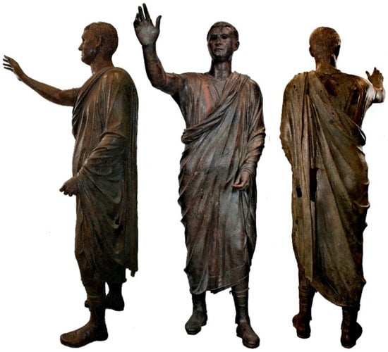
The Arringatore (Orator) is the renowned Etruscan large bronze of the Medici collections, which is exhibited at the National Archaeological Museum of Florence (Figure 1) [42,43,44]. Although the present work focuses on material analysis and interpretation of the execution processes of this masterpiece, it is useful to briefly preface such core topics with some essential historical information, which can support easier reading of the data interpretations.
Figure 1.
Views of the Arringatore soon after the restoration in 2008.
As indicated by the Etruscan inscription at the left mid-shin level [45], the artefact is a votive life-size (height about 179 cm) portrait of Aule Meteli. His clothing, whose tailoring and weaving details have been meticulously rendered in the bronze figure [46], has been interpreted as evidence of social prominence and romanization that, along with other formal aspects of the portrait, played a crucial role for its archeological dating.
The Etruscan citizen wears a toga draped around the body, the typical Etruscan and Roman wool mantle [46]. The latter leaves the right shoulder and arm uncovered, where the underlying sleeveless tunic with one of the typical two purple narrow stripes running vertically from the shoulder down until disappearing beneath the diagonal draped folds of the toga can be seen. The present type of stripe was called by Romans angustus clavus, although it cannot be considered uniquely Roman [46]. This, along with the distinctive shoes (calcei senatorii) denotes the high rank of Aule Meteli, who since the eighteenth century has been named “the Arringatore” because of its apparently oratory gesture (silentium manu facere).
The most widely accepted stylistic dating for this sculpture ranges within the 2nd century BCE [47], while its provenience has been debated till 2019 when new archival research definitively clarified that it was excavated in Pila near Perugia, Italy, in July 1567 by a farmer. Soon after, he sold the statue to the goldsmith Giulio Danti and eventually his son Ignazio (cosmographer of Cosimo I) resold it to the Duke and brought it to Florence at the beginning of September 1567 [48]. According to the sources, when the masterpiece was discovered, it presented a greenish appearance and had a broken arm (the right one), which was likely restored by Giulio Danti before moving the sculpture to Florence. There is not any information about such a restoration work but in a letter by Giorgio Vasari to Vincenzo Borghini dated 20 September 1567 the archeological find was described as complete without anything missing (“il duca ha auto una statua di bronzo intera che non gli manca niente …”/“the Duke received a whole bronze statue that lacks nothing…”) [48], thus confirming it was previously restored.
Within the Medici–Lorena collections the Arringatore underwent maintenance interventions and likely consolidation of the right arm, whose last restoration was probably carried out during the second half of 1930s [42] (see below).
2.2. Methods
Fifteen samples of stratified mineral and organic materials, each one consisting of one or more microfragments or powders, were collected from apparently well-preserved zones before or after the removal of the undesired modern coats. They were taken in a minimally invasive manner by applying light pressure with a micro-chisel. Hereafter, the microstratigraphic results achieved on the most representative samples are discussed.
X-ray diffraction analyses were carried out on all microfragments (sizes 1–3 mm, thickness mostly <500 μm) or powder samples using a Philips X.Pert PRO PW 3040 (Amsterdam, Netherland) diffractometer (Bragg–Brentano geometry) operating at 40 kV and 40 mA, equipped with a PW3015 X.Celerator detector, using CuKα radiation in the 5–80° 2θ range. The samples were positioned on a Zero Background Holder, the measurement was carried out using the software X’Pert Data Collector, and the following data analysis was performed using the software X’Pert HighScore 2.2.
Polished cross-sections and thin cross-sections of the fragments were prepared and examined in reflection or transmission by means of polarized light microscopy (Zeiss Axioskop 40 Pol, Oberkochen, Germany), as well as SEM-EDX (Philips XL30 equipped with EDAX-DX4 EDS) following their carbon coating. FTIR measurements were also performed along with μRaman (XploRA, Horiba, Kyoto, Japan) mapping that was exploited to reconstruct the microstratigraphic distribution of mineral phases.
Naked eye, photographic, and X-ray metallotechnical examinations were carried out using a 300 kV industrial X-ray tube by Gilardoni (Lecco, Italy) toinvestigate casting and assembling processes of the present artefacts, by exploiting the literature information and the skill gained in previous investigations on large bronzes. As known, such a reverse engineering problem is often very difficult and without univocal solution. Thus, the aim of the metallotechnical examination is hence to point out multiple converging lines of evidence allowing one to credibly restrict the range of practical crafting possibilities.
As mentioned above, the alloys were investigated using the elemental depth profile approach based on a homemade portable LIBS prototype whose setup and technical details were reported in [19]. Briefly, it was equipped with a QS Nd:YAG laser, a mirror collector, and fibre-coupled spectrometers and used by positioning its probe on a tripod. As demonstrated in the latter and several subsequent works [13,21,24], the careful calibration of this instrument using a set of reference samples allowed reliable elemental quantification of the major elements of the copper alloys, which offered significant advantages with respect to alternative techniques. The data were processed for quantitative evaluation according to the calibration described in [19] and considering its depth dependence [13].
Thus, LIBS analyses of the alloys were carried out considering primarily the areas that were apparently better preserved. Further limitations were associated with the scarce accessibility of some zones due to the positioning of the artwork and the presence of several supports. Finally, some elemental depth profiles were ruled out during data elaboration due to the lack of the basic conditions for measuring bulk composition using LIBS, which are: (1) stabilization of the elemental content after a sufficiently high number of laser pulses to overcome the outer mineralization region; (2) decrease to zero of the signals associated with exogenous elements, such as Ca or Ca + Cl [49]. The presence of Cl represents a direct corrosion marker, while the penetration of Ca beneath the surface due to micro-karst effects indirectly highlights the presence of microporosity produced by corrosion phenomena that prevent analysis of the metal bulk.
3. Results: Material Layers
3.1. Macroscopic Observation
Before the last restoration of the outer surface of the Arringatore presented an almost homogeneous dark-brown appearance conferred by modern (i.e., applied after the excavation) patination films. Such an overall coating appeared esthetically very similar to those previously found on several Florentine sculptures [50,51,52], which testifies to a well-established maintenance/restoration practice of the local antiquarian tradition between the end of the 18th and the 19th century [53]. In a previous work [50], such a kind of brown coating has been defined as “bronze-like patination” (bronzatura) to underline its original aim to make the artworks resemble the appearance of bronze statues according to the esthetic taste at that time. After the former application, the coat was usually “renewed” using waxes and other materials, and sometimes it was reapplied.
The restoration of Florentine sculptural heritage carried out throughout the last fifty years included the removal of these patination layers, which were questioned since the end of the 19th century, in order to uncover surfaces closer to the original ones of the artworks or, at least, not disguised and visually flattened by relatively thick and dark layers. The conservation treatments applied to the Arringatore during the last restoration intervention fall within this line of thought.
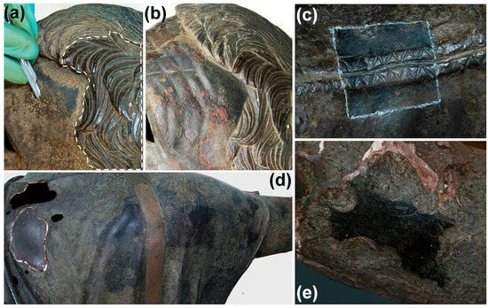
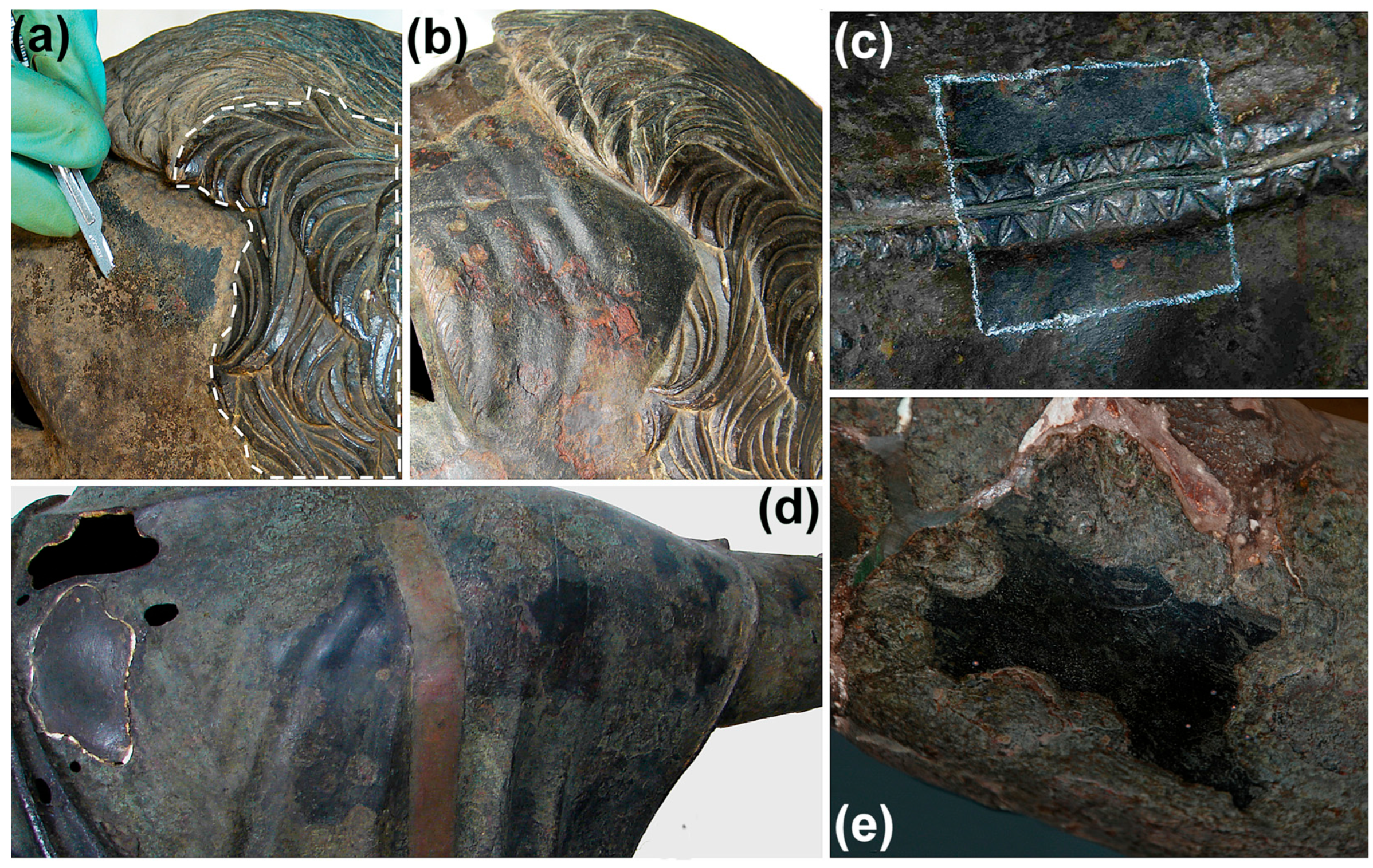
In Figure 2, a set of pictures taken during laser-assisted removal of such undesired coatings is displayed. Figure 2a shows the beginning of the mechanical removal using a scalpel after laser induced photothermal decomposition and partial ablation. As shown, these treatments gradually unveiled the blackish surface underneath. The result over the frontotemporal zone (Figure 2b) was very surprising, since the uncovered surface was mostly darker than the former, while the details of the chiseling of the original bronze surface were much more legible than before. This was not the only well-preserved zone.
Figure 2.
Removal of the modern patination layers: (a) scalpel finishing after laser irradiation; (b) corresponding result; (c) seams of the tunic uncovered onhe right deltoid; (d) right shoulder after the removal of the coats; and (e) well-preserved area surrounded by deep corrosion craters.
The conservation treatments mostly returned large areas without relevant traces of the original surface, which was completely destroyed by nodular corrosion, exfoliation, and craterisation/material losses phenomena. Nevertheless, several relics of well-preserved bronze surface were unequivocally recognizable. Beside the mentioned one of the face, these were also found, for example, over the hair and calcei, reinforced front hem of the toga, back of the right shoulder (Figure 2d) and over a relatively small but very meaningful area, such as that in proximity of the right elbow shown in Figure 2e. All these well-preserved areas appeared slightly raised with respect to the surrounding, which was lowered by the mentioned corrosion and material losses, very smooth, and decidedly blackish (all clear in Figure 2e). Interestingly, also the surface of clavus (Figure 2d) was well preserved but its colour was reddish rather than blackish.
3.2. Microstratigraphies
The examination of cross-sections and thin sections under polarized light microscopy of samples of the upper part of the stratification (i.e., not including the metal substrate) that were taken before or after the above-mentioned uncovering treatments from areas apparently well preserved revealed rather repeatable microstratigraphy. The following types of material layers were observed in the latter (from the outermost to the innermost layer):
(L1) Thin, amber-coloured layers (one or more) composed of prevailing organic matter with minimal mineral content;
(L2) Brownish bronze-like patination layers (one or more from multiple applications) located soon beneath the previous ones;
(L3) Blackish layer, ranging from dull black to silvery grey, sometimes split in two layers by the following;
(L4) Greenish-yellowish layer apparently resulting from the alteration of L3 progressing upwards from its basal zone;
(L5) Compact reddish layer ranging from brilliant red to orange, sometimes with a silvery lustre, with minor greenish and yellowish spots.
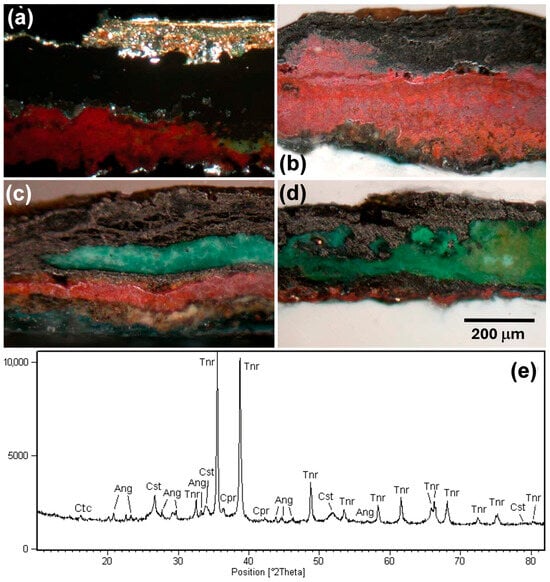
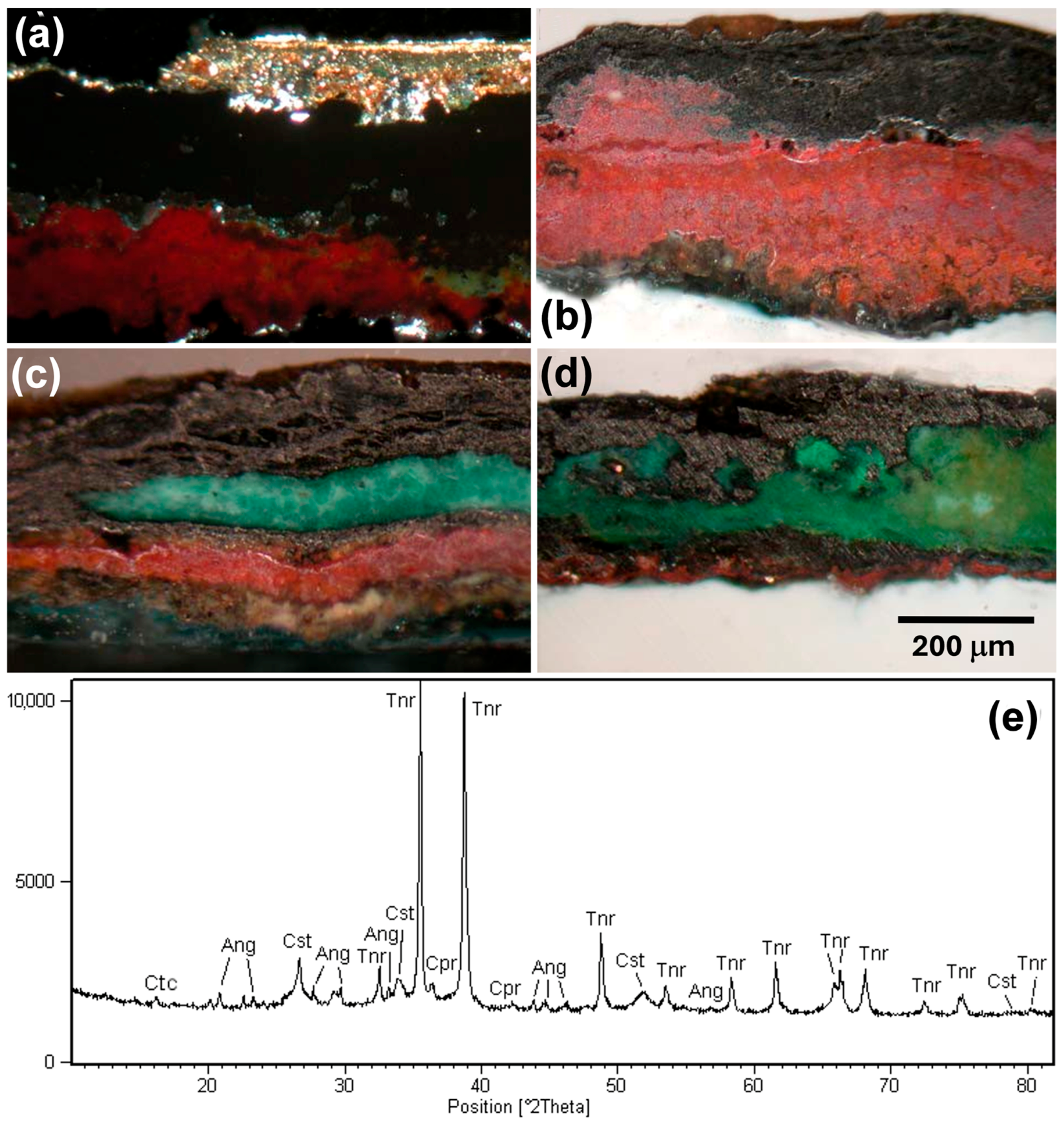
Apart from possible minor differences observed among the cross-sections, the fifteen material samples P1–P15 analyzed confirmed that the qualitative sequence just listed represented the general outer microstratigraphy of the Arringatore, which could be described in more detail in terms of possible sublayers. For example, Figure 3 reports images of the thin cross-sections of sample P6 (Figure 3a), which was a small flake collected from the toga and P8 (Figure 3d) from the left calceus, along with the cross-sections of P12 (Figure 3b) and P14 (Figure 3c) from the tunic.
Figure 3.
Representative stratigraphies and XRD pattern of the black layer: (a) thin cross-section of P6 (crossed polarizers); (b) cross-section of P12; (c) cross-section of P14; (d) thin section of P8 (see the text); and (e) diffraction pattern of the blackish layer sampled by scalpel scraping. Tnr: tenorite. Cst: cassiterite. Cpr: cuprite. Ang: anglesite, Ctc: clinoatacamite.
As shown in Figure 3a, P6 included all five layers listed above, which represented the initial situation before the laser and mechanical ablative treatments; the other samples were taken after partial (Figure 3b,c) or complete (Figure 3d) removal of the modern coats.
At the top of P6, two very thin organic sub-layers were recognized in L1 (L1a and L1b, Figure 3a), likely deriving from polishing operations using bees wax, as revealed by FTIR. Just underneath, the flattening bronze-like patination, L2, was similarly well-recognizable, which mainly included silicates, Ca-carbonate, gypsum, and pigment load of black carbon and ochres in organic binder, in agreement with previous studies on similar patinations [50,51,52]. Furthermore, FTIR of the samples P6, P5 and P7 also returned vibration bands of lead salts (1546, 1577 cm−1), nitrates (1384 cm−1), Ca-oxalate (1650–1620, 1324, 780 cm−1), and Cu-oxalate (medium-intense 1366 cm−1, according to the criteria defined in [54]) attributable to the partial mineralization of the organic binders.
Thanks to the latter and the waxing, laser irradiation allowed the thermal disaggregation of such undesired coating and its easy mechanical removal, as clearly shown in Figure 2a,b, where the final surface uncovered exhibited, locally, a good status of preservation of fine finishing details, such as the intact meticulous seams of the tunic displayed in Figure 2c.
Considering the mineral stratigraphy beneath the patination (L3–L5 displayed in Figure 3), at the first glance, the reddish and greenish layers superposition qualitatively reflected the most typical situation encountered for excavated bronze artefacts, where cuprite (Cu2O) produced by primary oxidation of the original metal surface is usually covered by a set of possible greenish copper minerals (Cu-clorides, Cu-carbonates, Cu-sulphates, etc.). Conversely, the recurrent blackish layer, L3, between the modern patination layers and the cuprite layer was unusual and needed further insights.
L3 and the reddish layer underneath (L5) were often strictly contiguous in the samples analyzed, as shown in the cross-section of P12 (Figure 3b), while the intermediate greenish layer, L4, was detected in just a few samples, such as P14 (Figure 3c), P8 (Figure 3d) and to a minimal extent in P6 (Figure 3a). As mentioned above, L4 appeared as secondary crystallization growing around the basal zone of L3.
To analyze the latter, it was tentatively mechanically detached from a fragment of P12 using a scalpel and analyzed using XRD. Figure 3e displays the corresponding diffraction pattern showing a prevalent content of tenorite (CuO) along with minor contents of cuprite (likely from L5), anglesite (PbSO4), and clinoatacamite (Cu2(OH)3Cl). Thus, the blackish appearance of some of the restored areas (Figure 2) was attributable to tenorite.
To collect a more general compositional picture, XRD was extended to all samples and allowed the identification of the main mineral phases they contained (Table 1). In particular, tenorite was identified in twelve samples, which was associated with cuprite in ten of them, also including such deepest layer (L5). The latter appeared correlated to the presence of cassiterite, while anglesite diffraction peaks were detected when cuprite, cassiterite, and tenorite were also detected, although it did not always accompany these minerals.
Table 1.
Mineral phases identified in the XRD patterns of the sample bulks.
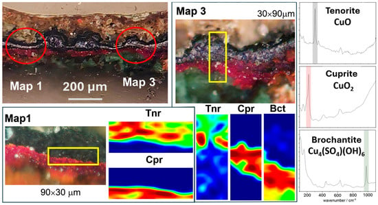
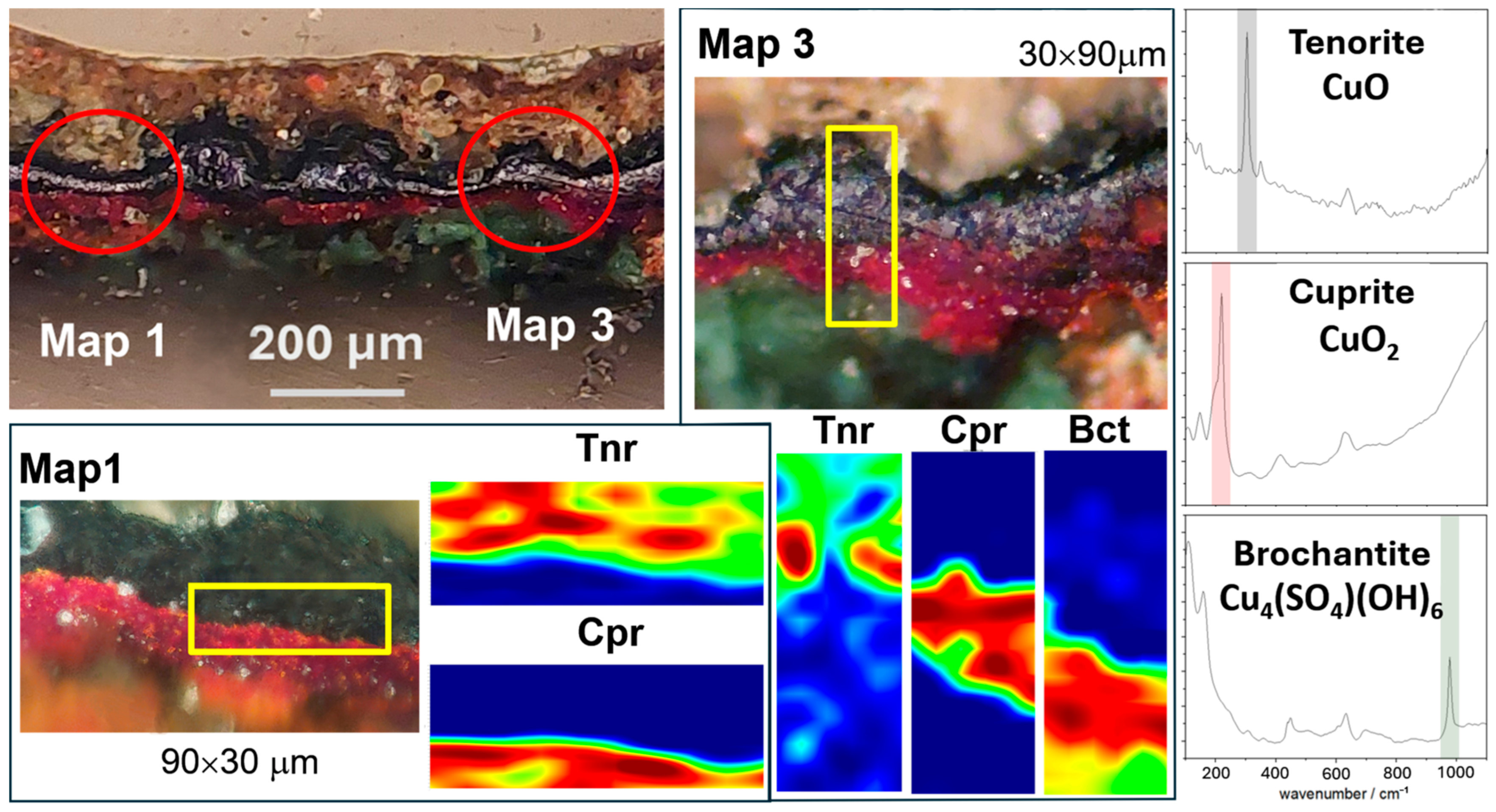
The association of the blackish appearance with tenorite represented an important datum, which needed microstructural insight to further verify the correspondence with the layer L3, the spatial distribution and degree of continuity of tenorite. μRaman spectroscopy was hence carried out on the cross-section of a microfragment of the sample P6. Figure 4 shows the compositional results of two scans over small transition areas (30 × 90 μm2, 1800 measurement spots) where the prevalent content of tenorite in the blackish layer, L3, was confirmed, as well as its continuity.
Figure 4.
μRaman mapping showing the transition between cuprite (L5) and tenorite (L3) layers within the areas framed with the yellow rectangles. Tnr: tenorite. Cpr: Cuprite. Bct: Brochantite.
The technique also pointed out the presence of brochantite beneath the cuprite layer, which was likely attributable to relatively recent indoor sulphation.
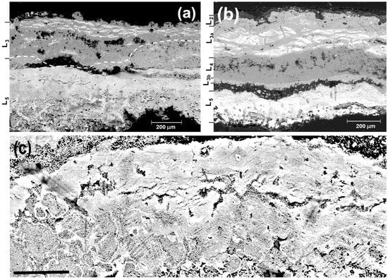
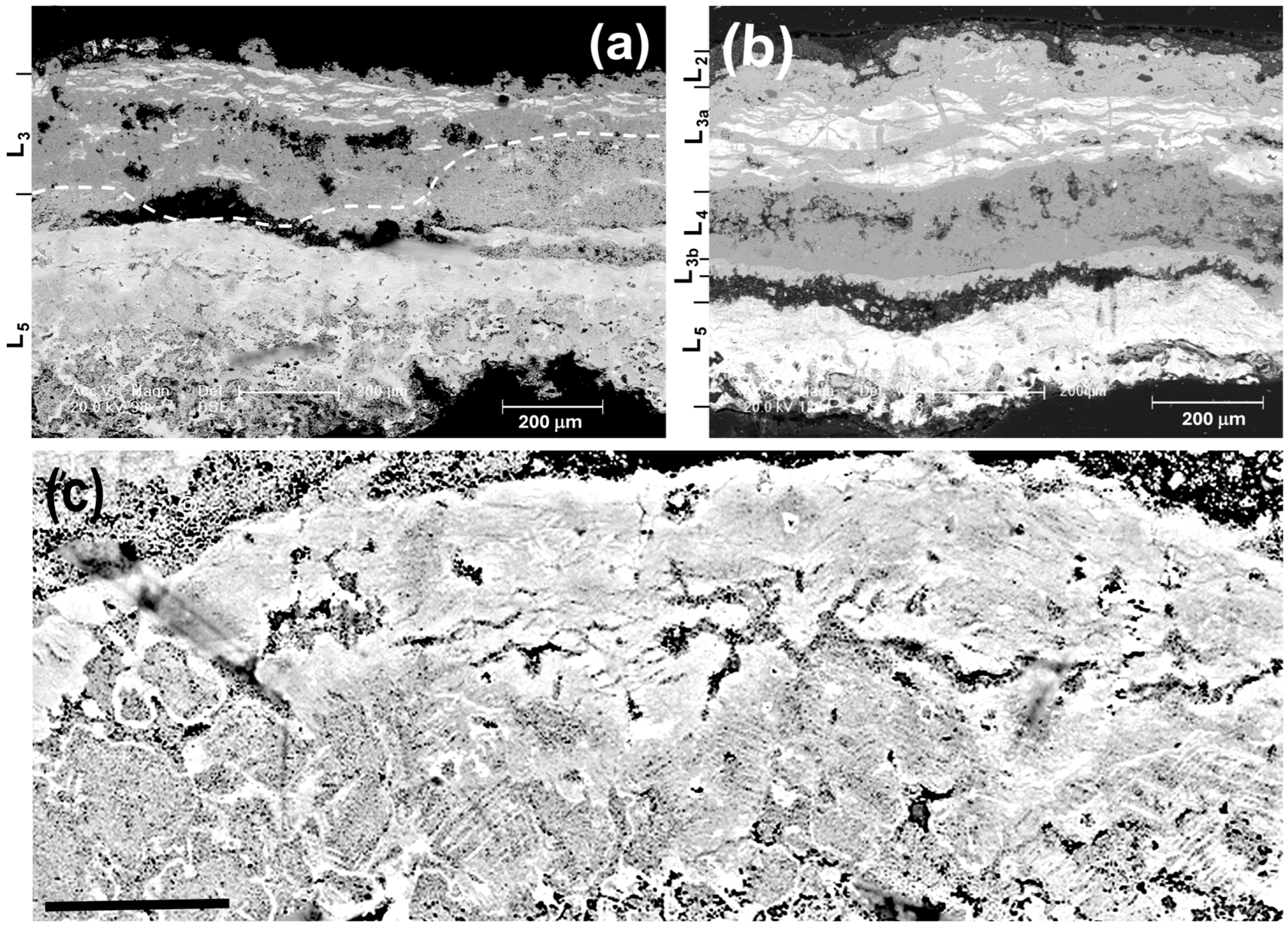
Finally, complementary information was achieved through SEM-EDX micrographic examinations (Figure 5). These allowed us to confirm the association of prevalent Cu (II)-oxide, Cu-chlorides (prevalently clinoatacamite), and Cu (I) oxide with the blackish, greenish, and reddish layers L3, L4, and L5, respectively, and to point out interesting features of their microstructure. Apart from the late patination layers at the top of the samples P12 and P14, made scarcely visible by the high contrast in Figure 5a and Figure 5b, respectively, both tenorite layers, L3, included significant quantities of lead and tin in proximity to their outer surface. According to the result of XRD, these mainly contained cassiterite and anglesite, where the latter likely represents the sulphation product of Pb-carbonate. White lead and tin rich micro-areas in BS SEM images appeared as segmented, narrow, elongated (i.e., with a high aspect ratio), and almost parallel to the surface, rather than in the form of disordered spots, as usually observed. The well-known mobility of lead during corrosion typically produces accumulation spots. The singularity of the present lead and tin distribution appeared even more pronounced, considering that lead content in the present Cu alloys was not so high, as detailed in the following.
Figure 5.
Back scattering-SEM images of cross-section of the sample P12 (a) and P14 (b), respectively, with the indication of the corresponding layers and a detail of the former (c) showing microstructures inherited by those of the dendritic segregation of the copper alloy (black bar correspond to 100 μm). See the text.
SEM examinations also allowed us to demonstrate the typical dendritic microstructures of bronze were partially reproduced in the cuprite layer (L5), as shown in Figure 5c, whereas such correlation was missing in the tenorite layer (L3), although in some cases minute compositional modulations could still suggest some vague similarities. Thus, cuprite represents the result of direct oxidation of the original metal and tenorite its following oxidation step. Both cuprite and tenorite layers appeared relatively well preserved but detachment gaps between them (Figure 5a,b) and destructive effects of the cyclic corrosion and/or sulphation (see L4 in Figure 5b grown at the basal level of L3 that was split in two sub-layers) were also evident.
4. Results: Metallotechnical Examination
The visual inspection of the artwork, examination of about 2000 photos documenting its surfaces before, during, and after the restoration work, and several tens of 30 × 40 cm2 X-ray radiographic plates allowed us to obtain chromatic, morphological, and structural information that along with the stratigraphic data presented above supported the interpretation of the ancient foundry processes.
The first feature which immediately appeared to the naked eye was the small thicknesses of the bronze walls, which were visible along the borders of a few lacunas. They mostly ranged within a few millimetres (about 2 mm were measured in some zones), apart from the legs, bottom edge of the toga, shoulders, repair area, and other areas that were thicker.
Crafting such a large and thin hollow figure using lost wax casting processes represented a difficult technical challenge for the bronzesmith, which was addressed almost perfectly, considering that a few small lacunas mentioned above can be considered a minor problem. The main lacunas were well visible on the neck, neckline, left shoulder, left buttock and flank (Figure 1 and Figure 2d) and they were left untouched in the present restoration that did not involve any surface structural integration.
4.1. Cast as Individual Pieces
The Arringatore was cast as a set of individual pieces and then assembled through the formation of joints by pouring molten bronze in metal gaps suitably prepared. As usually, and as suggested by the present examinations, after shaping the full wax for casting this was cut in several pieces to be cast as individual bronze pieces.
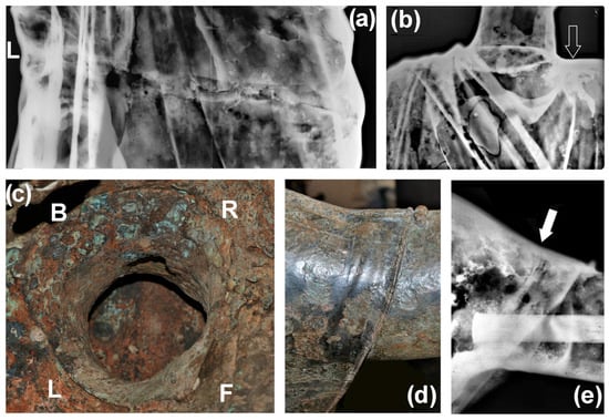
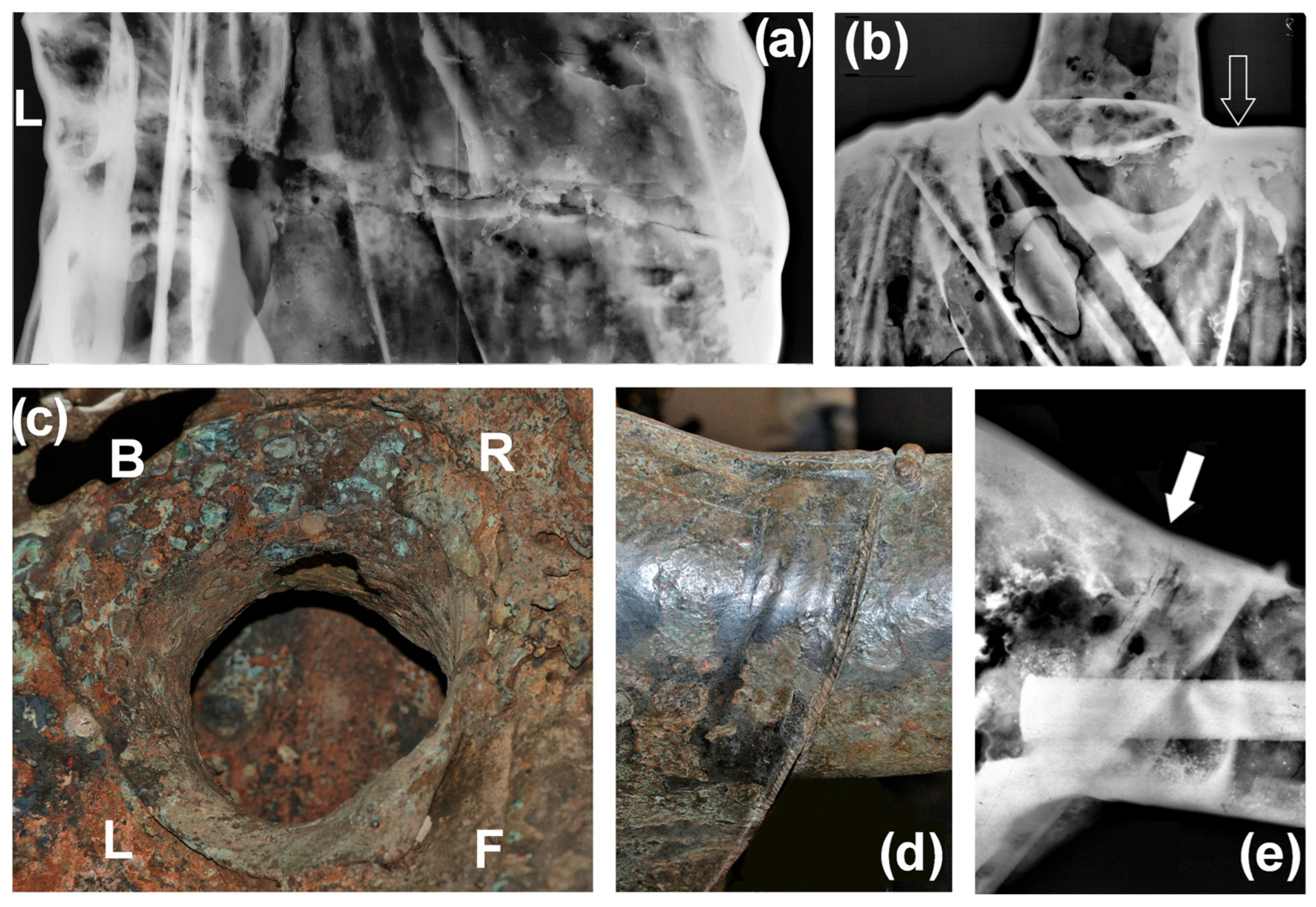
The observation of the internal surface of the figure allowed us to assess that its head, right arm, hands, upper-mid trunk, part from lower trunk to the legs, and part of the backside bottom edge of the tunic were cast as independent pieces and then assembled through such mentioned cast-on operations, as proven by visible and X-ray images of Figure 6. The clavus on the right shoulder was inlaid using the same technique as that of damascening, although the metal inlaid was not precious.
Figure 6.
Radiographic (a,b,e) and photographic (c,d) views of the main assembling zones: (a) joint between the mid-upper trunk and lower part of the figure; (b) joint along the neckline (the hollow arrow indicates the zone with abundant residues of poured metal); (c) internal view showing the joint of the head with abundant traces of metal pouring at the right side (R), as well as at the front side (F), and other zones along the neckline; (d,e) joint of the right arm (full arrow). F, L, B, and R indicate the front, left, back, and right side of the figure, respectively.
Reasonably, the slightly tilted full-circle joint of the trunk extending from the right side of the pelvis to the left forearm (Figure 6a) was executed after joining the head, right arm and hand to the upper-mid trunk and the left hand to the lower part, in order to better exploit the internal access. Such a slope of the main joint was needed to avoid intercepting the left hand, whose independent casting was evidenced by naked eye observation and some dark stretches barely distinguishable at the bottom-right corner of the X-ray image of Figure 6a, which correspond to empty zones in the joint. The cast-on joint connecting the head along the neckline was clearly visible in the corresponding radiography (Figure 6b) and visual examination from the inside of the sculpture (Figure 6c), and it was even arguable by very narrow gap stretches recognizable along the neckline. Similarly, also the rather narrow joint of the right arm was identified (Figure 6d,e) although it was partially shielded in the radiography by the iron bar used to consolidate the arm itself (Figure 6e), likely in the modern restoration of the last century mentioned above. According to the literature, this was probably carried out around 1935 to repair the arm once again after Danti’s restoration in 1567, perhaps due to accidental damage [42]. The latter probably involved also the index of the left hand, which now appears fixed with a screw.
Significant efforts were dedicated to deciphering whether the part below the trunk joint was achieved through assembling one or three independent pieces (lower trunk and legs), as reported in the metallotechnical study from the 1960s [42,43]. The thorough examination carried out provided evidence that supports the former option.
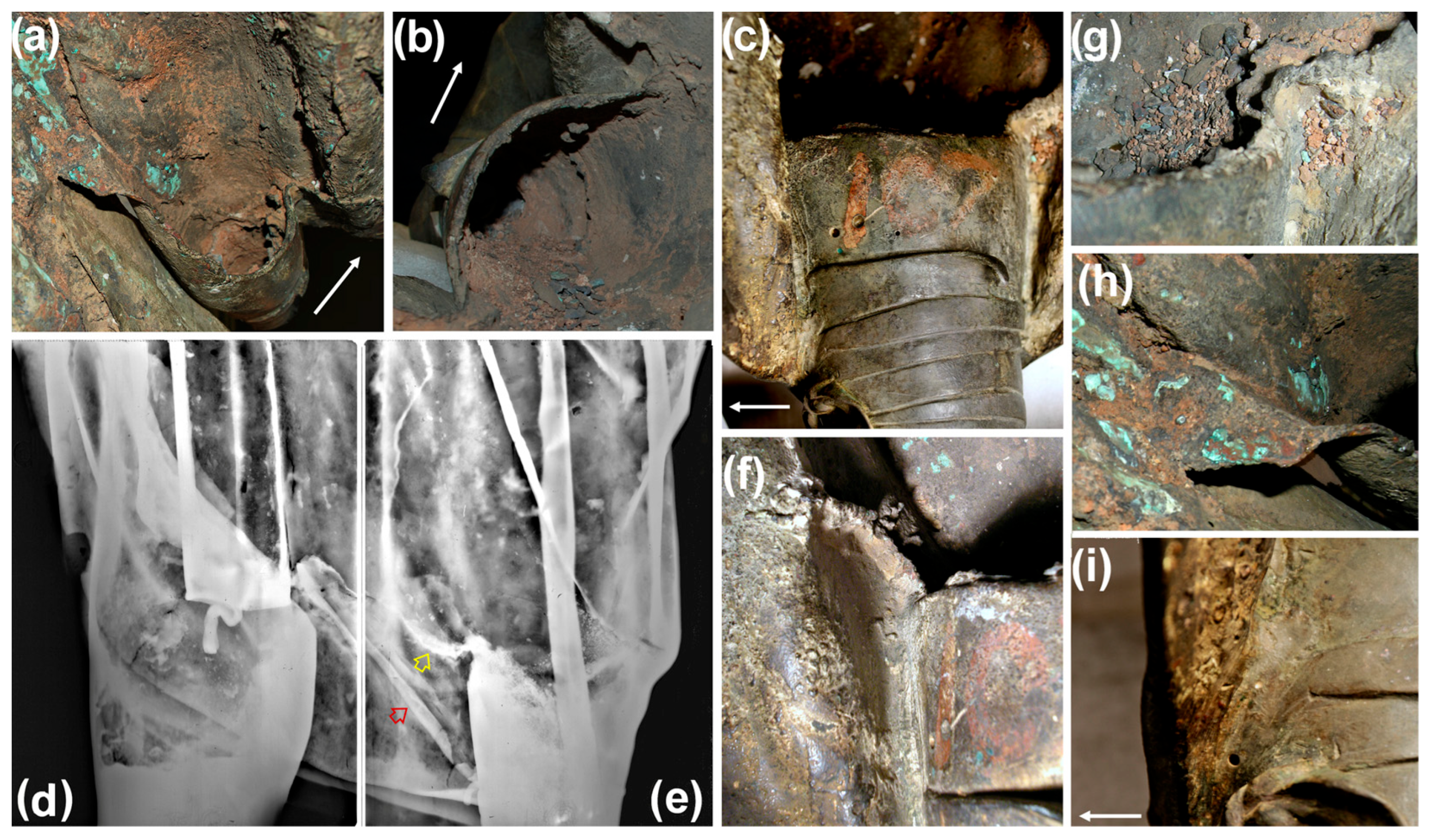
First, the bronze walls of the upper edges of the legs under the toga were not all round and did not present any finishing feature, i.e., they were semi-circular and as cast (Figure 7a,b). These features alone would have been sufficient to rule out independent execution of the legs, but there was more that supported the unified casting of the legs with the toga.
Figure 7.
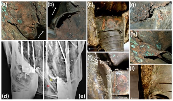
Photographic and radiographic evidence of the connections between the legs and the toga: top views from the inside of the left (a) and right (b) leg, lateral view of the right leg connection with the toga (c), radiographic plates showing the continuity of the left (d) and right (e) leg with the toga, upper part of the right leg connected with the toga (f), continuous flash between the left connection of the right leg and the toga (g), contact zone between the left leg and the toga (h), and bottom part of the front connection shown in c (i). The straight arrow indicates the front direction. The red hollow arrow indicates the anchoring while the yellow one the continuous flashing mentioned in the text.
Figure 7c shows two apparent bronze joints connecting the right leg to the internal surface of the toga, but a careful examination of their morphology (see the magnifications of Figure 7f,i) led us to conclude that these corresponded to joints of the wax model for casting. Thus, likely, after preparing the waxes of the legs independently, their lateral parts were suitably cut along an almost longitudinal section and welded to the inner surface of the lower part of the toga to be cast together with the latter. As mentioned above, this interpretation was supported by Figure 7a–c, as well as by the corresponding radiographic images of Figure 7d,e (see the darker areas of the legs and lack of the typical drips of molten metal). Last but not least, continuous flashing running from the top of the legs to the inner surface of the toga for several centimetres (Figure 7a,b,e–g) was observed, which unequivocally demonstrated once again that the lower trunk and the legs were cast together. At the same time, the careful examination of the lower part of the figure also allowed us to discover that the bottom edge of the toga at the back side of the figure was an independent piece, which was assembled with a pair of cast-on bronze joints at the edges and mechanical anchoring (red hollow arrow in Figure 7e) masked by a fold running from the right leg to the lacinia.
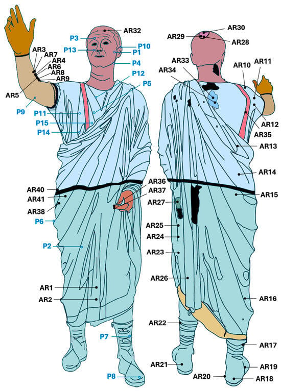
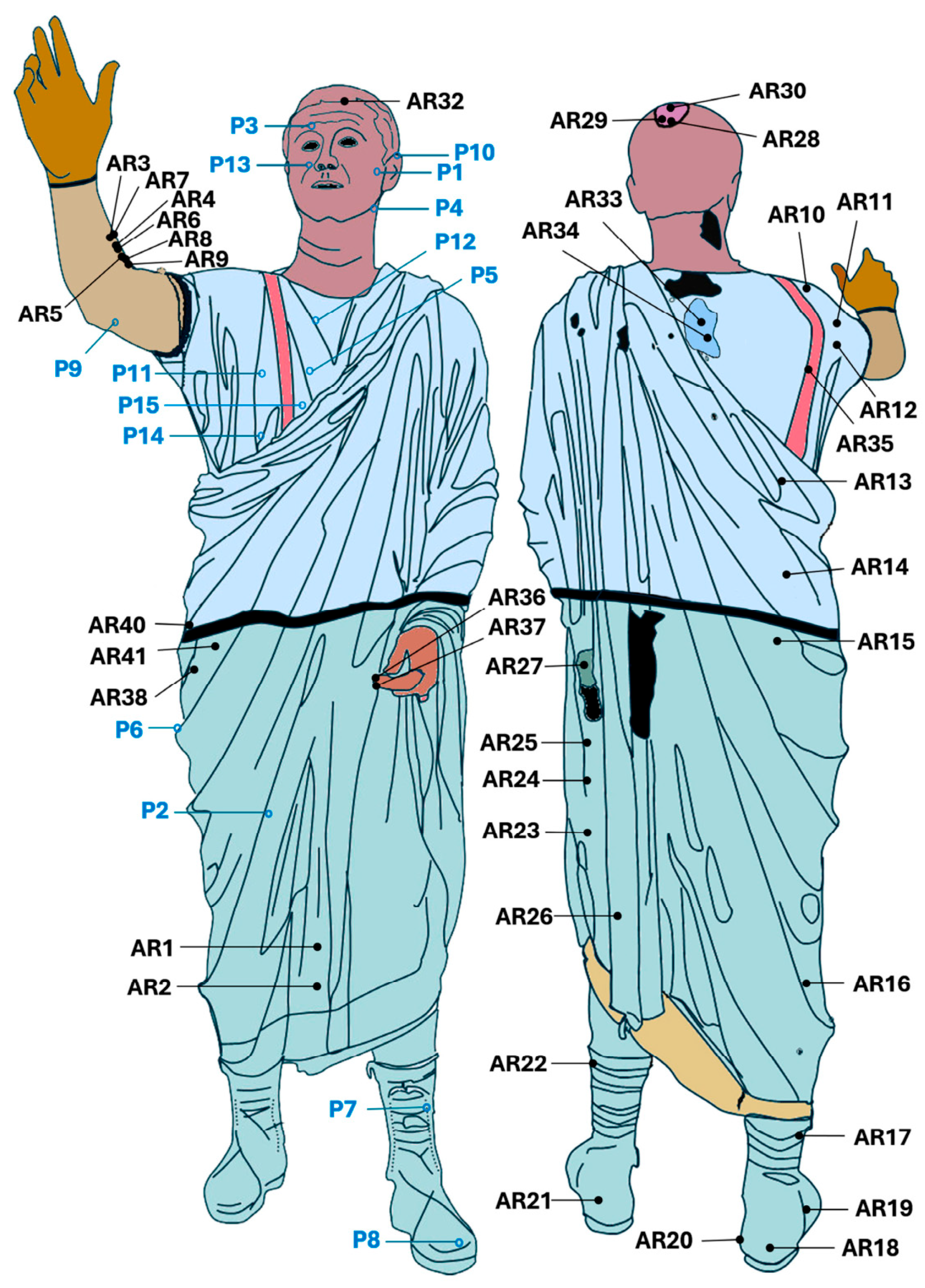
Finally, X-ray inspection allowed to observe a large repair at the right wrist which was interpretable as a further cast-on to joint the right hand and forearm (Figure 9a), then to conclude the Arringatore was assembled from eight independent pieces (head, right arm, right hand, clavus, upper-mid trunk, left hand, lower trunk with legs, and bottom edge of the toga) as highlighted in Figure 8, also reporting the positions/sites of the sampling and LIBS measurements (ARn).
Figure 8.
Pieces composing the Arringatore highlighted using different colours along with the map of the encrustation sampling (P1–P15) and LIBS (ARn) measurement sites.
4.2. Wax Models for Casting
As well known, a wax model for casting may be created either by direct modelling or using a mould taken from an original model (indirect method). The discrimination between these two methods and the associated interpretation of the corresponding operational phases represent important objectives of the metallotechnical examination, although it is often difficult to support the conclusions with really convincing evidence.
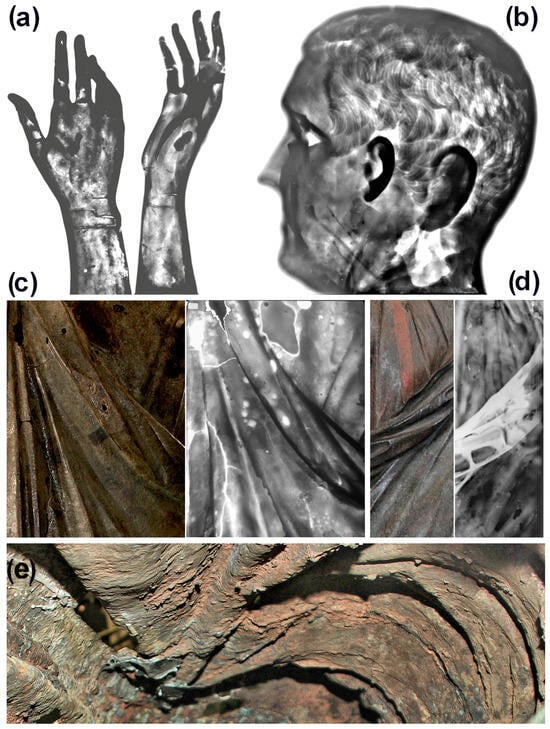
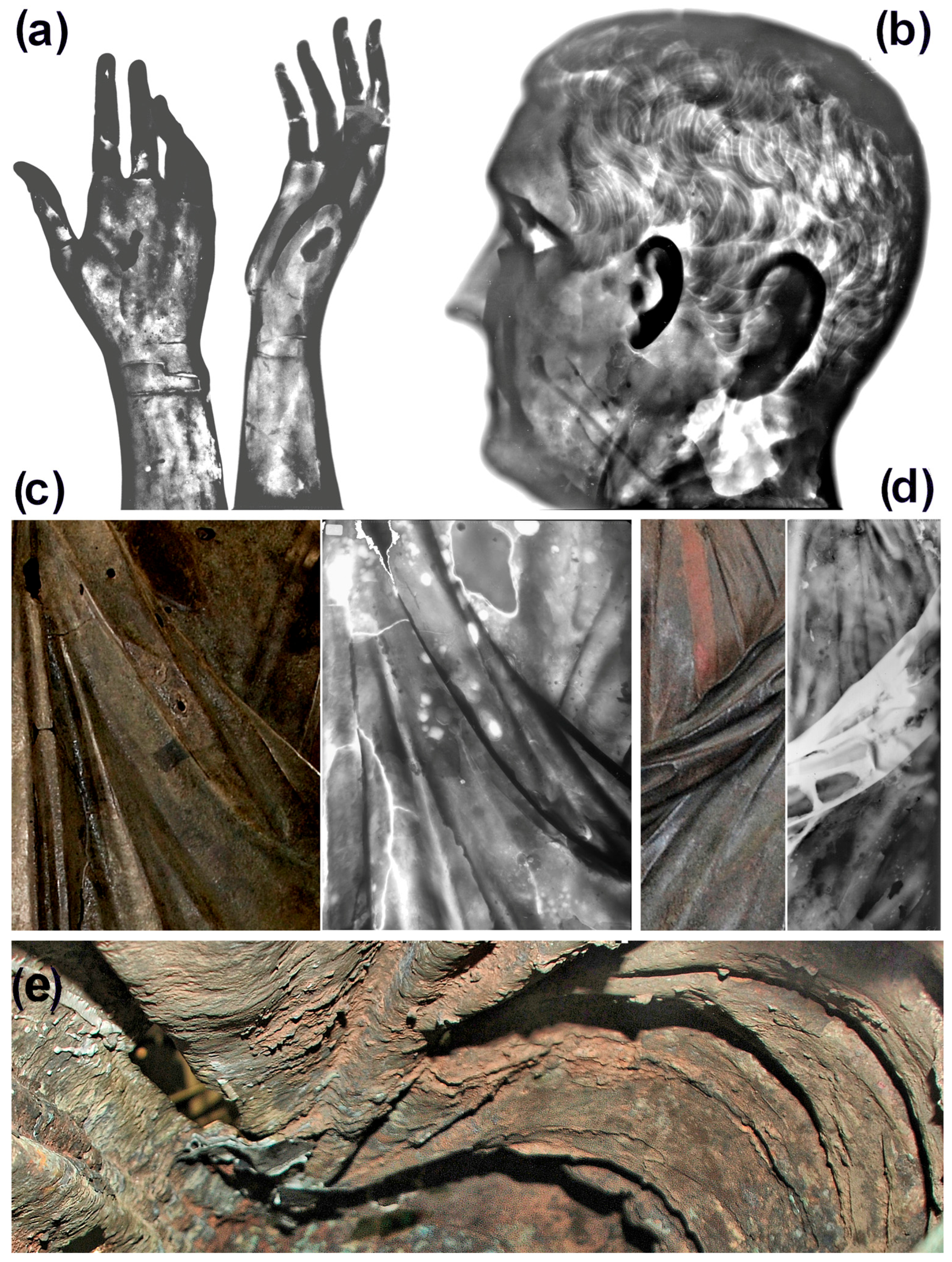
According to tradition, for the present portrait, the creation of the complete starting model using clay, gypsum, or similar materials and then rendering it in bronze through the indirect method represents the first hypothesis to be considered. The X-ray examination of the right arm and head proved that their waxes were likely prepared in corresponding moulds. Typical smooth and rounded wax solidification profiles usually observed in slush casting were found in the former (see, for example, the thumb in the two views of the right hand in Figure 9a), while pronounced signs of wax smearing were pointed out on the internal surface and the hair of the latter (Figure 9b). Probably, also the waxes of the left hand and legs were formed in moulds, although the high thicknesses of these sculptural parts did not allow us to demonstrate this hypothesis. Conversely, thickness modulations of the toga were less pronounced in several zones. For example, apart from the corrosion spots and small lacunas, a rather homogeneous thickness despite the presence of many folds is shown in Figure 9c. Gentle wax applications were also recognizable, but no fingerprints or grooves were observed. In Figure 9d, strong modulations were found due to a cast-on repair of the front draped folds in proximity of the strip but just below, regular behaviour was observed again.
Figure 9.
Details of the bronze walls providing indications about the shaping of the wax models for casting: negative X-ray images of the right hand showing its joint to the forearm and a core nail in the central part of the palm (a) and head (b); photos and corresponding negative X-ray images of areas of the front (c) and back (d) side of the figure, respectively, including many folds; and view of the inner surface of the statue with marks of wax application and drips (e). See the text.
Apart from flashes, repairs and other textural features, the profile of the figure followed that of the outer one also in areas of the toga including folds with tight curvatures, such as some of those included in Figure 9c. However, no convincing markers of indirect lost-wax casting were found. Neither intrinsic nor after casting-reinforcement filleting effects on wax thickness were noted, which are instead usually observed whenever slush moulding is involved in the production of a bronze artefact. Conversely, here, as already observed, the thickness appeared relatively homogeneous throughout the folds (Figure 9c). At the same time, marks of meticulous thickening of the wax wall after shaping were unequivocably observed all over the inner surface of the clothing. Apparently, thickness increase was achieved by local pouring of small amounts of molten wax and spreading it gently (see marks of transverse drips with respect to the longitudinal direction of the folds at the bottom-right corner of Figure 9e and hand application at the opposite side).
These features make it reasonable to consider improbable the use of the indirect method based on wax slush casting or mould lining while considering the direct shaping realistic.
5. Results: Alloy Compositions
As described above, despite most of the outer surface of the Arringatore exhibiting deep corrosion and erosion phenomena with significant material losses, it also presented relatively well-preserved zones that were selected for LIBS elemental analyses. Thus, it was possible to achieve several representative elemental depth profiles by collecting about 2000–3000 consecutive spectra at each site investigated corresponding to typical crater depths of about 0.5–1 mm in cases of successful measurements on archeological findings [55]. Here, this approach allowed us to derive quantitative alloy compositions at 38 measurement sites (ARn) (Table 2), whose positions are mapped in Figure 8, along with those of the analyzed samples.
Table 2.
Alloy compositions, as derived from LIBS elemental depth profiles. Backgroung colors are those of the corresponding pieces in Figure 8.
The measured independent pieces composing the Arringatore were cast using leaded bronze alloys (i.e., Cu-Sn-Pb ternary alloys) with relatively high Sn and low Pb contents with significant traces of Fe and Ni and minimal traces of Zn. As reported in Table 2, weighted average Sn contents of the upper half of the figure (head, right arm, upper-mid trunk) were practically coincident (Sn 12.1–12.5 wt%) and the differences in Pb content were relatively low (Pb 3.9–5.8 wt%). The lower trunk and legs were slightly more alloyed (Sn 13.8 wt%) and had similar low lead content (Pb 3.9 wt%), while the left hand exhibited a further increasing trend, although the error was also higher.
These data suggest that the casting processes could have included two main melting operations. The overall weighted average of the measured pieces returned a Sn 13.5 wt%, Pb 4.0 wt% ternary alloy, whereas quaternary alloys (Cu-Sn-Zn-Pb) were measured for the repair patches of two lacunas at the backside (central area between the shoulders and left flank, respectively) and the one covering the hole at the top of the head likely left by the typical iron bar used to build the mould. These patches appeared mechanically applied and did not exhibit relevant corrosion phenomena, which suggests attributing them to modern integrations. Furthermore, considering their composition and similarities, it seems reasonable to associate such partial repair with a single restoration work and hypothesize that it was carried out well after the excavation of the statue since the present alloy (w. av. Sn 6.3 wt%, Pb 5.3 wt%, Zn 4.3 wt%) recalls in mind a precursor of the well known industrial alloy Cu 85 wt%, Sn-5 wt%, Pb 5 wt%, Zn 5 wt%. Furthermore, the corresponding LIBS profiles allow to argue that the mentioned patches were likely applied in recent centuries.
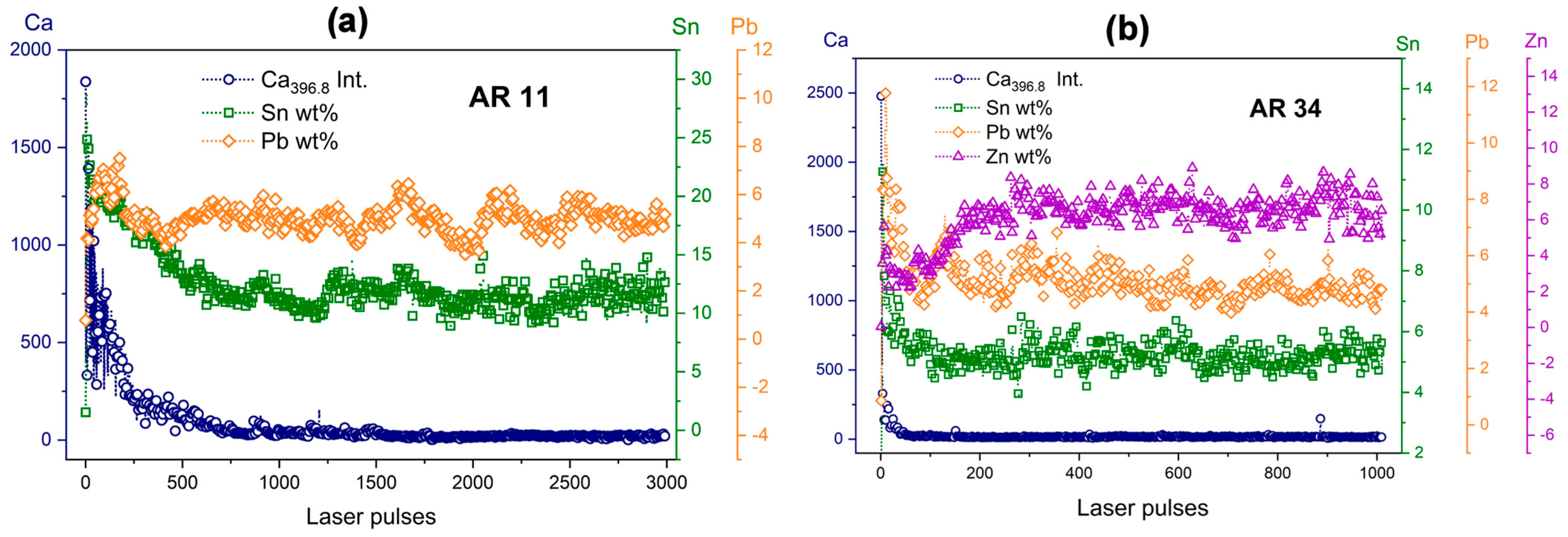
Figure 10 displays the comparison between the measurements carried out in the site AR11 (Figure 10a, original ternary alloy) and AR34 (Figure 10b, quaternary alloy), respectively. As shown, ruling out the first few laser pulses, surface tin enrichment up to around twice the average bulk content was observed for both (from about 12 to 23 wt%, and from about 5 to 8 wt%, respectively). However, the corresponding decreases to the bulk content were very different. About 700 laser pulses were needed in AR11 for the stabilization of the profile and the full decay of the Ca signal, whereas around 50 laser pulses were sufficient in AR34. These behaviours are typical for archeological and modern Cu alloys, respectively [49]. Very interestingly, further qualitative information was provided by the zinc profile in Figure 10b, which exhibited dezincification within 200 laser pulses that, along with the mentioned tin enrichment, is qualitatively congruent with initial ageing of the alloy spanning a few centuries.
Figure 10.
LIBS elemental dept profile of the measurement sites AR11 (a) and AR34 (b). See the text.
Finally, the alloys of the present patches are rather different from those of the Mannerist bronze statuary. Thus, considering the whole, very probably the present patches were added by the sculptor Francesco Carradori in 1785 within the well-known restoration intervention he carried out on the large bronzes of the Medici collection.
The alloy of the narrow stripe inlaid on the tunic that appeared red after the restoration (Figure 1, Figure 2 and Figure 9) was essentially pure copper (Sn 0.1 wt%, Pb 0.5 wt%). The examination also pointed out the presence of a double strip at the left side running from the edge of the draped folds to the lower edge of the toga along with around ten sections of stripes randomly distributed over the toga and the tunic. Unlike the clavus, they did not present metal polychromy/inlay and some of them were previously described as two lateral grooves and a central ridge, which could represent folds [46]. Most of such stripe segments were created on the wax model, while some appeared engraved. In our opinion besides the tentative interpretations previously formulated, the possibility that they represent decorative patterns woven into the fabric should be considered.
6. Discussion
The data collected provide a comprehensive metallotechnical, compositional, and microstructural picture of the Arringatore of Pila that allows us to formulate realistic interpretations of its execution processes and to extend the knowledge of unknown aspects of the Etruscan art foundry practices.
As shown, like most of the ancient large bronzes, the Arringatore was cast as individual pieces to limit casting defects and then carefully assembled by cast-on bronze joints. However, the bronzesmith decided against the precaution of casting the legs individually, likely because he considered crafting safe joints between the latter and the vertical wall of the toga harder than casting the whole lower part in a single piece. The high technical skill in executing the latter, as well as the thin walls and safe and hardly recognizable joints appeared evident upon observing the surfaces of the present work of art.
Along the border of the lacunas, thicknesses even below 2 mm were measured, but the presence of thicker parts was also evident (legs, shoulders, left hand, and others). A rough estimation of the average thickness, t, can be achieved as follows:
where w is the weight of the statue (w ≅ 80 kg), ρ is the approximate density of the present alloy (ρ ≅ 8780 kg/m3), and A is the statue-surface area. The latter can be estimated starting from the human Body-Surface Area [56]:
where h ≅ 179 cm is the height, by considering a 20–30% increase to account for the draped garments. This allows us to obtain correspondent average thickness estimations t ≅ 3.5–3.8 mm. Taking into account the mentioned thicker parts and the spatially concentrated repair materials that provided a weight contribution higher than that of the missing metal of the lacunas, it was concluded that most of the metal walls had thicknesses lower than the estimated average. Thin metal walls increased the risk of premature freezing during mould filling and the formation of lacunas but they also allowed the final cost of the work to be limited.
In presenting the morphological data supporting the interpretation of the wax model for casting, besides the indirect moulding of the head, right arm, left hand and legs the possible direct shaping of the remaining clothed parts was also hypothesized. This was suggested by a set of features including anomalous undulations of the toga attributable to manual shaping of wax sheets, along with marks of cutting and joining of the latter, marks of tools, and some signs of finger deformation. The overall examination suggested that waxed fabric could have been used for such manual shaping. Thin waxed fabric is easily moldable and, if needed, can gently be thickened.
LIBS elemental analyses showed the use of a slightly leaded (low lead) ternary alloy with average tin and lead contents of 13.2 ± 0.2 wt% and 4.1 ± 0.2 wt%, respectively, as derived from the three main groups of measurements in Table 2. According to the commonly used rule of thumb, which assumes lead content as incidental below about 2 wt% or an intentional addition above 3 wt%, the alloys of the Arringatore were intentionally leaded.
As emphasized in the literature [57] the metallotechnical features can provide information on dating ancient large bronzes, thanks to chronological trends in alloy compositions, assembling and repair techniques. Here, the medium tin and low lead contents, respectively, distance the present artefact from those where only traces of lead were found, such as, for example, in the Riace Bronzes [2], the Minerva of Arezzo [58], and the Victorious Youth [59], as well as from those with high lead contents produced during the Roman Empire [37], such as, for example, the Roman statue known as Togatus of Brindisi (portrait of Polydeukion presenting some similarities with the Arringatore), where Pb content is significantly higher than Sn content (Sn 5.6 wt%, Pb 18.1 wt% were measured during the restoration at AST). Assuming such a criterion of medium tin (ex. 8–16 wt%) and low (intentional) lead (3–6 wt%) strictly, even, for example, the Croatian Apoxyomenos (Sn 8–10 wt%, Pb 8–13 wt% [60]) does not match.
Conversely, considering the available data, the present composition aligns the Arringatore with other bronzes found in Tuscany, such as the Chimera of Arezzo [60], the Lamp of Cortona [61] (slightly higher in tin), and the Lupa of Fiesole [62], which represent an encouraging congruence. The respective compositions are reported in Table 3 along with those of some other important artefacts from different provenience presenting medium Sn-low Pb compositions. This type of alloy is usually associated with Hellenistic production although, obviously, there are no compelling reasons why the diachronic evolution of the art foundry should be strictly related to that of art. For this reason, the inclusion in Table 3 of artworks not interpreted as Hellenistic or dated to the corresponding period should not be considered surprising.
Table 3.
Some meaningful comparison of the present alloy composition with those of other important bronze sculptures.
Finally, the blackish appearance of the present artwork (Figure 2, Figure 3 and Figure 4) after the removal of modern patinations deserves special attention. This peculiar feature recalls in mind the well-known cases of black bronzes encountered in different cultures (Egyptian Hsmn-km, Corinthium aes mentioned by Pliny the Elder, and more recent Japanese shakudō), which were achieved through patination of copper alloys containing not negligible fractions of gold and/or silver and were prevalently characterized by the presence of a surface cuprite layer [66,67]. This is not the case of the Arringatore where the blackish appearance was determined by a relatively thick tenorite layer. This was generally contiguous to similarly thick underlying cuprite layer (Figure 3, Figure 4 and Figure 5, Table 1) unless alterations mainly due to the crystallization of chlorides or sulphates were observed. As already observed, microstructures resembling those of the alloy dendrites were clearly visible in the cuprite layer (Figure 5c) and in some spots they were vaguely recognizable also in the tenorite layer, which demonstrated that such oxide bilayer represented the primary oxidation of the original metal surface.
The formation of a Cu2O/CuO bilayer can be induced on clean copper alloy surfaces through heating and/or increasing the availability of oxygen, whereas it can hardly be produced by natural corrosion processes in burial conditions [68]. The multi-crystallinity and complex texture of the present tenorite layer, as well as the presence of inclusions and of ramified networks of thin infiltration channels with clayey minerals proved that “it was ancient”. The alternative of a possible heat treatment carried out after the excavation of the statue in 1567 could not be substantiated in any way, since tenorite was detected only on well-preserved areas of the outer surface and no evidence of deep cleaning or combustion residues was found. Conversely, it was evident that all the restoration works of the last centuries were based on cold assembling (right arm), cold-worked metal additions by Carradori, and the application of organic matrix coatings. Thus, the present oxidation bilayer testified that the Arringatore was heated in the distant past. Furthermore, the microstructural data indicated that it should have been a moderate and controlled heating since intentional or accidental overheating would have also produced massive recrystallizations, with consequent cancellation of traces of “pseudomorphism” within the oxidation bilayer and, likely, also partial or complete homogenization of the alloys. Conversely, dendritic segregations of the alloys and similar microstructures inherited by cuprite were abundant in the present case.
Native oxidation of copper alloys is dominated by the formation of Cu2O while CuO forms under more oxidizing conditions, such as those determined by high temperatures and increased availability of oxygen. Oxidation significantly accelerates when the temperature [69] and/or humidity [70] increase/s. The formation of the Cu2O/CuO bilayer can be observed already around 100 °C after a sufficiently long time [69] and more efficiently above this temperature [71].
Such results of rigorous scientific experiments provide general information, which is not intended as a satisfactory explanation of the oxidation bilayer detected in several preserved areas of the Arringatore. Apart from swellings, detachments, and/or partial destruction of the tenorite layer by corrosion produced by the depositional dynamics, the data indicate that the original thickness of the CuO layer was in the order of ~100 mm (i.e., much larger than those of the mentioned experiments). This suggests that heating represented the crucial part for inducing the formation of the oxidation bilayer but, likely, specific assists and treatment cycles were needed to produce such a thick tenorite layer. Assuming this possibility, the logical conclusion is that the statue was intentionally patinated to return a blackish appearance given by tenorite and likely finishing treatments. Tenorite produced by heating is dull black that could esthetically be improved during its formation using specific substances or after the heat-assisted process by applying further organic treatments. Unfortunately, the data do not offer sufficient clues to say more about the patination procedure, but they support the intentionality of the present tenorite layer. In particular, further data are needed to thoroughly interpret the abundance and the iso-orientation of lead and tin mineral inclusions shown in the SEM images of Figure 5 and to discriminate whether they were the result of natural or artificial processes.
7. Conclusions
The archaeometallurgical results reported in the present non-invasive study mainly based on photonic techniques reveal unknown aspects of the Arringatore, a bronze portrait of the prominent Etruscan citizen Aule Meteli. Furthermore, some of the results have general implications within context of the knowledge of ancient art foundry.
There are factual and congruent analytical clues (microscopy, XRD and Raman spectroscopy) along with naked eye evidence allowing us to conclude that the statue had a blackish intentional patina in the far past, likely created through heating and surface treatment processes that produced a relatively thick tenorite layer. This is the first time this type of patination is hypothesized and analytically documented for an ancient bronze. According to the literature and the analytical results described above, an alternative natural origin of such an overall black mineralization appears less probable.
The study of the alloy compositions using LIBS confirmed once again the high effectiveness of this technique for quantitative measurement of copper alloys through elemental depth profiles. Besides the mentioned advantages, it is perhaps useful to further underline that the repeatability and possibility to verify the quality of the analytical data before considering them valid are unique and crucial in order to extract representative average compositions. Here, this allowed us to demonstrate that the Arringatore was cast using a medium-tin–low-lead ternary alloys and the similarity with other important bronze artefacts was preliminarily discussed. In line with the general trend, we believe the quantification of the major elements can indeed support rough dating over long time periods, provided that the elemental quantifications are truly reproducible and therefore reliable and representative. At the same time, it should also be considered that artisanal traditions endure for centuries (circumstances permitting) and, in many cases, their geographic variation is more pronounced than the temporal one.
Finally, the metallotechnical examination has enabled a detailed description of the execution processes of the present Etruscan masterpiece, refining earlier interpretations—which remain noteworthy nonetheless, having been formulated without the use of advanced tools such as those employed in the present study. Thin walls, extensive use of casting in pieces, high-quality cast-on joints (not visible in normal fruition conditions), complex execution of the lower part of the figure, refined cold finishing, and polychromy (blackish patina with possible highlights provided by white Pb and Sn minerals, red clavus, realistic glass paste eyes, and perhaps other) allow this work to be placed at the pinnacle of the ancient bronze statuary.
Author Contributions
Conceptualization, S.S.; methodology, S.S. and S.M.; investigation, S.M., J.A., I.O., D.C., M.G. and S.S.; formal analysis, J.A. and S.S.; validation J.A.; resources, S.S., A.A.M. and M.G.; writing—original draft preparation, S.S. and S.M.; writing—review and editing, S.S., L.M. and S.M.; funding acquisition, S.S.; project administration, S.S.; supervision, S.S. All authors have read and agreed to the published version of the manuscript.
Funding
The present work was supported by the project iPhotoCult (Horizon Europe, Grant agreement ID: 101132448).
Data Availability Statement
Data will be made available on request.
Acknowledgments
The authors would first like to thank Fulvia Lo Schiavo, at the time, Superintendent of AST, for entrusting them with this investigation. Without her initiative and encouraging confidence, the present study and the restoration of the Arringatore of Pila would not have been carried out. Sincere thanks are extended to Marcello Miccio for having executed the radiographies of the present artwork, provided data on other bronze artefacts, and allowed to overcome operative problems, as well as to the restorers Stefano Sarri, Elisa Pucci, and Donato Scapati for their passionate collaboration.
Conflicts of Interest
The authors declare no conflicts of interest.
Abbreviations
The following abbreviations are used in this manuscript:
| AST | Archaeological Superintendence of Tuscany |
| LIBS | Laser-Induced Breakdown Spectroscopy |
| XRD | X-Ray Diffraction |
| AAS | Atomic Absorption Spectrosopy |
| ICP-OES | Inductively Coupled Plasma-Optical Emission Spectroscopy |
References
- Caley, E.R. The Early History of Chemistry in the Service of Archaeology. J. Chem. Educ. 1967, 44, 120–123. Available online: https://pubs.acs.org/jceda8/article-pdf/44/3/120/319616/ed044p120.pdf (accessed on 10 April 2026). [CrossRef] [Scilit]
- Soprintendenza Archeologica per la Toscana, Relazione sulle analisi. In Due Bronzi da Riace. Rinvenimento, Restauro, Analisi e Ipotesi di Interpretazione, Bollettino d’Arte; Serie Speciale III; Ministero per i Beni Culturali: Roma, Italy, 1984; Volume 1, pp. 85–106.
- Formigli, E. La tecnica di costruzione delle statue di Riace. In Due Bronzi da Riace. Rinvenimento, Restauro, Analisi e Ipotesi di Interpretazione, Bollettino d’Arte; Serie Speciale III; Ministero per i Beni Culturali: Roma, Italy, 1984; Volume 1, pp. 107–142. [Google Scholar]
- Scott, D.A. Copper and Bronze in Art: Corrosion, Colorants, Conservation; Getty Publications: Los Angeles, CA, USA, 2002; Available online: https://www.getty.edu/publications/virtuallibrary/temp/9780892366385.pdf (accessed on 10 April 2026).
- Papadopoulou, O.; Vassiliou, P. The Influence of Archaeometallurgical Copper Alloy Castings Microstructure towards Corrosion Evolution in Various Corrosive Media. Corros. Mater. Degrad. 2021, 2, 227–247. [Google Scholar] [CrossRef] [Scilit]
- Bernabale, M.; Cognigni, F.; Mura, F.; Nigro, L.; Montanari, D.; Rossi, M.; De Vito, C. 3D imaging of micro-segregation and corrosion behavior of alloying elements in archaeological artefacts from Motya (Sicily, Italy). Corros. Sci. 2023, 211, 110900. [Google Scholar] [CrossRef] [Scilit]
- Bourgarit, D.; Mille, B. The elemental analysis of ancient copper-based artefacts by inductively-coupled-plasma atomic-emission spectrometry: An optimized methodology reveals some secrets of the Vix crater. Meas. Sci. Technol. 2003, 14, 1538–1555. [Google Scholar] [CrossRef] [Scilit]
- Cesareo, R.; Sciuti, S.; Marabelli, M. Non-destructive analysis of ancient bronzes. Stud. Conserv. 1973, 18, 64–80. [Google Scholar] [CrossRef] [Scilit]
- Ferretti, M.; Miazzo, L.; Moioli, P. The application of a non-destructive XRF method to identify different alloys in the bronze statue of the Capitoline Horse. Stud. Conserv. 1997, 42, 241–246. [Google Scholar] [CrossRef] [Scilit]
- Pershin, S.M.; Colao, F.; Spizzichino, V. Quantitative analysis of bronze samples by laser-induced breakdown spectroscopy (LIBS): A new approach, model, and experiment. Laser Phys. 2006, 16, 455–467. [Google Scholar] [CrossRef] [Scilit]
- De Giacomo, A.; Dell’Aglio, M.; De Pascale, O.; Gaudiuso, R.; Santagata, A.; Teghil, R. Laser Induced Breakdown Spectroscopy methodology for the analysis of copper-based-alloys used in ancient artworks. Spectrochim. Acta Part B 2008, 63, 585–590. [Google Scholar] [CrossRef] [Scilit]
- Elhassan, A.; Giakoumaki, A.; Anglos, D.; Ingo, G.M.; Robbiola, L.; Harith, M.A. Nanosecond and femtosecond Laser Induced Breakdown Spectroscopic analysis of bronze alloys. Spectrochim. Acta B At. Spectrosc. 2008, 63, 504–511. [Google Scholar] [CrossRef] [Scilit]
- Agresti, J.; Siano, S. Depth-dependent calibration for quantitative elemental depth profiling of copper alloys using laser-induced plasma spectroscopy. Appl. Phys. A 2014, 117, 217–221. [Google Scholar] [CrossRef] [Scilit]
- Botto, A.; Campanella, B.; Legnaioli, S.; Lezzerini, M.; Lorenzetti, G.; Pagnotta, S.; Poggialini, F.; Palleschi, V. Applications of laser-induced breakdown spectroscopy in cultural heritage and archaeology: A critical review. J. Anal. At. Spectrom. 2019, 34, 81–103. [Google Scholar] [CrossRef] [Scilit]
- Fortes, F.J.; Cabalín, L.M.; Laserna, J.J. Fast and In-Situ Identification of Archaeometallurgical Collections in the Museum of Malaga Using Laser-Induced Breakdown Spectroscopy and a New Mathematical Algorithm. Heritage 2020, 3, 1330–1343. [Google Scholar] [CrossRef] [Scilit]
- Ferretti, M.; Cristoforetti, G.; Legnaioli, S.; Palleschi, V.; Salvetti, A.; Tognoni, E.; Console, E.; Palaia, P. In situ study of the Porticello Bronzes by portable X-ray fluorescence and laser-induced breakdown spectroscopy. Spectrochim. Acta Part B 2007, 62, 1512–1518. [Google Scholar] [CrossRef] [Scilit]
- Bertolini, A.; Carelli, G.; Francesconi, F.; Francesconi, M.; Marchesini, L.; Marsili, P.; Sorrentino, F.; Cristoforetti, G.; Legnaioli, S.; Palleschi, V.; et al. Modì: A new mobile instrument for in situ double-pulse LIBS analysis. Anal. Bioanal. Chem. 2006, 385, 240–247. [Google Scholar] [CrossRef] [Scilit]
- Goujon, J.; Giakoumaki, A.; Piñon, V.; Musset, O.; Anglos, D.; Georgiou, E.; Boquillon, J.P. A compact and portable laser-induced breakdown spectroscopy instrument for single and double pulse applications. Spectrochim. Acta B At. Spectrosc. 2008, 63, 1091–1096. [Google Scholar] [CrossRef] [Scilit]
- Agresti, J.; Mencaglia, A.A.; Siano, S. Development and application of a portable LIPS system for characterising copper alloy artefacts. Anal. Bioanal. Chem. 2009, 395, 2255–2262. [Google Scholar] [CrossRef] [Scilit]
- Siano, S.; Bartoli, L.; Kockelmann, W.; Zoppi, M.; Miccio, M. Neutron metallography of archaeological bronzes. Phys. B Condens. Matter. 2004, 350, 123–126. [Google Scholar] [CrossRef] [Scilit]
- Agresti, J.; Osticioli, I.; Guidotti, M.C.; Kardjilov, N.; Siano, S. Non-invasive archaeometallurgical approach to the investigations of bronze figurines using neutron, laser, and X-ray techniques. Microchem. J. 2016, 124, 765–774. [Google Scholar] [CrossRef] [Scilit]
- Senesi, G.S.; Harmon, R.S.; Hark, R.R. Field-portable and handheld laser-induced breakdown spectroscopy: Historical review, current status and future prospects. Spectrochim. Acta B At. Spectrosc. 2021, 175, 106013. [Google Scholar] [CrossRef] [Scilit]
- Mattiello, S.; De Pascale, O.; Palleschi, V.; Fiorentino, G.; Senesi, G.S. Application of handheld/portable spectroscopic tools to the identification, inner stratigraphy and mapping of archaeological metal artefacts. J. Phys. Photonics 2024, 6, 035005. [Google Scholar] [CrossRef] [Scilit]
- Siano, S.; Agresti, J. Archaeometallurgical characterisation of Donatello’s Florentine copper alloy masterpieces using portable laser-induced plasma spectroscopy and traditional techniques. Stud. Conserv. 2015, 60, S106–S119. [Google Scholar] [CrossRef] [Scilit]
- Siano, S.; Agresti, J.; Cacciari, I.; Ciofini, D.; Mascalchi, M.; Osticioli, I.; Mencaglia, A.A. Laser cleaning in conservation of stone, metal, and painted artifacts: State of the art and new insights on the use of the Nd:YAG lasers. Appl. Phys. A 2012, 106, 419–446. [Google Scholar] [CrossRef] [Scilit]
- Vandenabeele, P.; Edwards, H.G.M.; Jehlička, J. The role of mobile instrumentation in novel applications of Raman spectroscopy: Archaeometry, geosciences, and forensics. Chem. Soc. Rev. 2014, 43, 2628–2649. [Google Scholar] [CrossRef] [Scilit]
- Frost, R.L.; Martens, W.; Kloprogge, J.T.; Williams, P.A. Raman spectroscopy of the basic copper chloride minerals atacamite and paratacamite: Implications for the study of copper, brass and bronze objects of archaeological significance. J. Raman Spectrosc. 2002, 33, 801–806. [Google Scholar] [CrossRef] [Scilit]
- Hayez, V.; Guillaume, J.; Hubin, A.; Terryn, H. Micro-Raman spectroscopy for the study of corrosion products on copper alloys: Setting up of a reference database and studying works of art. J. Raman Spectrosc. 2004, 35, 732–738. [Google Scholar] [CrossRef] [Scilit]
- Bouchard, M.; Smith, D.C. Catalogue of 45 reference Raman spectra of minerals concerning research in art history or archaeology, especially on corroded metals and coloured glass. Spectrochim. Acta Part A 2003, 59, 2247–2266. [Google Scholar] [CrossRef] [Scilit]
- Hayez, V.; Segato, T.; Hubin, A.; Terryn, H. Study of copper nitrate-based patinas. J. Raman Spectrosc. 2006, 37, 1211–1220. [Google Scholar] [CrossRef] [Scilit]
- Bertolotti, G.; Bersani, D.; Lottici, P.P.; Alesiani, M.; Malcherek, T.; Schlüter, J. Micro-Raman study of copper hydroxychlorides and other corrosion products of bronze samples mimicking archaeological coins. Anal. Bioanal. Chem. 2012, 402, 1451–1457. [Google Scholar] [CrossRef] [Scilit]
- Privitera, A.; Corbascio, A.; Calcani, G.; Della Ventura, G.; Ricci, M.A.; Sodo, A. Raman approach to the forensic study of bronze patinas. J. Arch. Sci. Rep. 2021, 39, 103115. [Google Scholar] [CrossRef] [Scilit]
- Mikic, D.; Otmacic Curkovic, H.; Kosec, T.; Peko, N. An Electrochemical and Spectroscopic Study of Surfaces on Bronze Sculptures Exposed to Urban Environment. Materials 2021, 14, 2063. [Google Scholar] [CrossRef] [Scilit]
- Kwon, H.; Cho, N. In-situ non destructive investigation of contemporary outdoor bronze sculptures. Her. Sci. 2024, 12 167. [Google Scholar] [CrossRef] [Scilit]
- Luvidi, L.; Casanova Municchia, A.; Konstantakopoulou, E.; Orazi, N.; Ferretti, M.; Caruso, G. Combined Use of Non-Destructive Analysis Techniques to Investigate Ancient Bronze Statues. Sensors 2025, 5, 1727. [Google Scholar] [CrossRef] [Scilit]
- Craddock, P. The composition of the copper alloys used by the Greek, Etruscan and Roman civilisations: 2. The Archaic, Classical and Hellenistic Greeks. J. Arch. Sci. 1977, 4, 103–123. [Google Scholar] [CrossRef] [Scilit]
- Riederer, J. La composizione dei grandi bronzi Romani. In I Grandi Bronzi; Formigli, E., Ed.; Nuova Immagine Editrice: Siena, Italy, 1999; pp. 271–274. [Google Scholar]
- Formigli, E. (Ed.) I Grandi Bronzi Antichi: Le Fonderie e le Tecniche di Lavorazione Dall’età Arcaica al Rinascimento. Atti dei Seminari di Studi ed Esperimenti; (Murlo 24-30 luglio 1993 e 1-7 luglio); Nuova Immagine Editrice: Siena, Italy, 1999. [Google Scholar]
- Craddock, P.; Giumlia-Mair, A. Beauty is skin deep: Evidence for the original appearance of classical statuary. In Metal Plating and Patination, Cultural, Technical and Historical Developments; Butterworth-Heinemann: Oxford, UK, 1993; pp. 30–38. [Google Scholar] [CrossRef] [Scilit]
- Colacicchi Alessandri, O.; Caruso, G.; Orazi, N.; Ferretti, M. A Technical Examination of the Great Bronzes of the Museo Nazionale Romano. Stud. Conserv. 2023, 68, 326–342. [Google Scholar] [CrossRef] [Scilit]
- Bourgarit, D.; Bassett, J.; Bewer, F.G.; Heginbotham, A.; Lacey, A.; Motture, P. (Eds.) Guidelines for the Technical Examination of Bronze Sculpture; Getty Publications: Los Angeles, CA, USA, 2025; Available online: https://www.getty.edu/publications/bronze-guidelines/_assets/downloads/BronzeGuidelines.pdf (accessed on 9 June 2026).
- Dohrn, T. L’Arringatore, capolavoro del museo archeologico di Firenze. Boll. d’Arte 1964, 2, 97–114. Available online: https://bollettinodarte.cultura.gov.it/wp-content/uploads/Importer/1483444588808_04_Dohrn_97.pdf (accessed on 3 April 2026).
- Dohrn, T. Der Arringatore Bronzestatue im Museo Archeologico von Florenz; Gebr. Mann Verlag: Berlin, Germany, 1968. [Google Scholar]
- Colonna, G. Il posto dell’Arringatore nell’arte etrusca di età ellenistica. Studi Etruschi 1989–1990, LVI, 99–122. Available online: https://www.studietruschi.org/wp-content/uploads/2021/08/SE56_08.pdf (accessed on 3 April 2026).
- Pallottino, M. Nota sull’iscrizione dell’Arringatore. Boll. d’Arte 1964, 49, 115–116. [Google Scholar]
- Granger-Taylor, H. Weaving Clothes to Shape in the Ancient World: The Tunic and Toga of the Arringatore. Text. Hist. 1982, 13, 3–25. [Google Scholar] [CrossRef]
- Pulcinelli, L. Lo stato delle ricerche sull’Arringatore e le nuove acquisizioni emerse. Boll. d’Arte 2019, 42, 7–12. [Google Scholar]
- Sartore, A.M. L’Arringatore a Pila. La storia del ritrovamento nei nuovi documenti d’archivio. Boll. d’Arte 2019, 42, 13–44. Available online: https://www.academia.edu/44820308/Novit%C3%A0_sul_ritrovamento_della_statua_in_bronzo_dellArringatore_LArringatore_a_Pila_La_storia_del_ritrovamento_nei_nuovi_documenti_darchivio_Alberto_Maria_Sartore (accessed on 25 August 2026).
- Siano, S.; Bartoli, L.; Mencaglia, A.A.; Miccio, M.; Agresti, J. Use of neutron diffraction and laser-induced plasma spectroscopy in integrated authentication methodologies of copper alloy artefacts. Nuovo Cim. B 2009, 124, 671–686. [Google Scholar] [CrossRef] [Scilit]
- Siano, S.; Casciani, A.; Giusti, A.; Matteini, M.; Pini, R.; Porcinai, S.; Salimbeni, R. The Santi Quattro Coronati by Nanni di Banco: Cleaning of the gilded decorations. J. Cult. Herit. 2003, 4, 123s–128s. [Google Scholar] [CrossRef] [Scilit]
- Siano, S. The Birth and Second Life of the Minerva of Arezzo. In The Restoration of Ancient Bronzes: Naples and Beyond; Sounders, D., Risser, E., Eds.; Getty Publications: Los Angeles, CA, USA, 2013; pp. 64–82. Available online: https://www.getty.edu/publications/virtuallibrary/1606061542.html (accessed on 25 August 2026).
- Siano, S.; Nicolai, M.L.; Porcinai, S. Verrocchio’s David: Characterization and conservation treatments. In Verrocchio’s David restored: A Renaissance bronze from the National Museum of the Bargello; Radke, G.M., Brown, D.A., Nicolai, M.L., Paoletti, J.T., Paolozzi Strozzi, B., Porcinai, S., Siano, S., Vaccari, M.G., Eds.; High Museum in association with Giunti Gruppo Editoriale: Florence, Italy; Atlanta, GA, USA, 2023; pp. 97–109. [Google Scholar]
- Giusti, A.; Lalli, C.; Lanterna, G.; Matteini, M.; Porcinai, S.; Rizzi, M.; Siano, S. Indagini storiche e diagnostiche per il restauro dei Quattro Santi Coronati dalla facciata di Orsanmichele a Firenze. OPD Restauro 2001, 13, 143–150. [Google Scholar]
- Nevin, A.; Melia, J.L.; Osticioli, I.; Gautier, G.; Colombini, M.P. The identification of copper oxalates in a 16th century Cypriot exterior wall painting using micro FTIR, micro Raman spectroscopy and Gas Chromatography-Mass Spectrometry. J. Cult. Herit. 2008, 9, 154–161. [Google Scholar] [CrossRef] [Scilit]
- Siano, S.; Cacciari, I.; Mencaglia, A.; Agresti, J. Spatially calibrated elemental depth profiling using LIPS and 3D digital microscopy. Eur. Phys. J. Plus 2011, 126, 120. [Google Scholar] [CrossRef] [Scilit]
- Mosteller, R.D. Simplified Calculation of Body-Surface Area. N. Engl. J. Med. 1987, 317, 1098. [Google Scholar] [CrossRef] [Scilit]
- Formigli, E. La datazione tecnologica dei grandi bronzi antichi: Il caso della Lupa Capitolina. In Bronzes Grecs et Romains, Recherches Récentes. Hommage à Claude Rolley; Denoyelle, M., Descamps-Lequime, S., Mille, B., Verger, S., Eds.; Publications de l’Institut National d’Histoire de l’Art: Paris, France, 2012; pp. 182–201. [Google Scholar] [CrossRef] [Scilit]
- Siano, S.; Miccio, M. La tecnica esecutiva. In La Minerva d.Arezzo, (Catalogue of the Exhibition at the National Archaeological Museum of Arezzo, July 2008–January 2009); Cygielman, M., Ed.; Comune di Arezzo (Ufficio Cultura): Arezzo, Italy, 2008; pp. 175–190. Available online: https://www.researchgate.net/publication/400854448_La_tecnica_esecutiva_della_Minerva_di_Arezzo (accessed on 25 August 2026).
- Podany, J.; Scott, D. The Getty Victorious Youth reconsidered: Initial report on the scientific and technical re-examination. J. Roman Archaeol. 2000, 1, 179–191. [Google Scholar]
- Mudronja, D.; Jakšić, M.; Fazinić, S.; Božičević, I.; Desnica, V.; Woodhead, J.; Stos-Gale, Z.A. Croatian Appoxiomenos alloy composition and lead provenance study. J. Arch. Sci. 2010, 37, 1396–1402. [Google Scholar] [CrossRef] [Scilit]
- Siano, S.; Miccio, M.; Giamello, M.; Mugnaini, S.; Agresti, J. Journey through the material layers of the Chimaera from Arezzo. In Myth, Allegory Emblem—The many lives of the Chimaera of Arezzo, Proc. of the Int. Colloquium—Malibu, The J. Paul Getty Museum (Dec. 4-5, 2009); Cianferoni, G.C., Iozzo, M., Iozzo, E.S.M., Eds.; Aracne Editrice: Roma, Italy, 2012; pp. 185–222. [Google Scholar]
- van der Meer, L.B. The Etruscan Bronze Lamp of Cortona, its Cosmic Program and its Attached Inscription. Latomus 2014, 73, 289–302. [Google Scholar] [CrossRef]
- Warden, P.G. The Chimaera of Arezzo: Made in Etruria? Am. J. Archaeol. 2011, 115, 1–5. [Google Scholar] [CrossRef]
- Ammannati, N.; Martellucci, E.; Parisi Presicce, C.; Carruba, A. Il restauro della “Lupa Capitolina”: Analisi del bronzo e delle patine di corrosione tramite microscopio ottico ed elettronico a scansione. LA Metall. Ital. 2001, 1, 43–50. [Google Scholar]
- Siano, S.; Melillo, L.; Sarri, S.; Agresti, J. The Bronze Sculpture of Alexander the Great on Horseback: An Archaeometallurgical Study. In Artistry in Bronze The Greeks and Their Legacy XIXth Int. Cong. on Ancient Bronzes; Daehner, J.M., Lapatin, K., Spinelli, A., Eds.; Getty Publications: Los Angeles, CA, USA, 2017; Volume 44, Available online: https://www.getty.edu/publications/artistryinbronze/conservation-and-analysis/44-siano/ (accessed on 25 August 2026).
- Craddock, P.T.; Giumlia-Mair, A. Hsmn-km, Corinthian bronze, shakudo: Black patinated bronze in the ancient world. In Metal Plating and Patination: Cultural and Historical Developments; La Niece, S., Craddock, P.T., Eds.; Butterworth–Heinemann: Oxford, UK, 1993; pp. 102–127. [Google Scholar] [CrossRef] [Scilit]
- Benzonelli, A.; Freestone, I.C.; Martinón-Torres, M. A better shade of black: Effects of manufacturing parameters on the development of ancient black bronzes. Archaeometry 2017, 59, 1034–1049. [Google Scholar] [CrossRef] [Scilit]
- Scott, D.A. Copper compounds in metals and colorants: Oxides and hydroxides. Stud. Conserv. 1997, 42, 93–100. [Google Scholar] [CrossRef] [Scilit]
- Aromaa, J.; Kekkonen, M.; Mousapour, M.; Jokilaakso, A.; Lundström, M. The Oxidation of Copper in Air at Temperatures up to 100 °C. Corros. Mater. Degrad. 2021, 2, 625–640. [Google Scholar] [CrossRef] [Scilit]
- Aastrup, T.; Wadsak, M.; Schreiner, M.; Leygraf, C. Experimental in situ studies of copper exposed, to humidiÆed air. Corros. Sci. 2000, 42, 957–967. [Google Scholar] [CrossRef] [Scilit]
- Lee, S.-K.; Hsu, H.-C.; Tuan, W.-H. Oxidation Behavior of Copper at a Temperature below 300 °C and the Methodology for Passivation. Mater. Res. 2016, 19, 51–56. [Google Scholar] [CrossRef] [Scilit]

Disclaimer/Publisher’s Note: The statements, opinions and data contained in all publications are solely those of the individual author(s) and contributor(s) and not of MDPI and/or the editor(s). MDPI and/or the editor(s) disclaim responsibility for any injury to people or property resulting from any ideas, methods, instructions or products referred to in the content. |
© 2026 by the authors. Licensee MDPI, Basel, Switzerland. This article is an open access article distributed under the terms and conditions of the Creative Commons Attribution (CC BY) license.