


Abstract
Background and Clinical Significance: A longitudinal epiphyseal bracket (delta phalanx) is a rare congenital abnormality in which the epiphysis extends longitudinally rather than transversely, restricting growth and producing angular deformity of the affected bone. The condition commonly involves the hands and may be associated with substantial thumb deformity and functional limitation. Surgical management includes corrective osteotomy and growth-plate restoration by physiolysis; Case Presentation: We report a three-year-old girl with bilateral thumb deformity first noticed at approximately one year of age and progressing over time. The patient had difficulty actively flexing, opposing, and pinching with the thumbs. Clinical examination showed radial deviation and hyperextension at the interphalangeal joints. Radiographs demonstrated a nearly complete longitudinal epiphyseal bracket of each proximal thumb phalanx, with a C-shaped epiphysis and joint subluxation. The preoperative lateral deviation angles were approximately 63° in the left thumb and 65° in the right thumb. She underwent bilateral ulnar closing-wedge corrective osteotomy of the proximal phalanx with limited physiolysis performed through the osteotomy window and temporary Kirschner-wire fixation. Clinical and radiographic follow-up showed improved alignment. Three-month radiographs showed improvement in deviation from approximately 63° to 16.4° on the left and from approximately 65° to 19.1° on the right. At follow-up, thumb movement and object grasp were observed clinically, and parents reported satisfaction with hand use and appearance; quantitative functional measurements were not recorded; Conclusions: Combined closing-wedge osteotomy and physiolysis achieved satisfactory early correction of severe bilateral thumb deformity in this patient. Longer follow-up is needed to evaluate the growth and durability of correction.
1. Introduction and Clinical Significance
Thumb deformities are typically congenital conditions and can play a key role in diagnosing several rare disorders [1]. The rare hereditary condition known as epiphyseal bracket causes incomplete growth of primary ossification centers, resulting in abnormalities of the hand or foot [2]. It is an uncommon condition of the ossification of the hand’s tubular bones, along with several other thumb anomalies [2,3]. It is usually bilateral and inherited in an autosomal dominant manner, primarily affecting the hands [2].
The epiphysis runs longitudinally rather than transversely, which is the underlying problem. Additionally, small and long bones in the hands and feet may also have longitudinal epiphyseal brackets. Bracket epiphysis and delta phalanx are two other commonly used terms for the epiphyseal bracket [4,5]. Jones provided the first explanation of the delta phalanx in 1964 [5]. Moreover, congenital angular deformity, delta bone, and longitudinal bracketed diaphysis are less commonly used terms for the same phenomenon [6]. The epiphyseal bracket has been linked to several anomalies, such as Apert’s syndrome, clinodactyly and polydactyly in the hands, hallux varus in feet, fibrodysplasia ossificans progressiva, and Rubinstein–Taybi syndrome [1,2,3,4].
A four-stage radiographic maturation scheme for longitudinal epiphyseal brackets, summarized by Choo and Mubarak [2], distinguishes: stage I, a radiolucent cartilaginous epiphysis with a delta-shaped diaphysis; stage II, secondary ossification centers at the proximal and distal ends; stage III, ossification of the longitudinal bracket along one side of the diaphysis; and stage IV, closure of the bracket physis. These are developmental radiographic stages, not separate morphological types of thumb deformity [2,4].
For this condition, there are two possible treatments: treating the associated abnormalities and treating the epiphyseal bracket [4]. Treatment options for the epiphyseal bracket include corrective osteotomy of thumb deformity [4,5,6,7]. The other is growth plate restoration surgery (physiolysis or resection of the bracket physis).
We report a three-year-old girl with bilateral longitudinal epiphyseal brackets of the proximal thumb phalanges treated with ulnar closing-wedge corrective osteotomy and physiolysis.
2. Case Presentation
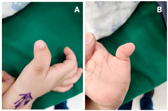
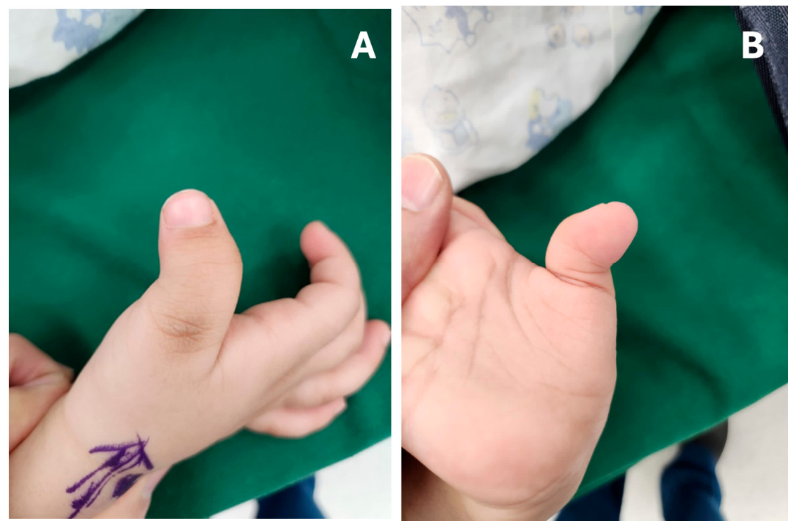
This 3-year-old girl had no known genetic disorder or long-term health issue. The patient presented to our pediatric orthopedic clinic, and the family reported concerns regarding the abnormal shape of both thumbs, first noticed when she was one year old. The patient’s bilateral thumb deformity progressed over time, making it difficult to actively flex, oppose, or pinch using her thumbs. Figure 1 illustrates hyperextension at the interphalangeal joints and radial deviation in both thumbs.
Figure 1.
(A,B) Clinical photographs demonstrating hyperextension at the thumb interphalangeal joints and radial deviation of both thumbs.
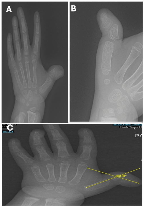
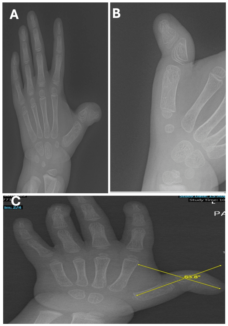
Radiographs showed subluxation of the thumb interphalangeal joint on both sides, a nearly complete epiphyseal bracket of the proximal phalanx, and a C-shaped epiphysis. In the proximal phalanx, the epiphysis ossified in a proximal-to-distal direction. The lateral deviation angle was defined as the angle between the longitudinal axes of the distal thumb phalanx and the first metacarpal. The preoperative deviation measured approximately 63° in the left thumb and 65° in the right thumb (Figure 2). The findings were considered consistent with a stage III longitudinal epiphyseal bracket bilaterally in the four-stage radiographic scheme summarized by Choo and Mubarak [2]. There was no family history of similar digital deformities, and no other anomalies or syndromic features were identified on clinical assessment.
Figure 2.
Preoperative radiographs demonstrating longitudinal epiphyseal bracket deformity of the thumbs: (A) anteroposterior (AP) view of the left hand, including the left thumb; (B) focused AP-to-slightly oblique view of the right thumb and first ray, showing subluxation at the thumb interphalangeal joint, the C-shaped bracket epiphysis, and a delta-shaped proximal phalanx; (C) AP view showing measurement of the preoperative radial lateral deviation angle (63.8°).
2.1. Surgical Procedure
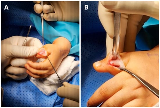
Preoperative antimicrobial prophylaxis consisted of intravenous cefazolin 500 mg (30 mg/kg), administered 30 min before skin incision. The operation was performed under general anesthesia with the patient in the supine position. We began with the right thumb. An incision was made on the ulnar aspect of the thumb at the dorsal edge of the flexion crease (Figure 3).
Figure 3.
(A,B) Intraoperative photographs of the right thumb showing the ulnar-sided incision and surgical exposure.
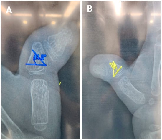
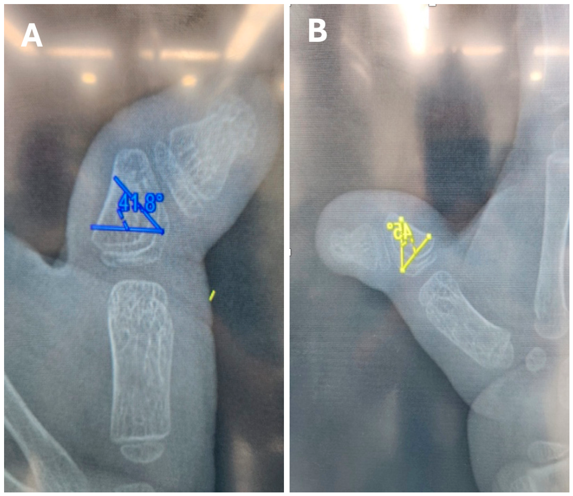
The incision was deepened to expose the proximal phalanx; the periosteum was identified; and the neurovascular structures were protected during deeper dissection (Figure 4).
Figure 4.
(A,B) Preoperative radiographic planning images illustrating angular deformity and the intended correction; panel (A), left thumb; panel (B), right thumb.
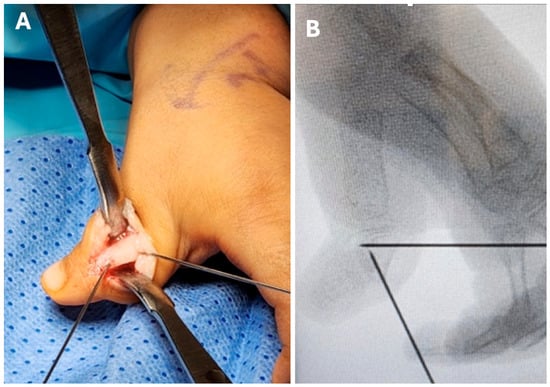
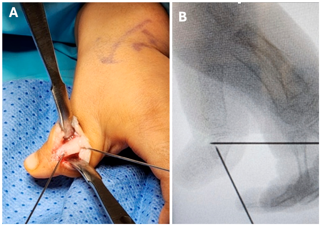
Based on preoperative X-ray planning and the estimation of angulation deformity, two K wires were inserted on the intended osteotomy site (Figure 5).
Figure 5.
(A,B) Intraoperative images of the right thumb showing localization of the planned osteotomy.
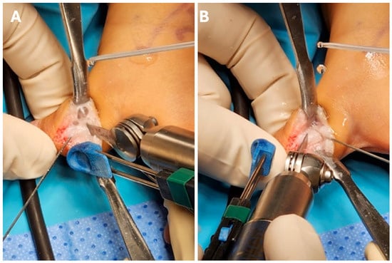
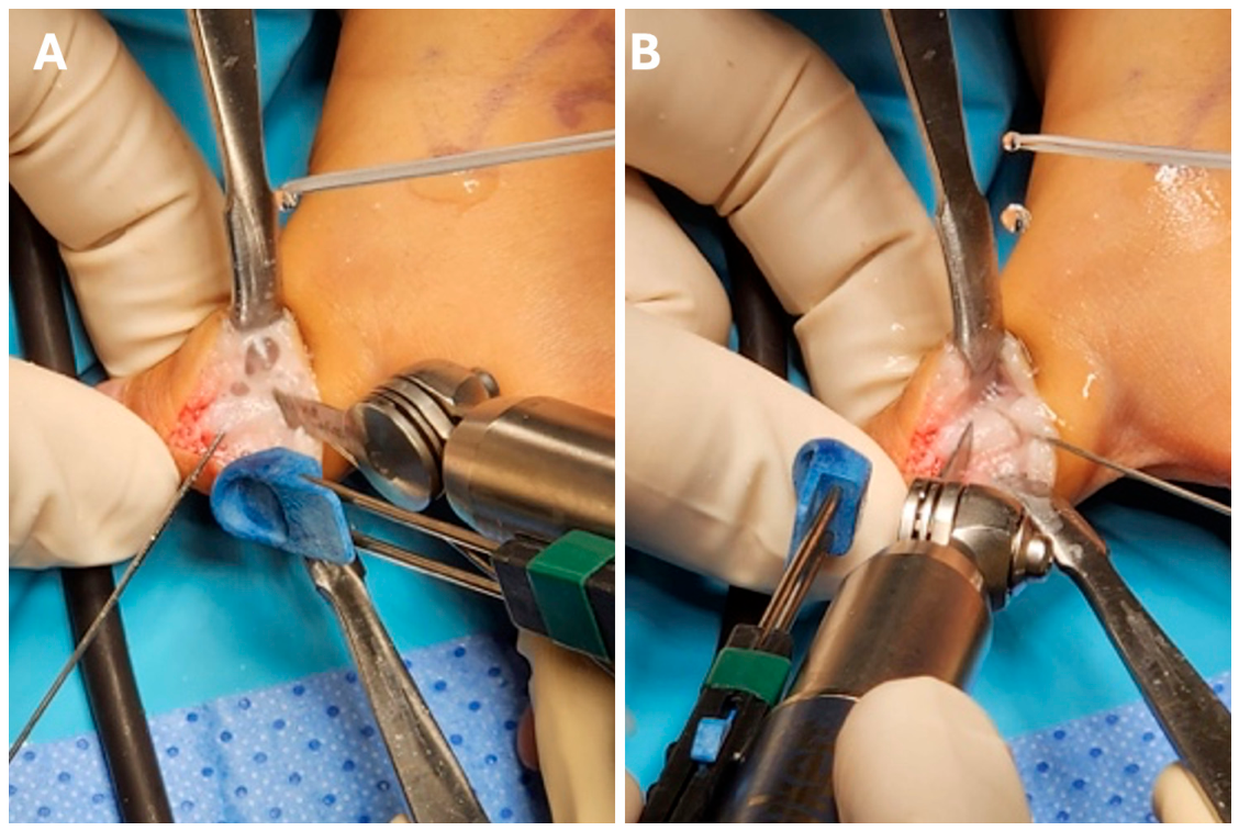
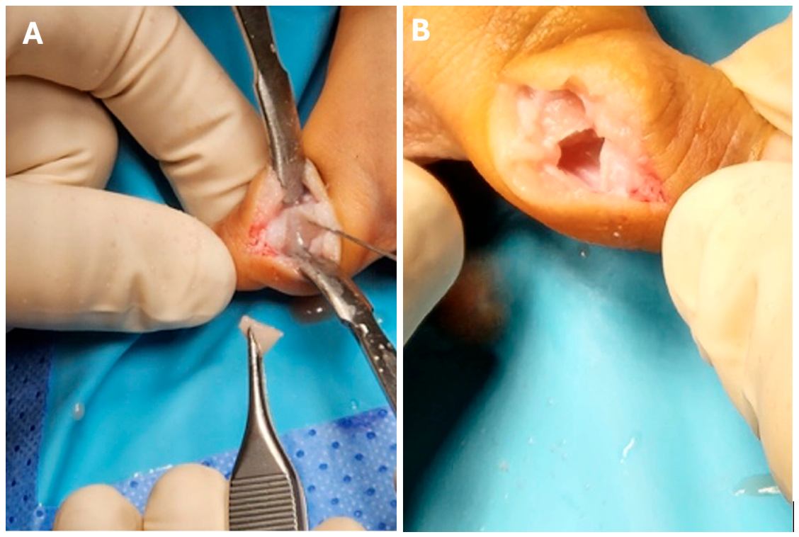
The osteotomy was planned with preoperative radiographs, and two Kirschner wires were placed as guides; intraoperative fluoroscopy was used to confirm their position relative to the ends of the longitudinal bracket (Figure 5). A powered saw was used to remove a bone wedge from the proximal thumb phalanx (Figure 6). The closing-wedge osteotomy was performed in the proximal phalanx of the thumb at the planned site localized fluoroscopically between the proximal and distal extent of the longitudinal epiphyseal bracket. An ulnar-based bone wedge was fashioned, with the osteotomy cuts oriented perpendicularly as planned to correct the radial lateral-deviation angle while avoiding disruption of the adjacent metacarpophalangeal and interphalangeal articular surfaces. Following resection of the planned bone wedge, the osteotomy was closed by approximating the opposing osseous surfaces to achieve correction of the angular deformity. The corrected alignment was subsequently stabilized using transphalangeal Kirschner-wire (1.0 mm) fixation, with no residual osteotomy gap at definitive fixation.
Figure 6.
(A,B) Intraoperative images of the right thumb showing removal of the bone wedge during the closing-wedge osteotomy.
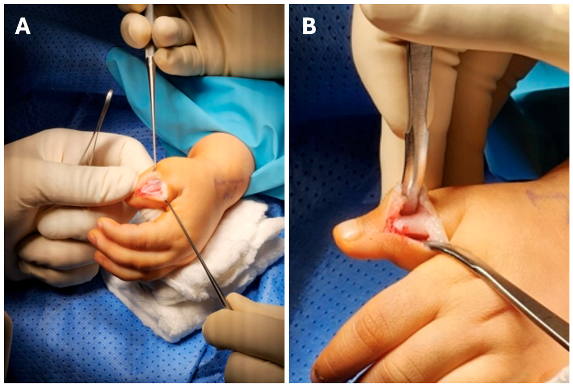
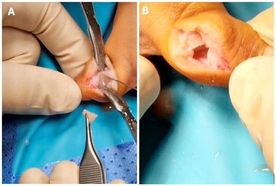
Next, we performed physiolysis on the radial side of the phalanx using the osteotomy window (Figure 7).
Figure 7.
(A,B) Intraoperative images of the right thumb demonstrating limited physiolysis through the osteotomy window.
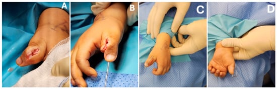
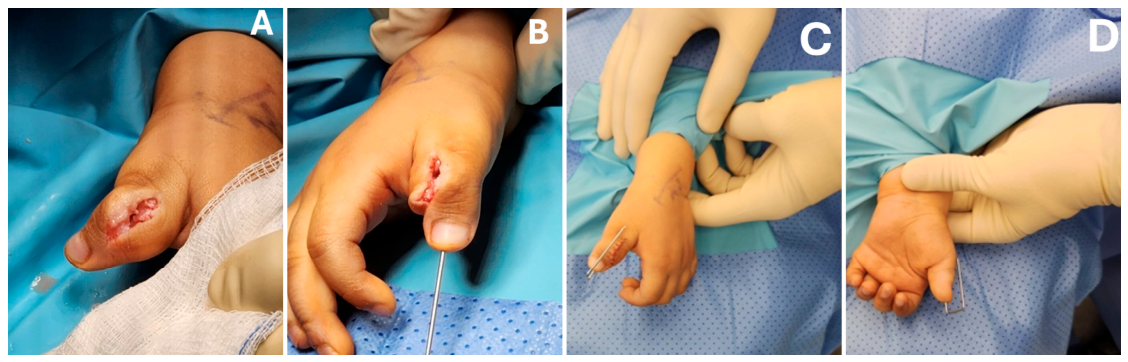
Limited physiolysis was performed on the radial side of the proximal phalanx through the osteotomy window (Figure 7). The planned correction and extent of bone/physeal resection were estimated from preoperative radiographs and the angular deformity (approximately 63° in the left thumb and 65° in the right thumb). Intraoperative fluoroscopy guided the placement of localization K-wires and confirmed the proximal and distal boundaries of the longitudinal, C-shaped epiphyseal bracket. Within these localized boundaries, a limited section of the abnormal epiphysis and physeal-cartilage bridge was removed using a 2-mm curette or fine rongeur. This targeted approach was intended to minimize unnecessary disturbance of the remaining physis and adjacent articular surface; the exact depth of excision was not recorded. No fat, muscle, polymethylmethacrylate (PMMA), bone graft, or other interposition material was placed within the physiolysis defect following excision of the abnormal physeal bridge; this defect was left unfilled. The separate closing-wedge osteotomy was nevertheless definitively closed by approximation of its cut osseous surfaces, with alignment maintained by transphalangeal Kirschner-wire fixation. Definitive fixation was achieved with one Kirschner wire per thumb, introduced retrograde from the distal phalanx across the interphalangeal joint into the proximal phalanx (Figure 8).
Figure 8.
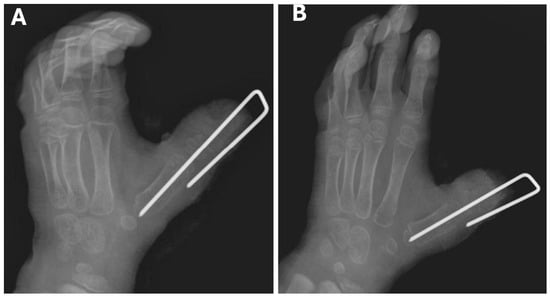
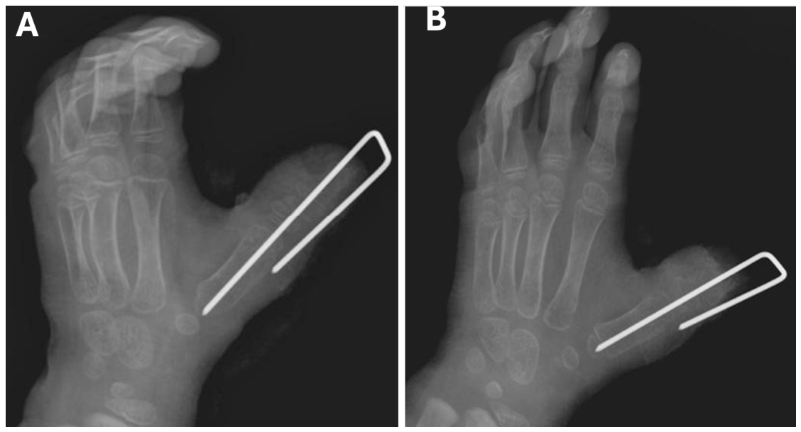
(A–D) Intraoperative photographs of the right thumb showing final alignment and Kirschner-wire fixation immediately after fixation.
After phalanx alignment was corrected, the osteotomy surfaces were approximated to close the wedge and secured with trans-phalangeal Kirschner-wire fixation (Figure 8). Final inspection showed a properly aligned phalanx without additional angulation or displacement. The operated thumb was dressed with soft bandages. The left thumb underwent the same procedure as the right thumb.
2.2. Follow-Up
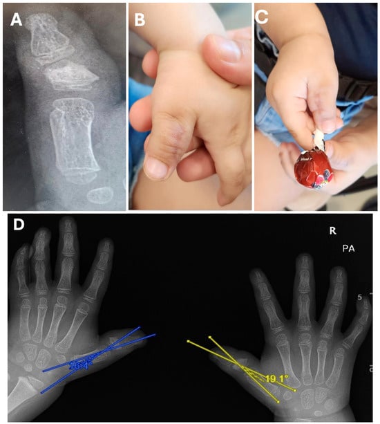
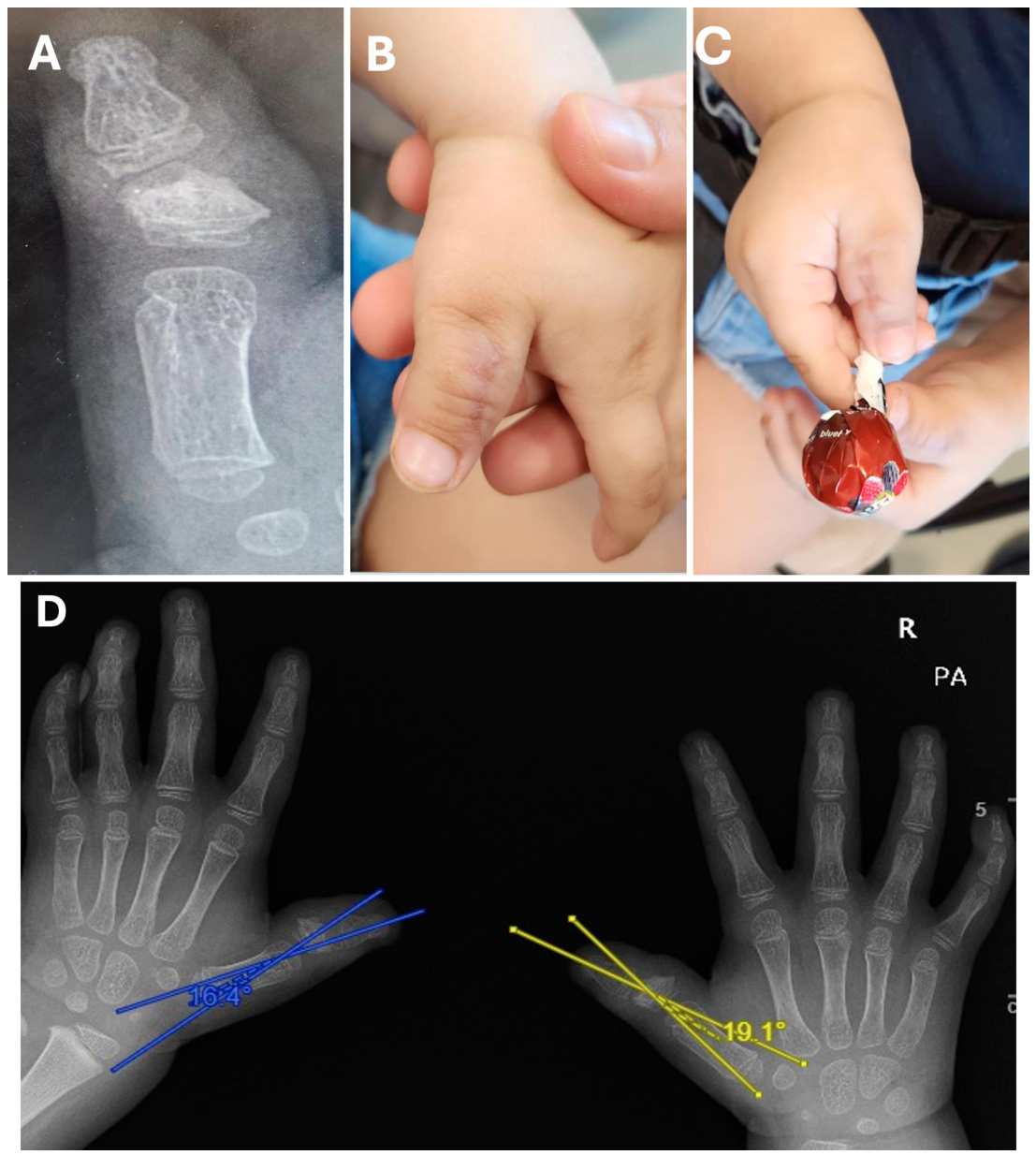
Postoperative follow-up was performed at three weeks (Figure 9), six weeks, and three months (Figure 10), the last recorded visit. Both Kirschner wires and the sutures were removed three weeks after surgery. Mobilization commenced after K-wire removal at three weeks following physiotherapy referral, with assisted, passive, and active range-of-motion exercises. Using the angle between the longitudinal axes of the thumb distal phalanx and first metacarpal, deviation improved from approximately 63° to 16.4° on the left and from approximately 65° to 19.1° on the right on the three-month postoperative radiographs (Figure 10D). At the final three-month clinical assessment, thumb movement and object grasp were observed clinically, and the parents reported satisfactory hand use and appearance. Interphalangeal range of motion, opposition, and pinch were not assessed with recorded quantitative measurements or a standardized functional instrument.
Figure 9.
(A,B) Postoperative follow-up radiographs at approximately three weeks (following removal of the Kirschner wire and sutures); panel (A), left thumb; panel (B), right thumb.
Figure 10.
(A–C) Three-month postoperative images of the right thumb: (A) radiograph; (B) clinical photograph; (C) clinical photograph illustrating observed object grasp. (D) Three-month postoperative radiographs of both thumbs with angular measurements.
3. Discussion
Physiolysis has been advocated as an early option for selected longitudinal epiphyseal brackets while growth remains [2,7]. In the present report, the term stage III refers to the four-stage radiographic developmental scheme of longitudinal epiphyseal brackets summarized by Choo and Mubarak [2], in which the longitudinal bracket epiphysis is ossified along one side of the diaphysis. Evidence specifically addressing operative management of thumb longitudinal epiphyseal brackets remains limited, with small and heterogeneous clinical series. Surgical reconstruction may be required to achieve a stable and functional thumb. Reported osteotomy techniques include opening-wedge, closing-wedge, and reverse-wedge osteotomies and, separately, dome-shaped osteotomy; physiolysis is a distinct growth-plate procedure [5,8,9]. Physiolysis aims to release the longitudinal growth restriction and permit remodeling in a growing child [2,10].
A non-systematic search of Pub-Med-indexed publications available up to 5 November 2026 was performed using combinations of the terms: “longitudinal epiphyseal bracket”, “delta phalanx”, “thumb”, “physiolysis”, and “osteotomy”, and a review of the references cited in relevant papers was conducted. Of the bibliography, seven primary clinical reports were chosen for discussion, including syndromic delta phalanges, congenital ulnar-deviated thumbs, triphalangeal thumbs and thumb duplication with epiphyseal brackets. The seven selected thumb-related reports and the present case are summarized in Table 1. For broader context, a systematic review of congenital clinodactyly conducted in 2026 found eight studies of surgery for congenital clinodactyly, including 119 patients and 165 digits, but the population was not restricted to thumb epiphyseal brackets and no uniform criteria for selection for physiolysis or osteotomy were found [8].
Caouette-Laberge and colleagues examined thirty-five fingers that underwent physiolysis. The average degree of correction attained at a follow-up of 3.2 years was 11.1 degrees. Compared to older children (6.5°), children who underwent surgery before the age of six had a mean degree of correction that was higher (17.9°). Two cases of premature fusion of the proximal transverse physis were reported among ten reoperated fingers in that series [10]. The degree of severe phalanx deformity present before surgery and the patient’s age at the time of the procedure determines whether or not surgery is indicated [7]. According to studies, surgery may be an effective potential treatment for the deformity. Even so, a chance of some residual deformity exists [8]. A. Jain, S. Rehman and G. Smith reported five of thirteen thumbs with residual radial deviation following corrective osteotomy, all of which were in the dome-osteotomy group [9]. No validated preoperative threshold predicting recurrence is established by these studies. A recent review showed a variety of operative indications, with some studies defining severe deformity as a deviation of more than 20–25° rather than just as a criterion for intervention [8].
Wood and Rubinstein reported surgery on 35 thumbs in 20 patients with Rubinstein–Taybi syndrome; the preferred surgical procedure was closing-wedge osteotomy, typically combined with Z-plasty, and eight (possibly eleven) thumbs developed recurrence of angulation or stiffness [11]. They preferred early correction before the age of 2 years, in opposition to a suggested blanket recommendation to delay surgery beyond the age of 3 years [11]. Other authors have described early physiolysis and later or primary corrective osteotomy, but the choice of procedure should be tailored to the growth potential, deformity and functional impairment [2,7,8,9]. The 63° and 65° deviations in this patient are marked angulation used descriptively, not as a validated treatment cutoff; there is no unique numeric threshold that defines advanced angular deformity [8].
In the present patient, the deformity was consistent with stage III longitudinal epiphyseal bracket of both thumbs. A combined closing-wedge osteotomy and limited physiolysis was chosen because the child was 3 years old and had severe angular deformity (approximately 63° on the left and 65° on the right), which was considered unlikely to be corrected adequately by physiolysis alone. The closing-wedge osteotomy provided immediate restoration of mechanical alignment, while the limited physiolysis was intended to address the abnormal longitudinal physeal tether. The authors considered this approach less technically demanding and not requiring bone grafting; however, these practical considerations should be interpreted cautiously because they are based on a single case. At six weeks, both thumbs were described as stable and better aligned. At three months, the deviation angles measured 16.4° on the left and 19.1° on the right. At the final documented clinical assessment at three months, thumb movement and object grasp were observed clinically, and the parents reported satisfaction with hand use and appearance; objective range-of-motion, opposition, and pinch measurements were not recorded.
Recent literature provides additional context: Han and colleagues evaluated surgically treated congenital ulnar-deviated thumbs [12], while Vanhoenacker and colleagues reviewed congenital and developmental abnormalities of the hand and wrist [13]. More recent reports address longitudinal epiphyseal brackets in hallux varus [14], bilateral delta-phalangeal clinodactyly [15], soft-tissue reconstruction during correction of hand clinodactyly [16], and first-metatarsal bracket deformities [17]. Long-term observation of another clinodactyly phenotype has also been reported [18]. A case report described closing-wedge osteotomy for a triphalangeal thumb with a small delta phalanx [19], and a 2026 series described early treatment of congenital thumb duplication accompanied by longitudinal epiphyseal bracket [20]. These reports involve differing presentations and techniques and therefore provide context rather than direct comparative evidence for the present operation.
Table 1.
Published thumb-related clinical reports and series compared with the present case.
Limitations
This is a single-patient report with a short period of radiographic and clinical follow-up (three months). There were no recorded quantitative preoperative or postoperative measurements of thumb interphalangeal range of motion, opposition, or pinch, and no standardized functional instrument or formal cosmetic score was administered. Observed thumb movement and object grasp, illustrated in Figure 10C, are qualitative clinical observations; satisfaction with hand use and appearance reflects parental report. No objective comparison of functional gain is possible. The available follow-up is insufficient to determine whether longitudinal growth is preserved, whether partial physeal arrest develops, or whether angular deformity recurs. Longer-term radiographic and clinical surveillance is required.
4. Conclusions
Longitudinal epiphyseal brackets are uncommon growth-plate abnormalities that may cause progressive thumb deformity and functional impairment. In this case, bilateral closing-wedge osteotomy with limited physiolysis was associated with satisfactory early alignment and parent-reported improvement in hand use and appearance. This single case does not establish a general age-based recommendation. Continued follow-up is required to evaluate growth, recurrent angulation, and physeal status.
Author Contributions
Writing—original draft preparation, M.S.A. (Introduction), A.M.A. (Case Presentation), A.A.A. (Case Presentation) and M.J.A. (Discussion); writing—review and editing, A.N.B.; supervision, A.N.B. All authors have read and agreed to the published version of the manuscript.
Funding
This research received no external funding.
Institutional Review Board Statement
Ethical review and approval for this study were not required by the Institutional Review Board of Almoosa private hospital because case reports are not considered research in their jurisdiction.
Informed Consent Statement
Written informed consent has been obtained from the patient to publish this paper.
Data Availability Statement
The original contributions presented in this study are included in the article. Further inquiries can be directed to the corresponding author.
Conflicts of Interest
The authors declare no conflicts of interest.
References
- Patel, N.K.; Toyoda, Y.; Grunzweig, K.A.; Shah, A.S.; Mendenhall, S.D. Recent advancements in the diagnosis and treatment of congenital hand differences. J. Am. Acad. Orthop. Surg. 2023, 31, 766–782. [Google Scholar] [CrossRef] [Scilit] [PubMed]
- Choo, A.D.; Mubarak, S.J. Longitudinal epiphyseal bracket. J. Child. Orthop. 2013, 7, 449–454. [Google Scholar] [CrossRef] [Scilit] [PubMed]
- McCarter, J.H.; Zeledon, R.A.; Cole, S.H.; Layon, S.A.; Nguyen, J.L. Common pediatric hand anomalies. Semin. Plast. Surg. 2023, 37, 275–286. [Google Scholar] [CrossRef] [Scilit] [PubMed]
- Minkowitz, B.; Spingarn, C.M.; Bertsch, A.J.; Fekete, J.M.; Rienzo, J.A.; Davis, E.D.; Shah, N.; Mubarak, S.J. Physiolysis for metatarsal bracketed epiphysis. J. Pediatr. Orthop. Soc. N. Am. 2023, 5, 773. [Google Scholar] [CrossRef] [Scilit] [PubMed]
- Alelyani, R.H.; Moazin, O.M.; Alazzmi, H.A.; Alfadhel, E.A.; Alobaidi, H.; Alaqil, S.; Bhat, T.A.; Nawwab, E.; Mirza, M.Y. Unusual presentation of familial unilateral postaxial polydactyly with metacarpal delta phalanx: A report of a rare case. Cureus 2023, 15, e49484. [Google Scholar] [CrossRef] [Scilit] [PubMed]
- Sferopoulos, N.K. Congenital clinodactyly of the thumb. Br. J. Med. Med. Res. 2014, 4, 2874–2883. [Google Scholar] [CrossRef] [Scilit]
- Iba, K.; Wada, T.; Yamashita, T. Correction of thumb angulations after physiolysis of delta phalanges in a child with Rubinstein–Taybi syndrome: A case report. Case Rep. Plast. Surg. Hand Surg. 2015, 2, 12–14. [Google Scholar] [CrossRef] [Scilit] [PubMed]
- Devetzis, K.; Beni, R.; McGhee, J.; To, E.Y.H.; Lam, W.L.; Horwitz, M.D. Outcomes of the surgical management of clinodactyly: A systematic review. J. Hand Surg. Am. 2026. advance online publication. [Google Scholar] [CrossRef] [Scilit] [PubMed]
- Jain, A.; Rehman, S.; Smith, G. Long-term results following osteotomy of the thumb delta phalanx in Rubinstein–Taybi syndrome. J. Hand Surg. Eur. 2010, 35, 296–301. [Google Scholar] [CrossRef] [Scilit] [PubMed]
- Caouette-Laberge, L.; Laberge, C.; Egerszegi, E.P.; Stanciu, C. Physiolysis for correction of clinodactyly in children. J. Hand Surg. Am. 2002, 27, 659–665. [Google Scholar] [CrossRef] [Scilit] [PubMed]
- Wood, V.E.; Rubinstein, J.H. Surgical treatment of the thumb in the Rubinstein–Taybi syndrome. J. Hand Surg. Br. 1987, 12, 166–172. [Google Scholar] [CrossRef] [Scilit] [PubMed]
- Han, B.; Shen, K.; Wang, Z.; Xu, Y. Clinical characteristics and surgical outcomes of congenital ulnar-deviated thumbs: Delta triphalangeal thumbs and irregular epiphyses. J. Pediatr. Orthop. 2021, 41, 28–32. [Google Scholar] [CrossRef] [Scilit] [PubMed]
- Vanhoenacker, F.M.; Simoni, P.; Bosmans, F.; Boutry, N. Congenital and developmental abnormalities of the hand and wrist. Semin. Musculoskelet. Radiol. 2021, 25, 260–271. [Google Scholar] [CrossRef] [Scilit] [PubMed]
- Kadıoğlu, B.; Güven, M.; Akman, B.; Özkan, N.K. Longitudinal epiphyseal bracket as an overlooked etiology of congenital-residual hallux varus: A case report. J. Am. Podiatr. Med. Assoc. 2021, 111, 20021. [Google Scholar] [CrossRef] [Scilit] [PubMed]
- Afshar, A.; Tabrizi, A. Bilateral clinodactyly of the index and middle fingers because of proximal delta phalanx. World J. Plast. Surg. 2022, 11, 95–97. [Google Scholar] [CrossRef] [Scilit] [PubMed]
- Kaempf, R.; Brunelli, J.P.F.; Aita, M.A.; Delgado, P.J.; Soldado, F.; Vergara, E. Bilobed flap in hand clinodactyly reconstruction: Technique description and result appraisal. Rev. Bras. Ortop. 2022, 57, 642–648. [Google Scholar] [CrossRef] [Scilit] [PubMed]
- Marcarelli, M.; Errichiello, C.; Germano, M.; Risitano, S.; Deledda, D.; Thomas, M. Longitudinal epiphyseal bracket of the first metatarsal: A case series on treatment of deformities associated. J. Am. Podiatr. Med. Assoc. 2022, 112, 21076. [Google Scholar] [CrossRef] [Scilit] [PubMed]
- Teodonno, F.; Maffeis, J.; Guero, S. Spontaneous improvement of bilateral clinodactyly of the little finger with trapezoidal phalanx: A case report with a 15-year follow-up. J. Hand Surg. Eur. 2024, 49, 106–108. [Google Scholar] [CrossRef] [Scilit] [PubMed]
- Okhovatpour, M.A.; Adibi, A. Correction of triphalangeal thumb with small delta phalanx by closed wedge osteotomy of the proximal phalanx: A case report. J. Orthop. Spine Trauma 2024, 10, 124–126. [Google Scholar] [CrossRef] [Scilit]
- Liao, W.; Yin, Z.; Liu, L.; Zheng, P. Early surgical correction of congenital thumb duplication combined with longitudinal epiphyseal bracket: A prospective case series of 15 infants and toddlers. J. Orthop. Surg. Res. 2026, 21, 645. [Google Scholar] [CrossRef] [Scilit] [PubMed]
Disclaimer/Publisher’s Note: The statements, opinions and data contained in all publications are solely those of the individual author(s) and contributor(s) and not of MDPI and/or the editor(s). MDPI and/or the editor(s) disclaim responsibility for any injury to people or property resulting from any ideas, methods, instructions or products referred to in the content. |
© 2026 by the authors. Licensee MDPI, Basel, Switzerland. This article is an open access article distributed under the terms and conditions of the Creative Commons Attribution (CC BY) license.