


Abstract
The Tau protein is traditionally recognized for stabilizing axonal microtubules in neurons. However, emerging evidence indicates that astrocytes also show basal expression of the microtubule-associated protein Tau (MAPT) gene, with potential implications for central nervous system (CNS) homeostasis. Under physiological conditions, astrocytes support functions essential to CNS integrity, including metabolic support, neurotransmitter uptake, blood–brain barrier maintenance, and immune surveillance, all of which may be influenced by endogenous Tau expression. This review examines the current literature on the mechanisms that govern astrocytic Tau regulation, mainly in primary tauopathies with glial cytopathology. Available data indicate that Tau accumulation in these glial cells arises from endogenous synthesis, uptake from neighboring neurons, or a combination of these pathways, while failed extracellular clearance may favor this process by increasing Tau availability in the extracellular space. Once internalized, Tau can be processed, degraded through proteolytic and autophagic-lysosomal pathways, or re-released into the extracellular space, potentially facilitating its propagation; insufficient degradation, in contrast, promotes reactive astrogliosis and disrupts vital processes such as metabolic support and neurotransmitter handling. Taken together, this evidence suggests that distinguishing endogenous synthesis from neuronal uptake is essential to define glial-mediated pathology and recognize astrocytic Tau as a relevant contributor to neurodegenerative disease progression.
1. Introduction
Two main cell classes are recognized in the central nervous system (CNS): neurons and glia. The complexity of the CNS depends on the complementary interaction between neurons and glial cells. Both cell types have co-evolved to support the increasing structural, metabolic, and functional demands of the CNS [1]. Among glial cells, astrocytes and microglia are the best characterized. Astrocytes, in particular, perform functions such as neurotransmitter recycling (glutamate, GABA, and glycine), lipid metabolism, immune defense, and structural support [2]. They also help maintain the blood–brain barrier by modulating extracellular ionic homeostasis through their peri-vascular terminal processes, also known as astrocytic perivascular end-feet, which interact with endothelial cells, pericytes, and the basement membrane [3]. Microglia, in turn, are the immune cells of the CNS. They respond to disturbances of the brain parenchyma, such as neuronal death, tissue damage, accumulation of misfolded proteins, and ischemia, changing their morphology and functional state as they proliferate and migrate toward the site of injury through the expression of inflammatory mediators such as the interleukins IL-6 and IL-1β and tumor necrosis factor α (TNF-α) [4]. Several pathological and structural proteins disrupt glial cell function (astrocytes and microglia), including alpha-synuclein, Tau protein, amyloid-beta peptide, and mutated glial fibrillary acidic protein (GFAP) [5,6]. Particularly, Tau protein plays a significant role in astrocytes, as its accumulation in these cells is implicated in disruptions to glutamate regulation, metabolic support, proteostasis, the extracellular microenvironment, and functional communication with neurons [5,6].
The Tau protein was first described in the 1970s by Marc Kirschner’s group [7]. During neuronal development, Tau is distributed throughout the soma and dendrites; however, as the neuron matures, Tau becomes localized to the axon, where it binds directly to tubulin dimers and regulates microtubule assembly, stability, and dynamics [8,9,10]. It was initially regarded as a structural protein of the neuronal cytoskeleton, but was later identified as the main component of neurofibrillary tangles (NFTs) in Alzheimer’s disease (AD) [11]. This finding contributes to recognizing tauopathies as a growing and heterogeneous class of neurodegenerative diseases. Tau is now recognized as an amyloidogenic protein, that is, a protein capable of shifting from its soluble conformation to an abnormal, β-sheet–rich conformation. This structure can bind identical molecules and form aggregates that progress from monomer to oligomer, from oligomer to protofibril, from protofibril to amyloid fibril, and finally to an amyloid deposit [12]. Amyloidogenic proteins play an important role in tauopathies, a group of neurodegenerative diseases characterized by abnormal post-translational modifications, aberrant folding, aggregation, and intracellular accumulation of Tau [13,14,15].
Tauopathies are classified into three main groups according to the type of cell affected in the CNS: neuronal predominance, glial predominance (astrocytes and oligodendrocytes), and mixed (neuronal and glial), although the cell with the greatest pathological burden does not necessarily represent the cell that initiates the process [16]. Furthermore, according to the pathogenic role of the Tau protein, tauopathies are classified as primary and secondary. In primary tauopathies, Tau constitutes the central molecular lesion, and glial involvement may be prominent; Thus, progressive supranuclear palsy and corticobasal degeneration present neuronal inclusions along with specific astrocytic and oligodendroglial phenotypes, while globular glial tauopathy (GGT) represents the most characteristic example of a primary glial-predominant tauopathy [17]. In contrast, secondary tauopathies correspond to those in which Tau deposits occur as a phenomenon associated with another abnormality or an initial driving protein (such as amyloid in AD).
Tau expression has been studied almost exclusively from a neuronal perspective. This view has begun to change; however, considering evidence that astrocytes also endogenously express the microtubule-associated protein Tau (MAPT) gene [18,19]. The presence of Tau in glial cells, including gliomas and astrocytic tumor cells, indicates that its expression is not limited to neurons, although these models reflect a pathological context distinct from that of mature astrocytes under physiological conditions [20]. Current evidence indicates that astrocytes express MAPT basally, and it remains unclear whether Tau accumulation arises from endogenous MAPT expression, from the uptake of extracellular Tau released by neurons, or from a combination of both mechanisms [4,18,21]. This shift in how Tau is viewed broadens the picture and raises the possibility that astrocytes contribute to the accumulation, processing, and propagation of pathological Tau species.
Although Tau has been detected in astrocytes, the processes by which these cells process, clear, or propagate the protein remain incompletely understood, as do the cellular mechanisms underlying its astrocytic accumulation. It is therefore necessary to examine the available evidence on endogenous Tau expression in astrocytes; the mechanisms involved in the uptake, processing, clearance, and propagation of extracellular Tau; and the functional consequences of Tau accumulation in these glial cells.
2. The Tau Protein and Its Dynamic Binding to Microtubules
The MAPT gene, located on chromosome 17q21.31, encodes the Tau protein [22,23]. In the adult human CNS, alternative splicing of exons 2, 3, and 10 gives rise to six main protein isoforms: 0N3R, 0N4R, 1N3R, 1N4R, 2N3R, and 2N4R [24,25,26] (Figure 1). The 0N, 1N, and 2N nomenclature refers to the number of amino-terminal inserts (N) present: the 0N isoforms contain none of these inserts, whereas the 1N isoforms include a 29-amino-acid insert encoded by exon 2. The 2N isoforms, in turn, contain the inserts encoded by exons 2 and 3; they therefore carry two N-terminal inserts, adding a total of 58 amino acids to Tau [24,25,26].
Figure 1.
Alternative splicing of MAPT and structural organization of the six Tau isoforms expressed in the adult human brain. (A) Schematic representation of MAPT exons, highlighting exons 2, 3, and 10, which undergo alternative splicing. The exons are not shown to scale. (B) Inclusion or exclusion of exons 2 and 3 generates Tau isoforms with zero, one, or two N-terminal insertions (0N, 1N, and 2N), while alternative splicing of exon 10 produces isoforms containing three or four microtubule-binding repeats (3R and 4R). (C) Tau structural domains showing the PHF6* motif (VQIINK, residues 275–280) within R2 and the PHF6 motif (VQIVYK, residues 306–311) within R3. PHF6 is present in all six isoforms, while PHF6* is restricted to the 4R isoforms, because R2 is encoded by exon 10. For clarity, only selected exons relevant to the 0N/1N/2N and 3R/4R classification are represented in panels (A) and (B). Residue numbering in panel C corresponds to the longest human Tau isoform, 2N4R (441 amino acids). Created with BioRender.
The 3R or 4R designation indicates the number of carboxy-terminal repeats (R) that make up the microtubule-binding domain. Exclusion of exon 10 produces the 3R isoforms, composed of repeats R1, R3, and R4, whereas its inclusion produces the 4R isoforms, which contain repeat R2 in addition to R1, R3, and R4. These structural differences can alter Tau’s interactions with tubulin and microtubules, affecting its ability to promote assembly and regulate the stability and dynamics of these structures. Alterations in the 3R and 4R isoforms are therefore linked to the tauopathies [24,26].
Structurally, the longest human Tau isoform, 2N4R, comprises 441 amino acids. It is a highly soluble, intrinsically disordered protein whose sequence is organized into four main functional regions: the N-terminal region, the proline-rich region, the microtubule-binding domain, and the C-terminal region. The N-terminal end participates in Tau interaction with the cell membrane, whereas the proline-rich region contains several phosphorylation sites that, together with the microtubule-binding domain, contribute to its interaction with tubulin and the microtubule surface [27,28].
The R1–R4 repeats of the binding domain participate in the assembly and regulation of microtubule stability through continuous cycles of association and dissociation [27,28,29]. This binding is not permanent: each Tau molecule associates transiently with the microtubule surface through noncovalent, reversible interactions with the repeated tubulin regions distributed along the protofilaments. Tau remains bound to a given site for only a few milliseconds and can then move by diffusion to bind another nearby site on the same microtubule or on an adjacent one [30]. A single Tau molecule can interact with roughly 20–30 different sites per second, an interaction known as “kiss-and-hop” [30,31].
The duration of these interactions also depends on Tau phosphorylation. The addition of phosphate groups in regions close to the binding domain can reduce its affinity and shorten its binding time to the microtubule, whereas dephosphorylation, mediated mainly by protein phosphatase 2A (PP2A), favors its interaction with tubulin [32]. This exchange allows Tau to modulate microtubule polymerization and dynamics without hindering the movement of kinesins and dynein during axonal transport. Transient interaction enables Tau to promote the incorporation of α/β-tubulin dimers and to modulate the growth and shortening phases of microtubules without remaining continuously anchored to their surface.
Most of the available information on the dynamic interaction between Tau and microtubules comes from studies in neurons, particularly on axonal microtubules. In astrocytes, which lack an axonal compartment, the subcellular distribution and binding dynamics of endogenous Tau to microtubules are even less well characterized. However, astroglial Tau pathology exhibits a disease-specific intracellular distribution. In progressive supranuclear palsy, phosphorylated Tau 4R accumulates mainly in the soma and proximal processes of tufted astrocytes, whereas in corticobasal degeneration, it is predominantly located in distal astroglial processes, where it forms astrocytic plaques. These patterns suggest that cytoskeletal organization and intracellular transport in astrocytes may influence the localization of Tau deposits; however, direct differences in Tau-microtubule binding dynamics between astrocytes and neurons have not yet been demonstrated [33].
When post-translational modifications or conformational changes persistently reduce Tau interaction with tubulin and microtubules, the fraction of protein not associated with the cytoskeleton increases. Under certain conditions, this cytosolic Tau can expose regions with amyloidogenic potential, such as PHF6 and PHF6*, and interact with other Tau molecules, favoring the formation of oligomeric and fibrillar species [15,34]. These pathological forms can disrupt microtubule organization, intracellular transport, mitochondrial function, and synaptic communication. Within the microtubule-binding domain lie two sequences with a high intrinsic propensity to adopt β-sheet conformations: PHF6, composed of valine-glutamine-isoleucine-valine-tyrosine-lysine (VQIVYK, residues 306–311), located at the start of R3 and present in both the 3R and 4R isoforms; and PHF6*, composed of valine–glutamine–isoleucine–isoleucine–asparagine–lysine (VQIINK, residues 275–280), located at the start of R2 and present in the 4R isoforms [35,36].
Exposure of these motifs promotes interactions between Tau molecules, facilitating initial nucleation and subsequent assembly into oligomers and paired helical filaments (PHFs). The importance of these regions has been assessed using truncated Tau constructs; under these experimental conditions, retention of R2 or R3, together with their respective hexapeptides, allowed aggregate formation, whereas a short sequence such as VQI was insufficient to confer this amyloidogenic capacity [37]. In addition, all six recombinant human Tau isoforms formed oligomers and filaments that were morphologically similar to those observed in tauopathies. To this end, the aggregation kinetics of the 0N3R, 1N3R, and 2N3R isoforms were compared with those of the 0N4R, 1N4R, and 2N4R isoforms, and aggregation was monitored using thioflavin T fluorescence. Under these experimental conditions, the 3R isoforms aggregated faster than the 4R isoforms. Although R2 contains the PHF6* motif and can contribute to Tau nucleation, the initial stage of aggregation, its presence does not necessarily accelerate aggregation of the full-length protein; however, the N-terminal regions influence the rate of this process. Once stable Tau nucleation is established, it acts as a template that facilitates the progressive incorporation of other Tau molecules, promoting the formation of oligomers, protofilaments, and, ultimately, paired helical filaments [4].
3. Post-Translational Modifications and Loss of Affinity for Microtubules
The transition of Tau toward pathological forms does not depend on a single post-translational modification or alteration, but on the combination of post-translational modifications, conformational changes, and the presence of molecules or aggregates that facilitate its assembly (Table 1).
Table 1.
Post-translational modifications of Tau and their implications in tauopathies.
Post-translational modifications of Tau also occur in astrocytes, although their characterization is less complete than in neurons. In primary 4R tauopathies, astroglial inclusions contain abnormally phosphorylated Tau 4R and acquire disease-specific morphologies, including the tufted astrocytes of progressive supranuclear palsy and the astrocytic plaques of corticobasal degeneration. Phosphorylation is the best-characterized modification in these astroglial lesions and can be detected by epitopes such as AT8, which recognizes phosphorylated Tau at Ser202 and Thr205. However, this immunoreactivity identifies a specific phosphoepitope and not the complete profile of post-translational modifications of each Tau molecule. The specific contribution of acetylation, ubiquitination, truncation, and other modifications to astroglial Tau is still not clearly defined; therefore, findings obtained in neuronal Tau should not be directly attributed to astrocytic Tau [47,48].
In particular, acetylation of lysine residues located in the fibril core (the ordered, compact region of a Tau filament) can modify the charge and the interactions between the β-strands that make up the protofilaments [49]. Ubiquitination of certain lysines, in turn, can provide additional contact points between two Tau protofilaments, favoring their association and stabilizing the packing of the fibril [43]. Because the position and orientation of these contact points vary among the tauopathies, this modification may contribute to the formation of distinct fibrillar structures in AD and in other tauopathies.
These modifications can favor the formation of soluble species, including dimers, trimers, and larger Tau oligomers, some of which show greater seeding capacity and toxicity than the filaments. Neurofibrillary tangles should therefore not be regarded as the only form of Tau responsible for cell-to-cell spreading [50,51]. As Tau becomes more insoluble, the amount of functional soluble Tau may decrease, contributing to microtubule destabilization and impaired axonal transport. However, the accumulation of insoluble Tau and neurofibrillary tangles may not be the sole or direct cause of toxicity, as soluble oligomeric Tau species, which can coexist with tangle formation, can also disrupt axonal transport and contribute to neurodegeneration [52,53].
Phosphorylation is one of the most important post-translational modifications of Tau. Phosphorylation of serine or threonine residues, located mainly in the proline-rich region and the C-terminal region, introduces negative charges that alter the electrostatic interactions of Tau and can decrease its affinity for the microtubule surface, which is also negatively charged [15,51]. As a result, the amount of Tau not bound to microtubules increases in the cytosol; however, its detachment and hyperphosphorylation are not sufficient on their own to form fibrils. In in vitro models, the formation of full-length Tau fibrils requires cofactors such as RNA, DNA, arachidonic acid, or polyphosphates, which alter internal electrostatic interactions and favor the exposure and alignment of the PHF6 and PHF6* motifs into β-sheet structures [51].
In Tau, this post-translational modification not only changes the protein’s electrical charge but also alters its molecular conformation, its affinity for other molecules, and its supramolecular organization, in which several nearby Tau molecules organize as a compact region on the microtubule [38]; here, kinases add phosphate groups to serine, threonine, and, less frequently, tyrosine residues, whereas phosphatases remove them [40,54]. Under physiological conditions, this process is dynamic and regulates Tau localization and its interaction with microtubules, contributing to the maintenance of cytoskeletal dynamics and intracellular transport [38,55,56].
Notably, an average of 1.9 mol of phosphate per mol of Tau has been estimated in the healthy human brain, whereas the Tau present in the pathological filaments of AD can contain between 6 and 8 mol of phosphate per mol of protein. This represents a three- to fourfold increase in the degree of phosphorylation, not in Tau concentration relative to normal levels [34]. When phosphorylation occurs excessively and at multiple sites, it is termed hyperphosphorylation. In AD, Tau can acquire a hyperphosphorylated state, characterized by an abnormal increase in phosphate at the various sites of the protein [38]. In the pathological state, Tau shows decreased affinity for microtubules and redistributes from the axon to the soma and cytosol, forming tangles and neuritic threads (considered swollen, Tau-laden axons with or without myelin sheaths, or other glial cytoplasmic protrusions). Once this cytosolic accumulation is established, Tau can assemble into neurofibrillary tangles and neuropil threads, Tau-positive filamentous structures located in neuronal processes [57]. Finally, these filaments cluster into neurofibrillary tangles, causing cell death (Figure 2). In addition, phosphorylation in the flexible N-terminal and C-terminal regions of the filaments, known as the fuzzy coat, not only accompanies aggregation but can also increase the capacity of pathological Tau to act as a seed and induce the aggregation of endogenous Tau in other neurons [40,58,59].
Figure 2.
Tau hyperphosphorylation decreases its affinity for microtubules. In tauopathies, the Tau protein aggregates abnormally: it begins as monomers, forms paired helical filaments (PHFs), and progresses to neurofibrillary tangles (NFTs), leading to cell death. Created with BioRender.
Phosphorylation is associated with the pathological intracellular burden and with the presence of soluble p-Tau in the extracellular space. Three-dimensional in vitro models of human nervous tissue generated from human induced pluripotent stem cells (iPSCs) enabled observation of the contributions of both neurons and astroglial-lineage cells to Tau phosphorylation and release. Amyloidogenic conditions, such as increased β-amyloid, are associated with greater Tau phosphorylation in both neurons and astrocytes, which increases the release of soluble Tau (associated with AD). This suggests that Tau hyperphosphorylation is not an exclusively neuronal phenomenon [56]. Tau hyperphosphorylation thus represents not only a biochemical change in Tau but also a process that links cytoskeletal dysfunction to protein aggregation and neurodegeneration [55].
4. Astrocytes in the CNS
Some authors suggest that astrocytes are the most abundant cell type in the adult CNS [60]. Historically, these cells were regarded as structural support for neurons and the blood–brain barrier [61]. Astrocytes help maintain CNS homeostasis by regulating neurotransmitters and extracellular ions; they are essential for supplying energy and metabolic and neurotrophic support to neurons, thereby contributing to their survival, synaptic activity, and plasticity [61]. They also possess mechanisms that enable them to recognize, internalize, and degrade cellular debris and extracellular proteins. These functions are relevant for understanding what happens when astrocytes come into contact with Tau released by neurons: uptake favors clearance of the protein, whereas accumulation favors its subsequent propagation.
4.1. Regulation of the Neuronal Microenvironment
Astrocytes participate in the development, maintenance, and remodeling of synaptic connections. During synaptogenesis, they secrete molecules such as the glypicans GPC4 and GPC6, the protein SPARCL1, and the thrombospondins THBS1 and THBS2, which favor the formation and maturation of synapses [62]. Likewise, astrocyte morphology and their association with neuronal processes are regulated by cell adhesion molecules, among them the astroglial neuroligins NL1, NL2, and NL3, which interact with the neurexins present in the neuronal membrane [63].
The development and maturation of neuronal circuits depend not only on the formation of new synapses but also on their constant remodeling. Astrocytes actively participate in this refinement, pruning synapses through phagocytosis, a process that helps maintain the circuit’s homeostasis and efficiency. At the molecular level, the capacity of astrocytes to phagocytose synaptic material and eliminate synapses is critically mediated by the expression of specific phagocytic receptors on the astroglial membrane, mainly multiple epidermal growth factor-like domains protein 10 (MEGF10) and MER tyrosine kinase (MERTK) [64]. Likewise, the classical complement cascade participates in synapse elimination and pruning; basal signaling of complement components, such as the C3/C3aR pathway, appears to play an important physiological role in maintaining normal dendritic arborization and in stimulating neurogenesis [65]. However, when this pathway is excessively or persistently activated, it can also contribute to the abnormal elimination of synaptic connections.
Astrocytes also participate in regulating cerebral blood flow, water homeostasis, and communication between the brain parenchyma and blood vessels through the distribution of the proteins AQP4 and Kir4.1 at their perivascular end-feet [66,67]. In particular, AQP4 enables water movement across the astroglial membrane, whereas Kir4.1 potassium channels participate in the uptake and redistribution of the potassium released during neuronal activity [68].
Astrocytes are a central component of the glymphatic system, a network of perivascular channels specialized in the efficient clearance of soluble proteins and metabolic byproducts from the brain’s interstitial fluid, including β-amyloid, extracellular Tau, and lactate [69,70]. They participate in both beneficial and pathological processes in AD, including cytokine secretion, β-amyloid peptide clearance, and Tau pathology; that is, these glial cells can influence how Tau accumulates, propagates, and causes damage. Astroglial autophagy can indirectly influence these functions by regulating metabolism, the inflammatory response, and the availability of membrane proteins at their end-feet [70].
4.2. Proteostasis, Phagocytosis, and Protein Clearance
Proteostasis comprises the mechanisms that regulate the synthesis, folding, transport, and clearance of proteins. In astrocytes, this function includes both the maintenance of their own proteins and organelles and the uptake of components from the extracellular space. Through endocytosis and phagocytosis, these cells can internalize synapses, dendritic fragments, cellular debris, axonal mitochondria, and protein aggregates associated with neurodegenerative diseases [39,71,72].
Endocytosis allows the internalization of small molecules and particles through invagination of the plasma membrane and the formation of intracellular vesicles. Phagocytosis, by contrast, allows larger molecules to be surrounded and internalized through membrane protrusions that depend on reorganization of the actin cytoskeleton. Although microglia are considered the professional phagocytes of the CNS, astrocytes can also remove synapses, synaptosomes, neuronal fragments, and apoptotic cell bodies [64].
The endosome-lysosome system sorts internalized molecules and recycles components back to the plasma membrane. Late endosomes fuse with lysosomes, which contain hydrolytic enzymes such as proteases and cathepsins B, D, and L, responsible for fragmenting proteins, as well as lipases, glycosidases, nucleases, phosphatases, and sulfatases, responsible for degrading proteins, lipids, and other cellular components [72,73].
With respect to Tau, cathepsins B and D participate in its lysosomal processing; however, the efficiency of degradation depends on the protein’s conformation, size, and degree of aggregation. Astrocytes can also internalize monomeric Tau, although its subsequent intracellular fate and the efficiency of its degradation have not yet been determined [73,74].
Autophagy is another degradation mechanism and is closely related to the endosomal-lysosomal system, but it is neither endocytosis nor phagocytosis. Macroautophagy mainly removes aggregated proteins, damaged organelles, and other cytoplasmic components. During this process, an initial membrane called the phagophore forms, expands around the material destined for degradation, and closes to give rise to a double-membrane autophagosome. This autophagosome can fuse with a late endosome to form an amphisome, or fuse directly with a lysosome to form an autolysosome, in which lysosomal enzymes degrade its contents and release components that may or may not be used by the cell [70,72,75].
In a pathological context, astrocytes are important regulators of immune signaling in the CNS; in response to insults such as trauma, neurodegeneration, or infection, they rapidly change their morphology, gene expression, and function, in a process called reactive astrogliosis [19]. In this process, astrocytes acquire multiple context-dependent reactive states, characterized by distinct molecular and functional changes. Among these, populations with homeostatic, inflammatory, protective, or dysfunctional characteristics can be distinguished, which may simultaneously exhibit the loss of some homeostatic functions and the acquisition of protective or detrimental activities. These astrocytes release chemokines, cytokines, and neurotrophic factors, and they are involved in inflammation, tissue repair, and regulating neuronal homeostasis [69]. Reactive astrogliosis is also linked to changes in autophagy and in cytoskeletal organization. During this process, the expression of intermediate filaments, such as GFAP and vimentin, increases, and changes in the distribution of microtubules and actin filaments are observed. Cytoskeletal remodeling modifies astrocyte morphology and regulates the positioning and mobility of vesicles and organelles. Because the transport of autophagosomes and lysosomes depends on cytoskeletal organization, persistent disruption of this network could compromise autophagic-lysosomal degradation, although this mechanism has not yet been directly elucidated in astrocytes [75].
In tauopathies, Tau accumulation could increase the proteostatic burden on astrocytes and impair the function of their autophagic and lysosomal systems. If these mechanisms prove insufficient, misfolded proteins, damaged mitochondria, and reactive oxygen species can accumulate, favoring the activation of inflammatory pathways. However, the direct involvement of astroglial autophagy in the degradation of the various Tau species still requires specific experimental studies.
4.3. Endogenous MAPT Expression in Astrocytes
Although Tau expression in astrocytes was once considered absent, several authors have noted that these cells can also transcribe the MAPT gene. However, astroglial expression levels are markedly lower under basal conditions, estimated to be between five and one hundred times lower than those observed in neuronal cells [18,19,76,77]. This evidence changes the traditional interpretation that all Tau protein found within astrocytes necessarily derives from the uptake of protein released by neurons, because endogenous synthesis and extracellular uptake could jointly contribute to its accumulation.
In acutely purified human astrocytes, including fetal astrocyte progenitors and mature astrocytes isolated from the juvenile and adult cerebral cortex, RNA sequencing analyses generated cell-type-specific transcriptomic profiles, and the authors documented lower MAPT expression than in neurons [78]. In a complementary approach, studies using post-mortem human brain tissue employed in situ hybridization with RNAscope, combined with immunofluorescence for phosphorylated Tau and single-nucleus RNA sequencing, to identify and quantify MAPT transcripts in neurons, astrocytes, and oligodendrocytes [68]. These analyses showed that MAPT transcripts are located in both the nucleus and the cytoplasm of glial cells and that their expression persists even in astrocytes and oligodendrocytes containing Tau inclusions in progressive supranuclear palsy. The authors also proposed that conserved MAPT expression could provide a reservoir for local Tau production or contribute to physiological Tau susceptible to incorporation into aggregates nucleated by internalized misfolded Tau; however, this interpretation corresponds to a proposed mechanism and not to direct evidence [68]. Taken together, these findings show that glial cells express MAPT mRNA, which may contribute to local Tau synthesis and to the susceptibility of these cells to pathological aggregation, although the presence of mRNA alone does not demonstrate autonomous production or aggregation of the Tau protein. Even so, detecting MAPT mRNA alone does not allow us to determine whether the Tau protein detected in glial cells is synthesized endogenously and whether MAPT expression is sufficient to induce Tau aggregation [68,76,78].
Additionally, evidence from astrocytes derived from human iPSCs shows the presence of intracellular and phosphorylated Tau at residues associated with AD, such as Ser202, Thr205, Ser396, and Ser404. Moreover, MAPT mutations induced by CRISPR/Cas9 that prevent Tau phosphorylation at Thr181, Thr205, or Thr217 decrease both the phosphorylated-Tau signal and the levels of p-Tau181, p-Tau217, and p-Tau231. These results support the hypothesis that astrocytes not only take up extracellular Tau from neurons but also possess the transcriptional machinery to synthesize endogenous Tau and contribute to the extracellular reservoir of phosphorylated Tau; astroglial Tau should therefore be considered relevant to Tau pathology and to potential biomarkers associated with AD [56].
More direct evidence of the presence of MAPT in human astrocytes came from studies that combined RNAscope, an in situ hybridization technique that allows individual RNA molecules to be visualized within the tissue under analysis, with specific markers to identify each cell type [68]. They used a probe for MAPT, together with ALDH1L1 (astroglial marker), RBFOX3 (neuronal marker), and OLIG2 (oligodendroglial marker); through these procedures, they identified nuclear and cytoplasmic MAPT transcripts in astrocytes of the human brain without neurodegenerative diseases. This expression was not limited to a specific brain region but was detected in glial cells from several brain regions, including the frontal cortex, the entorhinal cortex, the hippocampus (CA1 region and dentate gyrus), the temporal cortex, the basal ganglia, and the cerebellum [68].
Jackson and colleagues confirmed these results by RNAscope using SLC1A2 (the gene encoding the astroglial transporter EAAT2) to identify astrocytes and SYP (the gene encoding synaptophysin) to recognize neurons. The authors identified a considerable number of MAPT transcripts in the nucleus and cytoplasm of astrocytes, although their number was significantly lower than in neurons. In this study, the number of transcripts per astrocyte was approximately 30–50% of that observed in neurons. In addition, expression was higher in striatal astrocytes than in those of the frontal cortex; approximately 70% of striatal astrocytes contained more than five MAPT transcripts, whereas in the frontal cortex this proportion was close to 35% [76].
The detection of mRNA indicates that astrocytes have the capacity for endogenous Tau synthesis; however, it does not automatically determine the amount of protein produced, because RNA abundance, translation, and protein stability are independently regulated at distinct levels [78]. Likewise, the presence of basal expression does not exclude that a proportion of astroglial Tau derives from the uptake of extracellular species.
4.4. MAPT Under Pathological Conditions
The pathogenesis of the tauopathies reveals that the accumulation and propagation of Tau protein in astrocytes constitute a complex, multidimensional pathophysiological mechanism, as they involve both the cell’s own gene expression and the extracellular uptake of the pathological protein [21].
In primary tauopathies, astrocytic expression undergoes transcriptional and biological alterations. Genetic mutations associated with the MAPT gene, such as the intron 10 + 16 mutation, do not directly change the Tau sequence but alter how MAPT mRNA is spliced: when exon 10 is included in the mRNA, the 4R Tau isoforms are produced, whereas its exclusion produces the 3R isoforms of the protein [21,79]. When this mutation is present, the proportion of 4R Tau relative to 3R Tau increases. An increased 4R:3R Tau ratio has been reported in astrocytes and neurons derived from iPSCs obtained from two asymptomatic female carriers of the 10 + 16 mutation, because this mutation alters MAPT splicing and favors inclusion of exon 10. Compared with controls, both cell lines showed an increased 4R:3R ratio in both MAPT mRNA and Tau protein. In astrocytes, this increase was maintained across different periods of cell maturation, including cultures of 100, 200, and 300 days. Over longer periods, between 370 and 620 days, the 4R:3R ratio remained elevated in astrocytes, whereas in carrier neurons it gradually decreased; this imbalance has been associated with 4R tauopathies and with some forms of frontotemporal dementia [19].
Taken together, these results indicate that the 10 + 16 mutation can cause an autonomous alteration of MAPT splicing in astrocytes and favor persistent expression of 4R Tau. Therefore, in tauopathies associated with this mutation, astroglial Tau would not be explained solely by the uptake of neuronal protein but could also originate from the endogenous synthesis of 4R isoforms within astrocytes.
Additionally, human astrocytes differentiated from iPSCs of individuals carrying the IVS10 + 16 mutation have been studied. These astrocytes showed greater expression of 4R Tau than the isogenic controls in which the mutation had been corrected. The increase in 4R Tau was accompanied by greater expression of genes associated with astroglial reactivity, a reduced capacity to clear extracellular glutamate, and greater vulnerability to oxidative stress. These results show that an alteration of MAPT splicing can directly modify astrocyte function, even without Tau initially deriving from a neuron [21].
A similar result was observed in astrocytes derived from induced pluripotent cells carrying the MAPT N279K mutation. Unlike IVS10 + 16, N279K is located in exon 10 and modifies both the Tau sequence and its splicing. Hallmann and colleagues found that astrocytes carrying this mutation had total mRNA and full-length Tau protein levels similar to those of controls, but showed a significant increase in the levels of isoforms containing exon 10. This indicates that a pathological alteration in the proportion of 4R Tau can exist without necessarily increasing the total amount of MAPT. Most of the 4R Tau detected corresponded to the 0N4R isoform, suggesting that astrocytes derived from pluripotent cells may retain maturation features distinct from those of the adult human brain [80].
In addition, Tau expression has been identified in cell models of astroglial origin. In particular, rat C6 glioma cells have been shown to express total Tau and phosphorylated Tau as basal signals in cell lysates. After stimulation with lipopolysaccharide (LPS), Tau phosphorylation at residue Ser262 (pTau-Ser262) increased significantly between 6 and 24 h. These results show that C6 cells possess endogenous Tau; however, because this is a rodent tumor line, these findings should be considered complementary experimental evidence rather than a direct representation of the physiological expression of MAPT in human astrocytes [81].
Further evidence comes from the murine astroglial line C8D1A, derived from postnatal brain tissue and used as an in vitro model of astrocytes. In this cell line, total Tau and the specific phosphoproteoforms pTau-Thr181, pTau-Ser199, pTau-Thr231, pTau-Ser262 and pTau-Ser396 were detected. When the expression of the enzyme 3β-hydroxysterol-Δ24-reductase (DHCR24) was reduced, the abundance of these phosphorylated Tau species increased. Phosphorylation at Ser396 showed the greatest increase, reaching approximately 2.38-fold the level of controls, whereas Thr181 showed an increase of about 1.59-fold. Because these changes occurred in an astroglial population cultured without added extracellular Tau, the study supports the presence of an endogenous Tau reservoir susceptible to phosphorylation by the astrocyte’s own intracellular mechanisms [82].
Tau presence in astrocytes has also been supported in animal models, such as mice expressing human Tau with the K257T and P301S mutations under the control of the natural murine Tau promoter, rather than an artificial neuron- or astrocyte-specific promoter. With this design, transgene distribution depended on the physiological activity of the Tau promoter in the different cell types. The authors detected mutated human Tau in astrocytes and observed that these cells acquired a dystrophic morphology, reduced their capacity to phagocytose β-amyloid, and showed reduced vascular endothelial growth factor (VEGF) levels. Although this model uses mutated Tau and does not represent the basal expression of the wild-type protein, it shows that the natural Tau promoter can drive its expression in astrocytes in vivo, supporting astroglial transcriptional regulation of the gene [83].
Taken together, this evidence shows that Tau protein levels depend not only on changes in MAPT abundance but also on how its mRNA is processed, as well as on mutations that can lead to an imbalance and promote reactive changes that decrease glutamate uptake and increase vulnerability to oxidative stress in this cell type.
4.5. Mechanisms of Tau Accumulation and Propagation in Astrocytes
The accumulation and propagation of Tau protein in astrocytes are complex pathophysiological processes in which endogenous Tau synthesis, the uptake of extracellular species, intracellular processing, and subsequent propagation to other cells may converge. As explained above, astrocytes express the MAPT gene at lower levels than neurons and, in certain tauopathies, this expression can change.
In progressive supranuclear palsy, for example, some populations of astrocytes with Tau aggregates contain more MAPT mRNA than astrocytes without these protein aggregates, suggesting that endogenous synthesis could contribute to the intracellular accumulation of Tau [18,21]. Extracellular Tau can arise through neuronal synthesis and depends on neuronal activity, the release of extracellular vesicles, and/or the exposure of intracellular contents following neuronal damage and death. When this protein reaches the extracellular space, astrocytes can internalize it through an endocytic pathway, although neither a specific protein nor a receptor has yet been identified; fibrils and other aggregates are taken up by macropinocytosis or phagocytosis [74,84,85].
Recent evidence has identified activity-regulated cytoskeleton-associated protein (Arc) as a mediator of neuronal Tau uptake and release via extracellular vesicles. Arc binds directly to Tau, with a higher affinity for its phosphorylated forms, and promotes its uptake into neuronal extracellular vesicles. The absence of Arc reduced Tau release via these vesicles, its seeding capacity, and its transfer between neurons; at the same time, it promoted intracellular Tau accumulation. Furthermore, Arc and Tau were detected in the same extracellular vesicles isolated from post-mortem human brain tissue. In post-mortem samples from individuals with Alzheimer’s disease, vesicles with higher levels of Arc also exhibited higher levels of phosphorylated Tau, supporting the potential involvement of this mechanism in Tau propagation [86].
Furthermore, the uptake of Tau oligomers can involve specific proteins on the astroglial surface; extracellular Tau oligomers are internalized more rapidly and are more abundant in astrocytes than in neurons. In this model, amyloid precursor protein (APP) was required for internalization; in contrast, APP-knockout mice did not internalize Tau oligomers. However, this result does not show that APP is the only receptor for internalizing all Tau species; rather, it participates in the uptake of oligomeric Tau under these experimental conditions [87].
After Tau internalization, the protein can be directed to early and late endosomes, as well as to lysosomes. When the endosomal-lysosomal system functions efficiently, astrocytes contribute to the reduction in extracellular Tau; activation of transcription factor EB (TFEB), a regulator of lysosomal biogenesis and function, increases the uptake, lysosomal transport, and degradation of Tau fibrils in astrocytes and decreases their propagation in certain models [72,74]. Astroglial uptake may therefore function initially as a mechanism for clearing and protecting the brain microenvironment.
In post-mortem human brain tissue, epigenomic analysis of individual nuclei identified TFEB dysregulation in astrocytes from progressive supranuclear palsy and corticobasal degeneration. In progressive supranuclear palsy, decreased TFEB-associated regulatory activity was observed, accompanied by reduced cathepsin D expression in tufted astrocytes containing Tau. These results suggest impaired lysosomal degradation in this cell population. In contrast, in corticobasal degeneration, TFEB-associated regulatory activity increased, but this was not accompanied by a significant increase in cathepsin D in astrocytic plaques, indicating that the autophagic-lysosomal response may be insufficient or inefficient [88].
However, internalization of Tau protein does not guarantee its complete degradation. In a study of human astrocytes (iPSCs) exposed to Tau fibrils isolated from AD or control brains, the astrocytes internalized Tau, but their capacity to degrade it was limited. The fibrils remained stored for at least 12 days and concentrated in intracellular inclusions located around the nucleus. These results indicate that fibrillar structures can resist lysosomal processing and accumulate when the astrocyte’s degradative capacity is insufficient [15].
Subsequently, astrocytes transferred part of the accumulated Tau to neighboring cells through secretion and membrane nanotubes. These nanotubes are thin cytoplasmic projections that connect two cells and allow the exchange of vesicles, proteins, and organelles. In co-culture experiments, astrocytes containing pathological Tau transmitted this material to initially healthy human neurons and promoted its appearance in them. In addition, the Tau forms released by astrocytes showed a high seeding capacity; that is, they retained the ability to induce the misfolding and aggregation of other Tau molecules [4,14,89]. However, the evidence supporting the involvement of membrane nanotubes in Tau transfer comes primarily from cell culture systems. The formation and maintenance of these structures are highly dependent on in vitro conditions, whereas models based on Tau overexpression or inflammatory stimulation could artificially promote their formation and increase intercellular Tau transfer. Furthermore, there is still little robust in vivo evidence demonstrating that nanotubes contribute significantly to the spread of Tau from astrocytes to neurons [90].
In this context, it is important to distinguish between seeding and propagation. Propagation corresponds to the movement of Tau from one cell to another, whereas seeding occurs when a misfolded species acts as a template and induces other Tau molecules to adopt a similar conformation and become incorporated into the aggregate. Tau released by astrocytes retains its seeding capacity in experimental cell systems; however, it has not yet been conclusively shown that internalized Tau directly induces abnormal folding of endogenous Tau within astrocytes. Therefore, basal MAPT expression may contribute to astroglial Tau pathology, but it should not yet be considered a fully demonstrated requirement for the formation of astroglial inclusions [9,18,21,91,92].
4.6. Tau Seeding
Tau seeding is a prion-like mechanism, although Tau is not considered a prion protein. The term refers to the ability of misfolded Tau to act as a conformational template that induces soluble Tau molecules to adopt abnormal structures, cluster, and aggregate, thereby acquiring seeding capacity and contributing to pathology.
Experimental evidence indicates that not all forms of Tau have the same seeding capacity; rather, this capacity depends on biochemical and structural features, including conformation, post-translational modifications, oligomeric state, proteolytic processing, and the Tau isoform present in the recipient cell. These differences can modify a seed’s ability to recruit a specific Tau isoform [30,93,94]. In addition, certain phosphorylated Tau species, particularly p-Tau-Thr217, p-Tau-Ser262, and p-Tau-Ser396, are associated with greater seeding activity, suggesting that hyperphosphorylation not only reflects neuronal damage but also participates in the propagation of Tau pathology in the AD brain [59].
Seeding capacity also depends on the compatibility between the seed’s molecular conformation and the Tau isoform present in the recipient cell. Huang and McEwan showed that Tau extracts derived from human brains can induce aggregation in cells expressing full-length, non-mutant human Tau, and that this response depends on the seed isoform and conformation. This is relevant because it indicates that seeds derived from human tissue retain biological properties that are not always reproduced in synthetic aggregates generated under artificial conditions [94].
The relevance of pathological seeds derived from human tissue has also been supported by amplification studies. A Tau seed amplification assay has been developed to detect seeds from the AD brain even after a one-hundred-million-fold dilution relative to the initial inoculum [95]. This assay enabled the distinction between AD and control samples, supporting the existence of Tau species with high seeding capacity. That is, these species can induce soluble Tau molecules to undergo an abnormal conformational change and aggregate. Nonetheless, these assays quantify a sample’s ability to induce aggregation under controlled conditions and, on their own, do not demonstrate intercellular propagation of Tau in vivo [95].
Likewise, as mentioned above, selectivity has been observed in cell and animal models. Smith and colleagues showed that insoluble Tau aggregates obtained from SPAM-transgenic mice carrying the S320F and P301S mutations can induce the misfolding and aggregation of human and murine Tau, both mutated and wild-type. These seeds retained their activity after serial passaging and showed a preference for recruiting 4R Tau isoforms [96].
In astrocytes, seeding must be considered carefully: although they express basal levels of MAPT, endogenous astroglial Tau could act as a substrate for amplifying the pathology upon contact with misfolded species. However, the presence of Tau within the astrocyte does not by itself demonstrate that seeding has occurred. It is therefore worth noting in this review that basal MAPT expression could favor intracellular amplification of the pathology, but this mechanism remains unsupported by direct evidence in human astrocytes.
4.7. Functional Consequences of Pathological Tau in Astrocytes
Insufficient clearance of Tau in astrocytes can turn an initially protective response into a mechanism that favors its propagation. When Tau is phagocytosed by the astrocyte, it can be directed to the endosomal-lysosomal system for degradation. If lysosomal capacity is adequate, astrocytes can help reduce extracellular Tau levels. In fact, overexpression of TFEB favors lysosomal trafficking of Tau and decreases its propagation in certain experimental models [72,74]. However, when the amount, size, or structural resistance of internalized Tau exceeds the astrocyte’s degradative capacity, the protein can accumulate in intracellular compartments. Neuropathological evidence and imaging studies in humans show synaptic alterations in primary tauopathies with prominent astroglial pathology. In particular, some neuropathological studies suggest that the accumulation of Tau in astrocytes may contribute to synapse loss and impaired synaptic support [33,97,98].
Furthermore, this accumulation can disrupt astroglial functions necessary for synaptic communication, as astrocytes have been shown to rapidly and abundantly internalize extracellular Tau oligomers, and this uptake is APP-dependent in their experimental model. Following internalization, astrocytes exhibit a decrease in the amplitude and frequency of intracellular calcium signals, as well as a reduction in the release of calcium-dependent gliotransmitters, primarily ATP. These alterations are accompanied by a reduction in pre- and postsynaptic proteins, a decrease in synaptic vesicle release, and a reduction in the frequency and amplitude of excitatory postsynaptic currents in nearby neurons [87]. However, these results do not demonstrate that APP acts directly as a Tau receptor. Subsequent evidence identified glycan-4 (GPC4), a heparan sulfate proteoglycan present on the astroglial surface, as a mediator of Tau oligomer internalization. Blocking GPC4 reduced Tau uptake by astrocytes and prevented alterations in calcium signaling, ATP release, and synaptic function. Furthermore, GPC4 expression was dependent on APP and its intracellular domain (AICD), which was associated with the Gpc4 promoter. Therefore, APP could indirectly regulate Tau internalization by astrocytes by controlling GPC4 expression [99].
A study integrating post-mortem histological quantification with lifetime imaging of patients with progressive supranuclear palsy identified extensive, but interregional, loss of structurally preserved synapses. The most marked reductions were observed in the globus pallidus, thalamus, substantia nigra, and midbrain tegmentum, although synaptic loss was also detected in several cortical regions. Synaptic density decreased with increasing Tau pathology burden and stage and was associated with greater cognitive impairment. Furthermore, post-mortem-measured synaptic density correlated with the 11C-UCB-J signal obtained by positron emission tomography, supporting its use as an indicator of synaptic loss in vivo. However, these results are from a preprint with a small cohort and still require validation in peer-reviewed studies with a larger number of patients [98].
In another study with post-mortem samples from progressive supranuclear palsy and corticobasal degeneration, astroglial Tau pathology was found to be related to impaired synaptic maintenance. In progressive supranuclear palsy, a general decrease in excitatory and inhibitory synapses was detected in the frontal cortex, although this loss did not show a direct and consistent association with tufted astrocytes. In contrast, in corticobasal degeneration, the density of excitatory synapses decreased in domains containing Tau-positive astrocytic plaques compared to astrocyte domains without inclusions. These results suggest that Tau accumulation in astrocytes may compromise their ability to maintain synaptic integrity, particularly in corticobasal degeneration, although they do not allow us to attribute all the synaptic loss observed in these tauopathies exclusively to astroglial pathology [33].
Additionally, in human astrocytes, Tau fibrils derived from AD brains remained stored for several days, increased GFAP and vimentin, and promoted the secretion of inflammatory mediators such as interleukin-8 (IL-8) and monocyte chemoattractant protein-1 (MCP-1) [9].
As a result, astrocytes with Tau accumulation can lose essential homeostatic functions. These alterations include reduced capacity for glutamate reuptake at the synapse, greater vulnerability to oxidative stress, and altered regulation of the extracellular microenvironment. This dysfunction can contribute to neuronal hyperexcitability and to the death of surrounding neurons, thereby accelerating disease progression [100,101]. In chronic traumatic encephalopathy, perivascular pathology of phosphorylated Tau can be observed not only in neurons but also in astrocytes. However, this evidence refers to the pathological accumulation of Tau in reactive astrocytes, not necessarily to a basal physiological expression of Tau [21].
The participation of astrocytes in Tau propagation should not be framed as a single or linear process. After uptake, this protein can be degraded, stored, modified by enzymes, or released back into the extracellular medium. If degradation does not occur, astrocytes can act as reservoirs of pathological Tau species and contribute to their propagation between cells. Overall, astroglial dysfunction could favor the progression of tauopathies not only through the loss of homeostatic functions but also by facilitating the propagation of Tau with seeding capacity.
5. Discussion
Although the Tau protein has been studied mainly from a neuronal functional perspective, given its binding affinity for microtubules, which it promotes assembly of and contributes to cytoskeletal stability, primarily in the axon [7,64], the evidence reviewed here indicates that its function cannot be understood from a neuronal perspective alone. It is also important to consider the role of glial cells, specifically astrocytes, in tauopathies. These cells do not act merely as a reactive response to neuronal damage but may also actively participate in the accumulation, alteration, and propagation of pathological Tau [18,21].
Under physiological conditions, Tau contributes to the structural organization of the cell through its interactions with microtubules. This function can be altered by post-translational modifications, particularly phosphorylation. Although Tau phosphorylation is necessary to regulate its dynamic interaction with microtubules, its pathological increase can decrease Tau’s affinity for tubulin, favor its cytosolic accumulation, and promote the formation of oligomers, paired helical filaments, and neurofibrillary tangles. Hyperphosphorylation is therefore a critical step in the transition of Tau from a functional protein to one with neurotoxic potential.
The relevance of astrocytes, not only as support elements but also as regulators of CNS homeostasis, together with their participation in various functions such as inflammatory responses and neurovascular communication, suggests that any alteration in their function could amplify neuronal damage. In diseases such as the secondary tauopathies, for example, Alzheimer’s disease, reactive astrogliosis, cytokine release, and changes in gene expression could generate a cellular response that favors synaptic dysfunction, neuroinflammation, and the progression of neuronal deterioration [79,84]. An important limitation remains, however: it is not yet entirely clear whether the Tau protein accumulated in astrocytes is endogenous or derives from extracellular uptake from neurons, and it could result from a combination of both mechanisms.
While this review focuses on astrocytes, it is important to note that they are not the only glial cells involved in tauopathies. Tau oligodendroglial pathology is a significant component of several primary tauopathies. In progressive supranuclear palsy and corticobasal degeneration, phosphorylated Tau 4R isoforms accumulate in the soma and processes of oligodendrocytes, where they form the characteristic coiled bodies and glial threads present in both gray and white matter [102,103,104]. Furthermore, experimental studies indicate that oligodendrocytes can develop seed-induced Tau aggregates and participate in the spread of oligodendroglial pathology along white matter tracts, even in the absence of extensive neuronal Tau transmission. This process has been associated with myelin alterations and oligodendrocyte loss, suggesting that Tau oligodendroglial pathology could directly contribute to the white matter dysfunction and neurodegeneration observed in primary tauopathies [105,106,107].
6. Conclusions
Tau protein plays a more intricate role in astrocytes than merely serving as a structural element. Although its expression levels are lower than in neurons, astrocytes can uptake, accumulate, and influence the spread or removal of abnormal Tau forms. Therefore, astrocytes should be viewed not just as passive victims of tauopathies, but as active participants in their progression. Initially, they may function to capture and clear extracellular Tau, but when their capacity is exceeded, Tau buildup can lead to astrogliopathy and neuronal impairment. Understanding the shift from a potentially protective to a neurotoxic response is essential for developing therapies targeting the interactions among Tau, astrocytes, and the brain environment. However, because much of the causal evidence stems from murine injection models and limited neuropathological research, it remains unclear in humans whether glial pathology is the primary cause of the disease or, in some cases, results from a reciprocal interaction between neuronal and glial Tau.
Author Contributions
Conceptualization, K.G.L.-H., L.I.G. and D.C.-C.; writing—original draft preparation, K.G.L.-H. and D.C.-C.; writing—review and editing, K.G.L.-H., L.B.-P., C.M.-V., M.L.L.-M., L.I.G. and D.C.-C.; visualization, K.G.L.-H.; supervision, L.I.G. and D.C.-C.; project administration, D.C.-C.; funding acquisition, L.I.G. and D.C.-C. All authors have read and agreed to the published version of the manuscript.
Funding
K.G.L.-H. was supported by the Secretaría de Ciencia, Humanidades, Tecnología e Innovación (SECIHTI), Mexico, fellowship CVU-2100699. The funder played no role in the study design, data collection, data analysis, or interpretation of this manuscript’s writing.
Institutional Review Board Statement
Not applicable.
Informed Consent Statement
Not applicable.
Data Availability Statement
No new data were created or analyzed in this study. Data sharing is not applicable to this article.
Acknowledgments
K.G.L.-H. thanks to the SECIHTI and the Institute of Brain Research. D.C.-C thanks to the SIREI project No. 385002026103.
Conflicts of Interest
The authors declare no conflicts of interest.
Abbreviations
The following abbreviations are used in this manuscript:
| AD | Alzheimer’s disease |
| ALDH1L1 | Aldehyde dehydrogenase 1 family member L1 |
| APP | Amyloid precursor protein |
| AQP4 | Aquaporin 4 |
| C3/C3aR | Complement component 3/Complement component 3a receptor |
| CNS | Central nervous system |
| DHCR24 | 3β-hydroxysterol-Δ24-reductase |
| DNA | Deoxyribonucleic acid |
| EAAT2 | Excitatory amino acid transporter 2 |
| GABA | Gamma-aminobutyric acid |
| GFAP | Glial fibrillary acidic protein |
| GPC4/GPC6 | Glypican 4/Glypican 6 |
| IL-1β/IL-6/IL-8 | Interleukin 1 beta/Interleukin 6/Interleukin 8 |
| iPSCs | Induced pluripotent stem cells |
| Kir4.1 | Inwardly rectifying potassium channel 4.1 |
| LPS | Lipopolysaccharide |
| MAPT | Microtubule-associated protein Tau |
| MCP-1 | Monocyte chemoattractant protein 1 |
| MEGF10 | Multiple epidermal growth factor-like domains protein 10 |
| MERTK | MER tyrosine kinase |
| NFTs | Neurofibrillary tangles |
| NL1/NL2/NL3 | Neuroligin 1/Neuroligin 2/Neuroligin 3 |
| OLIG2 | Oligodendrocyte transcription factor 2 |
| PHFs | Paired helical filaments |
| PP2A | Protein phosphatase 2A |
| p-Tau | Phosphorylated Tau |
| RBFOX3 | RNA binding fox-1 homolog 3 |
| RNA | Ribonucleic acid |
| SLC1A2 | Solute carrier family 1 member 2 |
| SPARCL1 | SPARC-like protein 1 |
| SYP | Synaptophysin |
| TFEB | Transcription factor EB |
| THBS1/THBS2 | Thrombospondin 1/Thrombospondin 2 |
| TNF-α | Tumor necrosis factor alpha |
| VEGF | Vascular endothelial growth factor |
References
- Vellucci, L.; Mazza, B.; Barone, A.; Nasti, A.; De Simone, G.; Iasevoli, F.; de Bartolomeis, A. The role of astrocytes in the molecular pathophysiology of schizophrenia: Between neurodevelopment and neurodegeneration. Biomolecules 2025, 15, 615. [Google Scholar] [CrossRef] [Scilit] [PubMed]
- Sobral, A.F.; Costa, I.; Teixeira, V.; Silva, R.; Barbosa, D.J. Molecular motors in blood-brain barrier maintenance by astrocytes. Brain Sci. 2025, 15, 279. [Google Scholar] [CrossRef] [Scilit] [PubMed]
- Wang, S.; Wang, S.; Chen, H.; Xu, J. Microglia-neuron crosstalk: An intimate molecular conversation in neurodegeneration. Int. J. Mol. Sci. 2026, 27, 2011. [Google Scholar] [CrossRef] [Scilit] [PubMed]
- Giusti, V.; Kaur, G.; Giusto, E.; Civiero, L. Brain clearance of protein aggregates: A close-up on astrocytes. Mol. Neurodegener. 2024, 19, 5. [Google Scholar] [CrossRef] [Scilit] [PubMed]
- Badina, A.M.; Tahiri, H.; Ouarour, A.; Ceyzériat, K.; Millet, P.; Tournier, B.B.; Chakir, I. Glial cells are involved in day–night protein dysregulation in the hippocampus of a mouse model of Alzheimer’s disease. Sci. Rep. 2026, 16, 1263. [Google Scholar] [CrossRef] [Scilit] [PubMed]
- Bandara, E.M.S.; Asih, P.R.; Pedrini, S.; Hone, E.; Bin Fernando, W.M.A.D.; Martins, R.N. The Role of Glial Fibrillary Acidic Protein in the Neuropathology of Alzheimer’s Disease and Its Potential as a Blood Biomarker for Early Diagnosis and Progression. Mol. Neurobiol. 2025, 62, 15576–15608. [Google Scholar] [CrossRef] [Scilit] [PubMed]
- Parra Bravo, C.; Naguib, S.A.; Gan, L. Cellular and pathological functions of Tau. Nat. Rev. Mol. Cell Biol. 2024, 25, 845–864. [Google Scholar] [CrossRef] [Scilit] [PubMed]
- Beckman, D.; Diniz, G.B.; Ott, S.; Hobson, B.; Chaudhari, A.J.; Muller, S.; Chu, Y.; Takano, A.; Schwarz, A.J.; Yeh, C.-L.; et al. Temporal progression of Tau pathology and neuroinflammation in a rhesus monkey model of Alzheimer’s disease. Alzheimers Dement. 2024, 20, 5198–5219. [Google Scholar] [CrossRef] [Scilit] [PubMed]
- Eltom, K.; Mothes, T.; Libard, S.; Ingelsson, M.; Erlandsson, A. Astrocytic accumulation of Tau fibrils isolated from Alzheimer’s disease brains induces inflammation, cell-to-cell propagation and neuronal impairment. Acta Neuropathol. Commun. 2024, 12, 34. [Google Scholar] [CrossRef] [Scilit] [PubMed]
- Iwata, M.; Watanabe, S.; Yamane, A.; Miyasaka, T.; Misonou, H. Regulatory mechanisms for the axonal localization of Tau in neurons. bioRxiv 2018. [Google Scholar] [CrossRef] [Scilit]
- Sugiyama, M.; Kosik, K.S.; Panagiotou, E. Geometry based prediction of Tau protein sites and motifs associated with misfolding and aggregation. Sci. Rep. 2025, 15, 10283. [Google Scholar] [CrossRef] [Scilit] [PubMed]
- Pandey, P.; Kumar, C.; Khandia, R. The Insight into the Role of Protein Misfolding and Tau Oligomers in Alzheimer’s Disease. In Proteostasis: Investigating Molecular Dynamics in Neurodegenerative Disorders; Puranik, N., Ed.; Springer: Singapore, 2025; pp. 253–275. [Google Scholar] [CrossRef] [Scilit]
- Holper, S.; Watson, R.; Yassi, N. Tau as a Biomarker of Neurodegeneration. Int. J. Mol. Sci. 2022, 23, 7307. [Google Scholar] [CrossRef] [Scilit] [PubMed]
- Gao, Y.-L.; Wang, N.; Sun, F.-R.; Cao, X.-P.; Zhang, W.; Yu, J.-T. Tau in neurodegenerative disease. Ann. Transl. Med. 2018, 6, 175. [Google Scholar] [CrossRef] [Scilit] [PubMed]
- Miller, S.J. Astrocyte Heterogeneity in the Adult Central Nervous System. Front. Cell. Neurosci. 2018, 12, 401. [Google Scholar] [CrossRef] [Scilit] [PubMed]
- Kahlson, M.A.; Colodner, K.J. Glial Tau Pathology in Tauopathies: Functional Consequences. J. Exp. Neurosci. 2015, 9, 43–50. [Google Scholar] [CrossRef] [Scilit] [PubMed]
- Kovacs, G.G.; Ghetti, B.; Goedert, M. Classification of diseases with accumulation of Tau protein. Neuropathol. Appl. Neurobiol. 2022, 48, e12792. [Google Scholar] [CrossRef] [Scilit] [PubMed]
- Tremblay, M.-È. A diversity of cell types, subtypes and phenotypes in the central nervous system: The importance of studying their complex relationships. Front. Cell. Neurosci. 2020, 14, 628347. [Google Scholar] [CrossRef] [Scilit] [PubMed]
- Setó-Salvia, N.; Esteras, N.; de Silva, R.; de Pablo-Fernandez, E.; Arber, C.; Toomey, C.E.; Polke, J.M.; Morris, H.R.; Rohrer, J.D.; Abramov, A.Y.; et al. Elevated 4R-Tau in astrocytes from asymptomatic carriers of the MAPT 10+16 intronic mutation. J. Cell. Mol. Med. 2022, 26, 1327–1331. [Google Scholar] [CrossRef] [Scilit] [PubMed]
- Gargini, R.; Segura-Collar, B.; Sánchez-Gómez, P. Novel functions of the neurodegenerative-related gene Tau in cancer. Front. Aging Neurosci. 2019, 11, 231. [Google Scholar] [CrossRef] [Scilit] [PubMed]
- Ezerskiy, L.A.; Schoch, K.M.; Sato, C.; Beltcheva, M.; Horie, K.; Rigo, F.; Martynowicz, R.; Karch, C.M.; Bateman, R.J.; Miller, T.M. Astrocytic 4R Tau expression drives astrocyte reactivity and dysfunction. JCI Insight 2022, 7, e152012. [Google Scholar] [CrossRef] [Scilit] [PubMed]
- García-Cruz, V.M.; Coria, R.; Arias, C. Role of saturated fatty acid metabolism in posttranslational modifications of the Tau protein. Mol. Cell. Biochem. 2025, 480, 4613–4626. [Google Scholar] [CrossRef] [Scilit] [PubMed]
- d’Errico, P.; Meyer-Luehmann, M. Mechanisms of pathogenic Tau and Aβ protein spreading in Alzheimer’s disease. Front. Aging Neurosci. 2020, 12, 265. [Google Scholar] [CrossRef] [Scilit] [PubMed]
- Goedert, M.; Spillantini, M.G.; Jakes, R.; Rutherford, D.; Crowther, R.A. Multiple isoforms of human microtubule-associated protein Tau: Sequences and localization in neurofibrillary tangles of Alzheimer’s disease. Neuron 1989, 3, 519–526. [Google Scholar] [CrossRef] [Scilit] [PubMed]
- Andreadis, A.; Brown, W.M.; Kosik, K.S. Structure and novel exons of the human Tau gene. Biochemistry 1992, 31, 10626–10633. [Google Scholar] [CrossRef] [Scilit] [PubMed]
- Buchholz, S.; Zempel, H. The six brain-specific TAU isoforms and their role in Alzheimer’s disease and related neurodegenerative dementia syndromes. Alzheimers Dement. 2024, 20, 3606–3628. [Google Scholar] [CrossRef] [Scilit] [PubMed]
- Hernández, F.; Ferrer, I.; Pérez, M.; Zabala, J.C.; del Río, J.A.; Ávila, J. Tau Aggregation. Neuroscience 2023, 518, 64–69. [Google Scholar] [CrossRef] [Scilit] [PubMed]
- Saito, Y.; Ruberu, N.N.; Sawabe, M.; Arai, T.; Tanaka, N.; Kakuta, Y.; Yamanouchi, H.; Murayama, S. Staging of argyrophilic grains: An age-associated Tauopathy. J. Neuropathol. Exp. Neurol. 2004, 63, 911–918. [Google Scholar] [CrossRef] [Scilit] [PubMed]
- Goode, B.L.; Denis, P.E.; Panda, D.; Radeke, M.J.; Miller, H.P.; Wilson, L.; Feinstein, S.C. Functional interactions between the proline-rich and repeat regions of Tau enhance microtubule binding and assembly. Mol. Biol. Cell 1997, 8, 353–365. [Google Scholar] [CrossRef] [Scilit] [PubMed]
- Janning, D.; Igaev, M.; Sündermann, F.; Brühmann, J.; Beutel, O.; Heinisch, J.J.; Bakota, L.; Piehler, J.; Junge, W.; Brandt, R. Single-molecule tracking of Tau reveals fast kiss-and-hop interaction with microtubules in living neurons. Mol. Biol. Cell 2014, 25, 3541–3551. [Google Scholar] [CrossRef] [Scilit] [PubMed]
- Bakota, L.; Tulva, K.; Trushina, N.I.; Brandt, R. Beyond microtubule regulation: The multifaceted roles of Tau in neuronal function and dysfunction. Transl. Psychiatry 2026, 16, 223. [Google Scholar] [CrossRef] [Scilit] [PubMed]
- Sontag, J.-M.; Nunbhakdi-Craig, V.; White, C.L., 3rd; Halpain, S.; Sontag, E. The protein phosphatase PP2A/Bα binds to the microtubule-associated proteins Tau and MAP2 at a motif also recognized by the kinase Fyn: Implications for Tauopathies. J. Biol. Chem. 2012, 287, 14984–14993. [Google Scholar] [CrossRef] [Scilit] [PubMed]
- Briel, N.; Pratsch, K.; Roeber, S.; Arzberger, T.; Herms, J. Contribution of the astrocytic tau pathology to synapse loss in progressive supranuclear palsy and corticobasal degeneration. Brain Pathol. 2021, 31, e12914. [Google Scholar] [CrossRef] [Scilit] [PubMed]
- Joseph, U.G.; Oyovwi, M.O.; Jeroh, E.; Esuku, D.T.; Ben-Azu, B. Dysfunctional astrocyte metabolism: A driver of imbalanced excitatory/inhibitory tone and support for therapeutic intervention targets. J. Mol. Pathol. 2025, 6, 12. [Google Scholar] [CrossRef] [Scilit]
- von Bergen, M.; Barghorn, S.; Li, L.; Marx, A.; Biernat, J.; Mandelkow, E.M.; Mandelkow, E. Mutations of Tau protein in frontotemporal dementia promote aggregation of paired helical filaments by enhancing local beta-structure. J. Biol. Chem. 2001, 276, 48165–48174. [Google Scholar] [CrossRef] [Scilit] [PubMed]
- Aillaud, I.; Funke, S.A. Tau aggregation inhibiting peptides as potential therapeutics for Alzheimer disease. Cell. Mol. Neurobiol. 2023, 43, 951–961. [Google Scholar] [CrossRef] [Scilit] [PubMed]
- Wu, L.; Wang, Z.; Lad, S.; Gilyazova, N.; Dougharty, D.T.; Marcus, M.; Henderson, F.; Ray, W.K.; Siedlak, S.; Li, J.; et al. Selective detection of misfolded Tau from postmortem Alzheimer’s disease brains. Front. Aging Neurosci. 2022, 14, 945875. [Google Scholar] [CrossRef] [Scilit] [PubMed]
- Siahaan, V.; Weissova, R.; Karhanova, A.; Lanska, E.; Ruiz-Estrada, M.J.; Pukajová, B.; Dostál, V.; Henriot, V.; Janke, C.; Libusová, L.; et al. Tau phosphorylation impedes functionality of protective Tau envelopes. Nat. Chem. Biol. 2026, 22, 759–769. [Google Scholar] [CrossRef] [Scilit] [PubMed]
- Yang, C.-Z.; Wang, S.-H.; Zhang, R.-H.; Lin, J.-H.; Tian, Y.-H.; Yang, Y.-Q.; Liu, J.; Ma, Y.-X. Neuroprotective effect of astragalin via activating PI3K/Akt-mTOR-mediated autophagy on APP/PS1 mice. Cell Death Discov. 2023, 9, 15. [Google Scholar] [CrossRef] [Scilit] [PubMed]
- Kasen, A.; Lövestam, S.; Breton, L.; Meyerdirk, L.; McPhail, J.A.; Piche, K.; Louwrier, A.; Capan, C.D.; Lee, H.; Scheres, S.H.W.; et al. Seed structure and phosphorylation in the fuzzy coat impact Tau seeding competency. Nat. Commun. 2025, 16, 9240. [Google Scholar] [CrossRef] [Scilit] [PubMed]
- Chakraborty, P.; Rivière, G.; Hebestreit, A.; de Opakua, A.I.; Vorberg, I.M.; Andreas, L.B.; Zweckstetter, M. Acetylation discriminates disease-specific Tau deposition. Nat. Commun. 2023, 14, 5919. [Google Scholar] [CrossRef] [Scilit] [PubMed]
- Sabir, U.; Csubak, B.A.; Ilchenko, S.; Ansari, M.Y.; Kanaan, N.M.; Tsai, T.-H.; Srinivasan, D.; Dengler-Crish, C.M.; Kasumov, T. Sex-Specific Effects of Acetylation on Tauopathy in Aging hTau Mice. Sci. Rep. 2026, 16, 11862. [Google Scholar] [CrossRef] [Scilit] [PubMed]
- Puangmalai, N.; Sengupta, U.; Bhatt, N.; Gaikwad, S.; Montalbano, M.; Bhuyan, A.; Garcia, S.; McAllen, S.; Sonawane, M.; Jerez, C.; et al. Lysine 63-linked ubiquitination of Tau oligomers contributes to the pathogenesis of Alzheimer’s disease. J. Biol. Chem. 2022, 298, 101766. [Google Scholar] [CrossRef] [Scilit] [PubMed]
- Martín-Ávila, A.; Modak, S.R.; Rajamohamedsait, H.B.; Dodge, A.; Shamir, D.B.; Krishnaswamy, S.; Sandusky-Beltran, L.A.; Walker, M.; Lin, Y.; Congdon, E.E.; et al. Clearing truncated Tau protein restores neuronal function and prevents microglia activation in Tauopathy mice. Cell Rep. 2025, 44, 116291. [Google Scholar] [CrossRef] [Scilit] [PubMed]
- Guedjdal, S.; Leghay, C.; Derisbourg, M.; Eddarkaoui, S.; Lecerf, S.; Vermon, F.; Caillierez, R.; Begard, S.; Regost, C.; Laloux, C.; et al. N-terminally acetylated Met11-Tau: A new pathological truncated Tau species with functional relevance in Alzheimer’s disease. Transl. Neurodegener. 2026, 15, 19. [Google Scholar] [CrossRef] [Scilit] [PubMed]
- Liu, K.; Liu, Y.; Li, L.; Qin, P.; Iqbal, J.; Deng, Y.; Qing, H. Glycation alters the process of Tau phosphorylation to change Tau isoforms aggregation property. Biochim. Biophys. Acta 2016, 1862, 192–201. [Google Scholar] [CrossRef] [Scilit] [PubMed]
- Ferrer, I.; López-González, I.; Carmona, M.; Arregui, L.; Dalfó, E.; Torrejón-Escribano, B.; Diehl, R.; Kovacs, G.G. Glial and neuronal tau pathology in tauopathies: Characterization of disease-specific phenotypes and tau pathology progression. J. Neuropathol. Exp. Neurol. 2014, 73, 81–97. [Google Scholar] [CrossRef] [Scilit] [PubMed]
- Yoshida, M. Astrocytic inclusions in progressive supranuclear palsy and corticobasal degeneration. Neuropathology 2014, 34, 555–570. [Google Scholar] [CrossRef] [Scilit] [PubMed]
- Arakhamia, T.; Lee, C.E.; Carlomagno, Y.; Duong, D.M.; Kundinger, S.R.; Wang, K.; Williams, D.; DeTure, M.; Dickson, D.W.; Cook, C.N.; et al. Posttranslational modifications mediate the structural diversity of Tauopathy strains. Cell 2020, 180, 633–644.e12. [Google Scholar] [CrossRef] [Scilit] [PubMed]
- Niewiadomska, G.; Niewiadomski, W.; Steczkowska, M.; Gasiorowska, A. Tau oligomers neurotoxicity. Life 2021, 11, 28. [Google Scholar] [CrossRef] [Scilit] [PubMed]
- Zheng, H.; Sun, H.; Cai, Q.; Tai, H.-C. The enigma of Tau protein aggregation: Mechanistic insights and future challenges. Int. J. Mol. Sci. 2024, 25, 4969. [Google Scholar] [CrossRef] [Scilit] [PubMed]
- Swanson, E.; Breckenridge, L.; McMahon, L.; Som, S.; McConnell, I.; Bloom, G.S.; Alonso, A. Extracellular tau oligomers induce invasion of endogenous tau into the somatodendritic compartment and axonal transport dysfunction. J. Alzheimers Dis. 2017, 58, 803–820. [Google Scholar] [CrossRef] [Scilit] [PubMed]
- Fossati, G.; Matteoli, M.; Menna, E. Astrocytic factors controlling synaptogenesis: A team play. Cells 2020, 9, 2173. [Google Scholar] [CrossRef] [Scilit] [PubMed]
- Sun, S.; Chen, S.; He, J.; Yang, B.; Zhang, Y.; Li, N.; Wang, H.; Tam, K.Y.; Ying, Z. Pharmacological interventions on GSK3β phosphorylation-mediated Tau aggregation by modulating phase separation of Tau proline-rich domain. Biomed. Pharmacother. 2025, 189, 118290. [Google Scholar] [CrossRef] [Scilit] [PubMed]
- Donison, N.; Palik, J.; Volkening, K.; Strong, M.J. Cellular and molecular mechanisms of pathological Tau phosphorylation in traumatic brain injury: Implications for chronic traumatic encephalopathy. Mol. Neurodegener. 2025, 20, 56. [Google Scholar] [CrossRef] [Scilit] [PubMed]
- Zhang, R.; Dong, X.; Pei, G.; Huang, S. Engineered Alzheimer organoids validate the link between intracellular and soluble p-Tau biomarkers and highlight the contribution of astrocytic Tau. Neurosci. Bull. 2026. advance online publication. [Google Scholar] [CrossRef] [Scilit] [PubMed]
- Miyasaka, T.; Watanabe, A.; Saito, Y.; Murayama, S.; Mann, D.M.A.; Yamazaki, M.; Ravid, R.; Morishima-Kawashima, M.; Nagashima, K.; Ihara, Y. Visualization of Newly Deposited Tau in Neurofibrillary Tangles and Neuropil Threads. J. Neuropathol. Exp. Neurol. 2005, 64, 665–674. [Google Scholar] [CrossRef] [Scilit] [PubMed]
- Nakata, R.; Torii, T.; Miyasaka, T.; Misonou, H. Graded regulation of microtubule-binding of Tau by the phosphorylation state of the proline-rich region in living neurons. Neurosci. Res. 2026, 222, 104987. [Google Scholar] [CrossRef] [Scilit] [PubMed]
- Yang, Y.; Guan, H.; Zhang, Z.; Wu, H.; Xiong, Y.; Xu, C.; Mao, H.; You, W.; Kuang, Y.; Zhang, Q.; et al. Regional variations in seeding activity, phosphorylation, and protein levels of Tau in Alzheimer’s disease brain. npj Biosens. 2025, 2, 37. [Google Scholar] [CrossRef] [Scilit]
- Lawal, O.; Ulloa Severino, F.P.; Eroglu, C. The role of astrocyte structural plasticity in regulating neural circuit function and behavior. Glia 2022, 70, 1467–1483. [Google Scholar] [CrossRef] [Scilit] [PubMed]
- Demmings, M.D.; da Silva Chagas, L.; Traetta, M.E.; Rodrigues, R.S.; Acutain, M.F.; Barykin, E.; Datusalia, A.K.; German-Castelan, L.; Mattera, V.S.; Mazengenya, P.; et al. (Re)building the nervous system: A review of neuron-glia interactions from development to disease. J. Neurochem. 2025, 169, e16258. [Google Scholar] [CrossRef] [Scilit] [PubMed]
- Pekna, M.; Pekny, M. The complement system: A powerful modulator and effector of astrocyte function in the healthy and diseased central nervous system. Cells 2021, 10, 1812. [Google Scholar] [CrossRef] [Scilit] [PubMed]
- Xu, S.; Lu, J.; Shao, A.; Zhang, J.H.; Zhang, J. Glial cells: Role of the immune response in ischemic stroke. Front. Immunol. 2020, 11, 294. [Google Scholar] [CrossRef] [Scilit] [PubMed]
- Santiago-Balmaseda, A.; Aguirre-Orozco, A.; Valenzuela-Arzeta, I.E.; Villegas-Rojas, M.M.; Pérez-Segura, I.; Jiménez-Barrios, N.; Hurtado-Robles, E.; Rodriguez-Hernández, L.D.; Rivera-German, E.R.; Guerra-Crespo, M.; et al. Neurodegenerative diseases: Unraveling the heterogeneity of astrocytes. Cells 2024, 13, 921. [Google Scholar] [CrossRef] [Scilit] [PubMed]
- Giovannoni, F.; Quintana, F.J. The role of astrocytes in CNS inflammation. Trends Immunol. 2020, 41, 805–819. [Google Scholar] [CrossRef] [Scilit] [PubMed]
- Brehar, F.-M.; Costea, D.; Tataru, C.P.; Rădoi, M.P.; Ciurea, A.V.; Munteanu, O.; Tulin, A. The fluidic connectome in brain disease: Integrating aquaporin-4 polarity with multisystem pathways in neurodegeneration. Int. J. Mol. Sci. 2025, 26, 11536. [Google Scholar] [CrossRef] [Scilit] [PubMed]
- Zurolo, E.; de Groot, M.; Iyer, A.; Anink, J.; van Vliet, E.A.; Heimans, J.J.; Reijneveld, J.C.; Gorter, J.A.; Aronica, E. Regulation of Kir4.1 expression in astrocytes and astrocytic tumors: A role for interleukin-1β. J. Neuroinflamm. 2012, 9, 280. [Google Scholar] [CrossRef] [Scilit] [PubMed]
- Forrest, S.L.; Lee, S.; Nassir, N.; Martinez-Valbuena, I.; Sackmann, V.; Li, J.; Ahmed, A.; Tartaglia, M.C.; Ittner, L.M.; Lang, A.E.; et al. Cell-specific MAPT gene expression is preserved in neuronal and glial Tau cytopathologies in progressive supranuclear palsy. Acta Neuropathol. 2023, 146, 395–414. [Google Scholar] [CrossRef] [Scilit] [PubMed]
- Andreadis, A. Tau gene alternative splicing: Expression patterns, regulation and modulation of function in normal brain and neurodegenerative diseases. Biochim. Biophys. Acta 2005, 1739, 91–103. [Google Scholar] [CrossRef] [Scilit] [PubMed]
- Lee, J.H.; Chang, W.; Min, S.S.; Song, D.Y.; Yoo, H.I. Beyond support cells: Astrocytic autophagy as a central regulator of CNS homeostasis and neurodegenerative diseases. Cells 2025, 14, 1342. [Google Scholar] [CrossRef] [Scilit] [PubMed]
- Lee, S.Y.; Chung, W.-S. The roles of astrocytic phagocytosis in maintaining homeostasis of brains. J. Pharmacol. Sci. 2021, 145, 223–227. [Google Scholar] [CrossRef] [Scilit] [PubMed]
- Wong, C.-O. Endosomal-lysosomal processing of neurodegeneration-associated proteins in astrocytes. Int. J. Mol. Sci. 2020, 21, 5149. [Google Scholar] [CrossRef] [Scilit] [PubMed]
- Jiang, S.; Bhaskar, K. Degradation and transmission of Tau by autophagic-endolysosomal networks and potential therapeutic targets for Tauopathy. Front. Mol. Neurosci. 2020, 13, 586731. [Google Scholar] [CrossRef] [Scilit] [PubMed]
- Martini-Stoica, H.; Cole, A.L.; Swartzlander, D.B.; Chen, F.; Wan, Y.-W.; Bajaj, L.; Bader, D.A.; Lee, V.M.Y.; Trojanowski, J.Q.; Liu, Z.; et al. TFEB enhances astroglial uptake of extracellular Tau species and reduces Tau spreading. J. Exp. Med. 2018, 215, 2355–2377. [Google Scholar] [CrossRef] [Scilit] [PubMed]
- Potokar, M.; Jorgačevski, J. Targeting Autophagy in Astrocytes: A Potential for Neurodegenerative Disease Intervention. Front. Cell. Neurosci. 2025, 19, 1584767. [Google Scholar] [CrossRef] [Scilit] [PubMed]
- Jackson, R.J.; Melloni, A.; Fykstra, D.P.; Serrano-Pozo, A.; Shinobu, L.; Hyman, B.T. Astrocyte Tau deposition in progressive supranuclear palsy is associated with dysregulation of MAPT transcription. Acta Neuropathol. Commun. 2024, 12, 132. [Google Scholar] [CrossRef] [Scilit] [PubMed]
- Zhang, Y.; Sloan, S.A.; Clarke, L.E.; Caneda, C.; Plaza, C.A.; Blumenthal, P.D.; Vogel, H.; Steinberg, G.K.; Edwards, M.S.B.; Li, G.; et al. Purification and Characterization of Progenitor and Mature Human Astrocytes Reveals Transcriptional and Functional Differences with Mouse. Neuron 2016, 89, 37–53. [Google Scholar] [CrossRef] [Scilit] [PubMed]
- Schwanhäusser, B.; Busse, D.; Li, N.; Dittmar, G.; Schuchhardt, J.; Wolf, J.; Chen, W.; Selbach, M. Global quantification of mammalian gene expression control. Nature 2011, 473, 337–342. [Google Scholar] [CrossRef] [Scilit] [PubMed]
- Reid, M.J.; Beltran-Lobo, P.; Johnson, L.; Pérez-Nievas, B.G.; Noble, W. Astrocytes in Tauopathies. Front. Neurol. 2020, 11, 572850. [Google Scholar] [CrossRef] [Scilit] [PubMed]
- Hallmann, A.-L.; Araúzo-Bravo, M.J.; Mavrommatis, L.; Ehrlich, M.; Röpke, A.; Brockhaus, J.; Missler, M.; Sterneckert, J.; Schöler, H.R.; Kuhlmann, T.; et al. Astrocyte pathology in a human neural stem cell model of frontotemporal dementia caused by mutant TAU protein. Sci. Rep. 2017, 7, 42991. [Google Scholar] [CrossRef] [Scilit] [PubMed]
- Martínez-Díaz, J.A.; Hernández-Aguilar, M.E.; Rojas-Durán, F.; Herrera-Covarrubias, D.; Garcia-Hernández, L.I.; Mestizo-Gutiérrez, S.L.; Aranda-Abreu, G.E. Expression of proteins linked to Alzheimer’s disease in C6 rat glioma cells under the action of lipopolysaccharide (LPS), nimesulide, resveratrol and citalopram. Turk. J. Biochem. 2020, 45, 793–801. [Google Scholar] [CrossRef] [Scilit]
- Mai, M.; Guo, X.; Huang, Y.; Zhang, W.; Xu, Y.; Zhang, Y.; Bai, X.; Wu, J.; Zu, H. DHCR24 Knockdown Induces Tau Hyperphosphorylation at Thr181, Ser199, Ser262, and Ser396 Sites via Activation of the Lipid Raft-Dependent Ras/MEK/ERK Signaling Pathway in C8D1A Astrocytes. Mol. Neurobiol. 2022, 59, 5856–5873. [Google Scholar] [CrossRef] [Scilit] [PubMed]
- Farfara, D.; Sooliman, M.; Avrahami, L.; Royal, T.G.; Amram, S.; Rozenstein-Tsalkovich, L.; Trudler, D.; Blanga-Kanfi, S.; Eldar-Finkelman, H.; Pahnke, J.; et al. Physiological expression of mutated TAU impaired astrocyte activity and exacerbates β-amyloid pathology in 5xFAD mice. J. Neuroinflamm. 2023, 20, 174. [Google Scholar] [CrossRef] [Scilit] [PubMed]
- Perea, J.R.; López, E.; Díez-Ballesteros, J.C.; Ávila, J.; Hernández, F.; Bolós, M. Extracellular monomeric Tau is internalized by astrocytes. Front. Neurosci. 2019, 13, 442. [Google Scholar] [CrossRef] [Scilit] [PubMed]
- Fleeman, R.M.; Proctor, E.A. Astrocytic propagation of Tau in the context of Alzheimer’s disease. Front. Cell. Neurosci. 2021, 15, 645233. [Google Scholar] [CrossRef] [Scilit] [PubMed]
- Tyagi, M.; de Hoog, E.; Grega, M.; Sullivan, K.R.; Walker, A.C.; Chadha, R.; Northrop, A.; Fábián, B.; Hummer, G.; Fuxreiter, M.; et al. Arc mediates intercellular tau transmission via extracellular vesicles. Cell 2026, 189, 4706–4723. [Google Scholar] [CrossRef] [Scilit] [PubMed]
- Piacentini, R.; Li Puma, D.D.; Mainardi, M.; Lazzarino, G.; Tavazzi, B.; Arancio, O.; Grassi, C. Reduced gliotransmitter release from astrocytes mediates Tau-induced synaptic dysfunction in cultured hippocampal neurons. Glia 2017, 65, 1302–1316. [Google Scholar] [CrossRef] [Scilit] [PubMed]
- Briel, N.; Ruf, V.C.; Pratsch, K.; Roeber, S.; Widmann, J.; Mielke, J.; Dorostkar, M.M.; Windl, O.; Arzberger, T.; Herms, J.; et al. Single-nucleus chromatin accessibility profiling highlights distinct astrocyte signatures in progressive supranuclear palsy and corticobasal degeneration. Acta Neuropathol. 2022, 144, 615–635. [Google Scholar] [CrossRef] [Scilit] [PubMed]
- Mothes, T.; Portal, B.; Konstantinidis, E.; Eltom, K.; Libard, S.; Streubel-Gallasch, L.; Ingelsson, M.; Rostami, J.; Lindskog, M.; Erlandsson, A. Astrocytic uptake of neuronal corpses promotes cell-to-cell spreading of Tau pathology. Acta Neuropathol. Commun. 2023, 11, 97. [Google Scholar] [CrossRef] [Scilit] [PubMed]
- Tardivel, M.; Bégard, S.; Bousset, L.; Dujardin, S.; Coens, A.; Melki, R.; Buée, L.; Colin, M. Tunneling nanotube (TNT)-mediated neuron-to-neuron transfer of pathological Tau protein assemblies. Acta Neuropathol. Commun. 2016, 4, 117. [Google Scholar] [CrossRef] [Scilit] [PubMed]
- Amro, Z.; Yool, A.J.; Collins-Praino, L.E. The potential role of glial cells in driving the prion-like transcellular propagation of Tau in Tauopathies. Brain Behav. Immun. Health 2021, 14, 100242. [Google Scholar] [CrossRef] [Scilit] [PubMed]
- Smethurst, P.; Franklin, H.; Clarke, B.E.; Sidle, K.; Patani, R. The role of astrocytes in prion-like mechanisms of neurodegeneration. Brain 2022, 145, 17–26. [Google Scholar] [CrossRef] [Scilit] [PubMed]
- Wang, Z.; Wu, L.; Gerasimenko, M.; Gilliland, T.; Shah, Z.S.A.; Lomax, E.; Yang, Y.; Gunzler, S.A.; Donadio, V.; Liguori, R.; et al. Seeding activity of skin misfolded Tau as a biomarker for Tauopathies. Mol. Neurodegener. 2024, 19, 92. [Google Scholar] [CrossRef] [Scilit] [PubMed]
- Huang, M.; McEwan, W.A. Sensitive detection and propagation of brain-derived Tau assemblies in HEK293-based wild-type Tau seeding assays. J. Biol. Chem. 2025, 301, 108245. [Google Scholar] [CrossRef] [Scilit] [PubMed]
- Gorski, D.; Evans, H.; Allison, T.; Barria, C.; Harrison, D.; Banerjee, V.; Mendez, N.; Shahnawaz, M.; Kaalund, S.; Folke, J.; et al. Design and application of a Tau seed amplification assay for screening inhibitors of Tau seeding. Alzheimers Res. Ther. 2025, 17, 214. [Google Scholar] [CrossRef] [Scilit] [PubMed]
- Smith, E.D.; Paterno, G.; Bell, B.M.; Gorion, K.-M.M.; Prokop, S.; Giasson, B.I. Tau from SPAM transgenic mice exhibit potent strain-specific prion-like seeding properties characteristic of human neurodegenerative diseases. Neuromol. Med. 2025, 27, 44. [Google Scholar] [CrossRef] [Scilit] [PubMed]
- Scarpa, M.; Vallera, E.; Ausellé-Bosch, S.; Rocha, F.M.; Mercan, B.E.; Roy, A.; Nordberg, A.; Kumar, A. Post mortem validation and mechanistic study of UCB-J in progressive supranuclear palsy patients’ brains. Alzheimers Dement. 2025, 21, e14409. [Google Scholar] [CrossRef] [Scilit] [PubMed]
- Nolan, G.; Holland, N.; Yang, S.W.; Dall’Ò, G.M.; Chen, Q.; Allinson, K.; Savulich, G.; Halliday, K.; Naessens, M.; Hong, Y.T.; et al. Synapse loss in progressive supranuclear palsy post-mortem reflects clinical and pathological disease severity and 11C-UCB-J PET in vivo. medRxiv 2026. [Google Scholar] [CrossRef] [Scilit]
- Puliatti, G.; Li Puma, D.D.; Aceto, G.; Lazzarino, G.; Acquarone, E.; Mangione, R.; D’Adamio, L.; Ripoli, C.; Arancio, O.; Piacentini, R.; et al. Intracellular accumulation of tau oligomers in astrocytes and their synaptotoxic action rely on Amyloid Precursor Protein Intracellular Domain-dependent expression of Glypican-4. Prog. Neurobiol. 2023, 227, 102482. [Google Scholar] [CrossRef] [Scilit] [PubMed]
- Kim, H.Y.; Kim, S.; Akaydin, A.N.; Kim, S.; Hyeon, S.J.; Lee, J.; Ryu, H. The rise of astrocytes: Are they guardians or troublemakers of the brain disorder? Exp. Mol. Med. 2026, 58, 301–318. [Google Scholar] [CrossRef] [Scilit] [PubMed]
- D’Sa, K.; Choi, M.L.; Wagen, A.Z.; Setó-Salvia, N.; Kopach, O.; Evans, J.R.; Rodrigues, M.; López-Garcia, P.; Lachica, J.; Clarke, B.E.; et al. Astrocytic RNA editing regulates the host immune response to alpha-synuclein. Sci. Adv. 2025, 11, eadp8504. [Google Scholar] [CrossRef] [Scilit] [PubMed]
- Komori, T. Tau-positive glial inclusions in progressive supranuclear palsy, corticobasal degeneration and Pick’s disease. Brain Pathol. 1999, 9, 663–679. [Google Scholar] [CrossRef] [Scilit] [PubMed]
- Forman, M.S.; Zhukareva, V.; Bergeron, C.; Chin, S.S.; Grossman, M.; Clark, C.; Lee, V.M.Y.; Trojanowski, J.Q. Signature Tau neuropathology in gray and white matter of corticobasal degeneration. Am. J. Pathol. 2002, 160, 2045–2053. [Google Scholar] [CrossRef] [Scilit] [PubMed]
- Arai, T.; Ikeda, K.; Akiyama, H.; Shikamoto, Y.; Tsuchiya, K.; Yagishita, S.; Beach, T.; Rogers, J.; Schwab, C.; McGeer, P.L. Distinct isoforms of tau aggregated in neurons and glial cells in brains of patients with Pick’s disease, corticobasal degeneration and progressive supranuclear palsy. Acta Neuropathol. 2001, 101, 167–173. [Google Scholar] [CrossRef] [Scilit] [PubMed]
- Narasimhan, S.; Guo, J.L.; Changolkar, L.; Stieber, A.; McBride, J.D.; Silva, L.V.; He, Z.; Zhang, B.; Gathagan, R.J.; Trojanowski, J.Q.; et al. Pathological Tau strains from human brains recapitulate the diversity of tauopathies in nontransgenic mouse brain. J. Neurosci. 2017, 37, 11406–11423. [Google Scholar] [CrossRef] [Scilit] [PubMed]
- Ferrer, I.; Aguiló García, M.; Carmona, M.; Andrés-Benito, P.; Torrejón-Escribano, B.; Garcia-Esparcia, P.; Del Rio, J.A. Involvement of oligodendrocytes in Tau seeding and spreading in tauopathies. Front. Aging Neurosci. 2019, 11, 112. [Google Scholar] [CrossRef] [Scilit] [PubMed]
- Narasimhan, S.; Changolkar, L.; Riddle, D.M.; Kats, A.; Stieber, A.; Weitzman, S.A.; Zhang, B.; Li, Z.; Roberson, E.D.; Trojanowski, J.Q.; et al. Human Tau pathology transmits glial Tau aggregates in the absence of neuronal Tau. J. Exp. Med. 2020, 217, e20190783. [Google Scholar] [CrossRef] [Scilit] [PubMed]
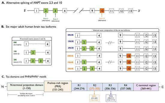
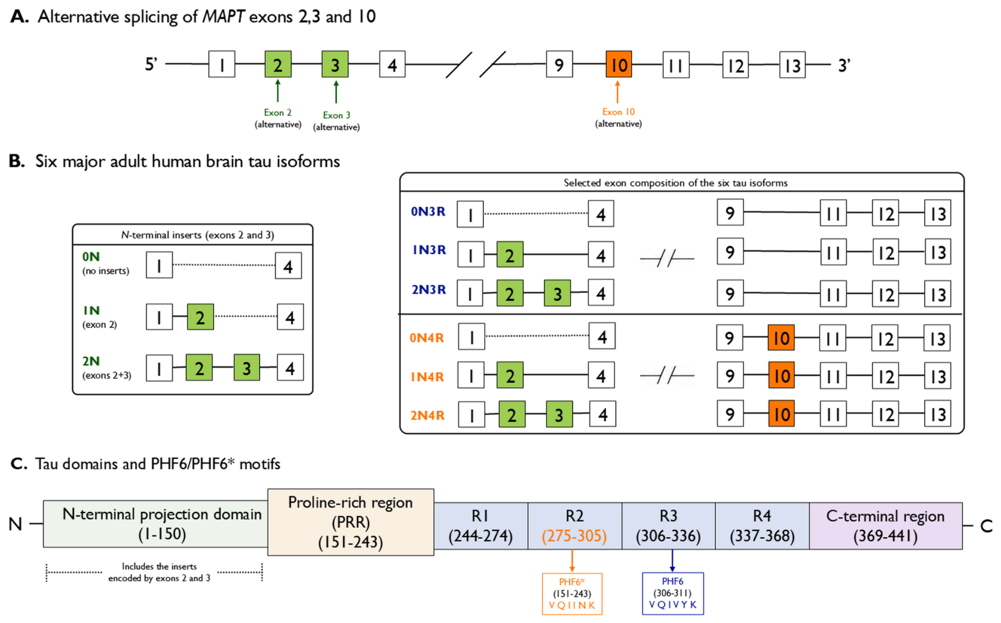
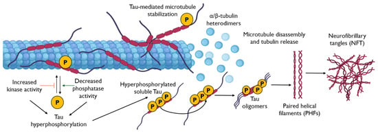
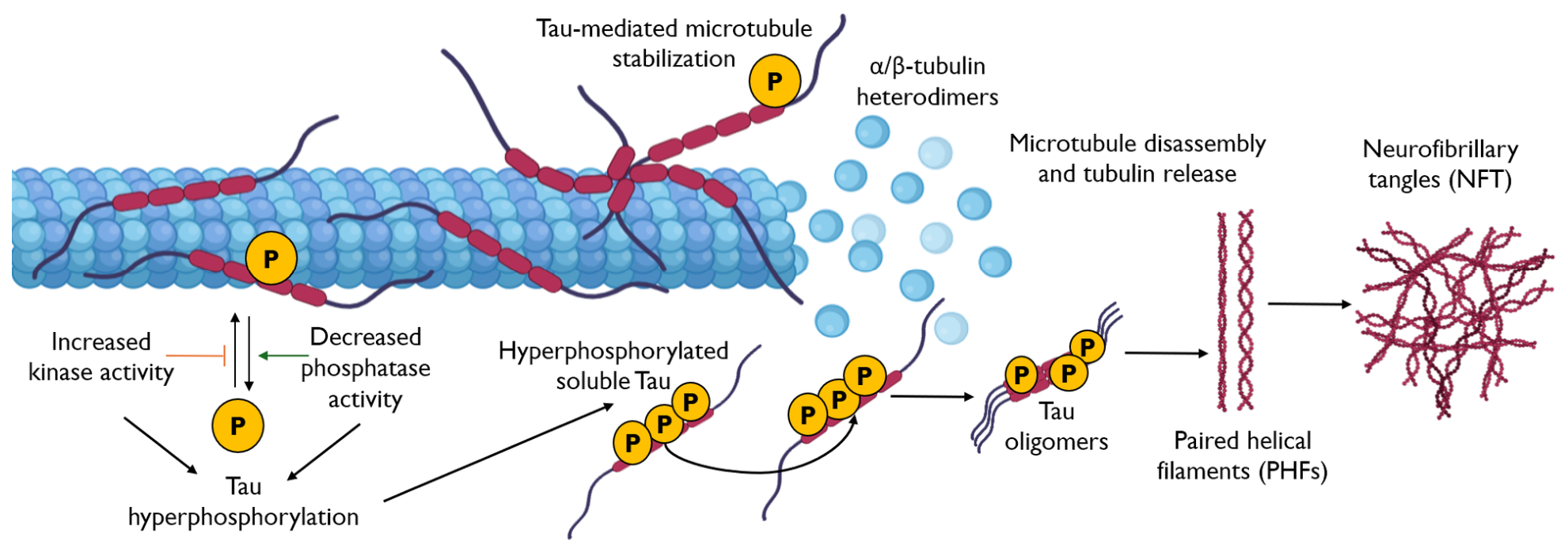

Disclaimer/Publisher’s Note: The statements, opinions and data contained in all publications are solely those of the individual author(s) and contributor(s) and not of MDPI and/or the editor(s). MDPI and/or the editor(s) disclaim responsibility for any injury to people or property resulting from any ideas, methods, instructions or products referred to in the content. |
© 2026 by the authors. Licensee MDPI, Basel, Switzerland. This article is an open access article distributed under the terms and conditions of the Creative Commons Attribution (CC BY) license.