


Abstract
Cold Atmospheric Plasma (CAP) is gaining interest as a biomedical technology due to its ability to modulate cellular behaviour through the controlled delivery of reactive oxygen and nitrogen species (RONS), UV photons, and electric fields. This is particularly relevant to wound healing, where redox modulation can reduce microbial burden and promote tissue repair. Mesenchymal stem/stromal cells (MSCs) are key regulators of immune response and tissue regeneration, suggesting potential synergy between CAP and MSC-based approaches. However, before CAP can be incorporated into MSC-based approaches, it is essential to establish whether it compromises core MSC characteristics. This study investigated the effects of CAP on MSC viability, morphology, phenotype and multilineage differentiation potential. Adipose-derived immortalised MSCs were exposed to CAP using a helium plasma jet under optimised conditions and assessed for up to 72 h. CAP exposure produced treatment-time-dependent effects on metabolic activity and morphology, enabling the identification of a non-cytotoxic treatment window. Under mild conditions, CAP-treated MSCs maintained the characteristic MSC phenotype (CD73-, CD90-, and CD105-positive; HLA-DR- negative) and retained adipogenic, osteogenic, and chondrogenic differentiation capacity. These findings indicate that controlled CAP exposure preserves key MSC properties, supporting further investigation of CAP as a complementary tool for MSC-based wound healing applications.
1. Introduction
The skin is the largest organ of the body and serves as a physical barrier that protects internal tissues and organs. Disruptions to the skin barrier by an external force or trauma are classified as acute or chronic wounds, distinguished by the time they take to restore physiological and structural integrity of the injury site [1]. Wound healing is a complex and highly coordinated process involving dynamic interactions between resident dermal and epidermal cells, ultimately resulting in wound closure and the restoration of damaged tissue [2]. The physiological process is generally categorised into four overlapping stages: homeostasis, inflammation, proliferation and remodelling. Dysregulation of these stages can give rise to chronic, non-healing wounds, representing a major global healthcare burden that affects millions of individuals worldwide [3]. The aetiology and clinical manifestations of non-healing wounds vary and can be classified as arterial, diabetic, pressure and venous ulcers [4]. Despite differences in their underlying pathophysiology, they are commonly associated with prolonged inflammation and reduced angiogenesis at the injury site [5].
Chronic wound management and care have seen major advances in recent years with a move from conventional methods such as hydrocolloids and gauze to smart biomaterial dressings and hydrogels [6,7] and cell-based therapies [8], especially mesenchymal stem/stromal cells (MSCs) [9]. MSCs are a class of cells distinguished by their natural capacity for self-renewal, differentiation into specialised tissue cells, immunomodulation and targeted migration to injury sites [10]. The International Society for Cell & Gene Therapy (ISCT) defines MSCs as spindle-shaped, adherent cells that express CD105, CD90, and CD73 and lack CD45, CD34, CD11b, CD79α, and HLA-DR surface markers and maintain trilineage differentiation capacity in cell culture media [11]. MSCs contribute to multiple stages of the wound healing process [12]. Through their injury-homing capacity, MSCs localise to sites of tissue damage, where they modulate the activity of inflammatory cells via paracrine signalling [13,14]. They also support the proliferative phase of wound repair through engraftment at the injury site and the secretion of regenerative and anti-fibrotic factors, thereby promoting enhanced tissue and skin repair [15].
Despite the potential of MSCs in therapeutic applications, translating their unique abilities in clinic is complex and limited by their variability. MSCs from different donors differ in their proliferation and differentiation capacity, and further differences arise from the tissue they are isolated from as well as the isolation protocol [16]. This variability is particularly challenging when aiming to define treatment parameters and understand their influence on cells for a new technology, as the effects of treatment need to be distinguished from the differences between cell batches. Immortalisation with human telomerase reverse transcriptase (hTERT) has been used to overcome some of these limitations of primary MSC cultures [17]. hTERT-immortalised adipose-derived MSCs were therefore used in this study to provide a stable and reproducible cell model.
Non-thermal or Cold Atmospheric Plasma (CAP) is partially ionised gas composed of excited atoms, electrons, positive ions, electromagnetic fields, UV radiations, reactive species and free radicals, which result from direct and indirect collisions between electrons and molecules as well as the loss of electrons following the supply of energy to a neutral gas [18]. Electrons are the first to take up the energy, which they then transfer into gas molecules (e.g., Ar, He, N2, CO2, O2) in the form of vibrational excitation. The generation of plasma in air or in noble gases such as argon or helium, which then interact with surrounding air molecules, results in the creation of reactive species such as reactive oxygen species (ROS), which include singlet oxygen, superoxide anion (O2−), hydrogen peroxide (H2O2), hydroxyl radical (OH●) and ozone (O3), as well as reactive nitrogen species (RNS) such as nitric oxide (NO) and nitrogen dioxide (NO2) [19,20]. The various components of CAP contribute to its complex chemical kinetics that lay down the basis of its clinical potential.
The application of CAP in medicine has evolved rapidly, including disinfection, wound healing, cancer treatment and tissue regeneration. The unique mix of RONS, ions, electrons, free radicals, electromagnetic fields and UV radiation that CAP generates contributes to its interactions with living cells [21,22]. CAP has also been demonstrated to effectively reduce planktonic bacterial cells and biofilms through a number of mechanisms such as lipid peroxidation and DNA damage [23]. This capability has positioned CAP as a promising alternative for wound disinfection, as it combats antibiotic-resistant infections commonly associated with chronic wounds. Extensive research has been conducted on CAP in chronic wounds and ulcers, with studies spanning animal models [24,25] and clinical applications [26,27]. In addition to alleviating the infection load, CAP can stimulate skin cells (keratinocytes, fibroblasts) to proliferate and enhance their migration, leading to accelerated wound healing [28]. Given that both CAP and MSC-based therapies are being developed for chronic wound management, their combined use is of growing interest. However, before the two approaches can be combined, it is essential to establish whether CAP preserves the key characteristics of MSCs.
The aim of this study was to characterise the reactive species generated by a helium-based plasma jet and to evaluate their possible contribution to the response of MSCs to CAP. To achieve this, the main reactive species produced by the plasma jet were first quantified. The CAP treatment window was then determined using dose–response analysis, and the relative contribution of reactive species was examined by comparing direct treatment, plasma activated medium (PAM) and medium replacement after exposure. Finally, the expression of selected MSC surface markers and trilineage differentiation capacity were evaluated to determine whether key MSC characteristics were preserved following CAP treatment.
2. Materials and Methods
2.1. Plasma Source: J-Plasma Radiofrequency (RF) Helium Atmospheric Pressure Plasma Jet (APPJ)
The Apyx Medical Ultimate® Electro-surgical generator, an electrosurgical FDA-approved (FDA: K192867) helium RF CAP jet (Apyx Medical, Clearwater, FL, USA), was used. J-plasma consists of a 15 cm handpiece connected to a control unit and a gas line, enabling the regulated flow of helium through the device, which generates a helium corona discharge at room temperature. The jet nozzle was stabilised at a 2 cm distance from the support platform for direct MSC treatment and activation of media. All treatments were conducted at 40% power (approx. 6 W; [29,30]) and a 4 Standard Litre per Minute (SLM) gas flow.
2.2. Chemical Analysis of J-Plasma
Plasma activated water (PAW) was generated in a 24-well plate using deionised water (1 mL, dH2O) at 2 cm from platform. The dH2O was exposed to a J-plasma device for varying durations: 30, 60, 120, 180 s. Following treatment, the pH of PAW was measured using FiveEasy ™ Plus pH/mV bench meter (Mettler Toledo, Greifensee, Switzerland).
The concentration of H2O2 in plasma-activated dH2O was determined using titanium oxysulfate (TiOSO4, Merck, Arklow, Ireland). An amount of 100 μL of PAW or H2O2 standard sample was incubated with 10 μL of TiOSO4 for 10 min in the dark, after which absorbance readings were taken at 405 nm wavelength (FilterMax F5, Multi-Mode MicroPlate Reader, Molecular Devices, San Jose, CA, USA). Data were acquired with SoftMax Pro (version 6.4; Molecular Devices). A standard curve of known H2O2 concentrations was included on each plate and used to convert the absorbance of samples into concentration H2O2 in μM.
Total oxidative species were measured using potassium iodide (KI, Merck, Arklow, Ireland). A total of 50 μL of PAW or H2O2 standard sample was mixed with 50 μL dH2O (non-buffered conditions) or 50 μL 10 mM phosphate buffer (buffered conditions) and 100 μL 1 M KI solution and incubated in the dark for 20 min. The absorbance was read at a 405 nm wavelength (FilterMax F5, Multi-Mode MicroPlate Reader, Molecular Devices, San Jose, CA, USA).
All chemical measurements were performed in three independent experiments, each in three technical replicates.
2.3. Cell Culture of ASC52telo hTERT Immortalised Adipose-Derived Mesenchymal Stem Cells
Human ASC52telo hTERT-immortalised adipose-derived MSCs were kindly provided by Dr. Jessica Whelan and Dr. Susan McDonnell, University College Dublin, Dublin, Ireland. Cells were cultured in Mesenchymal Stem Cell Medium (Cat. No. 7501 MSCM, ScienCell, Carlsbad, CA, USA) supplemented with 5% Foetal Bovine Serum (Cat. No. 0025, FBS, ScienCell), 1% MSC growth supplement (MSCGS, Cat. No. 7552, ScienCell) and a 1% penicillin/streptomycin solution (Cat. No. 0503, P/S, ScienCell) in a humidified incubator at 37 °C and 5% CO2. Cells were routinely subcultured once a confluence of 85% was reached using a 0.05% trypsin/EDTA Solution (Cat. No. 0183, Trypsin/EDTA, ScienCell) and reseeded at 2.5 × 10⁴ cs/mL.
2.4. Direct and Indirect Plasma Treatment Set-Ups
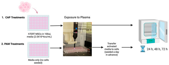
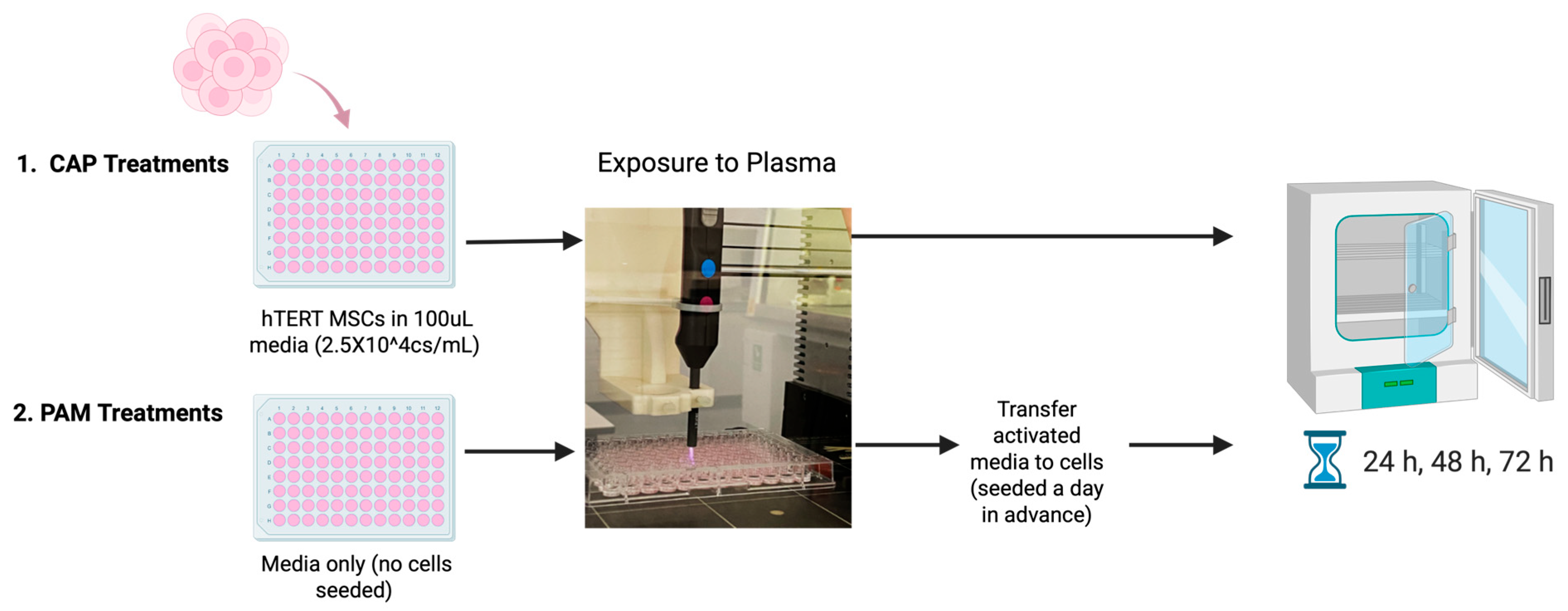
All treatments were conducted at 2 cm distance from the platform, with a gas flow rate of 4 SLM and power of 40% (approx. 6 W; [30,31]) (Figure 1). For direct treatments, cells were seeded 24 h prior to treatment (at a concentration of 2.5 × 10⁴ cs/mL in 100 µL). Before plasma exposure, culture media was removed, cells were washed with PBS, and 100 µL of fresh media was added, resulting in a liquid depth of approximately 3.4 mm above the cell monolayer. Cells were then exposed to plasma for treatment durations of 1, 2, 5, 10, 15, 20, 30, 60, and 90 s and the untreated control was kept in the same conditions except for being exposed to plasma. Each treatment condition was performed in triplicate. For plasma activated media (PAM) treatments, the same procedure, treatment times and set-up were used; however, the media was treated separately before being immediately added to the cells.
Figure 1.
Schematic diagram of the experimental flowchart of cold plasma treatments.
2.5. Resazurin/Alamar Blue Assay
The response of hTERT-MSCs was analysed using the colorimetric resazurin metabolic activity assay (resazurin sodium salt, Cat. No. R7017-1G, Merck, Arklow, Ireland). After the specified exposure and incubation times, media was removed and cells were washed with sterile Phosphate-Buffered Saline (PBS) (Cat. No. P4417-50TAB, Merck, Arklow, Ireland) and 100 μL of resazurin media solution was added (final concentration of 8 μg/mL resazurin) followed by a 2.5 h incubation at 37 °C and in 5% CO2. Absorbance was measured using a spectrophotometric plate reader (FilterMax F5, Multi-Mode MicroPlate Reader) at 570 nm and 600 nm as a reference wavelength. Results are expressed in the percentage of metabolic activity of control cells, which are non-treated cells.
2.6. Live Cell Imaging
Changes in hTERT-MSC fibroblast-like morphology were investigated following each treatment for the duration of the incubation (24, 48, 72 h) using the Incucyte® Live-Cell Analysis Instrument (Sartorius, Göttingen, Germany) in phase-contrast mode with the S3/SX 1 G/R Optical Module.
2.7. Phenotyping of ASC52telo hTERT-MSCs
To determine the MSC phenotype of hTERT-MSCs, the expression of positive markers CD105, CD90 and CD73 and the negative marker HLA/DR was analysed by flow cytometry using an MSC Phenotyping Cocktail Kit, anti-human, REAfinity (Miltenyi Biotec, Bisley, UK). Samples were analysed using the Cytoflex LX flow cytometer (Beckman Coulter, Brea, CA, USA) using CytExpert software (version 2.6.0.105; Beckman Coulter). Cells were harvested for phenotypic analysis 72 h following CAP treatment (20 s and 30 s) by pooling the cells harvested from 60 wells of a 96-well plate, each treated for the appropriate exposure time.
Flow cytometry data were analysed using a sequential gating strategy to accurately identify viable MSC populations and assess surface marker expression. Initially cells were gated based on forward scatter (FSC-A) versus side scatter (SSC-A) to exclude debris and select the main cell population based on size and granularity. Doublets were then excluded by plotting FSC-A versus FSC-H and live singlets were gated. Fluorescent gates were then established using unstained controls, and positive and negative gates were selected based on these unstained controls. Percentages of cells expressing CD73, CD90 and CD105, as well as cells lacking HLA-DR, were calculated within viable singlet population.
2.8. Osteogenesis Differentiation Potential Assay
Following CAP treatment (72 h) cells were seeded at 5 × 10³ cs/cm2 in a 96-well plate for 2 days prior to adding complete osteogenesis differentiation media (StemPro Osteocyte/Chondrocyte Differentiation Kit, Cat. No. A10072-01, Gibco, Paisley, UK). Media was changed every 3–4 days, and cells were incubated for a total of 21 days. Cells were then gently washed with PBS and fixed with 4% paraformaldehyde (PFA) at room temperature. Calcium-rich deposits were assessed using an Alizarin Red S staining kit (Cliniscience, Cat. No. 0223-SC, Nanterre, France) according to the manufacturer’s instructions. Stained cultures were imaged using brightfield microscopy.
2.9. Adipogenesis Differentiation Potential Assay
Following CAP treatment (72 h), cells were seeded at 1 × 10⁴ cs/cm2 in a 96-well plate for 2 days prior to adding complete adipogenesis differentiation media (StemPro Adipogenesis Differentiation Kit, Cat. No. A10070-01, Gibco, Paisley, UK). Media was changed every 3–4 days and cells were incubated for a total of 14 days. Cells were then gently washed with PBS and fixed with fixative (Cliniscience, Cat. No. 0843, Nanterre, France) at room temperature. Intracellular lipid droplet accumulation was assessed using Red Oil O staining (Cliniscience, Cat. No. 0843a, Nanterre, France) according to the manufacturer’s instructions. Stained cultures were imaged using brightfield microscopy.
2.10. Chondrogenesis Differentiation Potential Assay
Following CAP treatment (72 h), cells were resuspended at a density of 1 × 10⁷ cs/mL, and 5 μL droplets were dispensed into wells of a 24-well plate to generate micromasses. The micromass cultures were incubated for 2 h at 37 °C without media to allow cells to adhere and form a compact micromass before adding media. Complete chondrogenesis media (StemPro Chondrogenesis Differentiation Kit, Cat. No. A10071-01, Gibco, Paisley, UK) was added. Media was changed every 2–3 days, and cells were incubated for a total of 14 days. Cells were then gently washed with PBS and fixed with 4% paraformaldehyde (PFA) at room temperature. Sulphated proteoglycans were detected using Alcian Blue staining (Cliniscience, Cat. No. 8378-SC, Nanterre, France) according to the manufacturer’s instructions. Stained cultures were imaged using brightfield microscopy.
2.11. Statistical Analysis
All experiments were performed at least three independent times. Statistical analysis was carried out using GraphPad Prism (version 9.5.1, GraphPad Software, San Diego, CA, USA). Data are presented as the mean ± standard deviation (SD). Multiple comparison analyses were performed using two-way ANOVA followed by Sidak’s multiple comparison test.
3. Results
3.1. pH Measurements and Chemical Properties
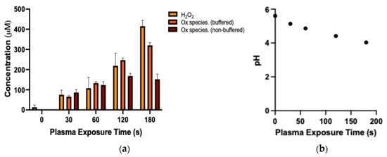
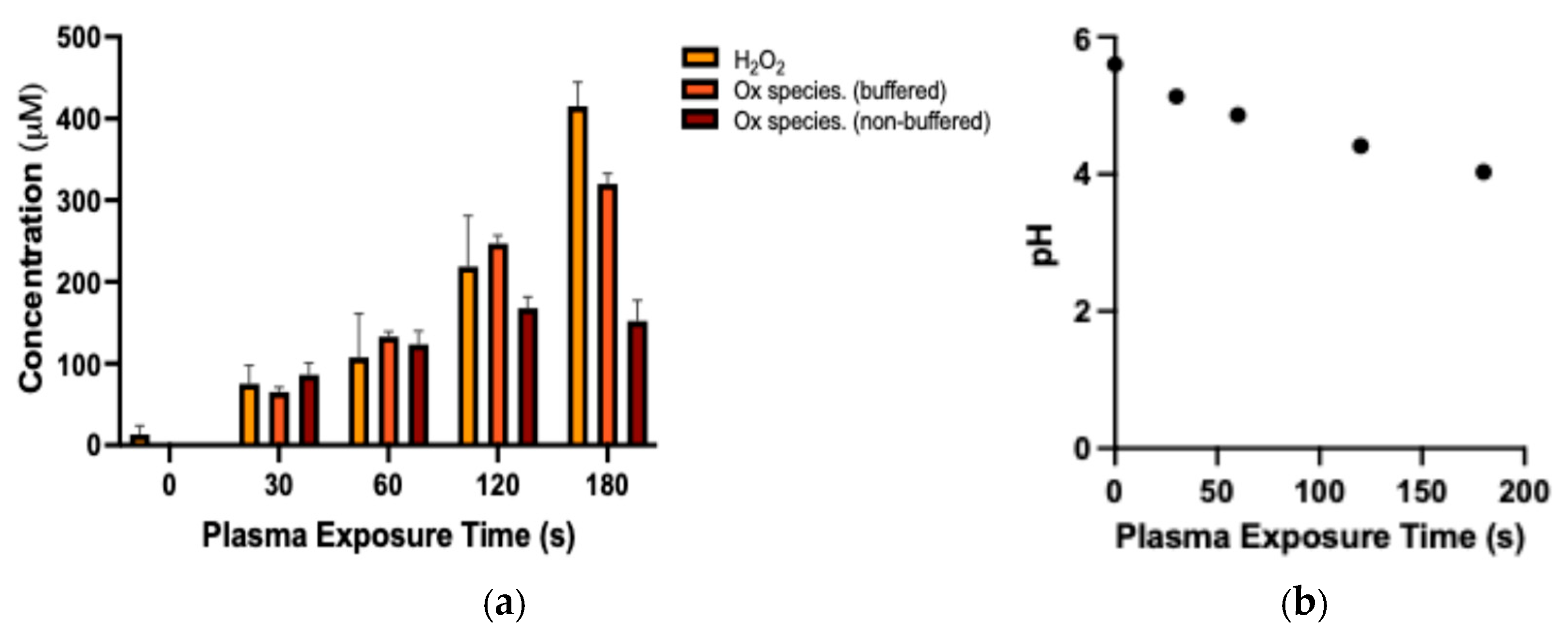
Upon interacting with a liquid surface, reactive species originally present in the gas phase both from the plasma and surrounding air generate short and long-lived reactive species within the liquid they encounter. These RONS play a crucial role in mediating CAP’s effects on biological entities, such as cells in culture and tissues. The concentration of hydrogen peroxide generated in deionised water (dH2O) was directly correlated with exposure time, where it increased from 57.5 μM with 30 s exposure to 429 μM at 180 s treatment time (Figure 2a). Similarly, total oxidative species generated in dH2O also demonstrated a direct relationship between exposure time and concentration when measured under non-buffered (reactive species that are stable at acidic conditions) and buffered (reactive species that are stable at neutral conditions) conditions. Concentrations of nitrites and nitrates were also measured but showed minimal RNS generation by this plasma system (<3 μM at 3 min exposure), in line with findings of other studies [31]. The pH of deionised water decreased as plasma exposure time increased, from pH 5.3 at 30 s exposure to pH 4.03 recorded at 180 s exposure (Figure 2b).
Figure 2.
(a) Chemical composition of PAW after exposure to Apyx J-Plasma APPJ for exposure times of 30, 60, 120 and 180 s. Oxidative species refer to species able to oxidise potassium iodide (KI) at acidic pH (non-buffered) or neutral pH (buffered). H2O2 is quantified using TiOSO4. (b) Measurements of pH in water following exposure to Apyx J-Plasma APPJ for exposure times of 30, 60, 120 and 180 s. Results are presented as the mean ± S.D. of three experiments.
3.2. Optimisation of CAP Treatment Parameters Based on Biological Response
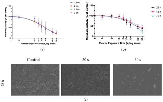
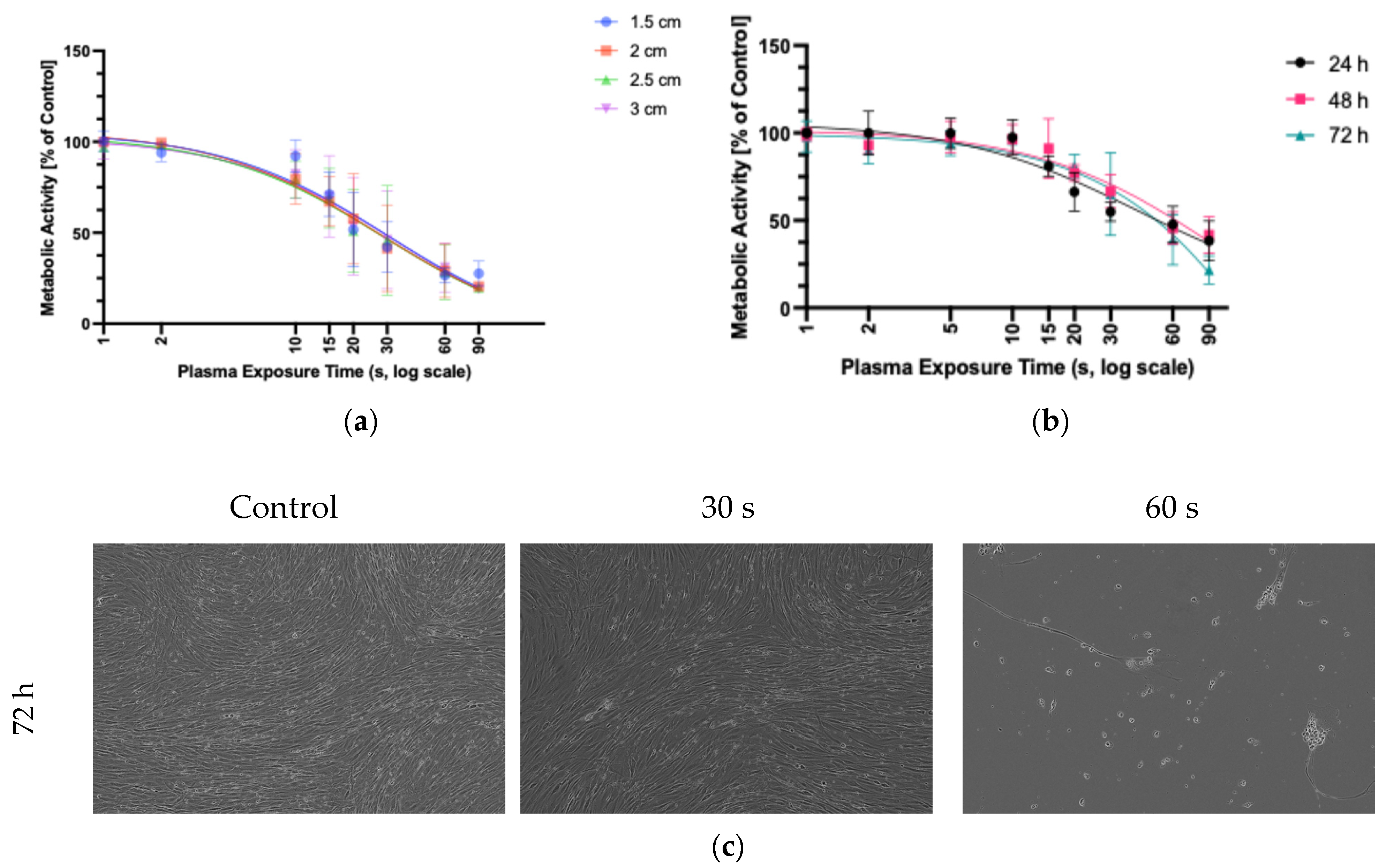
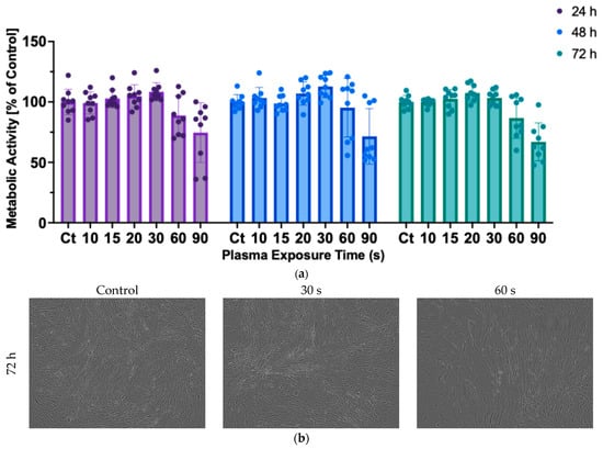
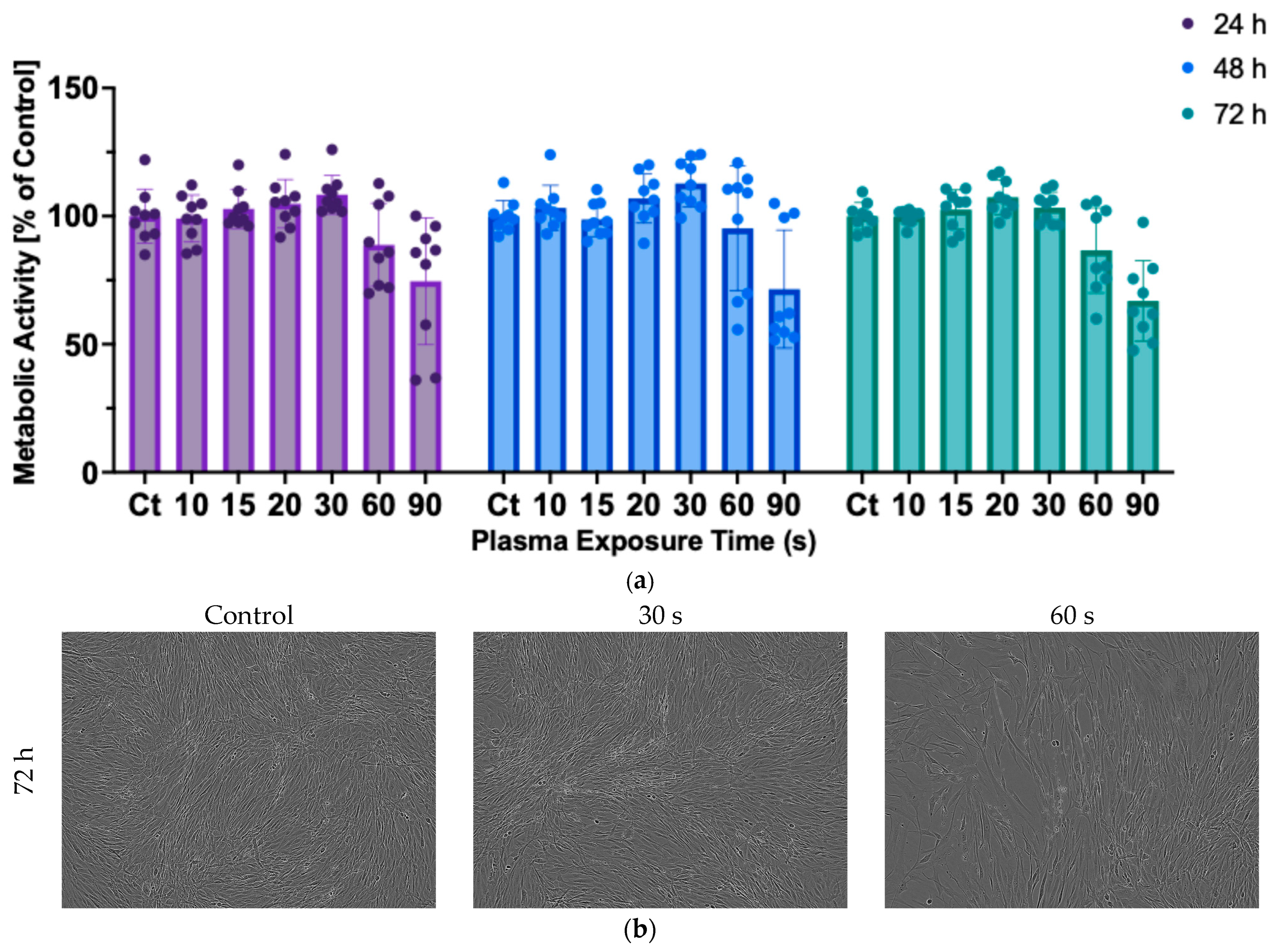
In order to investigate MSCs’ sensitivity to plasma and its potential use as a tool to modulate MSC activity, it is essential to identify safe CAP parameters. Immortalised human mesenchymal stem cells (hTERT-MSCs) were used to determine dose responses to CAP conditions, as their ability to expand indefinitely eliminates the time constraints associated with primary MSC cell lines. Following the initial characterisation of plasma-generated reactive species and associated pH changes, the influence of distance (1.5–3 cm) was investigated. Dose–response curves were generated for each treatment distance (Figure 3a). As treatment distance did not markedly alter the viability of cells, a distance of 2 cm was selected as an intermediate and representative treatment condition for subsequent experiments. Subsequently, a broader exposure range of 1 s–1.5 min was evaluated, and cells were incubated for 24, 48 or 72 h post-treatment (Figure 3b). hTERT-MSC metabolic activity was reduced to 50% at an exposure time of about 40 s. Comparison between 24, 48 and 72 h incubation post-treatment showed similar viability trends, suggesting that the majority of cell death at higher exposure times is sustained over the measured time points (24, 48 and 72 h) after interaction with plasma and not over prolonged incubation post-treatment. Morphology and confluency of hTERT-MSCs were monitored by live-cell imaging over 72 h, and Figure 3c shows representative phase-contrast images acquired at 72 h following 30 s, 60 s and control treatments. Cells exposed to 30 s CAP maintained normal spindle-shaped morphology and confluence, whereas higher treatment (60 s) resulted in reduced cell density and visible morphological changes consistent with decreased viability. Based on these results, 30 s was defined as a mild treatment at which metabolic activity remained above 50% of control and cells retained proliferative capacity along with displaying no visible morphological changes. This exposure was used as an upper limit for ‘mild treatment’ conditions in the subsequent analysis.
Figure 3.
Changes in (a) metabolic activity in response to CAP exposure at varying distances (1.5, 2, 2.5, 3 cm at 72 h post-treatment. Changes in (b) metabolic activity following exposure to CAP at 2 cm distance and incubation for 24 h, 48 h and 72 h following treatment. Results are presented as the mean ± S.D. of three experiments and relative to the untreated control. (c) Changes in MSC’s spindle-shaped morphology at 60 s, 30 s and control at 72 h, taken at 10× objective by IncuCyte S3.
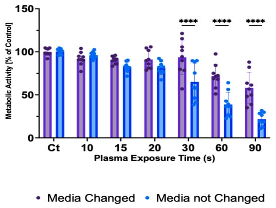
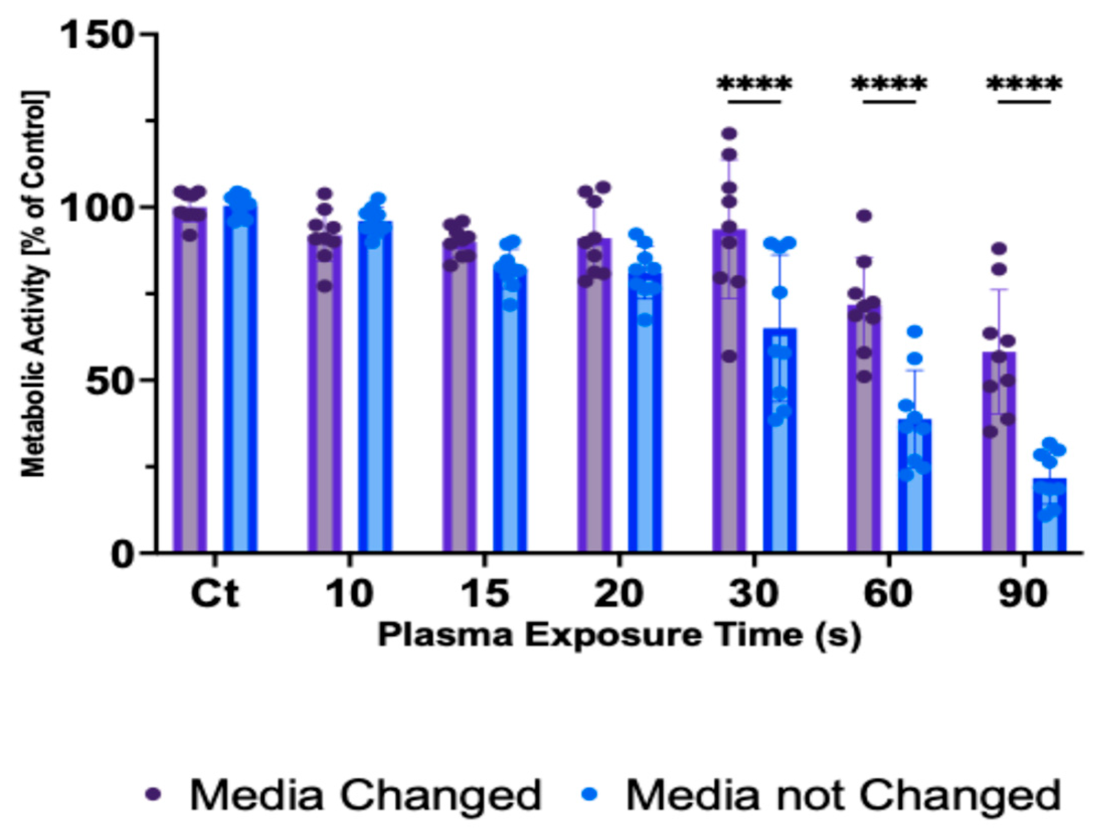
CAP influences living cells through its chemical and physical components. Different plasma devices vary in their design, gas inlet composition, and power, leading to differences in the way they generate and deliver these components. These variations lead to different cellular responses; hence, it is essential to understand the specific mode of action of each device. In this section, cells were treated directly with CAP, and media was changed straight after treatment, followed by a 72 h incubation. Immediate replacement of the culture media after plasma exposure reduces cellular interactions with long-lived reactive species (e.g., H2O2). Significantly higher metabolic activity can be seen with media change at higher exposure times (30 s–90 s) compared to no media changes (Figure 4), suggesting that removing these reactive species and other byproducts of the initial interaction with plasma attenuates the reduction in metabolic activity compared to not changing the media following exposure. This points to the potential contribution of long-lived ROS in mediating the effects of the Apyx J-plasma on MSCs.
Figure 4.
Changes in metabolic activity in response to direct CAP exposure, followed by immediate media change or no media change and a 72 h incubation. Metabolic activity is obtained via resazurin. The results are presented as the mean ± S.D. of three experiments and relative to the untreated control. **** p < 0.0001.
3.3. Effects of Plasma Activated Medium (PAM) on hTERT-MSCs
In addition to direct CAP treatment, PAM was also investigated. While direct CAP treatments have shown effects on MSCs, there is a growing interest in the use of PAM as a more practical and versatile approach for biomedical applications. Avoiding the higher cytotoxicity caused by increased oxidative stress and UV radiation, PAM may offer an alternative by harnessing CAP’s benefits in a controlled and accessible manner. Cell culture medium was activated in a 96-well plate (100 μL per well) and then immediately transferred to cells that had been seeded 24 h in advance in a 96-well plate. By using PAM, the treatment circumvents the exposure to short-lived species, UV emissions, and electromagnetic fields. Compared to direct treatment, PAM elicited a less distinct dose–response pattern in MSCs and less cytotoxicity overall (Figure 5a). Additionally, phase-contrast live images at 72 h (Figure 5b) indicate that cell morphology remained unchanged following PAM treatment. The differences in responses between PAM and direct CAP may be attributed to the stability and lifespan of the reactive species involved, as direct treatment also exposes cells to short-lived species and other plasma components.
Figure 5.
(a) Changes in metabolic activity obtained via resazurin of hTERT-MSC in response to PAM followed by a 24 h, 48 h, and 72 h incubation. (b) MSC’s spindle-shaped morphology following PAM treatments at 60 s, 30 s and control at 72 h, taken at 10× objective by IncuCyte S3. Results are presented as the mean ± S.D. of three experiments and relative to the untreated control.
3.4. Effects of CAP on hTERT-MSC Phenotypical Marker Expression
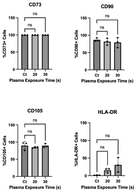
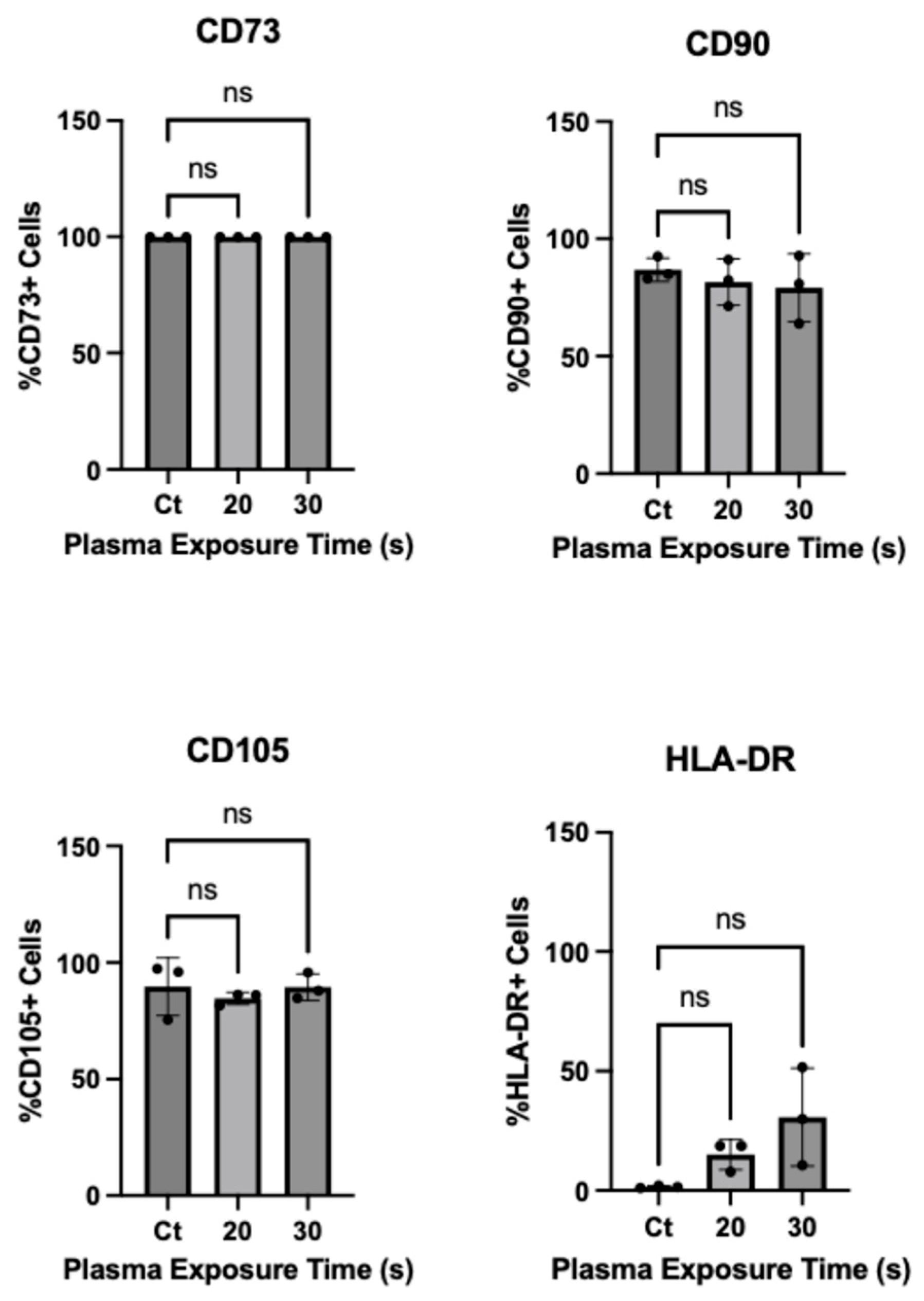
To evaluate the impact of CAP on the phenotypic properties of MSCs, the expression of established MSC surface markers (positive markers: CD73, CD90, CD105; negative markers: HLA-DR) between mild CAP-treated and untreated cells was investigated using flow cytometry. The results (Figure 6 and Supplementary Figure S1) showed that CAP-treated cells at parameters (20 s, 30 s) with low cytotoxicity continued to express MSC markers in a similar manner to untreated control, with no significant increase or decrease in phenotypical characteristics. Although not statistically significant, HLA-DR showed an exposure-time-dependent increased expression, which warrants further investigation.
Figure 6.
Phenotypical analysis of MSC surface markers (positive markers: CD105, CD90, CD73; negative marker: HLA-DR) for MSCs treated for 20 s and 30 s compared to the untreated control. Percentages of cells expressing CD73, CD90, CD105 and HLA-DR, presented as the mean ± S.D. of three experiments and relative to the untreated control at 72 h post-treatment. Viability was determined using trypan blue (≈99%).
3.5. Effects of CAP on hTERT-MSC Differentiation Potential
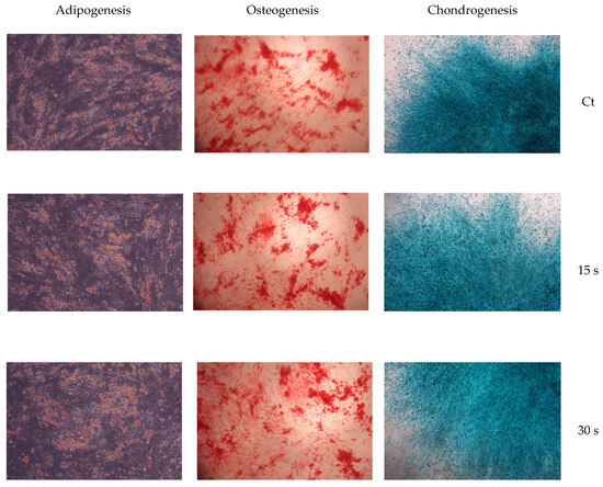
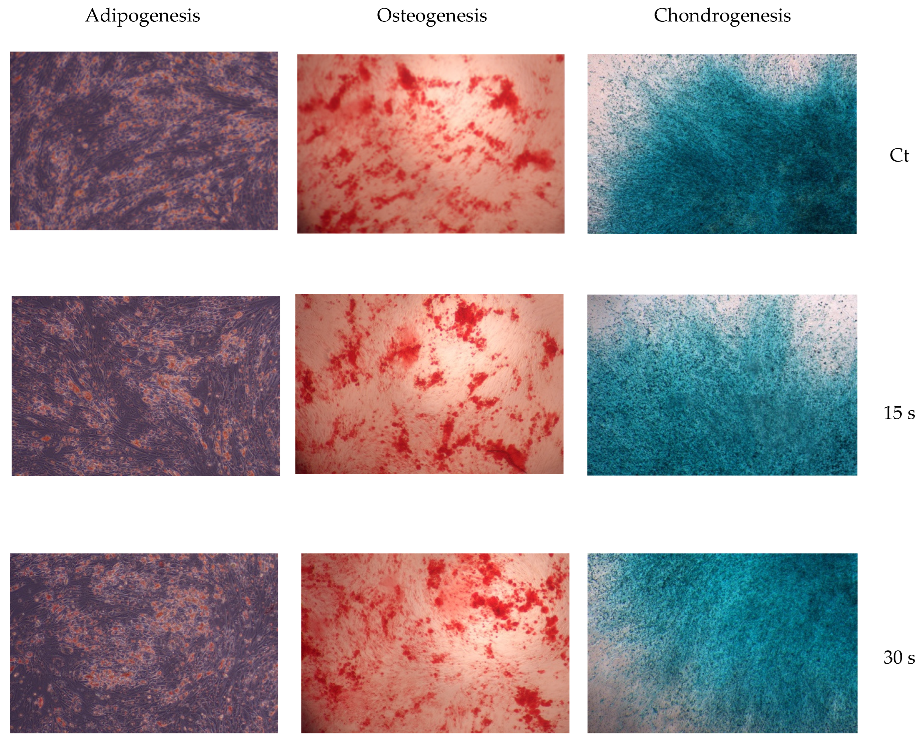
To evaluate whether CAP exposure affects the multipotent capacity of MSCs, adipogenic, osteogenic and chondrogenic differentiation assays were performed following treatment (Figure 7). Cells treated with mild exposure times (15, 30 s) retained their ability to differentiate along all lineages. Under adipogenic conditions, CAP-treated hTERT-MSCs formed lipid droplets similar to the untreated control. Similarly osteogenic differentiation potential was preserved at 15 and 30 s treatment times, where treated cells formed calcium deposits. Chondrogenesis differentiation was also maintained, with CAP-treated cells showing a proteoglycan-rich matrix. These findings suggest that, at the parameters tested, CAP exposure did not prevent hTERT-MSCs from undergoing trilineage differentiation. In contrast, cells exposed to 60 s of CAP exhibited markedly reduced viability (as shown in Section 3.2) prior to the induction of differentiation. As a result, differentiation capacity at this exposure time and higher was diminished, primarily due to cell death.
Figure 7.
Adipogenic, osteogenic and chondrogenic differentiation potential of hTERT-MSCs following CAP treatments at 15 s and 30 s and untreated control. (left) Images of adipogenic differentiation showing intracellular lipid accumulation stained with Oil Red O. (middle) Images of osteogenic differentiation demonstrating calcium-rich mineralised matrix deposition stained with Alizarin Red S. (right) Images of chondrogenic differentiation demonstrating acidic polysaccharides stained with Alcian Blue.
4. Discussion
Cold plasma holds significant promise for applications in tissue regeneration and wound healing. To safely translate these into therapeutic effects, it is essential to understand how MSCs respond to plasma to define the limits of their sensitivity and determine safe exposure thresholds. Emerging research suggests that cold plasma can effectively stimulate the proliferation of adipose-derived, bone-marrow-derived and hematopoietic stem cells compared to untreated controls [32,33]. Furthermore, studies have demonstrated that combining CAP with MSC therapy can enhance wound healing in animal skin models [34,35]. To investigate plasma as a tool for modulating MSCs and understanding its effect on these cells, it is essential to identify safe treatment parameters and determine their tolerance to CAP [36].
Direct exposure to CAP can cause cellular damage and apoptosis [37,38,39]. Therefore, establishing safe operative parameters is crucial when translating CAP into regenerative medicine applications. Dose–response analysis was carried out to identify the threshold at which MSCs lose their viability. Cells exhibited a less than 50% decrease in viability for all treatment durations under 30 s (40% power (approx. 6 W), 4.0 SLM), identifying this as a treatment window for investigating plasma effects on MSC function. No significant difference was observed in cell viability when incubated for longer than 24 h post-treatment. However, Ermakov et al. (2021) showed that longer incubation times result in decreased cell viability of MSCs post-plasma treatment [40]. This contrasts with other studies, which showed an increase in cell proliferation following plasma treatment that further increased with incubation time [32]. The discrepancy between these studies and our results may stem from variations in plasma source, treatment parameters and experimental conditions. This also highlights the challenges of standardising plasma treatments across studies to ensure reproducibility and facilitate comparison. Additionally, it was previously shown that hTERT overexpression in human fibroblasts is associated with improved mitochondrial function and reduced response to oxidative stress [41]. Given that the present study used hTERT-immortalised MSCs, it is possible that primary MSCs that lack hTERT overexpression could respond differently to CAP-induced oxidative stress.
Previous studies on MSCs have performed direct and indirect cold plasma treatments using different approaches and achieved different outcomes [32,42,43]. For CAP to be used as a tool for tissue regeneration, understanding its mechanism of action and assessing both immediate and delayed cellular responses is crucial in order to differentiate between transient and sustained effects of plasma exposure on cell behaviour. Chemical characterisation of J-plasma in deionised water shows that H2O2 is the main long-lived species generated, which could suggest it plays a vital role in the loss of metabolic activity observed in MSCs. However, to confirm whether J-plasma-generated ROS are the main drivers of its effect on cells, a detailed analysis of the ROS generated in media is required. Our results demonstrated that cells maintained significantly higher viability at longer exposure times when media was replaced immediately, indicating a potential contribution of CAP-generated long-lived ROS to cytotoxicity. These findings further support the role of ROS as mediators of the plasma-induced cellular responses [44,45]. Previously, CAP-generated ROS have been shown to induce intra- and extracellular ROS accumulation, leading to oxidative stress and DNA damage that diminish cell metabolic activity and induce apoptosis [46,47]. However, H2O2 levels have been reported to deplete in the presence of enzymes found in media and sera [48]. Understanding CAP’s effect on MSCs is therefore more challenging and multifaceted, requiring consideration of not only individual reactive species like H2O2.
Treatment with PAM eliminates any physical interaction between cells and plasma, resulting in a reduced level of cytotoxicity compared to that observed with direct treatment. This could be due to the combined effects of short- and long-lived reactive species and the UV and electromagnetic field in CAP, which can lead to increased oxidative stress and potential DNA damage [44], while PAM relies only on the effects of longer-lived reactive species that may exert more controlled and prolonged biological effects. Hahn et al. (2024) showed that PAM also induces an oxidative response in MSCs [43]. The lifetime and concentration of reactive species could differ between direct and indirect plasma treatment, where the direct treatment generates an intense burst of ROS at the cell surface, potentially triggering apoptosis [43]. Treatment with PAM also allows for a more uniform treatment distribution across cells, avoiding localised overexposure that may occur with direct treatment.
Treatment with CAP did not result in changes in expression profiles of the positive markers CD105, CD90 and CD73, suggesting that while CAP alters cells’ oxidative state, metabolism and survival, it does not alter these MSCs’ phenotypical characteristics at short exposure times. These results suggest that the expression of the selected MSC surface markers was maintained following short plasma exposure, despite the changes observed in metabolic activity. Phenotypic stability is an important consideration for downstream applications of CAP, as shifts in marker expression could affect MSC function or introduce variability. These findings are consistent with previous reports demonstrating that controlled CAP treatment can modulate cellular behaviour without adversely affecting surface markers [32,49]. Oxidative stress has previously been reported to induce senescence in MSCs, even at non-cytotoxic levels of exposure [50]. Similarly, CAP has been shown to increase the expression of senescence-associated factor p16 in fibroblasts and adipose-derived stem cells (ASCs) [51]. Interestingly, our findings demonstrate that low-cytotoxic direct CAP treatment did not impair the adipogenic, osteogenic or chondrogenic differentiation capacity of hTERT-MSCs. This may be attributed to treatment conditions used in the present study, as well as the potential scavenging and degradation of CAP-generated ROS by media components, such as FBS, present in the culture media [52]. As differentiation was not evaluated quantitatively, changes in differentiation capacity cannot be excluded and further investigation is required.
This study demonstrated that short exposures (<30 s) to a helium APPJ jet (J-plasma) preserved the MSC phenotype of hTERT-MSCs, supporting further investigation of this plasma source as a potential tool for MSC-based therapeutic applications. While CAP treatment modulated viability in a dose-dependent manner, exposure times with low cytotoxic effect preserved MSC phenotype and trilineage differentiation capacity. Collectively this supports the potential use of CAP as a pre-conditioning or adjuvant therapeutic strategy in regenerative medicine and wound healing applications. Future studies should further investigate the molecular mechanisms underlying CAP-MSC interactions, particularly in primary MSC cell models, as well as explore long-term effects of CAP exposure, such as cellular senescence, DNA damage and sustained proliferative capacity. In addition, the effect of CAP on MSC secretome and extracellular vesicle (EV) production and the molecular composition of EV cargo (e.g., proteins, lipids and microRNAs) should be analysed, as well as the downstream signalling pathways responsible for mediating the regenerative and immunomodulatory responses of MSCs.
Supplementary Materials
The following supporting information can be downloaded at https://www.mdpi.com/article/10.3390/plasma9040043/s1, Figure S1: Representative dot plots of flow cytometry data illustrating surface marker expression profiles (positive CD73, CD90, CD105) and negative (HLA-DR)_ for hTERT-MSCs treated for 20 s and 30 s compared to untreated control at 72 h post treatment.
Author Contributions
Conceptualisation, S.A. and D.B.; methodology, S.A. and D.B.; software, S.A. and D.B.; validation, S.A. and D.B.; formal analysis, S.A. and D.B.; investigation, S.A. and D.B.; resources, S.A. and D.B.; data curation, S.A. and D.B.; writing—original draft preparation, S.A.; writing—review and editing, S.A. and D.B.; visualisation, S.A. and D.B.; supervision, D.B.; project administration, D.B.; funding acquisition, D.B. All authors have read and agreed to the published version of the manuscript.
Funding
This research was funded by the University College Dublin Ad Astra Funding and Stem Challenge Fund, grant number R24349 and R25144.
Institutional Review Board Statement
Not applicable.
Informed Consent Statement
Not Applicable.
Data Availability Statement
All data generated or analysed during this study are included in this published article and are available from the corresponding author upon reasonable request.
Acknowledgments
The authors would like to thank Dr. Alfonso Blanco for technical support and guidance in flow cytometry, which was performed at the UCD Conway Institute Flow Cytometry Core Facility. The authors also acknowledge TheraDep Limited (Clonmel, Ireland) for the loan of the J-plasma jet and their valuable support with system set-up.
Conflicts of Interest
The authors declare no conflicts of interest.
Abbreviations
The following abbreviations are used in this manuscript:
| CAP | Cold Atmospheric Plasma |
| APPJ | Atmospheric Pressure Plasma Jet |
| PAM | Plasma Activated Media |
| MSCs | Mesenchymal Stem/Stromal Cells |
| RONS | Reactive Oxygen and Nitrogen Species |
| SLM | Standard Litre per Min |
References
- Rodrigues, M.; Kosaric, N.; Bonham, C.A.; Gurtner, G.C. Wound Healing: A Cellular Perspective. Physiol. Rev. 2019, 99, 665–706. [Google Scholar] [CrossRef] [Scilit]
- Heras, K.L.; Igartua, M.; Santos-Vizcaino, E.; Hernandez, R.M. Chronic wounds: Current status, available strategies and emerging therapeutic solutions. J. Control. Release 2020, 328, 532–550. [Google Scholar] [CrossRef] [Scilit]
- La Monica, F.; Campora, S.; Ghersi, G. Collagen-Based Scaffolds for Chronic Skin Wound Treatment. Gels 2024, 10, 137. [Google Scholar] [CrossRef] [Scilit]
- Bowers, S.; Wounds, E.F.C. Chronic Wounds: Evaluation and Management. Am. Fam. Physician 2020, 101, 159–166. [Google Scholar]
- Pastar, I.; Balukoff, N.C.; Marjanovic, J.; Chen, V.Y.; Stone, R.C.; Tomic-Canic, M. Molecular Pathophysiology of Chronic Wounds: Current State and Future Directions. Cold Spring Harb. Perspect. Biol. 2023, 15, a041243. [Google Scholar] [CrossRef] [Scilit]
- Liu, Y.; Ge, L. Smart Biomaterials in Wound Healing: Advances, Challenges, and Future Directions in Intelligent Dressing Design. Bioengineering 2025, 12, 1178. [Google Scholar] [CrossRef] [Scilit]
- Ning, X.; Lu, H.; Zeng, H.; Zhou, Z.; Hu, P. Multifunctional self-healing hydrogels for chronic wound repair: Design, mechanisms, and applications. J. Control. Release 2025, 388, 114282. [Google Scholar] [CrossRef] [Scilit]
- Nayak, M.; Banerjee, D.; Venugopal, V.; Nethi, S.K.; Barui, A.K.; Mukherjee, S. Cell-engineered technologies for wound healing and tissue regeneration. npj Biomed. Innov. 2025, 2, 38. [Google Scholar] [CrossRef] [Scilit]
- Dehkordi, A.N.; Babaheydari, F.M.; Chehelgerdi, M.; Dehkordi, S.R. Skin tissue engineering: Wound healing based on stem-cell-based therapeutic strategies. Stem Cell Res. Ther. 2019, 10, 111. [Google Scholar] [CrossRef] [Scilit]
- Ding, D.-C.; Shyu, W.-C.; Lin, S.-Z. Mesenchymal stem cells. Cell Transpl. 2011, 20, 5–14. [Google Scholar] [CrossRef] [Scilit]
- Dominici, M.; Le Blanc, K.; Mueller, I.; Slaper-Cortenbach, I.; Marini, F.C.; Krause, D.S.; Deans, R.J.; Keating, A.; Prockop, D.J.; Horwitz, E.M. Minimal criteria for defining multipotent mesenchymal stromal cells. The International Society for Cellular Therapy position statement. Cytotherapy 2006, 8, 315–317. [Google Scholar] [CrossRef] [Scilit]
- Nasadiuk, K.; Kolanowski, T.; Kowalewski, C.; Wozniak, K.; Oldak, T.; Rozwadowska, N. Harnessing Mesenchymal Stromal Cells for Advanced Wound Healing: A Comprehensive Review of Mechanisms and Applications. Int. J. Mol. Sci. 2025, 26, 199. [Google Scholar] [CrossRef] [Scilit]
- Gao, M.; Guo, H.; Dong, X.; Wang, Z.; Yang, Z.; Shang, Q.; Wang, Q. Regulation of inflammation during wound healing: The function of mesenchymal stem cells and strategies for therapeutic enhancement. Front. Pharmacol. 2024, 15, 1345779. [Google Scholar] [CrossRef] [Scilit]
- Zhang, Q.-Z.; Su, W.-R.; Shi, S.-H.; Wilder-Smith, P.; Xiang, A.P.; Wong, A.; Nguyen, A.L.; Kwon, C.W.; Le, A.D. Human gingiva-derived mesenchymal stem cells elicit polarization of m2 macrophages and enhance cutaneous wound healing. Stem Cells 2010, 28, 1856–1868. [Google Scholar] [CrossRef] [Scilit]
- Wu, Y.; Chen, L.; Scott, P.G.; Tredget, E.E. Mesenchymal stem cells enhance wound healing through differentiation and angiogenesis. Stem Cells 2007, 25, 2648–2659. [Google Scholar] [CrossRef] [Scilit]
- Česnik, A.B.; Švajger, U. The issue of heterogeneity of MSC-based therapy medicinal products—A review. Front. Cell Dev. Biol. 2024, 12, 1400347. [Google Scholar] [CrossRef] [Scilit]
- Kang, S.K.; Putnam, L.; Dufour, J.; Ylostalo, J.; Jung, J.S.; Bunnell, B.A. Expression of telomerase extends the lifespan and enhances osteogenic differentiation of adipose tissue-derived stromal cells. Stem Cells 2004, 22, 1356–1372. [Google Scholar] [CrossRef] [Scilit]
- Adamovich, I.; Baalrud, S.D.; Bogaerts, A.; Bruggeman, P.J.; Cappelli, M.; Colombo, V.; Czarnetzki, U.; Ebert, U.; Eden, J.G.; Favia, P.; et al. The 2017 Plasma Roadmap: Low temperature plasma science and technology. J. Phys. D Appl. Phys. 2017, 50, 323001. [Google Scholar] [CrossRef] [Scilit]
- Dharini, M.; Jaspin, S.; Mahendran, R. Cold plasma reactive species: Generation, properties, and interaction with food biomolecules. Food Chem. 2023, 405, 134746. [Google Scholar] [CrossRef] [Scilit]
- Fridman, A. Plasma Chemistry, 1st ed.; Cambridge University Press: Cambridge, UK, 2008. [Google Scholar] [CrossRef] [Scilit]
- Fridman, G.; Peddinghaus, M.; Balasubramanian, M.; Ayan, H.; Fridman, A.; Gutsol, A.; Brooks, A. Blood Coagulation and Living Tissue Sterilization by Floating-Electrode Dielectric Barrier Discharge in Air. Plasma Chem. Plasma Process 2006, 26, 425–442. [Google Scholar] [CrossRef] [Scilit]
- Laroussi, M.; Leipold, F. Evaluation of the roles of reactive species, heat, and UV radiation in the inactivation of bacterial cells by air plasmas at atmospheric pressure. Int. J. Mass Spectrom. 2004, 233, 81–86. [Google Scholar] [CrossRef] [Scilit]
- Das, S.; Gajula, V.P.; Mohapatra, S.; Singh, G.; Kar, S. Role of cold atmospheric plasma in microbial inactivation and the factors affecting its efficacy. Health Sci. Rev. 2022, 4, 100037. [Google Scholar] [CrossRef] [Scilit]
- Hung, Y.-W.; Lee, L.-T.; Peng, Y.-C.; Chang, C.-T.; Wong, Y.-K.; Tung, K.-C. Effect of a nonthermal-atmospheric pressure plasma jet on wound healing: An animal study. J. Chin. Med. Assoc. 2016, 79, 320. [Google Scholar] [CrossRef] [Scilit]
- Melotti, L.; Martinello, T.; Perazzi, A.; Martines, E.; Zuin, M.; Modenese, D.; Cordaro, L.; Ferro, S.; Maccatrozzo, L.; Iacopetti, I.; et al. Could cold plasma act synergistically with allogeneic mesenchymal stem cells to improve wound skin regeneration in a large size animal model? Res. Vet. Sci. 2021, 136, 97–110. [Google Scholar] [CrossRef] [Scilit]
- Brehmer, F.; Haenssle, H.; Daeschlein, G.; Ahmed, R.; Pfeiffer, S.; Görlitz, A.; Simon, D.; Schön, M.; Wandke, D.; Emmert, S. Alleviation of chronic venous leg ulcers with a hand-held dielectric barrier discharge plasma generator (PlasmaDerm(®) VU-2010): Results of a monocentric, two-armed, open, prospective, randomized and controlled trial (NCT01415622). J. Eur. Acad. Dermatol. Venereol. 2015, 29, 148–155. [Google Scholar] [CrossRef] [Scilit]
- Mirpour, S.; Fathollah, S.; Mansouri, P.; Larijani, B.; Ghoranneviss, M.; Tehrani, M.M.; Amini, M.R. Cold atmospheric plasma as an effective method to treat diabetic foot ulcers: A randomized clinical trial. Sci. Rep. 2020, 10, 10440. [Google Scholar] [CrossRef] [Scilit]
- Braný, D.; Dvorská, D.; Halašová, E.; Škovierová, H. Cold Atmospheric Plasma: A Powerful Tool for Modern Medicine. Int. J. Mol. Sci. 2020, 21, 2932. [Google Scholar] [CrossRef] [Scilit]
- Holcomb, J.D.; Schucker, A. Helium Plasma Skin Regeneration: Evaluation of Skin Tissue Effects in a Procine Model and Comparison to Nitrogen Plasma Skin Regeneration. Lasers Surg. Med. 2020, 52, 23–32. [Google Scholar] [CrossRef] [Scilit]
- Madigan, C.D.; O’Sullivan, D.; O’Neill, L.; Kavanagh, D.F. A comparison of two cold atmospheric helium plasma devices which utilise the same RF power generator. Clin. Plasma Med. 2020, 19–20, 100108. [Google Scholar] [CrossRef] [Scilit]
- Thompson, T.P.; Connelly, A.; Kelly, S.; Duncan, R.M.; Maybin, J.; McDonnell, C.; Melvage, A.; McClenaghan, L.A.; Dedeloudi, A.; Lamprou, D.A.; et al. Biomedical Application of Cold Plasma: Navigating Through Plasma Types and Devices by Antimicrobial Effectiveness and Tissue Tolerance. Adv. Ther. 2025, 8, 2400339. [Google Scholar] [CrossRef] [Scilit]
- Park, J.; Lee, H.; Lee, H.J.; Kim, G.C.; Kim, S.-S.; Han, S.; Song, K. Non-thermal atmospheric pressure plasma is an excellent tool to activate proliferation in various mesoderm-derived human adult stem cells. Free Radic. Biol. Med. 2019, 134, 374–384. [Google Scholar] [CrossRef] [Scilit]
- Park, J.; Lee, H.; Lee, H.J.; Kim, G.C.; Kim, D.Y.; Han, S.; Song, K. Non-Thermal Atmospheric Pressure Plasma Efficiently Promotes the Proliferation of Adipose Tissue-Derived Stem Cells by Activating NO-Response Pathways. Sci. Rep. 2016, 6, 39298. [Google Scholar] [CrossRef] [Scilit]
- Cui, H.S.; Joo, S.Y.; Cho, Y.S.; Park, J.H.; Kim, J.-B.; Seo, C.H. Effect of Combining Low Temperature Plasma, Negative Pressure Wound Therapy, and Bone Marrow Mesenchymal Stem Cells on an Acute Skin Wound Healing Mouse Model. Int. J. Mol. Sci. 2020, 21, 3675. [Google Scholar] [CrossRef] [Scilit]
- Choi, B.B.R.; Song, K.W.; Lee, H.J.; Park, S.R.; Kim, G.C. Effects of no-ozone cold plasma and mouse mesenchymal stem cell treatments on wound healing in a mouse skin model. Biochem. Biophys. Res. Commun. 2024, 738, 150562. [Google Scholar] [CrossRef] [Scilit]
- Li, Y.; Choi, E.H.; Han, I. Regulation of Redox Homeostasis by Nonthermal Biocompatible Plasma Discharge in Stem Cell Differentiation. Oxidative Med. Cell. Longev. 2019, 2019, 2318680. [Google Scholar] [CrossRef] [Scilit]
- Yan, D.; Wang, Q.; Malyavko, A.; Zolotukhin, D.B.; Adhikari, M.; Sherman, J.H.; Keidar, M. The anti-glioblastoma effect of cold atmospheric plasma treatment: Physical pathway v.s. chemical pathway. Sci. Rep. 2020, 10, 11788. [Google Scholar] [CrossRef] [Scilit]
- Peeters, S.; Wu, P.B.; Haist, B.; Armellini, A.; Kim, W.J.; Chen, Z.; Obenchain, R.; Ayad, G.; Ge, W.; Bhaduri, A.; et al. Cold atmospheric plasma effectively kills chordoma cells through induction of intracellular reactive oxygen species. Sci. Rep. 2025, 15, 20838. [Google Scholar] [CrossRef] [Scilit]
- Roshan, M.; Farnia, P.; Ghomi, H.; Jafari, S. Apoptotic effects of cold atmospheric pressure plasma on A549 and LL/2 lung carcinoma cell lines. Sci. Rep. 2025, 15, 19567. [Google Scholar] [CrossRef] [Scilit]
- Ermakov, A.M.; Ermakova, O.N.; Afanasyeva, V.A.; Popov, A.L. Dose-Dependent Effects of Cold Atmospheric Argon Plasma on the Mesenchymal Stem and Osteosarcoma Cells In Vitro. Int. J. Mol. Sci. 2021, 22, 6797. [Google Scholar] [CrossRef] [Scilit]
- Ahmed, S.; Passos, J.F.; Birket, M.J.; Beckmann, T.; Brings, S.; Peters, H.; Birch-Machin, M.A.; von Zglinicki, T.; Saretzki, G. Telomerase does not counteract tolemere shortening but protects mitochondrial function under oxidative stress. J. Cell Sci. 2008, 121, 1046–1053. [Google Scholar] [CrossRef] [Scilit]
- Park, J.; Suh, D.; Tang, T.; Lee, H.J.; Roe, J.-S.; Kim, G.C.; Han, S.; Song, K. Non-thermal atmospheric pressure plasma induces epigenetic modifications that activate the expression of various cytokines and growth factors in human mesoderm-derived stem cells. Free Radic. Biol. Med. 2020, 148, 108–122. [Google Scholar] [CrossRef] [Scilit]
- Hahn, O.; Waheed, T.O.; Sridharan, K.; Huemerlehner, T.; Staehlke, S.; Thürling, M.; Boeckmann, L.; Meister, M.; Masur, K.; Peters, K. Cold Atmospheric Pressure Plasma-Activated Medium Modulates Cellular Functions of Human Mesenchymal Stem/Stromal Cells In Vitro. Int. J. Mol. Sci. 2024, 25, 4944. [Google Scholar] [CrossRef] [Scilit]
- Turrini, E.; Laurita, R.; Stancampiano, A.; Catanzaro, E.; Calcabrini, C.; Maffei, F.; Gherardi, M.; Colombo, V.; Fimognari, C. Cold Atmospheric Plasma Induces Apoptosis and Oxidative Stress Pathway Regulation in T-Lymphoblastoid Leukemia Cells. Oxid. Med. Cell Longev. 2017, 2017, 4271065. [Google Scholar] [CrossRef] [Scilit]
- Yan, D.; Cui, H.; Zhu, W.; Talbot, A.; Zhang, L.G.; Sherman, J.H.; Keidar, M. The Strong Cell-based Hydrogen Peroxide Generation Triggered by Cold Atmospheric Plasma. Sci. Rep. 2017, 7, 10831. [Google Scholar] [CrossRef] [Scilit]
- Ma, Y.; Ha, C.S.; Hwang, S.W.; Lee, H.J.; Kim, G.C.; Lee, K.-W.; Song, K. Non-Thermal Atmospheric Pressure Plasma Preferentially Induces Apoptosis in p53-Mutated Cancer Cells by Activating ROS Stress-Response Pathways. PLoS ONE 2014, 9, e91947. [Google Scholar] [CrossRef] [Scilit]
- Lopes, B.P.; O’neill, L.; Bourke, P.; Boehm, D. Combined Effect of Plasma-Activated Water and Topotecan in Glioblastoma Cells. Cancers 2023, 15, 4858. [Google Scholar] [CrossRef] [Scilit]
- Rubio, C.P.; Cerón, J.J. Spectrophotometric assays for evaluation of Reactive Oxygen Species (ROS) in serum: General concepts and applications in dogs and humans. BMC Vet. Res. 2021, 17, 226. [Google Scholar] [CrossRef] [Scilit]
- Wu, Y.; Yu, S.; Zhang, X.; Wang, X.; Zhang, J. The Regulatory Mechanism of Cold Plasma in Relation to Cell Activity and Its Application in Biomedical and Animal Husbandry Practices. Int. J. Mol. Sci. 2023, 24, 7160. [Google Scholar] [CrossRef] [Scilit]
- Brandl, A.; Meyer, M.; Bechmann, V.; Nerlich, M.; Angele, P. Oxidative stress induces senescence in human mesenchymal stem cells. Exp. Cell Res. 2011, 317, 1541–1547. [Google Scholar] [CrossRef] [Scilit]
- Bourdens, M.; Jeanson, Y.; Taurand, M.; Juin, N.; Carrière, A.; Clément, F.; Casteilla, L.; Bulteau, A.-L.; Planat-Bénard, V. Short exposure to cold atmospheric plasma induces senescence in human skin fibroblasts and adipose mesenchymal stromal cells. Sci. Rep. 2019, 9, 8671. [Google Scholar] [CrossRef] [Scilit]
- Francis, G.L. Albumin and mammalian cell culture: Implications for biotechnology applications. Cytotechnology 2010, 62, 1–16. [Google Scholar] [CrossRef] [Scilit]
Disclaimer/Publisher’s Note: The statements, opinions and data contained in all publications are solely those of the individual author(s) and contributor(s) and not of MDPI and/or the editor(s). MDPI and/or the editor(s) disclaim responsibility for any injury to people or property resulting from any ideas, methods, instructions or products referred to in the content. |
© 2026 by the authors. Licensee MDPI, Basel, Switzerland. This article is an open access article distributed under the terms and conditions of the Creative Commons Attribution (CC BY) license.